Note : Les descriptions sont présentées dans la langue officielle dans laquelle elles ont été soumises.
CA 03171479 2022-08-16
WO 2021/168218 PCT/US2021/018737
COMPOSITIONS COMPRISING AXITINIB AND METHODS OF TREATING
OCULAR DISORDERS
CROSS-REFERENCE TO RELATED APPLICATIONS
[0001] This application claims priority to U. S. Provisional Application No.
62/978,540, filed
February 19, 2020; which is incorporated herein by reference in its entirety.
BACKGROUND OF THE INVENTION
[0002] Neovascular age-related macular degeneration (nAMD), also known as
"wet" AMD, is the
leading cause of vision loss in the industrialized world. It is caused by
abnormal blood vessel
growth in the choriocapillaris, a layer of capillaries situated immediately
below Bruch's
membrane. Choroidal neovascularization leads to the leakage of blood, lipids,
and serum into the
retinal layers, which can result in permanent damage to light-sensitive
retinal cells and irreversible
central vision distortions.
[0003] Although not fully elucidated, much is known regarding the pathogenesis
of nAMD. The
vascular endothelial growth factor (VEGF) signaling pathway has been shown to
be centrally
involved in the 10 to 15% of AMD diagnoses classified as the neovascular type
(nAMD). In this
pathway, VEGF signaling ligands bind to different isoforms of VEGF receptors
(VEGFR) to
activate cellular processes that promote growth of new vasculature.
Specifically, VEGF-A, which
acts at VEGFR-1 and -2, has been shown to promote abnormal blood vessel growth
and is therefore
an optimal target for treatment. Currently, anti-VEGF-A drugs are the standard
of care for the
treatment of nAMD; however, an unmet need remains for significantly improving
and maintaining
visual acuity in patients
[0004] Choroidal neovascularization (CNV) is the creation of new blood vessels
in the choroid
layer of the eye. Choroidal neovascularization is a common cause of, and/or
can be associated
with, nAMD. CNV can occur rapidly in individuals with defects in Bruch's
membrane, the
innermost layer of the choroid. It is also associated with excessive amounts
of VEGF. CNV can
also occur frequently with the rare genetic disease pseudoxanthoma elasticum
and rarely with the
more common optic disc drusen. CNV has also been associated with extreme
myopia or malignant
1
CA 03171479 2022-08-16
WO 2021/168218 PCT/US2021/018737
myopic degeneration, where in choroidal neovascularization occurs primarily in
the presence of
cracks within the retinal macular tissue known as lacquer cracks. CNV can
create a sudden
deterioration of central vision, noticeable within a few weeks. Other symptoms
which can occur
include color disturbances, and metamorphopsia (distortions in which straight
lines appears wavy).
Hemorrhaging of the new blood vessels can accelerate the onset of symptoms of
CNV.
[0005] Currently, even the most successful treatments of nAMD and CNN/ do not
preclude
reoccurrence, making multiple treatments likely in addition, currently
available treatments do not
restore vision that has already been lost Therefore, there is a need in the
art for treatment
breakthroughs, in order to maintain vision for a longer period of time without
repeated laser use.
SUMMARY OF THE INVENTION
[0006] Methods and compositions for the treatment of neovascular age-related
macular
degeneration (nAMD; also referred to herein as wet AMD), choroidal
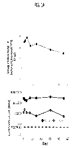
neovascularization (CNV),
nAMD associated with CNV, retinopathy, diabetic retinopathy, diabetic macular
edema (DME),
and related diseases are provided. The compositions comprise one or more
tyrosine kinase
inhibitors and are delivered to the suprachoroidal space of the eye via a non-
surgical means. In
some embodiments the tyrosine kinase inhibitor has activity against vascular
endothelial growth
factor (VEGF) and/or platelet derived growth factor (PDGF). In some
embodiments, the tyrosine
kinase inhibitor is axitinib. In some embodiments, the present disclosure
provides an axitinib
formulation, CLS-AX.
[0007] In some embodiments, the present disclosure provides methods for
treating nAMD,
choroidal neovascularization (CNV), nAMD associated with CNV, and/or DME
comprising
administering a formulation comprising axitinib to the suprachoroidal space of
an eye of a subject
in need thereof. In some embodiments, the method comprises administering about
0.01 mg to about
3.0 mg of axitinib to the eye. In some embodiments, the method comprises
administering about
0.01 mg to about 0.5 mg of axitinib to the eye. In some embodiments, the
method comprises
administering about 0.01 mg to about 0.3 mg of axitinib to the eye. In further
embodiments, the
method comprises administering about 0.03 to about 0.1 mg of axitinib to the
eye. In some
embodiments, the method comprises administering about 0.01, about 0.02, about
0.03, about 0.04,
about 0.05, about 0.06, about 0.07, about 0.08, about 0.09, about 0.10, about
0.2, about 0.3, about
2
CA 03171479 2022-08-16
WO 2021/168218 PCT/US2021/018737
0.4, about 0.5, about 1.0, about 2.0, or about 3.0 mg of axitinib to the eye.
In some embodiments,
the method comprises administering about 0.03 mg of axitinib to the eye. In
some embodiments,
the method comprises administering about 0.06 mg of axitinib to the eye. In
some embodiments,
the method comprises administering about 0.1 mg of axitinib to the eye. In
some embodiments,
the method comprises administering about 0.3 mg of axitinib to the eye. In
some embodiments,
the axitinib is administered in a volume of about 100 L. Thus, in some
embodiments, the method
comprises administering an axitinib formulation in a concentration of about
0.1 mg/mL, to about
1 mg/mL, or about 0.1 mg/mL, about 0.3 mg/mL, about 0.6 mg/mL, or about 1.0
mg/mL. In some
embodiments, the method comprises administering the axitinib to the eye non-
surgically. For
example, in embodiments, the method comprises administering the axitinib to
the eye non-surgical
surpachoroidal injection. In embodiments, the non-surgical suprachoroidal
injection is achieved
by administering the axitinib using an injection device comprising a needle,
wherein the needle
has an effective length of about 500 to about 2000 microns.
[0008] In some embodiments, the method comprises administering axitinib to the
eye in at least
one dose. In some embodiments, the method comprises administering axitinib to
the eye in two
doses, wherein the two doses are spaced apart by at least about 1, at least
about 2, at least about 3,
at least about 4, at least about 5, at least about 6, at least about 7, at
least about 8, at least about 9,
at least about 10, at least about 11, or at least about 12 months.
[0009] In some embodiments, following administration of axitinib to the eye of
the subject, the
subject experiences no loss in visual acuity as measured by best corrected
visual acuity (BCVA).
In some embodiments, following administration of axitinib to the eye of the
subject, the subject
experiences minimal loss in visual acuity as measured by BCVA. In embodiments,
the minimal
loss in visual acuity is a loss of no more than 2 letters. In further
embodiments, the subject
experiences an improvement in BCVA of > 10 letters, > 15 letters or > 25
letters, as compared to
the subject's visual acuity prior to the administration of the axitinib
formulation. In embodiments,
the subject experiences no loss in visual acuity as measured by BCVA for 2
months, 3, months, 4
months, 5 months, 6 months, or longer after administration of the axitinib
formulation. In
embodiments, the subject experiences minimal loss in visual acuity as measured
by BCVA for 2
months, 3, months, 4 months, 5 months, 6 months, or longer after
administration of the axitinib
formulation. In embodiments, the subject experiences an improvement in visual
acuity as
3
CA 03171479 2022-08-16
WO 2021/168218 PCT/US2021/018737
measured by BCVA for 2 months, 3, months, 4 months, 5 months, 6 months, or
longer after
administration of the axitinib formulation.
[0010] In some embodiments, following administration of axitinib to the eye of
the subject, the
subject experiences a decrease in retinal thickness from baseline (e.g.,
retinal thickness such as
central subfield thickness (CST) prior to treatment), at any given time point
after administration
of a drug provided herein, e.g., a decrease of least about 20 p.m, or at least
about 40 p.m, or at least
about 50 p.m, or at least about 100 p.m, or at least about 150 p.m or at least
about 200 p.m, or from
about 50-100 p.m, and all values in between. In another embodiment, the
patient experiences a >
5%,> 10%,> 15%,> 20%,> 25% decrease in retinal thickness (e.g., CST)
subsequent to
administration of axitinib. In one embodiment, change in retinal thickness
from baseline is
measured as a change in CST, for example, by spectral domain optical coherence
tomography (SD-
OCT). In embodiments, the subject experiences the decrease in retinal
thickness for 2 months, 3,
months, 4 months, 5 months, 6 months, or longer after administration of the
axitinib formulation.
In embodiments, the subject experiences no increase in retinal thickness for 2
months, 3, months,
4 months, 5 months, 6 months, or longer after administration of the axitinib
formulation.
[0011] In some embodiments, the axitinib is Form IX axitinib. In some
embodiments, the axitinib
is present in the formulation in an amount of about 0.5 mg/mL to about 100
mg/mL, e.g., about 1
mg/mL to about 10 mg/mL or about 20 mg/mL to about 80 mg/mL. In embodiments,
the axitinib
is present in the formulation in an amount of about 1 mg/mL or about 40 mg/mL.
In some
embodiments, the present disclosure provides formulations comprising axitinib
and Polysorbate
80. In further embodiments, the present disclosure provides formulations
comprising
carboxymethylcellulose sodium, Polysorbate 80, sodium chloride, and sodium
phosphate. In
further embodiments, the formulation comprises Polysorbate 80 at a w/v of
about 0.025% to about
0.2%. In some embodiments, the Polysorbate 80 is present in the formulation at
an amount of about
0.05% to about 0.1%. In some embodiments, the Polysorbate 80 is present in the
formulation at an
amount of about 0.05% or about 0.1%. In some embodiments, formulation
comprises about 0.04%
w/v to about 0.07% w/v sodium phosphate (monobasic monohydrate). In further
embodiments,
the formulation comprises about 0.059% w/v sodium phosphate (monobasic
monohydrate). In
some embodiments, the formulation comprises about 0.05% w/v to about 0.09% w/v
sodium
phosphate (dibasic, anhydrous). In some embodiments, the formulation comprises
about 0.079%
4
CA 03171479 2022-08-16
WO 2021/168218 PCT/US2021/018737
w/v sodium phosphate (dibasic, anhydrous). In some embodiments, the
formulation comprises
about 0.5% w/v to about 1.0% w/v sodium chloride. In further embodiments, the
formulation
comprises about 0.7% w/v to about 0.9% w/v sodium chloride. In some
embodiments, the
formulation comprises about 0.79% w/v sodium chloride. In some embodiments,
the formulation
comprises about 0.25% w/v to about 0.75% w/v carboxymethylcellulose sodium. In
further
embodiments, the formulation comprises about 0.3% w/v to about 0.7% w/v
carboxymethylcellulose sodium. In some embodiments, the formulation comprises
about 0.5%
w/v carboxymethylcellulose sodium. In some embodiments, the axitinib
formulation comprises
microparticles comprising axitinib. In some embodiments, the microparticles
are about 1-20
microns in size for the D90 distribution. In some embodiments, the
microparticles are about 1-10
microns in size for the Dso distribution. In some embodiments, the
microparticles are about 1-4
microns in size for the Dio distribution. In some embodiments, the
microparticles are about 3-5
microns in size for the D90 distribution. In some embodiments, the
microparticles are about 3
microns in size for the D90 distribution. In some embodiments, the
microparticles are about 10
microns in size for the D90 distribution. In some embodiments, the clinical
formulation of CLS-
AX comprises axitinib microparticles of about 10 microns in size for the D90
distribution. In some
embodiments, the clinical formulation of CLS-AX comprises axitinib
microparticles of about 2
microns in size for the Dm distribution. In some embodiments, the clinical
formulation of CLS-
AX comprises axitinib microparticles of about 5 microns in size for the Dso
distribution. In some
embodiments, the clinical formulation of CLS-AX comprises axitinib
microparticles of about 10
microns for the D90 distribution.
BRIEF DESCRIPTION OF THE DRAWINGS
[0012] FIGS. 1A and 1B show the mean ( SD) CLS-AX concentrations in the
indicated tissues
following suprachoroidal injection with CLS-AX into the eyes of male Dutch
Belted rabbits. FIG.
1A shows the drug remaining (mg) in sclera/choroid/RPE (top panel) and the
concentration (in
nM) of drug in the retina and vitreous, at the indicated time points up to Day
91 (bottom panel).
FIG. 1B shows the concentration of axitinib (ng/g or ng/mL) in the vitreous
humor,
sclera/chorid/RPE, or retina at the lower (0.03 mg/eye) or higher (0.1 mg/eye)
dose.
CA 03171479 2022-08-16
WO 2021/168218 PCT/US2021/018737
[0013] FIG. 2A shows the fluorescein angiography (FA) grading scale for the
laser CNV model
in rats. FIG. 2B shows the % of Grade IV lesions at Day 21 in control animals
vs. CLS-AX
recipient animals. *, Fischer's Exact p-value = 0.0002.
[0014] FIGS. 3A and 3B show the results of the vascular leakage studies in the
CNV model in
pigs. FIG. 3A is a bar graph showing the area of vascular leakage 7 days or 14
days after laser
treatment to induce retinal lesions. OS = left eye; OD = right eye; BSS =
balanced salt solution;
SCS = suprachoroidal space. The OS-BSS treatment is shown as the left bar at
both time points
and the OD-CLS-AX treatment is shown as the right bar at both time points.
FIG. 3B provides
representative images from B SS-treated eyes and CLS-AX treated eyes.
[0015] FIGS. 4A and 4B show the mean ( SEM) isolectin IB4 on retinal flat
mount. FIG. 4A
provides representative images. Scale bar = 100 p.m. FIG. 4B shows the mean
SEM Isolectin IB4
signal. Signal in the CLS-AX group was significantly less than that of the BSS
treated group
(p=0.0297; t-test [27 sites measured OS; 354 sites measured OD]).
DETAILED DESCRIPTION OF THE INVENTION
[0016] This disclosure is generally related to ophthalmic therapies, and more
particularly to
methods and devices that allow for infusion of a fluid drug formulation into
posterior ocular tissues
for targeted, localized treatment, for example, for the treatment of diseases
and disorders of the
eye associated with neovascularization. For example, the diseases and
disorders include
neovascular age-related macular degeneration (nAMD; also referred to herein as
wet AMD),
nonexudative AMID, choroidal neovascularization (CNV), retinal vein occlusion
(RVO), nAMD
associated with RVO, nAMD associated with CNV, retinopathy, diabetic
retinopathy, and diabetic
macular edema (DME).
[0017] In some embodiments, the formulations comprise one or more tyrosine
kinase inhibitors
and are administered to the suprachoroidal space (SCS) of the eye via a non-
surgical means, for
example via a hollow microneedle, and/or an injection device comprising a
hollow needle wherein
the needle has an effective length of about 500 to about 2000 microns. The
methods and
6
CA 03171479 2022-08-16
WO 2021/168218 PCT/US2021/018737
formulations provided herein allow for effective posterior segment drug
delivery, and generally
embody the following characteristics: (1) the methods are non-surgical and
thus minimally
invasive and safe; (2) the drug formulations are administered in such a way
that they are well
targeted to the posterior segment of the eye and/or the suprachoroidal space
(SCS) of the eye and/or
the supraciliary space of the eye and/or the supraretinal space of the eye
and/or the subretinal space
of the eye, while simultaneously limiting drug exposure to the anterior
segment or other regions
of the eye; (3) the methods and formulations are capable of delivering drug in
a sustained and/or
controlled manner; (4) the methods and devices are user-friendly. The non-
surgical SCS delivery
methods and drug formulations for SCS delivery set forth herein achieve these
desired
characteristics.
[0018] Axitinib is a tyrosine kinase inhibitor (TKI) that antagonizes the
vascular endothelial
growth factor receptors VEGFR-1, VEGFR-2, and VEGFR-3, as well as of the
platelet-derived
growth factor receptors (PDGFR) and c-Kit receptors. Axitinib was initially
approved in 2012 as
an oral tablet formulation (INLYTA ) at a dose of 5 mg given twice daily for
the treatment of
advanced renal cell carcinoma after failure of one prior systemic therapy. An
axitinib formulation
suitable for delivery to the eye is provided herein. In some embodiments, the
axitinib formulation
suitable for delivery to the eye provided herein is "CLS-AX." Exemplary CLS-AX
formulations
are provided in Table 1A and Table 1B.
[0019] The term "suprachoroidal space," is used interchangeably herein with
suprachoroidal, SCS,
suprachoroid, suprachoroidia, and the like; and describes the potential space
in the region of the
eye disposed between the sclera and choroid. This region primarily is composed
of closely packed
layers of long pigmented processes derived from each of the two adjacent
tissues; however, a space
can develop in this region as a result of fluid or other material buildup in
the suprachoroidal space
and the adjacent tissues. Those skilled in the art will appreciate that the
suprachoroidal space
frequently is expanded by fluid buildup because of some disease state in the
eye or as a result of
some trauma or surgical intervention. In the present description, however, in
some embodiments,
the fluid buildup is intentionally created by infusion of a drug formulation
into the suprachoroid
to create the suprachoroidal space (which is filled with drug formulation).
Not wishing to be bound
by theory, it is believed that the SCS region serves as a pathway for
uveoscleral outflow (i.e., a
7
CA 03171479 2022-08-16
WO 2021/168218 PCT/US2021/018737
natural process of the eye moving fluid from one region of the eye to the
other through) and
becomes a real space in instances of choroidal detachment from the sclera.
[0020] As used herein, "non-surgical" ocular drug delivery devices and methods
refer to methods
and devices for drug delivery that do not require general anesthesia and/or
retrobulbar anesthesia
(also referred to as a retrobulbar block). Alternatively or additionally, a
"non-surgical" ocular drug
delivery method is performed with an instrument having a needle with an
effective length of less
than 2000 microns; and or an instrument having a needle with a diameter of 28
gauge or smaller.
Alternatively or additionally, "non-surgical" ocular drug delivery methods do
not require a
guidance mechanism that is typically required for ocular drug delivery via a
shunt or cannula. Non-
surgical ocular drug delivery methods provided herein may be used in a clinic
or out-patient setting
and do not require a hospital setting. As used herein, "surgical" ocular drug
delivery includes
insertion of devices or administration of drugs by surgical means, for
example, via incision to
expose and provide access to regions of the eye including the posterior
region, and/or via insertion
of a stent, shunt, or cannula and/or via a method that requires anesthesia
(e.g., general or
retrobulbar anesthesia).
[0021] The surgical and non-surgical posterior ocular disorder treatment
methods and devices
described herein are particularly useful for the local delivery of drugs to
the posterior region of the
eye, for example the retinochoroidal tissue, macula, retinal pigment
epithelium (RPE) and optic
nerve in the posterior segment of the eye. In one embodiment, the non-surgical
methods provided
herein can be used to target drug delivery to specific posterior ocular
tissues or regions within the
eye or in neighboring tissue. In one embodiment, the methods described herein
deliver drug
specifically to the sclera, the choroid, the Brach's membrane, the retinal
pigment epithelium, the
subretinal space, the retina, the macula, the optic disk, the optic nerve, the
ciliary body, the
trabecular meshwork, the aqueous humor, the vitreous humor, and/or other
ocular tissue or
neighboring tissue in the eye of a human subject in need of treatment. The
methods provided
herein, in one embodiment, can be used to target drug delivery to specific
posterior ocular tissues
or regions within the eye or in neighboring tissue.
[0022] As provided throughout, in one embodiment, the methods described herein
are carried out
with a puncture member, which may comprise a hollow or solid microneedle, for
example, a rigid
8
CA 03171479 2022-08-16
WO 2021/168218 PCT/US2021/018737
microneedle. As used herein, the term "microneedle" refers to a conduit body
having a base, a
shaft, and a tip end suitable for insertion into the sclera and other ocular
tissue and has dimensions
suitable for minimally invasive insertion and drug formulation infusion as
described herein. Both
the "length" and "effective length" of the microneedle encompass the length of
the shaft of the
microneedle and the bevel height of the microneedle. In some embodiments, the
needles used
herein are microneedles in that they have an effective length of less than
2000 microns. For
example, in some embodiments, the needles useful in the methods described
herein are
microneedles in that they have an effective length of about 50 microns to
about 2000 microns, or
about 500 microns to about 1800 microns, or about 700 microns to about 1500
microns, or about
900 microns to about 1200 microns. In some embodiments, the needles useful in
the methods
described herein are microneedles in that they have an effective length of
about 800 microns, about
900 microns, about 1000 microns, about 1100 microns, or about 1200 microns. In
some
embodiments, the device used to carry out the methods described herein
comprises one of the
devices disclosed in U.S. Patent No. 9,539,139, issued January 10, 2017 or
International Patent
Application Publication No. W02014/179698 (Application No. PCT/US2014/036590),
filed May
2, 2014 and entitled "Apparatus and Method for Ocular Injection," each of
which is incorporated
by reference herein in its entirety for all purposes. In some embodiments, the
device used to carry
out the methods described herein comprises one of the devices disclosed in
International Patent
Application Publication No. W02014/036009 (Application No. PCT/US2013/056863),
filed
August 27, 2013 and entitled "Apparatus and Method for Drug Delivery Using
Microneedles,"
incorporated by reference herein in its entirety for all purposes. In some
embodiments, the
microneedle is an SCS microinjector as described herein.
[0023] As used herein, the term "hollow" includes a single, straight bore
through the center of the
microneedle, as well as multiple bores, bores that follow complex paths
through the microneedles,
multiple entry and exit points from the bore(s), and intersecting or networks
of bores. That is, a
hollow microneedle has a structure that includes one or more continuous
pathways from the base
of the microneedle to an exit point (opening) in the shaft and/or tip portion
of the microneedle
distal to the base.
[0024] The microneedle device in one embodiment, comprises a fluid reservoir
for containing the
therapeutic formulation (e.g., drug or cell formulation), e.g., as a solution
or suspension, and the
9
CA 03171479 2022-08-16
WO 2021/168218 PCT/US2021/018737
drug reservoir (which can include any therapeutic formulation) being in
operable communication
with the bore of the microneedle at a location distal to the tip end of the
microneedle. The fluid
reservoir may be integral with the microneedle, integral with the elongated
body, or separate from
both the microneedle and elongated body.
[0025] In some embodiments, features of the devices, formulations, and methods
are provided in
U.S. Patent No. 9,636,332, U.S. Patent Application Publication No. 2018-
0042765, International
Patent Application Publication Nos. W02014/074823 (Application No.
PCT/U52013/069156),
W02015/195842 (Application No. PCT/U52015/036299), U.S. Patent Publication No.
2019-
0269702, WO 2017/120600 (Application No. PCT/U52017/012755), and/or
W02017/120601
(Application No. PCT/U52017/012757), each of which is hereby incorporated by
reference in its
entirety for all purposes.
[0026] In one embodiment, the device used to carry out one of the methods
described herein
comprises the device described in U.S. Design Patent Application Serial No.
29/506,275 entitled,
"Medical Injector for Ocular Injection," filed October 14, 2014, the
disclosure of which is
incorporated herein by reference in its entirety for all purposes. In one
embodiment, the device
used to carry out one of the methods described herein comprises the device
described in U.S. Patent
Publication No. 2015/0051581 or U.S. Patent Publication No. 2017/0095339,
which are each
incorporated herein by reference in their entireties for all purposes. In some
embodiments, such a
device is an SCS microinjector as described herein.
[0027] As used herein, the terms "about" and "approximately" generally mean
plus or minus 10%
of the value stated. For example, about 0.5 would include 0.45 and 0.55, about
10 would include
9 to 11, about 1000 would include 900 to 1100.
[0028] Further details of possible manufacturing techniques for the
microneedles and/or
microinjectors provided herein are described, for example, in U.S. Patent
Application Publication
No. 2006/0086689, U.S. Patent Application Publication No. 2006/0084942, U.S.
Patent
Application Publication No. 2005/0209565, U.S. Patent Application Publication
No.
2002/0082543, U.S. Patent No. 6,334,856, U.S. Patent No. 6,611,707, U.S.
Patent No. 6,743,211
and PCT/U52014/36590, filed May 2, 2014, all of which are incorporated herein
by reference in
their entireties for all purposes.
CA 03171479 2022-08-16
WO 2021/168218 PCT/US2021/018737
[0029] Any of the methods described herein can be performed use any suitable
injector of the
types shown and described herein. In some embodiments, in accordance with the
methods
described herein, the dose of drug has a delivered volume of at least about 20
at least about 50
uL, at least about 100 tL, at least about 200 tL or at least about 500 L. In
one embodiment, the
amount of therapeutic formulation delivered into the suprachoroidal space from
the devices
described herein is from about 10 tL to about 200 tL, e.g., from about 50 tL
to about 150 L. In
another embodiment, from about 10 tL to about 500 tL, e.g., from about 50 tL
to about 250
is non-surgically administered to the suprachoroidal space. In some
embodiments, about 100 tL
of a drug formulation is non-surgically administered to the suprachoroidal
space. In some
embodiments, the drug formulation comprises 100 tL of an axitinib formulation,
e.g., CLS-AX.
[0030] The SCS drug delivery methods provided herein allow for the delivery of
drug formulation
over a larger tissue area and to more difficult to target tissue in a single
administration as compared
to previously known needle devices. Not wishing to be bound by theory, it is
believed that upon
entering the SCS the drug formulation flows circumferentially from the
insertion site toward the
retinochoroidal tissue, macula, and optic nerve in the posterior segment of
the eye as well as
anteriorly toward the uvea and ciliary body. In addition, a portion of the
infused drug formulation
may remain in the SCS as a depot, or remain in tissue overlying the SCS, for
example the sclera,
near the microneedle insertion site, serving as additional depot of the drug
formulation that
subsequently can diffuse into the SCS and into other adjacent posterior
tissues.
[0031] The term "subject" is used interchangeably herein with the term
"patient." The subject may
be any mammal. Preferably, the subject is a human subject. The human subject
treated with the
methods and devices provided herein may be an adult or a child. In one
embodiment, the subject
presents with a retinal thickness of greater than 300 p.m (e.g., central
subfield thickness as
measured by optical coherence tomography). In another embodiment, the subject
in need of
treatment has a BCVA score of > 20 letters read in each eye (e.g., 20/400
Snellen approximate).
In yet another embodiment, the subject in need of treatment has a BCVA score
of > 20 letters read
in each eye (e.g., 20/400 Snellen approximate), but < 70 letters read in the
eye in need of treatment.
[0032] Therapeutic response, in one embodiment, is assessed via a visual
acuity measurement at
one and/or two months post treatment (e.g., by measuring the mean change in
best corrected visual
11
CA 03171479 2022-08-16
WO 2021/168218 PCT/US2021/018737
acuity (BCVA) from baseline, i.e., prior to treatment). In one embodiment, a
patient treated by
one or more of the methods provided herein experiences an improvement in BCVA
from baseline,
at any given time point (e.g., 2 weeks after administration, 4 weeks after
administration, 2 months
after administration, 3 months after administration), of at least 2 letters,
at least 3 letters, at least 5
letters, at least 8 letters, at least 12 letters, at least 13 letters, at
least 15 letters, at least 20 letters,
and all values in between, as compared to the patient's BCVA prior to
administration of axitinib.
[0033] In one embodiment, the patient gains about 5 letters or more, about 10
letters or more, 15
letters or more, about 20 letters or more, about 25 letters or more in a BCVA
measurement after
administration of axitinib, compared to the patient's BCVA measurement prior
to undergoing
treatment. In even a further embodiment, the patient gains from about 5 to
about 30 letters, 10 to
about 30 letters, from about 15 letters to about 25 letters or from about 15
letters to about 20 letters
in a BCVA measurement, compared to the patient's BCVA measurement prior to
treatment with
axitinib. In one embodiment, the BCVA gain is about 2 weeks, about 1 month,
about 2 months,
about 3 months or about 6 months after administration of axitinib. In another
embodiment, the
BCVA is measured at least about 2 weeks, at least about 1 month, at least
about 2 months, at least
about 3 months or at least about 6 months after administration of axitinib.
[0034] In one embodiment, the BCVA is based on the Early Treatment of Diabetic
Retinopathy
Study (ETDRS) visual acuity charts and is assessed at a starting distance of 4
meters.
[0035] In another embodiment, the patient subjected to a treatment method,
e.g., with one of the
devices provided herein substantially maintains his or her vision subsequent
to the treatment (e.g.,
a single administration or multiple administrations of axitinib to the
suprachoroidal space of the
eye), as measured by losing fewer than 15 letters in a best-corrected visual
acuity (BCVA)
measurement, compared to the patient's BCVA measurement prior to undergoing
treatment. In a
further embodiment, the patient loses fewer than 10 letters, fewer than 8
letters, fewer than 6 letters,
fewer than 5 letters, or fewer than 2 letters letters in a BCVA measurement,
compared to the
patient's BCVA measurement prior to undergoing treatment. In some embodiments,
the patient
experiences no further loss of vision subsequent to treatment with axitinib
(e.g., the patient
experiences a loss of 0 letter as measured by BCVA). In some embodiments, the
patient
experiences a gain in vision subsequent to treatment (e.g., a single
administration or multiple
12
CA 03171479 2022-08-16
WO 2021/168218 PCT/US2021/018737
administrations of axitinib to the suprachoroidal space of the eye). For
example, in some
embodiments, the patient experiences a gain of at least 2, at least 5, at
least 10, at least 15, at least
20, or more letters. In some embodiments, "minmal loss of vision" as used
herein means losing
no more than 1 letter, no more than 2 letters, no more than 3 letters, no more
than 4 letters, no
more than 5 letters, no more than 6 letters, no more than 7 letters, no more
than 8 letters, no more
than 9, letters, no more than 10 letters, no more than 12 letters, or no more
than 15 letters. In some
embodiments, the patient experiences a minimal loss of vision, relative to the
patient's baseline
vision prior to treatment, over 2, 6, 12, 18, or 24 months. In some
embodiments, the patient
experiences a gain in vision, relative to the patient's baseline vision prior
to treatment, over 2, 6,
12, 18, or 24 month.
[0036] A decrease in retina thickness and/or macula thickness is one
measurement of treatment
efficacy of the methods provided herein. For example, in one embodiment,
patient suffering from
nAMD treated by one of the methods provided herein (e.g., administration of a
drug (e.g., axitinib)
to the suprachoroidal space of an eye) experiences a decrease in retinal
thickness from baseline.
For example, in one embodiment, the patient experiences a decrease in central
subfield thickness
(CST) at any given time point after administration of the drug, e.g., a
decrease of least about 20
p.m, or at least about 40 p.m, or at least about 50 p.m, or at least about 100
p.m, or at least about
150 p.m or at least about 200 p.m, or from about 50-100 p.m, and all values in
between. In another
embodiment, the patient experiences a > 5%,> 10%,> 15%,> 20%,> 25% decrease in
retinal
thickness (e.g., CST) subsequent to administration of the drug. In some
embodiments, the patient
experiences a decrease in CST, relative to the patient's baseline CST prior to
treatment, for at least
2,6, 12, 18, or 24 months.
[0037] In one embodiment, a patient treated by the methods provided herein
experiences a
decrease in retinal thickness from baseline (i.e., retinal thickness prior to
treatment), at any given
time point, of from about 20 p.m to about 200 p.m, at from about 40 p.m to
about 200 p.m, of from
about 50 p.m to about 200 p.m, of from about 100 p.m to about 200 m, or from
about 150 p.m to
about 200 p.m. In one embodiment, change in retinal thickness from baseline is
measured as a
change in CST, for example, by spectral domain optical coherence tomography
(SD-OCT).
13
CA 03171479 2022-08-16
WO 2021/168218 PCT/US2021/018737
[0038] In one embodiment, the decrease in retinal thickness is measured about
2 weeks, about 1
month, about 2 months, about 3 months, about 6 months, about 12 months, about
18 months, or
about 24 months after administration of the drug. In another embodiment, the
decrease in retinal
thickness is measured at least about 2 weeks, at least about 1 month, at least
about 2 months, at
least about 3 months, at least about 6 months, at least about 12 months, at
least about 18 months,
or at least about 24 months after administration of the drug. In one
embodiment, where multiple
dosing sessions are employed, a decrease in retinal thickness is sustained by
the patient for at least
about 2 weeks, at least about 1 month, at least about 2 months, at least about
3 months or at least
about 6 months after each drug administration.
[0039] A reduction in the frequency and/or severity in ocular lesions within
the eye is also one
measurement of treatment efficacy of the methods provided herein.
[0040] In one embodiment, the suprachoroidal drug dose sufficient to achieve a
therapeutic
response in a human subject treated with the non-surgical SCS drug delivery
method is less than
the intravitreal, parenteral, intracameral, topical, or oral drug dose
sufficient to elicit the identical
or substantially identical therapeutic response. In a further embodiment, the
suprachoroidal drug
dose is at least 10 percent less than the oral, parenteral or intravitreal
dose sufficient to achieve the
identical or substantially identical therapeutic response.
In a further embodiment, the
suprachoroidal dose is about 10 percent to about 25 percent less, or about 10
percent to about 50
percent less than the oral, parenteral, intracameral, topical, or intravitreal
dose sufficient to achieve
the identical or substantially identical therapeutic response.
[0041] In some embodiments, the non-surgical administration of axitinib
according to the methods
described herein reduces the number and/or frequency of administration of a
VEGF modulator to
the subject. Thus, in some embodiments, the administration of the axitinib
increases the
effectiveness and/or durability of the VEGF modulator treatment. For example,
in some
embodiments, the SCS administration of axitinib results in a need for fewer
administrations of a
VEGF modulator, and/or results in a longer period of time between
administrations of a VEGF
modulator.
[0042] In some embodiments, the non-surgical administration of axitinib
according to the methods
described herein results in maintenance of the improved BCVA and/or the
improved CST in the
14
CA 03171479 2022-08-16
WO 2021/168218 PCT/US2021/018737
subject for at least 12 weeks, at least 16 weeks, at least 20 weeks, at least
24 weeks, at least 30
weeks, at least 36 weeks, at least 40 weeks, at least 44 weeks, at least 48
weeks, or longer after the
initial dose of axitinib. In some embodiments, the non-surgical administration
of axitinib according
to the methods described herein results in maintenance of the improved BCVA
and/or the
improved CST in the subject for at least 12 weeks, at least 16 weeks, at least
20 weeks, at least 24
weeks, at least 30 weeks, at least 36 weeks, at least 40 weeks, at least 44
weeks, at least 48 weeks,
at least 52 weeks, or longer after a second dose of axitinib.
[0043] In one embodiment, the non-surgical administration of axitinib to the
eye according to the
methods provided herein results in a decreased number of deleterious side
effects or clinical
manifestations in the treated patient as compared to the number of side
effects or clinical
manifestations caused by the same drug dose administered intravitreally,
intracamerally, orally or
parenterally; or results in a decreased number of deleterious side effects or
clinical manifestations
in the treated patient as compared to those caused by administration of a drug
previously used to
treat the disease.
[0044] Examples of side effects and clinical manifestations that can be
reduced or ameliorated
include, but are not limited to, inflammation, gastrointestinal side effects
(e.g., diarrhea, nausea,
gastroenteritis, vomiting, gastrointestinal, rectal, and duodenal hemorrhage,
hemorrhagic
pancreatitis, large intestine perforation black or bloody stools, and/or
coughing up blood);
hematologic side effects (e.g., leucopenia, anemia, pancytopenia and
agranulocytosis,
thrombocytopenia, neutropenia, pure red cell aplasia (PRCA), deep venous
thrombosis easy
bruising, and /or unusual bleeding from the nose, mouth, vagina, or rectum);
immunologic side
effects/clinical manifestations (e.g., immunosuppression, immunosuppression
resulting in sepsis,
opportunistic infections (herpes simplex virus ,herpes zoster, and invasive
candidal infections),
and/or increased infection); oncologic side effects/clinical manifestations
(e.g., lymphoma,
lymphoproliferative disease and/or non-melanoma skin carcinoma); renal side
effects/clinical
manifestations (e.g. dysuria, urgency, urinary tract infections, hematuria,
kidney tubular necrosis,
and/or BK virus-associated nephropathy); metabolic side effects/clinical
manifestations (e.g.
edema, hyperphosphatemia, hypokalemia, hyperglycemia, hyperkalemia. swelling,
rapid weight
gain, and/or enlarged thyroid); respiratory side effects/clinical
manifestations (e.g., respiratory
infection, dyspnea, increased cough, primary tuberculosis dry cough, wheezing,
and/or stuffy
CA 03171479 2022-08-16
WO 2021/168218 PCT/US2021/018737
nose); dermatologic side effects/clinical manifestations (e.g., acne, rash,
dyshidrotic eczema,
papulosquamous psoriatic-like skin eruption rash, blisters, oozing, mouth
sores, and/or hair loss);
muscoskeletal side effects/clinical manifestations (e.g. myopathy and/or
muscle pain), hepatic side
effects/clinical manifestations (e.g. hepatoxicity and/or jaundice), abdominal
pain, increased
incidence of first trimester pregnancy loss, missed menstrual periods, severe
headache, confusion,
change in mental status, vision loss, seizure (convulsions), increased
sensitivity to light, dry eye,
red eye, itchy eye, and/or high blood pressure.
[0045] As provided throughout, the compositions administered herein in one
embodiment, the
methods described herein comprise administering a tyrosine kinase inhibitor.
Exemplary tyrosine
kinase inhibitors for use in the methods described herein include, but are not
limited to, Alectinib
(Alecensag); angiokinase inhibitors such as Nintedanib (Vargatevg), Afatinib
(Gilotrifg), and
Motesanib; Apatinib; Axitinib; Cabozantinib (Cometriqg); Canertinib;
Crenolanib;
Damnacanthal; Foretinib; Fostamatinib; growth factor receptor inhibitor;
Ibrutinib (Imbruvicag);
Icotinib; Imatinib (Gleevecg); Linifanib; Mubritinib; Radotinib; T790M; V600E;
Vatalanib;
Vemurafenib (Zelborafg); AEE788 (TKI, VEGFR-2, EGFR: Novartis); ZD6474 (TKI,
VEGFR-
1, -2, -3, EGFR: Zactima: AstraZeneca); AZD2171 (TKI, VEGFR-1, -2:
AstraZeneca); SU 11248
(TKI, VEGFR-1, -2, PDGFR: Sunitinib: Pfizer); AG13925 (TKI, VEGFR-1, -2:
Pfizer);
AG013736 (TKI, VEGFR-1, -2: Pfizer); CEP-7055 (TKI, VEGFR-1, -2, -3:
Cephalon); CP-
547,632 (TKI, VEGFR-1, -2: Pfizer); GW7S6024 (TKL VEGFR-1, -2, -3:
GlaxoSmithKline);
GW786034 (TKI, VEGFR-1, -2, -3: GlaxoSmithKline); sorafenib (TKI, Bay 43-9006,
VEGFR-1,
-2, PDGFR: Bayer/Onyx); SU4312 (TKI, VEGFR-2, PDGFR: Pfizer); AMG706 (TKI,
VEGFR-
1, -2, -3: Amgen); XL647 (TKI, EGFR, HER2, VEGFR, ErbB4: Exelixis); XL999
(TKI, FGFR,
VEGFR, PDGFR, FII-3: Exelixis); PKC412 (TKI, KIT, PDGFR, PKC, FLT3, VEGFR-2:
Novartis); AEE788 (TKI, EGFR, VEGFR2, VEGFR-1: Novartis): OSI-030 (TKI, c-kil,
VEGFR:
OSI Pharmaceuticals); OS1-817 (TKI c-kit, VEGFR: OSI Pharmaceuticals); DMPQ
(TKI, ERGF,
PDGFR, ErbB2. p56. pkA, pkC); MLN518 (TKI, Flt3, PDGFR, c-KIT (T53518:
Millennium
Pharmaceuticals); lestaurinib (TKI, FLT3, CEP-701, Cephalon); ZD 1839 (TKI,
EGFR: gefitinib,
Iressa: AstraZcneca); OSI-774 (TKI, EGFR: Erlotininb: Tarceva: OSI
Pharmaceuticals); lapatinib
(TKI, ErbB-2, EGFR, and GD-2016: Tykerb: GlaxoSmithKline).
16
CA 03171479 2022-08-16
WO 2021/168218 PCT/US2021/018737
[0046] Axitinib is a potent tyrosine kinase inhibitor of vascular endothelial
growth factor receptors
VEGFR-1, VEGFR-2, and VEGFR-3. These receptors are implicated in pathologic
angiogenesis,
tumor growth, and metastatic progression of cancer. Axitinib has been shown to
potently and
selectively inhibit VEGF-mediated signaling and endothelial cell proliferation
and survival at
picomolar concentrations. Axitinib also inhibits other RTKs at low nanomolar
concentrations,
including PDGFR-a, PDGFR-f3, and c-Kit. The most common metabolites of
axitinib, the N-
glucuronide (M7) and the sulfoxide (M12) were > 400-fold less potent against
VEGFR-2.
[0047] "CLS-AX" is used herein to describe a formulation comprising axitinib.
Exemplary
formulations are provided in Tables 1A and 1B and throughout the disclosure.
[0048] In one embodiment, the tyrosine kinase inhibitor (e.g., axitinib) may
be used in
combination with one or more agents listed herein or with other agents known
in the art, either in
a single or multiple formulations. In one embodiment, the agent is a VEGF
modulator and is
administered intravitreally to the patient in need of treatment. In one
embodiment, the VEGF
modulator is a VEGF antagonist. In one embodiment, the second drug is a VEGF
antagonist
including, without limitation, a VEGF-receptor kinase antagonist, an anti-VEGF
antibody or
fragment thereof, an anti-VEGF receptor antibody, an anti-VEGF aptamer, a
small molecule
VEGF antagonist, a thiazolidinedione, a quinoline or a designed ankyrin repeat
protein (DARPin).
In one embodiment, the VEGF antagonist includes, but is not limited to,
aflibercept, ziv-
aflibercept, bevacizumab, sonepcizumab, VEGF sticky trap, cabozantinib,
foretinib, vandetanib,
nintedanib, regorafenib, cediranib, ranibizumab, lapatinib, sunitinib,
sorafenib, plitidepsin,
regorafenib, verteporfin, bucillamine, axitinib, pazopanib, fluocinolone
acetonide, nintedanib,
AL8326, 2C3 antibody, AT001 antibody, XtendVEGF antibody, HuMax-VEGF antibody,
R3
antibody, AT001/r84 antibody, HyBEV, ANG3070, APX003 antibody, APX004
antibody,
ponatinib, BDM-E, VGX100 antibody, VGX200, VGX300, COSMIX, DLX903/1008
antibody,
ENMD2076, INDUS815C, R84 antibody, KD019, NM3, MGCD265, MG516, MP0260, NT503,
anti-DLL4/VEGF bispecific antibody, PAN90806, Palomid 529, BD0801 antibody,
XV615,
lucitanib, motesanib diphosphate, AAV2-sFLT01, soluble Flt1 receptor, AV-951,
Volasertib,
CEP11981, KH903, lenvatinib, lenvatinib mesylate, terameprocol, PF00337210,
PRS050, SP01,
carboxyamidotriazole orotate, hydroxychloroquine, linifanib, ALG1001,
AGN150998, MP0112,
AMG386, ponatinib, PD173074, AVA101, BMS690514, KH902, golvatinib (E7050),
dovitinib,
17
CA 03171479 2022-08-16
WO 2021/168218 PCT/US2021/018737
dovitinib lactate (TKI258, CHIR258), ORA101, ORA102, Axitinib (Inlyta,
AG013736), PTC299,
pegaptanib sodium, troponin, EG3306, vatalanib, Bmab100, GSK2136773, Anti-
VEGFR
Alterase, Avila, CEP7055, CLT009, ESBA903, GW654652, HMPL010, GEM220, HYB676,
JNJ17029259, TAK593, Nova21012, Nova21013, CP564959, smart Anti-VEGF antibody,
AG028262, AG13958, CVX241, SU14813, PRS055, PG501, PG545, PTI101, TG100948,
ICS283, XL647, enzastaurin hydrochloride, BC194, COT601M06.1, C0T604M06.2,
MabionVEGF, Apatinib, RAF265 (CHIR-265), Motesanib Diphosphate (AMG-706),
Lenvatinib
(E7080), TSU-68 (SU6668, Orantinib), Brivanib (BMS-540215), MGCD-265, AEE788
(NVP-
AEE788), ENMD-2076, OSI-930, CYC116, Ki8751, Telatinib, KRN 633, SAR131675,
Dovitinib
(TKI-258) Dilactic Acid, Apatinib, BMS-794833, Brivanib Alaninate (BMS-
582664), Golvatinib
(E7050), Semaxanib (SU5416), ZM 323881 HC1, Cabozantinib malate (XL184), ZM
306416,
AL3818, AL8326, 2C3 antibody, AT001 antibody, HyBEV, bevacizumab (Avasting),
ANG3070,
APX003 antibody, APX004 antibody, ponatinib (AP24534), BDM-E, VGX100 antibody
(VGX100 CIRCADIAN), VGX200 (c-fos induced growth factor monoclonal antibody),
VGX300,
COSMIX, DLX903/1008 antibody, ENMD2076, sunitinib malate (Sutentg), INDUS815C,
R84
antibody, KDO19, NM3, allogenic mesenchymal precursor cells combined with an
anti-VEGF
antagonist (e.g., anti-VEGF antibody), MGCD265, MG516, VEGF-Receptor kinase
inhibitor,
MP0260, NT503, anti-DLL4/VEGF bispecific antibody, PAN90806, Palomid 529,
BD0801
antibody, XV615, lucitanib (AL3810, E3810), AMG706 (motesanib diphosphate),
AAV2-
sFLT01, soluble Fltl receptor, cediranib (RecentinTm), AV-951, tivozanib (KRN-
951),
regorafenib (Stivargag), volasertib (BI6727), CEP11981, KH903, lenvatinib
(E7080), lenvatinib
mesylate, terameprocol (EM1421), ranibizumab (Lucentisg), pazopanib
hydrochloride
(Votrientrm), PF00337210, PRS050, SPO1 (curcumin), carboxyamidotriazole
orotate,
hydroxychloroquine, linifanib (ABT869, RG3635), fluocinolone acetonide
(Iluvieng), ALG1001,
AGN150998, DARPin MP0112, A1V1G386, ponatinib (AP24534), AVA101, nintedanib
(Vargatefrm), BMS690514, KH902, golvatinib (E7050), everolimus (Afinitorg),
dovitinib lactate
(TKI258, CHIR258), ORA101, ORA102, axitinib (Inlytag, AG013736), plitidepsin
(Apliding),
PTC299, aflibercept (Zaltrapg, Eyleag), pegaptanib sodium (MacugenTm,
LI900015), verteporfin
(Visudyneg), bucillamine (Rimatil, Lamin, Brimani, Lamit, Boomiq), R3
antibody, AT001/r84
antibody, troponin (BLS0597), EG3306, vatalanib (PTK787), Bmab100, GSK2136773,
Anti-
VEGFR Alterase, Avila, CEP7055, CLT009, ESBA903, HuMax-VEGF antibody,
GW654652,
18
CA 03171479 2022-08-16
WO 2021/168218 PCT/US2021/018737
HMPL010, GEM220, HYB676, JNJ17029259, TAK593, XtendVEGF antibody, Nova21012,
Nova21013, CP564959, Smart Anti-VEGF antibody, AG028262, AG13958, CVX241,
SU14813,
PRS055, PG501, PG545, PTI101, TG100948, ICS283, XL647, enzastaurin
hydrochloride
(LY317615), BC194, quinolines, COT601M06.1, C0T604M06.2, MabionVEGF, SIR-
Spheres
coupled to anti-VEGF or VEGF-R antibody, Apatinib (YN968D1), or AL3818.
[0049] In embodiments, the compositions and methods provided herein are for
use in treating
ocular disease and disorders. Exemplary ocular diseases and disoders include,
without limitation,
wet AMD, nonexudative AMD, CNV, RVO (including central RVO, hemi-RVO, branch
RVO),
retinopathy, diabetic retinopathy, and diabetic macular edema (DME). In
embodiments, the disease
or disorder is macular edema (ME). ME may occur in association with and/or due
to central RVO,
hemiretianl RVO, branch RVO, inflammation, uveitis, or CNV. In embodiments,
the disease or
disorder is a geographic atrophy, for example, from AMD, degenerative retinal
disorders, or
hereditay retinal disorders. In embodiments, the disease or disorder is
retinal neovascularization.
For example, in embodiments, retinal neovascularization can result form
ischemic causes such as
diabetic retinopathy, central RVO, hemiretinal RVO, branch RVO, central
retinal artery occlusion,
branch retinal artery occlusion, sickle cell retinopathy, or retinpaty of
prematurity. In
emboidments, retinal neovascularization can result form inflammatory and
uveitic disorders.
[0050] Particular conditions in which choroidal neovascularization may occur
include wet AMD,
angloid streaks, anterior ischemic optic neuropathy, bacterial endocarditis,
Best disease, birdshot
retinochroidopathy, choroidal hemanioma, chorodial nevi, choroidal
nonperfusion, choroidal
osteomas, choroidal rupture, choroidermia, chronic retinal deteachment,
coloboma of the retina,
diabetes mellitus, drusen, endogenous candida endophthalmitis, extrapapillary
hematomas of the
retinal pigment epithelium, fundus flavimaculatus, an idiopathic condition,
macular hole,
malignant melanoma, membranoproliferative glomerulonephritis (type II),
metallic intraocular
foreign body, morning-glory disc syndrome, retinitis pigmentosa,
retinochoroidal coloboma,
Rubella, sarcoidosis, serpiginous or geographic choroiditis, subretinal fluid
drainage, tilted disc
syndrome, toxoplasma retinochoroiditis, tuberculosis, Vogt-Koyanagi-Harada
syndrome,
idiopathic polypoidal choroidal vasculopathy, ocular ischemic syndrome, and
carotid stenosis.
EXAMPLES
19
CA 03171479 2022-08-16
WO 2021/168218 PCT/US2021/018737
[0051] The present invention is further illustrated by reference to the
following Examples.
However, it should be noted that these Examples, like the embodiments
described above, are
illustrative and are not to be construed as restricting the scope of the
invention in any way.
Example 1. Axitinib Formulation
[0052] One exemplary Axitinib formulation, denoted CLS-AX, includes the
following
components.
Table 1A. Exemplary Axitinib Formulation
Ingredient CLS-AX (%w/v
or as noted)
Axitinib 40 mg/mL
Carboxymethylcellulose Sodium (NaCMC) 0.5%
Polysorbate 80 (PS-80) 0.1%
Sodium chloride 0.79%
Sodium phosphate, monobasic, monohydrate 0.059%
Sodium phosphate, dibasic, anhydrous 0.079%
Water for Injection q.s.
Sodium hydroxide / Hydrochloric acid Adjust pH to 6.8
[0053] One exemplary axitinib formulation, also denoted in the table below as
CLS-AX, is a 1
mg/mL formulation and includes the following components.
Table 1B. Exemplary Axitinib Formulation
Component Composition Quantity per Batch (5029g)
( /0 w/w)
Axitnib 0.0994 5.00 g + 0.05 g
Super Refined Tween 0.050 2.51 g + 0.05 g
80-LQ-(MH),
Polysorbate 80
7 MF PH Sodium 0.497 30.00 g + 0.30 g
Carboxymethylcellulose
CA 03171479 2022-08-16
WO 2021/168218 PCT/US2021/018737
Sodium Chloride, 0.794 47.92 g + 0.48 g
Powder
Sodium Phosphate, 0.060 3.62 g + 0.04 g
Monobasic,
Monohydrate
Sodium Phosphate, 0.080 4.83 g + 0.05 g
Dibasic, Anhydrous,
Extra Pure
Water for Injection 98.4194 q.s. to 5,029.0 g + 1%
(WFI)
[0054] In exemplary axitinib formulations, the axitinib is present at a
concentration of 1 mg/mL
or 10 mg/mL. In addition, in some exemplary axitinib formulations, the
polysorbate 80 is present
in the formulation at about 0.04% w/v to about 0.05% w/v. Futher, in some
exemplary
embodiments, the sodium chloride is present at a concentration of 0.8%, and/or
the sodium
phosphate (monobasic, monohydrate) is present at 0.05% w/v; and/or the sodium
phosphate
(dibasic, anhydrous) is present at 0.085% w/v. Accordingly, the polysorbate 80
concentration may
range from about 0.04% w/v to about 0.1% w/v, the sodium chloride
concentration may range
from about 0.7% w/v to about 0.9% w/v, the sodium phosphate (monobasic,
monohydrate)
concentration may range from about 0.05% to about 0.06% w/v, and the sodium
phosphate
(dibasic, anhydrous) may range from about 0.075% w/v to about 0.085% w/v.
[0055] In some embodiments, the NaCMC or a similar compound is included in the
formulation
as a viscosidy modifier. In some embodiments, the polysorbate 80 (PS-80) or a
similar agent is
included in the formulation as a surfactant wetting agent for the active
pharmaceutical ingredient,
axitinib. In some embodiments, the sodium chloride is included in the
formulation as a tonicity
adjuster. In some embodiments, the sodium phosphate, monobasic, monohydrate;
and/or the
sodium phosphate, dibasic, anhydrous are included as pH buffers. In some
embodiments, the
sodium hydroxide/hydrochloric acid is included in the formulation as a pH
adjuster. In some
embodiments, the water for injection is the solvent of the formulation.
[0056] In some embodiments, no preservative is present. In some embodiments,
the formulation
is terminally sterilized via autoclave.
21
CA 03171479 2022-08-16
WO 2021/168218 PCT/US2021/018737
[0057] Further information providing the solubility, particle size, viscosity,
and other product
profile parameters of exemplary CLS-AX compositions (active pharmaceutical
ingredient (API);
and drug product) are provided in Table 1C.
Table 1C. Exemplary Product Profile
API Chemical formula C22H18N40S
Molecular weight 386.47
(g/mol)
Melting point ( C) 213-215
Boiling Point ( C) 668.9 55.0
Density (g/mL) 1.4
Solubility DMSO
0.2 g/mL in water
Particle size ( 2) D50 - 2um
D90 - 3um
Drug Particle size ( 2) Preclinical Clinical
Product D50 - 'um D10 - 2um
D90 - 3um D50 - 5um
D90 - 10um
Viscosity (cP) 6
Example 2. Ocular Distribution of CLS-AX Following Suprachoroidal
Administration to
Pigmented Rabbits
[0058] The purpose of the following studies was to assess the pharmacokinetics
and ocular tissue
distribution following a single bilateral suprachoroidal microneedle injection
of CLS-AX to male
pigmented Dutch Belted rabbits. In one study, CLS-AX was administered at a
dose of 4 mg/eye
(100 4/injection). In a separate study, CLS-AX was administered at a dose of
0.1 mg/eye or 0.03
mg/eye. The animals' eyes were examined by a board certified veterinary
ophthalmologist using
a slitlamp biomicroscope and an indirect ophthalmoscope. The exams occurred
predose and on
the indicated study days prior to sacrifice, as applicable. On specified days,
at least two
animals/time point were euthanized for the collection of blood (for plasma)
and ocular tissues
(aqueous humor, vitreous humor, retina, and sclera/choroid-RPE). Plasma and
ocular tissues were
analyzed for concentrations of CLS-AX using liquid chromatography/mass
spectrometry.
22
CA 03171479 2022-08-16
WO 2021/168218 PCT/US2021/018737
[0059] For the initial study, the animals were acclimated to study conditions
for 16 days prior to
dose administration. At dosing, the animals weighed 1721 to 1941 g and were 5
months of age.
All animals were housed in individual, suspended, stainless steel cages during
acclimation and the
test period. Certified Hi-Fiber Rabbit Diet #5325 (PMI) was provided. Water
was provided fresh
daily, ad libitum. All animals were housed in individual, suspended, stainless
steel cages during
acclimation and the test period. Environmental controls for the animal room
were set to maintain
a temperature of 16 to 22 C (Deviation), a relative humidity of 50 20%
(Deviation), and a 12-
hour light/12-hour dark cycle. As necessary, the 12-hour dark cycle was
interrupted to
accommodate study procedures. Each animal was assigned a temporary
identification number. At
selection, permanent animal numbers were assigned (Deviation). Each animal was
uniquely
identified with an individually numbered cage card prior to animal selection
and an implantable
microchip identification device upon assignment to the study. Immediately
prior to dosing,
animals were anesthetized with an intramuscular (IM) injection of ketamine,
dexmedetomidine,
and glycopyrrolate. Following application of topical anesthetic, eyes were
rinsed with an iodine
solution followed by a saline rinse. Animals were not fasted prior to dose
administration.
[0060] The dosing formulation was drawn up into a 1-mL luer-lock syringe using
a standard
21-gauge, 1-inch needle; any bubbles were expressed, and the standard needle
was replaced by a
30-gauge microneedle 700 p.m in length. A single suprachoroidal injection of
100 lit was given
over approximately 5-10 seconds to each eye (3-4 mm from the limbus, in the
superior temporal
quadrant) by an OSOD representative according to a study-specific procedure.
Following the
injection, the needle was kept in the eye for approximately 20 seconds before
being withdrawn.
Upon withdrawal of the microneedle, a cotton-tipped applicator (CTA, dose
wipe) was placed over
the injection site for approximately 10 seconds; the dose wipe was discarded.
The eye was
inspected to confirm accuracy of injection by an OSOD representative. The
right eye was dosed
first; all postdose times were based on the time of dosing of the second
(left) eye. Any dosing
observations were recorded.
[0061] Animals were given an intramuscular (IM) injection of flunixin (2
mg/kg) prior to sedation
and approximately 24 hours (0.08 mL per animal) post the first flunixin
administration, as
applicable. Buprenorphine sustained release (SR, 0.2 mg/kg) was administered
subcutaneously
(SQ) and bland ophthalmic ointment was applied to each eye upon recovery.
Animals were also
23
CA 03171479 2022-08-16
WO 2021/168218 PCT/US2021/018737
given neo-poly-bac ointment and atropine ointment topical ocular to both eyes
once following
dosing on Study Day 1 and twice daily on Study Days 2 and 3.
[0062] Twice daily (a.m. and p.m.), animals were observed for mortality and
signs of pain and
distress. Cageside observations for general health and appearance, with
particular attention paid
to the eyes, were done once daily.
[0063] Body weights were taken on the day of arrival, at the time of animal
selection, on the day
of dose administration, and weekly throughout the remainder of the study, as
applicable.
[0064] A board-certified veterinary ophthalmologist conducted ophthalmic
examinations predose
and on Study Days 4, 15, 28, and 91. At each time point, using a slitlamp
biomicroscope to
examine the adnexa and anterior portion of each eye, an external examination
was conducted. In
addition, the eyes were dilated with a mydriatic agent, and the ocular fundus
of each eye was
examined using an indirect ophthalmoscope.
[0065] The following samples were collected for analysis.
[0066] Blood and Plasma: Two animals/time point were euthanized with an
overdose of sodium
pentobarbital and blood (approximately 5 mL) was collected via cardiac
puncture into tubes
containing K2EDTA at 24, 72, and 168 hours postdose and on Study Days 15, 29,
61, and 91.
Samples were maintained on wet ice until centrifuged to obtain plasma. All
plasma samples were
placed on dry ice prior to storage at approximately -70 C until analyzed. The
cellular fraction was
discarded. Additional blood was collected and discarded to facilitate
collection of ocular tissues.
[0067] Ocular Tissues: At the time of sacrifice, both eyes were immediately
enucleated. The
aqueous humor was collected and each eye was flash frozen in liquid nitrogen
for 15 to 20 seconds,
and subsequently placed on dry ice for at least 2 hours (Deviation). Within
approximately one
day, the aqueous humor, retina, sclera/choroid (including retinal pigmented
epithelium), and
vitreous humor tissues were collected. The ocular tissues were rinsed with
saline and blotted dry,
as appropriate, weighed, and placed on dry ice until stored at approximately -
70 C until analyzed.
Residual carcasses and remaining ocular tissues were discarded
24
CA 03171479 2022-08-16
WO 2021/168218 PCT/US2021/018737
[0068] Plasma and ocular tissues were analyzed for concentrations of CLS-AX
using liquid
chromatography/mass spectrometry.
[0069] Values from instruments such as balances are reported as generated by
(or recorded from)
each instrument. Unless otherwise noted, calculated values for mean and
standard deviation are
reported to three significant figures. Statistical analyses were limited to
descriptive statistics such
as mean and standard deviation. Because the data were computer-generated and
rounded
appropriately for inclusion in the report, the use of reported values to
calculate subsequent
parameters will, in some instances, yield minor variations from those listed
in the tables. Dose
tables were compiled with values calculated using Excel, Version 14.0
(Microsoft Corporation).
As applicable, for individual animals, the maximum concentration (Cmax) in
plasma, aqueous
humor, retina, sclera/choroid-RPE (SCR), and vitreous humor and the time to
reach maximum
concentration (Tmax) were obtained by visual inspection of the raw data.
Pharmacokinetic
parameters calculated included half-life (tv2), area under the concentration-
time curve from time
0 to the last measurable time point (AUCo-t), and area under the concentration-
time curve from 0
to infinity (AUC0--). Pharmacokinetic parameters were calculated by using
Phoenix Winnonlin,
version 6.2.1 (Pharsight Corporation).
[0070] All animals appeared clinically healthy throughout acclimation and were
released from
acclimation and approved for use on the study. The suprachoroidal
administration of CLS-AX did
not have a deleterious effect on body weight over the duration of the study.
[0071] Sporadic instances of low food consumption were noted throughout the
study. Infrequent
occurrences of these observations are considered normal for the species in a
laboratory
environment.
[0072] Results. All animals were free of ophthalmologic findings prior to test
article
administration. Suprachoroidal administration of CLS-AX at 4 mg/eye (100
4/injection) was
well tolerated through Study Day 91. Subconjunctival white plaques, likely
representing test
article were commonly observed at later intervals once the initial
conjunctival response to the
injection had resolved. The observed white plaques were consistent with the
dosing observations
above, indicating the plaques may be from the minor reflux into the
subconjunctival space of CLS-
CA 03171479 2022-08-16
WO 2021/168218 PCT/US2021/018737
AX following dosing. Clear subconjunctival channels filled with a clear fluid
were also observed
in some eyes near the injection site on Study Day 15 and thereafter. Although
the exact nature of
these channels is unclear, they likely represent congested aqueous veins or
lymphatic-like vessels.
RPE pigment mottling was observed in one eye on Day 91 of the dosing phase,
suggesting that
this tissue was disturbed by the injection or test article.
[0073] White deposits were observed during tissue collections. Up to 61 days
postdose, white
deposits were observed on the exterior of the eye and could be removed with
the bulbar
conjunctiva. However, on Study Day 91, attempts were made to dislodge the
white deposits from
adhering to the sclera once the conjunctiva was removed. These attempts were
unsuccessful,
suggesting that the deposit was sub-scleral in location. It is possible the
appearance of the deposits
were more pronounced because the white test article is located between the
translucent sclera and
the deeply pigmented choroid. These white deposits were not seen upon
examination of the
fundus, indicating the deposit was most likely CLS-AX located in the
suprachoroidal space, which
could be observed during external examination of the eyes.
[0074] Following many of the injections, small amount of test material may
have been trapped
under the conjunctiva or within the sclera upon needle withdrawal. The
refluxed material may
have appeared as subconjunctival white plaques to the examiner. In agreement
with the postdose
observations and exam findings, white deposits were observed during tissue
collections. Up to 61
days postdose, white deposits were observed on the exterior of the eye and
could be removed with
the bulbar conjunctiva until Study Day 91, suggesting that the deposit was in
part suprachoroidal
in location.
[0075] The results of the 4mg/eye study are provided graphically in FIG. 1A.
Pharmacokinetic
analysis results are presented in Table 2.The results of the 0.1 mg/eye or
0.03 mg/eye study are
provided graphically in FIG. 1B.
[0076] Following a single bilateral suprachoroidal administration of CLS-AX (4
mg/eye), the
analyte was not observed at quantifiable levels in either plasma or aqueous
humor samples. CLS-
AX was quantifiable at all time points in vitreous humor, retina, and
sclera/choroid-RPE (SCR)
after administration of 4 mg/eye. A concentration gradient of CLS-AX in
tissues was present, with
the dose depot (SCR) the highest, followed by the retina, and finally the
vitreous humor with the
26
CA 03171479 2022-08-16
WO 2021/168218 PCT/US2021/018737
lowest concentrations (FIG. 1A). The dose depot (SCR) extrapolated initial
concentration (Co)
was 17.2 mg/g. The mean concentrations of CLS-AX in the SCR rose slightly on
Study Day 4,
most likely due to interanimal variability. The dose depot (SCR)
concentrations of CLS-AX were
the highest early in the study, and then started to decline beyond Study Day 8
through Study Day
91 (FIG. 1A, top panel). The high levels of CLS-AX remaining coincided with
observation of
sub-scleral white plaques, suggesting that the plaques were remaining dose
depot. The elimination
half-life (tv2) was calculated to be 102 days; more than 60% of CLS-AX
remained in the SCR at
3 months post injection (FIG. 1A, top panel). The level of CLS-AX in the
retina was more than
100,000 fold above the IC50 of axitinib for its receptors (VEGFr and PDGFr)
(FIG. 1A, bottom
panel). The observed exposure (AUCo-t) value of the dose depot was 1260
[tg*day/g.
[0077] Although CLS-AX is dosed as a suspension, with limited solubility in
aqueous solvent, the
immediate presence of CLS-AX 24 hours postdose in retina and vitreous humor
indicated a burst
release of the test article into tissue following dosing. CLS-AX levels in
retina and vitreous humor
increased over time, to a maximal mean concentration (Cmax) of 325 [tg/g and
0.857 [tg/mL,
respectively. The Cmax levels were reached in the vitreous humor on Study Day
4 (Tmax), and then
declined thereafter albeit with some variability. The mean concentrations in
retina were similar
from Study Day 2 up to Study Day 15, and then increased approximately 3-fold
on Study Day 29.
The concentrations of CLS-AX in retina were similar to the Cmax through Study
Day 91. The
observed exposure to CLS-AX (AUCo-t) in retina and vitreous humor was
consistent with the
concentration gradient between the two tissues. The retina concentrations of
CLS-AX remained
above 44 nig throughout the duration of the study.
[0078] Individual concentrations of CLS-AX in plasma, aqueous humor, retina,
sclera/choroid-RPE (SCR), and vitreous humor are presented in Table 2. The
concentrations of
CLS-AX were determined using liquid chromatography with tandem mass
spectrometric
(LC-MS/MS) methods. Sample analysis was performed using a verified method.
Analyst
software (Version 1.6.2) was used to capture the LC-MS/MS data and integrate
the peak areas.
Watson LIMS software (Version 7.4.1) was used for data storage, management and
reporting.
[0079] As noted above, concentration of CLS-AX was below the limit of
quantitation (BLQ) in
all plasma samples and aqueous humor samples. In retina samples,
concentrations of CLS-AX
27
CA 03171479 2022-08-16
WO 2021/168218 PCT/US2021/018737
ranged from 2750 ng/mL to 60,100 ng/mL of homogenate and 44,500 ng/g to
1,220,000 ng/g
tissue. In SCR, concentrations of CLS-AX ranged from 1,870,000 ng/mL to
3,360,000 ng/mL
homogenate and to 9,790,000 ng/g to 24,400,000 ng/g tissue. In the vitreous
humor, concentrations
of CLS-AX ranged from 60 ng/mL to 6,030 ng/mL.
[0080] In summary, following suprachoroidal administration of CLS-AX (4
mg/eye), the analyte
was not observed at quantifiable levels (lower limit of quantitation = 1
ng/mL) in either plasma or
aqueous humor samples throughout the duration of the study. However, CLS-AX
was quantifiable
at all time points in vitreous humor, retina, and sclera/choroid-RPE (SCR).
[0081] In the second study, CLS-AX was detectable in the retina and choroid-
RPE/sclera well
above the IC50 for the full length of the study (67 days) for both doses (FIG.
1B). The
concentration detected in the vitreous was significantly lower compared to
that in the posterior
tissues, and by day 14 CLS-AX was no longer detectable in the vitreous (FIG.
1B).
[0082] Additional data from the second study are provided below and in Table
1C. Mean axitinib
concentrations were maximal on day 1 in SCR, retina, and vitreous humor for
both doses.
Elimination ty2 values of 257 and 379 hours were calculated for 0.03 and 0.1
mg/eye SCR drug
depot, respectively. The 0.1 mg/eye mean Cmax and AUG), values were
approximately 6- and 7-
fold higher than 0.03 mg/eye parameters, respectively.
[0083] Following bilateral suprachoroidal administration, axitinib mean retina
concentrations
reached Cmax values of 4480 and 6260 ng/g on study day 2 (tmax = 24 hours
postdose) for 0.03
mg/eye and 0.1 mg/eye, respectively. Beyond Study day 2, the lower dose group
mean retina
concentrations dropped sharply and were essentially below the limit of
quantitation (16.7 ng/g) by
day 15, with only 1 of 4 eyes on days 31 and 61 having quantifiable levels of
axitinib. The higher
dose mean retina concentrations also dropped sharply beyond 24 hours postdose,
and were
quantifiable in only 1 of 4 eyes on Days 45 through 66. The higher dose mean
Cmax and AUCot
values were 1.4- and 1.6-fold higher than the lower dose parameters,
respectively. These
concentration and exposure ratios are less than dose proportional.
[0084] Beyond day 2, mean vitreous humor concentrations were quantifiable in
only a few eyes
in each group on Day 8. The mean Cmax values for vitreous humor for the two
dose groups were
28
CA 03171479 2022-08-16
WO 2021/168218 PCT/US2021/018737
very similar, suggesting that CLS-AX does not readily partition into vitreous
humor, independent
of dose level.
Table 1D. Pharmacokinetic parameters of Axitinib in rabbits dosed via SCS
injection with
CLS-AX doses of 0.03 mg/eye or 0.1 mg/eye
Dose Co ( g/g Cmax (jagjg tmax (hour) AUCo-t AUCo-.
t1/4 (hour)
Administered or mL)' or mL)' ( g=day/g ( g=day/g
(mg/eye) or mL) or mL)
SCR
0.03 48.40 37.20 24.0 8,120.00 8,160.00 257
0.1 269.00 232.00 24.0 55,600.00 57,300.00 379 a
Retina
0.03 NC 4.48 24.0 414.00 NC NC
0.1 NC 6.26 24.0 674.00 NC NC
Vitreous Humor
0.03 NC 0.004 24.0 NC NC NC
0.1 NC 0.0046 24.0 NC NC NC
[0085] Taken together, the results of the studies showed that a single
bilateral administration of
CLS-AX suspension to the suprachoroidal space was well tolerated at all doses
tested (4 mg/eye,
0.1 mg/eye, 0.03 mg/eye) and exhibited favorable ocular distribution and
pharmacokinetics for
durability of treatment of a posterior ocular disorder. Minimal or no systemic
exposure, minimal
anterior segment exposure, high absorption into the posterior segment, and a
much longer half life
than expected was achieved. The longer half-life reduces or eliminates the
need for repeated
injections of axitinib. The problem of effective drug administration to the
posterior segment of the
eye without detrimental side effects of repeated injections or side effects
related to systemic
exposure has been a difficult problem for those in the field to solve. With
the SCS delivery of an
axitinib drug formulation for treatment of a posterior ocular disorder
disclosed in the present
application and stated in the claims, an unexpected improvement in the method
for treatment of a
posterior ocular disorder is provided.
29
CA 03171479 2022-08-16
WO 2021/168218 PCT/US2021/018737
Table 2: Mean pharmacokinetic parameters in retina, sclera/choroid-RPE, and
vitreous
humor of male Dutch Belted rabbits dosed via suprachoroidal injection with
Axitinib (4
mg/eye)
CO Cmax Tmax t1/2 AUCo-t
Matrix (itg/g) (itg/g or itg/mL) (days) (days) (itg*day/g
or itg*day/mL)
Sclera/Choroid-RPE 17200 22000 NC 102a 1260
Retina NC 325 61 NC 23.9
Vitreous Humor NC 0.857 4 NC 0.0314
NC Not calculated.
RPE Retinal pigmented epithelium.
a The calculated half-life value should be interpreted with caution as
the value was calculated
over less than two half-lives.
Example 3. Rat Laser-Induced Choroidal Neovascularization Model
[0086] The purpose of this study was to determine the potential efficacy of
suprachoroidal
injection of CLS-AX in a laser-induced choroidal neovascularization model with
Brown Norway
rats using a modified microneedle/SCS microinjector.
[0087] On Day 1, prior to the choroidal neovascularization procedure,
mydriatic drops were
applied to both eyes, and the animals were anesthetized prior to initiation of
the following
procedures. A 4-spot pattern was made between the major retinal vessels around
the optic disc of
each eye. Laser parameters were adjusted as required to ensure rupture of
Bruch' s membrane.
Thereafter, groups of male rats (10/group) were administered the control item
(sodium chloride
for injection, Unites States Pharmacopeia [USP]) or CLS-AX (batch No. 50311-1,
40 mg/mL)
under anesthesia into both eyes by freehand bilateral suprachoroidal injection
using a 100 pL
Hamilton syringe equipped with a modified Clearside microneedle (33 gauge,
<400 pm). Dosing
was performed on Days 1 and 8 at a dose of 0 or 0.4 mg/eye/dose (0.2 mg/eye
per each dose
administration), respectively. The target dose volume for each animal was 5
pL/eye/injection.
[0088] Parameters evaluated during the study included mortality, clinical
signs, body weight, food
consumption, ophthalmology (indirect ophthalmoscopy and slit-lamp
biomicroscopy) once
prestudy and on Day 1 post-dose; and fluorescein angiography (FA) once
prestudy and on Days 7
(predose), 14, and 21. Fluorescein angiography is a technique used to assess
vascular leakage from
CA 03171479 2022-08-16
WO 2021/168218
PCT/US2021/018737
ocular neovascularization lesions. The individual laser spots on the still
images were evaluated for
leakage semiquantitatively on a scale of Grade 0 to Grade IV by 2 independent
readers, who
subsequently determined a consensus score. The scale is shown at FIG. 2A. The
animals were
sacrificed on Day 22, and the eyes and optic nerves were collected and
preserved in Davidson's
fixative.
[0089] The study design is presented in Table 3.
Table 3: Rat Laser-Induced
Choroidal Neovascularization Study Design
Group Test Material Dose Level Dose Volume Dose
Number of
Number (mg/eye/dose) (itL)
Concentration Animals
(mg/mL) (Male)
la Saline for 0 5 0 10
Injection USP
2b CLS-AX 0.4 5 40 10
Abbreviations: USP=United States Pharmacopeia; 'Group 1 was dosed on Days 1
and 8. bGroup
2 was dosed on Days 1 and 8 (0.2 mg/eye on each occasion).
[0090] There were no deaths, and no treatment-related clinical signs or
effects on body weights,
body weight gains, or food consumption. Minor retinal iatrogenic trauma
resulted from the force
applied during the injection procedure, but otherwise no clinically important
ocular complications
were noted immediately following the first injection.
[0091] Administration of CLS-AX using the microneedle resulted in successful
suprachoroidal
and/or subretinal injections of CLS-AX in most animals, although assessment of
the post-injection
results in the control group was complicated by the transparency of the
control item. CLS-AX
appeared white upon ophthalmic examination, and in Group 2, a total of 11/20
eyes had white test
item in the SCS as noted from indirect ophthalmoscopy, and 9/20 had subretinal
test item. In 4
eyes, the CLS-AX was observed in both subretinal and suprachoroidal spaces. In
5/20 eyes, the
test item could be observed near the needle tip and appeared to have been
injected subsclerally or
suprachoroidally, but in these eyes, possible visualization of the test item
upon funduscopy was
negative. Exact localization of CLS-AX in these eyes remained uncertain but
was unlikely to be
subconjunctival as the conjunctiva did not elevate during the injection.
31
CA 03171479 2022-08-16
WO 2021/168218 PCT/US2021/018737
[0092] Reflux was minimal in most eyes, and similar in the reference group and
CLS-AX group.
Six eyes in the reference group and 10 eyes in the CLS-AX group were observed
with noted reflux.
The higher incidence in the CLS-AX group may have been due to the fact that
visualization of the
CLS-AX was much easier due to its white color. Reflux observed in this study
is likely a result of
the difficulty of freehand injection in rats, and is not expected to occur
when administering CLS-
AX to humans using the microinjector.
[0093] Control animals generally showed no decrease in leakage from Days 7 to
21, with the
exception of 2 of 20 eyes, from 2 different animals. This rate is typical of
the laser-induced
choroidal neovascularization model in rats and indicates the model was
performing as expected.
Decreases in the incidence of clinically important lesions as assessed using
FA (scores of 3 or 4)
were noted in rats administered CLS-AX, in which 8 of 20 eyes showed
improvement (scores of
0 to 2). Comparing the mean scores for control versus treated eyes
demonstrated that CLS-AX-
treated animals had statistically significant lower scores by Day 21,
supporting the activity of CLS-
AX in the model. Moreover, the percent of Grade IV lesions at day 21 was
significantly less in the
CLS-AX group compared to the control group (63.8% and 88.8%, respectively;
FIG. 2B). Drug
reflux did not appear to correlate with a decreased benefit from CLS-AX
treatment.
[0094] The study demonstrated that administration of CLS-AX at 0.4 mg/eye/dose
once weekly
for 2 weeks by suprachoroidal injection was well tolerated in rats and
resulted in significant
reduction in clinically important lesions by Day 21 compared to control rats
treated with saline.
Example 4. Pig Laser-Induced Choroidal Neovascularization Model
[0095] The purpose of this study was to evaluate the effect of a single SC
injection of CLS-AX on
the development of choroidal neovascularization in a laser-induced porcine
model of choroidal
neovascularization. This is a model of human ocular posterior segment
neovascular diseases
occurring in the choroid, such as nAMD.
[0096] The potential efficacy of a single SC injection of CLS-AX was assessed
in 8 male weanling
pigs. On Day 1, mydriatic drops were applied to both eyes and the animals were
anesthetized prior
to the choroidal neovascularization procedure. The diode laser energy was
delivered via a laser
32
CA 03171479 2022-08-16
WO 2021/168218 PCT/US2021/018737
indirect headset to create 6 uniform white laser lesions surrounding the optic
nerve in each eye.
Immediately after laser treatment pigs were administered the control item
(balanced salt solution
[BSS]) in the left eye and CLS-AX (concentration 40 mg/mL) at a dose of 4 mg
in the right eye
by freehand SC injection using a syringe equipped with the microneedle (30
gauge, <400 um) at
a dose volume of 100 pL/eye.
[0097] Parameters evaluated during the in-life portion of the study included
ocular fundus
photography and FA immediately following treatment as well as 1 and 2 weeks
post treatment.
The pigs were euthanized 2 weeks following treatment and the eyes were
enucleated and processed
for retinal flat mounts. Isolectin IB4 staining, a technique used to evaluate
neovascularization, was
performed to quantify neo-formed vessels in retinal flat mounts.
[0098] The experimental design is provided in Table 4.
Table 4: Pig Laser-Induced Choroidal Neovascularization Study Design
Group N (Animals) Laser (OU) Drug Injection Sample Collection
Times
Volume
N=8 animals per time point
1 8 6 spots around OS: BSS
100 uL Ocular fundus photography (0, 1,
optic nerve OD: CLS- and 2 weeks after
laser)
AX 4 mg
Fluorescein angiography (0, 1,
and 2 weeks after laser)
Retinal flat mounts (2 weeks
after laser [n=16 eyes])
Abbreviations: BSS=balanced salt solution; OD=right eye; OS=left eye; OU=both
eyes
[0099] The laser-induced retinal lesions were readily observed by fundus
photography and FA.
All lesions became less white and defined over time, but there was no visible
difference between
eyes dosed with BSS (OS [left eye]) or CLS-AX (OD [right eye]) as evaluated
immediately after
laser treatment and at 1 and 2 weeks after laser treatment. On FA, at 1 and 2
weeks after laser
treatment, the eyes treated with CLS-AX had a 10.5% (P=0.009) and 16.0%
(P=0.0015) mean
reduction in the area of fluorescence, respectively, compared to the control
eyes given BSS (FIG.
3A), indicating treatment with CLS-AX significantly reduced vascular leakage
and reduced
growth of new blood vessels at the site of the retinal laser lesion.
Representative images of a BSS
treated eye and a CLS-AX treated eye are provided in FIG. 3B.
33
CA 03171479 2022-08-16
WO 2021/168218 PCT/US2021/018737
[00100] Quantification of neoformed vessels performed on retinal flat
mount tissue
(measured by isolectin IB4 signal ¨ a marker of neoformed vasculature)
revealed that eyes treated
with CLS-AX had reduced isolectin IB4 signal area by 18% relative to BSS-
treated eyes
(Representative sample; FIG. 4A; Quantification of mean IB4 signal, FIG. 4B).
These data
indicated that CLS-AX treatment significantly suppressed neovascularization
relative to the
control eyes administered BSS (P=0.0297).
[00101] Thus, a single SC S injection of CLS-AX at a dose of 4 mg/eye
significantly reduced
vascular leakage (as evidenced by reduced fluorescein signal) and growth of
new blood vessels (as
assessed by reduced isolectin IB4 signal) at the site of the retinal laser
lesion in pigs as compared
with the control treatment of B S S.
[00102] Taken together, the pharmacokinetic and pharmacodynamic data
provided herein
shows that CLS-AX is generally well tolerated in rats, rabbits, and pigs upon
SCS administration
with no overt signs of toxicity. Sustained and high exposure of axitinib was
observed in ocular
tissues throughout the PK study with highest levels in the sclera/choroid/RPE,
followed by the
retina, followed by the vitreous. No quantifiable axitinib was detected in
either plasma or aqueous
humor. In rats, CLS-AX decreased the incidence of clinically important
neovascular lesions
(scores of 3 or 4) where 8/20 eyes (40%) showed a general improvement with
reduction in
clinically important lesions by Day 21 compared to control group. In pigs, CLS-
AX significantly
reduced fluorescein leakage (105% and 16% at week 1) and growth of new blood
vessels (18%)
at the site of the retinal laser lesion as compared to saline treatment.
Accordingly, is a potent anti-
angiogenic/ anti--neovascularization treatment for nAMD, and his highly
effective when non-
surgically administered to the SC S of the eye. CLS-AX has potential as a
therapy, e.g., a bi-annual
therapy, for nAMD, given its pan-VEGF inhibition through receptor blockade as
well as the
pharmacodynamics effect, prolonged duration, ability to directly target
affected tissue layers, and
high potency as uncovered in the studies provided herein.
Example 5. Clinical Study
[00103] A Phase 1/2a clinical study will be performed to assess the safety
and efficacy of
non-surgical administration of axitinib to the suprachoroidal space of the eye
of nAMD patients.
34
CA 03171479 2022-08-16
WO 2021/168218 PCT/US2021/018737
In part, the doses to be tested are in line with the data obtained in the
pharmacodynamics studies
disclosed herein.
[00104] In developed countries, neovascular age-related macular
degeneration (nAMD) is
the leading cause of irreversible central blindness (Santarelli, 2015).
Although AMD pathogenesis
is complex and still not fully understood, many of the mechanisms involved are
already partially
known and, specifically for the 10-15% of AMD classified as the wet type
(nAMD), include the
vascular endothelial growth factor (VEGF) signaling pathway. In nAMD, abnormal
blood vessel
growth, choroidal neovascularization (CNV) in the choriocapillaris,
immediately below Bruch's
membrane, under the retina and macula leads to leakage of blood, lipids, and
serum into the retinal
layers and causes the macula to bulge or lift up from its normal position,
distorting or destroying
central vision. In nAMD, VEGF-A, which acts at VEGF-receptors 1 and 2 (VEGFR-
1, VEGFR-
2) has been shown to promote abnormal blood vessel growth and is therefore an
optimal target for
treatment. Currently, anti-VEGF drugs are the standard of care for this
condition; however
significant unmet need remains for significantly improving and maintaining
visual acuity in most
patients (Martin, 2012). Furthermore, the current treatment paradigm of
frequent intravitreal
injections is burdensome; for example, a recent "realworld" retrospective
study of nearly 50,000
eyes with wet AN/ID demonstrated that patients are undertreated with only 7.3
injections on
average, yielding only a 1-letter gain at 1 year (Ciulla, 2019).
[00105] Primary Endpoint: To assess safety and tolerability of CLS-AX in
subjects with
neovascular age-related macular degeneration who show stable visual acuity (no
loss) following
>3 injections with an intravitreal anti-VEGF therapy in the preceding 5
months.
[00106] Secondary Endpoint: To evaluate and compare the effect of 3 cohort
regimens of
CLS-AX over 3 months on visual function and anatomy and the need for
additional treatment with
intravitreal aflibercept.
[00107] Study Design: This is a Phase 1/2a open-label, dose-escalation
study to assess the
safety and tolerability of single doses of CLS-AX administered
suprachoroidally following at least
3 prior treatments (the last of which will be administered at the Screening
visit) with an intravitreal
(IVT) anti-VEGF agent in nAMD subjects. The study design includes 3 dose
cohorts of 5 subjects
each. Subject eligibility will be established at Visit 1, Screening (Day -28
3 days). Eligible
CA 03171479 2022-08-16
WO 2021/168218 PCT/US2021/018737
subjects will receive an IVT injection of aflibercept, 2 mg (0.05 mL), at
Visit 1, Screening (Day -
28 3 days), followed by a suprachoroidal injection of CLS-AX at Visit 2,
Baseline (Day 0).
Subjects return for safety and tolerability assessments, visual function and
ocular anatomy
assessments, and the need for additional treatment at Visits 3, 4, and 5
(Weeks 4, 8 and 12),
(Follow-up Period). Further, additional treatments will be administered at
Visits 3 and 4 (Weeks
4 and 8) based on PRN criteria and will consist of aflibercept (2 mg (0.05 mL)
administered by
IVT injection unless other therapy is medically necessary. All subjects will
be followed until Visit
(Week 12, Study Exit) regardless of whether additional therapy is given or
not. The 3 dose cohorts
will include the following:
= Cohort 1 (lowest dose arm): 0.03 mg suprachoroidal injection of CLS-AX
= Cohort 2 (mid dose arm): 0.06 mg suprachoroidal injection of CLS-AX
= Cohort 3 (high dose arm): 0.10 mg suprachoroidal injection of CLS-AX
[00108] All cohorts will be assessed for safety and tolerability and
effects on visual function
and anatomy as outlined in the time and events schedule.
[00109] Number of subjects (planned): Approximately 15.
[00110] Inclusion criteria: Subjects are eligible for participation in
this study if s/he meets
all of the following criteria at the Screening visit (Visit 1) and Baseline
visit (Visit 2):
1. Diagnosis of neovascular age-related macular degeneration in the study eye
2. Active subfoveal choroidal neovascularization (CNV) secondary to AMD of any
lesion type in
the study eye with photos and/or fluorescein angiography (FA) and/or spectral-
domain optical
coherence tomography (SD-OCD) showing:
a. the total lesion area (including blood,
neovascularization and scar/atrophy) <30 mm2, b. CNV compoenent area of >50%
of total leasion
area, c. CNV must not be associated with subfoveal hemorrhage, subfoveal
fibrosis or subfoveal
atrophy.
3. At Screening, two or more anti-VEGF treatments (aflibercept, ranibizumab,
bevacizumab,
brolucizumab) in the 4 months preceding Screening (Visit 1), with the last
treatment administered
36
CA 03171479 2022-08-16
WO 2021/168218 PCT/US2021/018737
at least 4 weeks prior to Screening (Visit 1), for neovascular AMD in the
study eye with a
meaningful response, for example, an improvement in vision and/or exudation,
based on the
Investigator's opinion..
4. Early Treatment Diabetic Retinopathy Study (ETDRS) best-corrected visual
activity (BCVA)
score of > 20 letters read (20/400 Snellen equivalent) and < 75 letters read
(20/32 Snellen
equivalent) in the study eye with less than 5 letters change in BCVA between
Visit 1 and Visit 2
(Screening and Baseline)
5. Understands the language of the informed consent; willing and able to
provide written informed
consent prior to any study procedures; willing to comply with the instructions
and attend all
scheduled study visits
6. At least 50 years of age
[00111] Ophthalmic Exclusion criteria: Subjects are ineligible for
participation in this study
if s/he meets any of the following criteria:
1. Any atrophy or fibrosis in the fovea of the study eye as assessed by
spectral-domain optical
coherence tomography
2. Has significant media opacity in the study eye precluding evaluation of the
retina and vitreous
3. Has macular edema or CNV in the study eye with etiology other than AMD; has
any active
ocular disease or infection in the study eye other than AMD; has CNV in the
study eye with any
of the following on photos and/or FA and/or SD-OCT: a. Total lesion area
(including CNV,
hemorrhage, fibrosis, atrophy) >30 mm2, b. CNV component area of < 50% of
total lesion area,
c. Subfoveal hemorrhage, subfoveal fibrosis or subfoveal atrophy, d. Retinal
pigment epithelial
tear, Retinal angiomatous proliferation (RAP)
4. Other than IVT injected aflibercept, ranibizumab, bevacizumab, or
brolucizumab, prior
treatment for CNV in the study eye.
37
CA 03171479 2022-08-16
WO 2021/168218 PCT/US2021/018737
5. Intraocular pressure > 25 mmHg or uncontrolled glaucoma (open angle or
angle closure) in the
study eye at Visit 1; subjects are not excluded if intraocular pressure (TOP)
is < 25 mmHg in the
study eye with no more than 1 TOP lowering medications; or cup-to-disc ratio
>0.8
6. History of any vitreoretinal surgery (examples include but are not limited
to scleral buckle, pars
plana vitrectomy, retrieval of a dropped nucleus or intraocular lens) or
history of panretinal or
macular laser photocoagulation in the study eye; intravitreal injections are
acceptable; prior
cataract extraction, Yttrium-Aluminum-Garnet laser capsulotomy is allowed, but
must not have
been within 3 months of Screening (Visit 1)
7. Has had cyclodestructive procedures or filtration surgeries in the study
eye in the 3 months prior
to Visit 1
8. Has high myopia in the study eye defined as a spherical equivalent > -6
diopters or an axial
length > 26 mm. Has a prior history of high myopia, if pseudophakic
9. Has had photocoagulation or cryotherapy in the study eye within the 6
months prior to Visit 1
10. In the study eye, any topical ocular corticosteroid in the 10 days prior
to treatment at Visit 2
(Day 0); any intraocular or periocular corticosteroid injection
11. Concomitant therapy with any drug that may be toxic to the lens, retina,
or optic nerve
12. At the time of screening, is receiving anti-VEGF therapy in the fellow
eye, or is expected to
receive anti-VEGF therapy in the fellow eye during the study.
[00112] General Exclusion Criteria: Subjects are ineligible for
participation in this study if
s/he meets any of the following criteria:
12. Female subjects who are pregnant, lactating or planning a pregnancy.
Females of childbearing
potential must agree to submit to a pregnancy test at screening and agree to
use an acceptable
method of contraception throughout participation in the study. Acceptable
methods of
contraception include double barrier methods (condom with spermicide or
diaphragm with
spermicide), hormonal methods (oral contraceptives, implantable, transdermal,
or injectable
contraceptives), or an intrauterine contraceptive device with a documented
failure rate of less than
38
CA 03171479 2022-08-16
WO 2021/168218 PCT/US2021/018737
1% per year. Abstinence may be considered an acceptable method of
contraception at the
discretion of the Investigator, but the subject must agree to use one of the
acceptable birth control
methods if she becomes sexually active.
13. Any uncontrolled systemic disease that, in the opinion of the
Investigator, would preclude
participation in the study (e.g., unstable medical status including
uncontrolled elevated blood
pressure, cardiovascular disease, and glycemic control) or put the subject at
risk due to study
treatment or procedures
14. Has taken systemic corticosteroids at doses greater than 20 mg per day for
oral prednisone (or
equivalent for other corticosteroids) in the 2 weeks prior to Visit 2;
subjects on 20 mg or less per
day can be enrolled if no increase in dosing is anticipated for the duration
of the study
15. Likely need for hospitalization or surgery within the study period,
including planned elective
surgery or hospitalization that cannot be deferred
16. Hypersensitivity to any component of the CLS-AX, fluorescein, or to
topical anesthetics
17. Currently enrolled in an investigational drug or device study or has used
an investigational
drug or device within 30 days of Visit 1
[00113] Investigational product, dosage and mode of administration: CLS-
AX, axitinib
injectable suspension, doses at 0.03 mg, 0.06 mg and 0.10 mg in 100 [EL into
the suprachoroidal
space, administered with the SCS Microinjector
[00114] Criteria for evaluation include incidence of treatment-emergent
adverse events
(TEAEs) and serious adverse events, grouped by organ system, relatedness to
study treatment, and
severity (primary); and incidence/descriptive statistics of changes in ocular
safety parameters,
number of additional aflibercept treatments following CLS-AX administration,
mean change from
baseline in central subfield thickness, mean change from baseline in ETDRS
BCVA letter score,
and the systemic and ocular outcomes (secondary).
[00115] All subjects will receive IVT injected aflibercept, 2 mg (0.05
mL), at Screening
(Visit 1) followed by a single dose of CLS-AX administered suprachoroidally at
Baseline (Visit
39
CA 03171479 2022-08-16
WO 2021/168218 PCT/US2021/018737
2). Safety assessments from the visits at Week 4 (Visit 3), 8 (Visit 4) and 12
(Visit 5) following
CLS-AX SC injection will be reviewed. Subjects will be assessed for additional
therapy at Visits
3, 4, and 5 (Weeks 4, 8, and 12). Additional therapy will be administered at
Visits 3 and 4 (Weeks
4 and 8), consisting of IVT injection of aflibercept 2 mg (0.05 mL) (unless
other therapy is
medically necessary), based on the "Additional Therapy Criteria" provided
below.
[00116] The following procedures must be performed before the SC CLS-AX
injection (the
same day as the injection): assess adverse events, review changes to
concomitant medications,
perform resting heart rate and blood pressure measurements, collect blood for
PK analysis, collect
urine for pregnancy test in females of child-bearing potential, and perform
ophthalmic assessments
on the study eye. Ophthalmic assessments include ETDRS BCVA, slit-lamp
biomicroscopy
including dilated lens grading, intraocular pressure (TOP) measurement in both
eyes, dilated
indirect ophthalmoscopy, SD-OCT, and OCT-A. The following photographic
evaluations will be
performed: fluorescein angiograph and fundus photography.
[00117] Eligible subjects will receive suprachoroidal injection of 100 [IL
of CLS-AX. The
following assessments will occur after the suprachoroidal injection: assess
adverse events, review
changes to concomitant medications, perform resting heart rate (resting 5
minutes) and blood
pressure measurements at least 30 minutes after injection, perform 12-lead
electrocardiogram at
least 30 minutes after injection, collect blood approximately 60 minutes after
injection for
pharmacokinetic analysis, and perform ophthalmic assessments. Ophthalmic
assessments include:
indirect ophthalmoscopy immediately after the injection, slit-lamp
biomicroscopy, and evaluate
TOP 10 to 30 minutes after injection. If TOP remains elevated, the subject
must remain on site until
the TOP is under control according to the Investigator's best medical
judgment. If TOP is <30
mmHg, then the subject may leave the clinic. At non-dosing visit days, the
Investigator will contact
the subject and assess adverse events and review changes to concomitant
medications.
[00118] At Visit 3 (Week 4) and Visit 4 (Week 8) and Visit 5 (Week 12),
the following
procedures are performed: assess adverse events, review changes to concomitant
medications,
perform resting heart rate and blood pressure measurements, perform 12-lead
electrocardiogram,
collect blood for PK analysis, collect blood and urine samples for central
laboratory tests before
fluorescein angiogram, and perform ophthalmic assessments on the study eye.
Ophthalmic
CA 03171479 2022-08-16
WO 2021/168218 PCT/US2021/018737
assessments include ETDRS BCVA, slit-lamp biomicroscopy including dilated lens
grading,
intraocular pressure (lOP) measurement in both eyes, dilated indirect
ophthalmoscopy, SD-OCT,
and OCT-A. The following photographic evaluations will be performed:
fluorescein angiograph
and fundus photography. Subjects will be evaluated for the need of additional
therapy with
aflibercept.
[00119] If the predefined criteria described below ("Additional Therapy
Criteria") are met,
then IVT aflibercept will be administered. Following administration, the
following assessments
will be performed: assess adverse events, review changes to concomitant
medications, perform
ophthalmic assessments on the study eye. Ophthalmic assessments will include:
assess the study
eye by indirect ophthalmoscopy immediately after the injection, perform slit-
lamp biomicroscopy,
and evaluate TOP 10 to 30 minutes after injection. If IOP remains elevated,
the subject must remain
on site until IOP is under control according to the Investigator's best
medical judgment. If IOP is
<30 mmHg, then the subject may leave the clinic.
[00120] Description of Study Drug: Treatment in each cohort will consist
of a single
unilateral injection of IVT aflibercept in the study eye at Visit 1
(Screening; Day -14 to -1) and a
single unilateral suprachoroidal injection of CLS-AX in 100 pL administered
(dosing levels of
0.03 mg, 0.06 mg, or 0.10 mg) on Visit 2 (Day 0). Subjects will be assessed
for additional treatment
at Visits 3, 4, and 5 (Weeks 4, 8, and 12). Additional treatments will be
administered based on
PRN criteria below ("Additional Therapy Criteria") and will consist of IVT
aflibercept 2 mg unless
other therapy is medically necessary.
[00121] Enrollment into the next higher dose cohort will be based on the
recommendation
of the SMC following review of the safety data. Five subjects will be enrolled
into each cohort.
[00122] Additional Therapy Criteria: At Visits 3, 4, and 5 (Weeks 4, 8,
and 12), subjects
will be evaluated for the need for additional therapy for neovascular AMD
based on the following
criteria. If any of the following criteria are met in the study eye at Visit 3
(Week 4) and Visit 4
(Week 8), then IVT aflibercept (2 mg (0.05mL) will be administered:
= Loss of 10 or more letters in BCVA compared to the best prior study-
assessed BCVA in
the study eye that is attributed to intra- or sub-retinal fluid observed by
the Investigator, or
41
CA 03171479 2022-08-16
WO 2021/168218 PCT/US2021/018737
= Increase in central subfield retinal thickness greater than 75 microns
from Baseline (Visit
2) in the study eye, or
= Presence of vision-threatening hemorrhage due to AMD in the study eye.
[00123] Additional treatment will be IVT aflibercept 2 mg unless other
therapy is medically
necessary; even if additional therapy is given the subjects will still be
followed until study exit.
[00124] Axitinib systemic blood concentrations will be collected from
those subjects who
consent, by venipuncture by qualified study personnel, and will be used to
estimate standard
population PK parameters. A single blood sample will be obtained from each
subject at the
following time points: Visit 2 (Day 0) pre-dose, Visit 2 (Day 0) approximately
60 minutes post-
dose, and Visits 3 and 5 (Weeks 4 and 12).
[00125] Best corrected visual acuity (BCVA) will be evaluated by ETDRS
using
standardized lighting and standardized lanes. The results shall be reported as
the number of letters
read. Visual acuity testing should precede any examination requiring contact
with the eye. In order
to provide standardization and well-controlled assessments of BCVA during the
study, all BCVA
assessments must be performed by trained staff who are certified on the study
procedure using
certified VA equipment/lanes.
[00126] Retinal thickness and disease characterization will be assessed
via Spectral Domain
Optical Coherence Tomography (SD-OCT). The SD-OCT instrument and technician
must be
certified before screening any subjects. The technician is encouraged to use
the same certified
equipment throughout the subject's study participation. All images should be
taken by the same
technician, whenever possible, on each subject per research site. Images will
be sent to the Central
Reading Center for analysis and interpretation.
[00127] Choroidal neovascular membranes will be classified and follow-up
structural
changes after the suprachoroidal injection will be assessed via Optical
Coherence Tomography
Angiography (OCT-A). The OCT-A instrument and technician must be certified
before screening
any subjects. The technician is encouraged to use the same certified equipment
throughout the
42
CA 03171479 2022-08-16
WO 2021/168218 PCT/US2021/018737
subject's study participation. All images should be taken by the same
technician, whenever
possible, on each subject per research site.
[00128] Intraocular pressure (TOP) will be measured by applanation
tonometry and results
will be recorded in mmHg. Where available, Goldmann applanation tonometry
should be used at
all visits. Tonopens may be used for post-injection pressure checks and in
cases where no
Goldmann is available. The technician is encouraged to use the same tonometry
method
throughout the subject's study participation. At baseline, where
suprachoroidal CLS-AX is to be
administered, TOP will be measured 2 times: before suprachoroidal CLS-AX
injection, and after
suprachoroidal CLS-AX injection. Tonometers must be calibrated for accuracy
before the first
subject screening at that site and according to the manufacturer
specifications during the study,
until the last subject has exited the study at that site.
[00129] Slit-lamp biomicroscopy, including magnification, will be
performed consistent
with standard clinical practice. This procedure should be conducted in the
same manner for all
subjects and will include an assessment of each of the following as normal or
abnormal: eyelids,
sclera and conjunctiva, cornea, anterior chamber, iris, and lens. All abnormal
findings will be
described. Slit lamp examination of the iris is to rule out neovascularization
of the iris (NVI).
[00130] Cataract Lens Grading: if an abnormal finding of cataract is noted
during the slit-
lamp examination, the cataract should be graded for nuclear opalescence,
cortical opacity, and
posterior subcapsular opacity. Graders must verify training on the grading
procedures. Cataract
classification will be based on the Lens Opacities Classification System III
(LOCS III) grading
scale (Chylack, 1993).
[00131] Indirect ophthalmoscopy should be performed according to the
Investigator's
standard procedure. This procedure should be the same for all subjects
observed at the
Investigator's site. The fundus will be examined thoroughly, and the following
variables will be
assessed as normal or abnormal (including but not limited to): vitreous,
retina, choroid, and optic
nerve/disc, appearance of vessels, and absence of neovascularization.
[00132] Fluorescein angiography will be performed for anatomic assessments
and will
include the area of fluorescein leakage, area of capillary nonperfusion, the
presence of retinal
43
CA 03171479 2022-08-16
WO 2021/168218 PCT/US2021/018737
vascular and optic nerve head staining, and retinal pigment epithelium
abnormalities. Digital
equipment will be registered, and photographers certified for the imaging
procedures. De-
identified images will be uploaded to the Central Reading Center.
[00133] Color fundus photographs will be obtained. It is recommended that
the fundus
photographs should be taken prior to the fluorescein angiography. All
photographs should be taken
by the same photographer, whenever possible, on all subjects per research
site. Digital equipment
will be registered, and photographers certified for the imaging procedures. De-
identified images
will be uploaded to the Central Reading Center.
[00134] Statistical methods: This is an open-label Phase 1 study. The
observations and
change from baseline will be summarized descriptively for each cohort.
Categorical variables will
be summarized by counts and percentages and continuous variables by
descriptive statistics (n,
mean, standard deviation, standard error, median, minimum and maximum).
Baseline is defined
as the last assessment prior to administration of CLS-AX.
[00135] Dose-escalation criteria: All subjects will receive IVT
aflibercept at Screening
(Visit 1) followed by CLS-AX administered suprachoroidally at Baseline (Visit
2). Safety data
will be assessed 4, 8 and 12 weeks post CLS-AX injection. Dose escalation will
not proceed to the
next highest dose if:
= A treatment related serious adverse event is reported
= >2 subjects experience a Grade 3 or higher non-serious treatment relate
adverse event,
based on the CTCAE severity grading scale
= >3 subjects within the same dose cohort experience a Grade 2 or higher
non-serious
treatment related adverse event in the same MedDRA system organ class
= >4 subjects within the same dose cohort experience a Grade 2 or higher
non-serious
treatment related adverse event in any MedDRA system organ class
= 30 letter decrease in BCVA compared with pre-injection visual activity
44
CA 03171479 2022-08-16
WO 2021/168218 PCT/US2021/018737
= Sustained (> 15 minutes) loss of light perception due to elevated TOP
= 20 mmHg increase or decrease in TOP that does not return to pre-injection
levels within 7
days of the onset of the event
* * * * * * *
References
Busbee BG, Ho AC, Brown DM, Heier JS, Sufier IJ, Li Z, Rubio RG, Lai P, HARBOR
Study Group.
Twelve-month efficacy and safety of 0.5 mg or 2.0 mg ranibizumab in patients
with subfoveal
neovascular age-related macular degeneration. Ophthalmology. 2013;120:1046-
1056.
Cabral T, Lima LH, Mello LGM, Polido J, Correa EP, Akiyoshi Oshima, Jimmy
Duong, Pedro
Serracarbassa, Caio V. Regatieri, Vinit B. Mahajan, Rubens Belfor. Bevacizumab
injection in patients
with neovascular age-related macular degeneration increases angiogenic
biomarkers. Ophthalmol Retina.
2018; 2(1): 31-37. doi:10.1016/j.oret.2017.04.004.
Chylack LT, Wolfe JK, Singer DM, Leske MC, Bullimore MA, Bailey IL, Friend J,
Wu SY. The lens
opacities classification system III. The longitudinal study of cataract study
group. Archives of
ophthalmology. 1993; 111(6): 831-836.
Ciulla TA, Hussain RM, Pollack JS, Williams DF. Visual Acuity Outcomes and
Anti-Vascular
Endothelial Growth Factor Therapy Intensity in Neovascular Age-Related Macular
Degeneration Patients:
A Real-World Analysis of 49,485 Eyes. Ophthalmol Retina. 2019 May 25. pii:
S2468- 6530(19)30280-5.
Giddabasappa A, Lalwani K, Norberg R, Gukasyan HJ, Paterson D, Schachar RA,
Rittenhouese K,
Klamerus K, Mosyak L, Eswaraka J. Axitinib inhibits retinal and choroidal
neovascularization in in-vitro
and in-vivo models. Exp Eye Res. 2016, 145: 373-379.
Heier JS, Brown DM, Chong V, Korobelnik JF, Kaiser PK, Nguyen QD, Kirchhof B,
Ho A. Ogura Y,
Yancopoulos GD, Stahl N, Vitti R, Berliner AJ, Soo Y, Anderesi M, Groetzbach
G, Sommerauer B,
Sandbrink R, Simader C, Schmidt-Erfurth U. Intravitreal aflibercept (VEGF trap-
eye) in wet age-related
macular degeneration. Ophthalmology. 2012;119:2537-2548.
Hu-Lowe D, Zou HY, Grazzini ML, Hallin ME, Wickman GR, Amundson K, Chen JH,
Rewolinski DA,
Yamazaki S, Wu EY, McTigue MA, Murray BW, Kania RS, O'Connor P, Shalinsky DR,
Bender SL.
Nonclinical antiangiogenesis and antitumor activities of axitinib (AG-13736),
an oral, potent, and
selective inhibitor of vascular endothelial growth factor receptor tyrosine
kinases 1, 2, 3. Clin Cancer Res.
2008; 14(22): 7272-7283.
Kang S, Roh CR, Cho WK, Park KC, Yang KJ, Choi HS, So-Hee K, Roh YJ.
Antiangiogenic effects of
Axitinib, an inhibitor of vascular endothelial growth factor receptor tyrosine
kinase, on laser-induced
choroidal neovascularization in mice. Curr Eye Res. 2012; 8(1): 119-127.
CA 03171479 2022-08-16
WO 2021/168218 PCT/US2021/018737
Lieu CH, Tran H, Jiang ZQ, Mao M, Overman MJ, Lin E, Eng C, Morris J, Ellis L,
Heymach JV, Kopetz
S. The Association of alternate VEGF ligands with resistance to anti-VEGF
therapy in metastatic
colorectal cancer. PLoS ONE. 2013; 8(10): e77117.
Martin DF, Maureen GM, Fine SL, Ying G, Jaffe GJ, Grunwald JE, Toth C, Redford
M, Ferris FL.
Ranibizumab and Bevacizumab for treatment of neovascular age-related macular
degeneration.
Ophthalmology. 2012;119(7):1388-98. doi: 10.1016/j .ophtha.2012.03.053.
Nakano A, Nakahara T, Mori A, Ushikubo H, Sakamoto K, Ishii K. Short-term
treatment with VEGF
receptor inhibitors induces retinopathy of prematurity-like abnormal vascular
growth in neonatal Rats.
Exp Eye Res. 2016; 143: 120-131.
Rao P, Lum F, Wood K, Salman C, Burugapalli B, Hall R, Singh S, Parke II DW,
Williams GA. Real-
world vision in age-related macular degeneration patients treated with single
anti-VEGF drug type for 1
year in the IRIS Registry. Ophthalmology. 2018;125:522e528.
Riquelme ML, Campos-Mollo E, Fernandez-Sanchez L. Topical axitinib is a potent
inhibitor of corneal
neovascularization. Clinical and Experimental Ophthalmology 2018; 46: 1063-
1074.
Santarelli M, Diplotti L, Samassa F, Veritti D, Kuppermann BD, Lanzetta P.
Advances in
pharmacotherapy for wet age-related macular degeneration, Expert Opinion on
Pharmacotherapy. 2015;
16(12): 1769-1781, DOT: 10.1517/14656566.2015.1067679.
Schmidt-Erfurth U, Kaiser PK, Korobelnik JF, Brown DM, Chong V, Nguyen QD, Ho
AC, Ogura Y,
Simader C, Jaffe GJ, Slakter JS, Yancopoulos GD, Stahl N, Vitti R, Berliner
AJ, Soo Y, Anderesi M,
Sowade 0, Zeitz 0, Norenberg C, Sandbrink R, Heier JS. Intravitreal
aflibercept injection for neovascular
age-related macular degeneration: ninety-six-week results of the VIEW studies.
Ophthalmology.
2014;121(1):193-201.
Thiele S, Liegl RG, Konig S, Siedlecki J, Langer J, Eibl K, Haritoglou C,
Kampik A, Kernt M.
Multikinase Inhibitors as a New Approach in Neovascular Age-Related Macular
Degeneration (AMD)
Treatment: In Vitro Safety Evaluations of Axitinib, Pazopanib and Sorafenib
for Intraocular Use. Klin
Monatsbl Augenheilkd 2013; 230: 247-254.
Yuan X, Marcano DC, Shin CS, Hua X, Isenhart LC, Pflugfelder SC, Acharya G.
Ocular drug delivery
nanowafer with enhanced therapeutic efficacy. ACS Nano. 2015;9(2):1749-58.
[00136] Publications, patents and patent applications cited herein are
specifically
incorporated by reference in their entireties. While the described invention
has been described
with reference to the specific embodiments thereof it should be understood by
those skilled in the
art that various changes may be made and equivalents may be substituted
without departing from
the true spirit and scope of the invention. In addition, many modifications
may be made to adopt
a particular situation, material, composition of matter, process, process step
or steps, to the
objective spirit and scope of the described invention. All such modifications
are intended to be
within the scope of the claims appended hereto.
46