Note : Les descriptions sont présentées dans la langue officielle dans laquelle elles ont été soumises.
WO 2021/191870
PCT/1B2021/052542
1
EX VIVO USE OF MODIFIED CELLS OF LEUKEMIC ORIGIN FOR ENHANCING THE
EFFICACY OF ADOPTIVE CELL THERAPY
RELATED APPLICATIONS
This application claims priority to U.S. Provisional Patent Application Serial
Nos.
63/001,189, filed March 27, 2020, and 63/110,003, filed November 5, 2020, the
entire
disclosures of which are hereby incorporated by reference herein
BACKGROUND
The use of chimeric antigen receptor (CAR) and T cell receptor (TCR)
engineered T
cells has recently been the subject of much preclinical and clinical research.
These genetically
modified T cells combine the principles of basic immunology with current
advances in
immunotherapy and provide a promising approach to utilize the body's own
immune system
to attack diseases such as cancer. Adoptive cell therapies generally involve
the collection of
a patient's own immune cells, ex vivo expansion and genetic modification of
the immune cells
to encode a tumor antigen-specific receptor. In some cases, the immune cells
may be
obtained from an allogeneic source. The genetically modified immune cells are
infused back
into the patient resulting in effective tumor clearance. Current
immunotherapies based on the
infusion of ex vivo expanded immune cells have shown remarkable success in
cancer
treatment, particularly in hematological malignancies. For example, clinical
trials in patients
with advanced B cell leukemias and lymphomas treated with CD19-specific CAR T
cells have
induced durable remissions in adults and children.
Genetically modified immune cells infused back into the patients (in
particular
autologous cells) are mainly terminally differentiated and often fail in
maintaining long-lasting
memory responses against tumor. Further, despite impressive clinical
effectiveness, adoptive
cell therapies face challenges as durable clinical responses are affected by
inadequate in vivo
expansion, survival and long-term persistence of the engineered cells after
treatment. One
challenge arises in the propensity of T cells to become exhausted, a
phenomenon defined by
the development of suboptimal effector function, increased expression of
inhibitory receptors
and the development of an expression profile that is distinct from that of
functional effector or
memory T cells. T cell exhaustion leads to reduced effector functions such as
cytotoxicity
against disease-causing cells and cytokine expression.
There is a need in the art for genetically modified immune cells with improved
function
and therapeutic effectiveness, and methods for making the same. The present
disclosure
addresses and satisfies this need.
CA 03172449 2022- 9- 20
WO 2021/191870
PCT/1B2021/052542
2
SUMMARY
The present disclosure is based, at least in part, on the finding that certain
cells of
leukemic origin can improve the expansion, efficacy and/or functionality of
certain modified
immune cells (e.g., autologous patient derived CAR-T cells) employed in
adoptive cell therapy
when these cells are combined together ex vivo. In certain embodiments, immune
cells that
are expanded and co-cultured in the presence of the cells of leukemic origin
exhibit improved
expansion and persistence following subsequent administration to a patient by
adoptive cell
transfer. In other embodiments, immune cells exposed to the modified cells of
leukemic origin
demonstrate improved CD4 help, e.g., based on CD4 phenotype and CD4/CD8
ratios. In still
other aspects, and without being bound to any particular theory, it is thought
that the exposure
to "background" anti-tumor immunity enables prolonged T cell activation and
survival of the
modified immune cell post-infusion. In certain embodiments, the modified
immune cell may
be exhibit prolonged post-infusion survival due to co-culturing the modified
immune cell ex
vivo with a cell of leukemic origin that expresses the same tumor antigen that
the modified
immune cells is designed to target in the patient. Accordingly, the methods of
the present
disclosure address one of the main bottlenecks in CAR-T and other adoptive T
cell therapies,
namely the limited expansion capacity of T cells, particularly patient derived
autologous T
cells.
In certain aspects, a method for activating, stimulating and and/or expanding
a
population of immune cells, comprising: obtaining a population of cells
comprising immune
cells; contacting the population of cells with a modified cell of leukemic
origin, wherein the
modified cell comprises a mature dendritic cell phenotype and is non-
proliferating; and co-
culturing the population of cells and the modified cell of leukemic origin
under conditions
suitable to stimulate proliferation of the immune cells, thereby activating
and expanding the
population of immune cells, is provided.
In other aspects, a method for generating a population of memory T cells,
comprising:
obtaining a population of cells comprising immune cells; contacting the
population of cells with
a modified cell of leukemic origin, wherein the modified cell comprises a
mature dendritic cell
phenotype and is non-proliferating; and co-culturing the population of cells
and the modified
cell of leukemic origin under conditions suitable to stimulate proliferation
of the immune cells,
thereby generating the population of memory T cells, is provided.
In certain exemplary embodiments, the population of cells comprise immune
cells
comprising an engineered immune receptor. In certain exemplary embodiments,
the
engineered immune receptor is a chimeric antigen receptor (CAR) or a T cell
receptor (TCR).
In other aspects, a method for generating a population of autologous T cells
with
enhanced activation status, comprising: obtaining a population of autologous T
cells from a
patient suffering from a cancer; modifying the population of autologous T
cells to express an
CA 03172449 2022- 9- 20
WO 2021/191870
PCT/1B2021/052542
3
engineered immune receptor selected from a chimeric antigen receptor (CAR) or
a T cell
receptor (TCR) which binds a tumor antigen in the patient; contacting the
population of
modified autologous T cells with a modified cell of leukemic origin, wherein
the modified cell
comprises a mature dendritic cell phenotype and is non-proliferating; and co-
culturing the
population of modified autologous T cells and the modified cell of leukemic
origin under
conditions suitable to stimulate proliferation of the modified autologous T
cells, thereby
generating the population of autologous T cells with enhanced activation
status, is provided.
In certain exemplary embodiments, the method is for treating the patient
suffering from
the cancer, the method further comprising administering the population of
autologous cells
with enhanced activation status to the patient suffering from the cancer.
In other aspects, a method for expanding a population of autologous T cells
comprising
anti-tumor antigen specificity, comprising: obtaining a population of
autologous T cells from a
patient suffering from a cancer; modifying the population of autologous T
cells to express an
engineered immune receptor selected from a chimeric antigen receptor (CAR) or
a T cell
receptor (TCR) which binds a tumor antigen on a tumor cell in the patient;
contacting the
population of modified autologous T cells with a modified cell of leukemic
origin, wherein the
modified cell comprises a mature dendritic cell phenotype and is non-
proliferating; and co-
culturing the population of modified autologous T cells and the modified cell
of leukemic origin
under conditions suitable to expand and stimulate the population of modified
autologous T
cells, thereby generating a population of modified autologous T cells
comprising anti-tumor
antigen specificity, wherein the population of modified autologous T cells
comprising anti-
tumor antigen specificity is capable of reacting with tumor cells of the
patient.
In certain exemplary embodiments, the population of modified autologous cells
is
capable of reacting with tumor cells of the patient that do not express the
tumor antigen to
which the engineered immune receptor binds.
In certain exemplary embodiments, the modified cell comprises at least one
tumor
antigen selected from the group consisting of VVT-1, RHAMM, FRAME, MUC-1, p53,
and
Survivin.
In certain exemplary embodiments, the immune cells are activated following
exposure
to the endogenous cells expressed by the modified cell of leukemic origin.
In certain exemplary embodiments, the modified cell is CD34-positive, CD1a-
positive,
CD83-positive, and CD14-negative. In certain exemplary embodiments, the
modified cell
further comprises a cell surface marker selected from the group consisting of
DC-SIGN,
Langerin, CD40, CD70, CD80, CD83, CD86, and any combination thereof. In
certain
exemplary embodiments, the modified cell is further: CD40-positive, CD80-
positive, and
0D86-positive. In certain exemplary embodiments, the modified cell
comprises a
costimulatory molecule. In certain exemplary embodiments, the costimulatory
molecule is
CA 03172449 2022- 9- 20
WO 2021/191870
PCT/1B2021/052542
4
CD70. In certain exemplary embodiments, the modified cell comprises an MHC
class I
molecule. In certain exemplary embodiments, the modified cell comprises an MHC
class ll
molecule.
In certain exemplary embodiments, the modified cell is loaded with an
exogenous
antigen or peptide fragments thereof. In certain exemplary embodiments, the
exogenous
antigen is a tumor-associated antigen (TAA) or non-tumor-associated antigen.
In certain
exemplary embodiments, the modified cell is capable of expressing the
exogenous antigen.
In certain exemplary embodiments, the modified cell is not capable of
expressing the
exogenous antigen. In certain exemplary embodiments, the exogenous antigen is
provided
in the form of a peptide, a nucleotide sequence, whole protein, or tumor
lysate. In certain
exemplary embodiments, the exogenous antigen is matched with the antigen to
which the
engineered immune receptor binds. In certain exemplary embodiments, the
exogenous
antigen is different from the antigen to which the engineered immune receptor
binds.
In certain exemplary embodiments, the modified cell of leukemic origin is
loaded with
the exogenous antigen or peptide fragments thereof prior to its exhibiting a
mature dendritic
cell phenotype. In certain exemplary embodiments, the modified cell of
leukemic origin is
loaded with the exogenous antigen or peptide fragments thereof during
transition of the
modified cell of leukemic origin to a mature dendritic cell phenotype. In
certain exemplary
embodiments, the modified cell of leukemic origin is loaded with the exogenous
antigen or
peptide fragments thereof prior to, after the modified cell of leukemic origin
exhibits a mature
dendritic cell phenotype.
In certain exemplary embodiments, the modified cell comprises a genetic
aberration
between chromosome 11p15.5 to 11p12. In certain exemplary embodiments, the
genetic
aberration encompasses about 16 Mb of genomic regions.
In certain exemplary embodiments, the modified cell has been irradiated.
In certain exemplary embodiments, the conditions suitable to stimulate
proliferation of
the immune cells or autologous T cells comprises providing signal-1 to the
immune cells. In
certain exemplary embodiments, signal-1 is provided by the modified cell. In
certain
exemplary embodiments, signal-1 comprises activation of a TCR/CD3 complex.
In certain exemplary embodiments, the conditions suitable to stimulate
proliferation of
the immune cells or autologous T cells comprises providing signal-2 to the
immune cells. In
certain exemplary embodiments, signal-2 is provided by the modified cell. In
certain
exemplary embodiments, signal-2 comprises activation of a costimulatory
molecule. In certain
exemplary embodiments, the costimulatory molecule is CD70.
In certain exemplary embodiments, the population of cells or autologous T
cells is
derived from a human. In certain exemplary embodiments, the population of
cells or
autologous T cells comprise both CD4+ and CD8+ cells, and wherein the method
results in
CA 03172449 2022- 9- 20
WO 2021/191870
PCT/1B2021/052542
combined stimulation of both the CD4+ and CD8+ cells. In certain exemplary
embodiments,
the population of cells or autologous T cells comprise both CD4+ and CD8+
cells, and wherein
the method results in an increased ratio of CD4+ to CD8+ cells. In certain
exemplary
embodiments, the population of cells or autologous T cells comprise non-
stimulated T cells.
5 In
certain exemplary embodiments, the population of cells or autologous T cells
comprise a functional endogenous TCR repertoire.
In certain exemplary embodiments, the population of cells or autologous T
cells
engineered to target the exogenous antigen of the modified immune cell of
leukemic origin.
In certain exemplary embodiments, the population of cells or autologous T
cells is
engineered to target the same tumor-associated antigen (TAA) of the modified
cell of leukemic
origin.
In certain exemplary embodiments, the population of cells or autologous T
cells is
cross-reactive with non-tumor derived antigens displayed by the modified
immune cell of
leukemic origin.
In certain exemplary embodiments, the non-tumor derived antigens are viral or
vaccine-derived recall antigens.
In certain exemplary embodiments, the engineered immune cells are Epstein Barr
Virus (EBV)-specific T cells.
In certain exemplary embodiments, the CAR comprises an antigen binding domain,
a
transmembrane domain, and an intracellular domain comprising a costimulatory
domain and
a primary signaling domain. In certain exemplary embodiments, the antigen
binding domain
comprises a full-length antibody or antigen-binding fragment thereof, a Fab, a
single-chain
variable fragment (scFv), or a single-domain antibody. In certain exemplary
embodiments,
the antigen binding domain is specific for a tumor-associated antigen (TAA) or
a non-tumor-
associated antigen.
In certain exemplary embodiments, the modified cell comprises an exogenous
antigen
or peptide fragments thereof, and wherein the antigen binding domain is
specific for a tumor-
associated antigen (TAA) or non-tumor-associated antigen that is distinct from
the exogenous
antigen.
In certain exemplary embodiments, the CAR further comprises a hinge region. In
certain exemplary embodiments, the hinge region is a hinge domain selected
from the group
consisting of an Fc fragment of an antibody, a hinge region of an antibody, a
CH2 region of
an antibody, a CH3 region of an antibody, an artificial hinge domain, a hinge
comprising an
amino acid sequence of CD8, or any combination thereof. In certain exemplary
embodiments,
the transmembrane domain is selected from the group consisting of an
artificial hydrophobic
sequence, a transmembrane domain of a type I transmembrane protein, an alpha,
beta, or
zeta chain of a T cell receptor, CD28, CD3 epsilon, CD45, CD4, CD5, CD8, CD9,
CD16, CD22,
CA 03172449 2022- 9- 20
WO 2021/191870
PCT/1B2021/052542
6
CD33, CD37, CD64, CD80, CD86, 0X40 (CD134), 4-1 BB (CD137), ICOS (CD278), or
CD154,
and a transmembrane domain derived from a killer immunoglobulin-like receptor
(KIR). In
certain exemplary embodiments, the intracellular domain comprises a
costimulatory signaling
domain and an intracellular signaling domain. In certain exemplary
embodiments, the
costimulatory signaling domain comprises one or more of a costimulatory domain
of a protein
selected from the group consisting of proteins in the TNFR superfamily, CD27,
CD28, 4-1 BB
(CD137), 0X40 (CD134), PD-1, CD7, LIGHT, CD83L, DAP10, DAP12, CD27, CD2, CD5,
ICAM-1, LFA-1, Lck, TNFR-I, TNFR-II, Fas, CD30, CD40, ICOS (CD278), NKG2C, B7-
H3
(CO276), and an intracellular domain derived from a killer immunoglobulin-like
receptor (KIR),
or a variant thereof. In certain exemplary embodiments, the intracellular
signaling domain
comprises an intracellular domain selected from the group consisting of
cytoplasmic signaling
domains of a human CD3 zeta chain (CD3), FcyRIII, FcsRI, a cytoplasmic tail of
an Fc
receptor, an immunoreceptor tyrosine-based activation motif (ITAM) bearing
cytoplasmic
receptor, TCR zeta, FcR gamma, CD3 gamma, CD3 delta, CD3 epsilon, CD5, CD22,
CD79a,
CD79b, and CD66d, or a variant thereof.
In certain exemplary embodiments, the TCR is endogenous to the immune cells or
autologous T cells. In certain exemplary embodiments, the TCR is exogenous to
the immune
cells or autologous T cells. In certain exemplary embodiments, the TCR
comprises a TCR
alpha chain and a TCR beta chain. In certain exemplary embodiments, the TCR is
selected
from the group consisting of a wildtype TCR, a high affinity TCR, and a
chimeric TCR. In
certain exemplary embodiments, the TCR is selected from the group consisting
of a full-length
TCR, a dimeric TCR, and a single-chain TCR.
In certain exemplary embodiments, the modified cell comprises an exogenous
antigen
or peptide fragments thereof, and wherein the TCR is specific for a tumor-
associated antigen
(TAA) or non-tumor-associated antigen that is distinct from the exogenous
antigen. In certain
exemplary embodiments, the modified cell comprises an exogenous antigen or
peptide
fragments thereof, and wherein the TCR is specific for a tumor-associated
antigen (TAA) or
non-tumor-associated antigen that is the same as the exogenous antigen.
In other aspects, a method for generating an antigen-specific immune cell,
comprising
inducing generation of the antigen-specific immune cell by contacting an
immune cell with a
modified cell of leukemic origin, wherein the modified cell comprises a mature
dendritic cell
phenotype and is non-proliferating, is provided.
In certain exemplary embodiments, the modified cell comprises a target
antigen. In
certain exemplary embodiments, the target antigen is endogenous to the
modified cell and
selected from the group consisting of WT-1, RHAMM, PRAME, MUG-I, p53,
Survivin, and any
combination thereof. In certain exemplary embodiments, the target antigen is
exogenous to
CA 03172449 2022- 9- 20
WO 2021/191870
PCT/1B2021/052542
7
the modified cell. In certain exemplary embodiments, the target antigen is a
tumor-associated
antigen (TAA) or a non-tumor-associated antigen.
In certain exemplary embodiments, the modified cell is CD34-positive, CD1a-
positive,
0D83-positive, and CD14-negative. In certain exemplary embodiments, the
modified cell
further comprises a cell surface marker selected from the group consisting of
DC-SIGN,
Langerin, CD40, CD70, CD80, CD83, CD86, and any combination thereof. In
certain
exemplary embodiments, the modified cell is further: CD40-positive, CD80-
positive, and
CD86-positive. In certain exemplary embodiments, the modified cell
comprises a
costimulatory molecule. In certain exemplary embodiments, the costimulatory
molecule is
CD70. In certain exemplary embodiments, the modified cell comprises an MHC
class I
molecule. In certain exemplary embodiments, the modified cell comprises an MHC
class ll
molecule. In certain exemplary embodiments, the modified cell comprises a
genetic aberration
between chromosome 11p15.5 to 11p12. In certain exemplary embodiments, the
genetic
aberration encompasses about 16 Mb of genomic regions.
In certain exemplary embodiments, the modified cell has been irradiated.
In other aspects, a method for expanding a population of modified immune
cells,
comprising: obtaining a population of modified immune cells, wherein the
modified immune
cells comprise an immune receptor; contacting the population of cells with a
modified cell of
leukemic origin, wherein the modified cell comprises a mature dendritic cell
phenotype and is
non-proliferating; and culturing the population of modified immune cells under
conditions
suitable to stimulate proliferation of the modified immune cells, thereby
expanding the
population of modified immune cells, is provided.
In certain exemplary embodiments, the modified cell comprises a target
antigen. In
certain exemplary embodiments, the target antigen is endogenous to the
modified cell and
selected from the group consisting of VVT-1, RHAMM, PRAME, MUC-1, p53,
Survivin, and any
combination thereof. In certain exemplary embodiments, the target antigen is
exogenous to
the modified cell. In certain exemplary embodiments, the target antigen is a
tumor-associated
antigen (TAA) or a non-tumor-associated antigen.
In certain exemplary embodiments, the modified cell is CD34-positive, CD1a-
positive,
CD83-positive, and CD14-negative. In certain exemplary embodiments, the
modified cell
further comprises a cell surface marker selected from the group consisting of
DC-SIGN,
Langerin, CD40, CD70, CD80, CD83, CD86, and any combination thereof. In
certain
exemplary embodiments, the modified cell is further: CD40-positive, CD80-
positive, and
CD86-positive. In certain exemplary embodiments, the modified cell
comprises a
costimulatory molecule. In certain exemplary embodiments, the costimulatory
molecule is
CD70. In certain exemplary embodiments, the modified cell comprises an MHC
class I
molecule. In certain exemplary embodiments, the modified cell comprises an MHC
class ll
CA 03172449 2022- 9- 20
WO 2021/191870
PCT/1B2021/052542
8
molecule. In certain exemplary embodiments, the modified cell comprises a
genetic aberration
between chromosome 11p15.5 to 11p12. In certain exemplary embodiments, the
genetic
aberration encompasses about 16 Mb of genomic regions.
In certain exemplary embodiments, the modified cell has been irradiated.
In certain exemplary embodiments, the conditions suitable to stimulate
proliferation of
the immune cells comprises providing signal-1 to the immune cells. In certain
exemplary
embodiments, signal-1 is provided by the modified cell. In certain exemplary
embodiments,
signal-1 comprises activation of a TCR/CD3 complex.
In certain exemplary embodiments, the conditions suitable to stimulate
proliferation of
the immune cells comprises providing signal-2 to the immune cells. In certain
exemplary
embodiments, signal-2 is provided by the modified cell. In certain exemplary
embodiments,
signal-2 comprises activation of a costimulatory molecule. In certain
exemplary embodiments,
the costimulatory molecule is CD70.
In other aspects, a method for treating a disease or disorder in a subject in
need
thereof, comprising: administering to the subject a modified immune cell
produced by any one
of the methods of the preceding claims, is provided.
In certain exemplary embodiments, the disease or disorder is a cancer.
In certain exemplary embodiments, the modified cell is an autologous cell
derived from
the patient suffering from the cancer.
In certain exemplary embodiments, the cancer is a tumor. In certain exemplary
embodiments, the tumor is a liquid tumor. In certain exemplary embodiments,
the tumor is a
solid tumor.
In other aspects, a method for treating a tumor in a subject in need thereof,
comprising:
administering to the subject a modified immune cell produced by any one of the
preceding
methods, is provided.
In certain exemplary embodiments, the immune cell comprises specificity for
the
exogenous antigen or peptide fragments thereof. In certain exemplary
embodiments, the
immune cell comprises an engineered immune receptor comprising specificity for
the
exogenous antigen or peptide fragments thereof. In certain exemplary
embodiments, the
engineered immune receptor is a chimeric antigen receptor (CAR) or a T cell
receptor (TCR).
In certain exemplary embodiments, the method further comprises a tumor-marking
step comprising administering a composition to the subject at the tumor site,
wherein the
composition comprises an exogenous antigen or peptide fragments thereof.
In certain exemplary embodiments, the exogenous antigen is a tumor-associated
antigen (TAA) or a non-tumor-associated antigen.
In certain exemplary embodiments, the tumor marking-step comprises
administering
the composition into the tumor or proximal to the tumor. In certain exemplary
embodiments,
CA 03172449 2022- 9- 20
WO 2021/191870
PCT/1B2021/052542
9
the tumor-marking step is performed after the modified immune cell is
administered. In certain
exemplary embodiments, the tumor-marking step is performed before the modified
immune
cell is administered.
In certain exemplary embodiments, the immune cell is a T cell. In certain
exemplary
embodiments, the immune cell is an autologous T cell.
In certain exemplary embodiments, the non-tumor-associated antigen is of a
viral, a
bacterial, or a fungal origin. In certain exemplary embodiments, the non-tumor-
associated
antigen is an allergen, a toxin, or a venom. In certain exemplary embodiments,
the non-tumor-
associated antigen is an allergen, a toxin, or a venom. In certain exemplary
embodiments,
the non-tumor-associated antigen is a diphtheria toxin or a non-toxic variant
thereof. In certain
exemplary embodiments, the non-tumor-associated antigen is CRM197 or a variant
thereof.
In certain exemplary embodiments, the non-tumor-associated antigen is a
peptide derived
from cytomegalovirus (CMV). In certain exemplary embodiments, the non-tumor-
associated
antigen is a pp65 peptide.
Other embodiments will become apparent from a review of the ensuing detailed
description, drawings and accompanying claims.
BRIEF DESCRIPTION OF THE DRAWINGS
The foregoing and other features and advantages of the present disclosure will
be
more fully understood from the following detailed description of illustrative
embodiments taken
in conjunction with the accompanying drawings. The file of this patent
contains at least one
drawing/photograph executed in color. Copies of this patent with color
drawing(s)/photograph(s) will be provided by the Office upon request and
payment of the
necessary fee.
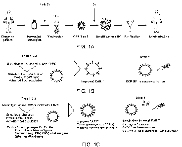
FIG. 1A shows that DCOne mDCs could be added at two different steps in the CAR
T
manufacturing process to: 1) Improve the enrichment and activation status of T
cells (memory
phenotype); 2) Induce additional tumor-targeting specificity in the adoptive T
cell pool (based
on endogenous or exogenous antigens); and/or 3) Improve the expansion of CAR
expressing
T cells (phenotype, viability and CAR expression levels).
FIG. 1B is a schematic depicting the use of DCOne mDCs according to an
embodiment
of the disclosure. FIG. 1C is a schematic depicting the use of DCOne mDCs
according to an
embodiment of the disclosure.
FIG. 2 depicts a plot showing the expression profile of DCOne progenitors and
DCOne
cells with a mature dendritic cell phenotype (mDCs).
FIG. 3 depicts a graph showing the percentage of proliferating cells resulting
from the
addition of DCOne progenitors or DCOne mDCs.
CA 03172449 2022- 9- 20
WO 2021/191870
PCT/1B2021/052542
FIGs. 4A-4G depict plots demonstrating the release of inflammatory and
effector
cytokines in PBMCs stimulated with DCP-001. In particular, the plots depict
the release of IL-
113 (FIG. 4A), GM-CSF (FIG. 4B), IFNy (FIG. 4C), IL-2 (FIG. 4D), TNFa (FIG.
4E), IL-8 (FIG.
4F), and RANTES (FIG. 4G).
5 FIGs.
5A-5C depict plots demonstrating that DCP-001 stimulated T cell proliferation
of
CD3 T cells (FIG. 5A), CD4+ T cells (FIG. 5B) and CD8+ T cells (FIG. 5C) in
healthy donor
and ovarian cancer patients PBMC.
FIGs. 6A-60 depict plots demonstrating the response of antigen specific T cell
clones
against antigens expressed by DCOne mDCs (DCP-001). FIG. 6A shows the response
of
10 PRAME
T cell clones to DCP-001; FIG. 6B shows the response of WT-1 T cell clones to
DCP-
001; FIG. 6C shows the response of MUC-1 T cell clones to DCP-001, and FIG. 60
shows the
response of RHAMM T cell clones to DCP-001.
FIGs. 7A-7B depict a graph demonstrating that DCOne mDCs loaded with exogenous
antigens were a potent stimulator of antigen-specific T cells in vitro as
measured by IFNy
expression. FIG. 7A shows stimulation of WT-1 specific T cells by DCOne mDCs
loaded with
exogenous antigens including matched exogenous antigen (WT-1). FIG. 7B shows
stimulation of NY-ES0-1 specific T cells by DCOne mDCs loaded with exogenous
antigens
including matched exogenous antigen (NY-ES0-1) IFN response the induction of
IFNy in
response to NY-ES0-1 specific T cells.
FIGs. 8A-80 depict graphs showing that in vitro stimulation of PBMCs with DCP-
001
lead to an increased CD45R0 expression in PBMCs from both ovarian cancer
patients (0C
patients; FIG. 8A and FIG. 8C) and healthy donors (FIG. 8B and FIG. 80).
FIGs. 9A-90 depict graphs showing that in vitro stimulation of PBMCs with DCP-
001
triggered an increased CD4+ / CD8+ ratio in PBMCs from both ovarian cancer
patients (00
patients; FIG. 9A and FIG. 9C) and healthy donors (FIG. 9B and FIG. 9D).
FIGs. 10A-10B depicts a graph showing that DCP001 induced T cell activation
and
myeloma-specific immunity in PBMCs of multiple myeloma (MM) patients as
measured by
DCOne RNA uptake (FIG. 10A) and granzyme B killing (FIG. 10B).
FIG. 11 depicts a graph showing that in vitro stimulation of PBMC with DC One
induced
cytotoxic T cells responses towards a variety of leukemic cancer cell lines.
FIG. 12 depicts a graph showing that DCOne mDCs induced cytotoxic T cell
responses
towards the SKOV ovarian cancer cell line from ovarian cancer patients.
FIG. 13 depicts a graph showing the therapeutic rationale for combining DCP-
001 and
adoptive cell therapies in vivo.
FIG. 14 is a plot showing the percent uptake in DCOne mDC cells of CMVpp65-
FITC
or CRM197-CMVpp65-FITC peptides.
CA 03172449 2022- 9- 20
WO 2021/191870
PCT/1B2021/052542
11
FIG. 15 is a plot showing the level of IFN-7 detected in the media of DCOne
mDCs
loaded as indicated.
FIGs. 16A-16C are plots showing the percent uptake of CMVpp65-FITC or CRM197-
CMVpp65-FITC peptides in OVCAR3 (FIG. 16A), 0V90 (FIG. 16B), and U87MG (FIG.
16C)
cells.
FIGs. 17A-17C are plots showing a CMVpp65 T cell clone stimulated with or
without
CRM-CMVpp65 conjugate-pulsed DCOne mDC incubated with HLA-A2+ U87-MG tumor
cells
marked with CRM197-CMVpp65 conjugate/peptide at 5:1 effector : target (E:T)
ratio, and
effector cytokine IFN-y analyzed in the supernatants by ELISA (FIG. 17A).
Stimulation of
CMVpp65-specific CD8 T cells by tumor cells marked with CMVpp65 peptide lead
to an
increase in CD107a expression (FIG. 17B) and lysis of the tumor cells (FIG.
17C).
DETAILED DESCRIPTION
Provided herein are methods for improving the stimulation and expansion of
immune
cells, as well as methods for generating antigen-specific immune cells and
immune cells of a
memory phenotype. Methods for enhancing the effect of genetically modified
immune cells
are also provided. In certain embodiments, the methods comprise contacting a
population of
cells (e.g., comprising immune cells) with a modified cell of leukemic origin.
Methods of
treating a disease or disorder are also provided, comprising the
administration of a non-
proliferating modified cell of leukemic origin into a subject who has
undergone adoptive cell
therapy. Such methods may prolong the duration of the clinical effect of a
genetically modified
immune cell, and/or function to stabilize subjects following adoptive cell
therapy. In certain
embodiments, the modified cell of leukemic origin is non-proliferating (e.g.,
via irradiation). In
certain embodiments, the non-proliferating modified cell of leukemic origin is
a non-
proliferating DCOne derived cell.
It is to be understood that the methods described herein are not limited to
particular
methods and experimental conditions disclosed herein as such methods and
conditions may
vary. It is also to be understood that the terminology used herein is for the
purpose of
describing particular embodiments only, and is not intended to be limiting.
The methods
described herein use conventional molecular and cellular biological and
immunological
techniques that are well within the skill of the ordinary artisan. Such
techniques are well known
to the skilled artisan and are explained in the scientific literature.
A. DEFINITIONS
Unless otherwise defined, scientific and technical terms used herein have the
meanings that are commonly understood by those of ordinary skill in the art.
In the event of
CA 03172449 2022- 9- 20
WO 2021/191870
PCT/1B2021/052542
12
any latent ambiguity, definitions provided herein take precedent over any
dictionary or extrinsic
definition. Unless otherwise required by context, singular terms shall include
pluralities and
plural terms shall include the singular. The use of "or" means "and/or" unless
stated otherwise.
The use of the term "including," as well as other forms, such as "includes"
and "included," is
not limiting.
Generally, nomenclature used in connection with cell and tissue culture,
molecular
biology, immunology, microbiology, genetics and protein and nucleic acid
chemistry and
hybridization described herein is well-known and commonly used in the art. The
methods and
techniques provided herein are generally performed according to conventional
methods well
known in the art and as described in various general and more specific
references that are
cited and discussed throughout the present specification unless otherwise
indicated.
Enzymatic reactions and purification techniques are performed according to
manufacturer's
specifications, as commonly accomplished in the art or as described herein.
The
nomenclatures used in connection with, and the laboratory procedures and
techniques of,
analytical chemistry, synthetic organic chemistry, and medicinal and
pharmaceutical chemistry
described herein are those well-known and commonly used in the art. Standard
techniques
are used for chemical syntheses, chemical analyses, pharmaceutical
preparation, formulation,
and delivery, and treatment of patients.
That the disclosure may be more readily understood, select terms are defined
below.
The articles "a" and "an" are used herein to refer to one or to more than one
(i.e., to at
least one) of the grammatical object of the article. By way of example, "an
element" means
one element or more than one element.
"About" as used herein when referring to a measurable value such as an amount,
a
temporal duration, and the like, is meant to encompass variations of 20% or
10%, e.g., 5%,
1%, or 0.1% from the specified value, as such variations are appropriate to
perform the
disclosed methods.
"Activation," as used herein, refers to the state of a T cell that has been
sufficiently
stimulated to induce detectable cellular proliferation. Activation can also be
associated with
induced cytokine production, and detectable effector functions. The term
"activated T cells"
refers to, among other things, T cells that are undergoing cell division.
As used herein, to "alleviate" a disease means reducing the severity of one or
more
symptoms of the disease.
The term "antigen" as used herein is defined as a molecule that provokes an
immune
response. This immune response may involve either antibody production, or the
activation of
specific immunologically-competent cells, or both. The skilled artisan will
understand that any
macromolecule, including virtually all proteins or peptides, can serve as an
antigen.
CA 03172449 2022- 9- 20
WO 2021/191870
PCT/1B2021/052542
13
The term "antigen" or "antigenic," as used in relation to a polypeptide as
described
herein, refers generally to a biological molecule which contains at least one
epitope specifically
recognized by a T cell receptor, an antibody, or other elements of specific
humoral and/or
cellular immunity. The whole molecule may be recognized, or one or more
portions of the
molecule, for instance following intracellular processing of a polypeptide
into an MHC peptide
antigen complex and subsequent antigen presentation. The term "antigenic
polypeptide" is
interchangeable with "polypeptide antigen." This terminology includes
antigenic parts of said
polypeptides, for instance produced after intracellular processing of a
polypeptide and in the
context of a MHC peptide antigen complex. The term "antigen" or "antigenic"
includes
reference to at least one, or more, antigenic epitopes of a polypeptide as
described herein. In
certain embodiments, a "non-tumor antigen" refers to herein as an antigen that
is not derived
from a tumor. For example, in certain embodiments, a non-tumor antigen may be
a foreign
antigen.
A "tumor-independent antigen" refers to herein as an antigen that is not
derived from
a tumor that a subject is currently suffering from. For example, in certain
embodiments, a
tumor-independent antigen may be a foreign antigen. A tumor-independent
antigen may be
human or non-human. In certain embodiments, in the context of marking a tumor
of a human
subject with a tumor-independent antigen, the tumor-independent antigen may be
of a non-
human origin. In certain embodiments, in the context of marking a tumor of a
host subject
with a tumor-independent antigen, the tumor-independent antigen may be of a
non-host origin.
In certain embodiments, a tumor-independent antigen may be an antigen that is
not expressed
by a tumor that the subject is currently suffering from. For example, if a
subject is currently
suffering from pancreatic cancer, a tumor-independent antigen is a pancreatic-
cancer
independent antigen. In such an example, a pancreatic-cancer independent
antigen can be
an antigen derived from a non-pancreatic cancer that is not expressed by the
pancreatic
cancer, e.g., an ovarian cancer antigen that is not expressed by the
pancreatic cancer.
Similarly, when a certain antigen is associated with a strong immune response
within a certain
tumor type, such antigen could be introduced in tumors of the same type which
do not express
such antigen. This could, e.g., be the case for testis-associated antigens
like NY-ESO-1 in
ovarian cancer.
The tumor-independent antigen can be a recall antigen. The term "recall
antigen," as
used herein, refers to an antigen (e.g., an antigenic polypeptide) which has
previously (e.g.,
prior to the occurrence of a tumor in the subject or prior to a tumor-marking
step) been
encountered by a subject. Recall antigens are those which have previously been
encountered
by the subject and for which there exists pre-existing memory lymphocytes in
the subject (e.g.,
memory T cells and/or memory B cells). In certain embodiments, a recall
antigen refers to an
antigen (e.g., antigenic polypeptide) for which pre-existing memory
lymphocytes exist in the
CA 03172449 2022- 9- 20
WO 2021/191870
PCT/1B2021/052542
14
subject, e.g., as a result of prior infections or vaccinations. In certain
embodiments, a recall
antigen refers to an antigenic polypeptide which has previously been
encountered by a subject
via vaccination. In certain embodiments, the recall antigen is an antigenic
polypeptide for
which there is pre-existing immunity in said subject.
Furthermore, antigens can be derived from recombinant or genomic DNA. A
skilled
artisan will understand that any DNA, which comprises a nucleotide sequences
or a partial
nucleotide sequence encoding a protein that elicits an immune response
therefore encodes
an "antigen" as that term is used herein. Furthermore, one skilled in the art
will understand
that an antigen need not be encoded solely by a full length nucleotide
sequence of a gene. It
is readily apparent that the present disclosure includes, but is not limited
to, the use of partial
nucleotide sequences of more than one gene and that these nucleotide sequences
are
arranged in various combinations to elicit the desired immune response.
Moreover, a skilled
artisan will understand that an antigen need not be encoded by a "gene" at
all. It is readily
apparent that an antigen can be generated synthesized or can be derived from a
biological
sample. Such a biological sample can include, but is not limited to a tissue
sample, a tumor
sample, a cell or a biological fluid.
As used herein, the term "autologous" is meant to refer to any material
derived from
the same individual to which it is later to be re-introduced into the
individual.
A "co-stimulatory ligand" refers to a molecule on an antigen presenting cell
that
specifically binds a cognate co-stimulatory molecule on a T cell, thereby
providing a signal
which, in addition to the primary signal provided by, for instance, binding of
a TCR/CD3
complex with an MHC molecule loaded with peptide, mediates a T cell response,
including,
but not limited to, proliferation activation, differentiation and the like. A
co-stimulatory ligand
can include, but is not limited to, CD7, B7-1 (CD80), B7-2 (CD86), PD-Li, PD-
L2, 4-1 BBL,
OX4OL, inducible costimulatory ligand (ICOS-L), intercellular adhesion
molecule (ICAM,
CD3OL, CD40, CD70, CD83, HLA-G, MICA, M1CB, HVEM, lymphotoxin beta receptor,
3/TR6,
ILT3, ILT4, an agonist or antibody that binds Toll ligand receptor and a
ligand that specifically
binds with B7-H3. A co-stimulatory ligand also encompasses, inter alia, an
antibody that
specifically binds with a co-stimulatory molecule present on a T cell, such
as, but not limited
to, CD27, CD28, 4-IBB, 0X40, CD30, CD40, PD-1, ICOS, lymphocyte function-
associated
antigen-1 (LFA-1), CD2, CD7, LTGHT, NKG2C, B7-H3, and a ligand that
specifically binds
with CD83.
A "co-stimulatory molecule" refers to the cognate binding partner on a T cell
that
specifically binds with a co-stimulatory ligand, thereby mediating a co-
stimulatory response by
the cell, such as, but not limited to proliferation. Co-stimulatory molecules
include, but are not
limited to, an MHC class I molecule, BTLA and Toll ligand receptor. Examples
of costimulatory
molecules include CD27, CD28, CD8, 4-1BB (CD137), 0X40, CD30, CD40, PD-1,
ICOS,
CA 03172449 2022- 9- 20
WO 2021/191870
PCT/1B2021/052542
lymphocyte function-associated antigen-1 (LFA-1), CD2, CD7, LIGHT, NKG2C, B7-
H3, a
ligand that specifically binds with CD83, and the like.
A "co-stimulatory signal," as used herein, refers to a signal, which in
combination with
a primary signal, such as TCR/CD3 ligation, leads to T cell proliferation
and/or upregulation or
5
downregulation of key molecules. In certain exemplary embodiments, the co-
stimulatory
signal is CD70.
A "disease" is a state of health of an animal wherein the animal cannot
maintain
homeostasis, and wherein if the disease is not ameliorated then the animal's
health continues
to deteriorate. In contrast, a "disorder" in an animal is a state of health in
which the animal is
10 able
to maintain homeostasis, but in which the animal's state of health is less
favorable than
it would be in the absence of the disorder. Left untreated, a disorder does
not necessarily
cause a further decrease in the animal's state of health.
"Effective amount" or "therapeutically effective amount" are used
interchangeably
herein, and refer to an amount of a compound, formulation, material, or
composition, as
15
described herein effective to achieve a particular biological result or
provides a therapeutic or
prophylactic benefit. Such results may include, but are not limited to an
amount that when
administered to a mammal, causes a detectable level of immune suppression or
tolerance
compared to the immune response detected in the absence of the composition of
the
disclosure. The immune response can be readily assessed by a plethora of art-
recognized
methods. The skilled artisan would understand that the amount of the
composition
administered herein varies and can be readily determined based on a number of
factors such
as the disease or condition being treated, the age and health and physical
condition of the
mammal being treated, the severity of the disease, the particular compound
being
administered, and the like.
"Encoding" refers to the inherent property of specific sequences of
nucleotides in a
polynucleotide, such as a gene, a cDNA, or an mRNA, to serve as templates for
synthesis of
other polymers and macromolecules in biological processes having either a
defined sequence
of nucleotides (i.e., rRNA, tRNA and mRNA) or a defined sequence of amino
acids and the
biological properties resulting therefrom. Thus, a gene encodes a protein if
transcription and
translation of mRNA corresponding to that gene produces the protein in a cell
or other
biological system. Both the coding strand, the nucleotide sequence of which is
identical to the
mRNA sequence and is usually provided in sequence listings, and the non-coding
strand,
used as the template for transcription of a gene or cDNA, can be referred to
as encoding the
protein or other product of that gene or cDNA.
As used herein "endogenous" refers to any material from or produced inside an
organism, cell, tissue or system.
CA 03172449 2022- 9- 20
WO 2021/191870
PCT/1B2021/052542
16
As used herein, the term "exogenous" refers to any material introduced from or
produced outside an organism, cell, tissue or system.
The term "expand" as used herein refers to increasing in number, as in an
increase in
the number of T cells. In one embodiment, the T cells that are expanded ex
vivo increase in
number relative to the number originally present in the culture. In another
embodiment, the T
cells that are expanded ex vivo increase in number relative to other cell
types in the culture.
The term "ex vivo," as used herein, refers to cells that have been removed
from a living
organism, (e.g., a human) and propagated outside the organism (e.g., in a
culture dish, test
tube, or bioreactor).
The term "expression" as used herein is defined as the transcription and/or
translation
of a particular nucleotide sequence driven by its promoter.
An "expression vector" refers to a vector comprising a recombinant
polynucleotide
comprising expression control sequences operatively linked to a nucleotide
sequence to be
expressed. An expression vector comprises sufficient cis-acting elements for
expression;
other elements for expression can be supplied by the host cell or in an in
vitro expression
system. Expression vectors include all those known in the art, such as
cosmids, plasmids
(e.g., naked or contained in liposomes) and viruses (e.g., Sendai viruses,
lentiviruses,
retroviruses, adenoviruses, and adeno-associated viruses) that incorporate the
recombinant
polynucleotide.
The term "immune response," as used herein, includes T cell mediated and/or B
cell
mediated immune responses. Exemplary immune functions of T cells include,
e.g., cytokine
production and induction of cytotoxicity in other cells. B cell functions
include antibody
production. In addition, the term includes immune responses that are
indirectly affected by T
cell activation, e.g., antibody production and activation of cytokine
responsive cells, e.g.,
macrophages. Immune cells involved in the immune response include lymphocytes,
such as
B cells and T cells (CD4+ and CD8+ cells); antigen presenting cells (e.g.,
professional antigen
presenting cells such as dendritic cells, macrophages, B lymphocytes,
Langerhans cells, and
non-professional antigen presenting cells such as keratinocytes, endothelial
cells, astrocytes,
fibroblasts, oligodendrocytes); natural killer cells; myeloid cells, such as
macrophages,
eosinophils, mast cells, basophils, and granulocytes. In certain embodiments,
the term refers
to a T cell mediated immune response. The immune response may in some
embodiments be
a T cell-dependent immune response. The skilled person understands that the
phrase
"immune response against a tumor" also includes immune responses against a non-
human
antigenic polypeptide that is introduced into the tumor micro-environment by
intratumoral
administration, such as intratumoral administration of (i) dendritic cells,
including autologous
or allogeneic dendritic cells, loaded with said polypeptide or (ii) viruses
comprising a nucleic
acid encoding said polypeptide.
CA 03172449 2022- 9- 20
WO 2021/191870
PCT/1B2021/052542
17
The term "T cell dependent immune response," as used herein, refers to an
immune
response wherein either T cells, B cells or both T cell and B cell populations
are activated, and
wherein T cells further assist T and B cells and other immune cells in
executing their function.
The term "immunosuppressive" is used herein to refer to reducing overall
immune
response.
"Insertion/deletion," commonly abbreviated "indel," is a type of genetic
polymorphism
in which a specific nucleotide sequence is present (insertion) or absent
(deletion) in a genome.
"Isolated" means altered or removed from the natural state. For example, a
nucleic
acid or a peptide naturally present in a living animal is not "isolated," but
the same nucleic acid
or peptide partially or completely separated from the coexisting materials of
its natural state is
"isolated." An isolated nucleic acid or protein can exist in substantially
purified form, or can
exist in a non-native environment such as, for example, a host cell.
A "Ientivirus" as used herein refers to a genus of the Retroviridae family.
Lentiviruses
are unique among the retroviruses in being able to infect non-dividing cells.
They can deliver
a significant amount of genetic information into the DNA of the host cell, so
they are one of
the most efficient methods of a gene delivery vector. HIV, Sly, and FIV are
all examples of
lentiviruses. Vectors derived from lentiviruses offer the means to achieve
significant levels of
gene transfer in vivo.
By the term "modified" as used herein, is meant a changed state or structure
of a
molecule or cell of the disclosure. Molecules may be modified in many ways,
including
chemically, structurally, and functionally. Cells may be modified through the
introduction of
nucleic acids.
By the term "modulating," as used herein, is meant mediating a detectable
increase or
decrease in the level of a response in a subject compared with the level of a
response in the
subject in the absence of a treatment or compound, and/or compared with the
level of a
response in an otherwise identical but untreated subject. The term encompasses
perturbing
and/or affecting a native signal or response thereby mediating a beneficial
therapeutic
response in a subject, e.g., a human.
Unless otherwise specified, a "nucleotide sequence encoding an amino acid
sequence" includes all nucleotide sequences that are degenerate versions of
each other and
that encode the same amino acid sequence. The phrase nucleotide sequence that
encodes
a protein or an RNA may also include introns to the extent that the nucleotide
sequence
encoding the protein may in some version contain an intron(s).
"Parenteral" administration of an immunogenic composition includes, e.g.,
subcutaneous (s.c.), intravenous (i.v.), intramuscular (i.m.), intradermal,
intraperitoneal, or
intrasternal injection, or infusion techniques.
CA 03172449 2022- 9- 20
WO 2021/191870
PCT/1B2021/052542
18
The term "polynucleotide," as used herein, is defined as a chain of
nucleotides.
Furthermore, nucleic acids are polymers of nucleotides.
Thus, nucleic acids and
polynucleotides as used herein are interchangeable. One skilled in the art has
the general
knowledge that nucleic acids are polynucleotides, which can be hydrolyzed into
the
monomeric "nucleotides." The monomeric nucleotides can be hydrolyzed into
nucleosides.
As used herein polynucleotides include, but are not limited to, all nucleic
acid sequences which
are obtained by any means available in the art, including, without limitation,
recombinant
means, i.e., the cloning of nucleic acid sequences from a recombinant library
or a cell genome,
using ordinary cloning technology and PCR, and the like, and by synthetic
means.
As used herein, the terms "peptide," "polypeptide," and "protein" are used
interchangeably, and refer to a compound comprised of amino acid residues
covalently linked
by peptide bonds. A protein or peptide must contain at least two amino acids,
and no limitation
is placed on the maximum number of amino acids that can comprise a protein's
or peptide's
sequence. Polypeptides include any peptide or protein comprising two or more
amino acids
joined to each other by peptide bonds. As used herein, the term refers to both
short chains,
which also commonly are referred to in the art as peptides, oligopeptides and
oligomers, for
example, and to longer chains, which generally are referred to in the art as
proteins, of which
there are many types. "Polypeptides" include, for example, biologically active
fragments,
substantially homologous polypeptides, oligopeptides, homodimers,
heterodimers, variants of
polypeptides, modified polypeptides, derivatives, analogs, fusion proteins,
among others. The
polypeptides include natural peptides, recombinant peptides, synthetic
peptides, or a
combination thereof.
By the term "specifically binds," as used herein with respect to an antibody,
is meant
an antibody which recognizes a specific antigen, but does not substantially
recognize or bind
other molecules in a sample. For example, an antibody that specifically binds
to an antigen
from one species may also bind to that antigen from one or more species. But,
such cross-
species reactivity does not itself alter the classification of an antibody as
specific. In another
example, an antibody that specifically binds to an antigen may also bind to
different allelic
forms of the antigen. However, such cross reactivity does not itself alter the
classification of
an antibody as specific. In some instances, the terms "specific binding" or
"specifically
binding," can be used in reference to the interaction of an antibody, a
protein, or a peptide with
a second chemical species, to mean that the interaction is dependent upon the
presence of a
particular structure (e.g., an antigenic determinant or epitope) on the
chemical species. For
example, an antibody recognizes and binds to a specific protein structure
rather than to
proteins generally. If an antibody is specific for epitope "A," the presence
of a molecule
containing epitope A (or free, unlabeled A), in a reaction containing labeled
"A" and the
antibody, will reduce the amount of labeled A bound to the antibody.
CA 03172449 2022- 9- 20
WO 2021/191870
PCT/1B2021/052542
19
By the term "stimulation," is meant a primary response induced by binding of a
stimulatory molecule (e.g., a TCR/CD3 complex) with its cognate ligand thereby
mediating a
signal transduction event, such as, but not limited to, signal transduction
via the TCR/CD3
complex. Stimulation can mediate altered expression of certain molecules, such
as
downregulation of TGF-beta, and/or reorganization of cytoskeletal structures,
and the like.
A "stimulatory molecule," as the term is used herein, means a molecule on a T
cell that
specifically binds with a cognate stimulatory ligand present on an antigen
presenting cell.
A "stimulatory ligand," as used herein, means a ligand that when present on an
antigen
presenting cell (e.g., an aAPC, a dendritic cell, a B cell, and the like) can
specifically bind with
a cognate binding partner (referred to herein as a "stimulatory molecule") on
a T cell, thereby
mediating a primary response by the T cell, including, but not limited to,
activation, initiation of
an immune response, proliferation, and the like. Stimulatory ligands are well-
known in the art
and encompass, inter alia, an MHC Class I molecule loaded with a peptide, an
anti-CD3
antibody, a superagonist anti-CD28 antibody, and a superagonist anti-CD2
antibody.
The term "subject," as used herein, refers to the recipient of a method as
described
herein, i.e., a recipient that can mount a cellular immune response, and is a
mammal. In
certain embodiments, the subject is a human. In certain embodiments, the
subject is a
domesticated animal, e.g., a horse, a cow, a pig, a sheep, a dog, a cat, etc.
The terms "patient"
and "subject" may be used interchangeably. In certain embodiments, the subject
is a human
suffering from a tumor (e.g., a solid tumor). In certain embodiments, the
subject is a
domesticated animal suffering from a tumor (e.g., a solid tumor).
As used herein, the term "T cell receptor" or "TCR" refers to a complex of
membrane
proteins that participate in the activation of T cells in response to the
presentation of antigen.
The TCR is responsible for recognizing antigens bound to major
histocompatibility complex
molecules. TCR is composed of a heterodimer of an alpha (a) and beta (13)
chain, although
in some cells the TCR consists of gamma and delta (y/6) chains. TCRs may exist
in alpha/beta
and gamma/delta forms, which are structurally similar but have distinct
anatomical locations
and functions. Each chain is composed of two extracellular domains, a variable
and constant
domain. In some embodiments, the TCR may be modified on any cell comprising a
TCR,
including, for example, a helper T cell, a cytotoxic T cell, a memory T cell,
regulatory T cell,
natural killer T cell, and gamma delta T cell.
The term "therapeutic" as used herein means a treatment and/or prophylaxis. A
therapeutic effect is obtained by suppression, remission, or eradication of a
disease state.
The term "transfected" or "transformed" or "transduced" as used herein refers
to a
process by which exogenous nucleic acid is transferred or introduced into the
host cell. A
"transfected" or "transformed" or "transduced" cell is one which has been
transfected,
CA 03172449 2022- 9- 20
WO 2021/191870
PCT/1B2021/052542
transformed or transduced with exogenous nucleic acid. The cell includes the
primary subject
cell and its progeny.
To "treat" a disease as the term is used herein, means to reduce the frequency
or
severity of at least one sign or symptom of a disease or disorder experienced
by a subject.
5 The
term "tumor," as used herein, includes reference to cellular material, e.g., a
tissue,
proliferating at an abnormally high rate. A growth comprising neoplastic cells
is a neoplasm,
also known as a "tumor," and generally forms a distinct tissue mass in a body
of a subject. A
tumor may show partial or total lack of structural organization and functional
coordination with
the normal tissue. As used herein, a tumor is intended to encompass
hematopoietic tumors
10 as well
as solid tumors. In certain embodiments, the tumor is a solid tumor. The term
"tumor,"
as used herein, includes reference to the tumor micro-environment or tumor
site, i.e., the area
within the tumor and the area directly outside the tumorous tissue. In certain
embodiments,
the tumor micro-environment or tumor site includes an area within the
boundaries of the tumor
tissue. In certain embodiments, the tumor micro-environment or tumor site
includes the tumor
15
interstitial compartment of a tumor, which is defined herein as all that is
interposed between
the plasma membrane of neoplastic cells and the vascular wall of the newly
formed
neovessels. As used herein, the terms "tumor micro-environment" or "tumor
site" refers to a
location within a subject in which a tumor resides, including the area
immediately surrounding
the tumor.
20 A tumor
may be benign (e.g., a benign tumor) or malignant (e.g., a malignant tumor or
cancer). Malignant tumors can be broadly classified into three major types:
those arising from
epithelial structures are called carcinomas, those that originate from
connective tissues such
as muscle, cartilage, fat or bone are called sarcomas, and those affecting
hematopoietic
structures (structures pertaining to the formation of blood cells) including
components of the
immune system, are called leukemias and lymphomas. Other tumors include, but
are not
limited to, neurofibromatosis. In certain exemplary embodiments, the tumor is
a glioblastoma.
In certain exemplary embodiments, the tumor is an ovarian cancer (e.g., an
epithelial ovarian
cancer, which can be further subtyped into a serous, a clear cell, an
endometrioid, a nrrucinous,
or a mixed epithelial ovarian cancer).
Solid tumors are abnormal masses of tissue that can be benign or malignant. In
certain
embodiments, solid tumors are named for the type of cells that form them (such
as sarcomas,
carcinomas, and lymphomas). Examples of solid tumors, such as sarcomas and
carcinomas,
include, but are not limited to, liposarcoma, fibrosarcoma, chondrosarcoma,
osteosarcoma,
myxosarcoma, and other sarcomas, mesothelioma, synovioma, leiomyosarcoma,
Ewing's
tumor, colon carcinoma, rhabdomyosarcoma, pancreatic cancer, lymphoid
malignancy, lung
cancers, breast cancer, prostate cancer, ovarian cancer, hepatocellular
carcinoma,
adenocarcinoma, basal cell carcinoma, sweat gland carcinoma, squamous cell
carcinoma,
CA 03172449 2022- 9- 20
WO 2021/191870
PCT/1B2021/052542
21
medullary thyroid carcinoma, pheochromocytomas sebaceous gland carcinoma,
papillary
thyroid carcinoma, papillary adenocarcinomas, papillary carcinoma, medullary
carcinoma,
bronchogenic carcinoma, hepatoma, renal cell carcinoma, bile duct carcinoma,
Wilms' tumor,
choriocarcinoma, cervical cancer, seminoma, testicular tumor, bladder
carcinoma, melanoma,
CNS tumors (e.g., a glioma, e.g., brainstem glioma and mixed gliomas,
glioblastoma (e.g.,
glioblastoma multiforrne), germinoma, astrocytoma, craniopharyngioma,
nnedulloblastoma,
ependymoma, Schwannoma, CNS lymphoma, acoustic neuroma, pinealoma,
hemangioblastoma, meningioma, oligodendroglioma, retinoblastoma,
neuroblastoma, and
brain metastases), and the like.
Carcinomas that can be amenable to therapy by a method disclosed herein
include,
but are not limited to, squamous cell carcinoma (various tissues), basal cell
carcinoma (a form
of skin cancer), esophageal carcinoma, bladder carcinoma, including
transitional cell
carcinoma (a malignant neoplasm of the bladder), hepatocellular carcinoma,
colorectal
carcinoma, bronchogenic carcinoma, lung carcinoma, including small cell
carcinoma and non-
small cell carcinoma of the lung, colon carcinoma, thyroid carcinoma, gastric
carcinoma,
breast carcinoma, ovarian carcinoma, adrenocortical carcinoma, pancreatic
carcinoma, sweat
gland carcinoma, prostate carcinoma, papillary carcinoma, adenocarcinoma,
sebaceous
gland carcinoma, medullary carcinoma, papillary adenocarcinoma, ductal
carcinoma in situ or
bile duct carcinoma, cystadenocarcinoma, renal cell carcinoma,
choriocarcinoma, Wilm's
tumor, seminoma, embryonal carcinoma, cervical carcinoma, testicular
carcinoma,
nasopharyngeal carcinoma, osteogenic carcinoma, epithelial carcinoma, uterine
carcinoma,
and the like.
Sarcomas that can be amenable to therapy by a method disclosed herein include,
but
are not limited to, myxosarcoma, chondrosarcoma, chordoma, osteogenic sarcoma,
liposarcoma, fibrosarcoma, angiosarcoma, lymphangiosarcoma, endotheliosarcoma,
osteosarcoma, mesothelioma, Ewing's sarcoma, leiomyosarcoma, rhabdomyosarcoma,
lymphangioendotheliosarcoma, synovioma, and other soft tissue sarcomas.
A "vector" is a composition of matter which comprises an isolated nucleic acid
and
which can be used to deliver the isolated nucleic acid to the interior of a
cell. Numerous
vectors are known in the art including, but not limited to, linear
polynucleotides,
polynucleotides associated with ionic or amphiphilic compounds, plasmids, and
viruses. Thus,
the term "vector" includes an autonomously replicating plasmid or a virus. The
term should
also be construed to include non-plasmid and non-viral compounds which
facilitate transfer of
nucleic acid into cells, such as, for example, polylysine compounds,
liposomes, and the like.
Examples of viral vectors include, but are not limited to, Sendai viral
vectors, adenoviral
vectors, adeno-associated virus vectors, retroviral vectors, lentiviral
vectors, and the like.
CA 03172449 2022- 9- 20
WO 2021/191870
PCT/1B2021/052542
22
The term "immunogenic composition," as used herein, refers to a substance
which
induces a specific immune response against an immunogen in a subject who is in
need of an
immune response against said immunogen. The composition may include an
adjuvant and
optionally one or more pharmaceutically-acceptable carriers, excipients and/or
diluents. The
immunogenic composition can be employed in prime-boost vaccination, such as at
least 2, 3,
4 or at least 5 immunizations separated in time. The immunogenic composition
can be an
(allogeneic) dendritic cell comprising said immunogen.
The term "immunogen," as used herein, refers to a compound such as a
polypeptide
capable of eliciting an immune response that is specifically directed against
an antigenic
polypeptide as described herein. An immunogen is also an antigen, i.e., an
antigenic
polypeptide. In contrast, an antigen is not necessarily an immunogen. In
certain embodiments,
the immunogen is used for vaccination (in an immunogenic composition such as a
vaccine
composition), and the antigenic polypeptide prepared for intratumoral delivery
is instead used
for marking a tumor as a target for an immune response to be elicited, or as a
target for an
immune response that is already elicited, in a subject. The term "immunogen"
is also used to
refer to a nucleic acid which encodes the non-human antigenic polypeptide as
described
herein. In addition, embodiments that describe the antigenic polypeptide, also
apply to an
immunogen as described herein.
The term "non-human," as used herein in the context of an antigenic
polypeptide,
includes polypeptides that are not of human origin, including a bacterial
polypeptide, a
polypeptide of an organism of the Archaea domain, a fungal polypeptide and a
viral
polypeptide. Also included are plant polypeptides and non-human mammalian
polypeptides
such as polypeptides of non-human primates, rodents (e.g., mice and rats),
rabbits, pigs,
sheep, goats, cows, pigs, horses and donkeys, and birds (e.g., chickens,
turkeys, ducks,
geese and the like). Also included are polypeptides of snails or other
mollusks, including
Megathura crenulata. The term "non-human" also encompasses synthetic
polypeptides, i.e.,
polypeptides that have an artificial sequence designed by man and that do not
occur in nature
or are not yet identified in nature. In addition, the term comprises human
polypeptides
comprising an amino acid alteration from the native sequence, the alteration
providing for
immunogenicity in a human subject.
The term "intratumoral," as used herein, refers to delivery or transport of
the antigenic
polypeptide, or the nucleic acid encoding said polypeptide, into a tumor. One
example of
intratumoral delivery, or transport, of an antigenic polypeptide as described
herein is by
intratumoral administration, a route of administration generally known in the
art. As an
alternative route for intratumoral administration, the antigen may be
delivered to the tumor via
a tumor-specific carrier, such as an oncolytic virus or a gene therapy vector,
which have been
broadly developed to deliver gene sequences to tumors. The use of such
vehicles allows for
CA 03172449 2022- 9- 20
WO 2021/191870
PCT/1B2021/052542
23
multiple routes of administration, in addition to intratumoral administration,
such by as
intravenous or intraperitoneal administration, subsequently resulting in the
delivery of the
nucleic acid encoding said polypeptide, into the tumor (Lundstrom, Diseases,
6(2):42 (2018);
Alemany, Biomedicines, 2, p.36-49 (2014); Twumasi-Boateng et al., Nature
Reviews Cancer
18, p.419-432 (2018).
The phrase "prepared for intratumoral delivery," as used herein, refers to an
antigenic
polypeptide as described herein, or a nucleic acid encoding said polypeptide
as described
herein, that is adapted for intratumoral delivery and/or is in a formulation
that allows for
intratumoral delivery. The preparation used for intratumoral delivery may be
composed such
that it has a beneficial effect on the interaction between the immune system
and the tumor.
For instance, dendritic cells, such as autologous or allogeneic dendritic
cells, can be loaded
with said polypeptide and upon intratumoral administration may provide for
additional immune
stimulation via direct interaction with T cells entering the tumor and/or
indirectly by recruiting
bystander antigen-presenting cells (Laurell et al., Journal for Immunotherapy
of Cancer, 5:52
(2017); Wallgren et al., Scandinavian Journal of Immunology, 62, p.234-242
(2005). Another
example of such preparation is that the polypeptide or nucleic acid as
described herein can
be comprised in a tumor-delivery vehicle such as a tumor-targeted vehicle
including a tumor-
specific virus such as an oncolytic virus (or any other virus that selectively
replicates in tumor
cells) that infects a tumor cell and which allows for (i) expression of said
nucleic acid in a tumor
cell, and (ii) (subsequently) intracellular processing and antigen
presentation (MHC) of said
(expressed) polypeptide by said tumor cell. The skilled person is well aware
of other methods
and means for preparing a polypeptide, or a nucleic acid encoding said
polypeptide, for
intratumoral delivery. For instance, the skilled person can apply other tumor-
targeted delivery
vehicles such as a tumor-specific nanoparticle or he can apply intratumoral
administration
through intratumoral injection in order to deliver said polypeptide or nucleic
acid into a tumor.
In certain embodiments, the polypeptide or nucleic acid prepared for
intratumoral delivery as
described herein, is comprised in a tumor-targeted vehicle.
As used herein, the term "extratumoral" refers to a location, e.g., in the
body of a
subject, that is away (e.g., distal) from a tumor and immediately surrounding
tissue (e.g., that
may make up the tumor micro-environment).
The compositions for use as described herein, elicit an immune response
specifically
directed against a tumor in a subject. The skilled person understands that
"specifically
directed" refers to an immune response that is specific for a tumor. The
specificity is
introduced by a step of marking a tumor with a non-human antigenic polypeptide
as a target
for an immune response, and by eliciting an immune response against an
antigenic part of
said non-human antigenic polypeptide (i.e., the target). Thus, in certain
embodiments, the
compositions for use as described herein, is for use in eliciting an immune
response against
CA 03172449 2022- 9- 20
WO 2021/191870
PCT/1B2021/052542
24
a tumor marked as a target for said immune response. In certain embodiments,
the
compositions for use as described herein, is for use in eliciting an immune
response against
a tumor that is marked as a target for said immune response; wherein said
target is a non-
human antigenic polypeptide as described herein.
In certain embodiments, the non-human antigenic polypeptide, or a nucleic acid
encoding said polypeptide, prepared for intratumoral delivery as described
herein, serves the
purpose of marking the tumor as a target for an immune response
(polypeptide/nucleic acid
for marking a tumor). Thus, in certain embodiments, said polypeptide or said
nucleic acid
prepared for intratumoral delivery marks the tumor as a target for an immune
response
following intratumoral delivery.
As used herein, the term "vaccination step" refers to a step in a method
(vaccination
strategy) as described herein, wherein a composition comprising an antigenic
polypeptide
(e.g., a non-tumor antigen) or a nucleic acid encoding an antigenic
polypeptide is administered
to a subject at a site distal to a tumor site. In certain embodiments, in a
vaccination step of a
method as described herein, the composition is administered at a site that is
not the site in
which the tumor resides (e.g., not the tumor site). In certain embodiments, in
a vaccination
step of a method as described herein, the composition is administered at an
extratumoral site.
As used herein, the term "booster step" refers to a step in a method
(vaccination
strategy) as described herein, wherein a booster composition comprising an
antigenic
polypeptide (e.g., a non-tumor antigen) or a nucleic acid encoding the
antigenic polypeptide
is administered to a subject at a site distal to a tumor site. In certain
embodiments, a booster
step is performed after a vaccination step, wherein the vaccination step
results in an immune
response against the antigen, and the booster step enhances the immune
response against
the antigen. In certain embodiments, a booster step results in an enhanced
immune response
in a subject having pre-existing immunity against, e.g., an antigenic
polypeptide (e.g., a non-
tumor antigen). In certain embodiments, the vaccination step in a method as
described herein
is a booster stem, e.g., when the subject has pre-existing immunity against,
e.g., a non-tumor
recall antigen.
The term "marking," "mark" or "marked," as used herein, refers to active
manipulation
of the antigenic state of a tumor by intratumoral delivery of an antigenic
polypeptide, or a
nucleic acid encoding said polypeptide, as described herein. This provides for
direct labelling
of a tumor cell through intracellular delivery and subsequent processing and
presentation of
said polypeptide by said tumor cell, or provides for indirect labelling of a
tumor via: (i)
intracellular delivery and subsequent processing and presentation of said
polypeptide by a
non-tumor cell in said tumor; or (ii) extracellular delivery of said antigenic
polypeptide to said
tumor (i.e., extracellular to the cells present in said tumor before marking),
for instance by
using a dendritic cell that comprises a nucleic acid encoding said polypeptide
or that is loaded
CA 03172449 2022- 9- 20
WO 2021/191870
PCT/1B2021/052542
with said antigenic polypeptide. As used herein, the term "tumor-marking step"
refers to a step
in a method (e.g., a vaccination strategy) as described herein, wherein a
composition
comprising an antigenic polypeptide (e.g., a non-tumor antigen) or a nucleic
acid encoding an
antigenic polypeptide is administered to a subject at a tumor site.
5 The term "modified cell of leukemic origin," as used herein, refers to
a cell that can
take up an antigen such as an antigenic polypeptide into its cell, and
presents the antigen, or
an immunogenic part thereof together with an MHC class I complex or MHC class
II complex.
In certain embodiments, the modified cell of leukemic origin is a cell derived
from cell line
DCOne as deposited under the conditions of the Budapest treaty with the DSMZ
under
10 accession number DSMZ ACC3189 on 15 November 2012. The process of
obtaining mature
cells from the deposited DCOne cell line is, for instance, described in
EP2931878131.
Ranges: throughout this disclosure, various aspects of the disclosure can be
presented
in a range format. It should be understood that the description in range
format is merely for
convenience and brevity and should not be construed as an inflexible
limitation on the scope
15 of the disclosure. Accordingly, the description of a range should be
considered to have
specifically disclosed all the possible subranges as well as individual
numerical values within
that range. For example, description of a range such as from 1 to 6 should be
considered to
have specifically disclosed subranges such as from 1 to 3, from 1 to 4, from 1
to 5, from 2 to
4, from 2 to 6, from 3 to 6 etc., as well as individual numbers within that
range, for example,
20 1, 2, 2.7, 3, 4, 5, 5.3, and 6. This applies regardless of the breadth
of the range.
B. MODIFIED CELL OF LEUKEMIC ORIGIN
Provided herein are methods comprising the use of a modified cell of leukemic
origin
to stimulate and expand immune cells, generate antigen-specific immune cells,
and for
25 methods of treatment. As used herein, the term "modified cell of
leukemic origin" refers to a
cell capable of taking up an antigen such as an antigenic polypeptide, and
capable of
presenting the antigen, or an immunogenic part thereof, together with an MHC
class I complex
or MHC class ll complex. A modified cell of leukemic origin provided herein
comprises a
mature dendritic cell phenotype. The term "dendritic cell," as used herein,
refers to a
professional antigen presenting cell (APC) that can take up an antigen such as
an antigenic
polypeptide into its cell, and presents the antigen, or an immunogenic part
thereof together
with an MHC class I complex or MHC class II complex. Having a mature dendritic
cell
phenotype means that the modified cell of leukemic origin is capable of
performing similar
functions to those of a mature dendritic cell. The term includes both immature
dendritic cells
("imDC") and mature dendritic cells ("mDC"), depending on maturity.
In certain embodiments, the modified cell of leukemic origin is derived from
leukemia
cells. In certain embodiments, the modified cell of leukemic origin is derived
from a patient
CA 03172449 2022- 9- 20
WO 2021/191870
PCT/1B2021/052542
26
having leukemia. In certain embodiments, the modified cell of leukemic origin
is derived from
the peripheral blood of a patient having leukemia. In certain embodiments, the
modified cell
of leukemic origin is derived from the peripheral blood of a patient having
acute myeloid
leukemia. The skilled artisan will recognize that a modified cell of leukemic
origin can be
derived from any patient obtained peripheral blood, wherein the patient has
any type of
leukemia, given that the modified cell of leukemic origin thus derived
comprises the
characteristics disclosed herein.
In certain embodiments, the modified cell of leukemic origin is CD34-positive,
CD1a-
positive, and CD83-positive. In certain embodiments, the modified cell of
leukemic origin
comprises a cell surface marker selected from the group consisting of CD14, DC-
SIGN,
Langerin, CD40, CD70, CD80, CD83, CD86, and any combination thereof. In
certain
embodiments, the modified cell of leukemic origin comprises an MHC class I
molecule. In
certain embodiments, the modified cell of leukemic origin comprises an MHC
class II molecule.
In certain embodiments, the modified cell of leukemic origin is CD34-positive,
CD1a-positive,
0D83-positive, and CD14-negative. In certain embodiments, the modified cell of
leukemic
origin is CD40-positive, CD80-positive, and CD86-positive. In certain
embodiments, the
modified cell of leukemic origin is CD34-positive, CD1a-positive, CD83-
positive, CD40-
positive, CD80-positive, CD86-positive, and CD14-negative.
In certain embodiments, the modified cell of leukemic origin comprises a
genetic
aberration between chromosome 11p15.5 to 11p12. In certain embodiments, the
genetic
aberration encompasses about 16 Mb of genomic regions (e.g., from about 20.7
Mb to about
36.6 Mb). In certain embodiments, the genetic aberration contains a loss of
about 60 known
and unknown genes
In certain embodiments, the modified cell of leukemic origin comprises a co-
stimulatory
molecule. In certain embodiments, the co-stimulatory molecule includes,
without limitation, an
MHC class I molecule, BTLA and Toll ligand receptor. Examples of co-
stimulatory molecules
include CD70, CD80, CD86, 4-1BBL (CD137-ligand), 0X40L, CD3OL, CD40, PD-L1,
ICOSL,
ICAM-1, lymphocyte function-associated antigen 3 (LFA3 (CD58)), K12/SECTM1,
LIGHT,
HLA-E, 67-H3 and CD83.
In certain embodiments, the modified cell of leukemic origin comprises at
least one
endogenous antigen. Depending on the leukemic origin of the modified cell, the
modified cell
of leukemic origin may comprise at least one known endogenous antigen that is
specific to the
leukemic origin. In certain embodiments, the endogenous antigen is a tumor-
associated
antigen. In certain embodiments, an endogenous tumor-associated antigen may be
selected
from the group consisting of WT-1, RHAMM, PRAME, p53, Survivin, and MUC-1.
In certain embodiments, the modified cell of leukemic origin comprises an
exogenous
antigen or peptide fragments thereof. Such an exogenous antigen may be
provided to the
CA 03172449 2022- 9- 20
WO 2021/191870
PCT/1B2021/052542
27
modified cell of leukemic origin via various antigen loading strategies. For
example, strategies
for loading a modified cell of leukemic origin may include, without
limitation, the use of
synthetic long peptides, mRNA loading, peptide-pulsing, protein-loading, tumor
lysate-loading,
coculturing with a tumor cell, RNA/DNA transfection or viral transduction.
Other strategies for
loading a modified cell of leukemic origin are known to those of skill in the
art and may be used
to load a modified cell of leukemic origin with an exogenous antigen. In
general, the modified
cell of leukemic origin will process the exogenous antigen via particular
molecules, e.g., via
MHC I or MHC II. As such, an exogenous antigen comprised by the modified cell
of leukemic
origin may be an MHC class I antigen or an MHC class II antigen. In certain
embodiments,
the exogenous antigen is a tumor-associated antigen. For example, in certain
embodiments,
the modified cell of leukemic origin is loaded with NY-ESO-1 peptide and/or
VVT-1 peptide, or
a tumor-independent antigen such as CMVpp65. In certain embodiments, the
exogenous
antigen is associated with a disease or disorder, e.g., a non-cancer-
associated disease or
disorder. It will be appreciated by those of ordinary skill in the art that
any tumor-associated
antigen or antigen associated with a disease or disorder can be provided to
the modified cell
of leukemic origin described herein. As such, in certain embodiments, a
modified cell of
leukemic origin comprises any tumor-associated antigen or antigen associated
with a disease
or disorder contemplated by those skilled in the art.
In certain embodiments, the exogenous antigen is a non-tumor-associated
antigen
(i.e., a tumor-independent antigen). In certain embodiments, the modified cell
of leukemic
origin is loaded with a tumor-independent antigen, i.e. an antigen not
associated with a tumor.
For example, suitable tumor-independent antigens include, without limitation,
proteins of viral,
bacterial, fungal origin; allergens, toxins and venoms, or model antigens of
various sources
such as chicken egg ovalbumin and keyhole limpet hemocyanin from the giant
keyhole limpet,
Megathura crenulata. In certain embodiments, a suitable tumor-independent
antigen is of
bacterial origin. In certain embodiments, a suitable tumor-independent antigen
is a diphtheria
toxin. In certain embodiments, a suitable tumor-independent antigen is a non-
toxic variant of
diphtheria toxin. For example, in certain embodiments, a suitable tumor-
independent antigen
is CRM197 or a variant thereof. In certain embodiments, a modified cell of
leukemic origin
comprises CRM197 or a variant thereof. In certain embodiments, a suitable
tumor-
independent antigen is of viral origin. In certain embodiments, a suitable
tumor-independent
antigen is a peptide derived from cytomegalovirus (CMV), e.g., a peptide
derived from CMV
internal matrix protein pp65. In certain embodiments, a modified cell of
leukemic origin
comprises a pp65 peptide. It will be appreciated by those of ordinary skill in
the art that any
tumor-independent antigen can be provided to the modified cell of leukemic
origin described
herein. As such, in certain embodiments, a modified cell of leukemic origin
comprises any
tumor-independent antigen contemplated by those skilled in the art.
CA 03172449 2022- 9- 20
WO 2021/191870
PCT/1B2021/052542
28
In certain embodiments, loading a modified cell of leukemic origin with an
exogenous
antigen or peptide fragments thereof, includes use of a photochemical
processes (e.g.,
photochemical internalization). In certain embodiments, loading a modified
cell of leukemic
origin with an exogenous antigen or peptide fragments thereof is achieved with
the use of
photochemical internalization. In certain embodiments, photochemical
internalization may be
used to enhance the delivery of an antigen or peptide fragments thereof (e.g.,
an antigenic
polypeptide (e.g., a non-tumor antigen), or a nucleic acid encoding the
antigenic polypeptide)
into the modified cell of leukemic origin.
Photochemical internalization refers to a delivery method which involves the
use of
light and a photosensitizing agent for introducing otherwise membrane-
impermeable
molecules into the cytosol of a target cell, but which does not necessarily
result in destruction
or death of the target cell. In this method, the molecule to be internalized
or transferred is
applied to the cells in combination with a photosensitizing agent. Exposure of
the cells to light
of a suitable wavelength activates the photosensitizing agent which in turn
leads to disruption
of the intracellular compartment membranes and the subsequent release of the
molecule into
the cytosol. In photochemical internalization, the interaction between the
photosensitizing
agent and light is used to affect the cell such that intracellular uptake of
the molecule is
improved. Photochemical internalization as well as various photosensitizing
agents are
described in PCT Publication Nos. WO 96/07432, WO 00/54708, WO 01/18636, WO
02/44396, \NO 02/44395, and WO 03/020309, U.S. Patent. Nos. 6,680,301, U.S.
Pat. No.
5,876,989, the disclosures of which are incorporated by reference herein in
their entireties. In
certain embodiments, photochemical internalization is used to deliver an
antigen into the
cytosol of a tumor cell. In certain embodiments, photochemical internalization
is used to
enhance the delivery of an antigen into the cytosol of a tumor cell.
Loading of the modified cell of leukemic origin with an exogenous antigen or
peptide
fragments thereof may be performed at anytime. The skilled person will be able
to determine
and carry out the specific timing of loading of the modified cell of leukemic
origin to best suit
their needs. For example, in certain embodiments, the modified cell of
leukemic origin is
loaded with an exogenous antigen or peptide fragments thereof prior to its
exhibiting a mature
dendritic cell phenotype. In certain embodiments, the modified cell of
leukemic origin is loaded
with the exogenous antigen or peptide fragments thereof during transition of
the modified cell
of leukemic origin to a mature dendritic cell phenotype. In certain
embodiments, the modified
cell of leukemic origin is loaded with the exogenous antigen or peptide
fragments thereof after
the modified cell of leukemic origin exhibits a mature dendritic cell
phenotype.
In certain embodiments, the modified cell of leukemic origin is a cell of cell
line DCOne
as described in PCT Publication Nos. WO 2014/006058 and WO 2014/090795, the
disclosures of which are incorporated by reference herein in their entireties.
In certain
CA 03172449 2022- 9- 20
WO 2021/191870
PCT/1B2021/052542
29
embodiments, modified cell of leukemic origin is a cell of cell line DCOne and
comprises a
mature dendritic cell phenotype that is CD34-positive, Cola-positive, and CD83-
positive. In
certain embodiments, modified cell of leukemic origin is a cell of cell line
DCOne and is CD34-
positive, CD1a-positive, and CD83-positive. In certain embodiments, modified
cell of leukemic
origin is a cell of cell line DCOne and comprises a cell surface marker
selected from the group
consisting of C014, DC-SIGN, Langerin, CD80, CD86, CD40, CD70, and any
combination
thereof. In certain embodiments, modified cell of leukemic origin is a cell of
cell line DCOne
and comprises MHC class I. In certain embodiments, modified cell of leukemic
origin is a cell
of cell line DCOne and comprises MHC class II. In certain embodiments, the
modified cell of
leukemic origin is a cell of cell line DCOne and is CD34-positive, CD1a-
positive, CD83-
positive, and CD14-negative. In certain embodiments, the modified cell of
leukemic origin is
a cell of cell line DCOne and is CD40-positive, CD80-positive, and CD86-
positive. In certain
embodiments, the modified cell of leukemic origin is a cell of cell line DCOne
and is CD34-
positive, CD1a-positive, CD83-positive, CD40-positive, CD80-positive, C086-
positive, and
CD14-negative. In certain embodiments, modified cell of leukemic origin is a
cell of cell line
DCOne and comprises a genetic aberration between chromosome 11p15.5 to 11p12.
In
certain embodiments, modified cell of leukemic origin is a cell of cell line
DCOne and
comprises a genetic aberration that encompasses about 16 Mb of genomic regions
(e.g., from
about 20.7 Mb to about 36.6 Mb). In certain embodiments, modified cell of
leukemic origin is
a cell of cell line DCOne and comprises a genetic aberration that contains a
loss of about 60
known and unknown genes.
As provided herein, certain methods are directed to the use of a modified cell
of
leukemic origin, wherein the modified cell is non-proliferating. In certain
embodiments, the
modified cell of leukemic origin has been irradiated. In certain embodiments,
the modified cell
of leukemic origin has been irradiated prior to its use in a method disclosed
herein. Irradiation
can, for example, be achieved by gamma irradiation at 30 ¨ 150 Gy, e.g., 100
Gy, for a period
of 1 to 3 hours, using a standard irradiation device (Gammacell or
equivalent). Irradiation
ensures that any remaining progenitor cell in a composition comprising the
modified cell of
leukemic origin, e.g., a CD34 positive cell, cannot continue dividing. The
cells may, for
example, be irradiated prior to injection into patients, when used as a
vaccine, or immediately
after cultivating is stopped. In certain embodiments, the cells are irradiated
to inhibit their
capacity to proliferate and/or expand, while maintaining their immune
stimulatory capacity.
C. STIMULATION AND EXPANSION OF IMMUNE CELLS
Signaling through the T cell receptor (TCR) provides what is commonly referred
to as
signal-1, and is not sufficient for adequate T cell activation. Costimulatory
molecules provide
indispensable signals, commonly referred to as signal-2, for proliferation,
survival, and
CA 03172449 2022- 9- 20
WO 2021/191870
PCT/1B2021/052542
differentiation. Both signal-1 and signal-2 is required for full T cell
activation, and the strength
of these signals influence the size (e.g., number of T cells) in the resulting
T cell population.
Indeed, naïve T cells that only receive signal 1 without signal 2 are
unresponsive and/or die
through apoptosis.
5
Whether prior to or after modification of cells to express an immune receptor
(e.g., a T
cell receptor or a chimeric antigen receptor), the cells can be activated and
expanded in
number using methods as described, for example, in U.S. Patent Nos. 6,352,694;
6,534,055;
6,905,680; 6,692,964; 5,858,358; 6,887,466; 6,905,681; 7,144,575; 7,067,318;
7,172,869;
7,232,566; 7,175,843; 5,883,223; 6,905,874; 6,797,514; 6,867,041; and U.S.
Publication No.
10
20060121005. Generally, the immune cells (e.g., T cells, memory T cells) of
the disclosure
may be expanded by integrating the provision of signal-1 and signal-2.
In certain
embodiments, these signals are provided by contacting immune cells with a
surface having
attached thereto an agent that stimulates a CD3/TCR complex associated signal
(i.e., signal-
1) and a ligand that stimulates a costimulatory molecule on the surface of the
immune cells
15 (i.e.,
signal-2). For example, chemicals such as calcium ionophore A23187, phorbol 12-
myristate 13-acetate (PMA), or mitogenic lectins like phytohemagglutinin (PHA)
can be used
to create an activation signal for the immune cell.
Immune cell populations (e.g., T cell populations) may be stimulated in vitro
(e.g., ex
vivo) such as by contact with an anti-CD3 antibody, or antigen-binding
fragment thereof, or an
20 anti-
0O28 antibody immobilized on a surface, or by contact with a protein kinase C
activator
(e.g., bryostatin) in conjunction with a calcium ionophore. For co-stimulation
of an accessory
molecule on the surface of the immune cells (e.g., T cells), a ligand that
binds the accessory
molecule may be used. For example, a population of immune cells (e.g., T
cells) can be
contacted with an anti-CD3 antibody and an anti-CD28 antibody, under
conditions appropriate
25 for
stimulating proliferation of the immune cells. For example, the agents
providing each signal
may be in solution or coupled to a surface. As those of ordinary skill in the
art can readily
appreciate, the ratio of particles to cells may depend on particle size
relative to the target cell.
In certain embodiments, the immune cells (e.g., T cells), are combined with
agent-coated
beads (e.g., magnetic beads), the beads and the cells are subsequently
separated, and then
30 the
cells are cultured. In certain embodiments, prior to culture, the agent-coated
beads and
cells are not separated but are cultured together.
In certain exemplary embodiments, the foregoing conditions for stimulating and
expanding immune cells (e.g., T cells), may be provided by a modified cell of
leukemic origin
as described herein. Accordingly, provided herein is a method for activating
and expanding a
population of immune cells (e.g., T cells), comprising: obtaining a population
of cells
comprising immune cells; contacting the population of cells with a modified
cell of leukemic
CA 03172449 2022- 9- 20
WO 2021/191870
PCT/1B2021/052542
31
origin; and culturing the population of cells under conditions suitable to
stimulate proliferation
of the immune cells, thereby activating and expanding the population of immune
cells.
Due to the nature of the modified cell of leukemic origin, methods utilizing
the modified
cell of leukemic origin result in the enhanced generation of certain subsets
of immune cells.
As known in the art, conventional adaptive T cell subtypes include helper CD4+
T cells,
cytotoxic CD8+ T cells, memory T cells, and regulatory T cells. In certain
embodiments,
provided herein are methods for generating a population of memory immune cells
(e.g.,
memory T cells). Accordingly, provided herein is a method for generating a
population of
memory immune cells (e.g., memory T cells), comprising: obtaining a population
of cells
comprising immune cells (e.g., T cells); contacting the population of cells
with a modified cell
of leukemic origin; and culturing the population of cells under conditions
suitable to stimulate
proliferation of the immune cells, thereby generating the population of memory
immune cells.
In certain embodiments, provided herein are methods for generating a
population of memory
immune cells (e.g., memory T cells). Accordingly, provided herein is a method
for generating
a population of memory T cells, comprising: obtaining a population of cells
comprising immune
cells (e.g., T cells); contacting the population of cells with a modified cell
of leukemic origin;
and culturing the population of cells under conditions suitable to stimulate
proliferation of the
immune cells, thereby generating the population of memory T cells. As such,
the methods
provided herein can be used to enrich the memory T cell population from a
source, e.g.,
peripheral blood. Memory T cells are long-lived and can quickly expand to
large numbers of
effector T cells upon exposure to a cognate antigen. Through these
characteristics, memory
T cells can provide the immune system with memory function against previously
encountered
antigens. In general, memory T cells are characterized by the presence of
certain cell surface
markers, including, CD4+ or CD8+, and CD45RO, optionally lacking expression of
CD45RA.
Various memory T cell subsets have been identified and are each identified by
their own
distinguishing set of cell surface markers. Memory T cell subsets include,
without limitation,
central memory T cells, effector memory T cells, tissue resident memory T
cells, virtual
memory T cells, and stem memory T cells. Methods for further differentiation
of memory T
cells into the various memory T cell subsets are known to those of skill in
the art, and will be
recognized as an additional step in a method provided herein to further refine
the population
of memory T cells as such obtained.
CD4+ T cells assist other lymphocytes, for example, in the activation of
cytotoxic T
cells and macrophages. CD4+ T cells are characterized by cell surface
expression of CD4
and are activated when naïve T cells interact with MHC class ll molecules
(e.g., an MHC class
I molecule comprised by a modified cell of leukemic origin provided herein).
CD4+ T cell
subsets are known in the art and include, without limitation, Th1 cells, Th2
cells, Th17 cells,
Th9 cells, and Tfh cells, and are characterized largely by the type of
cytokines that are
CA 03172449 2022- 9- 20
WO 2021/191870
PCT/1B2021/052542
32
produced. For example, Th1 cells produce IFNy, and Th2 cells produce IL-4.
Cytotoxic CD8+
T cells are characterized by cell surface expression of CD8 and function to
attack targets that
express a cognate antigen. CD8+ T cells include, e.g., To cells, cytotoxic T-
lymphocytes, T-
killer cells, and killer T cells. CD8+ T cells recognize their targets by
binding to short peptides
associated with MHC class I molecules (e.g., an MHC class I molecule comprised
by a
modified cell of leukemic origin provided herein). CD8+ T cells are known to
produce key
cytokines such as IL-2 and IFNy.
Accordingly, provided herein are methods for the combined stimulation of CD4+
and
CD8+ immune cell (e.g., T cell) populations. In certain embodiments, provided
herein is a
method for generating a population of CD4+ and CD8+ T cells, comprising:
obtaining a
population of cells comprising immune cells (e.g., T cells); contacting the
population of cells
with a modified cell of leukemic origin; and culturing the population of cells
under conditions
suitable to stimulate proliferation of the immune cells, thereby generating
the population of
CD4+ and CD8+ T cells. In certain embodiments, provided herein is a method for
generating
a population of CD4+ T cells, comprising: obtaining a population of cells
comprising immune
cells (e.g., T cells); contacting the population of cells with a modified cell
of leukemic origin;
and culturing the population of cells under conditions suitable to stimulate
proliferation of the
immune cells, thereby generating the population of CD4+ T cells. In certain
embodiments,
provided herein is a method for generating a population of CD8+ T cells,
comprising: obtaining
a population of cells comprising immune cells (e.g., T cells); contacting the
population of cells
with a modified cell of leukemic origin; and culturing the population of cells
under conditions
suitable to stimulate proliferation of the immune cells, thereby generating
the population of
CD8+ T cells. Methods for further differentiation of CD4+ and/or CD8+ T cells
into various T
cell subsets are known to those of skill in the art, and will be recognized as
an additional step
in a method provided herein to further refine the population of CD4+ and/or
CD8+ T cells as
such obtained.
In the various methods provided herein for stimulating and expanding immune
cells,
conditions suitable to stimulate proliferation of the immune cells comprises
providing signal-1
and signal-2 to the immune cells. In certain embodiments, signal-1 comprises
activation of a
TCR/CD3 complex. In certain embodiments, signal-2 comprises activation of a
costimulatory
molecule. It is believed that a modified cell of leukemic origin as described
herein is capable
of providing both signal-1 and signal-2 to the immune cells, and thus
providing the conditions
suitable for the immune cells to stimulate and expand.
In certain embodiments, the various methods provided utilize a modified cell
of
leukemic origin that comprises a mature dendritic cell phenotype. In certain
exemplary
embodiments, the modified cell of leukemic origin is non-proliferating (e.g.,
via irradiation).
CA 03172449 2022- 9- 20
WO 2021/191870
PCT/1B2021/052542
33
The immune cells (e.g., T cells) are maintained under conditions necessary to
support
growth, for example, an appropriate temperature (e.g., 37 C) and atmosphere
(e.g., air plus
5% CO2). Immune cells (e.g., T cells) that have been exposed to varied
stimulation times may
exhibit different characteristics.
The population of immune cells (e.g., T cells, memory T cells, CD4+/CD8+ T
cells)
generated by the methods disclosed herein can be multiplied by about 10 fold,
20 fold, 30 fold,
40 fold, 50 fold, 60 fold, 70 fold, 80 fold, 90 fold, 100 fold, 200 fold, 300
fold, 400 fold, 500 fold,
600 fold, 700 fold, 800 fold, 900 fold, 1000 fold, 2000 fold, 3000 fold, 4000
fold, 5000 fold,
6000 fold, 7000 fold, 8000 fold, 9000 fold, 10,000 fold, 100,000 fold,
1,000,000 fold,
10,000,000 fold, or greater, and any and all whole or partial integers
therebetween. In one
embodiment, the immune cells expand in the range of about 20 fold to about 50
fold.
Following culturing, the immune cells (e.g., T cells) can be incubated in cell
medium in
a culture apparatus for a period of time or until the cells reach confluency
or high cell density
for optimal passage before passing the cells to another culture apparatus. The
culturing
apparatus can be of any culture apparatus commonly used for culturing cells in
vitro. In certain
exemplary embodiments, the level of confluence is 70% or greater before
passing the cells to
another culture apparatus. In certain exemplary embodiments, the level of
confluence is 90%
or greater. A period of time can be any time suitable for the culture of cells
in vitro. The cell
medium may be replaced during the culture of the immune cells at any time. In
certain
exemplary embodiments, the cell medium is replaced about every 2 to 3 days.
The immune cells are then harvested from the culture apparatus whereupon the
immune cells can be used immediately or cryopreserved to be stored for use at
a later time.
In certain embodiments, methods provided herein further include cryopreserving
the resulting
immune cell population. In embodiments where the stimulated and expanded
immune cells
are for use in downstream modification, fresh or cryopreserved immune cells
are prepared for
the introduction of genetic material into the immune cells (e.g., nucleic
acids encoding an
immune receptor, e.g., TCR or CAR). In certain embodiments, cryopreserved
immune cells
are thawed prior to the introduction of genetic material. In certain
embodiments, fresh or
cryopreserved immune cells are prepared for electroporation with RNA encoding
an immune
receptor (e.g., TCR or CAR).
Another procedure for ex vivo expansion of immune cells is described in U.S.
Patent
No. 5,199,942, the disclosure of which is incorporated by reference herein in
its entirety.
Methods for expanding and activating immune cells can also be found in U.S.
Patent Nos.
7,754,482, 8,722,400, and 9,555,105, the disclosures of which are incorporated
herein in their
entirety. Such art recognized expansion and activation methods can be an
alternative or in
addition to the methods described herein.
CA 03172449 2022- 9- 20
WO 2021/191870
PCT/1B2021/052542
34
The culturing step (e.g., contact with a modified cell of leukemic origin as
described
herein) can be short, for example less than 24 hours such as 1, 2, 3, 4, 5, 6,
7, 8, 9, 10, 11,
12, 13, 14, 15, 16, 17, 18, 19, 20, 21, 22, 0r23 hours. The culturing step
(e.g., contact with a
modified cell of leukemic origin as described herein) can be longer, for
example 1, 2, 3, 4, 5,
6, 7, 8, 9, 10, 11, 12, 13, 14, or more days.
In certain embodiments, the cells may be cultured for several hours (about 3
hours) to
about 14 days or any hourly integer value in between. Conditions appropriate
for immune cell
(e.g., T cell) culture include an appropriate media (e.g., Minimal Essential
Media or RPM!
Media 1640 or, X-vivo 15, (Lonza)) that may contain factors necessary for
proliferation and
viability, including serum (e.g., fetal bovine or human serum), insulin, IFN7,
interleukin-2 (IL-
2), IL-4, IL-7, IL-10, IL-15, GM-CSF, TGFI3, and TNF-a, or any other additives
for the growth
of cells known to the skilled artisan. For example, other additives may
include, without
limitation, surfactant, plasmanate, and reducing agents such as N-acetyl-
cysteine and 2-
mercaptoethanol. Media can include RPM! 1640, AIM-V, DMEM, MEM, a-MEM, F-12, X-
Vivo
10, and X-Vivo 20, Optimizer, with added amino acids, sodium pyruvate, and
vitamins, either
serum-free or supplemented with an appropriate amount of serum (or plasma) or
a defined
set of hormones, and/or an amount of cytokine(s) sufficient for the growth and
expansion of
immune cells. Antibiotics, e.g., penicillin and streptomycin, are included
only in experimental
cultures, not in cultures of cells that are to be infused into a subject.
D. ANTIGEN-SPECIFIC IMMUNE CELLS
Also provided herein are methods for generating antigen-specific immune cells.
Such
methods utilize modified cells of leukemic origin as described herein.
Accordingly, provided
herein is a method for generating an antigen-specific immune cell, comprising
inducing
generation of the antigen-specific immune cell by contacting an immune cell
with a modified
cell of leukemic origin.
Antigen specificity of the immune cells generated by a method described herein
may
be directed to an antigen that is endogenous to the modified cell of leukemic
origin. In certain
embodiments, the modified cell of leukemic origin comprises an endogenous
antigen selected
from the group consisting of WT-1, RHAMM, FRAME, MUC-1, p53, Survivin, and any
combination thereof. In certain embodiments, antigen specificity of the immune
cells
generated by a method described herein may be directed to an antigen that is
exogenous to
the modified cell of leukemic origin. An antigen exogenous to the modified
cell of leukemic
origin may be a tumor-associated antigen (TAA) or a non-tumor-associated
antigen. In such
embodiments, the method for generating an antigen-specific immune cell may
comprise
inducing generation of the antigen-specific immune cell by contacting an
immune cell with a
CA 03172449 2022- 9- 20
WO 2021/191870
PCT/1B2021/052542
modified cell of leukemic origin comprising an exogenous antigen or peptide
fragment thereof.
In certain embodiments, the method for generating an antigen-specific immune
cell comprises
inducing generation of the antigen-specific immune cell by contacting an
immune cell with a
modified cell of leukemic origin, wherein the modified cell of leukemic origin
has been loaded
5 with an exogenous antigen or peptide fragment thereof. In certain
embodiments, the antigen
exogenous to the modified cell of leukemic origin is a non-tumor-associated
antigen (e.g., a
tumor-independent antigen). Tumor-independent antigens such as recall antigens
are further
described herein, and are also described in, e.g., U.S. Provisional Patent
Application serial
no. 63/110,046, filed November 5,2020, the disclosure of which is incorporated
by reference
10 herein in its entirety.
In certain embodiments, the specificity of antigen-specific immune cells may
at least
be in part the result of an antigen-specific immune receptor. The antigen-
specific immune
receptor may be endogenous (e.g., an antigen-specific T cell receptor derived
from an
endogenous T cell receptor repertoire), or exogenous (e.g., a chimeric antigen
receptor
15 specific for an antigen), and is specific to an antigen comprised by a
modified cell of leukemic
origin described herein. Various methods of modifying immune cells to
comprise, e.g., an
immune receptor, are known to those in the art. In certain embodiments,
modification of an
immune cell to comprise an immune receptor is mediated by a transposon or a
viral vector.
Transposon-based methods are described in, e.g., US Patent No. 10,513,686; US
Patent
20 Publication No. US20180002397A1; and PCT Publication Nos. W02020014366A1;
W02019046815A1; and W02019173636A1, the disclosures of which are herein
incorporated
by reference in their entireties.
In certain embodiments, the antigen can be introduced into a tumor cell, e.g_,
via a
tumor-marking step as described herein.
E. T CELL RECEPTORS
Provided herein are compositions and methods for modified immune cells or
precursors thereof (e.g., modified T cells) comprising an immune receptor,
wherein the
immune receptor is a T cell receptor (TCR), e.g., an exogenous TCR. Thus, in
some
embodiments, the cell has been altered to contain specific T cell receptor
(TCR) genes (e.g.,
a nucleic acid encoding an alpha/beta TCR). TCRs or antigen-binding portions
thereof include
those that recognize a peptide epitope or T cell epitope of a target
polypeptide, such as an
antigen of a tumor, viral or autoimmune protein. In certain embodiments, the
TCR has binding
specificity for a non-tumor-associated antigen. In certain embodiments, the
TCR has binding
specificity for a tumor-associated antigen (TAA). In certain embodiments, the
antigen that the
TCR is specific for, or is matched to, an antigen comprised by a tumor cell.
CA 03172449 2022- 9- 20
WO 2021/191870
PCT/1B2021/052542
36
A TCR is a disulfide-linked heterodimeric protein comprised of six different
membrane
bound chains that participate in the activation of immune cells (e.g., T
cells) in response to an
antigen. Alpha/beta TCRs and gamma/delta TCRs are known. An alpha/beta TCR
comprises
a TCR alpha chain and a TCR beta chain. T cells expressing a TCR comprising a
TCR alpha
chain and a TCR beta chain are commonly referred to as alpha/beta T cells.
Gamma/delta
TCRs comprise a TCR gamma chain and a TCR delta chain. T cells expressing a
TCR
comprising a TCR gamma chain and a TCR delta chain are commonly referred to as
gamma/delta T cells.
The TCR alpha chain and the TCR beta chain are each comprised of two
extracellular
domains, a variable region and a constant region. The TCR alpha chain variable
region and
the TCR beta chain variable region are required for the affinity of a TCR to a
target antigen
(e.g., a TAA, or non-tumor-associated antigen). Each variable region comprises
three
hypervariable or complementarity-determining regions (CDRs) which provide for
binding to a
target antigen. The constant region of the TCR alpha chain and the constant
region of the
TCR beta chain are proximal to the cell membrane. A TCR further comprises a
transmembrane region and a short cytoplasmic tail. CD3 molecules are assembled
together
with the TCR heterodimer. CD3 molecules comprise a characteristic sequence
motif for
tyrosine phosphorylation, known as immunoreceptor tyrosine-based activation
motifs (ITAMs).
Proximal signaling events are mediated through the CD3 molecules, and
accordingly, TCR-
CD3 complex interaction plays an important role in mediating cell recognition
events.
Stimulation of TCR is triggered by major histocompatibility complex molecules
(MHCs)
on antigen presenting cells that present antigen peptides to T cells and
interact with TCRs to
induce a series of intracellular signaling cascades. Engagement of the TCR
initiates both
positive and negative signaling cascades that result in cellular
proliferation, cytokine
production, and/or activation-induced cell death.
A TCR can be a wild-type TCR, a high affinity TCR, and/or a chimeric TCR. A
high
affinity TCR may be the result of modifications to a wild-type TCR that
confers a higher affinity
for a target antigen compared to the wild-type TCR. A high affinity TCR may be
an affinity-
matured TCR. In certain embodiments, it may be desired to obtain a TCR of
lower affinity as
compared to the wild-type TCR. Such lower affinity TCRs may also be referred
to as affinity-
tuned TCRs. Methods for modifying TCRs and/or the affinity-maturation /
affinity-tuning of
TCRs are known to those of skill in the art. Techniques for engineering and
expressing TCRs
include, but are not limited to, the production of TCR heterodimers which
include the native
disulfide bridge which connects the respective subunits (Garboczi, et al.,
(1996), Nature
384(6605): 134-41; Garboczi, et al., (1996), J Immunol 157(12): 5403-10; Chang
et al., (1994),
PNAS USA 91: 11408-11412; Davodeau et al., (1993), J. Biol. Chem. 268(21):
15455-15460;
Golden et al., (1997), J. !nun. Meth. 206: 163-169; U.S. Patent No.
6,080,840).
CA 03172449 2022- 9- 20
WO 2021/191870
PCT/1B2021/052542
37
In certain embodiments, the exogenous TCR is a full TCR or an antigen-binding
fragment thereof. In certain embodiments, the TCR is an intact or full-length
TCR, including
TCRs in the a6 form or y5 form. In certain embodiments, the TCR is an antigen-
binding portion
that is less than a full-length TCR but that binds to a specific peptide bound
in an MHC
molecule, such as binds to an MHC-peptide complex. In certain embodiments, an
antigen-
binding portion or fragment of a TCR can contain only a portion of the
structural domains of a
full-length or intact TCR, but yet is able to bind the peptide epitope, such
as an MHC-peptide
complex, to which the full TCR binds. In certain embodiments, an antigen-
binding portion
contains the variable domains of a TCR, such as variable a chain and variable
6 chain of a
TCR, sufficient to form a binding site for binding to a specific MHC-peptide
complex.
Generally, the variable chains of a TCR contain complementarity determining
regions (CDRs)
involved in recognition of the peptide, MHC and/or MHC-peptide complex.
In certain embodiments, the variable domains of the TCR contain hypervariable
loops,
or CDRs, which generally are the primary contributors to antigen recognition
and binding
capabilities and specificity. In certain embodiments, a CDR of a TCR or
combination thereof
forms all or substantially all of the antigen-binding site of a given TCR
molecule. The various
CDRs within a variable region of a TCR chain generally are separated by
framework regions
(FRs), which generally display less variability among TCR molecules as
compared to the
CDRs (see, e.g., Jores et al, Proc. Nat'l Acad. Sci. U.S.A. 87:9138, 1990;
Chothia et al., EMBO
J. 7:3745, 1988; see also Lefranc et al., Dev. Comp. Immunol. 27:55, 2003). In
certain
embodiments, CDR3 is the main CDR responsible for antigen binding or
specificity, or is the
most important among the three CDRs on a given TCR variable region for antigen
recognition,
and/or for interaction with the processed peptide portion of the peptide-MHC
complex. In
certain embodiments, the CDR1 of the alpha chain can interact with the N-
terminal part of
certain antigenic peptides. In certain embodiments, CDR1 of the beta chain can
interact with
the C-terminal part of the peptide. In certain embodiments, CDR2 contributes
most strongly
to or is the primary CDR responsible for the interaction with or recognition
of the MHC portion
of the MHC-peptide complex. In certain embodiments, the variable region of the
6-chain can
contain a further hypervariable region (CDR4 or HVR4), which generally is
involved in
superantigen binding and not antigen recognition (Kotb (1995) Clinical
Microbiology Reviews,
8:411-426).
In certain embodiments, a TCR contains a variable alpha domain (Va) and/or a
variable
beta domain (Vp) or antigen-binding fragments thereof. In certain embodiments,
the a-chain
and/or I3-chain of a TCR also can contain a constant domain, a transmembrane
domain and/or
a short cytoplasmic tail (see, e.g., Janeway et al., Immunobiology: The Immune
System in
Health and Disease, 3 Ed., Current Biology Publications, p. 4:33, 1997). In
certain
embodiments, the a chain constant domain is encoded by the TRAC gene (IMGT
CA 03172449 2022- 9- 20
WO 2021/191870
PCT/1B2021/052542
38
nomenclature) or is a variant thereof. In certain embodiments, the p chain
constant region is
encoded by TRBC1 or TRBC2 genes (IMGT nomenclature) or is a variant thereof.
In certain
embodiments, the constant domain is adjacent to the cell membrane. For
example, in certain
embodiments, the extracellular portion of the TCR formed by the two chains
contains two
membrane-proximal constant domains, and two membrane-distal variable domains,
which
variable domains each contain CDRs.
It is within the level of a skilled artisan to determine or identify the
various domains or
regions of a TCR. In certain embodiments, residues of a TCR are known or can
be identified
according to the International lmmunogenetics Information System (IMGT)
numbering system
(see e.g. www.imgt.org; see also, Lefranc et al. (2003) Developmental and
Comparative
Immunology, 2&;55-77; and The T Cell Factsbook 2nd Edition, Lefranc and
LeFranc Academic
Press 2001). The IMGT numbering system should not be construed as limiting in
any way, as
there are other numbering systems known to those of skill in the art, and it
is within the level
of the skilled artisan to use any of the numbering systems available to
identify the various
domains or regions of a TCR.
In certain embodiments, the TCR may be a heterodimer of two chains a and 13
(or
optionally y and El) that are linked, such as by a disulfide bond or disulfide
bonds. In certain
embodiments, the constant domain of the TCR may contain short connecting
sequences in
which a cysteine residue forms a disulfide bond, thereby linking the two
chains of the TCR. In
certain embodiments, a TCR may have an additional cysteine residue in each of
the a and 13
chains, such that the TCR contains two disulfide bonds in the constant
domains. In certain
embodiments, each of the constant and variable domains contain disulfide bonds
formed by
cysteine residues.
In certain embodiments, the TCR is one generated from a known TCR sequence(s),
such as sequences of Va,I3 chains, for which a substantially full-length
coding sequence is
readily available. Methods for obtaining full-length TCR sequences, including
V chain
sequences, from cell sources are well known. In certain embodiments, nucleic
acids encoding
the TCR can be obtained from a variety of sources, such as by polymerase chain
reaction
(PCR) amplification of TCR-encoding nucleic acids within or isolated from a
given cell or cells,
or synthesis of publicly available TCR DNA sequences. In certain embodiments,
the TCR is
obtained from a biological source, such as from cells such as from a T cell
(e.g. cytotoxic T
cell), T cell hybridomas or other publicly available source. In certain
embodiments, the T cells
can be obtained from in vivo isolated cells. In certain embodiments, the T
cells can be
obtained from a cultured T cell hybridoma or clone. In certain embodiments,
the TCR or
antigen-binding portion thereof can be synthetically generated from knowledge
of the
sequence of the TCR. In certain embodiments, a high-affinity T cell clone for
a target antigen
(e.g., a cancer antigen) is identified, isolated from a patient, and
introduced into the cells. In
CA 03172449 2022- 9- 20
WO 2021/191870
PCT/1B2021/052542
39
certain embodiments, the TCR clone for a target antigen has been generated in
transgenic
mice engineered with human immune system genes (e.g., the human leukocyte
antigen
system, or HLA). See, e.g., tumor antigens (see, e.g., Parkhurst et al. (2009)
Clin Cancer
Res. 15: 169-180 and Cohen et al. (2005) J Immunol. 175:5799-5808).
In certain
embodiments, phage display is used to isolate TCRs against a target antigen
(see, e.g.,
Varela-Rohena et al. (2008) Nat Med. 14: 1390-1395 and Li (2005) Nat
Biotechnol. 23:349-
354).
In certain embodiments, the TCR or antigen-binding portion thereof is one that
has
been modified or engineered. In certain embodiments, directed evolution
methods are used
to generate TCRs with altered properties, such as with higher affinity for a
specific MHC-
peptide complex. In certain embodiments, directed evolution is achieved by
display methods
including, but not limited to, yeast display (Holler et al. (2003) Nat
Immunol, 4, 55-62; Holler
et al. (2000) Proc Natl Acad Sci U S A, 97, 5387-92), phage display (Li et al.
(2005) Nat
Biotechnol, 23, 349-54), or T cell display (Chervin et al. (2008) J Immunol
Methods, 339, 175-
84). In certain embodiments, display approaches involve engineering, or
modifying, a known,
parent or reference TCR. For example, in some cases, a wild-type TCR can be
used as a
template for producing mutagenized TCRs in which in one or more residues of
the CDRs are
mutated, and mutants with an desired altered property, such as higher affinity
for a desired
target antigen, are selected.
In certain embodiments, the TCR can contain an introduced disulfide bond or
bonds.
In certain embodiments, the native disulfide bonds are not present. In certain
embodiments,
the one or more of the native cysteines (e.g. in the constant domain of the a
chain and 13 chain)
that form a native interchain disulfide bond are substituted with another
residue, such as with
a serine or alanine. In certain embodiments, an introduced disulfide bond can
be formed by
mutating non-cysteine residues on the alpha and beta chains, such as in the
constant domain
of the a chain and 13 chain, to cysteine. Exemplary non-native disulfide bonds
of a TCR are
described in PCT Publication Nos. W02006/000830 and W02006/037960, the
disclosures of
which are incorporated herein by reference in their entirety. In certain
embodiments, cysteines
can be introduced at residue Thr48 of the a chain and Ser57 of the 13 chain,
at residue Thr45
of the a chain and Ser77 of the p chain, at residue Tyr10 of the a chain and
Ser17 of the p
chain, at residue Thr45 of the a chain and Asp59 of the 13 chain and/or at
residue Ser15 of the
a chain and Glul5 of the p chain. In certain embodiments, the presence of non-
native cysteine
residues (e.g. resulting in one or more non-native disulfide bonds) in a
recombinant TCR can
favor production of the desired recombinant TCR in a cell in which it is
introduced over
expression of a mismatched TCR pair containing a native TCR chain.
In certain embodiments, the TCR chains contain a transmembrane domain. In some
embodiments, the transmembrane domain is positively charged. In certain
embodiments, the
CA 03172449 2022- 9- 20
WO 2021/191870
PCT/1B2021/052542
TCR chain contains a cytoplasmic tail. In certain embodiments, each chain
(e.g. alpha or
beta) of the TCR can possess one N-terminal immunoglobulin variable domain,
one
immunoglobulin constant domain, a transmembrane region, and a short
cytoplasmic tail at the
C-terminal end. In certain embodiments, a TCR, for example via the cytoplasmic
tail, is
5 associated with invariant proteins of the CD3 complex involved in
mediating signal
transduction. In certain embodiments, the structure allows the TCR to
associate with other
molecules like CD3 and subunits thereof. For example, a TCR containing
constant domains
with a transmembrane region may anchor the protein in the cell membrane and
associate with
invariant subunits of the CD3 signaling apparatus or complex. The
intracellular tails of CD3
10 signaling subunits (e.g. CD3y, CD35, CD3s and CD3 chains) contain one or
more
immunoreceptor tyrosine-based activation motif or ITAM that are involved in
the signaling
capacity of the TCR complex.
In certain embodiments, the TCR is a full-length TCR. In certain embodiments,
the
TCR is an antigen-binding portion. In certain embodiments, the TCR is a
dimeric TCR (dTCR).
15 In certain embodiments, the TCR is a single-chain TCR (sc-TCR). A TCR
may be cell-bound
or in soluble form. In certain embodiments, the TCR is in cell-bound form
expressed on the
surface of a cell. In certain embodiments, a dTCR contains a first polypeptide
wherein a
sequence corresponding to a TCR a chain variable region sequence is fused to
the N terminus
of a sequence corresponding to a TCR a chain constant region extracellular
sequence, and a
20 second polypeptide wherein a sequence corresponding to a TCR p chain
variable region
sequence is fused to the N terminus a sequence corresponding to a TCR p chain
constant
region extracellular sequence, the first and second polypeptides being linked
by a disulfide
bond. In certain embodiments, the bond can correspond to the native interchain
disulfide
bond present in native dimeric a13 TCRs. In certain embodiments, the
interchain disulfide
25 bonds are not present in a native TCR. For example, in certain
embodiments, one or more
cysteines can be incorporated into the constant region extracellular sequences
of dTCR
polypeptide pair. In certain embodiments, both a native and a non-native
disulfide bond may
be desirable. In certain embodiments, the TCR contains a transmembrane
sequence to
anchor to the membrane. In certain embodiments, a dTCR contains a TCR a chain
containing
30 a variable a domain, a constant a domain and a first dimerization motif
attached to the C-
terminus of the constant a domain, and a TCR 13 chain comprising a variable 13
domain, a
constant p domain and a first dimerization motif attached to the C-terminus of
the constant p
domain, wherein the first and second dimerization motifs easily interact to
form a covalent
bond between an amino acid in the first dimerization motif and an amino acid
in the second
35 dimerization motif linking the TCR a chain and TCR p chain together.
In certain embodiments, the TCR is an scTCR, which is a single amino acid
strand
containing an a chain and a 13 chain that is able to bind to MHC-peptide
complexes. Typically,
CA 03172449 2022- 9- 20
WO 2021/191870
PCT/1B2021/052542
41
an scTCR can be generated using methods known to those of skill in the art,
see, e.g., PCT
Publication Nos. WO 96/13593, WO 96/18105, WO 99/18129, WO 04/033685, WO
2006/037960, WO 2011/044186; U.S. Patent No. 7,569,664; and Schlueter, C. J.
et al. J. Mol.
Biol. 256, 859 (1996). In certain embodiments, an scTCR contains a first
segment constituted
by an amino acid sequence corresponding to a TCR a chain variable region, a
second
segment constituted by an amino acid sequence corresponding to a TCR 13 chain
variable
region sequence fused to the N terminus of an amino acid sequence
corresponding to a TCR
13 chain constant domain extracellular sequence, and a linker sequence linking
the C terminus
of the first segment to the N terminus of the second segment. In certain
embodiments, an
scTCR contains a first segment constituted by an amino acid sequence
corresponding to a
TCR p chain variable region, a second segment constituted by an amino acid
sequence
corresponding to a TCR a chain variable region sequence fused to the N
terminus of an amino
acid sequence corresponding to a TCR a chain constant domain extracellular
sequence, and
a linker sequence linking the C terminus of the first segment to the N
terminus of the second
segment. In certain embodiments, an scTCR contains a first segment constituted
by an a
chain variable region sequence fused to the N terminus of an a chain
extracellular constant
domain sequence, and a second segment constituted by a 13 chain variable
region sequence
fused to the N terminus of a sequence p chain extracellular constant and
transmembrane
sequence, and, optionally, a linker sequence linking the C terminus of the
first segment to the
N terminus of the second segment. In certain embodiments, an scTCR contains a
first
segment constituted by a TCR 13 chain variable region sequence fused to the N
terminus of a
13 chain extracellular constant domain sequence, and a second segment
constituted by an a
chain variable region sequence fused to the N terminus of a sequence
comprising an a chain
extracellular constant domain sequence and transmembrane sequence, and,
optionally, a
linker sequence linking the C terminus of the first segment to the N terminus
of the second
segment. In certain embodiments, for the scTCR to bind an MHC-peptide complex,
the a and
13 chains must be paired so that the variable region sequences thereof are
orientated for such
binding. Various methods of promoting pairing of an a and p in an scTCR are
well known in
the art. In certain embodiments, a linker sequence is included that links the
a and p chains to
form the single polypeptide strand. In certain embodiments, the linker should
have sufficient
length to span the distance between the C terminus of the a chain and the N
terminus of the
p chain, or vice versa, while also ensuring that the linker length is not so
long so that it blocks
or reduces bonding of the scTCR to the target peptide-MHC complex. In certain
embodiments,
the linker of an scTCR that links the first and second TCR segments can be any
linker capable
of forming a single polypeptide strand, while retaining TCR binding
specificity. In certain
embodiments, the linker sequence may, for example, have the formula -P-AA-P-,
wherein P
is proline and AA represents an amino acid sequence wherein the amino acids
are glycine
CA 03172449 2022- 9- 20
WO 2021/191870
PCT/1B2021/052542
42
and serine. In certain embodiments, the first and second segments are paired
so that the
variable region sequences thereof are orientated for such binding. In certain
embodiments,
the linker can contain from or from about 10 to 45 amino acids, such as 10 to
30 amino acids
or 26 to 41 amino acids residues, for example 29, 30, 31 or 32 amino acids. In
certain
embodiments, an scTCR contains a disulfide bond between residues of the single
amino acid
strand, which, in some cases, can promote stability of the pairing between the
a and 13 regions
of the single chain molecule (see e.g. U.S. Patent No. 7,569,664). In certain
embodiments,
the scTCR contains a covalent disulfide bond linking a residue of the
immunoglobulin region
of the constant domain of the a chain to a residue of the immunoglobulin
region of the constant
domain of the 13 chain of the single chain molecule. In certain embodiments,
the disulfide bond
corresponds to the native disulfide bond present in a native dTCR. In certain
embodiments,
the disulfide bond in a native TCR is not present. In certain embodiments, the
disulfide bond
is an introduced non-native disulfide bond, for example, by incorporating one
or more
cysteines into the constant region extracellular sequences of the first and
second chain
regions of the scTCR polypeptide. Exemplary cysteine mutations include any as
described
above. In some cases, both a native and a non-native disulfide bond may be
present.
In certain embodiments, any of the TCRs, including a dTCR or an scTCR, can be
linked
to signaling domains that yield an active TCR on the surface of a T cell. In
certain
embodiments, the TCR is expressed on the surface of cells. In certain
embodiments, the TCR
contains a sequence corresponding to a transmembrane sequence. In certain
embodiments,
the transmembrane domain can be a Ca or CP transmembrane domain. In certain
embodiments, the transmembrane domain can be from a non-TCR origin, for
example, a
transmembrane region from CD3z, CD28 or B7.1. In certain embodiments, the TCR
contains
a sequence corresponding to cytoplasmic sequences. In certain embodiments, the
TCR
contains a CD3z signaling domain. In certain embodiments, the TCR is capable
of forming a
TCR complex with CD3. In certain embodiments, the TCR or antigen binding
portion thereof
may be a recombinantly produced natural protein or mutated form thereof in
which one or
more property, such as binding characteristic, has been altered. In certain
embodiments, a
TCR may be derived from one of various animal species, such as human, mouse,
rat, or other
mammal.
In certain embodiments, the TCR has affinity to a target antigen on a target
cell. The
target antigen may include any type of protein, or epitope thereof, associated
with the target
cell. For example, the TCR may comprise affinity to a target antigen on a
target cell that
indicates a particular disease state of the target cell. In certain
embodiments, the target
antigen is processed and presented by MHCs.
In certain embodiments, the immune receptor (e.g., TCR) provides specificity
to the
immune cell towards a target antigen. In certain embodiments, the TCR (e.g.,
exogenous
CA 03172449 2022- 9- 20
WO 2021/191870
PCT/1B2021/052542
43
TCR) provided target antigen specificity is the same as the target antigen
that the immune cell
is specific for. In such embodiments, the TCR specificity is said to be
matched with the
endogenous specificity of the immune cell. In certain embodiments, the TCR
(e.g., exogenous
TCR) provided target antigen specificity is different to the target antigen
that the immune cell
is specific for. In such embodiments, the TCR specificity is said to be
unmatched with the
endogenous specificity of the immune cell. As such, a TCR having unmatched
specificity with
the endogenous specificity of the immune cell gives rise to a multispecific
(e.g., bispecific)
immune cell.
F. CHIMERIC ANTIGEN RECEPTORS
Provided herein are compositions comprising and methods for using modified
immune
cells or precursors thereof (e.g., modified T cells) comprising an immune
receptor, wherein
the immune receptor is a chimeric antigen receptor (CAR). Thus, in certain
embodiments, the
immune cell has been genetically modified to express the CAR. CARs of the
present
disclosure comprise an antigen binding domain, a transmembrane domain, and an
intracellular domain.
The antigen binding domain may be operably linked to another domain of the
CAR,
such as the transmembrane domain or the intracellular domain, both described
elsewhere
herein, for expression in the cell. In certain embodiments, a first nucleic
acid sequence
encoding the antigen binding domain is operably linked to a second nucleic
acid encoding a
transmembrane domain, and further operably linked to a third a nucleic acid
sequence
encoding an intracellular domain. The antigen binding domains described herein
can be
combined with any of the transmembrane domains described herein, any of the
intracellular
domains or cytoplasmic domains described herein, or any of the other domains
described
herein that may be included in a CAR. In certain embodiments, a CAR may also
include a
hinge domain as described herein. In certain embodiments, a CAR may also
include a spacer
domain as described herein. In certain embodiments, each of the antigen
binding domain,
transmembrane domain, and intracellular domain is separated by a linker.
Antigen Binding Domain
The antigen binding domain of a CAR is an extracellular region of the CAR for
binding
to a specific target antigen including proteins, carbohydrates, and
glycolipids. In certain
embodiments, the CAR has affinity to a target antigen on a target cell. The
target antigen may
include any type of protein, or epitope thereof, associated with the target
cell. For example,
the CAR may have affinity to a target antigen on a target cell that indicates
a particular disease
state of the target cell.
CA 03172449 2022- 9- 20
WO 2021/191870
PCT/1B2021/052542
44
Depending on the desired antigen to be targeted, the CAR can be engineered to
include an appropriate antigen binding domain that is specific to the desired
antigen target. In
certain embodiments, such an antigen can be introduced into a tumor cell,
e.g., via a tumor-
marking step as described herein. In certain embodiments, the target cell
antigen is a tumor-
associated antigen (TAA). In certain embodiments, the target cell antigen is a
non-tumor-
associated antigen (non-TAA, e.g., a tumor independent antigen). A CAR having
specificity
for any target antigen is suitable for use in a method as provided herein. In
certain
embodiments, the antigen that the CAR is specific for is matched to an antigen
expressed by
a tumor cell.
In certain embodiments, the immune receptor (e.g., CAR) provides specificity
to the
immune cell towards a target antigen. In certain embodiments, the CAR provided
target
antigen specificity is the same as the target antigen that the immune cell is
specific for. In
such embodiments, the CAR specificity is said to be matched with the
endogenous specificity
of the immune cell. In certain embodiments, the CAR-provided target antigen
specificity is
different than the target antigen for which the immune cell is specific. In
such embodiments,
the CAR specificity is said to be unmatched with the endogenous specificity of
the immune
cell. As such, a CAR having unmatched specificity with the endogenous
specificity of the
immune cell gives rise to a multispecific (e.g., a bispecific) immune cell.
As described herein, a CAR having affinity for a specific target antigen on a
target cell
may comprise a target-specific binding domain. In certain embodiments, the
target-specific
binding domain is a murine target-specific binding domain, e.g., the target-
specific binding
domain is of murine origin. In certain embodiments, the target-specific
binding domain is a
human target-specific binding domain, e.g., the target-specific binding domain
is of human
origin.
In certain embodiments, a CAR may have affinity for one or more target
antigens on
one or more target cells. In certain embodiments, a CAR may have affinity for
one or more
target antigens on a target cell. In such embodiments, the CAR is a bispecific
CAR, or a
multispecific CAR. In certain embodiments, the CAR comprises one or more
target-specific
binding domains that confer affinity for one or more target antigens. In
certain embodiments,
the CAR comprises one or more target-specific binding domains that confer
affinity for the
same target antigen. For example, a CAR comprising one or more target-specific
binding
domains having affinity for the same target antigen could bind distinct
epitopes of the target
antigen. When a plurality of target-specific binding domains is present in a
CAR, the binding
domains may be arranged in tandem and may be separated by linker peptides. For
example,
in a CAR comprising two target-specific binding domains, the binding domains
are connected
to each other covalently on a single polypeptide chain, through an oligo
linker or a polypeptide
linker, an Fc hinge region, or a membrane hinge region.
CA 03172449 2022- 9- 20
WO 2021/191870
PCT/1B2021/052542
In certain embodiments, the antigen binding domain is selected from the group
consisting of an antibody, an antigen binding fragment (Fab), and a single-
chain variable
fragment (scFv). The antigen binding domain can include any domain that binds
to the antigen
and may include, but is not limited to, a monoclonal antibody, a polyclonal
antibody, a synthetic
5 antibody, a human antibody, a humanized antibody, a non-human antibody,
and any fragment
thereof. In some embodiments, the antigen binding domain portion comprises a
mammalian
antibody or a fragment thereof. The choice of antigen binding domain may
depend upon the
type and number of antigens that are present on the surface of a target cell.
As used herein, the term "single-chain variable fragment" or "scFv" is a
fusion protein
10 of the variable regions of the heavy (VH) and light chains (VL) of an
immunoglobulin (e.g.,
mouse or human) covalently linked to form a VH::VL heterodinner. The heavy
(VH) and light
chains (VL) are either joined directly or joined by a peptide-encoding linker,
which connects
the N-terminus of the VH with the C-terminus of the VL, or the C-terminus of
the VH with the
N-terminus of the VL. In certain embodiments, the antigen binding domain
(e.g., PSCA
15 binding domain) comprises an scFv having the configuration from N-
terminus to C-terminus,
VH ¨ linker ¨ VL. In certain embodiments, the antigen binding domain comprises
an scFv
having the configuration from N-terminus to C-terminus, VL ¨ linker ¨ VH.
Those of skill in the
art would be able to select the appropriate configuration for use in the
present disclosure.
The linker is usually rich in glycine for flexibility, as well as serine or
threonine for
20 solubility. The linker can link the heavy chain variable region and the
light chain variable region
of the extracellular antigen-binding domain. Non-limiting examples of linkers
are disclosed in
Shen et al., Anal. Chem. 80(6):1910-1917 (2008) and WO 2014/087010, the
contents of which
are hereby incorporated by reference in their entireties. Various linker
sequences are known
in the art, including, without limitation, glycine serine (GS) linkers. Those
of skill in the art
25 would be able to select the appropriate linker sequence for use in the
present disclosure. In
certain embodiments, an antigen binding domain of the present disclosure
comprises a heavy
chain variable region (VH) and a light chain variable region (VL), wherein the
VH and VL is
separated by a GS linker sequence.
Despite removal of the constant regions and the introduction of a linker, scFv
proteins
30 retain the specificity of the original immunoglobulin. Single chain Fv
polypeptide antibodies
can be expressed from a nucleic acid comprising VH- and VL-encoding sequences
as
described by Huston, et al. (Proc. Nat. Acad. Sci. USA, 85:5879-5883, 1988).
See, also, U.S.
Patent Nos. 5,091,513, 5,132,405 and 4,956,778; and U.S. Patent Publication
Nos.
20050196754 and 20050196754. Antagonistic scFvs having inhibitory activity
have been
35 described (see, e.g., Zhao et al., Hybridoma (Larchmt) 2008 27(6):455-
51; Peter et al., J
Cachexia Sarcopenia Muscle 2012 August 12; Shieh et al., J Immunol 2009
183(4):2277-85;
Giomarelli et al., Thronnb Haemost 2007 97(6):955-63; Fife eta., J Clin Invst
2006 116(8):2252-
CA 03172449 2022- 9- 20
WO 2021/191870
PCT/1B2021/052542
46
61; Brocks et al., Immunotechnology 1997 3(3):173-84; Moosmayer et al., Ther
Immunol 1995
2(10:31-40). Agonistic scFvs having stimulatory activity have been described
(see, e.g., Peter
et al., J Bioi Chem 2003 25278(38):36740-7; Xie et al., Nat Biotech 1997
15(8):768-71;
Ledbetter et al., Crit Rev Immunol 1997 17(5-6):427-55; Ho et al., BioChim
Biophys Acta 2003
1638(3):257-66).
As used herein, "Fab" refers to a fragment of an antibody structure that binds
to an
antigen but is monovalent and does not have a Fc portion, for example, an
antibody digested
by the enzyme papain yields two Fab fragments and an Fc fragment (e.g., a
heavy (H) chain
constant region; Fc region that does not bind to an antigen).
As used herein, "F(ab')2" refers to an antibody fragment generated by pepsin
digestion
of whole IgG antibodies, wherein this fragment has two antigen binding (ab')
(bivalent) regions,
wherein each (ab') region comprises two separate amino acid chains, a part of
a H chain and
a light (L) chain linked by an S¨S bond for binding an antigen and where the
remaining H
chain portions are linked together. A "F(ab')2" fragment can be split into two
individual Fab'
fragments.
In certain embodiments, the antigen binding domain may be derived from the
same
species in which the immune cell may be administered to. For example, for use
in humans,
the antigen binding domain of the CAR may comprise a human antibody or a
fragment thereof.
In certain embodiments, the antigen binding domain may be derived from a
different species
in which the immune cell may be administered to. For example, for use in
humans, the antigen
binding domain of the CAR may comprise a murine antibody or a fragment
thereof.
Transmembrane Domain
A CAR may comprise a transmembrane domain that connects the antigen binding
domain of the CAR to the intracellular domain of the CAR. The transmembrane
domain of a
CAR is a region that is capable of spanning the plasma membrane of a cell
(e.g., an immune
cell or precursor thereof). The transmembrane domain is for insertion into a
cell membrane,
e.g., a eukaryotic cell membrane. In certain embodiments, the transmembrane
domain is
interposed between the antigen binding domain and the intracellular domain of
a CAR.
In certain embodiments, the transmembrane domain is naturally associated with
one
or more of the domains in the CAR. In some embodiments, the transmembrane
domain can
be selected or modified by one or more amino acid substitutions to avoid
binding of such
domains to the transmembrane domains of the same or different surface membrane
proteins,
to minimize interactions with other members of the receptor complex.
The transmembrane domain may be derived either from a natural or a synthetic
source. Where the source is natural, the domain may be derived from any
membrane-bound
or transmembrane protein, e.g., a Type I transmembrane protein. Where the
source is
CA 03172449 2022- 9- 20
WO 2021/191870
PCT/1B2021/052542
47
synthetic, the transmembrane domain may be any artificial sequence that
facilitates insertion
of the CAR into a cell membrane, e.g., an artificial hydrophobic sequence.
Examples of the
transmembrane domain of particular use in this disclosure include, without
limitation,
transmembrane domains derived from (i.e. comprise at least the transmembrane
region(s) of)
the alpha, beta or zeta chain of the T cell receptor, CD28, CD3 epsilon, CD45,
CD4, CD5,
CD7, CD8, CD9, CD16, CD22, CD33, CD37, CD64, CD80, CD86, CD134 (0X-40), CD137
(4-1BB), CD154 (CD4OL), Toll-like receptor 1 (TLR1), TLR2, TLR3, TLR4, TLR5,
TLR6, TLR7,
TLR8, and TLR9. In certain embodiments, the transmembrane domain may be
synthetic, in
which case it will comprise predominantly hydrophobic residues such as leucine
and valine.
In certain exemplary embodiments, a triplet of phenylalanine, tryptophan and
valine will be
found at each end of a synthetic transmembrane domain.
The transmembrane domains described herein can be combined with any of the
antigen binding domains described herein, any of the intracellular domains
described herein,
or any of the other domains described herein that may be included in a CAR.
In certain embodiments, the transmembrane domain further comprises a hinge
region.
In certain embodiments, a CAR may also include a hinge region. The hinge
region of the CAR
is a hydrophilic region which is located between the antigen binding domain
and the
transmembrane domain. In certain embodiments, this domain facilitates proper
protein folding
for the CAR. The hinge region is an optional component for the CAR. The hinge
region may
include a domain selected from Fc fragments of antibodies, hinge regions of
antibodies, CH2
regions of antibodies, CH3 regions of antibodies, artificial hinge sequences
or combinations
thereof. Examples of hinge regions include, without limitation, a CD8a hinge,
artificial hinges
made of polypeptides which may be as small as, three glycines (Gly), as well
as CHI and CH3
domains of IgGs (such as human IgG4).
In certain embodiments, a CAR includes a hinge region that connects the
antigen
binding domain with the transmembrane domain, which, in turn, connects to the
intracellular
domain. The hinge region is typically capable of supporting the antigen
binding domain to
recognize and bind to the target antigen on the target cells (see, e.g.,
Hudecek et al., Cancer
Immunol. Res. (2015) 3(2): 125-135). In certain embodiments, the hinge region
is a flexible
domain, thus allowing the antigen binding domain to have a structure to
optimally recognize
the specific structure and density of the target antigens on a cell such as
tumor cell. Id. The
flexibility of the hinge region permits the hinge region to adopt many
different conformations.
In certain embodiments, the hinge region is an immunoglobulin heavy chain
hinge
region. In certain embodiments, the hinge region is a hinge region polypeptide
derived from
a receptor (e.g., a CD8-derived hinge region).
The hinge region can have a length of from about 4 amino acids (aa) to about
50 amino
acids (aa), e.g., from about 4 aa to about 10 aa, from about 10 aa to about 15
aa, from about
CA 03172449 2022- 9- 20
WO 2021/191870
PCT/1B2021/052542
48
15 aa to about 20 aa, from about 20 aa to about 25 aa, from about 25 aa to
about 30 aa, from
about 30 aa to about 40 aa, or from about 40 aa to about 50 aa. In some
embodiments, the
hinge region can have a length of greater than 5 aa, greater than 10 aa,
greater than 15 aa,
greater than 20 aa, greater than 25 aa, greater than 30 aa, greater than 35
aa, greater than
40 aa, greater than 45 aa, greater than 50 aa, greater than 55 aa, or more.
Suitable hinge regions can be readily selected and can be of any of a number
of
suitable lengths, such as from 1 amino acid (e.g., Gly) to 20 amino acids,
from 2 amino acids
to 15 amino acids, from 3 amino acids to 12 amino acids, including 4 amino
acids to 10 amino
acids, 5 amino acids to 9 amino acids, 6 amino acids to 8 amino acids, or 7
amino acids to 8
amino acids, and can be 1, 2, 3, 4, 5, 6, or 7 amino acids. Suitable hinge
regions can have a
length of greater than 20 amino acids (e.g., 30, 40, 50, 6001 more amino
acids).
For example, hinge regions include glycine polymers, glycine-serine polymers,
glycine-
alanine polymers, alanine-serine polymers, and other flexible linkers known in
the art. Glycine
and glycine-serine polymers can be used; both Gly and Ser are relatively
unstructured, and
therefore can serve as a neutral tether between components. Glycine polymers
can be used;
glycine accesses significantly more phi-psi space than even alanine, and is
much less
restricted than residues with longer side chains (see, e.g., Scheraga, Rev.
Computational.
Chem. (1992) 2: 73-142).
In certain embodiments, the hinge region is an immunoglobulin heavy chain
hinge
region. Immunoglobulin hinge region amino acid sequences are known in the art;
see, e.g.,
Tan et al., Proc. Natl. Acad. Sci. USA (1990) 87(1):162-166; and Huck et al.,
Nucleic Acids
Res. (1986) 14(4): 1779-1789.
The hinge region can comprise an amino acid sequence of a human IgG1, IgG2,
IgG3,
or IgG4, hinge region. In one embodiment, the hinge region can include one or
more amino
acid substitutions and/or insertions and/or deletions compared to a wild-type
(naturally-
occurring) hinge region. See, e.g., Yan et al., J. Biol. Chem. (2012) 287:
5891-5897.
Intracellular Signaling Domain
A CAR also includes an intracellular signaling domain. The terms
"intracellular
signaling domain" and "intracellular domain" are used interchangeably herein.
The
intracellular signaling domain of the CAR is responsible for activation of at
least one of the
effector functions of the cell in which the CAR is expressed (e.g., immune
cell). The
intracellular signaling domain transduces the effector function signal and
directs the cell (e.g.,
immune cell) to perform its specialized function, e.g., harming and/or
destroying a target cell.
Examples of an intracellular domain for use in the disclosure include, but are
not limited
to, the cytoplasmic portion of a surface receptor, co-stimulatory molecule,
and any molecule
CA 03172449 2022- 9- 20
WO 2021/191870
PCT/1B2021/052542
49
that acts in concert to initiate signal transduction in the T cell, as well as
any derivative or
variant of these elements and any synthetic sequence that has the same
functional capability.
Examples of the intracellular signaling domain include, without limitation,
the chain
of the T cell receptor complex or any of its homologs, e.g., q chain, FcsRly
and 13 chains, MB
1 (Iga) chain, B29 (Ig) chain, etc., human CD3 zeta chain, CD3 polypeptides
(A, 6 and E), syk
family tyrosine kinases (Syk, ZAP 70, etc.), src family tyrosine kinases (Lck,
Fyn, Lyn, etc.),
and other molecules involved in T cell transduction, such as CD2, CD5 and
CD28. In certain
embodiments, the intracellular signaling domain may be human CD3 zeta chain,
FcyRIII,
FcsRI, cytoplasmic tails of Fc receptors, an immunoreceptor tyrosine-based
activation motif
(ITAM) bearing cytoplasmic receptors, and combinations thereof.
In certain embodiments, the intracellular signaling domain of the CAR includes
any
portion of one or more co-stimulatory molecules, such as at least one
signaling domain from
CD2, CD3, CD8, CD27, CD28, ICOS, 4-i BB, PD-1, any derivative or variant
thereof, any
synthetic sequence thereof that has the same functional capability, and any
combination
thereof.
Other examples of the intracellular domain include a fragment or domain from
one or
more molecules or receptors including, but not limited to, TCR, CD3 zeta, CD3
gamma, CD3
delta, CD3 epsilon, CD86, common FcR gamma, FcR beta (Fc Epsilon Rib), CD79a,
CD79b,
FcyRIla, DAP10, DAP12, T cell receptor (TCR), CD8, CD27, CD28, 4-1BB (CD137),
0X9,
0X40, CD30, CD40, PD-1, ICOS, a KIR family protein, lymphocyte function-
associated
antigen-1 (LFA-1), CD2, CD7, LIGHT, NKG2C, B7-H3, a ligand that specifically
binds with
CD83, CDS, ICAM-1, GITR, BAFFR, HVEM (LIGHTR), SLAMF7, NKp80 (KLRF1), CD127,
CD160, CD19, CD4, CD8alpha, CD8beta, IL2R beta, IL2R gamma, IL7R alpha, ITGA4,
VLA1,
CD49a, ITGA4, CD49D, ITGA6, VLA-6, CD49f, ITGAD, CD11d, ITGAE, CD103, ITGAL,
CD11a, LFA-1, ITGAM, CDlib, ITGAX, CD11c, ITGBI, CD29, ITGB2, CD18, LFA-1,
ITGB7,
TNFR2, TRANCE/RANKL, DNAM1 (CD226), SLAMF4 (CD244, 2B4), CD84, 0D96 (Tactile),
CEACAM1, CRT AM, Ly9 (CD229), CD160 (BY55), PSGL1, CD100 (SEMA4D), CD69,
SLAMF6 (NTB-A, Ly108), SLAM (SLAMF1, CD150, IP0-3), BLAME (SLAMF8), SELPLG
(CD162), LTBR, LAT, GADS, SLP-76, PAG/Cbp, NKp44, NKp30, NKp46, NKG2D, Toll-
like
receptor 1 (TLR1), TLR2, TLR3, TLR4, TLR5, TLR6, TLR7, TLR8, TLR9, other co-
stimulatory
molecules described herein, any derivative, variant, or fragment thereof, any
synthetic
sequence of a co-stimulatory molecule that has the same functional capability,
and any
combination thereof.
Additional examples of intracellular domains include, without limitation,
intracellular
signaling domains of several types of various other immune signaling
receptors, including, but
not limited to, first, second, and third generation T cell signaling proteins
including CD3, B7
CA 03172449 2022- 9- 20
WO 2021/191870
PCT/1B2021/052542
family costimulatory, and Tumor Necrosis Factor Receptor (TNFR) superfamily
receptors
(see, e.g., Park and Brentjens, J. Clin. Oncol. (2015) 33(6): 651-653).
Additionally,
intracellular signaling domains may include signaling domains used by NK and
NKT cells (see,
e.g., Hermanson and Kaufman, Front. Immunol. (2015) 6: 195) such as signaling
domains of
5 NKp30
(B7-H6) (see, e.g., Zhang et al., J. Immunol. (2012) 189(5): 2290-2299), and
DAP 12
(see, e.g., Topfer et al., J. Immunol. (2015) 194(7): 3201-3212), NKG2D,
NKp44, NKp46,
DAP10, and CD3z.
Intracellular signaling domains suitable for use in a subject CAR of the
present
disclosure include any desired signaling domain that provides a distinct and
detectable signal
10 (e.g.,
increased production of one or more cytokines by the cell; change in
transcription of a
target gene; change in activity of a protein; change in cell behavior, e.g.,
cell death; cellular
proliferation; cellular differentiation; cell survival; modulation of cellular
signaling responses;
etc.) in response to activation of the CAR (i.e., activated by antigen and
dimerizing agent). In
certain embodiments, the intracellular signaling domain includes at least one
(e.g., one, two,
15 three,
four, five, six, etc.) ITAM motifs as described below. In certain embodiments,
the
intracellular signaling domain includes DAP10/CD28 type signaling chains. In
certain
embodiments, the intracellular signaling domain is not covalently attached to
the membrane
bound CAR, but is instead diffused in the cytoplasm.
Intracellular signaling domains suitable for use in a subject CAR of the
present
20 disclosure include immunoreceptor tyrosine-based activation motif (ITAM)-
containing
intracellular signaling polypeptides. In certain embodiments, an ITAM motif is
repeated twice
in an intracellular signaling domain, where the first and second instances of
the ITAM motif
are separated from one another by 6 to 8 amino acids. In certain embodiments,
the
intracellular signaling domain of a subject CAR comprises 3 ITAM motifs.
25 In
certain embodiments, intracellular signaling domains includes the signaling
domains
of human innmunoglobulin receptors that contain innmunoreceptor tyrosine based
activation
motifs (ITAMs) such as, but not limited to, FcyRI, FcyRIIA, FcyRIIC, FcyRIIIA,
and FcRL5 (see,
e.g., Gillis et al., Front. Immunol. (2014) 5:254).
A suitable intracellular signaling domain can be an ITAM motif-containing
portion that
30 is
derived from a polypeptide that contains an ITAM motif. For example, a
suitable intracellular
signaling domain can be an ITAM motif-containing domain from any ITAM motif-
containing
protein. Thus, a suitable intracellular signaling domain need not contain the
entire sequence
of the entire protein from which it is derived. Examples of suitable ITAM
motif-containing
polypeptides include, but are not limited to: DAP12, FCER1G (Fc epsilon
receptor I gamma
35
chain), CD3D (CD3 delta), CD3E (CD3 epsilon), CD3G (CD3 gamma), CD3Z (CD3
zeta), and
CD79A (antigen receptor complex-associated protein alpha chain).
CA 03172449 2022- 9- 20
WO 2021/191870
PCT/1B2021/052542
51
In certain embodiments, the intracellular signaling domain is derived from
DAP12 (also
known as TYROBP; TYRO protein tyrosine kinase binding protein; KARAP; PLOSL;
DNAX-
activation protein 12; KAR-associated protein; TYRO protein tyrosine kinase-
binding protein;
killer activating receptor associated protein; killer-activating receptor-
associated protein; etc.).
In certain embodiments, the intracellular signaling domain is derived from
FCER1G (also
known as FCRG; Fc epsilon receptor I gamma chain; Fc receptor gamma-chain; fc-
epsilon
RI-gamma; fcRy; fceRly; high affinity immunoglobulin epsilon receptor subunit
gamma;
immunoglobulin E receptor, high affinity, gamma chain; etc.). In certain
embodiments, the
intracellular signaling domain is derived from T cell surface glycoprotein CD3
delta chain (also
known as CD3D; CD3-DELTA; T3D; CO3 antigen, delta subunit; CD3 delta; CD3d
antigen,
delta polypeptide (T1T3 complex); OKT3, delta chain; T cell receptor T3 delta
chain; T cell
surface glycoprotein CD3 delta chain; etc.). In certain embodiments, the
intracellular signaling
domain is derived from T cell surface glycoprotein CD3 epsilon chain (also
known as CD3e,
T cell surface antigen T3/Leu-4 epsilon chain, T cell surface glycoprotein CD3
epsilon chain,
A1504783, CD3, CD3epsilon, T3e, etc.). In certain embodiments, the
intracellular signaling
domain is derived from T cell surface glycoprotein CD3 gamma chain (also known
as CD3G,
T cell receptor T3 gamma chain, CD3-GAMMA, T3G, gamma polypeptide (TiT3
complex),
etc.). In certain embodiments, the intracellular signaling domain is derived
from T cell surface
glycoprotein CD3 zeta chain (also known as CD3Z, T cell receptor T3 zeta
chain, CD247,
CD3-ZETA, CD3H, CD3Q, T3Z, TCRZ, etc.). In certain embodiments, the
intracellular
signaling domain is derived from CD79A (also known as B cell antigen receptor
complex-
associated protein alpha chain; CD79a antigen (immunoglobulin-associated
alpha); MB-1
membrane glycoprotein; ig-alpha; membrane-bound immunoglobulin-associated
protein;
surface IgM-associated protein; etc.). In certain embodiments, an
intracellular signaling
domain suitable for use in an FN3 CAR of the present disclosure includes a
DAP10/CD28 type
signaling chain. In certain embodiments, an intracellular signaling domain
suitable for use in
an FN3 CAR of the present disclosure includes a ZAP70 polypeptide. In certain
embodiments,
the intracellular signaling domain includes a cytoplasmic signaling domain of
TCR zeta, FcR
gamma, FcR beta, CD3 gamma, CD3 delta, CD3 epsilon, CD5, CD22, CD79a, CD79b,
or
CD66d. In certain embodiments, the intracellular signaling domain in the CAR
includes a
cytoplasmic signaling domain of human CD3 zeta.
While usually the entire intracellular signaling domain can be employed, in
many cases
it is not necessary to use the entire chain. To the extent that a truncated
portion of the
intracellular signaling domain is used, such truncated portion may be used in
place of the
intact chain as long as it transduces the effector function signal. The
intracellular signaling
CA 03172449 2022- 9- 20
WO 2021/191870
PCT/1B2021/052542
52
domain includes any truncated portion of the intracellular signaling domain
sufficient to
transduce the effector function signal.
The intracellular signaling domains described herein can be combined with any
of the
antigen binding domains described herein, any of the transmembrane domains
described
herein, or any of the other domains described herein that may be included in
the CAR.
G. MODIFIED IMMUNE CELLS AND METHODS OF PRODUCING THE SAME
Provided herein are methods for expanding modified immune cells or precursors
thereof (e.g., a T cell) comprising an immune receptor (e.g., a TCR or a CAR).
Accordingly,
such modified cells possess the specificity directed by the TCR and/or CAR
that is expressed
therein, and optionally in addition to the endogenous specificity provided by
the immune cell.
Also provided are methods for producing or generating a modified immune cell
or
precursor thereof (e.g., a T cell). The cells generally are engineered by
introducing one or
more genetically engineered nucleic acids encoding the immune receptors (e.g.,
a TCR and/or
a CAR).
In certain embodiments, the immune receptor (e.g., TCR and/or CAR) is
introduced
into a cell by an expression vector. Expression vectors comprising a nucleic
acid sequence
encoding a TCR and/or CAR are known in the art. Suitable expression vectors
include
lentivirus vectors, gamma retrovirus vectors, foamy virus vectors, adeno
associated virus
(AAV) vectors, adenovirus vectors, engineered hybrid viruses, naked DNA,
including but not
limited to transposon mediated vectors, such as Sleeping Beauty, piggyBac, and
Integrases
such as Phi31. Some other suitable expression vectors include Herpes simplex
virus (HSV)
and retrovirus expression vectors.
In certain embodiments, the nucleic acid encoding an immune receptor is
introduced
into the cell via viral transduction. In certain embodiments, the viral
transduction comprises
contacting the immune or precursor cell with a viral vector comprising the
nucleic acid
encoding the immune receptor.
Adenovirus expression vectors are based on adenoviruses, which have a low
capacity
for integration into genomic DNA but a high efficiency for transfecting host
cells. Adenovirus
expression vectors contain adenovirus sequences sufficient to: (a) support
packaging of the
expression vector and (b) to ultimately express the immune receptor in the
host cell. In certain
embodiments, the adenovirus genome is a 36 kb, linear, double stranded DNA,
where a
foreign DNA sequence (e.g., a nucleic acid encoding an exogenous TCR and/or
CAR) may
be inserted to substitute large pieces of adenoviral DNA in order to make the
expression vector
of the present disclosure (see, e.g., Danthinne and Imperiale, Gene Therapy
(2000) 7(20):
1707-1714).
CA 03172449 2022- 9- 20
WO 2021/191870
PCT/1B2021/052542
53
Another expression vector is based on an adeno associated virus (AAV), which
takes
advantage of the adenovirus coupled systems. This AAV expression vector has a
high
frequency of integration into the host genome. It can infect nondividing
cells, thus making it
useful for delivery of genes into mammalian cells, for example, in tissue
cultures or in vivo.
The AAV vector has a broad host range for infectivity. Details concerning the
generation and
use of AAV vectors are described in U.S. Patent Nos. 5,139,941 and 4,797,368.
Retrovirus expression vectors are capable of integrating into the host genome,
delivering a large amount of foreign genetic material, infecting a broad
spectrum of species
and cell types and being packaged in special cell lines. The retroviral vector
is constructed by
inserting a nucleic acid (e.g., a nucleic acid encoding an exogenous TCR
and/or CAR) into
the viral genome at certain locations to produce a virus that is replication
defective. Though
the retroviral vectors are able to infect a broad variety of cell types,
integration and stable
expression of the TCR and/or CAR requires the division of host cells.
Lentiviral vectors are derived from lentiviruses, which are complex
retroviruses that, in
addition to the common retroviral genes gag, pol, and env, contain other genes
with regulatory
or structural function (see, e.g., U.S. Patent Nos. 6,013,516 and 5,994, 136).
Some examples
of lentiviruses include the human immunodeficiency viruses (e.g., HIV-1, HIV-
2) and the
simian immunodeficiency virus (SIV). Lentiviral vectors have been generated by
multiply
attenuating the HIV virulence genes, for example, the genes env, vif, vpr, vpu
and nef are
deleted making the vector biologically safe. Lentiviral vectors are capable of
infecting non-
dividing cells and can be used for both in vivo and ex vivo gene transfer and
expression, e.g.,
of a nucleic acid encoding a TCR and/or CAR (see, e.g., U.S. Patent No.
5,994,136).
Expression vectors can be introduced into a host cell by any means known to
persons
skilled in the art. The expression vectors may include viral sequences for
transfection, if
desired. Alternatively, the expression vectors may be introduced by fusion,
electroporation,
biolistics, transfection, lipofection, or the like. The host cell may be grown
and expanded in
culture before introduction of the expression vectors, followed by the
appropriate treatment for
introduction and integration of the vectors. The host cells are then expanded
and may be
screened by virtue of a marker present in the vectors. Various markers that
may be used are
known in the art, and may include hprt, neomycin resistance, thymidine kinase,
hygromycin
resistance, etc. As used herein, the terms "cell," "cell line," and "cell
culture" may be used
interchangeably. In some embodiments, the host cell is an immune cell or
precursor thereof,
e.g., a T cell, an NK cell, or an NKT cell.
In certain embodiments, the modified immune cells are genetically engineered T
lymphocytes (T cells), naive T cells, memory T cells (for example, central
memory T cells
(TCM), effector memory cells (TEM)), memory B cells, natural killer cells (NK
cells), and
CA 03172449 2022- 9- 20
WO 2021/191870
PCT/1B2021/052542
54
macrophages capable of giving rise to therapeutically relevant progeny. In
certain
embodiments, the genetically engineered cells are autologous cells.
Modified immune cells (e.g., comprising a TCR and/or CAR) may be produced by
stably transfecting host cells with an expression vector including a nucleic
acid of the present
disclosure. Additional methods for generating a modified cell of the present
disclosure include,
without limitation, chemical transformation methods (e.g., using calcium
phosphate,
dendrimers, liposomes and/or cationic polymers), non-chemical transformation
methods (e.g.,
electroporation, optical transformation, gene electrotransfer and/or
hydrodynamic delivery)
and/or particle-based methods (e.g., impalefection, using a gene gun and/or
magnetofection).
Transfected cells expressing an immune receptor may be expanded ex vivo.
Physical methods for introducing an expression vector into host cells include
calcium
phosphate precipitation, lipofection, particle bombardment, nnicroinjection,
electroporation,
and the like. Methods for producing cells including vectors and/or exogenous
nucleic acids
are well-known in the art. See, e.g., Sambrook et al. (2001), Molecular
Cloning: A Laboratory
Manual, Cold Spring Harbor Laboratory, New York. Chemical methods for
introducing an
expression vector into a host cell include colloidal dispersion systems, such
as macromolecule
complexes, nanocapsules, microspheres, beads, and lipid-based systems
including oil-in-
water emulsions, micelles, mixed micelles, and liposomes.
Lipids suitable for use can be obtained from commercial sources. For example,
dimyristyl phosphatidylcholine ("DMPC") can be obtained from Sigma, St. Louis,
MO; dicetyl
phosphate ("DCP") can be obtained from K & K Laboratories (Plainview, NY);
cholesterol
("Choi") can be obtained from Calbiochem-Behring; dimyristyl ph
osphatidylglycerol ("D MPG")
and other lipids may be obtained from Avanti Polar Lipids, Inc. (Birmingham,
AL). Stock
solutions of lipids in chloroform or chloroform/methanol can be stored at
about -20 C.
Chloroform may be used as the only solvent since it is more readily evaporated
than methanol.
"Liposome" is a generic term encompassing a variety of single and mu
ltilamellar lipid vehicles
formed by the generation of enclosed lipid bilayers or aggregates. Liposomes
can be
characterized as having vesicular structures with a phospholipid bilayer
membrane and an
inner aqueous medium. Multilamellar liposomes have multiple lipid layers
separated by
aqueous medium. They form spontaneously when phospholipids are suspended in an
excess
of aqueous solution. The lipid components undergo self-rearrangement before
the formation
of closed structures and entrap water and dissolved solutes between the lipid
bilayers (Ghosh
et al., 1991 Glycobiology 5: 505-10). Compositions that have different
structures in solution
than the normal vesicular structure are also encompassed. For example, the
lipids may
assume a micellar structure or merely exist as nonuniform aggregates of lipid
molecules. Also
contemplated are lipofectamine-nucleic acid complexes.
CA 03172449 2022- 9- 20
WO 2021/191870
PCT/1B2021/052542
Regardless of the method used to introduce exogenous nucleic acids into a host
cell,
in order to confirm the presence of the nucleic acids in the host cell, a
variety of assays may
be performed. Such assays include, for example, molecular biology assays well
known to
those of skill in the art, such as Southern and Northern blotting, RT-PCR and
PCR;
5 biochemistry assays, such as detecting the presence or absence of a
particular peptide, e.g.,
by immunological means (ELISAs and Western blots) or by assays described
herein to identify
agents falling within the scope of the disclosure.
In one embodiment, the nucleic acids introduced into the host cell are RNA. In
another
embodiment, the RNA is mRNA that comprises in vitro transcribed RNA or
synthetic RNA.
10 The RNA may be produced by in vitro transcription using a polymerase
chain reaction (PCR)-
generated template. DNA of interest from any source can be directly converted
by PCR into
a template for in vitro mRNA synthesis using appropriate primers and RNA
polymerase. The
source of the DNA may be, for example, genomic DNA, plasmid DNA, phage DNA,
cDNA,
synthetic DNA sequence or any other appropriate source of DNA.
15 PCR may be used to generate a template for in vitro transcription of
mRNA which is
then introduced into cells. Methods for performing PCR are well known in the
art. Primers for
use in PCR are designed to have regions that are substantially complementary
to regions of
the DNA to be used as a template for the PCR. "Substantially complementary,"
as used
herein, refers to sequences of nucleotides where a majority or all of the
bases in the primer
20 sequence are complementary. Substantially complementary sequences are
able to anneal or
hybridize with the intended DNA target under annealing conditions used for
PCR. The primers
can be designed to be substantially complementary to any portion of the DNA
template. For
example, the primers can be designed to amplify the portion of a gene that is
normally
transcribed in cells (the open reading frame), including 5 and 3' UTRs. The
primers may also
25 be designed to amplify a portion of a gene that encodes a particular
domain of interest. In one
embodiment, the primers are designed to amplify the coding region of a human
cDNA,
including all or portions of the 5' and 3' UTRs. Primers useful for PCR are
generated by
synthetic methods that are well known in the art. "Forward primers" are
primers that contain
a region of nucleotides that are substantially complementary to nucleotides on
the DNA
30 template that are upstream of the DNA sequence that is to be amplified.
"Upstream" is used
herein to refer to a location 5, to the DNA sequence to be amplified relative
to the coding
strand. "Reverse primers" are primers that contain a region of nucleotides
that are
substantially complementary to a double-stranded DNA template that are
downstream of the
DNA sequence that is to be amplified. "Downstream" is used herein to refer to
a location 3' to
35 the DNA sequence to be amplified relative to the coding strand.
Chemical structures that have the ability to promote stability and/or
translation
efficiency of the RNA may also be used. The RNA typically has 5' and 3' UTRs.
In one
CA 03172449 2022- 9- 20
WO 2021/191870
PCT/1B2021/052542
56
embodiment, the 5 UTR is between zero and 3000 nucleotides in length. The
length of 5' and
3' UTR sequences to be added to the coding region can be altered by different
methods,
including, but not limited to, designing primers for PCR that anneal to
different regions of the
UTRs. Using this approach, one of ordinary skill in the art can modify the 5'
and 3' UTR lengths
required to achieve optimal translation efficiency following transfection of
the transcribed RNA.
The 5' and 3' UTRs can be the naturally occurring, endogenous 5' and 3' UTRs
for the
gene of interest. Alternatively, UTR sequences that are not endogenous to the
gene of interest
can be added by incorporating the UTR sequences into the forward and reverse
primers or by
any other modifications of the template. The use of UTR sequences that are not
endogenous
to the gene of interest can be useful for modifying the stability and/or
translation efficiency of
the RNA. For example, it is known that AU-rich elements in 3' UTR sequences
can decrease
the stability of mRNA. Therefore, 3' UTRs can be selected or designed to
increase the stability
of the transcribed RNA based on properties of UTRs that are well known in the
art.
In one embodiment, the 5' UTR can contain the Kozak sequence of the endogenous
gene. Alternatively, when a 5' UTR that is not endogenous to the gene of
interest is being
added by PCR as described above, a consensus Kozak sequence can be redesigned
by
adding the 5' UTR sequence. Kozak sequences can increase the efficiency of
translation of
some RNA transcripts, but does not appear to be required for all RNAs to
enable efficient
translation. The requirement for Kozak sequences for many mRNAs is known in
the art. In
other embodiments the 5' UTR can be derived from an RNA virus whose RNA genome
is
stable in cells. In other embodiments various nucleotide analogues can be used
in the 3' or
5' UTR to impede exonuclease degradation of the mRNA.
To enable synthesis of RNA from a DNA template without the need for gene
cloning,
a promoter of transcription should be attached to the DNA template upstream of
the sequence
to be transcribed. When a sequence that functions as a promoter for an RNA
polymerase is
added to the 5' end of the forward primer, the RNA polymerase promoter becomes
incorporated into the PCR product upstream of the open reading frame that is
to be
transcribed. In one embodiment, the promoter is a T7 polymerase promoter, as
described
elsewhere herein. Other useful promoters include, but are not limited to, T3
and SP6 RNA
polymerase promoters. Consensus nucleotide sequences for T7, T3 and SP6
promoters are
known in the art.
In one embodiment, the mRNA has both a cap on the 5' end and a 3' poly(A) tail
which
determine ribosome binding, initiation of translation and stability mRNA in
the cell. On a
circular DNA template, for instance, plasmid DNA, RNA polymerase produces a
long
concatameric product which is not suitable for expression in eukaryotic cells.
The transcription
of plasmid DNA linearized at the end of the 3' UTR results in normal sized
mRNA which is not
effective in eukaryotic transfection even if it is polyadenylated after
transcription.
CA 03172449 2022- 9- 20
WO 2021/191870
PCT/1B2021/052542
57
On a linear DNA template, phage T7 RNA polymerase can extend the 3 end of the
transcript beyond the last base of the template (Schenborn and Mierendorf, Nue
Acids Res.,
13:6223-36 (1985); Nacheva and Berzal-Herranz, Eur. J. Biochem., 270:1485-65
(2003).
The polyA/T segment of the transcriptional DNA template can be produced during
PCR
by using a reverse primer containing a polyT tail, such as 100T tail (size can
be 50-5000 T),
or after PCR by any other method, including, but not limited to, DNA ligation
or in vitro
recombination. Poly(A) tails also provide stability to RNAs and reduce their
degradation.
Generally, the length of a poly(A) tail positively correlates with the
stability of the transcribed
RNA. In one embodiment, the poly(A) tail is between 100 and 5000 adenosines.
Poly(A) tails of RNAs can be further extended following in vitro transcription
with the
use of a poly(A) polymerase, such as E. coli polyA polymerase (E-PAP). In one
embodiment,
increasing the length of a poly(A) tail from 100 nucleotides to between 300
and 400
nucleotides results in about a two-fold increase in the translation efficiency
of the RNA.
Additionally, the attachment of different chemical groups to the 3' end can
increase mRNA
stability. Such attachment can contain modified/artificial nucleotides,
aptarners and other
compounds. For example, ATP analogs can be incorporated into the poly(A) tail
using poly(A)
polymerase. ATP analogs can further increase the stability of the RNA.
5' caps also provide stability to RNA molecules. In a preferred embodiment,
RNAs
produced by the methods disclosed herein include a 5' cap. The 5' cap is
provided using
techniques known in the art and described herein (Cougot, et al., Trends in
Biochem. Sci.,
29:436-444 (2001); Stepinski, et al., RNA, 7:1468-95 (2001); Elango, et al.,
Biochim. Biophys.
Res. Commun., 330:958-966 (2005)).
In certain embodiments, RNA is electroporated into the cells, such as in vitro
transcribed RNA. Any solutes suitable for cell electroporation, which can
contain factors
facilitating cellular permeability and viability such as sugars, peptides,
lipids, proteins,
antioxidants, and surfactants can be included.
In some embodiments, a nucleic acid encoding an immune receptor is RNA, e.g.,
in
vitro synthesized RNA. Methods for in vitro synthesis of RNA are known in the
art; any known
method can be used to synthesize RNA comprising a sequence encoding an immune
receptor
(e.g., TCR and/or CAR). Methods for introducing RNA into a host cell are known
in the art.
See, e.g., Zhao et al. Cancer Res. (2010) 15: 9053. Introducing RNA comprising
a nucleotide
sequence encoding a TCR and/or CAR into a host cell can be carried out in
vitro, ex vivo or
in vivo. For example, a host cell (e.g., an NK cell, a cytotoxic T lymphocyte,
etc.) can be
electroporated in vitro or ex vivo with RNA comprising a nucleotide sequence
encoding a TCR
and/or CAR.
The disclosed methods can be applied to the modulation of T cell activity in
basic
research and therapy, in the fields of cancer, stem cells, acute and chronic
infections, and
CA 03172449 2022- 9- 20
WO 2021/191870
PCT/1B2021/052542
58
autoimmune diseases, including the assessment of the ability of the
genetically modified T cell
to kill a target cancer cell.
The methods also provide the ability to control the level of expression over a
wide
range by changing, for example, the promoter or the amount of input RNA,
making it possible
to individually regulate the expression level. Furthermore, the PCR-based
technique of mRNA
production greatly facilitates the design of the mRNAs with different
structures and
combination of their domains.
One advantage of RNA transfection methods of the disclosure is that RNA
transfection
is essentially transient and a vector-free. An RNA transgene can be delivered
to a lymphocyte
and expressed therein following a brief in vitro cell activation, as a minimal
expressing cassette
without the need for any additional viral sequences. Under these conditions,
integration of the
transgene into the host cell genome is unlikely. Cloning of cells is not
necessary because of
the efficiency of transfection of the RNA and its ability to uniformly modify
the entire
lymphocyte population.
Genetic modification of T cells with in vitro-transcribed RNA (IVT-RNA) makes
use of
two different strategies both of which have been successively tested in
various animal models.
Cells are transfected with in vitro-transcribed RNA by means of lipofection or
electroporation.
It is desirable to stabilize IVT-RNA using various modifications in order to
achieve prolonged
expression of transferred IVT-RNA.
Some IVT vectors are known in the literature which are utilized in a
standardized
manner as template for in vitro transcription and which have been genetically
modified in such
a way that stabilized RNA transcripts are produced. Currently protocols used
in the art are
based on a plasmid vector with the following structure: a 5' RNA polymerase
promoter
enabling RNA transcription, followed by a gene of interest which is flanked
either 3' and/or 5'
by untranslated regions (UTR), and a 3' polyadenyl cassette containing 50-70 A
nucleotides.
Prior to in vitro transcription, the circular plasmid is linearized downstream
of the polyadenyl
cassette by type II restriction enzymes (recognition sequence corresponds to
cleavage site).
The polyadenyl cassette thus corresponds to the later poly(A) sequence in the
transcript. As
a result of this procedure, some nucleotides remain as part of the enzyme
cleavage site after
linearization and extend or mask the poly(A) sequence at the 3' end. It is not
clear, whether
this non-physiological overhang affects the amount of protein produced
intracellularly from
such a construct.
In another aspect, the RNA construct is delivered into the cells by
electroporation. See,
e.g., the formulations and methodology of electroporation of nucleic acid
constructs into
mammalian cells as taught in US 2004/0014645, US 2005/0052630A1, US
2005/0070841A1,
US 2004/0059285A1, US 2004/0092907A1. The various parameters including
electric field
strength required for electroporation of any known cell type are generally
known in the relevant
CA 03172449 2022- 9- 20
WO 2021/191870
PCT/1B2021/052542
59
research literature as well as numerous patents and applications in the field.
See e.g., U.S.
Pat. No. 6,678,556, U.S. Pat. No. 7,171,264, and U.S. Pat. No. 7,173,116.
Apparatus for
therapeutic application of electroporation are available commercially, e.g.,
the MedPulserTM
DNA Electroporation Therapy System (Inovio/Genetronics, San Diego, Calif.),
and are
described in patents such as U.S. Pat. No. 6,567,694; U.S. Pat. No. 6,516,223,
U.S. Pat. No.
5,993,434, U.S. Pat. No. 6,181,964, U.S. Pat. No. 6,241,701, and U.S. Pat. No.
6,233,482;
electroporation may also be used for transfection of cells in vitro as
described e.g. in
US20070128708A1. Electroporation may also be utilized to deliver nucleic acids
into cells in
vitro. Accordingly, electroporation-mediated administration into cells of
nucleic acids including
expression constructs utilizing any of the many available devices and
electroporation systems
known to those of skill in the art presents an exciting new means for
delivering an RNA of
interest to a target cell.
In certain embodiments, the immune cells (e.g., T cells) can be incubated or
cultivated
prior to, during and/or subsequent to introducing the nucleic acid molecule
encoding the
immune receptor (e.g., the TCR and/or CAR). The cells (e.g., T cells) can be
incubated or
cultivated prior to, during or subsequent to the introduction of the nucleic
acid molecule
encoding the immune receptor, such as prior to, during or subsequent to the
transduction of
the cells with a viral vector (e.g. lentiviral vector) encoding the immune
receptor. In certain
embodiments, the method includes activating or stimulating cells with a
stimulating or
activating agent (e.g. a modified cell of leukemic origin) prior to
introducing the nucleic acid
molecule encoding the immune receptor. In certain embodiments, the method
includes
activating or stimulating cells with a stimulating or activating agent (e.g. a
modified cell of
leukemic origin) after introducing the nucleic acid molecule encoding the
immune receptor.
H. METHODS OF TREATMENT
In certain embodiments, immune cells obtained according to the methods of the
disclosure may be subsequently employed in an adoptive cell therapy. Adoptive
cell therapy
is an immunotherapy in which immune cells (e.g., T cells) are given to a
subject to fight
diseases, such as cancer. In general, T cells can be obtained from the
subject's own
peripheral blood or tumor tissue, stimulated and expanded ex vivo according to
the methods
of the disclosure, and then administered back to the subject (i.e., autologous
adaptive cell
therapy). In other embodiments, T cells can be obtained from a first subject
(e.g., from
peripheral blood or tumor tissue of the first subject), stimulated and
expanded ex vivo
according to the methods of the disclosure, and then administered to a second
subject (i.e.,
allogeneic adaptive cell therapy).
CA 03172449 2022- 9- 20
WO 2021/191870
PCT/1B2021/052542
In certain embodiments, the T cells can be further modified ex vivo (e.g.,
genetically
modified) to express an immune receptor (e.g., a TCR and/or a CAR). The term
"adoptive cell
therapy" refers to both T cell therapy without genetic modification, and T
cell therapy with
genetic modification to, e.g., express an immune receptor.
5 As
such, in certain embodiments, provided herein is a method for treating a
disease or
disorder in a subject in need thereof, comprising administering a composition
comprising a
modified immune cell of the disclosure, wherein the modified immune cell
comprises an
immune receptor. In certain embodiments, the immune receptor is a TCR and/or
CAR as
described elsewhere herein.
10 In
certain embodiments, the disease or disorder is a cancer. In certain
embodiments,
the cancer is a tumor. In certain embodiments, the cancer is a liquid tumor,
or a solid tumor.
In other aspects, provided herein is a method for treating a tumor in a
subject in need
thereof, comprising administering to the subject a modified immune cell
produced by any one
of the methods described herein.
15 In
certain embodiments, the modified cell of leukemic origin comprising an
exogenous
antigen directs the specificity of the modified immune cell towards the
exogenous antigen. In
certain embodiments, this is achieved by redirecting the specificity of an
immune receptor
(e.g., an engineered immune receptor) comprised by the modified immune cell.
In certain
embodiments, the immune receptor may be an exogenous receptor, e.g., a
chimeric antigen
20
receptor comprising an antigen binding domain specific for the exogenous
antigen, or an
exogenous T cell receptor (TCR) directed to the exogenous antigen. In certain
embodiments,
the immune receptor may be an endogenous receptor, e.g., a natural receptor,
e.g., a T cell
receptor derived from a natural and/or endogenous TCR repertoire
In certain embodiments, methods for treating a tumor provided herein further
comprise
25 a
tumor-marking step. In certain embodiments, the tumor-marking step serves to
mark the
tumor with the exogenous antigen in order to redirect (e.g., recruit) the
modified immune cells
to the site of the tumor. In certain embodiments, the tumor-marking step
comprises
administering a composition comprising the exogenous antigen at the tumor
site. In certain
embodiments, administering the composition at the tumor site comprises
intratumoral or
30
peritumoral administration. In certain embodiments, administering the
composition at the
tumor site comprises administration into the tumor or proximal to the tumor.
Various methods
of marking a tumor are known to those of skill in the art. In addition to
intratumoral delivery,
the exogenous antigen may be delivered to the tumor via a tumor-specific
carrier, such as an
oncolytic virus or a gene therapy vector, which have been broadly developed to
deliver gene
35
sequences to tumors. The use of such vehicles allows for multiple routes of
administration, in
addition to intratumoral administration, such by as intravenous or
intraperitoneal
administration, subsequently resulting in the delivery of the nucleic acid
encoding said
CA 03172449 2022- 9- 20
WO 2021/191870
PCT/1B2021/052542
61
polypeptide, into the tumor. Methods of tumor-marking are also described in
PCT Application
No. PCT/162020/053898 and PCT/NL19/50451, the disclosures of which are herein
incorporated by reference in their entireties.
In certain embodiments, the tumor-marking step is performed before the
modified
immune cell is administered. Accordingly, provided herein is a method for
treating a tumor in
a subject in need thereof, comprising the following sequential steps: (1)
administering to the
subject a modified immune cell produced by any one of the methods described
herein; and
(2) a tumor-marking step comprising administering a composition to the subject
at the tumor
site, wherein the composition comprises an exogenous antigen or peptide
fragments thereof.
In certain embodiments, the tumor-marking step is performed after the modified
immune cell is administered. Accordingly, provided herein is a method for
treating a tumor in
a subject in need thereof, comprising the following sequential steps: (1) a
tumor-marking step
comprising administering a composition to the subject at the tumor site,
wherein the
composition comprises an exogenous antigen or peptide fragments thereof; and
(2)
administering to the subject a modified immune cell produced by any one of the
methods
described herein
In certain embodiments, the composition administered in the tumor-marking step
comprises the modified cell of leukemic origin used to direct the specificity
of the modified
immune cell. For example, the tumor-marking step comprises administering a
composition to
the subject at the tumor site, wherein the composition comprises a modified
cell of leukemic
origin, wherein the modified cell is non-proliferating, and wherein the
modified cell comprises
an exogenous antigen or peptide fragments thereof.
In certain embodiments, the modified cell of leukemic origin is a cell of cell
line DCOne
as described in PCT Publication Nos. WO 2014/006058 and WO 2014/090795, the
disclosures of which are incorporated by reference herein in their entireties.
In certain
embodiments, modified cell of leukemic origin is a cell of cell line DCOne and
comprises a
mature dendritic cell phenotype (a DCOne mDC).
FIG. 'IA shows that DCOne nnDCs could be added at two different steps in a CAR
T
manufacturing process to: 1) Improve the enrichment and activation status of T
cells (memory
phenotype); 2) Induce additional tumor targeting specificity in the adoptive T
cell pool (based
on endogenous or exogenous antigens); and/or 3) Improve the expansion of CAR
expressing
T cells (phenotype, viability and CAR expression levels).
As illustrated in FIG. 113, in certain embodiments, a modified cell of
leukemic origin
(e.g., a DCOne mDC) is co-cultured with an immune cell (e.g., a T cell). The
immune cell may
be comprised within a population of peripheral blood mononuclear cells
(PBMCs). In certain
embodiments, a modified cell of leukemic origin (e.g., a DCOne mDC) is co-
cultured with a T
cell. In certain embodiments, co-culturing the modified cell of leukemic
origin with the immune
CA 03172449 2022- 9- 20
WO 2021/191870
PCT/1B2021/052542
62
cell stimulates immune cell proliferation (e.g., T cell proliferation). In
certain embodiments,
co-culturing the modified cell of leukemic origin with the T cell stimulates T
cell proliferation.
Co-culturing the modified cell of leukemic origin with the immune cell results
in an immune cell
with improved properties. In certain embodiments, co-culturing the modified
cell of leukemic
origin with the T cell results in a T cell with improved properties. For
example, in certain
embodiments, co-culturing the modified cell of leukemic origin with the T cell
increases the
ratio of CD4+ to CD8+ T cells. In certain embodiments, co-culturing the
modified cell of
leukemic origin with the immune cell activates the immune cell. In certain
embodiments, co-
culturing the modified cell of leukemic origin with the T cell activates the T
cell. Introducing an
immune receptor (e.g., a CAR and/or TCR) into such an immune cell with
improved properties,
results in an improved modified immune cell (e.g., an improved CAR-T or an
improved TCR-
T cell). Such improved modified immune cells find use in adoptive cell
therapies, resulting in
improved adoptive cell therapies. In certain embodiments, a DCOne based
vaccine (e.g.,
DCP-001 relapse vaccine) can be administered to a subject receiving an
improved adoptive
cell therapy as described herein. DCP-001 can further improve CAR-T function
and survival,
for example, by building immunological memory or boosting broader immune
control over any
residual disease.
In certain embodiments, a method of treating a disease or disorder (e.g.,
cancer)
comprises the steps illustrated in FIG. 1B. For example, a method of treating
a cancer (e.g.,
a solid tumor) comprises isolating PBMCs comprising T cells from a patient, co-
culturing the
isolated PBMCs with a modified cell of leukemic origin (e.g., a DCOne mDC)
resulting in at
least: 1) a stimulated T cell proliferation; 2) an increase in CD4+ to CD8+ T
cell ratio; and/or
3) an activated T cell population, introducing an immune receptor (e.g., a CAR
or a TCR) into
the T cells to generate improved CAR-T or TCR-T cells, administering the
improved CAR-T or
TCR-T cells to the patient, and simultaneously or subsequently administering
to the patient a
DCOne based vaccine (e.g., a DCP-001 relapse vaccination) that provides
improved adoptive
cell therapy efficacy by improving CAR-T or TCR-T function and survival,
improved
immunological memory, and/or improved immune control over residual disease.
FIG. 1C illustrates another embodiment of the disclosure. As shown, in certain
embodiments, an antigen-loaded modified cell of leukemic origin (e.g., a DCOne
mDC) is co-
cultured with an immune cell (e.g., a T cell). The immune cell may be
comprised within a
population of peripheral blood mononuclear cells (PBMCs). In certain
embodiments, an
antigen-loaded modified cell of leukemic origin (e.g., a DCOne mDC) is co-
cultured with a T
cell. In certain embodiments, co-culturing the antigen-loaded modified cell of
leukemic origin
with the immune cell stimulates immune cell proliferation (e.g., T cell
proliferation). In certain
embodiments, co-culturing the antigen-loaded modified cell of leukemic origin
with the T cell
stimulates T cell proliferation.
CA 03172449 2022- 9- 20
WO 2021/191870
PCT/1B2021/052542
63
Co-culturing the antigen-loaded modified cell of leukemic origin with the
immune cell
results in an immune cell with improved properties. In certain embodiments, co-
culturing the
antigen-loaded modified cell of leukemic origin with the T cell results in a T
cell with improved
properties. For example, in certain embodiments, co-culturing the antigen-
loaded modified
cell of leukemic origin with the T cell increases the ratio of CD4+ to CD8+ T
cells. In certain
embodiments, co-culturing the antigen-loaded modified cell of leukemic origin
with the immune
cell activates the immune cell. In certain embodiments, co-culturing the
antigen-loaded
modified cell of leukemic origin with the T cell activates the T cell. Use of
an antigen-loaded
modified cell of leukemic origin (e.g., an antigen-loaded DCOne mDC) provides
additional
improved qualities to the immune cell when co-cultured with the immune cell.
For example,
in certain embodiments, co-culturing immune cells with an antigen-loaded
modified cell of
leukemic origin enriches for antigen-specific immune cells. In certain
embodiments, co-
cultu ring T cells with an antigen-loaded modified cell of leukemic origin
enriches for antigen-
specific T cells.
It is readily appreciated by those of skill in the art that the antigen-loaded
modified cell
of leukemic origin can comprise any antigen. For example, an antigen-loaded
modified cell of
leukemic origin for use in the methods described herein can comprise, without
limitation, a
tumor-associated antigen, a non-tumor-associated antigen, a common viral
antigen (e.g., an
antigen derived from Epstein-Barr virus (EBV) or an antigen derived from
cytomegalovirus
(CMV)), or other recall antigens (e.g., CRM197). In certain embodiments, an
antigen-loaded
modified cell of leukemic origin for use in the methods described herein
comprises an EBV
derived antigen. In certain embodiments, an antigen-loaded modified cell of
leukemic origin
for use in the methods described herein comprises a CMV derived antigen. In
certain
embodiments, an antigen-loaded modified cell of leukemic origin for use in the
methods
described herein comprises a CRM197. In certain embodiments, an antigen-loaded
modified
cell of leukemic origin for use in the methods described herein comprises a
recall antigen.
Recall antigens are those which have previously been encountered by a host
subject and for
which there exists pre-existing memory lymphocytes (e.g., memory T cells
and/or memory B
cells) in the host. In certain embodiments, a recall antigen refers to a tumor-
independent
antigen for which pre-existing memory lymphocytes exist in the host. Pre-
existing immune
responses to recall antigens can exist as a result of prior infections or
vaccinations. In certain
embodiments, pre-existing immunity to a tumor-independent recall antigen is
developed as a
result of a prior infection, e.g., a viral infection. For example,
cytomegalovirus (CMV) is
commonly contracted without the subject knowing, as it rarely causes problems
in healthy
people. Subjects having had a prior CMV infection develop a strong immune
response against
CMV, resulting in having an immune system trained against CMV. As such, a
tumor-
independent antigen derived from CMV can be a recall antigen if used in a
method to treat a
CA 03172449 2022- 9- 20
WO 2021/191870
PCT/1B2021/052542
64
subject having had a prior CMV infection. In certain embodiments, pre-existing
immunity to a
tumor-independent recall antigen is developed as a result of a vaccination.
For example,
CRM197 is widely used as an immunogenic adjuvant in conjugate vaccines.
Subjects having
had prior vaccination where CRM197 is used as an immunogenic adjuvant will
have developed
an immune response against CRM197, resulting in having an immune system
trained against
CRM197. Further, subjects having had prior vaccination where CRM197 is used in
itself as a
vaccine, e.g., against diphtheria, will have developed an immune response
against CRM197,
resulting in having an immune system trained against CRM197. Other recall
antigens are
known to those of skill in the art, for example, without limitation, carrier
proteins, immunogenic
adjuvants, and immunogens known in the vaccine arts, and viral, bacterial, and
fungal
infections that are encountered. As used herein, the term "carrier" refers to
an immunogenic
adjuvant and/or a carrier vehicle. For example, in the context of a conjugate
vaccine, a carrier
refers to a carrier protein onto which antigens are covalently conjugated
thereto. In this
context, the carrier is an immunogenic adjuvant acting to potentiate and/or
modulate an
immune response to an antigen. A carrier may also refer to a vehicle by which
an antigen is
delivered. For example, in certain embodiments described herein, an antigen is
delivered via
a tumor-specific carrier, such as an oncolytic virus or a gene therapy vector.
In certain embodiments, the antigen-loaded modified cell of leukemic origin
redirects
the specificity of the immune cell to the antigen. In certain embodiments,
redirection of the
specificity of the immune cell is accomplished by inducing the production of
or enriching
immune cells having endogenous TCRs directed to the antigen. As such, in
certain
embodiments, co-culturing an antigen-loaded modified cell of leukemic origin
with an immune
cell results in an immune cell comprising an endogenous TCR having specificity
for the
antigen.
In certain embodiments, introducing an immune receptor (e.g., a CAR and/or a
TCR)
into an immune cell that has been co-cultured with an antigen-loaded modified
cell of leukemic
origin, results in an improved modified immune cell (e.g., an improved CAR-T
or an improved
TCR-T cell). Such improved modified immune cells may comprise both the
endogenous TCR
that has been produced in response to the antigen-loaded modified cell of
leukemic origin,
and the immune receptor that has been introduced to the immune cell. In such
cases, the
improved modified immune cell may have specificity for one or more antigens.
For example,
the improved modified immune cell may have a first specificity as directed by
the endogenous
TCR (produced in response to the antigen-loaded modified cell of leukemic
origin) and a
second specificity as directed by the immune receptor (that has been
introduced into the
immune cell, e.g., a CAR and or a TCR). In certain embodiments, use of an
antigen-loaded
modified cell of leukemic origin in methods of treatment disclosed herein may
result in recall
antigen-specific memory T cells. In certain embodiments, use of an antigen-
loaded modified
CA 03172449 2022- 9- 20
WO 2021/191870
PCT/1B2021/052542
cell of leukemic origin in methods of treatment disclosed herein may result in
recall antigen-
specific memory B cells. In certain embodiments, use of an antigen-loaded
modified cell of
leukemic origin in methods of treatment disclosed herein may result in virus-
specific memory
T cells. Use of virus-specific memory T cells for tumor immunotherapy has been
described,
5 see,
e.g., Rosato et al., Nature Communications (2019) 10:567. In certain
embodiments, use
of an antigen-loaded modified cell of leukemic origin in methods of treatment
disclosed herein
may result in virus-specific memory B cells.
Such embodiments provide an adoptive cell therapy with improved efficacy. In
certain
embodiments, a vaccination (e.g., a DCOne based vaccine, e.g., a DCP-001
relapse vaccine)
10 can be
administered to a subject receiving an improved adoptive cell therapy as
described
herein, to boost the efficacy of the improved modified immune cells. Boosting
of the efficacy
of the improved modified immune cells can be achieved in at least the
following manners: 1)
a vaccination that provides an immunogen matched to the antigen that the
endogenous TCR
is directed to can stimulate the improved modified immune cell via the
endogenous TCR; 2) a
15
vaccination that provides an irnrnunogen matched to the antigen that the
immune receptor
(e.g., CAR) is directed to can stimulate the improved modified immune cell via
the immune
receptor; and 3) a vaccination (e.g., a DCOne based vaccine) can further
improve the function
of the improved modified immune cell, for example, by building immunological
memory or
boosting broader immune control over any residual disease.
20 In
certain embodiments, the improved modified immune cell comprises a "stronger"
immune receptor, and a "weaker" immune receptor. The use of the terms stronger
and weaker
are not intended to qualify the actual strength of the immune receptors, but
merely to illustrate
the following concept The "stronger" immune receptor, e.g., a CAR, when
activated (i.e.,
when in contact with its cognate antigen), may result in a strong T cell
response, e.g., a strong
25
proliferative response, a strong cytotoxic response, etc. Due to this, the T
cell that comprises
the CAR may result in rapid T cell exhaustion (progressive loss of T cell
functions) and can
ultimately result in the destruction of the T cell via shifts in the balance
between apoptotic and
homeostatic regulatory factors. On the other hand, the "weaker" immune
receptor, in certain
embodiments, is activated by a recall antigen (e.g., a CMV derived antigen or
an EBV derived
30
antigen in a patient that has previously encountered CMV or EBV via infection
or vaccination).
As such, methods of the disclosure using a recall antigen-loaded modified cell
of leukemic
origin enriches for certain T cell populations that are able to respond to the
recall antigen, e.g.,
certain T cell populations comprising endogenous TCRs that have been developed
in
response to the recall antigen. Such T cell populations are trained T cell
populations as they
35 have
previously been developed due to the presence of the recall antigen, and
comprise
optimal immunity profiles, and are naturally viable populations. In certain
embodiments, such
T cell populations are naturally sustained, e.g., by chronic infections. In
certain embodiments,
CA 03172449 2022- 9- 20
WO 2021/191870
PCT/1B2021/052542
66
use of improved modified immune cells that have been co-cultured with an
antigen-loaded
modified cell of leukemic origin (e.g., a recall antigen-loaded modified cell
of leukemic origin)
provides a stronger anti-tumor effect when compared to use of modified immune
cells that
have not been co-cultured with an antigen-loaded modified cell of leukemic
origin.
In certain embodiments, a method of treating a disease or disorder (e.g.,
cancer)
comprises the steps illustrated in FIG. 1C. For example, a method of treating
a cancer (e.g.,
a solid tumor) comprises isolating PBMCs comprising T cells from a patient, co-
culturing the
isolated PBMCs with an antigen-loaded modified cell of leukemic origin (e.g.,
an antigen-
loaded DCOne mDC, a recall antigen-loaded DCOne mDC) resulting in at least: 1)
a
stimulated T cell proliferation; 2) an increase in CD4+ to CD8+ T cell ratio;
3) an activated T
cell population; and/or 4) enrichment for antigen-specific T cells (e.g.,
recall antigen-specific
T cells), introducing an immune receptor (e.g., a CAR or a TCR) into the T
cells to generate
improved CAR-T or TCR-T cells, administering the improved CAR-T or TCR-T cells
to the
patient, and simultaneously or subsequently administering to the patient a
vaccination (e.g., a
DCOne based vaccine, a DCP-001 relapse vaccination) that provides improved
adoptive cell
therapy efficacy by improving CAR-T or TCR-T function and survival, improved
immunological
memory, and/or improved immune control over residual disease.
In certain embodiments, use of an antigen-loaded modified cell of leukemic
origin (e.g.,
a tumor-independent antigen-loaded DCOne mDC) is in conjunction with any of
the various
methods described herein (e.g., tumor-marking methods).
In certain embodiments, a tumor-independent antigen-specific immune cell is
generated by introducing into an immune cell a tumor-independent antigen or
fragment thereof
via the use of a photochemical processes (e.g., photochemical
internalization). In certain
embodiments, introducing into an immune cell a tumor-independent antigen or
fragment
thereof is achieved with the use of photochemical internalization. In certain
embodiments,
photochemical internalization may be used to enhance the delivery of an
antigen or peptide
fragments thereof (e.g., an antigenic polypeptide (e.g., a non-tumor antigen),
or a nucleic acid
encoding the antigenic polypeptide) into the modified cell of leukemic origin.
Photochemical internalization refers to a delivery method which involves the
use of
light and a photosensitizing agent for introducing otherwise membrane-
impermeable
molecules into the cytosol of a target cell, but which does not necessarily
result in destruction
or death of the target cell. In this method, the molecule to be internalized
or transferred is
applied to the cells in combination with a photosensitizing agent. Exposure of
the cells to light
of a suitable wavelength activates the photosensitizing agent which in turn
leads to disruption
of the intracellular compartment membranes and the subsequent release of the
molecule into
the cytosol. In photochemical internalization, the interaction between the
photosensitizing
agent and light is used to affect the cell such that intracellular uptake of
the molecule is
CA 03172449 2022- 9- 20
WO 2021/191870
PCT/1B2021/052542
67
improved. Photochemical internalization as well as various photosensitizing
agents are
described in PCT Publication Nos. WO 96/07432, WO 00/54708, WO 01/18636, WO
02/44396, \NO 02/44395, and WO 03/020309, U.S. Patent. Nos. 6,680,301, U.S.
Pat. No.
5,876,989, the disclosures of which are incorporated by reference herein in
their entireties. In
certain embodiments, photochemical internalization is used to deliver a tumor-
independent
antigen into the cytosol of a tumor cell. In certain embodiments,
photochemical internalization
is used to enhance the delivery of a tumor-independent antigen into the
cytosol of a tumor
cell.
Methods for administration of immune cells for adoptive cell therapy are known
and
may be used in connection with the provided methods and compositions. For
example,
adoptive T cell therapy methods are described, e.g., in US Patent Application
Publication No.
2003/0170238 to Gruenberg et al; US Patent No. 4,690,915 to Rosenberg;
Rosenberg (2011)
Nat Rev Olin Oncol. 8(10):577-85). See, e.g., Themeli et al. (2013) Nat
Biotechnol. 31(10):
928-933; Tsukahara et al. (2013) Biochem Biophys Res Commun 438(1): 84-9;
Davila et al.
(2013) PLoS ONE 8(4): e61338. In certain embodiments, the cell therapy, e.g.,
adoptive T
cell therapy is carried out by autologous transfer, in which the cells are
isolated and/or
otherwise prepared from the subject who is to receive the cell therapy, or
from a sample
derived from such a subject. Thus, in certain embodiments, the cells are
derived from a
subject, e.g., patient, in need of a treatment and the cells, following
isolation and processing
are administered to the same subject.
In certain embodiments, the cell therapy, e.g., adoptive T cell therapy, is
carried out by
allogeneic transfer, in which the cells are isolated and/or otherwise prepared
from a subject
other than a subject who is to receive or who ultimately receives the cell
therapy, e g , a first
subject. In such embodiments, the cells then are administered to a different
subject, e.g., a
second subject, of the same species. In certain embodiments, the first and
second subjects
are genetically identical. In certain embodiments, the first and second
subjects are genetically
similar. In certain embodiments, the second subject expresses the same HLA
class or
supertype as the first subject.
In certain embodiments, the subject has been treated with a therapeutic agent
targeting the disease or condition, e.g. the tumor, prior to administration of
the cells or
composition containing the cells. In certain embodiments, the subject is
refractory or non-
responsive to the other therapeutic agent. In certain embodiments, the subject
has persistent
or relapsed disease, e.g., following treatment with another therapeutic
intervention, including
chemotherapy, radiation, and/or hematopoietic stem cell transplantation
(HSCT), e.g.,
allogenic HSCT. In certain embodiments, the administration effectively treats
the subject
despite the subject having become resistant to another therapy.
CA 03172449 2022- 9- 20
WO 2021/191870
PCT/1B2021/052542
68
In certain embodiments, the subject is responsive to the other therapeutic
agent, and
treatment with the therapeutic agent reduces disease burden. In certain
embodiments, the
subject is initially responsive to the therapeutic agent, but exhibits a
relapse of the disease or
condition over time. In certain embodiments, the subject has not relapsed. In
such
embodiments, the subject is determined to be at risk for relapse, such as at a
high risk of
relapse, and thus the cells are administered prophylactically, e.g., to reduce
the likelihood of
or prevent relapse. In certain embodiments, the subject has not received prior
treatment with
another therapeutic agent.
In certain embodiments, the subject has persistent or relapsed disease, e.g.,
following
treatment with another therapeutic intervention, including chemotherapy,
radiation, and/or
hematopoietic stem cell transplantation (HSCT), e.g., allogenic HSCT.
In certain
embodiments, the administration effectively treats the subject despite the
subject having
become resistant to another therapy.
The modified cell of leukemic origin and/or modified immune cells comprising
an
immune receptor can be administered to an animal, e.g., a mammal, e.g., a
human, to treat a
disease or disorder, e.g., a cancer. In addition, the cells of the present
disclosure can be used
for the treatment of any condition related to a cancer, especially a cell-
mediated immune
response against a tumor cell(s), where it is desirable to treat or alleviate
the disease. The
types of cancers to be treated using a method disclosed herein may be non-
solid tumors (such
as hematological tumors) or solid tumors. Adult tumors/cancers and pediatric
tumors/cancers
are also included. In certain embodiments, the cancer is a solid tumor or a
hematological
tumor. In certain embodiments, the cancer is a carcinoma. In certain
embodiments, the
cancer is a sarcoma In certain embodiments, the cancer is a leukemia
In certain
embodiments, the cancer is a solid tumor.
Solid tumors are abnormal masses of tissue that usually do not contain cysts
or liquid
areas. Solid tumors can be benign or malignant. Different types of solid
tumors are named for
the type of cells that form them (such as sarcomas, carcinomas, and
lymphomas).
The administration of the cells (e.g., a modified cell of leukemic origin,
and/or a
modified immune cell comprising an immune receptor) may be carried out in any
convenient
manner known to those of skill in the art. The cells may be administered to a
subject by aerosol
inhalation, injection, ingestion, transfusion, implantation or
transplantation. The compositions
described herein may be administered to a patient transarterially,
subcutaneously,
intradermally, intratumorally, intranodally, intramedullary, intramuscularly,
by intravenous (i.v.)
injection, or intraperitoneally. In certain embodiments, the cells of the
disclosure are injected
directly into a site of inflammation in the subject, a local disease site in
the subject, a lymph
node, an organ, a tumor, and the like.
CA 03172449 2022- 9- 20
WO 2021/191870
PCT/1B2021/052542
69
In certain embodiments, the cells are administered at a desired dosage, which
in some
aspects includes a desired dose or number of cells or cell type(s) and/or a
desired ratio of cell
types. Thus, the dosage of cells in some embodiments is based on a total
number of cells (or
number per kg body weight) and a desired ratio of the individual populations
or sub-types,
such as the CD4+ to CD8+ ratio for immune cell administration. In certain
embodiments, the
dosage of cells is based on a desired total number (or number per kg of body
weight) of cells
in the individual populations or of individual cell types. In certain
embodiments, the dosage is
based on a combination of such features, such as a desired number of total
cells, desired
ratio, and desired total number of cells in the individual populations.
In certain embodiments, for the administration of immune cells, the
populations or sub-
types of cells, such as CD8+ and CD4+ T cells, are administered at or within a
tolerated
difference of a desired dose of total cells, such as a desired dose of T
cells.
In certain embodiments, the desired dose is a desired number of cells or a
desired
number of cells per unit of body weight of the subject to whom the cells are
administered, e.g.,
cells/kg. In certain embodiments, the desired dose is at or above a minimum
number of cells
or minimum number of cells per unit of body weight. In certain embodiments,
among the total
cells, administered at the desired dose, the individual populations or sub-
types are present at
or near a desired output ratio (such as CD4+ to CD8+ ratio), e.g., within a
certain tolerated
difference or error of such a ratio.
In certain embodiments, the cells are administered at or within a tolerated
difference
of a desired dose of one or more of the individual populations or sub-types of
cells, such as a
desired dose of CD4+ cells and/or a desired dose of CD8+ cells. In certain
embodiments, the
desired dose is a desired number of cells of the sub-type or population, or a
desired number
of such cells per unit of body weight of the subject to whom the cells are
administered, e.g.,
cells/kg. In certain embodiments, the desired dose is at or above a minimum
number of cells
of the population or subtype, or minimum number of cells of the population or
sub-type per
unit of body weight. Thus, in certain embodiments, the dosage is based on a
desired fixed
dose of total cells and a desired ratio, and/or based on a desired fixed dose
of one or more,
e.g., each, of the individual sub-types or sub-populations. Thus, in certain
embodiments, the
dosage is based on a desired fixed or minimum dose of T cells and a desired
ratio of CD4' to
CD8+ cells, and/or is based on a desired fixed or minimum dose of CD4+ and/or
CD8+ cells.
In certain embodiments, the cells (e.g., modified cells of leukemic origin,
and/or
immune cells comprising an immune receptor), or individual populations of sub-
types of cells,
are administered to the subject at a range of about one million to about 100
billion cells, such
as, e.g., 1 million to about 50 billion cells (e.g., about 5 million cells,
about 25 million cells,
about 500 million cells, about 1 billion cells, about 5 billion cells, about
20 billion cells, about
30 billion cells, about 40 billion cells, about 50 million cells, or a range
defined by any two of
CA 03172449 2022- 9- 20
WO 2021/191870
PCT/1B2021/052542
the foregoing values), such as about 10 million to about 100 billion cells
(e.g., about 20 million
cells, about 30 million cells, about 40 million cells, about 60 million cells,
about 70 million cells,
about 80 million cells, about 90 million cells, about 1 0 billion cells, about
25 billion cells, about
50 billion cells, about 75 billion cells, about 90 billion cells, or a range
defined by any two of
5 the foregoing values), and in some cases about 100 million cells to about
50 billion cells (e.g.,
about 120 million cells, about 250 million cells, about 350 million cells,
about 450 million cells,
about 650 million cells, about 800 million cells, about 900 million cells,
about 3 billion cells,
about 30 billion cells, about 45 billion cells) or any value in between these
ranges.
In certain embodiments, the dose of total cells (e.g., modified cells of
leukemic origin,
10 and/or immune cells comprising an immune receptor) and/or dose of
individual sub-
populations of cells is within a range of between at or about lx1 05 cells/kg
to about lx1 011
cells/kg 104 and at or about 1011 cells/kilograms (kg) body weight, such as
between 10 and
106 cells / kg body weight, for example, at or about 1 x 10⁵ cells/kg, 1.5 x
105 cells/kg, 2 x 10⁵
cells/kg, or 1 x 10⁶ cells/kg body weight. For example, in certain
embodiments, the cells are
15 administered at, or within a certain range of error of, between at or
about 104 and at or about
10 T cells/kilograms (kg) body weight, such as between 105 and 1 06 T cells /
kg body weight,
for example, at or about 1 x 10⁵ T cells/kg, 1.5 x 10⁵ T cells/kg, 2 x 10⁵ T
cells/kg, or 1 x 10⁶ T
cells/kg body weight. In certain embodiments, a suitable dosage range of cells
for use in a
method provided herein includes, without limitation, from about 1x10⁵ cells/kg
to about 1x10
20 cells/kg, from about 1x10⁶ cells/kg to about 1x10⁷ cells/kg, from about
1x10⁷ cells/kg about
1x1 08 cells/kg, from about 1x10⁸ cells/kg about 1x10⁹ cells/kg, from about
1x1 09 cells/kg about
1x101 cells/kg, from about 1x1 010 cells/kg about 1x10¹¹ cells/kg.
In certain embodiments, the cells (e.g., immune cells comprising an immune
receptor)
are administered at or within a certain range of error of between at or about
104 and at or
25 about 10 CD4* and/or CD8+ cells/kilograms (kg) body weight, such as
between 105 and 106
CD4+ and/or CD8+cells / kg body weight, for example, at or about 1 x 10 CD4+
and/or CD8+
cells/kg, 1.5 x 10 CD4* and/or CDS* cells/kg, 2 x 10 CD4 and/or CD8*
cells/kg, or 1 x 10⁶
CD4. and/or CD8+ cells/kg body weight. In certain embodiments, the cells are
administered
at or within a certain range of error of, greater than, and/or at least about
1 x 10⁶, about 2.5 x
30 106, about 5 x 10⁶, about 7.5 x 10⁶, or about 9 x 10⁶ CD4' cells, and/or
at least about 1 x 10⁶,
about 2.5 x 10⁶, about 5 x 10⁶, about 7.5 x 10⁶, or about 9 x 10⁶ CD8+ cells,
and/or at least
about 1 x 10⁶, about 2.5 x 10⁶, about 5 x 10 , about 7.5 x 10⁶, or about 9 x
106 T cells. In
certain embodiments, the cells are administered at or within a certain range
of error of between
about 108 and 1012 or between about 1010 and 1011 T cells, between about 108
and 1012 or
35 between about 1010 and 1011 CD4+ cells, and/or between about 108 and 1
012 or between about
1010 and l011 CD8+ cells.
CA 03172449 2022- 9- 20
WO 2021/191870
PCT/1B2021/052542
71
In certain embodiments, for the administration of immune cells (e.g., immune
cells
comprising an immune receptor), the cells are administered at or within a
tolerated range of a
desired output ratio of multiple cell populations or sub-types, such as CD4+
and CD8+ cells
or sub-types. In certain embodiments, the desired ratio can be a specific
ratio or can be a
range of ratios, for example, in some embodiments, the desired ratio (e.g.,
ratio of CD4+ to
CD8+ cells) is between at or about 5: 1 and at or about 5: 1 (or greater than
about 1:5 and less
than about 5: 1), or between at or about 1:3 and at or about 3: 1 (or greater
than about 1:3
and less than about 3: 1), such as between at or about 2: 1 and at or about
1:5 (or greater
than about 1 :5 and less than about 2: 1, such as at or about 5: 1, 4.5: 1, 4:
1, 3.5: 1, 3: 1, 2.5:
1,2: 1,1.9: 1,1.8: 1, 1.7: 1, 1.6:1, 1.5: 1, 1.4: 1, 1.3:1, 1.2:1, 1.1: 1, 1:
1, 1: 1.1, 1: 1.2, 1:
1.3, 1:1.4, 1: 1.5, 1: 1.6, 1: 1.7, 1: 1.8, 1: 1.9: 1:2, 1:2.5, 1:3, 1:3.5,
1:4, 1:4.5, or 1:5. In certain
embodiments, the tolerated difference is within about 1%, about 2%, about 3%,
about 4%
about 5%, about 10%, about 15%, about 20%, about 25%, about 30%, about 35%,
about 40%,
about 45%, about 50% of the desired ratio, including any value in between
these ranges.
In certain embodiments, a dose of immune cells is administered to a subject in
need
thereof, in a single dose or multiple doses. In certain embodiments, a dose of
cells is
administered in multiple doses, e.g., once a week or every 7 days, once every
2 weeks or
every 14 days, once every 3 weeks or every 21 days, once every 4 weeks or
every 28 days.
For the prevention or treatment of disease, the appropriate dosage may depend
on the
type of disease to be treated, the type of cells or recombinant receptors, the
severity and
course of the disease, whether the cells are administered for preventive or
therapeutic
purposes, previous therapy, the subject's clinical history and response to the
cells, and the
discretion of the attending physician. The compositions and cells are in some
embodiments
suitably administered to the subject at one time or over a series of
treatments.
In certain embodiments, the cells are administered as part of a combination
treatment,
such as simultaneously with or sequentially with, in any order, another
therapeutic
intervention, such as an antibody or engineered cell or receptor or agent,
such as a cytotoxic
or therapeutic agent. The cells in certain embodiments are co-administered
with one or more
additional therapeutic agents or in connection with another therapeutic
intervention, either
simultaneously or sequentially in any order. In certain embodiments, the cells
are co-
administered with another therapy sufficiently close in time such that the
cell populations
enhance the effect of one or more additional therapeutic agents, or vice
versa. In certain
embodiments, the cells are administered prior to the one or more additional
therapeutic
agents. In certain embodiments, the cells are administered after the one or
more additional
therapeutic agents. In certain embodiments, the one or more additional agents
includes a
cytokine, such as IL-2, for example, to enhance persistence. In certain
embodiments, the
methods comprise administration of a chemotherapeutic agent.
CA 03172449 2022- 9- 20
WO 2021/191870
PCT/1B2021/052542
72
Following administration of the cells, the biological activity of the
engineered cell
populations in some embodiments is measured, e.g., by any of a number of known
methods.
Parameters to assess include specific binding of an modified or natural T cell
or other immune
cell to antigen, in vivo, e.g., by imaging, or ex vivo, e.g., by ELISA or flow
cytometry. In certain
embodiments, the ability of the modified immune cells to destroy target cells
can be measured
using any suitable method known in the art, such as cytotoxicity assays
described in, for
example, Kochenderfer et al., J. Immunotherapy, 32(7): 689-702 (2009), and
Herman et al. J.
Immunological Methods, 285(1): 25-40 (2004). In certain embodiments, the
biological activity
of the cells is measured by assaying expression and/or secretion of one or
more cytokines,
such as CD 107a, IFNy, IL-2, and TNF. In certain embodiments the biological
activity is
measured by assessing clinical outcome, such as reduction in tumor burden or
load, or
reduction in the occurrence of relapse.
In certain embodiments, the subject is provided a secondary treatment.
Secondary
treatments include but are not limited to chemotherapy, radiation, surgery,
and medications.
In certain embodiments, the subject can be administered conditioning therapy
prior to
adoptive cell therapy.
In certain embodiments, the conditioning therapy comprises
administering an effective amount of cyclophosphamide to the subject.
In certain
embodiments, the conditioning therapy comprises administering an effective
amount of
fludarabine to the subject. In certain embodiments, the conditioning therapy
comprises
administering an effective amount of a combination of cyclophosphamide and
fludarabine to
the subject. Administration of a conditioning therapy prior to adoptive cell
therapy may
increase the efficacy of the adoptive cell therapy. Methods of conditioning
patients for
adoptive cell therapy are described in U.S. Patent No. 9,855,298, which is
incorporated herein
by reference in its entirety.
Cells of the disclosure can be administered in dosages and routes and at times
to be
determined in appropriate pre-clinical and clinical experimentation and
trials. Cell
compositions may be administered multiple times at dosages within these
ranges.
Administration of the cells of the disclosure may be combined with other
methods useful to
treat the desired disease or condition as determined by those of skill in the
art.
It is known in the art that one of the adverse effects following infusion of
CAR T cells
is the onset of immune activation, known as cytokine release syndrome (CRS).
CRS is
immune activation resulting in elevated inflammatory cytokines. CRS is a known
on-target
toxicity, development of which likely correlates with efficacy. Clinical and
laboratory measures
range from mild CRS (constitutional symptoms and/or grade-2 organ toxicity) to
severe CRS
(sCRS; grade 3 organ toxicity, aggressive clinical intervention, and/or
potentially life
threatening). Clinical features include: high fever, malaise, fatigue,
myalgia, nausea, anorexia,
tachycardia/hypotension, capillary leak, cardiac dysfunction, renal
impairment, hepatic failure,
CA 03172449 2022- 9- 20
WO 2021/191870
PCT/1B2021/052542
73
and disseminated intravascular coagulation. Dramatic elevations of cytokines
including
interferon-gamma, granulocyte macrophage colony-stimulating factor, IL-10, and
IL-6 have
been shown following CAR T cell infusion. One CRS signature is elevation of
cytokines
including IL-6 (severe elevation), IFN-gamma, TNF-alpha (moderate), and IL-2
(mild).
Elevations in clinically available markers of inflammation including ferritin
and C-reactive
protein (CRP) have also been observed to correlate with the CRS syndrome. The
presence of
CRS generally correlates with expansion and progressive immune activation of
adoptively
transferred cells. It has been demonstrated that the degree of CRS severity is
dictated by
disease burden at the time of infusion as patients with high tumor burden
experience a more
sCRS.
Accordingly, the present disclosure provides for, following the diagnosis of
CRS,
appropriate CRS management strategies to mitigate the physiological symptoms
of
uncontrolled inflammation without dampening the antitumor efficacy of the
engineered cells
(e.g., CAR T cells). CRS management strategies are known in the art. For
example, systemic
corticosteroids may be administered to rapidly reverse symptoms of sCRS (e.g.,
grade 3 CRS)
without compromising initial antitumor response.
In some embodiments, an anti-IL-6R antibody may be administered. An example of
an anti-IL-6R antibody is the Food and Drug Administration-approved monoclonal
antibody
tocilizumab, also known as atlizumab (marketed as Actemra, or RoActemra).
Tocilizumab is
a humanized monoclonal antibody against the interleukin-6 receptor (IL-6R).
Administration
of tocilizumab has demonstrated near-immediate reversal of CRS.
CRS is generally managed based on the severity of the observed syndrome and
interventions are tailored as such CRS management decisions may be based upon
clinical
signs and symptoms and response to interventions, not solely on laboratory
values alone.
Mild to moderate cases generally are treated with symptom management with
fluid
therapy, non-steroidal anti-inflammatory drug (NSAID) and antihistamines as
needed for
adequate symptom relief.
More severe cases include patients with any degree of
hennodynamic instability. VVith any hemodynamic instability, the
administration of tocilizumab
is typically recommended. The first-line management of CRS may be tocilizumab,
in some
embodiments, at the labeled dose of 8 mg/kg IV over 60 minutes (not to exceed
800 mg/dose).
Tocilizumab treatment can be repeated Q8 hours. If a suboptimal response is
achieved after
the first dose of tocilizumab, additional doses of tocilizumab may be
considered. Tocilizumab
can be administered alone or in combination with corticosteroid therapy.
Patients with
continued or progressive CRS symptoms, inadequate clinical improvement in 12-
18 hours, or
poor response to tocilizumab, may be treated with high-dose corticosteroid
therapy, generally
hydrocortisone 100 mg IV or methylprednisolone 1-2 mg/kg. In patients with
more severe
hennodynamic instability or more severe respiratory symptoms, patients may be
administered
CA 03172449 2022- 9- 20
WO 2021/191870
PCT/1B2021/052542
74
high-dose corticosteroid therapy early in the course of the CRS. CRS
management guidance
may be based on published standards (Lee et al. (2019) Biol Blood Marrow
Transplant,
doi.org/10.1016/j.bbmt.2018.12.758; Neelapu et al. (2018) Nat Rev Clin
Oncology, 15:47;
Teachey et al. (2016) Cancer Discov, 6(6):664-679).
Features consistent with macrophage activation syndrome (MAS) or
hemophagocytic
lymphohistiocytosis (HLH) have been observed in patients treated with CAR-T
therapy
(Henter, 2007), coincident with clinical manifestations of the CRS. MAS
appears to be a
reaction to immune activation that occurs from the CRS, and should therefore
be considered
a manifestation of CRS. MAS is similar to HLH (also a reaction to immune
stimulation). The
clinical syndrome of MAS is characterized by high grade non-remitting fever,
cytopenias
affecting at least two of three lineages, and hepatosplenomegaly. It is
associated with high
serum ferritin, soluble interleukin-2 receptor, and triglycerides, and a
decrease of circulating
natural killer (NK) activity.
I. SOURCES OF IMMUNE CELLS
Prior to expansion, a source of immune cells is obtained from a subject for ex
vivo
manipulation. Sources of target cells for ex vivo manipulation may also
include, e.g.,
autologous or heterologous donor blood, cord blood, or bone marrow. For
example, the
source of immune cells may be from the subject to be treated with the modified
immune cells
of the disclosure, e.g., the subject's blood, the subject's cord blood, or the
subject's bone
marrow. Non-limiting examples of subjects include humans, dogs, cats, mice,
rats, and
transgenic species thereof. In certain exemplary embodiments, the subject is a
human.
Immune cells can be obtained from a number of sources, including blood,
peripheral
blood mononuclear cells, bone marrow, lymph node tissue, spleen tissue,
umbilical cord,
lymph, or lymphoid organs. Immune cells are cells of the immune system, such
as cells of the
innate or adaptive immunity, e.g., myeloid or lymphoid cells, including
lymphocytes, typically
T cells and/or NK cells. Other exemplary cells include stem cells, such as
multipotent and
pluripotent stem cells, including induced pluripotent stem cells (iPSCs).
In certain
embodiments, the cells are human cells. With reference to the subject to be
treated, the cells
may be allogeneic and/or autologous. The cells typically are primary cells,
such as those
isolated directly from a subject and/or isolated from a subject and frozen.
In certain embodiments, the immune cell is a T cell, e.g., a CD8+ T cell
(e.g., a CD8+
naive T cell, central memory T cell, or effector memory T cell), a CD4+ T
cell, a natural killer
T cell (NKT cells), a regulatory T cell (Treg), a stem cell memory T cell, a
lymphoid progenitor
cell a hematopoietic stem cell, a natural killer cell (NK cell) or a dendritic
cell. In certain
embodiments, the cells are monocytes or granulocytes, e.g., myeloid cells,
macrophages,
neutrophils, dendritic cells, mast cells, eosinophils, and/or basophils. In
certain embodiments,
CA 03172449 2022- 9- 20
WO 2021/191870
PCT/1B2021/052542
the target cell is an induced pluripotent stem (iPS) cell or a cell derived
from an iPS cell, e.g.,
an iPS cell generated from a subject, manipulated to alter (e.g., induce a
mutation in) or
manipulate the expression of one or more target genes, and differentiated
into, e.g., a T cell,
e.g., a CD8+ T cell (e.g., a CD8+ naive T cell, central memory T cell, or
effector memory T
5 cell), a CD4+ T cell, a stem cell memory T cell, a lymphoid progenitor
cell or a hematopoietic
stem cell.
In certain embodiments, the cells include one or more subsets of T cells or
other cell
types, such as whole T cell populations, CD4+ cells, CD8+ cells, and
subpopulations thereof,
such as those defined by function, activation state, maturity, potential for
differentiation,
10 expansion, recirculation, localization, and/or persistence capacities,
antigen specificity, type
of antigen receptor, presence in a particular organ or compartment, marker or
cytokine
secretion profile, and/or degree of differentiation. Among the sub-types and
subpopulations
of T cells and/or of CD4+ and/or of CD8+ T cells are naive T (TN) cells,
effector T cells (TEFF),
memory T cells and sub-types thereof, such as stem cell memory T (TSCM),
central memory
15 T (TCM), effector memory T (TEM), or terminally differentiated effector
memory T cells, tumor-
infiltrating lymphocytes (TIL), immature T cells, mature T cells, helper T
cells, cytotoxic T cells,
mucosa-associated invariant T (MAIT) cells, naturally occurring and adaptive
regulatory T
(Treg) cells, helper T cells, such as Th1 cells, Th2 cells, Th3 cells, Th17
cells, Th9 cells, Th22
cells, follicular helper T cells, alpha/beta T cells, and delta/gamma T cells.
In certain
20 embodiments, any number of T cell lines available in the art, may be
used.
In certain embodiments, the methods include isolating immune cells from the
subject,
preparing, processing, culturing, and/or engineering them. In certain
embodiments,
preparation of the engineered cells includes one or more culture and/or
preparation steps.
The cells for engineering as described may be isolated from a sample, such as
a biological
25 sample, e.g., one obtained from or derived from a subject. In certain
embodiments, the subject
from which the cell is isolated is one having the disease or condition or in
need of a cell therapy
or to which cell therapy will be administered. The subject in some embodiments
is a human
in need of a particular therapeutic intervention, such as the adoptive cell
therapy for which
cells are being isolated, processed, and/or engineered. Accordingly, the cells
in some
30 embodiments are primary cells, e.g., primary human cells. The samples
include tissue, fluid,
and other samples taken directly from the subject, as well as samples
resulting from one or
more processing steps, such as separation, centrifugation, genetic engineering
(e.g.
transduction with viral vector), washing, and/or incubation. The biological
sample can be a
sample obtained directly from a biological source or a sample that is
processed. Biological
35 samples include, but are not limited to, body fluids, such as blood,
plasma, serum,
cerebrospinal fluid, synovial fluid, urine and sweat, tissue and organ
samples, including
processed samples derived therefrom.
CA 03172449 2022- 9- 20
WO 2021/191870
PCT/1B2021/052542
76
In certain embodiments, the sample from which the cells are derived or
isolated is
blood or a blood-derived sample, or is or is derived from an apheresis or
leukapheresis
product. Exemplary samples include whole blood, peripheral blood mononuclear
cells
(PBMCs), leukocytes, bone marrow, thymus, tissue biopsy, tumor, leukemia,
lymphoma,
lymph node, gut associated lymphoid tissue, mucosa associated lymphoid tissue,
spleen,
other lymphoid tissues, liver, lung, stomach, intestine, colon, kidney,
pancreas, breast, bone,
prostate, cervix, testes, ovaries, tonsil, or other organ, and/or cells
derived therefrom.
Samples include, in the context of cell therapy, e.g., adoptive cell therapy,
samples from
autologous and allogeneic sources.
In certain embodiments, the cells are derived from cell lines, e.g., T cell
lines. The cells
in certain embodiments are obtained from a xenogeneic source, for example,
from mouse, rat,
non-human primate, and pig. In some embodiments, isolation of the cells
includes one or
more preparation and/or non-affinity based cell separation steps. In some
examples, cells are
washed, centrifuged, and/or incubated in the presence of one or more reagents,
for example,
to remove unwanted components, enrich for desired components, lyse or remove
cells
sensitive to particular reagents. In some examples, cells are separated based
on one or more
property, such as density, adherent properties, size, sensitivity and/or
resistance to particular
components.
In certain embodiments, cells from the circulating blood of a subject are
obtained, e.g.,
by apheresis or leukapheresis. The samples, in some aspects, contain
lymphocytes, including
T cells, monocytes, granulocytes, B cells, other nucleated white blood cells,
red blood cells,
and/or platelets, and in some aspects contains cells other than red blood
cells and platelets.
In certain embodiments, the blood cells collected from the subject are washed,
e.g., to remove
the plasma fraction and to place the cells in an appropriate buffer or media
for subsequent
processing steps. In some embodiments, the cells are washed with phosphate
buffered saline
(PBS). In certain embodiments, a washing step is accomplished by tangential
flow filtration
(TFF) according to the manufacturer's instructions. In certain embodiments,
the cells are
resuspended in a variety of biocompatible buffers after washing. In certain
embodiments,
components of a blood cell sample are removed and the cells directly
resuspended in culture
media. In certain embodiments, the methods include density-based cell
separation methods,
such as the preparation of white blood cells from peripheral blood by lysing
the red blood cells
and centrifugation through a Percoll or Ficoll gradient.
In certain embodiments, immune cells are obtained cells from the circulating
blood of
an individual are obtained by apheresis or leukapheresis. The apheresis
product typically
contains lymphocytes, including T cells, monocytes, granulocytes, B cells,
other nucleated
white blood cells, red blood cells, and platelets. The cells collected by
apheresis may be
washed to remove the plasma fraction and to place the cells in an appropriate
buffer or media,
CA 03172449 2022- 9- 20
WO 2021/191870
PCT/1B2021/052542
77
such as phosphate buffered saline (PBS) or wash solution lacks calcium and may
lack
magnesium or may lack many if not all divalent cations, for subsequent
processing steps.
After washing, the cells may be resuspended in a variety of biocompatible
buffers, such as,
for example, Ca-free, Mg-free PBS. Alternatively, the undesirable components
of the
apheresis sample may be removed and the cells directly resuspended in culture
media.
In certain embodiments, the isolation methods include the separation of
different cell
types based on the expression or presence in the cell of one or more specific
molecules, such
as surface markers, e.g., surface proteins, intracellular markers, or nucleic
acid. In certain
embodiments, any known method for separation based on such markers may be
used. In
certain embodiments, the separation is affinity- or immunoaffinity-based
separation. For
example, the isolation in certain embodiments includes separation of cells and
cell populations
based on the cells' expression or expression level of one or more markers,
typically cell
surface markers, for example, by incubation with an antibody or binding
partner that
specifically binds to such markers, followed generally by washing steps and
separation of cells
having bound the antibody or binding partner, from those cells having not
bound to the
antibody or binding partner.
Such separation steps can be based on positive selection, in which the cells
having
bound the reagents are retained for further use, and/or negative selection, in
which the cells
having not bound to the antibody or binding partner are retained. In certain
embodiments,
both fractions are retained for further use. In certain embodiments, negative
selection can be
particularly useful where no antibody is available that specifically
identifies a cell type in a
heterogeneous population, such that separation is best carried out based on
markers
expressed by cells other than the desired population. The separation need not
result in 100%
enrichment or removal of a particular cell population or cells expressing a
particular marker.
For example, positive selection of or enrichment for cells of a particular
type, such as those
expressing a marker, refers to increasing the number or percentage of such
cells, but need
not result in a complete absence of cells not expressing the marker. Likewise,
negative
selection, removal, or depletion of cells of a particular type, such as those
expressing a
marker, refers to decreasing the number or percentage of such cells, but need
not result in a
complete removal of all such cells.
In certain embodiments, multiple rounds of separation steps are carried out,
where the
positively or negatively selected fraction from one step is subjected to
another separation step,
such as a subsequent positive or negative selection. In certain embodiments, a
single
separation step can deplete cells expressing multiple markers simultaneously,
such as by
incubating cells with a plurality of antibodies or binding partners, each
specific for a marker
targeted for negative selection. Likewise, multiple cell types can
simultaneously be positively
CA 03172449 2022- 9- 20
WO 2021/191870
PCT/1B2021/052542
78
selected by incubating cells with a plurality of antibodies or binding
partners expressed on the
various cell types.
In certain embodiments, one or more of the T cell populations is enriched for
or
depleted of cells that are positive for (marker-'-) or express high levels
(marker) of one or
more particular markers, such as surface markers, or that are negative for
(marker-) or express
relatively low levels (marker-10w) of one or more markers. For example, in
certain embodiments,
specific subpopulations of T cells, such as cells positive or expressing high
levels of one or
more surface markers, e.g., CD28+, CD62L+, CCR7+, CD27+, CD127+, CD4+, CD8+,
CD45RA-, and/or CD45R0+ T cells, are isolated by positive or negative
selection techniques.
In certain embodiments, such markers are those that are absent or expressed at
relatively low
levels on certain populations of T cells (such as non-memory cells) but are
present or
expressed at relatively higher levels on certain other populations of T cells
(such as memory
cells). In one embodiment, the cells (such as the CD8+ cells or the T cells,
e.g., CD3+ cells)
are enriched for (i.e., positively selected for) cells that are positive or
expressing high surface
levels of CD45RO, CCR7, CD28, CD27, CD44, CD 127, and/or CD62L and/or depleted
of
(e.g., negatively selected for) cells that are positive for or express high
surface levels of
CD45RA. In certain embodiments, cells are enriched for or depleted of cells
positive or
expressing high surface levels of CD 122, CD95, 0D25, 0D27, and/or 1L7-Ra (CD
127). In
certain embodiments, CD8+ T cells are enriched for cells positive for CD45R0
(or negative
for CD45RA) and for CD62L. For example, CD3+, CD28+ T cells can be positively
selected
using CD3/CD28 conjugated magnetic beads (e.g., DYNABEADS M-450 CD3/CD28 T
Cell
Expander).
In certain embodiments, T cells are separated from a PBMC sample by negative
selection of markers expressed on non-T cells, such as B cells, monocytes, or
other white
blood cells, such as CD14. In certain embodiments, a CD4+ or CD8+ selection
step is used
to separate CD4+ helper and CD8+ cytotoxic T cells. Such CD4+ and CD8+
populations can
be further sorted into sub-populations by positive or negative selection for
markers expressed
or expressed to a relatively higher degree on one or more naive, memory,
and/or effector T
cell subpopulations. In certain embodiments, CD8+ cells are further enriched
for or depleted
of naive, central memory, effector memory, and/or central memory stem cells,
such as by
positive or negative selection based on surface antigens associated with the
respective
subpopulation. In certain embodiments, enrichment for central memory T (Tcm)
cells is
carried out to increase efficacy, such as to improve long-term survival,
expansion, and/or
engraftment following administration, which in some aspects is particularly
robust in such sub-
populations. In certain embodiments, combining Tcm-enriched CD8+ T cells and
CD4+ T
cells further enhances efficacy.
CA 03172449 2022- 9- 20
WO 2021/191870
PCT/1B2021/052542
79
In certain embodiments, memory T cells are present in both CD62L+ and CD62L-
subsets of CD8+ peripheral blood lymphocytes. PBMC can be enriched for or
depleted of
CD62L-CD8+ and/or CD62L+CD8+ fractions, such as using anti-CD8 and anti-CD62L
antibodies. In certain embodiments, a CD4+ T cell population and a CD8+ T cell
sub-
population, e.g., a sub-population enriched for central memory (Tcm) cells. In
certain
embodiments, the enrichment for central memory T (Tcm) cells is based on
positive or high
surface expression of CD45RO, CD62L, CCR7, CD28, CD3, and/or CD 127; in some
aspects,
it is based on negative selection for cells expressing or highly expressing
CD45RA and/or
granzyme B. In certain embodiments, isolation of a CD8+ population enriched
for TCM cells
is carried out by depletion of cells expressing CD4, CD14, CD45RA, and
positive selection or
enrichment for cells expressing CD62L. In certain embodiments, enrichment for
central
memory T (Tcm) cells is carried out starting with a negative fraction of cells
selected based
on CD4 expression, which is subjected to a negative selection based on
expression of CD14
and CD45RA, and a positive selection based on C062L. Such selections in
certain
embodiments are carried out simultaneously and in other aspects are carried
out sequentially,
in either order. In certain embodiments, the same CD4 expression-based
selection step used
in preparing the CD8+ cell population or subpopulation, also is used to
generate the CD4+ cell
population or sub-population, such that both the positive and negative
fractions from the CD4-
based separation are retained and used in subsequent steps of the methods,
optionally
following one or more further positive or negative selection steps.
CD4+ T helper cells are sorted into naive, central memory, and effector cells
by
identifying cell populations that have cell surface antigens. CD4+ lymphocytes
can be obtained
by standard methods. In certain embodiments, naive CD4+ T lymphocytes are
CD45RO-,
CD45RA+, CD62L+, CD4+ T cells. In certain embodiments, central memory CD4+
cells are
CD62L+ and CD45R0+. In certain embodiments, effector CD4+ cells are CD62L- and
CD45RO. In one example, to enrich for CD4+ cells by negative selection, a
monoclonal
antibody cocktail typically includes antibodies to CD14, CD20, CD11 b, CD16,
HLA-DR, and
CD8. In certain embodiments, the antibody or binding partner is bound to a
solid support or
matrix, such as a magnetic bead or paramagnetic bead, to allow for separation
of cells for
positive and/or negative selection.
In certain embodiments, the cells are incubated and/or cultured prior to or in
connection
with genetic engineering. The incubation steps can include culture,
cultivation, stimulation,
activation, and/or propagation. In certain embodiments, the compositions or
cells are
incubated in the presence of stimulating conditions or a stimulatory agent.
Such conditions
include those designed to induce proliferation, expansion, activation, and/or
survival of cells
in the population, to mimic antigen exposure, and/or to prime the cells for
genetic engineering,
such as for the introduction of a recombinant antigen receptor. The conditions
can include
CA 03172449 2022- 9- 20
WO 2021/191870
PCT/1B2021/052542
one or more of particular media, temperature, oxygen content, carbon dioxide
content, time,
agents, e.g., nutrients, amino acids, antibiotics, ions, and/or stimulatory
factors, such as
cytokines, chemokines, antigens, binding partners, fusion proteins,
recombinant soluble
receptors, and any other agents designed to activate the cells. In certain
embodiments, the
5
stimulating agents include IL-2, IL-7, IL-15 and/or IL-21, for example, an IL-
2 concentration of
at least about 10 units/mL.
In certain embodiments, T cells are isolated from peripheral blood by lysing
the red
blood cells and depleting the monocytes, for example, by centrifugation
through a
PERCOLLTM gradient. Alternatively, T cells can be isolated from an umbilical
cord. In any
10 event,
a specific subpopulation of T cells can be further isolated by positive or
negative
selection techniques.
The cord blood mononuclear cells so isolated can be depleted of cells
expressing
certain antigens, including, but not limited to, CD34, CD8, CD14, CD19, and
CD56. Depletion
of these cells can be accomplished using an isolated antibody, a biological
sample comprising
15 an
antibody, such as ascites, an antibody bound to a physical support, and a cell
bound
antibody.
Enrichment of a T cell population by negative selection can be accomplished
using a
combination of antibodies directed to surface markers unique to the negatively
selected cells.
A preferred method is cell sorting and/or selection via negative magnetic
immunoadherence
20 or
flow cytometry that uses a cocktail of monoclonal antibodies directed to cell
surface markers
present on the cells negatively selected. For example, to enrich for CD41-
cells by negative
selection, a monoclonal antibody cocktail typically includes antibodies to
CD14, CD20, CD11b,
CD16, HLA-DR, and CD8
For isolation of a desired population of cells by positive or negative
selection, the
25
concentration of cells and surface (e.g., particles such as beads) can be
varied. In certain
embodiments, it may be desirable to significantly decrease the volume in which
beads and
cells are mixed together (i.e., increase the concentration of cells), to
ensure maximum contact
of cells and beads. For example, in certain embodiments, a concentration of 2
billion cells/ml
is used. In one embodiment, a concentration of 1 billion cells/ml is used. In
a further
30 embodiment, greater than 100 million cells/ml is used. In a
further embodiment, a
concentration of cells of 10, 15, 20, 25, 30, 35, 40, 45, 01 50 million
cells/ml is used. In yet
another embodiment, a concentration of cells from 75, 80, 85, 90, 95, or 100
million cells/ml
is used. In further embodiments, concentrations of 125 or 150 million cells/ml
can be used.
Using high concentrations can result in increased cell yield, cell activation,
and cell expansion.
35 T
cells can also be frozen after the washing step, which does not require the
monocyte-
removal step. While not wishing to be bound by theory, the freeze and
subsequent thaw step
provides a more uniform product by removing granulocytes and to some extent
monocytes in
CA 03172449 2022- 9- 20
WO 2021/191870
PCT/1B2021/052542
81
the cell population. After the washing step that removes plasma and platelets,
the cells may
be suspended in a freezing solution. While many freezing solutions and
parameters are
known in the art and will be useful in this context, in a non-limiting
example, one method
involves using PBS containing 20% DMSO and 8% human serum albumin, or other
suitable
cell freezing media. The cells are then frozen to -80 C at a rate of 1 C per
minute and stored
in the vapor phase of a liquid nitrogen storage tank. Other methods of
controlled freezing may
be used as well as uncontrolled freezing immediately at -20 C or in liquid
nitrogen.
In certain embodiments, the population of immune cells (e.g., T cells) is
comprised
within cells such as peripheral blood mononuclear cells, cord blood cells, a
purified population
of T cells, and a T cell line. In certain embodiments, peripheral blood
mononuclear cells
comprise the population of T cells. In yet another embodiment, purified T
cells comprise the
population of T cells.
In certain embodiments, T regulatory cells (Tregs) can be isolated from a
sample. The
sample can include, but is not limited to, umbilical cord blood or peripheral
blood. In certain
embodiments, the Tregs are isolated by flow-cytometry sorting. The sample can
be enriched
for Tregs prior to isolation by any means known in the art. The isolated Tregs
can be
cryopreserved, and/or expanded prior to use. Methods for isolating Tregs are
described in
U.S. Patent Numbers: 7,754,482, 8,722,400, and 9,555,105, and U.S. Patent
Application No.
13/639,927, contents of which are incorporated herein in their entirety.
J. PHARMACEUTICAL COMPOSITIONS AND FORMULATIONS
Also provided are compositions including the cells for administration,
including
pharmaceutical compositions and formulations, such as unit dose form
compositions including
the number of cells for administration in a given dose or fraction thereof.
The pharmaceutical
compositions and formulations generally include one or more optional
pharmaceutically
acceptable carrier or excipient. In certain embodiments, the composition
includes at least one
additional therapeutic agent.
The term "pharmaceutical formulation" refers to a preparation which is in such
form as
to permit the biological activity of an active ingredient contained therein to
be effective, and
which contains no additional components which are unacceptably toxic to a
subject to which
the formulation would be administered. A "pharmaceutically acceptable carrier"
refers to an
ingredient in a pharmaceutical formulation, other than an active ingredient,
which is nontoxic
to a subject. A pharmaceutically acceptable carrier includes, but is not
limited to, a buffer,
excipient, stabilizer, or preservative. In certain embodiments, the choice
of carrier is
determined in part by the particular cell and/or by the method of
administration. Accordingly,
there are a variety of suitable formulations. For example, the pharmaceutical
composition can
contain preservatives. Suitable preservatives may include, for example,
methylparaben,
CA 03172449 2022- 9- 20
WO 2021/191870
PCT/1B2021/052542
82
propylparaben, sodium benzoate, and benzalkonium chloride. In certain
embodiments, a
mixture of two or more preservatives is used. The preservative or mixtures
thereof are
typically present in an amount of about 0.0001% to about 2% by weight of the
total
composition. Carriers are described, e.g., by Remington's Pharmaceutical
Sciences 16th
edition, Osol, A. Ed. (1980). Pharmaceutically acceptable carriers are
generally nontoxic to
recipients at the dosages and concentrations employed, and include, but are
not limited to:
buffers such as phosphate, citrate, and other organic acids; antioxidants
including ascorbic
acid and methionine; preservatives (such as octadecyldimethylbenzyl ammonium
chloride;
hexamethonium chloride; benzalkonium chloride; benzethonium chloride; phenol,
butyl or
benzyl alcohol; alkyl parabens such as methyl or propyl paraben; catechol;
resorcinol;
cyclohexanol; 3-pentanol; and m-cresol); low molecular weight (less than about
10 residues)
polypeptides; proteins, such as serum albumin, gelatin, or immunoglobulins;
hydrophilic
polymers such as polyvinylpyrrolidone; amino acids such as glycine, glutamine,
asparagine,
histidine, arginine, or lysine; monosaccharides, disaccharides, and other
carbohydrates
including glucose, mannose, or dextrins; chelating agents such as EDTA; sugars
such as
sucrose, mannitol, trehalose or sorbitol; salt-forming counter-ions such as
sodium; metal
complexes (e.g., Zn-protein complexes); and/or non-ionic surfactants such as
polyethylene
glycol (PEG).
Buffering agents in certain embodiments are included in the compositions.
Suitable
buffering agents include, for example, citric acid, sodium citrate, phosphoric
acid, potassium
phosphate, and various other acids and salts. In certain embodiments, a
mixture of two or
more buffering agents is used. The buffering agent or mixtures thereof are
typically present
in an amount of about 0_001% to about 4% by weight of the total composition_
Methods for
preparing administrable pharmaceutical compositions are known. Exemplary
methods are
described in more detail in, for example, Remington: The Science and Practice
of Pharmacy,
Lippincott Williams & Wilkins; 21st ed. (May 1, 2005).
The formulations can include aqueous solutions. The formulation or composition
may
also contain more than one active ingredient useful for the particular
indication, disease, or
condition being treated with the cells, e.g., those with activities
complementary to the cells,
where the respective activities do not adversely affect one another. Such
active ingredients
are suitably present in combination in amounts that are effective for the
purpose intended.
Thus, in some embodiments, the pharmaceutical composition further includes
other
pharmaceutically active agents or drugs, such as chemotherapeutic agents,
e.g.,
asparaginase, busulfan, carboplatin, cisplatin, daunorubicin, doxorubicin,
fluorouracil,
gemcitabine, hydroxyurea, methotrexate, paclitaxel, rituximab, vinblastine,
and/or vincristine.
The pharmaceutical composition in some embodiments contains the cells in
amounts effective
to treat or prevent the disease or condition, such as a therapeutically
effective or
CA 03172449 2022- 9- 20
WO 2021/191870
PCT/1B2021/052542
83
prophylactically effective amount. Therapeutic or prophylactic efficacy in
some embodiments
is monitored by periodic assessment of treated subjects. The desired dosage
can be delivered
by a single bolus administration of the cells, by multiple bolus
administrations of the cells, or
by continuous infusion administration of the cells.
Formulations include those for oral, intravenous, intraperitoneal,
subcutaneous,
pulmonary, transdermal, intramuscular, intranasal, buccal, sublingual, or
suppository
administration. In certain embodiments, the cell populations are administered
parenterally.
The term "parenteral," as used herein, includes intravenous, intramuscular,
subcutaneous,
rectal, vaginal, and intraperitoneal administration. In certain embodiments,
the cells are
administered to the subject using peripheral systemic delivery by intravenous,
intraperitoneal,
or subcutaneous injection. Compositions in certain embodiments are provided as
sterile liquid
preparations, e.g., isotonic aqueous solutions, suspensions, emulsions,
dispersions, or
viscous compositions, which may in some aspects be buffered to a selected pH.
Liquid
preparations are normally easier to prepare than gels, other viscous
compositions, and solid
compositions. Additionally, liquid compositions are somewhat more convenient
to administer,
especially by injection. Viscous compositions, on the other hand, can be
formulated within the
appropriate viscosity range to provide longer contact periods with specific
tissues. Liquid or
viscous compositions can comprise carriers, which can be a solvent or
dispersing medium
containing, for example, water, saline, phosphate buffered saline, polyol (for
example,
glycerol, propylene glycol, liquid polyethylene glycol) and suitable mixtures
thereof.
Sterile injectable solutions can be prepared by incorporating the cells in a
solvent, such
as in admixture with a suitable carrier, diluent, or excipient such as sterile
water, physiological
saline, glucose, dextrose, or the like. The compositions can contain auxiliary
substances such
as wetting, dispersing, or emulsifying agents (e.g., methylcellulose), pH
buffering agents,
gelling or viscosity enhancing additives, preservatives, flavoring agents,
and/or colors,
depending upon the route of administration and the preparation desired.
Standard texts may
in some aspects be consulted to prepare suitable preparations.
Various additives which enhance the stability and sterility of the
compositions,
including antimicrobial preservatives, antioxidants, chelating agents, and
buffers, can be
added. Prevention of the action of microorganisms can be ensured by various
antibacterial
and antifungal agents, for example, parabens, chlorobutanol, phenol, and
sorbic acid.
Prolonged absorption of the injectable pharmaceutical form can be brought
about by the use
of agents delaying absorption, for example, aluminum monostearate and gelatin.
The formulations to be used for in vivo administration are generally sterile.
Sterility
may be readily accomplished, e.g., by filtration through sterile filtration
membranes.
CA 03172449 2022- 9- 20
WO 2021/191870
PCT/1B2021/052542
84
The contents of the articles, patents, and patent applications, and all other
documents
and electronically available information mentioned or cited herein, are hereby
incorporated by
reference in their entirety to the same extent as if each individual
publication was specifically
and individually indicated to be incorporated by reference. Applicants reserve
the right to
physically incorporate into this application any and all materials and
information from any such
articles, patents, patent applications, or other physical and electronic
documents.
While the present disclosure has been described with reference to the specific
embodiments thereof, it should be understood by those skilled in the art that
various changes
may be made and equivalents may be substituted without departing from the true
spirit and
scope of the disclosure. It will be readily apparent to those skilled in the
art that other suitable
modifications and adaptations of the methods described herein may be made
using suitable
equivalents without departing from the scope of the embodiments disclosed
herein. In
addition, many modifications may be made to adapt a particular situation,
material,
composition of matter, process, process step or steps, to the objective,
spirit and scope of the
present disclosure. All such modifications are intended to be within the scope
of the claims
appended hereto. Having now described certain embodiments in detail, the same
will be more
clearly understood by reference to the following examples, which are included
for purposes of
illustration only and are not intended to be limiting.
K. ADDITIONAL EMBODIMENTS
The present disclosure is also described by the following embodiments.
Embodiment 1. A method for activating, stimulating and and/or
expanding a population
of immune cells, comprising: obtaining a population of cells comprising immune
cells;
contacting the population of cells with a modified cell of leukemic origin,
wherein the modified
cell comprises a mature dendritic cell phenotype and is non-proliferating; and
culturing the
population of cells under conditions suitable to stimulate proliferation of
the immune cells,
thereby activating and expanding the population of immune cells.
Embodiment 2. A method for generating a population of memory T
cells, comprising:
obtaining a population of cells comprising immune cells; contacting the
population of cells with
a modified cell of leukemic origin, wherein the modified cell comprises a
mature dendritic cell
phenotype and is non-proliferating; and culturing the population of cells
under conditions
suitable to stimulate proliferation of the immune cells, thereby generating
the population of
memory T cells.
Embodiment 3. A method for enhanced the activation status of a
population of
autologous T cells, comprising: obtaining a population of autologous T cells
from a patient
suffering from cancer; modifying the population of autologous T cells to
express an engineered
immune receptor selected from a chimeric antigen receptor (CAR) or a T cell
receptor (TCR)
CA 03172449 2022- 9- 20
WO 2021/191870
PCT/1B2021/052542
which binds a tumor antigen in the patient; contacting the population of
modified autologous
T cells with a modified cell of leukemic origin, wherein the modified cell
comprises a mature
dendritic cell phenotype and is non-proliferating; and co-culturing the
population of modified
immune cells and modified cells of leukemic origin under conditions suitable
to stimulate
5 proliferation of the immune cells, thereby generating the population of
autologous cells with
enhanced activation status.
Embodiment 4. A method for expanding the anti-tumor specificity
of population of
autologous T cells, comprising: obtaining a population of autologous T cells
from a patient
suffering from cancer; modifying the population of autologous T cells to
express an engineered
10 immune receptor selected from a chimeric antigen receptor (CAR) or a T
cell receptor (TCR)
which binds a tumor antigen on a tumor cell in the patient; contacting the
population of modified
autologous T cells with a modified cell of leukemic origin, wherein the
modified cell comprises
a mature dendritic cell phenotype and is non-proliferating; and co-culturing
the population of
modified immune cells and modified cells of leukemic origin under conditions
suitable to
15 expand the anti-tumor antigen specificity of the modified immune cells,
thereby generating the
population of autologous cells capable of reacting with tumor cells of the
patient that do not
express the tumor antigen to which the engineered receptor binds.
Embodiment 5. The methods of any of the previous Embodiments,
further comprising
administering the population of autologous cells with enhanced activation
status to the patient
20 suffering from cancer.
Embodiment 6. The method of any preceding Embodiment, wherein
the modified cell
comprises at least one tumor antigen selected from the group consisting of VVT-
1, RHAMM,
PRAME, MUC-1, p53, and Survivin.
Embodiment 7. The method of any preceding, wherein the immune
cells are activated
25 following exposure to the endogenous cells expressed by the modified
cell of leukemic origin.
Embodiment 8. The method of any one of the preceding
Embodiments, wherein the
modified cell is C034-positive, CD1a-positive, and CD83- positive.
Embodiment 9. The method of any one of the preceding
Embodiments, wherein the
modified cell comprises a cell surface marker selected from the group
consisting of CD14, DC-
30 SIGN, Langerin, CD40, CD70, CD80, CD83, CD86, and any combination
thereof.
Embodiment 10. The method of any one of the preceding
Embodiments, wherein the
modified cell comprises a costimulatory molecule.
Embodiment 11. The method of Embodiment 10, wherein the
costimulatory molecule is
CD70.
35 Embodiment 12. The method of any one of the preceding Embodiments,
wherein the
modified cell comprises an MHC class I molecule.
CA 03172449 2022- 9- 20
WO 2021/191870
PCT/1B2021/052542
86
Embodiment 13. The method of any one of the preceding
Embodiments, wherein the
modified cell comprises an MHC class II molecule.
Embodiment 14. The method of any one of the preceding
Embodiments, wherein the
modified cell is loaded with an exogenous antigen or peptide fragments
thereof.
Embodiment 15. The method of any one of the preceding Embodiments, wherein
the
exogenous antigen is a tumor-associated antigen (TAA) or non-tumor-associated
antigen.
Embodiment 16. The method of any one of the preceding
Embodiments, wherein the
modified cell is capable of expressing the exogenous antigen.
Embodiment 17. The method of any one of the preceding
Embodiments, wherein the
modified cell is not capable of expressing the exogenous antigen.
Embodiment 18. The method of any one of the preceding
Embodiments, wherein the
exogenous antigen is provided in the form of a peptide, a nucleotide sequence,
whole protein,
or tumor lysate.
Embodiment 19. The method of any one of the preceding
Embodiments, wherein the
exogenous antigen is matched with the antigen to which the engineered immune
receptor
binds.
Embodiment 20. The method of any one of the preceding
Embodiments, wherein the
exogenous antigen is different from the antigen to which the engineered immune
receptor
binds.
Embodiment 21. The method of any one of the preceding Embodiments, wherein
the
modified cell of leukemic origin is loaded with the exogenous antigen or
peptide fragments
thereof prior to its exhibiting a mature dendritic cell phenotype.
Embodiment 22. The method of any one of the preceding
Embodiments, wherein the
modified cell of leukemic origin is loaded with the exogenous antigen or
peptide fragments
thereof during transition of the modified cell of leukemic origin to a mature
dendritic cell
phenotype.
Embodiment 23. The method of any one of the preceding
Embodiments, wherein the
modified cell of leukemic origin is loaded with the exogenous antigen or
peptide fragments
thereof prior to, after the modified cell of leukemic origin exhibits a mature
dendritic cell
phenotype.
Embodiment 24. The method of any one of the preceding
Embodiments, wherein the
modified cell comprises a genetic aberration between chromosome 11p15.5 to
11p12.
Embodiment 25. The method of Embodiment 24, wherein the genetic
aberration
encompasses about 16 Mb of genomic regions.
Embodiment 26. The method of any one of the preceding Embodiments, wherein
the
modified cell has been irradiated.
CA 03172449 2022- 9- 20
WO 2021/191870
PCT/1B2021/052542
87
Embodiment 27. The method of any one of the preceding
Embodiments, wherein the
conditions suitable to stimulate proliferation of the immune cells comprises
providing signal-1
to the immune cells.
Embodiment 28. The method of Embodiment 27, wherein signal-1 is
provided by the
modified cell.
Embodiment 29. The method of Embodiment 27 or 28, wherein signal-
1 comprises
activation of a TCR/CD3 complex.
Embodiment 30. The method of any one of the preceding
Embodiments, wherein the
conditions suitable to stimulate proliferation of the immune cells comprises
providing signal-2
to the immune cells.
Embodiment 31. The method of Embodiment 30, wherein signal-2 is
provided by the
modified cell.
Embodiment 32. The method of Embodiment 29 or 30, wherein signal-
2 comprises
activation of a costimulatory molecule.
Embodiment 33. The method of Embodiment 32, wherein the costimulatory
molecule is
CD70.
Embodiment 34. The method of any one of the preceding
Embodiments, wherein the
population of cells is derived from a human.
Embodiment 35. The method of any one of the preceding
Embodiments, wherein the
population of cells comprise T cells.
Embodiment 36. The method of any one of the preceding
Embodiments, wherein the T
cells comprise both CD4+ and CD8+ cells, and wherein the method results in
combined
stimulation of both the CD4+ and CD8+ cells.
Embodiment 37. The method of any one of the preceding
Embodiments, wherein the T
cells comprise both CD4+ and CD8+ cells, and wherein the method results in an
increased
ratio of CD4+ to CD8+ cells.
Embodiment 38. The method of any one of the preceding
Embodiments, wherein the
population of cells comprise non-stimulated T cells.
Embodiment 39. The method of any one of the previous
Embodiments, wherein the
modified immune cell is an autologous cell derived from a patient suffering
from cancer.
Embodiment 40. The method of any one of the previous
Embodiments, wherein the
modified immune cells comprise a functional endogenous TCR repertoire.
Embodiment 41. The method of any one of the previous Embodiments, wherein
immune cell
is engineered to target the exogenous antigen of the modified immune cell of
leukemic origin.
Embodiment 42. The method of any one of the previous Embodiments, wherein
the
immune cells in engineered to target the same tumor-associated antigen (TAA)
of the modified
cell of leukemic origin.
CA 03172449 2022- 9- 20
WO 2021/191870
PCT/1B2021/052542
88
Embodiment 43. The method of any one of the previous
Embodiments, wherein the
immune cells are cross-reactive with non-tumor derived antigens displayed by
the modified
immune cell of leukemic origin.
Embodiment 44. The method of any one of the previous
Embodiments, wherein the non-
tumor derived antigens are viral or vaccine-derived recall antigens.
Embodiment 45. The method of any one of the previous
Embodiments, wherein the
engineered immune cells are Epstein Barr Virus (EBV)-specific T cells.
Embodiment 46. The method of any one of the preceding
Embodiments, wherein the
immune cells comprise an immune receptor.
Embodiment 47. The method of Embodiment 46, wherein the immune receptor is
a
chimeric antigen receptor (CAR) or a T cell receptor (TCR).
Embodiment 48. The method of Embodiment 47, wherein the CAR
comprises an antigen
binding domain, a transmembrane domain, and an intracellular domain comprising
a
costimulatory domain and a primary signaling domain.
Embodiment 49. The method of Embodiment 48, wherein the antigen binding
domain
comprises a full-length antibody or antigen-binding fragment thereof, a Fab, a
single-chain
variable fragment (scFv), or a single-domain antibody.
Embodiment 50. The method of Embodiment 48 or 49, wherein the
antigen binding
domain is specific for a tumor-associated antigen (TAA) or a non-tumor-
associated antigen.
Embodiment 51. The method of Embodiment 50, wherein the antigen binding
domain is
specific for a tumor-associated antigen (TAA) or non-tumor-associated antigen
that is distinct
from the exogenous antigen.
Embodiment 52. The method of Embodiment 50, wherein the antigen
binding domain is
specific for a tumor-associated antigen (TAA) or non-tumor-associated antigen
that is the
same as the exogenous antigen.
Embodiment 53. The method of any one of Embodiments 48-52,
wherein the CAR further
comprises a hinge region.
Embodiment 54. The method of Embodiment 53, wherein the hinge
region is a hinge
domain selected from the group consisting of an Fc fragment of an antibody, a
hinge region
of an antibody, a CH2 region of an antibody, a CH3 region of an antibody, an
artificial hinge
domain, a hinge comprising an amino acid sequence of CD8, or any combination
thereof.
Embodiment 55. The method of any one of Embodiments 48-54,
wherein the
transmembrane domain is selected from the group consisting of an artificial
hydrophobic
sequence, a transmembrane domain of a type I transmembrane protein, an alpha,
beta, or
zeta chain of a T cell receptor, CD28, CD3 epsilon, CD45, CD4, CD5, CD8, CD9,
CD16, CD22,
CD33, CD37, CD64, CD80, CD86, 0X40 (CD134), 4-1 BB (CD137), ICOS (CD278), or
CD154,
and a transmembrane domain derived from a killer immunoglobulin-like receptor
(KIR).
CA 03172449 2022- 9- 20
WO 2021/191870
PCT/1B2021/052542
89
Embodiment 56. The method of any one of Embodiments 48-55,
wherein the intracellular
domain comprises a costimulatory signaling domain and an intracellular
signaling domain.
Embodiment 57. The method of Embodiment 56, wherein the
costimulatory signaling
domain comprises one or more of a costimulatory domain of a protein selected
from the group
consisting of proteins in the TNFR superfamily, CD27, CD28, 4-1BB (CD137),
0X40 (CD134),
PD-1, CD7, LIGHT, CD83L, DAP10, DAP12, CD27, CD2, CD5, ICAM-1, LFA-1, Lck,
TNFR-I,
TNFR-II, Fas, CD30, CD40, ICOS (CD278), NKG2C, B7-H3 (CD276), and an
intracellular
domain derived from a killer immunoglobulin-like receptor (KIR), or a variant
thereof.
Embodiment 58. The method of Embodiment 56 or 57, wherein the
intracellular signaling
domain comprises an intracellular domain selected from the group consisting of
cytoplasmic
signaling domains of a human CD3 zeta chain (CD3), FcyRIII, FcsRI, a
cytoplasmic tail of an
Fc receptor, an immunoreceptor tyrosine-based activation motif (ITAM) bearing
cytoplasmic
receptor, TCR zeta, FcR gamma, CD3 gamma, CD3 delta, CD3 epsilon, CD5, CD22,
CD79a,
CD79b, and CD66d, or a variant thereof.
Embodiment 59. The method of Embodiment 47, wherein the TCR is endogenous
to the
immune cells.
Embodiment 60. The method of Embodiment 47, wherein the TCR is
exogenous to the
immune cells.
Embodiment 61. The method of Embodiment 47, wherein the TCR
comprises a TCR
alpha chain and a TCR beta chain.
Embodiment 62. The method of Embodiment 47, wherein the TCR is
selected from the
group consisting of a wildtype TCR, a high affinity TCR, and a chimeric TCR.
Embodiment 63. The method of Embodiment 47, wherein the TCR is
selected from the
group consisting of a full-length TCR, a dimeric TCR, and a single-chain TCR.
Embodiment 64. The method of any one of Embodiment 47 or 59-63, wherein the
TCR
is specific for a tumor-associated antigen (TAA) or non-tumor-associated
antigen that is
distinct from the exogenous antigen.
Embodiment 65. The method of any one of Embodiment 47 or 59-63,
wherein the TCR
is specific for a tumor-associated antigen (TAA) or non-tumor-associated
antigen that is the
same as the exogenous antigen.
Embodiment 66. A method for generating an antigen-specific
immune cell, comprising
inducing generation of the antigen-specific immune cell by contacting an
immune cell with a
modified cell of leukemic origin, wherein the modified cell comprises a mature
dendritic cell
phenotype and is non-proliferating.
Embodiment 67. The method of Embodiment 66, wherein the modified cell
comprises a
target antigen.
CA 03172449 2022- 9- 20
WO 2021/191870
PCT/1B2021/052542
Embodiment 68. The method of Embodiment 67, wherein the target
antigen is
endogenous to the modified cell and selected from the group consisting of WT-
1, RHAMM,
PRAME, MUC-1, p53, Survivin, and any combination thereof.
Embodiment 69. The method of any one of Embodiments 66-68,
wherein the target
5 antigen is exogenous to the modified cell.
Embodiment 70. The method of any one of Embodiments 66-69,
wherein the target
antigen is a tumor-associated antigen (TAA) or a non-tumor-associated antigen.
Embodiment 71. The method of any one of Embodiments 66-70,
wherein the modified
cell is C034-positive, CD1a-positive, and CD83-positive.
10 Embodiment 72. The method of any one of Embodiments 66-671, wherein
the modified
cell comprises a cell surface marker selected from the group consisting of
CD14, DC-SIGN,
Langerin, CD40, CD70, CD80, CD83, CD86, and any combination thereof.
Embodiment 73. The method of any one of Embodiments 66-62,
wherein the modified
cell comprises a costimulatory molecule.
15 Embodiment 74. The method of Embodiment 73, wherein the costimulatory
molecule is
CD70.
Embodiment 75. The method of any one of Embodiments 66-74,
wherein the modified
cell comprises an MHC class I molecule.
Embodiment 76. The method of any one of Embodiments 66-75,
wherein the modified
20 cell comprises an MHC class ll molecule.
Embodiment 77. The method of any one of Embodiments 66-76,
wherein the modified
cell comprises a genetic aberration between chromosome 11p15.5 to 11p12.
Embodiment 78. The method of Embodiment 77, wherein the genetic
aberration
encompasses about 16 Mb of genomic regions.
25 Embodiment 79. The method of any one of Embodiments 66-78, wherein
the modified
cell has been irradiated.
Embodiment 80. A method for expanding a population of modified
immune cells,
comprising: obtaining a population of modified immune cells, wherein the
modified immune
cells comprise an immune receptor; contacting the population of cells with a
modified cell of
30 leukemic origin, wherein the modified cell comprises a mature dendritic
cell phenotype and is
non-proliferating; and culturing the population of modified immune cells under
conditions
suitable to stimulate proliferation of the modified immune cells, thereby
expanding the
population of modified immune cells.
Embodiment 81. The method of Embodiment 80, wherein the modified
cell comprises a
35 target antigen.
CA 03172449 2022- 9- 20
WO 2021/191870
PCT/1B2021/052542
91
Embodiment 82. The method of Embodiment 81, wherein the target
antigen is
endogenous to the modified cell and selected from the group consisting of WT-
1, RHAMM,
PRAME, MUC-1, p53, Survivin, and any combination thereof.
Embodiment 83. The method of Embodiment 80, wherein the target
antigen is
exogenous to the modified cell.
Embodiment 84. The method of Embodiment 80, wherein the target
antigen is a tumor-
associated antigen (TAA) or a non-tumor-associated antigen.
Embodiment 85. The method of any one of Embodiments 80-84,
wherein the modified
cell is C034-positive, CD1a-positive, and CD83-positive.
Embodiment 86. The method of any one of Embodiments 80-85, wherein the
modified
cell comprises a cell surface marker selected from the group consisting of
CD14, DC-SIGN,
Langerin, CD40, CD70, CD80, CD83, CD86, and any combination thereof.
Embodiment 87. The method of any one of Embodiments 80-86,
wherein the modified
cell comprises a costimulatory molecule.
Embodiment 88. The method of Embodiment 87, wherein the costimulatory
molecule is
CD70.
Embodiment 89. The method of any one of Embodiments 80-88,
wherein the modified
cell comprises an MHC class I molecule.
Embodiment 90. The method of any one of Embodiments 80-88,
wherein the modified
cell comprises an MHC class ll molecule.
Embodiment 91. The method of any one of Embodiments 80-90,
wherein the modified
cell comprises a genetic aberration between chromosome 11p15.5 to 11p12.
Embodiment 92. The method of Embodiment 91, wherein the genetic
aberration
encompasses about 16 Mb of genomic regions.
Embodiment 93. The method of any one of Embodiments 80-92, wherein the
modified
cell has been irradiated.
Embodiment 94. The method of any one of Embodiments 80-93,
wherein the conditions
suitable to stimulate proliferation of the immune cells comprises providing
signal-1 to the
immune cells.
Embodiment 95. The method of Embodiment 94, wherein signal-1 is provided by
the
modified cell.
Embodiment 96. The method of Embodiment 94 or 95, wherein signal-
1 comprises
activation of a TCR/CD3 complex.
Embodiment 97. The method of any one of Embodiments 80-97,
wherein the conditions
suitable to stimulate proliferation of the immune cells comprises providing
signal-2 to the
immune cells.
CA 03172449 2022- 9- 20
WO 2021/191870
PCT/1B2021/052542
92
Embodiment 98. The method of Embodiment 97, wherein signal-2 is
provided by the
modified cell.
Embodiment 99. The method of Embodiment 98, wherein signal-2
comprises activation
of a costimulatory molecule.
Embodiment 100. The method of Embodiment 99, wherein the costimulatory
molecule is
CD70.
Embodiment 101. A method for treating a disease or disorder in a
subject in need thereof,
comprising: administering to the subject a modified immune cell produced by
any one of the
methods of the preceding Embodiments.
Embodiment 102. The method of Embodiment 101, wherein the disease or
disorder is a
cancer.
Embodiment 103. The method of Embodiment 102, wherein the cancer
is a tumor.
Embodiment 104. The method of Embodiment 103, wherein the tumor
is a liquid tumor.
Embodiment 105. The method of Embodiment 103, wherein the tumor
is a solid tumor.
Embodiment 106. The method of Embodiment 101, wherein the modified cell is
an
autologous cell derived from the patient suffering from the cancer.
L. EXPERIMENTAL EXAMPLES
The following examples are put forth so as to provide those of ordinary skill
in the art
with a complete disclosure and description of how to make and use the methods
and
compositions featured in the invention, and are not intended to limit the
scope of what the
inventors regard as their invention. Efforts have been made to ensure accuracy
with respect
to numbers used (e.g., amounts, temperature, etc.) but some experimental
errors and
deviations should be accounted for. Unless indicated otherwise, parts are
parts by weight,
molecular weight is average molecular weight, temperature is in degrees
Centigrade, and
pressure is at or near atmospheric.
Example 1: DCOne cells could be shifted towards a mature DC phenotype (mDC)
and used
as potent stimulators of T cell proliferation
FIG. 2 shows a shift in expression profile upon differentiation of DCOne
progenitor
cells into cells having a mature dendritic cell (mDC) phenotype. As shown in
FIG. 2, upon
differentiation into mDCs, the number of mDCs that were CD70+, 0080+, CD86+,
0040+, or
CD83+ was significantly higher as compared to DCOne progenitor cells assayed
for the same
expression profile.
Irradiated DCOne derived mDCs (DCP001) were found to be potent stimulators of
T
cell proliferation (FIG. 3). As shown in FIG. 3, mDCs significantly enhanced
the number of
CA 03172449 2022- 9- 20
WO 2021/191870
PCT/1B2021/052542
93
proliferating cells in a dose-dependent manner, as compared to the capability
of DCOne
progenitors. Further, DCP001 cells were found to trigger release of
inflammatory and effector
cytokines in PBMCs (FIGS. 4A-4G). DCP-001 ("DCP-001 + PBMC") had a strong
immunostimulatory effect on PBMCs whereas DCOne progenitor cells ("prog +
PBMC") lack
this immunostimulatory capacity. Furthermore, DCP-001 was found to produce IL-
113 (FIG.
4A), an immunostimulatory cytokine involved in DC activation. DCP-001 was
found to trigger
release of GM-CSF (FIG. 4B), IFNy (FIG. 4C), IL-2 (FIG. 4D), TNFa (FIG. 4E),
IL-8 (FIG. 4F),
and RANTES (FIG. 4G).
Finally, PBMC from healthy donors and ovarian cancer patients were co-cultured
with
increasing amounts of DCP-001. T cell proliferation was analysed using a 6-day
MLR assay.
FIGs. 5A-5C shows plots demonstrating that DCP-001 stimulates T cell
proliferation in healthy
donor and ovarian cancer patient PBMCs. CD3 T cells (FIG. 5A), CD4+ T cells
(FIG. 5B) and
CD8+ T cells (FIG. 5C) all proliferated in response to DCP-001. Data depicted
represent the
mean SD. HC represents data collected from PBMCs of healthy controls
(healthy donors),
and OC represents data collected from PBMCs of ovarian cancer patients.
Example 2: DCP-001 (DCOne derived mDCs) could stimulate T cells directed
against both
endoaenous and exoaenous antiaens ex vivo
DCOne mDCs were found to stimulate antigen-specific T cell clones directed
against
endogenous antigens expressed by the DCOne cell line (FIGS. 6A-6D). FIG. 6A
shows the
response of PRAME T cell clones to DCP-001. As shown in FIG. 6A, DCP-001 was
found to
stimulate DSK3, AAV46, and AAV54 PRAME T cell clones, but not a control T cell
clone that
recognizes a pp65 CMV antigen. FIG. 6B shows the response of VVT-1 T cell
clones to DCP-
001; FIG. 6C shows the response of MUC-1 T cell clones to DCP-001, and FIG. 6D
shows the
response of RHAMM T cell clones to DCP-001.
In FIG. 6A, irradiated DCOne progenitors or DCP-001 were incubated with three
PRAME-specific T cell clones and one CMV pp65-specific T cell clone, at a
stimulator :
responder ratio of 5: 1 in round-bottom 96-wells culture plates for 18 hours.
IFNy production
was analyzed in culture supernatants employing ELISA. T cell clones only,
without DCOne-
derived cells, served as negative control. Data shown were from 3 different
DCOne-derived
cell batches, each performed in duplicate. IFNy levels (pg/mL) are presented
as mean SD.
One-way ANOVA multiple comparison was used to calculate p-values. " = p < 0.05
In FIG. 6B, irradiated DCOne progenitor or DCP-001 cells were incubated with
HLA-
A2 restricted CD8+ T cell clone specific for WT[126-134], at a stimulator:
responder ratio of 1
: 5 in round-bottom 96-wells culture plates for 24 hours. IFNy production was
analyzed in
culture supernatants employing ELISA. T cell clone only, without DCOne-derived
cells, served
CA 03172449 2022- 9- 20
WO 2021/191870
PCT/1B2021/052542
94
as negative control. Horizontal lines indicate mean SD from n=8 experiments.
One-way
ANOVA multiple comparison was used to calculate p-values. *** = p < 0.0005.
In FIG. 6C, irradiated DCOne progenitor or DCP-001 cells were incubated with a
HLA-
A2 restricted CD8+ T cell clone specific for MUC-1[950-958], at a stimulator:
responder ratio
of 1:5 in round-bottom 96-wells culture plates for 24 hours. IFNy production
was analyzed in
culture supernatants employing ELISA. T cell clone only, without DCP-001,
served as
negative control. Data shown were from 4 different DCP-001 batches, each
performed in
triplicates. One-way ANOVA multiple comparison was used to calculate p-values.
*= p < 0.05
In FIG. 6D, irradiated DCP-001 cells were incubated with HLA-A2 restricted
CD8+ T
cell clone specific for RHAMM[165-173], at a stimulator : responder ratio of 1
: 5 in round-
bottom 96-wells culture plates for 24 hrs.
IFNy production was analyzed in culture
supernatants employing ELISA. T cell clone only, without DCP-001, served as
negative
control. Data shown were from 3 different DCP-001 batches, each performed in
triplicates.
One-way ANOVA multiple comparison was used to calculate p-values. * = p <
0.05.
Further, DCOne mDCs were found to stimulate antigen-specific T cell clones
directed
against exogenous antigens that are not expressed by the DCOne cell line, but
are present
on tumors targeted by the antigen-specific T cell clones (FIGs. 7A-713). In
particular, DCOne
cells did not express the tumor-specific antigens WT-1 or NY-ESO-1. DCOne
cells loaded
with exogenous VVT-1 antigen (FIG. 7A) or NY-ESO-1 peptide (FIG. 7B) were
found to be
potent and specific stimulators of WT-1-specific (FIG. 7A) or NY-ES0-1-
specific (FIG. 7B) T
cells derived from ovarian cancer patients.
Example 3: DCOne derived mDCs stimulated anti-tumor responses to autolodous
cells from
cancer patients ex vivo
FIGs. 8A-80 show that in vitro stimulation of PBMC with DCP-001 (DCOne mDC)
lead
to an increased CD45R0 expression, an important marker for T cell activation
and memory
formation. HC represents healthy controls (healthy donors; FIG. 8B and FIG.
8D), and OC
represents ovarian cancer patients (FIG. 8A and FIG. 8C). FIGs. 8A and 8B show
the
stimulation of CD45R0 in CD4+ T cells, in ovarian cancer patients and healthy
patients,
respectively. FIGs. 8C and 8D show the stimulation of CD45R0 in CD8+ T cells,
in ovarian
cancer patients and healthy patients, respectively. In FIGs. 8A-8D, "
indicates statistical
significance as calculated by one-way ANOVA with p<0.05; "" p<0.005; "'"
p<0.001; and
p<0.0001.
FIGs. 9A-9D show that DCOne triggered CD4+ and CD8+ T cell activation and
memory formation in PBMCs from healthy donors and ovarian cancer patients and
leads to
an increased CD4+/CD8+ ratio. The increased CD4+/CD8+ ratio generally improves
the
CA 03172449 2022- 9- 20
WO 2021/191870
PCT/1B2021/052542
quality of the T cell pool used for CAR-T generation and can improve the
efficacy of CAR-T
cell therapies in vivo (See e.g. Sommermeyer et al., Leukemia volume 30, 492-
500(2016),
Garfall of al., Blood Advances Volume 30, number 19 (2019)). HC represents
healthy controls
(healthy donors; FIGs. 9B and 9D), and OC represents ovarian cancer patients
(FIGs. 9A and
5 9C). FIGs. 9A and 9B show the change in percentage of CD4+ T cells, in
ovarian cancer
patients and healthy patients, respectively. FIGs. 9C and 9D show the change
in percentage
of CD8+ T cells, in ovarian cancer patients and healthy patients,
respectively. In FIGs. 9A-
9D, * indicates statistical significance as calculated by one-way ANOVA with
p<0.05; **
p<0.005; *** p<0.001; and **** p<0.0001.
10 FIGS. 10A-10B show that DCP-001 induced T cell activation and myeloma
specific
immunity in PBMCs of multiple myeloma (MM) patients. FIG. 10A shows that DCP-
001
ingested RNA dye was taken up by PBMCs of MM patients. FIG. 10B shows that DCP-
001
activated PBMCs from MM patients could kill autologous MM tumor cells, as
indicated by
detection of Granzyme B activity, but not healthy B cells (FIG. 10B). In FIG.
10A and FIG.
15 10B, * indicates statistical significance as calculated by paired-t-test
with p< 0.05.
FIG. 11 depicts a graph showing that in vitro stimulation of PBMC with DCP-001
induces T cell responses against a variety of leukemic cancer cell lines. The
cytotoxic capacity
of DCP-001-activated PBMC was determined in co-cultures with tumor target
cells K562-A2
(chronic myeloid leukemic tumor cell line) and MV4-11 (acute myeloid leukemic
tumor cell
20 line) using the GranToxiLux cell-based fluorogenic cytotoxicity assay,
that detect Granzyme B
activity. PBMCs were co-cultured with DCOne mDC cells for 6 days and tumor
cell cytotoxicity
was measured by incubation of the DCOne mDC-stimulated PBMCs (effector cells)
for 1 hour
with tumor cells (target cells) at a Target Effector ratio of 1 5 and 1 10.
Data from 5
independent experiments are shown; each dot represents the mean of results
obtained using
25 PBMC from one individual donor.
FIG. 12 depicts a graph showing that DCOne mDCs induced cytotoxic T cell
responses
in PBMCs from ovarian cancer patient towards the SKOV3 ovarian cancer cell
line. The
cytotoxic capacity of DCP-001-activated PBMC was determined in co-cultures
with ovarian
cancer target cells SKOV3. PBMCs from ovarian cancer patients (OC; n=8) or
heathy controls
30 (HC; n=7) were co-cultured with medium or DCP-001 for 21 days and tumour
cell cytotoxicity
was measured by incubation of the medium- or DCP-001-stimulated PBMCs
(effector cells)
for 5 to 6 hours with tumour cells (target cells) at a Target : Effector ratio
of 1 : 10 in the
presence of anti-CD107a antibody (marker for cytotoxicity). Cells were then
stained for T cell
surface markers followed by an intracellular IFNg staining, and measured by
flow cytometry.
35 Data from 5 independent experiments are shown; each dot represents the
mean of results
obtained using PBMC from one individual donor. HC represents healthy controls
(healthy
donors), and OC represents ovarian cancer patients.
CA 03172449 2022- 9- 20
WO 2021/191870
PCT/1B2021/052542
96
The above examples demonstrate that the induction of T cell responses directed
against multiple endogenous antigens and reactive towards different tumor
types, as
measured by exposing DCOne-stimulated PBMCs to tumor cell lines of different
origin, leads
to additional anti-tumor activity of the therapy. These findings support
reduced risk of antigen
escape and increased CAR-T survival/persistence as a result of the broader and
continued
immunogenic stimulation of the DCOne cells.
These results were especially pronounced in autologous CAR-T cells in which
the
endogenous T cell receptor repertoire remained intact. In this regard, the
DCOne stimulation
acted synergistically with the anti-tumor specificity of the recombinant
chimeric antigen
receptor (CAR) of the CAR-T cell, resulting in improved tumor control,
increased functionality
and increased viability of the CAR-Ts. Accordingly, the methods of the
disclosure address
one of the main bottlenecks in CAR-T and other adoptive T cell therapies,
namely the limited
expansion capacity of T cells, particularly patient derived autologous T
cells.
Example 4: DCOne relapse vaccination in vivo has the potential to expand the
efficacy of
CAR-T therapies
As noted herein, CAR-T therapy has the potency to bring cancer patients into
remission. However, clinical responses are limited in duration as a result of
the limited life
span of CAR-T and other cell therapies. Any residual tumor tissue may
therefore lead to
relapse.
FIG. 13 is a schematic showing that DCP-001 can also be used as a relapse
vaccine
in vivo to prolong the clinical response to CAR-T therapies_ For example,
exposure of CAR-
T therapies to DCP-001 can improve CAR-T function and survival, build
immunological
memory or boost broader immune control over residual disease. In another
example,
vaccination with a modified cell of leukemic origin (e.g., DCP-001) can deepen
and extend
clinical responses to CAR-T therapies and broaden anti-tumor immunity. Thus,
DCP-001 can
be combined with CAR-T therapy to obtain synergistic results.
Example 5: Generation of foreign antigen-specific T cells to boost tumor-
antigen-independent
anti-tumor responses
Due to unavailability of research-grade off-the-shelf engineered T cells
expressing
foreign antigen specific TCR or CAR-T cells with specificity for foreign-
antigen, a CMV-specific
T cell clone was used as a tool to address the efficacy of foreign-antigen
specific T cell to
induce effector T cell responses against tumors labelled with foreign antigen.
Materials and Methods
CA 03172449 2022- 9- 20
WO 2021/191870
PCT/1B2021/052542
97
Coupling CRM197 with CMVpp65 495-503 (FlTC-NLVPMVATV-GGC):
The CMVpp65 495-503 peptide has a C-terminal GGC, and an N-terminal FITC.
Coupling to CRM197-Maleimide occurs via free-cysteine. Different conditions
were assessed
for optimal coupling, as well as different ratios of CRM197-Maleimide and
CMVpp65 peptide.
After coupling, size exclusion chromatography using a Sephadex G25M column was
performed to separate the coupled CRM197-FITC-NLVPMVATV-GGC from uncoupled
FITC-
NLVPMVATV. Coupling QC was monitored via Western Blot.
Killing of HLA-A2+ tumor cells marked with CRM197- CMVpp65 peptide by CMVpp65
T cell
clone:
Tumor killing was assessed by culturing a CMVpp65 T cell clone with tumor
cells at
5:1 E : T ratio.
The killing of tumor cells by activated CMVpp65 specific T cells was evaluated
after 60
minutes of incubation time using the GranToxiLux assay (Oncolmmunin). This
assay
visualized the active amount of the cytolytic enzyme Granzyme B (GrzB) inside
the tumor
cells; the binding of a fluorochrome-labelled substrate (TFL4) to active GrzB
in tumor cells was
visualized by flow cytometry. Tumor target cells labelled with TFL4 and
incubated in the
absence of CMVpp65 specific T cells served as negative controls.
Assessment of CD107a expression, marker for CD8 T cell degranulation, a
prerequisite for
cytolysis, was performed using flow cytometry. Flow cytometric analysis was
performed using
a BD FACSVerse. Data was analyzed using FlowJo (BD Biosciences).
Experiments
DCOne mDC mediated internalization of CMVpp65 antigen coupled to CRM197
DCOne mDCs were cultured and loaded with CMVpp65-FITC or CRM197-CMVpp65-
FITC peptides for 4 hours and 24 hours. The cells were harvested washed and,
assessed by
flow cytometry, with or without Trypan blue (TB). Trypan blue quenches the
extracellular
binding of antigens and allows for the distinguishing between surface-bound
and internalized
antigens. FIG. 14 is a plot showing the percent uptake in DCOne mDC cells of
CMVpp65-
FITC or CRM197-CMVpp65-FITC peptides. Without being bound to any theory, the
HB-EGF
receptor on DCOne mDCs facilitated uptake of CMVpp65 peptides via conjugated
CRM197
ligand.
DCOne mDC mediated processing and presentation of CRM197-CMVpp65 to CMVpp65-
specific T cells clone:
DCOne mDC was cultured and loaded with CRM197-CMVpp65 conjugate, CRM197,
or CMVpp65 short peptide (SP) for 5 hours. After loading, the loaded DCOne
mDCs were co-
incubated with CMVpp65-T cells for 24 hours and IFN-y secreted in the medium
was assessed
CA 03172449 2022- 9- 20
WO 2021/191870
PCT/1B2021/052542
98
by ELISA. FIG. 15 is a plot showing the level of IFN-y detected in the media
of DCOne mDCs
loaded as indicated.
Internalization of CMVpp65 antigen coupled to CRM197 by tumor cell lines
Efficiency in labeling of various tumor cells, including OVCAR3, 0V90, and
U87MG
cells, was assessed by incubating the various tumor cells with CMVpp65-FITC
peptides and
CRM197-CMVpp65-FITC peptides for 4 hours and 24 hours. The cells were
harvested
washed and, assessed by flow cytometry, with or without Trypan blue (TB).
FIGs. 16A-16C
are plots showing the percent uptake of CMVpp65-FITC or CRM197-CMVpp65-FITC
peptides
in OVCAR3 (FIG. 16A), 0V90 (FIG. 16B), and U87MG (FIG. 16C) cells.
Evaluation of the cytotoxic ability of CMVpp65 T cell clone to kill HLA-A2+
tumor cells marked
with CRM197-CMVpp65 conjugate/peptide:
To study the cytotoxic capacity of a foreign antigen specific-T cell, CMVpp65
T cell
clone stimulated with or without CRM-CMVpp65 conjugate pulsed DCOne rnDC is
incubated
with HLA-A2+ U87-MG tumor cells marked with CRM197-CMVpp65 conjugate/peptide
at 5:1
effector : target (E:T) ratio and effector cytokine IFN-7 is analyzed in the
supernatants by
ELISA (FIG. 17A)
In another experiment, it was found that stimulation of CMVpp65-specific CD8+
T cells
by tumor cell lines marked with CMVpp65 peptide resulted in an increase in
CD107a
expression (FIG. 17B). CMVpp65-specific CD8+ T cells were cultured in the
presence or
absence of CRM197-CMVpp65 peptide conjugate loaded tumor cell lines for 24
hours and
subsequently analysed for intracellular cytolytic granules by measuring
expression of CD107a
using flow cytometry. The HLA deficient cell line K562 served as negative
control. In FIG.
17B, the data presented is in fold increase compared to medium control. Data
is presented
as mean SEM from 3-4 independent experiments.
To assay tumor cell killing, three different tumor cell lines were loaded
overnight with
the CRM197-CMVpp65 peptide conjugate. The killing of tumor cells by activated
CMVpp65
specific T cells were evaluated after 60 minutes of incubation time using the
GranToxiLux
assay (Oncolnrinnunin). This assay visualized the active amount of the
cytolytic enzyme
Granzyme B (GrzB) inside the tumor cells; and the binding of a fluorochrome-
labelled
substrate (TFL4) to active GrzB in tumor cells is visualized by flow
cytometry.
Tumor cells lines were labeled with fluorescent cell linker dye TFL4 and co-
incubated
with CMVpp65-specific CD8+ T cells for 1 hour at an effector : target ratio of
5:1 in the
presence of fluorogenic granzyme B substrate. As shown in FIG. 17C, co-
incubation with
CMVpp65-specific CD8+ T cells resulted in increased detection of fluorescence
in the tumor,
CA 03172449 2022- 9- 20
WO 2021/191870
PCT/1B2021/052542
99
as detected by multichannel flow cytometry. Fluorogenic Granzyme B activity in
the target
tumor cells after cleavage of the granzyme B substrate was measured by using
the
GranloxiLuxTM kit (Oncolmmunin, Inc., MD). The HLA deficient cell line K562
served as
negative control. In FIG. 17C, the data presented is in fold increase compared
to medium
control. Data is presented as mean SD from 4 independent experiments.
CA 03172449 2022- 9- 20