Note : Les descriptions sont présentées dans la langue officielle dans laquelle elles ont été soumises.
WO 2022/035889
PCT/U52021/045429
ULTRASONIC IMPLANT AND SYSTEM FOR MEASUREMENT OF
INTRAOCULAR PRESSURE
CROSS-REFERENCE TO RELATED APPLICATION
100011 This application claims the priority benefit to U.S. Provisional
Application No.
63/064,298, filed on August 11, 2020, which is incorporated herein by
reference for all
purposes.
FIELD OF THE DISCLOSURE
100021 The present invention relates to devices for sensing and reporting eye
conditions, such
as intraocular pressure, in a subject using ultrasonic backscatter
communication.
BACKGROUND OF THE DISCLOSURE
100031 Intraocular pressure (TOP) of a patient is typically monitored by an
eye care
professional to assess whether the patient has or is at risk for developing
glaucoma.
Glaucoma is an eye disease known to cause damage to the optic nerve, resulting
in vision
loss. The optic nerve can be affected by high TOP and thus early detection of
high IOP is
typically used to provide early treatment options for minimizing vision loss
associated with
high lOP. In general, regular monitoring of 10P can help identify' abnormal
10P readings
based on IOP trends of a patient. A widely accepted method for accurately
measuring LOP
requires assistance of an eye care professional to administer anesthetic eye
drops, fluorescent
dye, and measure intraocular pressure using specialized tonometry equipment.
The
specialized tonometry equipment includes a tip that is used to flatten the
cornea of an eye by
applying a calibrated amount of force. The reliance on an eye care
professional for IOP
monitoring limits the frequency oflOP monitoring to the number of patient
visits to an eye
care professional.
SUMMARY OF THE DISCLOSURE
100041 Described herein are devices, systems, and methods that allows for on-
demand
collection of intraocular pressure (10P) measurements. These devices, systems,
and methods
may be used outside of a clinical setting, allowing a patient to measure eye
pressures more
frequently and as desired. Regular use of the on-demand LOP measurement
collection can
play a key role in monitoring ocular disease progression and allows for fast
treatment
response times.
1
CA 03172888 2022- 9- 22
WO 2022/035889
PCT/US2021/045429
100051 In some embodiments, a device for measuring an intraocular pressure,
includes: a
pressure sensor configured to measure the intraocular pressure; an ultrasonic
transducer
electrically coupled to the pressure sensor and configured to receive
ultrasonic waves and
emit ultrasonic backscatter encoding a pressure measured by the pressure
sensor; and a
substrate attached to the pressure sensor and the ultrasonic transducer, and
configured to
interface a surface on or within an eye.
100061 In any of these embodiments, the substrate may have a partial or full
ring structure. In
some embodiments, the substrate is configured to apply a force to the
substrate, such as a
radial outward force. In some embodiments, the device is configured to be
implanted within a
capsular bag of the eye. In any of these embodiments, the substrate may
include one or more
apertures configured to secure a surgical tool for guiding the device during
implantation. In
any of these embodiments, the device may comprise a housing configured to
enclose the
pressure sensor and the ultrasonic transducer and interface the substrate. In
any of these
embodiments, the housing may be mounted on the substrate. In any of these
embodiments,
the substrate may have a partial or full ring structure, and may include a
mount configured to
mount the housing. In any of these embodiments, the mount may be configured to
extend
radially inwardly or radially outwardly from the substrate. In any of these
embodiments, the
housing may be hermetically sealed. In any of these embodiments, the housing
may include
an acoustic window. In any of these embodiments, the pressure sensor may be
positioned
within the housing, and the acoustic window may be configured to equilibrate a
pressure
inside the housing to a pressure outside the housing. In any of these
embodiments, the
housing may be filled with a liquid or gel configured to transmit ultrasonic
waves. In any of
these embodiments, the housing may be filled with silicone oil.
100071 In any of these embodiments, the device may include a temperature
sensor. In some
embodiments, the device is configured to calibrate the pressure measured by
the pressure
sensor using an eye temperature measured by the temperature sensor.
100081 In any of these embodiments, the ultrasonic transducer may have a
longest length
dimension of I mm or less.
100091 In any of these embodiments, the surface may include a capsular bag,
haptics of an
intraocular lens, or a contact lens.
100101 In any of these embodiments, the surface may include an iris.
100111 In any of these embodiments, the surface may include a lens capsule, an
episclera, or
on or near a pars plana of the eye.
2
CA 03172888 2022- 9- 22
WO 2022/035889
PCT/US2021/045429
100121 In any of these embodiments, the substrate may include one or more
fasteners for
attaching the substrate to the surface of the eye. in any of these
embodiments, the device may
include at least two fasteners positioned at opposite ends of the substrate.
In any of these
embodiments, the fasteners may include lateral hooks configured to attach to
eye tissue. In
any of these embodiments, the fasteners may include vertical hooks configured
to enter eye
tissue.
100131 In any of these embodiments, the ultrasonic transducer may be
configured to receive
ultrasonic waves that power the implantable device.
100141 In any of these embodiments, the ultrasonic waves may be transmitted by
an
interrogator external to the device.
100151 in any of these embodiments, the device may comprise an integrated
circuit in
electrical communication with the pressure sensor and the ultrasonic
transducer. In any of
these embodiments, the integrated circuit may be configured to power the
pressure sensor. In
any of these embodiments, wherein the integrated circuit may be configured to
encode the
measured pressure in the ultrasonic backscatter. In any of these embodiments,
the housing
may enclose the integrated circuit. In any of these embodiments, the
integrated circuit may be
coupled to a power circuit comprising a capacitor. In any of these
embodiments, the
ultrasonic transducer may receive ultrasonic waves that are converted into an
electrical
energy, which is stored by the power circuit. In any of these embodiments, the
integrated
circuit may selectively operate the device in a communication mode or power
storage mode.
100161 In any of these embodiments, the ultrasonic transducer may be a
piezoelectric crystal.
100171 In any of these embodiments, the device may be configured to be
implanted within
the eye of a subject. In any of these embodiments, the device may be
configured to be
implanted within an anterior chamber of the eye.
100181 in any of these embodiments, the device may be configured to be battery-
less.
100191 In some embodiments, a system for measuring intraocular pressure of an
eye, the
system includes: the device of any one of these embodiments and an
interrogator comprising:
a pressure sensor configured to measure ambient pressure; and one or more
ultrasonic
transducers configured to transmit the ultrasonic waves to implantable device,
and receive the
ultrasonic backscatter from the implantable device.
100201 In any of these embodiments, the interrogator may be configured to
determine the
measured intraocular pressure using on the received ultrasonic backscafter. In
any of these
embodiments, the interrogator may be configured to determine an adjusted
intraocular
3
CA 03172888 2022- 9- 22
WO 2022/035889
PCT/US2021/045429
pressure by calibrating the measured intraocular pressure further based on the
measured
ambient pressure.
100211 In any of these embodiments, the device may include a temperature
sensor positioned
on the device configured to measure eye temperature. Temperature detected by
the device
may be used, for example, to calibrate the pressure measurements made by the
pressure
sensor on the device. In any of these embodiments, the interrogator may be
configured to
determine the adjusted intraocular pressure by calibrating the measured
intraocular pressure
based on the measured ambient pressure and measured eye temperature.
100221 In any of these embodiments, the interrogator may include a force gauge
configured
to measure a force applied by the interrogator. In any of these embodiments,
the interrogator
may be configured to operate the device to determine a plurality of IOP
measurements as the
force gauge measures a decreasing force. In any of these embodiments, the
interrogator may
be configured to select an IOP measurement at a lowest measured force.
100231 In any of these embodiments, the ultrasonic transducer of the
interrogator may be
configured to transmit ultrasonic waves that power the implantable device.
100241 In some embodiments, a system for measuring intraocular pressure of an
eye,
comprising an interrogator includes: a pressure sensor configured to measure
ambient
pressure; and one or more ultrasonic transducers configured to transmit the
ultrasonic waves
and receive the ultrasonic backscatter encoding an intraocular pressure
measured by a device
on or in the eye; and wherein the interrogator is configured to determine a
measured
intraocular pressure based on the received ultrasonic backscafter, and
determine an adjusted
intraocular pressure by adjusting the measured intraocular pressure based on
the measured
ambient pressure.
100251 In any of these embodiments, the ultrasonic waves may be configured to
power the
device.
100261 In any of these embodiments, the ultrasonic waves may be configured to
encode
instructions for one or more of resetting and the device, designating a mode
of operation for
the device, setting device parameters for the device, and beginning a data
transmission
sequence from the device.
100271 In some embodiments, a method of measuring intraocular pressure of an
eye,
includes: transmitting ultrasonic waves from. one or more ultrasonic
transducers of an.
interrogator; receiving the ultrasonic waves transmitted by the one or more
ultrasonic
transducers of the interrogator at one or more ultrasonic transducers of a
device within or on
the eye; detecting an intraocular pressure using a pressure sensor on the
device; emitting
4
CA 03172888 2022- 9- 22
WO 2022/035889
PCT/US2021/045429
ultrasonic backscatter encoding the intraocular pressure from the ultrasonic
transducer of the
device; receiving the ultrasonic backscatter at the one or more ultrasonic
transducers of the
interrogator; determining the measured intraocular pressure from the
ultrasonic backscatter;
measuring an ambient pressure; and determining an adjusted intraocular
pressure by adjusting
the measured intraocular pressure based on the measured ambient pressure.
100281 In any of these embodiments, the device may be implanted in a capsular
bag of the
eye.
100291 In any of these embodiments, the method may include converting energy
from the
ultrasonic waves into an electrical energy that powers the device.
100301 In any of these embodiments, the method may include instructing the
device by the
interrogator to execute one or more of resetting the device, designating a
mode of operation
of the device, setting parameters of the device, and beginning a data
transmission sequence
from the device.
100311 In any of these embodiments, the pressure detection and measurement may
be
configured to occur during a time in which no ultrasonic waves are being
transmitted.
100321 In any of these embodiments, the method may include coupling the one or
more
ultrasonic transducers of the interrogator to an eyelid of the eye via a
couplant
100331 in any of these embodiments, the method may include applying a force by
the
interrogator to contact skin of an eyelid, skin over a brow bone, skin over a
nasal bone, or
skin over an eye socket, moving the interrogator away from the skin until
contact with the
skin is lost, and measuring by the interrogator a plurality of force
magnitudes while the
interrogator is in contact with the skin. In any of these embodiments, the
method may include
receiving by the interrogator a plurality of intraocular pressure measurements
while
measuring the plurality force magnitudes. In any of these embodiments, the
method may
include selecting from the plurality of intraocular pressure measurements a
final intraocular
pressure associated with a minimal force applied by the interrogator.
100341 In any of these embodiments, the method may include placing the
ultrasonic
transducer of the interrogator over an eyelid of the eye aiming towards the
device.
100351 In any of these embodiments, the method may include placing the
ultrasonic
transducer of the interrogator over skin of an eyelid, skin over a brow bone,
skin over a nasal
bone, or skin over eye socket.
100361 In any of these embodiments, the method may include detecting an
intraocular eye
temperature. In some embodiments, the detected intraocular eye temperature is
used to
calibrate the intraocular pressure measured by the device. In some
embodiments, the
CA 03172888 2022- 9- 22
WO 2022/035889
PCT/US2021/045429
intraocular temperature is encoded in the emitted ultrasonic backscatter, and
the intraocular
pressure detected by the device is calibrated by the interrogator. In some
embodiments, the
intraocular pressure detected by the device is calibrated by the device.
100371 in some embodiments, a method for treating a patient with an eye
disease, includes:
measuring an intraocular pressure using a system of any one of these
embodiments;
determining whether the measured intraocular pressure is above a threshold;
and upon
determination that the measured intraocular pressure is above the threshold,
administering a
therapeutic agent to the patient.
100381 In any of these embodiments, the eye disease may be glaucoma. or ocular
hypertension.
100391 in any of these embodiments, the therapeutic agent may decrease the
intraocular
pressure.
100401 In any of these embodiments, the threshold may be determined based at
least in part
on routine measurements of the intraocular pressure.
BRIEF DESCRIPTION OF THE DRAWINGS
100411 FIG. 1 shows an exemplary schematic of an exemplary system for
measuring
intraocular pressure.
100421 FIG. 2A shows a schematic of an exemplary device; according to some
embodiments.
100431 FIG. 2B shows a schematic of an exemplary device, according to some
embodiments.
100441 FIG. 2C illustrates an exploded view of the device of FIG. 2B. The
exploded view
shows the housing of the device detached from the substrate of the device,
according to some
embodiments.
100451 FIG. 3A shows an exemplary device having a substrate that includes
lateral fasteners,
the lateral fasteners are configured in an open position.
100461 FIG. 3B shows an exemplary device having a substrate that includes
lateral fasteners,
the lateral fasteners are configured in a closed position.
100471 FIG. 4A shows a perspective view of an exemplary device having a
substrate that
includes vertical fasteners.
100481 FIG. 4B shows a side view of the exemplary device of FIG. 4A.
100491 FIG. 5A shows an exemplary schematic of an exemplary device implanted
within an
eye.
100501 FIG. 5B shows an exemplary cross-sectional schematic of an exemplary
device
implanted within an eye at an exemplary location.
6
CA 03172888 2022- 9- 22
WO 2022/035889
PCT/US2021/045429
100511 FIG. 6A shows an exemplary board assembly for a device, which may be
enclosed in
a housing.
100521 FIG. 6B shows an exemplary board assembly for a device, which may be
enclosed in
a housing.
100531 FIG. 7 shows a board assembly for a body of a device that includes two
orthogonally
positioned ultrasonic transducers.
100541 FIG. 8 shows an interrogator in communication with a device. The
interrogator can
transmit ultrasonic waves. The device emits an ultrasonic backscatter, which
can be
modulated by the device to encode information.
100551 FIG. 9A shows an exemplary housing having an acoustic window that may
be
attached to the top of the housing, and a port that may be used to fill the
housing with an
acoustically conductive material.
100561 FIG. 9B shows an exploded view of a housing may be configured to house
a circuit
board.
100571 FIG. 10A shows an exemplary interrogator that can be used with a
device.
100581 FIG. 10B shows an exemplary schematic of an. exemplary interrogator.
100591 FIG. 11 shows an exemplary interrogator that can be used with a device.
100601 FIG. 12 shows a flowchart of an exemplary method for measuring lOP.
100611 FIG. 13 shows a flowchart of an exemplary method for treating an eye
disease.
100621 FIG. 14 shows a flowchart demonstrating a method for using a device for
monitoring
101).
100631 FIG. 15 shows a flowchart demonstrating a method for taking IOP
measurements with
a device mounted on or within an eye of a patient and an external
interrogator.
100641 FIG. 16 shows an example of a computing device according to examples or
the
disclosure.
DETAILED DESCRIPTION
100651 The devices disclosed herein are configured for measuring and
communicating KW
data The devices include a substrate, a sensor, and an ultrasonic transducer.
The substrate is
configured as a platform for mounting the device on or within an eye. The
devices are
configured to measure TOP data using the sensor and electrically communicate
the measured
TOP data to the ultrasonic transducer onboard the device.
100661 The systems disclosed herein include a device and an interrogator for
measuring and
communicating lOP data. The device is configured to be implanted within an eye
or mounted
on an eye. From its implanted or mounted location, the device is configured to
measure IOP
7
CA 03172888 2022- 9- 22
WO 2022/035889
PCT/US2021/045429
data using one or more sensors onboard the device, and communicate the
measured IOP data
to the interrogator using ultrasonic backscatter communication. The
interrogator is configured
to receive the measured 10P data, measure environmental conditions, determine
a final 10P
measurement by adjusting the measured IOP data using the measured
environmental
conditions, and communicate the final 10P measurement to a recipient external
to both the
interrogator and the device. The device, the interrogator, and the ultrasonic
commtmication
between the device and the interrogator are described further below according
to some
embodiments.
100671 The devices, systems, and methods disclosed herein enable quick and
efficient
monitoring of TOP outside a clinical setting, allowing a patient to measure
eye pressure
frequently and as desired. The capability of measuring eye pressure frequently
and as desired
enable an on-demand 10P measurement collection towards the prevention and
management
of glaucoma, ocular hypertension, and/or vision loss associated with abnormal
eye pressures.
Regular use of on-demand 101' sensing can be used to identify trends in 10P
data for early
detection of abnormal (high or low) TOP measurements. Furthermore, the
dimensions of the
device are configured to enable the device to be implanted within an eye via
minimally
invasive surgery requiring no sutures or mounted on the eye.
Definitions
100681 As used herein, the singular forms "a," "an." and "the" include the
plural relerence
unless the context clearly dictates otherwise.
100691 Reference to "about" a value or parameter herein includes (and
describes) variations
that are directed to that value or parameter per se. For example, description
referring to
"about X" includes description of "X".
100701 The terms "individual," "patient," and "subject" are used synonymously,
and refer to
a mammal.
10071.1 It is understood that aspects and variations of the invention
described herein include
"consisting" and/or "consisting essentially of" aspects and variations.
100721 When a range of values is provided, it is to be understood that each
intervening value
between the upper and lower limit of that range, and any other stated or
intervening value in
that states range, is encompassed within the scope of the present disclosure.
Where the stated
range includes upper or lower limits, ranges excluding either of those
included limits are also
included in the present disclosure.
8
CA 03172888 2022- 9- 22
WO 2022/035889
PCT/US2021/045429
100731 The section headings used herein are for organization purposes only and
are not to be
construed as limiting the subject matter described. The description is
presented to enable one
of ordinary skill in the art to make and use the invention and is provided in
the context of a
patent application and its requirements. Various modifications to the
described embodiments
will be readily apparent to those persons skilled in the art and the generic
principles herein
may be applied to other embodiments. Thus, the present invention is not
intended to be
limited to the embodiment shown but is to be accorded the widest scope
consistent with the
principles and features described herein.
100741 The figures illustrate processes according to various embodiments. In
the exemplary
processes, some blocks are, optionally, combined, the order of some blocks is,
optionally,
changed, and some blocks are, optionally, omitted. In some examples,
additional steps may
be performed in combination with the exemplary processes. Accordingly, the
operations as
illustrated (and described in greater detail below) are exemplary by nature
and, as such,
should not be viewed as limiting.
100751 In the following description of the disclosure and embodiments,
reference is made to
the accompanying drawings in which are shown, by way of illustration, specific
embodiments
that can be practiced. It is to be understood that other embodiments and
examples can be
practiced, and changes can be made, without departing from the scope of the
disclosure.
Device for Measuring Intraocular Pressure
100761 The device can include a substrate configured to interface a surface on
or within the
eye. The surface of an eye may include a natural surface of the eye or an
engineered surface
implanted within Or mounted on an eye (such as an intraocular lens implanted
within an eye,
a phakic intraocular lens implanted within an eye, or a contact lens mounted
on an eye). In
some embodiments, the substrate can include a flexible material configured to
interface with
the surface of an eye. In some embodiments, the device can include a housing
configured to
mount onto the substrate of the device and to house a pressure sensor of the
device. The
housing can include an acoustic window that allows ultrasonic waves to
penetrate and
equilibrate pressure external and internal to the housing. The equilibration
of pressure enables
accurate TOP measurements while protecting the sensor within the housing. The
device may
include an ultrasonic transducer for receiving the ultrasonic waves
penetrating the acoustic
window and emitting ultrasonic waves through the acoustic window. In some
embodiments,
the emitted ultrasonic waves include ultrasonic backscatter configured to be
received at a
device external to the device.
9
CA 03172888 2022- 9- 22
WO 2022/035889
PCT/US2021/045429
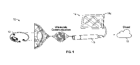
100771 FIG. I shows an exemplary schematic of an exemplary system 10 for
measuring 1013,
according to some embodiments. The system 10 may be configured to monitor TOP
in at least
two types of patients: those with early-to-late open-angle glaucoma who
require regular IOP
monitoring and, patients with normal-tension glaucoma with visual field loss
who require
frequent IOP monitoring. Users of the system may include surgeons implanting
or mounting
the device, clinicians training and assisting patients in taking TOP
measurements, and the
patients. In some embodiments, the system 10 may be used in a controlled
clinical
environment where the clinician can supervise the patient using the system 10.
In some
embodiments, the system 10 may be used outside a clinical environment, for
example in a
patient's home.
100781 in some embodiments, the system 10 may include a device 12 and an
ultrasonic
interrogator 14. The interrogator 14 may include a computer or graphical
display 14a
configured to process and display TOP data and a head 14b configured to
ultrasonically
couple to the implanted device 12. In FIG. 1, the device is implanted inside
the lens capsule
(i.e., capsular bag) of the patient. In other embodiments, the implantable
device may interface
with and/or be mounted on another surface on or within the eye. The implanted
device 12
may measure intraocultu- pressure data and communicate the measured data to
the
interrogator 14. The interrogator 14 may process the received measured data
before
communicating a final 10P measurement to a user.
100791 Optionally, the interrogator 14 can include an application configured
to receive
processed data from a cloud backend application 16, supply information to a
graphical user
interface 14a, and enable limited interactions with the ultrasonic
interrogator 14. The cloud
backend application 16 may be used fbr data aggregation and analytics.
100801 In some embodiments, a system for measuring IOP may include a plurality
of
operating states. For example the system 10 may include an OFF, Ready, Search,
Measurement Collection, Calibration, Complete, or Inactive or Fault state. In
the OFF state,
all system components may be powered OFF. In the Ready state, the interrogator
14 may be
powered on without active ultrasound. In the Ready state, the interrogator 14
may wait for a
user command to start ultrasound transmission. In the Search state, the
interrogator 14 may
search for, find, and power the device 12. In the Measurement Collection
state, the
interrogator may query the device 12 for data and perform the measurement
calculation,
while continuing to power the device. In the Measurement Calibration state,
the interrogator
may perform calibration of the pressure measurement. In the Measurement
Complete state,
the interrogator may notify the user that the measurement is complete via the
physical and
CA 03172888 2022- 9- 22
WO 2022/035889
PCT/US2021/045429
graphical user interfaces. In some embodiments, measurement data may be
displayed to the
user via a display 14a. In the Inactive or Fault state, an internal
interrogator diagnostics may
detect a fault and shut down the ultrasonic power while the interrogator
remains on. The
Inactive or Fault state is different from the Ready state because the
ultrasound will not be
able to be turned on by the user until the systems returns to the Ready state.
This may be the
case when there is a system fault sensed or when the interrogator deliberately
limits
ultrasound power output.
100811 In some embodiments, the system 10 may be configured to receive a
manual selection
from the user to change to a state where ultrasound power output is active. In
some
embodiments, the system 10 may automatically stop ultrasound output when the
IOP
measurement is complete.
100821 FIG. 2A shows an exemplary schematic of an exemplary device 12,
according to
some embodiments. The device 12 may be part of an. IOP measuring system as
shown in
system 10. In some embodiments, the device 12 may include a housing 14 that
encloses
internal components and the housing 14 may be hermetically sealed. In some
embodiments,
the device 12 may include a substrate 16 configured to attach to and support
the housing 14.
In some embodiments, the substrate 16 may be an annular member 16 made of a
flexible
material. In some embodiments, the substrate 16 may be an annular member 16
configured as
a tension ring. The annular member 16 may be configured to exert a radially
outward force
applied to the interfacing surface. For example, the annular member 16 may be
compressed
during implantation, generating an outward spring force when relaxed after
implantation. The
resulting outward force exerted by the annular member 16 can help stabilize
the device in
position after implantation. In some embodiments, the annular member 16 can be
made of
polymethylmethacrylate (PMMA). In some embodiments, the annular member 16 may
have a
full or partial ring structure. In some embodiments, annular member 16 can
form at least
50%, at least 60%, at least 70%, at least 80%, at least 90%, or at least 95%
of a circle, or a
complete circle.
100831 In some embodiments, the ring structure may include a mount (e.g., an
inwardly
extending portion) 18 configured to mount the housing 14. The mount 18 on the
exemplary
device sown in FIG. 2A extends inwardly, although in other configurations the
mount may
extend outwardly or may be positioned on top of the annular member 16. In some
embodiments, the size of the annular member 16 may be configured for a
particular range of
patient eye size. The annular member 16 may include a plurality of apertures
19 that can be
used to guide positioning of the device 12 during implantation or mounting. In
some
11
CA 03172888 2022- 9- 22
WO 2022/035889
PCT/US2021/045429
embodiments, for an annular member having a partial ring structure, each
aperture 19 may be
located at an end of the partial ring structure. In some embodiments, one or
more of the
apertures 19 may be spaced away from. an end of the partial ring structure.
The apertures 19
may be engaged by an external medical tool (such as a hook, forceps, etc.) for
placing the
device 12 properly within the eye. In some embodiments, the device 20 may
include a top
face 13a, a bottom face 13b, and a side face 13c.
100841 FIG. 2B shows a schematic of an exemplary device 20, according to some
embodiments. Device 20 may be part of an 10P measuring system such as system
10. Similar
to device 12, device 20 may include a housing 22, a substrate 24, an inwardly
extending
portion 26, and a plurality of apertures 28. FIG. 2B shows the device 20
interfaces with (e.g.,
may be mounted about) an intraocular lens 30. When implanted within an eye,
the intraocular
lens 30 may be a surface within the eye.
100851 In some embodiments, the device 20 may be implanted in one of the
patient's eyes
during the same surgery for intraocular lens placement. In some embodiments,
the device 20
may allow co-placement with an intraocular lens. An example of co-placement of
device 20
and an intraocular lens 30 is shown in FIG. 2B. In some embodiments, the
device 20 may be
co-placed with an intraocular lens (e.g., a commercially available intraocular
lens) such that
the substrate of the device 20 interfaces the arms (e.g., haptics 32) of the
intraocular lens 30.
The annular member 24 can exert a radial outward force against the haptics 32
of the
intraocular lens 30, which stabilizes the device 20 in position. When the
annular member 24
is co-placed with an intraocular lens, the placement of the annular member 24
does not
interfere with the line of sight of the eye or the functioning of the
intraocular lens. In some
embodiments, the housing 22, the substrate 24, and the plurality of apertures
28 may be
configured to not interfere with the haptics 32 of an intraocular lens 30.
100861 In some embodiments, the device 20 may be co-placed with an intraocular
lens such
that the top face 13a of the device 20 interfaces the intraocular lens 30. In
some
embodiments, the device 20 may be co-placed with an intraocular lens such that
the bottom
face 13b of the device 20 interfaces the intraocular lens 30. In some
embodiments, the device
20 may be co-placed with an intraocular lens such that the side face 13c of
the device 20
interfaces the intraocular lens 30. In some embodiments, the device 20 may be
co-placed with
an intraocular lens such that the device interfaces with the haptics of the
intraocular lens
without interfering with the function of the haptics. In other embodiments,
the device 20 may
be implanted within other areas of the eye such the posterior chamber and
anterior chamber
12
CA 03172888 2022- 9- 22
WO 2022/035889
PCT/US2021/045429
of the eye. The device 20 may be configured to maintain functional integrity
as an implanted
device for at least about 3 years, 4 years, 5 years, 6 years, 7 years, or
more.
100871 FIG. 2C illustrates an exploded view of device 20, according to some
embodiments.
The exploded view shows the housing 22 detached from the substrate 24,
according to some
embodiments. As shown in FIG. 2C, the housing 22 may include one or more
mounting
features 23 (e.g., snaps, clips, outwardly projecting members, etc.) to secure
the housing 22 to
a mount 34 positioned on the substrate 24 via corresponding features 25 (e.g.,
receiving
snaps. inwardly projecting members, etc.). In some embodiments, the
corresponding features
25 may be part of a radially extending portion configured to mount the
housing. In some
embodiments, the radially extending portion may include side walls 27
configured to at least
partially cover side walls 29 of the device 20. In some embodiments, a bottom
surface 31 of
the device 20 may be configured to interface a surface of the eye when the
housing 22 is
mounted on the substrate 24 that interfaces with (e.g., is mounted on) the
surface on or within
the eye, such as an intraocular lens.
100881 In some embodiments, the substrate 24 may be an annular member. In some
embodiments, the substrate 24 may be an annular member that is a tension ring.
1.n some
embodiments, when the device 20 is implanted within the capsular bag of an
eye, the annular
member 24 may be configured to apply a supporting force (i.e., a tension) to
the capsular bag.
In some embodiments, the supporting force may be enough to hold the tension
ring in place
within the eye.. In some embodiments, the annular member 24 may be held in
place within
the eye and retain its shape based on its size and position within the eye
within the capsular
bag of the eye. In some embodiments, the annular member 24 may interface a
perimeter of
the capsular bag.
100891 In some embodiments, the substrate may include fasteners to mount the
substrate to
the surface within the eye. In some embodiments, the fasteners may include a
plurality of
lateral clamps. FIGS. 3A and 3B show exemplary devices 300, 400, having a
respective
housing 310, 410 mounted onto a substrate 320, 420, according to some
embodiments. The
substrate may have a first side for mounting the substrate to a surface within
or an eye. For
example, FIG. 3A shows substrate 320 having a first side 322 for mounting
within or an eye.
The surface within the eye may be, for example, an iris, a lens capsule, an
episclera, an
intraocular lens implanted within an eye, or a phakic intraocular lens
implanted within an eye.
The substrate 320, 420 can include lateral clamps. A first lateral clamp 330,
430 can be
positioned at one end of the substrate 320, 420 and a second lateral clamp
340, 440 can be
positioned at an opposite end of the substrate 320, 420. Each lateral clamp
may be shaped by
13
CA 03172888 2022- 9- 22
WO 2022/035889
PCT/US2021/045429
a slit in the substrate and may include an open position in which eye tissue
of the surface
(such as iris 130) within the eye is positioned within the slit and a closed
position in which
the eye tissue positioned between th.e slit is clamped to mount the substrate
to the surface
within the eye. In some embodiments, the slit may be at least about 0.1, 0.2
mm, or 0.4 mm.
In some embodiments, the slit may be at most about 1 mm, 0.8 mm, or 0.6 mm. In
some
embodiments, the slit may be about 0.1-1 mm, 0.2-0.8, or 0.4-0.6 mm.
100901 FIG. 3A shows an example of the lateral clamps 330, 340 in an open
position in
which eye tissue (such as iris tissue 130) or outermost part of a surface may
be positioned
within slit 342 in the substrate 320, according to some embodiments. In some
embodiments,
the device 300 may be configured such that during placement of the device 300,
a surgeon
may move slit walls 344 to clamp onto eye tissue (such as iris tissue 130)
within the slit 342.
FIG. 3B shows an example of the lateral clamps 430, 440 in a position in which
eye tissue or
outermost part of a surface may be clamped within a thinner slit 442 (thinner
compared, for
example, to the slit 342), according to some embodiments. In some embodiments,
the device
400 may be configured such that during placement of the device 400, a surgeon
may pinch
eye tissue (such as iris tissue 130) to feed the pinched eye tissue through
the thinner slit 342.
In some embodiments, the lateral clamps may be made from polymer. In some
embodiments,
the positioning slits of slits 342, 442 may be configured to follow the radial
grain of the iris
fibers 130
100911 I.n some embodiments, each slit includes slit walls that are spaced
from each other in
the open position and the slit walls are movable towards each other for
clamping eye tissue in
a closed position. For example, slit wall 344 of slit 342 may be configured to
clamp onto eye
tissue. In some embodiments, the lateral clamps are configured to move from
the open
position (such as the open position of FIG. 3A.) to a closed position by a
force applied during
a surgical implantation or procedure. The lateral clamps may remain in the
closed position
until purposefully moved to an open position by a force applied during a
surgical procedure.
In some embodiments, each slit may extend into a circular aperture (such as
aperture 346) of
the substrate.
100921 In some embodiments, the substrate may be flexible and may be bonded to
the rigid
housing. In some embodiments, the housing may attach to the substrate by being
fixed on an
outer surface of the substrate. In some embodiments, the housing may attach to
the substrate
by extending through substrate. In some embodiments, the substrate may have a
second side
for attaching a mountable side of the housing to the substrate. For example,
FIG. 3A shows
the substrate having a second side 324 on which the housing 310 is mounted.
14
CA 03172888 2022- 9- 22
WO 2022/035889
PCT/US2021/045429
100931 In some embodiments, the fasteners may include a plurality of vertical
hooks. FIGS.
4A and 4B show an example of an exemplary device 500 mounted onto a substrate
550
having vertical hooks, according to some embodiments. In some embodiments, the
vertical
hooks may be insert molded. A first vertical hook 552 may be positioned at the
one end of the
substrate 550 and a second vertical hook 554 may be positioned at the opposite
end of the
substrate 550. Each vertical hook may be configured to extend from an interior
channel 510
of the substrate 550 that holds a first portion of the vertical hook within
the substrate 550. A
second portion of each vertical hook may extend in a first direction passed
the first side 556
of the substrate and away from the first side 556 of the substrate 550. The
second portion of
each hook may include an end that extends in a second direction, different
from the first
direction to form a hook shape. For example, hook 554 can include an end 558
configured to
catch eye tissue. Each vertical hook having a hook shape may be configured to
enter eye
tissue for mounting the substrate to the surface within the eye. For example,
the hooks 552,
554 are configured to pass through the tissue of an eye surface (such as iris
surface 130) to
mount the device 500 on the eye surface. When the hooks 552, 554 pass through
eye tissue or
outermost part of the eye surface, the hooks 552, 554 are configured to
prevent the device
500 from being unmounted from the eye surface. In some embodiments, the
vertical hooks
552, 554 may be pushed towards the surface within the eye to insert the
vertical hooks 552,
554 within the eye tissue. In some embodiments, the vertical hooks may be made
from
polymer.
100941 FIG. 5A and FIG. 58 show a schematic of an exemplary device 350 (such
as devices
300, 400) having an exemplary substrate 352 (such as 320, 420) for mounting
the device 350
within an eye 360 and an exemplary housing 354 (such as 310, 410) for housing
internal
components of the device, according to some embodiments. FIG. 5A shows an
exemplary
top-view of the device 350 mounted within the eye 360, according to some
embodiments. In
other embodiments, the device 350 may be configured to be mounted on an eye.
The device
350 may be configured to maintain functional integrity as a mounted or
implanted device for
at least about 3 years, 4 years, 5 years, 6 years, 7 years, or more.
100951 FIG. 5A shows possible exemplary locations for minimally invasive
incision sites 370
for mounting the device 350 within the eye 360 such that mounted device does
not interfere
with the line of sight of the eye 360. FIG. 5B shows an exemplary cross-
sectional schematic
displaying the exemplary device 350 mounted to a surface 380 within the eye
360, according
to some embodiments. As shown in FIG. 58, the surface 380 within the eye 360
may be a top
surface of the iris located in anterior chamber of an eye. Mounting the device
on the top
CA 03172888 2022- 9- 22
WO 2022/035889
PCT/US2021/045429
surface of the iris located in the anterior chamber as shown in FIG. 5B,
rather than mounting
on a bottom surface of the iris located in the posterior chamber of the eye,
is advantageous
because there is less risk of damaging the iris during implantation compared
to mounting on
the bottom surface of the iris. In some embodiments, the surface within the
eye may be on or
near a pars plana 382 of the ciliary body of the eye.
100961 In other embodiments, the device may be implanted within the capsular
bag. For
example, the device may be co-placed with an intraocular lens.
100971 The device is configured to measure KW data and encode 10P data via
ultrasonic
backscatter using internal components of the device, such as one or more
sensors, one or
more transducers, and an integrated circuit. Exemplary implantable devices
that are powered
by ultrasonic waves and can emit an ultrasonic backscatter encoding a detected
physiological
condition are described in WO 2018/009905 and WO 2018/009911.
100981 An integrated circuit of the device can electrically connect and
communicate with the
one or more sensors of the device and the wireless communication system (e.g.,
the one or
more ultrasonic transducers). The integrated circuit can include or operate a
modulation
circuit within the wireless communication system, which modulates an
electrical current
flowing through the wireless communication system (e.g., one or more
ultrasonic
transducers) to encode information in the electrical current. The modulated
electrical current
affects backscatter waves (e.g., ultrasonic backscatter waves) emitted by the
wireless
communication system, and the backscatter waves encode the information.
100991 FIG. 6A shows a side view of an exemplary board assembly of an
exemplary device,
which may be surrounded by a housing (such as housing 14, 22, 310, or 410) and
include an
integrated circuit, according to some embodiments. The device includes a
wireless
communication system (e.g., one or more ultrasonic transducers) 602 and an
integrated
circuit 604. In the illustrated embodiment, the integrated circuit 604
includes a power circuit
that includes a capacitor 606. In the illustrated embodiment, the capacitor is
an "off chip"
capacitor (in that it is not on the integrated circuit chip), but is still
electrically integrated into
the circuit. The capacitor can temporarily store electrical energy converted
from energy (e.g.,
ultrasonic waves) received by the wireless communication system, and can be
operated by the
integrated circuit 604 to store or release energy. The device further includes
one or more
sensors 608. The one or more sensors can include a pressure sensor. Since
ultrasound waves
transmitted to and from the device may affect sensor measurements, the one or
more sensors
of the device may be configured to measure 1OP data when ultrasound waves are
not being
transmitted. The one or more ultrasonic transducers 602, integrated circuit
604, the capacitor
16
CA 03172888 2022- 9- 22
WO 2022/035889
PCT/US2021/045429
606, and the one or more sensors 608 are mounted on a circuit board 610, which
may be a
printed circuit board. In some embodiments, the one or more ultrasonic
transducers 602,
integrated circuit 604, the capacitor 606, and the one or more sensors 608 are
adhered on the
circuit board 610. In some embodiments, the circuit board 610 may include
ports 612a-d.
Similar to FIG. 6A, FIG. 6B shows a side view of an exemplary board assembly
that may be
enclosed in a housing, according to some embodiments. The board assembly of
FIG. 6B
includes a piezoelectric transducer 602b and one or more sensors 608b adhered
on the circuit
board 610b, according to some embodiments.
101001 The wireless communication system of the device can be configured to
receive
instructions for operating the device. The instructions may be transmitted,
for example, by a
separate device, such as an interrogator. By way of example, ultrasonic waves
received by the
device (for example, those transmitted by the interrogator) can encode
instructions for
operating the device. The instructions may include, for example, a trigger
signal that instructs
the device to operate the pressure sensor to detect the intraocular pressure.
101011 An interrogator can transmit energy waves (e.g., ultrasonic waves),
which are
received by the wireless communication system of the device to generate an
electrical current
flowing through the wireless communication system (e.g., to generate an
electrical current
flowing through the ultrasonic transducer). The flowing current can then
generate backscatter
waves that are emitted by the wireless communication system. The modulation
circuit can be
configured to modulate the current flowing through the wireless communication
system to
encode the information. For example, the modulation circuit may be
electrically connected to
an ultrasonic transducer, which received ultrasonic waves from an
interrogator. The current
generated by the received ultrasonic waves can be modulated using the
modulation circuit to
encode the information, which results in ultrasonic backscatter waves emitted
by the
ultrasonic transducer to encode the information. The modulation circuit
includes one or more
switches, such as an on/off switch or a field-effect transistor (FET). An
exemplary FET that
can be used with some embodiments of the implantable device is a metal-oxide-
semiconductor field-effect transistor (MOSFED. The modulation circuit can
alter the
impedance of a current flowing through the wireless communication system, and
variation in
current flowing through the wireless communication system encodes the
information. In
some embodiments, information encoded in the backscatter waves includes
information
related to an electrical pulse emitted by the device, or a physiological
condition detected by
the one or more sensors of the device. In some embodiments, information
encoded in the
backscatter waves includes a unique identifier for the device. This can be
useful, for example,
17
CA 03172888 2022- 9- 22
WO 2022/035889
PCT/US2021/045429
to ensure the interrogator is in communication with the correct implantable
device when a
plurality of implantable devices is implanted in the subject. In some
embodiments, the
information encoded in the backscatter waves includes a verification signal
that verifies an
electrical pulse was emitted by the device. In some embodiments, the
information encoded in
the backscatter waves includes an amount of energy stored or a voltage in the
energy storage
circuit (or one or more capacitors in the energy storage circuit). In some
embodiments, the
information encoded in the backscatter waves includes a detected impedance.
Changes in the
impedance measurement can identify scarring tissue or degradation of the
electrodes over
time.
101021 In some embodiments, the modulation circuit is operated using a digital
circuit or a
mixed-signal integrated circuit (which may be part of the integrated circuit),
which can
actively encode the information in a digitized or analog signal. The digital
circuit or mixed-
signal integrated circuit may include a memory and one or more circuit blocks,
systems, or
processors for operating the implantable device. These systems can include,
for example, an
onboard microcontrol ler or processor, a finite state machine implementation,
or digital
circuits capable of executing one or more programs stored on the implant or
provided via
ultrasonic communication between interrogator and implantable device. In some
embodiments, the digital circuit or a mixed-signal integrated circuit includes
an analog-to-
digital converter (ADC), which can convert analog signal encoded in the
ultrasonic waves
emitted from the interrogator so that the signal can be processed by the
digital circuit or the
mixed-signal integrated circuit. The digital circuit or mixed-signal
integrated circuit can also
operate the power circuit, for example to generate the electrical pulse to
operate the pressure
sensor to detect TOP. In some embodiments, the digital circuit or the mixed
signal integrated
circuit receives the trigger signal. encoded in the ultrasonic waves
transmitted by the
interrogator, and operates the power circuit to discharge the electrical pulse
in response to the
trigger signal.
101031 In some embodiments, the one or more sensors 608 may a pressure sensor
configured
to measure 10P. The pressure sensor may implement capacitive or resistive
pressure sensing_
The measurement accuracy of the pressure sensor may be at least 0.1 mmHg, 0.2
mmHg, 0.3
mmHg, 0.4 mmHg, or 0.5 mmHg. The measurement accuracy of the pressure sensor
may be
at most 1.0 mmHg, 0.9 mmHg, 0.8 mmHg, 0.6 mmHg, or 0.7 mmHg. The measurement
accuracy of the pressure sensor may be 0.1-1.0mm Hg, 0.2-0.9 mm Hg, 0.3-0.8 mm
Hg, 0.4-
0.7 mm Hg, or 0.5-0.6 mmHg. In some embodiments, the measurement accuracy of
the
pressure sensor may be over a range of l. mmHg to 70 mmHg, 3 mmHg to 60 mmHg,
or 5
18
CA 03172888 2022- 9- 22
WO 2022/035889
PCT/US2021/045429
mmHg to 50 mmHg. In some embodiments, the pressure sensor may have a
sensitivity of
about 10 LIVN/mmHg, 20 [tVN/mnifig, or 30 [tVN/mmlig. In some embodiments, the
pressure sensor may have a sensitivity requirement dependent on the
sensitivity of the
readout electronics. In some embodiments, the pressure sensor may have a
measurement
accuracy and sensitivity range dependent on the sensitivity of the readout
electronics.
101041 In some embodiments, the pressure sensor may be temperature sensitive.
The pressure
sensor may be calibrated based on a temperature response of the temperature
sensor. The
calibration may be configured to ensure that a difference in pressure output
of the pressure
sensor is an actual different in pressure and not an artifact of a change in
temperature.
101051 In some embodiments, the one or more sensors may include a temperature
sensor
configured to measure an anterior chamber temperature of an eye. In some
embodiments, the
temperature sensor may have an accuracy of about 0.1-1 C, 0.2-0.8 C, or 0.3-
0.6 'C. In
some embodiments, the temperature sensor may monitor a range of temperature
inside the
eye from about 28 C to 46 C, 30 C to 44 C, or 32 C to 40 'C. In some
embodiments, the
temperature sensor data may be used for compensation purposes to increase
accuracy of the
final pressure measurement.
101061 Both the pressure data from the pressure sensor and temperature data
from the
temperature sensor may be reported to the external interrogator. The reported
pressure data
and the reported temperature data may be an averaged or processed result taken
from multiple
discrete measurements from the corresponding sensor. In some embodiments, the
temperature
measurement is used to calibrate the measured pressure at the device, and the
ultrasonic
backscatter can communicate a calibrated pressure. In some embodiments, the
pressure data
reported by the device may be equivalent to pressure outside of the device
with a lag of no
more than 1 second, 3 seconds, or 5 seconds. In some embodiments, the time
from when the
measurement command is received from the external interrogator to when the
measurement is
reported to the interrogator shall be no more than 2 seconds, 4 seconds, 6
seconds, or 8
seconds.
101071 In some embodiments, the wireless communication system includes one
ultrasonic
transducer that is an ultrasonic transceiver configured to convert mechanical
energy from
ultrasound waves to electrical current and vice versa. The ultrasonic
transducer may be
capable of harvesting energy originating from an. external ultrasonic
interrogator and capable
of producing a modulation depth detectable by an external interrogator.
10108! In some embodiments, the wireless communication system includes one or
more
ultrasonic transducers, such as one, two, or three or more ultrasonic
transducers. In some
19
CA 031721388 2022- 9- 22
WO 2022/035889
PCT/US2021/045429
embodiments, the wireless communication system includes a first ultrasonic
transducer
having a first polarization axis and a second ultrasonic transducer having a
second
polarization axis, wherein the second ultrasonic transducer is positioned so
that the second
polarization axis is orthogonal to the first polarization axis, and wherein
the first ultrasonic
transducer and the second ultrasonic transducer are configured to receive
ultrasonic waves
that power the device and emit an ultrasonic backscatter. In some embodiments,
the wireless
communication system includes a first ultrasonic transducer having a first
polarization axis, a
second ultrasonic transducer having a second polarization axis, and a third
ultrasonic
transducer having a third polarization axis, wherein the second ultrasonic
transducer is
positioned so that the second polarization axis is orthogonal to the first
polarization axis and
the third polarization axis, wherein the third ultrasonic transducer is
positioned so that the
third polarization axis is orthogonal to the first polarization and the second
polarization axis,
and wherein the first ultrasonic transducer and the second ultrasonic
transducer are
configured to receive ultrasonic waves that power the device and emit an
ultrasonic
backscatter. FIG. 7 shows a board assembly of a device that includes two
orthogonally
positioned ultrasonic transducers. The device includes a circuit board 702,
such as a printed
circuit board, and an integrated circuit 704, which a power circuit that
includes a capacitor
706. The device further includes a first ultrasonic transducer 708
electrically connected to the
integrated circuit 704, and a second ultrasonic transducer 710 electrically
connected to the
integrated circuit 704. The first ultrasonic transducer 708 includes a first
polarization axis
712, and the second ultrasonic transducer 710 includes a second polarization
axis 714. The
first ultrasonic transducer 708 and the second ultrasonic transducer are
positioned such that
the first polarization axis 712 is orthogonal to the second polarization axis
714.
101091 The one or more ultrasonic transducers, if included in the wireless
communication
system, can be a micro-machined ultrasonic transducer, such as a capacitive
micro-machined
ultrasonic transducer (CMUT) or a piezoelectric micro-machined ultrasonic
transducer
(PMUT), or can be a bulk piezoelectric transducer. Bulk piezoelectric
transducers can be any
natural or synthetic material, such as a crystal, ceramic, or polymer.
Exemplary bulk
piezoelectric transducer materials include barium titanate (BaTiO3), lead
zirconate titanate
(PZT), zinc oxide (ZO), aluminum nitride (AIN), quartz, berlinite (A1PO4),
topaz, langasite
(1.,a3Ga5Si014), gallium orthophosphate (GaPO4), lithium niobate (LiNb03),
lithium tantalite
(LiTa03), potassium niobate (KNb03), sodium tungstate (Na2W03), bismuth
ferrite
(BiFe03), polyvinylidene (di)fluoride (PVDF), and lead magnesium niobate-lead
titanate
(PlvIN-PT).
CA 03172888 2022- 9- 22
WO 2022/035889
PCT/US2021/045429
101101 In some embodiments, the bulk piezoelectric transducer is approximately
cubic (i.e.,
an aspect ratio of about 1: 1:1 (length:width:height). In some embodiments,
the piezoelectric
transducer is plate-like, with an. aspect ratio of about 5:5:1 or greater in
either the length or
width aspect, such as about 7:5:1 or greater, or about 10:10:1 or greater. In
some
embodiments, the bulk piezoelectric transducer is long and narrow, with an
aspect ratio of
about 3:1:1 or greater, and where the longest dimension is aligned to the
direction of the
ultrasonic backscatter waves (i.e., the polarization axis).
101111 In some embodiments, one dimension of the bulk piezoelectric transducer
is equal to
one half of the wavelength (.) corresponding to the drive frequency or
resonant frequency of
the transducer. At the resonant frequency, the ultrasound wave impinging on
either the face
of the transducer will undergo a 180" phase shift to reach the opposite phase,
causing the
largest displacement between the two faces. In some embodiments, the
piezoelectric crystal
may be assembled into the housing such that its poled direction is
perpendicular to an
acoustic window.
101121 in some embodiments, the height of the piezoelectric transducer is
about 10 p.m to
about 1000 1.1.M (such as about 40 1.1.M to about 40011M, about 10012M to
about 250 pm., about
250 p.m to about 500 gm, or about 500 p.m to about 1000 gm). In some
embodiments, the
height of the piezoelectric transducer is about 5 mm or less (such as about 4
mm or less,
about 3 nun or less, about 2 mm or less, about 1 mm or less, about 500 inn or
less, about 400
gm or less, 250 p.m or less, about 1001.1M. or less, or about 40 gm or less).
In some
embodiments, the height of the piezoelectric transducer is about 20 !um or
more (such as
about 40 pm or more, about 100 pm or more, about 250 p.m or more, about 400
1.1111 or more,
about 500 p.m or more, about 1 mm or more, about 2 mm or more, about 3 mm or
more, or
about 4 mm or more) in length. In some embodiments, the ultrasonic transducer
has a length
of about 5 mm or less such as about 4 ram. or less, about 3 mm or less, about
2 mm or less,
about 1 mm or less, about 500 pm or less, about 400 p.m or less, 250 inn or
less, about 100
p.m or less, or about 40 pm or less) in the longest dimension. In some
embodiments, the
ultrasonic transducer has a length of about 20 p.m or more (such as about 40
p.m or more,
about 100 pm or more, about 250 p.m or more, about 400 p.m or more, about 500
p.m or more,
about 1 mm or more, about 2 mm or more, about 3 mm or more, or about 4 mm or
more) in
the longest dimension.
101131 In some embodiments the micro-machined piezoelectric crystal can have
dimensions
of about at least 0.3 micrometer x 0.3 micrometer x 0.1 micrometer. In some
embodiments,
21
CA 03172888 2022- 9- 22
WO 2022/035889
PCT/US2021/045429
the piezoelectric crystal can have dimensions of about at most 1.2 micrometer
x 1.2
micrometer x 0.6 micrometer. In some embodiments, the piezoelectric aystal can
have
dimensions of about 0.3-1.2 micrometer x 0.3-1.2 micrometer x 0.1-0.6
micrometer.
101141 The one or more ultrasonic transducers, if included in the wireless
communication
system, can be connected to two electrodes to allow electrical communication
with the
integrated circuit. The first electrode is attached to a first face of the
transducer and the
second electrode is attached to a second face of the transducer, wherein the
first face and the
second face are opposite sides of the transducer along one dimension. In some
embodiments,
the electrodes comprise silver, gold, platinum, platinum-black, poly(3,4-
ethylenedioxythiophene (PEDOT), a conductive polymer (such as conductive PDMS
or
polyimide), or nickel. In some embodiments, the axis between the electrodes of
the
transducer is orthogonal to the motion of the transducer.
101151 The wireless communication system may be used to wireless receive the
energy, or a
separate system may be configured to receive the energy. For example, an
ultrasonic
transducer (which may be an ultrasonic transducer contained within the
wireless
communication system or a different ultrasonic transducer) can be configured
to receive
ultrasonic waves and convert energy from. the ultrasonic waves into an
electrical energy. The
electrical energy is transmitted to the integrated circuit to power the
device. The electrical
energy may power the device directly, or the integrated circuit may operate a
power circuit to
store the energy for later use.
101161 In some embodiments, the integrated circuit may be configured to
control the
harvesting of energy from the received ultrasonic waves, power the one or more
sensors, and
encode the eye-related data collected by the one or more sensors using
backscatter
modulation. The encoding or the eye-related data includes digitizing the eye-
related data
collected by the one or more sensors and modulating the characteristics of
electrical current
within the device for digital backscatter communication with the external
interrogator. In
some embodiments, the integrated circuit (such as integrated circuit 604, 704)
is an
application specific integrated circuit (ASIC). In some embodiments, the ASIC
operation
may be passive. The AS1C may power up and transmit messages only when
commanded by
the external interrogator. In some embodiments, there is no OFF command for
the ASIC
since the ASIC may be powered off by stopping ultrasound communication between
the
device and the external interrogator. The stopping of the ultrasound
communication may
quickly deplete the energy store of the device. When powered, the ASIC may
transmit data
bits or acknowledgments to the interrogator to allow for status evaluation of
the ultrasound
22
CA 03172888 2022- 9- 22
WO 2022/035889
PCT/US2021/045429
communication link. When a measurement command is received the ASIC may
perform the
command if it can complete the command with the available power.
101171 In some embodiments, power may be harvested from the received
ultrasonic waves
using the piezoelectric crystal of the ultrasonic transducer and the ASIC of
the device. The
ASIC may convert AC ultrasonic power to DC power, may be able to sustain
operation of the
device with a minimum average power, and may generate an TOP measurement
within a pre-
determined amount of time. In some embodiments, the minimum average power may
be
about 10 x 10-6 W, 20 x 10-6 W. or 30 x 10-6W average power. In some
embodiments, the
pre-determined amount of time may be about less than 1 second, 3 seconds, or 5
second.
101181 In some embodiments, the integrated circuit includes a power circuit,
which can
include an energy storage circuit. The energy storage circuit may include a
battey, or an
alternative energy storage device such as one or more capacitors. The device
may be
batteryless, and may rely on one or more capacitors. By way of example, energy
from
ultrasonic waves received by the device (for example, through the wireless
communication
system) is converted into a current, and can be stored in the energy storage
circuit. The
energy can be used to operate the device, such as providing power to the
digital circuit, the
modulation circuit, or one or more amplifiers, or can be used to generate an
electrical pulse.
In some embodiments, the power circuit further includes, for example, a
rectifier and/or a
charge pump.
[01191 in some embodiments, the piezoelectric crystal may be electrically and
mechanically
connected to the ASIC and substrate such that the Curie temperature, the
resonant frequency,
and resistance range at resonance are maintained within pre-determined ranges.
In some
embodiments, the Curie temperature may be at least about 1.80 C, 200 C, or
220 'C. In
some embodiments, the Curie temperature may be at most about 260 C, 250 C,
or 240 C.
In some embodiments, the Curie temperature may be about 180 to 60 C. 200 to
250 C, or
220 to 240 C. In some embodiments, the resonant frequency may be at least
about 1.2 MHz,
1.4 MHz, 1.6 MHz, or 1.8 MHz. In some embodiments, the resonant frequency may
be at
most about 2.8 MHz, 2.6 MHz, 2.4 MHz, or 2.2 MHz In some embodiments, the
resonant
frequency may be about 1.2 to 2.8 MHz, 1.4 to 2.6 MHz, 1.6 to 2.4 MHz, or 1.8
to 2.2 MHz.
In some embodiments, the resistance range at resonance may be at least about
0.11(0, 0.2 k52,
or 0.3 k. In some embodiments, the resistance range at resonance may be at
most about 1.7
kt/ , 1.5 , 1.3 kfl , or 1.1 ka. In some embodiments, the
resistance range at resonance
may be about 0.1 to 1.7 k1 ,0.2 to 1.5 kn 0.3 to 1.3 k/, or 0.3 to 1.1 Id1.
23
CA 03172888 2022- 9- 22
WO 2022/035889
PCT/US2021/045429
101201 FIG. 8 shows a schematic of an exemplary. device 700 having one or more
sensors 810
and a wireless communication system 820. The sensors or electrodes 810 may be
configured
to electrically communicate with the wireless communication system 820.
Additionally, the
wireless communication system 820 may be configured to communicate with an
external
device having a communication system. For example, the external device may be
an
interrogator 830 having a communication system that includes one or more
ultrasonic
transducers.
101211 In some embodiments, the housing may house the wireless communication
system,
the one or more sensors, and the integrated circuit. The housing of the device
can include a
base, one or more sidewalls, and a top for enclosing the internal components
of the device. In
some embodiments, the housing may be at most about 0.25 mm high, 0.5 mm high,
1 mm
high, or 2 mm high. In some embodiments, the housing may be at most 1 mm wide,
2 mm
wide, or 3 mm. wide. In some embodiments, the housing may be at most 1 mm
long, 2 mm
long, 3 mm long, 4 mm long, or 5 mm long. FIG. 9A shows an exploded view of an
exemplary housing 940, according to some embodiments. The housing is made from
a
bioinert material, such as a bioinert metal (e.g., steel or titanium) or a
bioinert ceramic (e.g.,
titania or alumina). In some embodiments, the housing may have no sharp
corners or edges
that could cause excessive reaction or inflammation beyond that caused by an
implanting
procedure. The housing is preferably hermetically sealed, which prevents body
fluids from
entering the body. In some embodiments, the hermetic seal may meet or exceed
an equivalent
leak rate of at least 2 x 10⁻⁸ atm-cc/sec Air, 5 x 10⁴ atm-cc/sec Air, or 8 x
10-8 atm-cc/sec
Air. The hermetically sealed housing may withstand shock, thermal cycling, and
pressure
change specifications identified by standards such as ISO 14708-1.
101221 In some embodiments, the housing can include an acoustic window that
serves at least
one or both of the following: 1) it allows ultrasonic waves to penetrate the
window and power
the piezoelectric crystal of the device, and 2) it provides a compliant
membrane that allows
changes in intraocular pressure to transfer to the MEMS prssure sensor. In
this way, the
acoustic window allows ultrasonic waves to penetrate and equilibrate pressure
external and
internal to the housing. In some embodiments, the acoustic window may have a
compliance
that is at least about 400 times, 600 times, or 800 times larger than the
compliance of a
pressure sensor membrane of the pressure sensor. In some embodiments, the
acoustic window
may have a compliance that is at most about 1600 times, 1400 times, or 1,200
times larger
than the compliance of a pressure sensor membrane of the pressure sensor. In
some
embodiments, the acoustic window may have a compliance that is at most about
400 to 1600
24
CA 03172888 2022- 9- 22
WO 2022/035889
PCT/US2021/045429
times, 600 to 1400 times, or 800 to 1,200 times larger than the compliance of
a pressure
sensor membrane of the pressure sensor. In some embodiments, the acoustic
window may be
oriented anterior to the Corona! Plane. The equilibration of pressure enables
accurate LOP
measurements while protecting the sensor within the housing. For example, the
top 944 of the
housing 940 can include an acoustic window. An acoustic window is a thinner
material (such
as a foil) that allows acoustic waves to penetrate the housing 940 so that
they may be
received by one or more ultrasonic transducers within the body of the device.
In some
embodiments, the housing (or the acoustic window of the housing) may be thin
to allow
ultrasonic waves to penetrate through the housing. In some embodiments, the
thickness of the
housing (or the acoustic window of the housing) is about 100 micrometers (pm)
or less in
thickness, such as about 75 pun or less, about 50 gm or less, about 25 p.m or
less, about 15 gm
or less, or about 10 p.m or less. In some embodiments, the thickness of the
housing (or the
acoustic window of the housing) is about 5 p.m. to about 10 gm, about 10 pm to
about 15 pm,
about 15 pm to about 25 pm, about 25 um to about 50 pm, about 50 pm to about
75 pm, or
about 75 pm to about 100 pm in thickness. In some embodiments, the acoustic
window can
be made from a metallic film.
101231 The housing of the device is relatively small, which allows for
comfortable and long-
term implantation while limiting tissue inflammation that is often associated
with implanting
devices. In some embodiments, the longest dimension of the housing of the
device is about 8
mm or less, about 7 mm or less, about 6 m or less, about 5 mm or less, about 4
mm or less,
about 3 mm or less, about 2 mm or less, about 1 mm or less, about 0.5 mm or
less, about 0.3
mm or less, about 0.1 mm or less in length. In some embodiments, the longest
dimension of
the housing of the device is about 0.05 mm or longer, about 0.1 mm or longer,
about 0.3 mm
or longer, about 0.5 mm or longer, about 1 mm or longer, about 2 mm or longer,
about 3 mm
or longer, about 4 mm or longer, about 5 mm or longer, about 6 mm or longer,
or about 7 mm
or longer in the longest dimension of the device. In some embodiments, the
longest
dimension of the housing of the device is about 0.3 mm to about 8 mm in
length, about 1 mm
to about 7 mm in length, about 2 mm to about 6 mm in length,. or about 3 mm to
about 5 mm
in length. In some embodiments, the housing of the implantable device has a
volume of about
mm3 or less (such as about 8 mm3 or less, 6 trun3 or less, 4 ttirn3 or less,
or 3 mm3 or less).
In some embodiments, the housing of the implantable device has a volume of
about 0.5 mm3
to about 8 mm3, about 1 mm3 to about 7 mm3, about 2 mm3 to about 6 mm3, or
about 3 mm3
to about 5 mm3.
CA 03172888 2022- 9- 22
WO 2022/035889
PCT/US2021/045429
101241 The housing may be filled with an acoustic medium and void of water,
moisture, or
air bubbles. The acoustic medium may have a density that avoids an impedance
mismatch
with surrounding tissue. The acoustic medium. may be electrically non-
conductive. For
example, the housing 940 may be filled with a polymer or oil (such as a
silicone oil). The
material can fill empty space within the housing to reduce acoustic impedance
mismatch
between the tissue outside of the housing and within the housing. Accordingly,
an interior of
the device is preferably void of air or vacuum. A port can be included on the
housing, for
example one of the sidewalls 942 of housing 940, there may be a port 946 to
allow the
housing to be filled with the acoustic medium. Once the housing 940 is filled
with the
material, the port 946 can be sealed to avoid leakage of the material after
implantation.
101251 FIG. 9B shows an exploded view of exemplary housing 950 that shows the
housing is
configured to house the circuit board 610b, according to some embodiments.
Similar to
housing 940, the housing 950 includes sidevvalls 952, port 956, and a top 954.
101261 In some embodiments, the housing 940, 950 may include externally
attached features
that allow placement and fixation of the device within or on an eye. The
externally attached
features do not interfere with ultrasound transmission, pressure transmission,
or mounting of
the device within or on the eye. For example, the housing may have externally
attached
features which allow placement and fixation into the lens capsule of the eye
without
interfering with the patient's line of sight or intraocular lens placement (if
applicable). In
some embodiments, the externally attached features may be free of sharp
corners or edges
that could cause excessive reaction or inflammation beyond that caused by the
mounting
procedure, or rough surfaces which are not required for the correct
functioning of the device.
In some embodiments, any externally attached features may not increase the
rigid dimensions
of the implant by more than 0.50 mm. in height, 1.00 mm in width, or 1.50 mm
in length.
Interrogator
101271 In some embodiments, the device may be configured to wirelessly
communicate with
components external to the device for IOP measuring operations. For example,
the device
may be configured to wireless!), communicate with an external interrogator.
Through the
wireless communication, the interrogator may be configured to instruct the
device to collect a
plurality of IOP measurements. The external interrogator may include one or
more
transducers, one or more sensors, and one or more force gauges.
101281 An exemplary interrogator 1000 is shown in FIG. 10A, according to some
embodiments. An exemplary schematic of the exemplary interrogator 1000 is
shown in FIG.
10B, according to some embodiments. The interrogator of FIG. 10A-B may be
configured to
26
CA 03172888 2022- 9- 22
WO 2022/035889
PCT/US2021/045429
wirelessly communicate with devices such as devices 300, 400, and 500. The
interrogator
1000 may include one or more transducers 1010 for wireless communication, one
or more
force gauges 1020 for measuring force applied by the interrogator, and one or
more sensors
1030 for measuring ambient conditions. In some embodiments, the one or more
transducers
1010 may include an ultrasonic transducer. The ultrasonic transducer may be
configured to
ultrasonically couple to skin of an eyelid, skin over a brow bone, skin over a
nasal bone, or
skin over an eye socket to facilitate ultrasonic communication between the
interrogator and
the device mounted on or within an eye. In some embodiments, an ultrasound
coupling gel or
an alternative couplant may be used to ultrasonically couple the interrogator
to the skin.
101291 Ultrasonically coupling the ultrasonic transducer to the skin includes
applying a
contact force by the interrogator on the skin. Since such an applied contact
force may
adversely affect IOP measurements from the device, it is preferable to use a
minimum
amount of contact force for a more accurate IOP measurement. In some
embodiments, the
interrogator may include a force gauge configured to measure a force applied
on the skin by
the interrogator. For example, the interrogator 1000 may include one or more
force gauges
1020 for this purpose. In some embodiments, the interrogator is configured to
operate the
device to determine a plurality of IOP measurements as the force gauge
measures a
decreasing force. The plurality of IOP measurements may be matched to
corresponding
gauge measurements to determine the IOP measurement collected at the lowest
measured
force.
1.01301 In some embodiments, the interrogator includes one or more sensors
configured to
measure ambient conditions. For example, interrogator 1000 may include one or
more
sensors 1030 as shown in FIG. 10. The one or more sensors of the interrogator
may include a
pressure sensor for measuring ambient pressure. Optionally, the interrogator
may further
include a temperature sensor for measuring ambient temperature, which can be
used to
calibrate the pressure sensor used for measuring ambient pressure. The
interrogator 1000 may
be configured to receive the IOP measurements collected by the one or more
sensors (such as
one or more sensors 608) of the device (such as devices 100,, 300, 400, 500),
measure
ambient conditions via the one or more sensors 1030 of the interrogator 1000,
determine a
final KW reading by compensating (as necessary) the IOP measurements with
ambient
measurements, and communicate the final IOP measurement to a recipient
external to both
the interrogator and the device. In some embodiments, the interrogator may
compensate the
101) measurements based on differences between the measured lOP and the
measured
ambient pressure. Since the difference between the TOP and ambient pressure is
a
27
CA 03172888 2022- 9- 22
WO 2022/035889
PCT/US2021/045429
biologically relevant value, in some embodiments, the compensation may simply
be the
di fference between the TOP and ambient pressure. In some embodiments, the
interrogator
may compensate the lop measurements using measured ambient pressure and a
measured
temperature inside the eye.
101311 In some embodiments, the interrogator 1000 may include ultrasound
receive and
transmit circuitry 1040, a data interface 1050, an embedded controller 1060,
and a power
source 1070. In some embodiments, the device may be configured to rely on
power
transmission from the external interrogator. The power transmission from the
interrogator
may be used to power the device to initiate IOP measurements collected by the
one or more
sensors of the device. In some embodiments, the ultrasonic transducer of the
interrogator may
be configured to transmit instructions to the device. The instructions from
the interrogator
may instruct the device to reset itself, enter a specific mode, set device
parameters, or begin a
transmission sequence.
101321 An exemplary interrogator is shown in FIG. 11, according to some
embodiments. The
illustrated interrogator shows a transducer array with a plurality of
ultrasonic transducers. In
some embodiments, the transducer array includes 1 or more, 2 or more, 3 or
more, 5 or more,
7 or more, 10 or more, 15 or more, 20 or more, 25 or more, 50 or more, 100 or
more 250 or
more, 500 or more, 1000 or more, 2500 or more, 5000 or more, or 10,000 or more
transducers. In some embodiments, the transducer array includes 100,000 or
fewer, 50,000 or
fewer, 25,000 or fewer, 10,000 or fewer, 5000 or fewer, 2500 or fewer, 1000 or
fewer, 500 or
fewer, 200 or fewer, 150 or fewer, 100 or fewer, 90 or fewer, 80 or fewer, 70
or fewer, 60 or
fewer, 50 or fewer, 40 or fewer, 30 or fewer, 25 or fewer, 20 or fewer, 15 or
fewer, 10 or
fewer, 7 or fewer or 5 or fewer transducers. The transducer array can be, for
example a chip
comprising 50 or more ultrasonic transducer pixels.
101331 The interrogator shown in FIG. 11 illustrates a single transducer
array; however the
interrogator can include 1 or more, 2 or more, or 3 or more separate arrays.
In some
embodiments, the interrogator includes 10 or fewer transducer arrays (such as
9, 8, 7, 6, 5, 4,
3, 2, or 1 transducer arrays). The separate arrays, for example, can be placed
at different
points of a subject, and can communicate to the same or different implantable
devices. In
some embodiments, the arrays are located on opposite sides of an implantable
device. The
interrogator can include an application specific integrated circuit (ASIC),
which includes a
channel for each transducer in the transducer array. In some embodiments, the
channel
includes a switch (indicated in FIG. 11 by "T/Rx"). The switch can
alternatively configure
the transducer connected to the channel to transmit ultrasonic waves or
receive ultrasonic
28
CA 03172888 2022- 9- 22
WO 2022/035889
PCT/US2021/045429
waves. The switch can isolate the ultrasound receiving circuit from the higher
voltage
ultrasound transmitting circuit.
101341 In some embodiments, the transducer connected to the channel is
configured only to
receive or only to transmit ultrasonic waves, and the switch is optionally
omitted from the
channel. The channel can include a delay control, which operates to control
the transmitted
ultrasonic waves. The delay control can control, for example, the phase shift,
time delay,
pulse frequency and/or wave shape (including amplitude and wavelength). The
delay control
can be connected to a level shifter, which shifts input pulses from the delay
control to a
higher voltage used by the transducer to transmit the ultrasonic waves. In
some embodiments,
the data representing the wave shape and frequency for each channel can be
stored in a 'wave
table'. This allows the transmit waveform on each channel to be different.
Then, delay
control and level shifters can be used to 'stream' out this data to the actual
transmit signals to
the transducer array. In some embodiments, the transmit waveform for each
channel can be
produced directly by a high-speed serial output of a microcontroller or other
digital system
and sent to the transducer element through a level shifter or high-voltage
amplifier. In some
embodiments, the ASIC includes a charge pump (illustrated in FIG. 11) to
convert a first
voltage supplied to the ASIC to a higher second voltage, which is applied to
the channel. The
channels can be controlled by a controller, such as a digital controller,
which operates the
delay control.
101351 in the ultrasound receiving circuit, the received ultrasonic waves are
converted to
current by the transducers (set in a receiving mode), which is transmitted to
a data capture
circuit. In some embodiments, an amplifier, an analog-to-digital converter
(ADC), a variable-
gain-amplifier, or a time-gain-controlled variable-gain-amplifier which
compensates for
tissue loss, and/or a band pass filter is included in the receiving circuit.
The ASIC can draw
power from a power supply, such as a battery (which is preferred for a
wearable embodiment
of the interrogator). In the embodiment illustrated in FIG. 11, a 1.8V supply
is provided to the
ASIC, which is increased by the charge pump to 32V, although any suitable
voltage can be
used. In some embodiments, the interrogator includes a processor and or a non-
transitory
computer readable memory. In some embodiments, the channel described above
does not
include a T/Rx switch but instead contains independent Tx (transmit) and Rx
(receive) with a
high-voltage Rx (receiver circuit) in the form of a low noise amplifier with
good saturation
recovery. In some embodiments, the T/Rx circuit includes a circulator. In some
embodiments,
the transducer array contains more transducer elements than processing
channels in the
interrogator transmit /receive circuitry, with a multiplexer choosing
different sets of
29
CA 03172888 2022- 9- 22
WO 2022/035889
PCT/US2021/045429
transmitting elements for each pulse. For example, 64 transmit receive
channels connected
via a 3:1 multiplexer to 192 physical transducer elements ¨ with only 64
transducer elements
active on a given pulse.
101361 in some embodiments, the interrogator is an external device (i.e., not
implanted, but
may be attached or held to an outer bodily surface). By way of example, the
external
interrogator can be a handheld interrogator (such as a wand), which may be a
held by a user
(such as the patient having the device implanted or mounted within or on
her/his eye, or
another person). The user may move the handheld external interrogator towards
the eye
having the implanted/mounted device to operate the implanted/mounted device.
For example,
the handheld interrogator may be placed on skin of an eyelid, skin over a brow
bone, skin
over a nasal bone, or skin over an eye socket to operate the implanted/mounted
device to take
the one or more measurements of KW. In some embodiments, aiming the external
interrogator towards the implanted/mounted device operates the device to take
one or more
measurements of 10P. In some embodiments, the handheld interrogator may
operate the
implanted/mounted device one or more times per day (such as 2-3 per day).
101371 Physical contact between the eye/eyelid of a patient and the
interrogator enables the
interrogator to receive measurements from the implanted/mounted device. In
some
embodiments, the interrogator may be physically fixed (not sutured or
implanted) to a patient.
For example, the interrogator may be fixed to a patient's face or patient's
skin surrounding
the eye having the implanted/mounted device via a strap, or the like. Skin
surrounding the
eye may include, skin of an eyelid, skin over a brow bone, skin over a nasal
bone, or skin
over an eye socket. Fixing the interrogator to the patient allows the
interrogator to
continuously monitor TOP without requiring the patient or another user to hold
the device in
place. The fixed interrogator may be configured to run a program designed to
activate the
implanted/mounted device to take a measurement over time. In some embodiments,
the fixed
interrogator may be used to monitor IOP while a patient sleeps.
101381 The specific design of the transducer array depends on the desired
penetration depth,
aperture size, and size of the individual transducers within the array. The
Rayleigh distance,
R, of the transducer array is computed as:
D2-2 D
R = D2
4A
where D is the size of the aperture and A. is the wavelength of ultrasound in
the propagation
medium. As understood in the art, the Rayleigh distance is the distance at
which the beam
radiated by the array is fully formed. That is, the pressure filed converges
to a natural focus at
CA 03172888 2022- 9- 22
WO 2022/035889
PCT/US2021/045429
the Rayleigh distance in order to maximize the received power. Therefore, in
some
embodiments, the implantable device is approximately the same distance from
the transducer
array as the Rayleigh distance.
101391 The individual transducers in a transducer array can be modulated to
control the
Raleigh distance and the position of the beam of ultrasonic waves emitted by
the transducer
array through a process of beamforming or beam steering. Techniques such as
linearly
constrained minimum variance (LCMV) beamforming can be used to communicate a
plurality of implantable devices with an external ultrasonic transceiver. See,
for example,
Bertrand et al., Beamforming Approaches for Untethered, Ultrasonic Neural Dust
Motes for
Cortical Recording: a Simulation Study, IEEE EMBC (Aug. 2014). In some
embodiments,
beam steering is performed by adjusting the power or phase of the ultrasonic
waves emitted
by the transducers in an array.
101401 In some embodiments, the interrogator includes one or more of
instructions for beam
steering ultrasonic waves using one or more transducers, instructions for
determining the
relative location of one or more implantable devices, instructions for
monitoring the relative
movement of one or more implantable devices, instructions for recording the
relative
movement of one or more devices (such as devices 100, 300, 400, 500) mounted
on or within
an eye, and instructions for deconvoluting backscatter from a plurality of
implantable
devices.
101.41.1 Optionally, the interrogator is controlled using a separate computer
system, such as a
mobile device (e.g., a srnartphone or a table). The computer system can
wirelessly
communicate to the interrogator, for example through a network connection, a
radiofrequency (RF) connection, or Bluetooth. The computer system may, for
example, turn
on or off the interrogator or analyze information encoded in ultrasonic waves
received by the
interrogator.
Ultrasonic Communication
101421 The device and the interrogator wirelessly communicate with each other,
for example
using ultrasonic waves. The communication may be a one-way communication (for
example,
the interrogator transmitting information to the device, or the device
transmitting information
to the interrogator), or a two-way communication (for example, the
interrogator transmitting
information to the device, or the device transmitting information to the
interrogator).
Information transmitted from the device to the interrogator may rely on, for
example, a
backscatter communication protocol. For example, the interrogator may transmit
ultrasonic
31
CA 03172888 2022- 9- 22
WO 2022/035889
PCT/US2021/045429
waves to the device, which emits backscatter waves that encode the
information. The
interrogator can receive the backscatter waves and decipher the information
encoded in the
received backscatter waves.
101431 in some embodiments, the one or more ultrasonic transducers of the
device may
include a piezoelectric crystal configured to receive commands from ultrasonic
energy
transmitted from the external interrogator. The device may decode pulse
interval encoded
commands transmitted from the external interrogator and may passively transmit
data to the
external interrogator via amplitude-modulated, backscatter communication. In
some
embodiments, the device receives ultrasonic waves from the interrogator
through one or more
ultrasonic transducers on the implantable device, and the received waves can
encode
instructions for operating the implantable device. For example, vibrations of
the ultrasonic
transducer(s) on the device generate a voltage across the electric terminals
of the transducer,
and current flows through the device, including the integrated circuit. The
current (which
may be generated, for example, using one or more ultrasonic transducers) can
be used to
charge an energy storage circuit, which can store energy to be used to emit an
electrical pulse,
for example after receiving a trigger signal. The trigger signal can be
transmitted from the
interrogator to the implantable device, signaling that an electrical pulse
should be emitted. In
some embodiments, the trigger signal includes information regarding the
electrical pulse to
be emitted, such as frequency, amplitude, pulse length, or pulse shape (e.g.,
alternating
current, direct current, or pulse pattern). A digital circuit can decipher the
trigger signal and
operate the electrodes and electrical storage circuit to emit the pulse.
101441 In some embodiments, ultrasonic backscatter is emitted from the device,
which can
encode information relating to the device. In some embodiments, a device is
configured to
detect a physiological condition describing IOP, and information regarding the
detected
physiological condition can be transmitted to the interrogator by the
ultrasonic backscatter.
To encode physiological condition in the backscatter, current flowing through
the ultrasonic
transducer(s) of the device is modulated as a function of the encoded
information, such as a
measured physiological condition. In some embodimentsõ modulation of the
current can be an
analog signal, which may be, for example, directly modulated by the detected
physiological
condition. In some embodiments, modulation of the current encodes a digitized
signal, which
may be controlled by a digital circuit in the integrated circuit. The
backscatter is received by
an external interrogator (which may be the same or different from the external
interrogator
that transmitted the initial ultrasonic waves). The information from the
electrophysiological
32
CA 03172888 2022- 9- 22
WO 2022/035889
PCT/US2021/045429
signal can thus be encoded by changes in amplitude, frequency; or phase of the
backscattered
ultrasound waves.
101451 In some embodiments, the ultrasound communication does not raise the
temperature
of any part of the eye more than about 1.5 C. at any time, in accordance with
ISO 14708-
01:2014 clause 17 which stipulates any surface of the implant shall not exceed
a temperature
increase of 2 C.
101461 In some embodiments, the ultrasound communication may be established
when the
piezoelectric crystal of the device is about 5mm +1- 20% distance from the
interrogator head.
In some embodiments, the ultrasound communication may be established when a
surface of
the piezoelectric crystal is at most about a 3 mm, 5mni, 7 nun, or 9 mm
distance from a
surface of the interrogator configured to touch skin of an eyelid, skin over a
brow bone, skin
over a nasal bone, or skin over an eye socket. In some embodiments, the
ultrasound
communication may be established when a surface of the piezoelectric crystal
is at least about
1 mm, 2mm, or 3 mm distance from the interrogator configured to touch skin of
an eyelid,
skin over a brow bone, skin over a nasal bone, or skin over an eye socket. In
some
embodiments, the ultrasound communication may be established when a surface of
the
piezoelectric crystal is about 1-9 mm, 2-7 mm, or 3-5 mm distance from the
interrogator
configured to touch skin of an eyelid, skin over a brow bone, skin over a
nasal bone, or skin
over an eye socket. Once established, the ultrasound communication may
tolerate typical
involuntary eye movement for the brief duration of the 1013 measurement.
101471 FIG. 8 shows an interrogator in communication with an implantable
device. The
external ultrasonic transceiver emits ultrasonic waves ("carrier waves"),
which can pass
through tissue. The carrier waves cause mechanical vibrations oti the
ultrasonic transducer
(e.g., a bulk piezoelectric transducer, a PUMT, or a CMUT). A voltage across
the ultrasonic
transducer is generated, which imparts a current flowing through an integrated
circuit on the
implantable device. The current flowing through to the ultrasonic transducer
causes the
transducer on the implantable device to emit backscatter ultrasonic waves. In
some
embodiments, the integrated circuit modulates the current flowing through the
ultrasonic
transducer to encode information, and the resulting ultrasonic backscatter
waves encode the
information. The backscatter waves can be detected by the interrogator, and
can be analyzed
to interpret information encoded in the ultrasonic backscatter.
101481 The instructions from the interrogator to the device can be carried by
the ultrasonic
carrier. Specifically, the ultrasonic carrier generated by the ultrasonic
transducer of the
interrogator may include a series of ultrasonic pulses that have a varying
number of carrier
33
CA 03172888 2022- 9- 22
WO 2022/035889
PCT/US2021/045429
periods. The number of carrier periods encode information specific to the
device. For
example, based on the number of carrier periods, the information may include
instructions for
the device to begin a data transmission sequence. The transmission sequence
can include
steps for measuring lOP data and encoding the 10P data as ultrasonic
backscatter. The
encoding includes backscattering the KM) data on the ultrasonic carrier to
modulate the
electrical current and converting the modulated current to ultrasonic
backscatter for
transmission to the interrogator. The number of carrier periods may encode
other information
related to the device. For example, the information may include instructions
for the device to
reset itself, enter a specific mode, or set device parameters.
101491 Communication between the interrogator and the implantable device can
use a pulse-
echo method of transmitting and receiving ultrasonic waves. In the pulse-echo
method, the
interrogator transmits a series of interrogation pulses at a predetermined
frequency, and then
receives backscatter echoes from the implanted device. In some embodiments,
the pulses are
square, rectangular, triangular, sawtooth, or sinusoidal. In some embodiments,
the pulses
output can be two-level (CiND and POS), three-level (OND, NEG, POS), 5-level,
or any
other multiplelevel (for example, if using 24-bit DAC). In some embodiments,
the pulses are
continuously transmitted by the interrogator during operation. In some
embodiments, when
the pulses are continuously transmitted by the interrogator a portion of the
transducers on the
interrogator are configured to receive ultrasonic waves and a portion of the
transducers on the
interrogator are configured to transmit ultrasonic waves. Transducers
configured to receive
ultrasonic waves and transducers configured to transmit ultrasonic waves can
be on the same
transducer array or on different transducer arrays of the interrogator. In
some embodiments, a
transducer on the interrogator can be configured to alternatively transmit or
receive the
ultrasonic waves. For example, a transducer can. cycle between transmitting
one or more
pulses and a pause period. The transducer is configured to transmit the
ultrasonic waves when
transmitting the one or more pulses, and can then switch to a receiving mode
during the pause
period.
101501 In some embodiments, the backscattered waves are digitized by the
implantable
device. For example, the implantable device can include an oscilloscope or
analog-to-digital
converter (ADC) and/or a memory, which can digitally encode information in
current (or
impedance) fluctuations. The digitized current fluctuations, which can encode
information,
are received by wireless communication system, which then transmits digitized
ultrasonic
waves. The digitized data can compress the analog data, for example by using
singular value
decomposition (SVD) and least squares-based compression. In some embodiments,
the
34
CA 03172888 2022- 9- 22
WO 2022/035889
PCT/US2021/045429
compression is performed by a correlator or pattern detection algorithm. The
backscatter
signal may go through a series of non-linear transformation, such as 4th order
Butterworth
bandpass filter rectification integration of backscatter regions to generate a
reconstruction
data point at a single time instance. Such transformations can be done either
in hardware (i.e.,
hard-coded) or in software.
10151.1 In some embodiments, the digitized data can include a unique
identifier. The unique
identifier can be useful, for example, in a system comprising a plurality of
implantable
devices and/or an implantable device comprising a plurality of electrode
pairs. For example,
the unique identifier can identify the implantable device of origin when from
a plurality of
implantable devices, for example when transmitting information from the
impla.ntable device
(such as a verification signal). The digitized circuit can encode a unique
identifier to identify
and/or verify which electrode pairs emitted the electrical pulse.
101521 In some embodiments, the digitized signal compresses the size of the
analog signal.
The decreased size of the digitized signal can allow for more efficient
reporting of
information encoded in the backscatter. By compressing the size of the
transmitted
information through digitization, potentially overlapping signals can be
accurately
transmitted.
101531 in some embodiments, an interrogator communicates with a plurality of
devices. This
can be performed, for example, using multiple-input, multiple output (MIMO)
system theory.
For example, communication between the interrogator and the plurality of
implantable
devices using time division multiplexing, spatial multiplexing, or frequency
multiplexing.
The interrogator can receive a combined backscatter from the plurality of the
implantable
devices, which can be deconvoluted, thereby extracting information from each
implantable
device. In some embodiments, interrogator focuses the ultrasonic waves
transmitted from a
transducer array to a particular implantable device through bearnsteering. The
interrogator
focuses the transmitted ultrasonic waves to a first device, receives
backscatter from the first
device, focuses transmitted ultrasonic waves to a second device, and receives
backscatter
from the second device. In some embodiments, the interrogator transmits
ultrasonic waves to
a plurality of devices, and then receives ultrasonic waves from the plurality
of devices.
101541 The wireless communication system, which can communicate with a
separate device
(such as an external interrogator or another device). For example, the
wireless
communication 420 may be configured to receive instructions for emitting
ultrasonic
backscatter associated with measured lop data from the one or more sensors.
The wireless
communication system can include, for example one or more ultrasonic
transducers. The
CA 03172888 2022- 9- 22
WO 2022/035889
PCT/US2021/045429
wireless communication system may also be configured to receive energy (for
example,
through ultrasonic waves) from another device, which can be used to power the
implantable
device.
101551 in addition to providing the device with instructions, in some
embodiments, the
ultrasonic carrier from the interrogator may transmit vibrational energy
configured to power
the device. That is, the ultrasonic pulses of the ultrasonic carrier is
delivered to the device at a
frequency suitable for imparting energy to power the ASIC.
101561 In some embodiments, the implantable device can also be operated to
transmit
information (i.e., uplink communication), which can be received by the
interrogator, through
the wireless communication system. In some embodiments, the wireless
communication
system is configured to actively generate a communication signal (e.g.,
ultrasonic waves) that
encode the information. In some embodiments, the wireless communication system
is
configured to transmit information encoded on backscatter waves (e.g.,
ultrasonic backscatter
waves). Backscatter communication provides a lower power method of
transmitting
information, which is particularly beneficial for small devices to minimize
energy sues. By
way of example, the wireless communication system may include one or more
ultrasonic
transducers configured to receive ultrasonic waves and emit an. ultrasonic
backscatter, which
can encode information transmitted by the implantable device. Current flows
through the
ultrasonic transducer, which can be modulated to encode the information. The
current may be
modulated directly, for example by passing the current through a sensor that
modulates the
current, or indirectly, for example by modulating the current using a
modulation circuit based
on a detected physiological condition such as 10P.
101571 The information vvirelessly transmitted using the wireless
communication system can
be received by an interrogator. In some embodiments, the information is
transmitted by being
encoded in backscatter waves (e.g., ultrasonic backscatter). The backscatter
can be received
by the interrogator, for example, and deciphered to determine the encoded
information.
Additional details about backscatter communication are provided herein, and
additional
examples are provided in WO 2018/009905; WO 2018/009908; WO 2018/0091010; WO
2018/009911; WO 2018/009912; International Patent Application No.
PCT/US2019/028381;
International Patent Application No. PCT/US2019/028385; and International
Patent
Application No. PCT/201.9/048647; each of which is incorporated herein by
reference for all
purposes. The information can be encoded by the integrated circuit using a
modulation
circuit. The modulation circuit is part of the wireless communication system,
and can be
operated by or contained within the integrated circuit.
36
CA 03172888 2022- 9- 22
WO 2022/035889
PCT/US2021/045429
Methods for Detecting Intraocular Pressure and/or Treating Eye Disease
101581 The interrogator and device may be configured to enable on-demand 10P
sensing. "Ihe
interrogator may be configured to initiate a device mounted on or within an
eye to measure
IOP. Based on instructions from the interrogator, the device may take a
plurality of IOP
measurements and transmit the messages encoded with the IOP measurements to
the
interrogator. The interrogator may be configured to decode the message and
adjust the IOP
measurements based on an ambient pressure measured by the interrogator. The
adjusted 10P
measurement may be communicated to a recipient external to both the
interrogator and the
device.
101591 FIG. 12 is a flowchart demonstrating a method 1200 of measuring
intraocular pressure
of an eye. At step 1202, ultrasonic waves are transmitted from an interrogator
to a device
external to the interrogator. The device may be mounted on or within an eye.
The interrogator
and the device may each include one or more ultrasonic transducers to receive
and transmit
ultrasonic waves. At step 1204, the ultrasonic waves are received by one or
more ultrasonic
transducers of the device. The ultrasonic waves may operate the device to
collect lop
measurements via a pressure sensor. At step 1206, IOP is detected via a
pressure sensor on
the device. In some embodiments, the device may collect two distinct values
with each
interrogation from the interrogator, one corresponding to the TOP measured
from the pressure
sensor and another corresponding to the intraocular temperature (10T) from the
temperature
sensor. The temperature sensor data may be used for compensation purposes to
increase
accuracy of a final pressure measurement, for example by calibrating the
pressure sensor. In
some embodiments, the pressure sensor is calibrated using the measured
temperature at the
device, and the device communicates the calibrated temperature to the
interrogator. In some
embodiments, the measurements of the pressure sensor and temperature sensor
may be
completed if there is power available to the device to complete the
measurements. In some
embodiments, the detected MP is encoded by the device as ultrasonic
backscatter. In some
embodiments, the detected 10P and IOT is encoded by the device as ultrasonic
backscatter.
At step 1208, the ultrasonic backscatter is emitted from the device. At step
1210, the
ultrasonic backscatter is received by one or more ultrasonic transducers of
the interrogator. At
step 1212, the measured LOP is determined from the ultrasonic backscatter. In
some
embodiments, the interrogator decodes the ultrasonic backscatter to determine
the measured
10P from the device. At step 1214, ambient pressure is measured by the
interrogator. In some
embodiments, the ambient pressure is pressure away from the body. At step
1216, an adjusted
37
CA 03172888 2022- 9- 22
WO 2022/035889
PCT/US2021/045429
1.013 is determined by adjusting the measured 10P based on the measured
ambient pressure. In
some embodiments, no adjustment is needed based on the measured ambient
pressure, in
which case the adjusted 1.0P equals the measured ambient pressure.
101601 in some embodiments, to perform 10P measuring operations, the
ultrasonic transducer
of the interrogator may be placed over an eyelid of an eye aiming towards the
device
implanted within or mounted on the eye. In some embodiments, the interrogator
is
ultrasonically coupled to the skin of an eyelid, skin over a brow bone, skin
over a nasal bone,
or skin over an eye socket by applying a force by the interrogator to the
skin. In some
embodiments, to perform TOP measuring operations, the interrogator is
contacted to the skin
and then moved away from the skin until the contact is lost. While the
interrogator is in
contact with the skin, the interrogator instructs the device to measure a
plurality of 10Ps
while the interrogator measures a plurality of force magnitudes applied to the
skin by the
interrogator. In some embodiments, the interrogator selects a final IOP
measurement from the
plurality oflOP measurements associated with a minimal force applied by the
interrogator.
101611 Regular monitoring of TOP can play a key role in monitoring and
preventing eye
disease related to high lOP, such as glaucoma or ocular hypertension. A high
10P for a given
patient may be determined based on whether the measured TOP is above a
threshold. The
threshold may be based on one or more oflOP trends of the patient and standard
10P values.
Thus, the threshold may vary from patient to patient. Regular monitoring of
LOP can enable
early detection of higher than normal LOP and allows the patient an
opportunity to receive
early treatment options for minimizing vision loss associated with high IOP.
101621 In the event high IOP is detected, the patient may be eligible for an
eye drop
medication, or other therapeutic agent, to decrease TOP. An effective amount
of the
therapeutic agent can. be administered to the patient to lower the intraocular
pressure (e.g., an
ocular antihypertensive). Depending on the patient and the eye condition, more
than one type
of eye drop may be used to decrease MP. Therapeutic agents that can lower IOP
include, for
example, prostaglandins, cannabinoid, beta blockers, alpha-adrenergic
agonists, carbonic
anhydrase inhibitors, rho kinase inhibitors, and rniotic of cholinergic
agents. Exemplary
therapeutic agents that can be used to treat glaucoma or ocular hypertension,
or to lower
intraocular pressure, include acetazolamide, apraclonidine, britnonidine (e.
.g, brimonidine
tartrate), carbachol, echothiphate (e.g., echothiphate iodide), methazolamide,
mitomycin,
nadolol, pilocarpine, and timolol (or a mixture of brimonidine and timolol).
101631 FIG. 13 is a flowchart demonstrating a method 1300 for treating a
patient with an eye
disease, such as glaucoma or ocular hypertension. At step 1302, TOP is
measured. The TOP
38
CA 03172888 2022- 9- 22
WO 2022/035889
PCT/US2021/045429
can be measured using, for example, a device (such as devices 12, 300, 400,
500) and an
interrogator (such as interrogator 1000). The TOP measured may be a final TOP
that is
determined based on an initial TOP measured by device and an. ambient pressure
measured by
the interrogator. At step 1304, the measured TOP is compared to a threshold.
If the measured
TOP is above the threshold, then the measured TOP is determined to be high. At
step 1306,
upon determination that the measured TOP is high, a therapeutic agent is
administered to the
patient to decrease IOP.
101641 FIG. 14 is a flowchart demonstrating a method 1400 for using a device
to monitor TOP
of a patient, according to some embodiments. At step 1410, the device may be
implanted in
one of the patient's eyes during surgery. For example, the device may be
implanted during
surgery for intraocular lens placement. At step 1420, a first measurement is
taken in presence
of clinician. The patient may be instructed to measure TOP once a day. At step
1430, the
patient will use an interrogator to take measurement as instructed. At step
1440, TOP
measurements are uploaded onto a cloud and analyzed using a backend
application. The
physician can use this information to help the patient make more informed
decisions about
their treatment.
101651 In some embodiments, the method 1400 may include a calibration step.
The
calibration may occur periodically after implantation, for example, to account
for sensor
reading drift. Calibration may involve recording TOP with a tonometer or
alternate standard
for measuring TOP. In some embodiments, the calibration may occur after a
patient healing
period, or if accuracy issues are suspected. In some embodiments, calibration
may occur
before implantation.
101661 FIG. 15 is a flowchart demonstrating a method 1500 for taking TOP
measurements
with a device mounted on or within an eye of a patient and an external
interrogator, according
to some embodiments. The method 1500 may include a setup step 1510, a search
step 1520,
and an TOP measurement step 1550, and a completion step 1540. The method 1500
may take
less than 2, 4, 6, 8, or 10 minutes. Al step 1510, the interrogator is turned
on and ultrasound
coupling medium is placed on the interrogator tip or eyelid. At step 1520, the
interrogator is
placed against the patient's eyelid and moved until it has successful
communication with the
device. At step 1530, the device will take TOP measurement. At step 1540, the
TOP
measurement is complete.
39
CA 03172888 2022- 9- 22
WO 2022/035889
PCT/US2021/045429
Exemplary Environmental Specifications
101671 The device, packaging of the device, and methods of using the device
comply with
standard medical procedures. For example, the bioburden testing method of th.e
device may
comply with standard medical specifications, such as ISO 11737-1. Fluid and
tissue
contacting components of the device may, based upon the nature of body contact
and contact
duration, meet the requirements of EN ISO 10993-1. In some embodiments, the
packaged
device may be sterilized in accordance with ISO 11135 in order to reach a
sterility assurance
level (SAL) of at least 1/1,000,000 according to the requirement in EN 556. In
some
embodiments, the device may meet the Ethylene Oxide (EO) sterilization
residual
requirements according to ISO 10993-7. The device may withstand at least five
cycles of EO
sterilization without any physical damage or material degradation. The
product's sterile
packaging may retain the sterility of the device for a minimum of 1 year.
101.681 The device may be constructed to withstand the changes of pressure
which can occur
during transit or normal conditions of use. The device components shall
withstand pressure
changes without irreversible deformation, cracking or tearing due to absolute
pressures of 70
kPa 3.5 kPa and 150 kPa-A., 7.5 kPa applied for not less than 1
hour per ISO 14708-1.. The
device may be configured so that no irreversible change will be caused by the
changes in
temperature to which they can be subjected during transportation or storage.
The device, in a
sterile pack, may be subjected to a test in accordance with IEC 60068-2-
14:2009, test Nb,
where the low temperature value is ---10 CA: 3 C and the high temperature
value is 55 C
2 C. The rate of change of temperature shall be 1 C/min It: 0.2 C/min. The
device may be
nonpyrogenic.
101691 FIG. 16 illustrates an example of a computing device 1600 in accordance
with some
embodiments (such as for operating interrogator 14 of system 10), or a
computing device for
implementing methods 1200 and 1300 using the interrogator). Computing device
1600 can
be a host computer connected to a network. Computing device 1600 can be a
client computer
or a server. As shown in FIG. 16, computing device 1600 can be any suitable
type of
microprocessor-based device, such as a personal computer, workstation, server,
Or handheld
computing device (portable electronic device) such as a phone or tablet. The
computing
device 1600 can include, for example, one or more of processor 1610, input
device 1620,
output device 1630, storage 1640, and communication device 1660. Input device
1620 and
output device 1630 can generally correspond to those described above and can
either be
connectable or integrated with the computer.
CA 03172888 2022- 9- 22
WO 2022/035889
PCT/US2021/045429
101701 Input device 1620 can be any suitable device that provides input, such
as a touch
screen, keyboard or keypad, mouse, or voice-recognition device. Output device
1630 can be
any suitable device that provides output, such as a touch screen, haptics
device, or speaker.
101711 Storage 1640 can be any suitable device that provides storage, such as
an electrical,
magnetic, or optical memory including a RAM, cache; hard drive, or removable
storage disk.
Communication device 1660 can include any suitable device capable of
transmitting and
receiving signals over a network, such as a network interface chip or device.
The
components of the computer can be connected in any suitable manner, such as
via a physical
bus or wirelessly.
101721 Software 1650, which can be stored in storage 1640 and executed by
processor 1610,
can include, for example, the programming that embodies the functionality of
the present
disclosure (e.g., as embodied in the devices as described above).
101731 Software 1650 can. also be stored and/or transported within any non-
transitory
computer-readable storage medium for use by or in connection with an
instruction execution
system, apparatus, or device, such as those described above, that can fetch
instructions
associated with the software from the instruction execution system, apparatus,
or device and
execute the instructions. In the context of this disclosure, a computer-
readable storage
medium can be any medium, such as storage 1640, that can contain or store
programming for
use by or in connection with an instruction execution system, apparatus, or
device.
101741 Software 1650 can also be propagated within any transport medium for
use by or in
connection with an instruction execution system, apparatus, or device, such as
those
described above, that can fetch instructions associated with the software from
the instruction
execution system, apparatus, or device and execute the instructions. In the
context of this
disclosure, a transport medi urn can be any medium. that can communicate,
propagate or
transport programming for use by or in connection with an instruction
execution system,
apparatus, or device. The transport readable medium can include, but is not
limited to, an
electronic, magnetic, optical, electromagnetic, or infrared wired or wireless
propagation
medium.
101751 Computing device 1600 may be connected to a network, which can be any
suitable
type of interconnected communication system. The network can implement any
suitable
communications protocol and can be secured by any suitable security protocol.
The network
can comprise network links of any suitable arrangement that can implement the
transmission
and reception of network signals, such as wireless network connections, T1 or
T3 lines, cable
networks. DSL, or telephone lines.
41
CA 03172888 2022- 9- 22
WO 2022/035889
PCT/US2021/045429
101761 Computing device 1600 can implement any operating system suitable for
operating on
the network. Software 1650 can be written in any suitable programming
language, such as C,
C++, Java, or Python. In various embodiments, application software embodying
the
functionality of the present disclosure can be deployed in different
configurations, such as in
a client/server arrangement or through a Web browser as a Web-based
application or Web
service, for example.
101771 In some embodiments, the computing device 1600 may store system
configuration
data and system calibration data. The computing device 1600 may also store and
be able to
report to the user the serial number and software and firmware versions for
the interrogator.
The computing device 1600 may have an event log. The computing device 1600 may
monitor
fault conditions. Fault conditions are any state where the system is unable to
perform in
accordance to product specifications.
101781 The foregoing description, for the purpose of explanation, has been
described with
reference to specific embodiments. However, the illustrative discussions above
are not
intended to be exhaustive or to limit the invention to the precise forms
disclosed. Many
modifications and variations are possible in view of the above teachings. The
embodiments
were chosen and described in order to best explain the principles of the
techniques and their
practical applications. Others skilled in the art are thereby enabled to best
utilize the
techniques and various embodiments with various modifications as are suited to
the particular
use contemplated.
101791 Although the disclosure and examples have been fully described with
reference to the
accompanying figures, it is to be noted that various changes and modifications
will become
apparent to those skilled in the art. Such changes and modifications are to be
understood as
being included within the scope or the disclosure and examples as defined by
the claims.
101801 The disclosures of all publications, patents, and patent applications
referred to herein
are each hereby incorporated by reference in their entireties. To the extent
that any reference
incorporated by reference conflicts with the instant disclosure, the instant
disclosure shall
control.
42
CA 03172888 2022- 9- 22