Note : Les descriptions sont présentées dans la langue officielle dans laquelle elles ont été soumises.
WO 2021/240392
PCT/IB2021/054587
1
METHOD OF CORRECTING HIGHER-ORDER ABERRATIONS USING
LASER VISION CORRECTION
TECHNICAL FIELD
[0001] The present invention generally relates to laser vision
correction and, in
particular, to a method of correcting higher-order aberrations using laser
vision
correction.
BACKGRO UND
[0002] Refractive surgery such as laser eye surgery or laser vision correction
has
opened new possibilities for treating nearsightedness, farsightedness,
astigmatism, and
other conditions of the eye. Laser eye surgery techniques such as
protorefractive
keratectory (PRK), laser-assisted in situ keratomileusis (LASIK), laser
epithlelial
keratomileusis (LASEK), automated lamellar keratoplasty (ALK), and small
incision
lenticule extraction (SMILE) have been developed to treat such conditions that
are also
known as lower-order aberrations (such as myopia, hyperopia, presbyopia, and
astigmatism). Higher-order aberrations (HOAs) are more complex refractive
errors
involving abnormal curvature and distortion of a cornea and crystalline lens
than the
lower-order aberrations. As such, treating HOAs requires different approaches.
SUMMARY
[00031 The disclosure provides a method for correcting higher-order
aberrations
including providing a laser radiation. The method also includes controlling a
location
of a beam focal point of the laser radiation by a system of scanners and
guiding the
beam focal point in such a way that the location of the beam focal point is in
a cornea
of an eye. The method further includes introducing the laser radiation into
the cornea
of the eye. The method includes cutting a lenslet, wherein a thickness of the
lenslet
t(X/Y) satisfies a following eqpuation: t(X/Y)=to-hAt(X,Y)/(n-1), where
At(X,Y)
represents a higher-order wavefront elevation and to represents the thickness
of the
lenslet having a spherical refractive power of D.
100041 The above method for correcting higher-order aberrations may be further
characterized by one or more of the following additional steps, which may be
combined
with one another or any other portion of the description in this
specification, including
specific examples, unless clearly mutually exclusive:
CA 03175489 2022- 10- 13
WO 2021/240392
PCT/IB2021/054587
2
i) the laser radiation may be a femtosecond laser;
ii) the system of scanners may comprise at least one transverse control
element and at least one longitudinal control element;
iii) the higher-order wavefront elevation may be measured with a wavefront
meter or a corneal topographer;
iv) the thickness of the lesnlet to may correct a spherical error of the
eye.
the higher-order wavefront elevation At(X,Y) may correct the higher-
order aberrations;
vi) the higher-order wavefront elevation At(X,Y) may be
expressed using
Zernike, Fourier, wavelet, Wiegner, or other orthogonal polynomials;
the lenslet may be cut using a spiral scanning of the femtosecond laser
beam; and
viii) the system of scanners may include 3D scanners.
[0oos] The disclosure provides a method for correcting higher-order
aberrations
including providing a laser radiation. The method also includes controlling a
location
of a beam focal point of the laser radiation by a scanner and guiding the beam
focal
point in such a way that the location of the beam focal point is in a cornea
of an eye.
The method further includes introducing the laser radiation into the cornea of
the eye.
The method includes cutting a lenslet, wherein a radius of the lenslet at any
X/Y point
satisfies a following equation: Ar(X/Y) = At(X/Y)* R/r, where At(X,Y)
represents a
higher-order wavefront elevation, R represents a curvature of the cornea, and
r/R
represents a slope of the curvature of the cornea.
[0006] The above method for correcting higher-order aberrations may be further
characterized by one or more of the following additional steps, which may be
combined
with one another or any other portion of the description in this
specification, including
specific examples, unless clearly mutually exclusive:
i) the laser radiation may be a femtosecond laser.
[0007] The disclosure provides a pulse laser device for correcting higher-
order
aberrations including a laser source that provides a laser radiation. The
pulse laser
device also includes a scanner that controls a location of a beam focal point
of the laser
radiation and guides the beam focal point in such a way that the location of
the beam
focal point is in a cornea of an eye. The pulse laser device further includes
a computer
CA 03175489 2022- 10- 13
WO 2021/240392
PCT/IB2021/054587
3
that generates instructions to the laser source and scanner to introduce the
laser radiation
into the cornea of the eye to cut a lenslet, wherein a thickness of the
lenslet t(X/Y)
satisfies a following equation: t(X/Y)=to+At(X,Y)/(n-1), where At(X,Y)
represents a
higher-order wavefront elevation and to represents the thickness of the
lenslet having a
spherical refractive power of D.
[0008] The above pulse laser device for correcting higher-order aberrations
may be
further characterized by one or more of the following additional elements,
which may
be combined with one another or any other portion of the description in this
specification, including specific examples, unless clearly mutually exclusive;
i) the laser radiation may be a femtosecond laser;
ii) the scanner may comprise at least one transverse control element and at
least
one longitudinal control element;
iii) the higher-order wavefront elevation may be measured with a wavefront
meter or a corneal topographer;
iv) the thickness of the lesnlet to may correct a spherical error of the
cornea;
v) the higher-order wavefront elevation At(X,Y) may correct the higher-
order
aberrations;
vi) the higher-order wavefront elevation At(X,Y) may be a Zernike or
Fourier
polynomials;
vii) the lenslet may be cut using a spiral scanning of the femtosecond
laser beam;
and
viii) the scanner may be a 3D scanner.
BRIEF DESCRIPTION OF THE DRAWINGS
[0009] Embodiments of the present disclosure are described by way of example
in
greater detail with reference to the attached figures, which are not
necessarily to scale,
and in which:
[0010] FIG. 1 is a schematic diagram of a pulsed laser system;
[0011] FIG. 2A is an illustration of a cross-sectional view of a
cornea depicting a
lenslet cut geometry for a small incision lenslet extraction procedure;
CA 03175489 2022- 10- 13
WO 2021/240392
PCT/IB2021/054587
4
[0012] FIG. 2B is an illustration of a cross-sectional view of a
cornea depicting a
lenslet cut geometry for a small incision lenslet extraction procedure with
respect to
axes;
[0013] FIG. 3 illustrates aberration modes based on Zernike polynomial
functions;
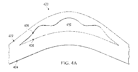
[0014] FIG. 4A is an illustration of a cross-sectional view of a
cornea depicting a
lenslet cut geometry for higher-order aberrations;
[0015] FIG. 4B is an illustration of a cross-sectional view of
cornea depicting a lenslet
cut geometry for higher-order aberrations with respect to axes;
[0016] FIG. 5 is a schematic plan view of a cornea illustrating a
lenslet cutting
geometry for higher-order aberrations;
[0017] FIG. 6 illustrates wavefront elevation maps generated from a wavefront
meter
and corneal topographer;
Foot si FIG. 7 illustrates a method for correcting higher-order
aberrations: and
[0019] FIG. 8 illustrates another variation of the method for correcting
higher-order
aberrations.
DETAILED DESCRIPTION
[0020] Embodiments of the present disclosure are directed to laser
vision correction.
More particularly, embodiments of the present disclosure are directed to a
method of
cutting a lenslet (a portion of the cornea that is removed during vision
correction
surgery and also called a lenticule) using a femtosecond laser to correct
higher-order
aberrations (HOAs). Embodiments of the present disclosure allow for correcting
HOAs
without creating a flap by cutting through the corneal epithelium and Bowman's
membrane with a femtosecond laser.
[0021] FIG. 1 is a schematic diagram of a pulsed laser system 100 for eye
surgery,
including refractive eye surgery such as laser vision correction. The pulsed
laser system
100 may be a separate surgical tool, or part of a larger eye surgery system,
which may
include other laser systems, patient or eye positioning systems, viewing
systems, or any
combinations thereof. In particular, the pulsed laser system 100 may be part
of a
CA 03175489 2022- 10- 13
WO 2021/240392
PCT/IB2021/054587
surgical suite designed to provide substantially all computer-assisted devices
for
performing a given eye surgery.
[0022] A pulsed laser system 100 includes a laser source 102,
which generates laser
radiation 104. The laser radiation 104 (a laser beam) may include laser
radiations used
to cut eye tissues including such as corneal stroma through vaporization (a
laser
scalpel). For example, the laser radiation 104 generated from the laser source
102 may
include a femtosecond, picosecond, nanosecond, or attosecond laser.
[0023] A pulsed laser system 100 includes a scanner 106 for
controlling a radiation
focal points 108 during surgery in the cornea of the patient's eye. The
scanner 106
provides transverse control axis (X- and Y-axes), longitudinal control axis (Z-
axis) of
radiation focal points 108. "Transverse" refers to a direction at a right
angle to the
propagation direction of laser beam 104. "Longitudinal- refers to the
propagation
direction of the laser beam 104. The scanner 106 may be 3D scanner.
[0024] Although the pulsed laser system 100 in FIG.1 does not show various
other
radiation control components, the scanner 106 may control radiation focal
points 108
in a longitudinal direction using a longitudinal control element. For example,
longitudinal control element may include a longitudinally adjustable lens.
Alternatively, longitudinal control element may include a variable refractive
power
lens. Also, alternatively, longitudinal control element may include a
deformable mirror.
Further, the scanner 106 may contain more than one transverse control element,
more
than one longitudinal control element, or more than one of both. In addition,
the
transverse control element and the longitudinal control element may be
separate
devices. Although scanner 106 shown in FIG. 1 is depicted as one component,
such a
configuration is merely provided for illustrative purposes. The embodiments of
the
present disclosure may be configured to include multiple scanners (a system of
scanners) to allow for more precise control of the radiation focal points 108.
[0025] The laser source 102 and scanner 106 are controlled by
computer 110. For
example, the computer 110 may control which wavelength of laser radiation 104
is
generated from the laser source 102. For instance, the computer may configure
the laser
source 102 to generate a femtosecond laser 104. Further, the computer 110 may
control
CA 03175489 2022- 10- 13
WO 2021/240392
PCT/IB2021/054587
6
the length of the laser radiation 104. Additionally, the computer 110 may
control the
scanner 106 to change movements of the radiation focal points 108.
[0026] The computer 110 includes at least a processing resource
able to execute code
to generate instructions to control a lenslet cut geometry and a lenslet cut
location in
the cornea of a patient's eye. The computer 110 may be in physical or wireless
communication with laser source 102 and scanner 106. The computer 110 may
further
include a memory, particularly a memory for storing instructions for the
processing
resource, a communications module for communicating with laser source 102 and
scanner 106, and other components.
[0027] For simplicity, not all potential components of the pulsed
laser system 100 are
illustrated in FIG. 1. For example, the pulsed laser system may include
various
components for directing, focusing, or otherwise manipulating laser beam, such
as
scanners, mirrors, beam expanders, or lenses. The pulsed laser system 100 may
further
include housings and other equipment to protect and position its components as
well as
patient-interface peripherals, which may be disposable.
[00281 Referring now to FIG. 2A, a schematic depiction 200 of cut geometry for
a
small incision lenslet extraction procedure (such as SMILE ) is described. The
human
eye has a cornea, which is a transparent front part of the eye that covers the
iris, pupil,
and anterior chamber. For laser vision correction surgery, a portion of stroma
(such as
lenslet 210) within the cornea is removed to change a thickness of the
patient's cornea
to correct vision. The schematic depiction of cut geometry shown in FIG. 2A is
a cross-
sectional view of a cornea of a human eye. In general, for a SMILE procedure,
alenslet
210 is created with a femtosecond laser in a shape corresponding to a desired
refractive
correction. The femtosecond incisions for the SMILE procedure include four
cuts: 1)
cornea posterior cut; 2) side cut for the lenslet; 3) cap cut; and 4) side cut
for the opening
incision. The four cuts are performed in succession in an integrated manner.
Then, the
lenslet is subsequently accessed and removed through the opening incision. The
cornea
includes anterior cornea 202 and posterior cornea 204. The lenslet cut creates
anterior
spherical surface 206 of the lenslet 210 and posterior spherical surface 208
of the lenslet
210.
CA 03175489 2022- 10- 13
WO 2021/240392
PCT/IB2021/054587
7
[0029] Referring now to FIG. 2B, a schematic depiction 250 of cut geometry for
small
incision lenslet extraction procedure (such as SMILE ) with respect to X-, Y-,
and Z-
axes is described. The lenslet is cut using a femtosecond laser. The spatial
position of
the laser radiation is controlled by three scanners: X-, Y-, and Z- scanners.
[0030] In general, X and Y scanners are galvanometric scanners. The lenslet is
cut
using spiral scanning of the femtosecond laser beam. The spiral is typically
nearly a
circle, because the radial line separation of consecutive spirals is around 5
urn and the
radius of the scanning is several thousand microns. For example, a diameter of
a circle
may be 4 mm (such as 4000 pm). Thus, the next outer circle of the spiral would
have a
diameter of 4010 p.m.
[0031] The spherical refractive power of the lenslet is determined
by the radii of the
curvature of the anterior R1 and posterior R2 curvature of the lenslet surface
as defined
by the following equation:
D = (n-1)*(1/R2-1/R1) (eq 1)
where D is the spherical refractive power of the lenslet and n is the
refractive index of
the cornea.
[0032] A thickness of the lenslet to at the radial position r can
be calculated by the
following equation:
to= 0.5*r2(1/R2-1/R1) = (r2/2)*D/(n-1) (eq 2)
[0033] The Z scanner is typically an axially adjustable telescope.
Due to mechanical
inertia the Z scanner is slow and not able change position, speed, or
acceleration nearly
as rapidly as the scanners movable in the x and y planes. However, the circle
time of
scanning in Z axis is about 20 ms and within 20 ms, the Z position can be
moved by a
few microns, allowing the lenslet to be cut with a spherical shape.
[0034] Surfaces derived from a high order azimuthal Zernike
polynomial presently
cannot be cut using the Z scanner of femtosecond laser, since the rotation
time of a (1)=
5mm circle having a typical 5 vim spot separation at 150 kHz laser rep rate=
CA 03175489 2022- 10- 13
WO 2021/240392
PCT/IB2021/054587
8
T=5000*n/(5*150000)=21 ms. Within 21 ms the Z scanner is incapable of moving
up
and down several times to cut a high order azimuthal surface.
[0035] Now referring to FIG. 3, aberration modes based on Zernike polynomial
functions are illustrated. Aberrations are focusing errors that prohibit the
formation of
high resolution One of the ways to characterize the aberrations includes
wavefront
aberrations, which characterizes complex optical errors in focus produced by
an optical
system. The Zernike polynomial series is used to decompose complex wavefront
aberrations into a collection of polynomial basis functions (such as modes),
which are
shown in FIG. 3.
[0036] Each Zernike mode includes two components: 1) radial order (n) and 2)
meridional frequency (f). In ophthalmology, radial orders of Zernike
polynomial series
are categorized as either low-order aberrations or high-order abrasions. Low-
order
aberrations are Zernike modes having second order or lower (n<2). High-order
aberrations are Zemike modes having third order or higher (n>3). Low-order
aberrations which correspond to Zernike defocus (4 in FIG. 3) and astigmatism
modes
(3 and 5 in FIG. 3) are typically corrected with prescription spectacle lenses
or contact
lenses, while correction of high-order aberrations requires more complex
procedures
The higher the radial order and/or meridional frequency is, the more complex a
Zernike
polynomial mode becomes. Cutting a lenslet for one of the high-order
aberrations which
is shown in FIG.3 is an arduous task with a slow z scanner of the femtosecond
laser.
[0037] Now referring to FIG. 4A, an illustration depicting a lenslet cut
geometry, in
accordance with one or more embodiments of the present disclosure is shown.
The
schematic depiction of cut geometry shown in FIG. 4A is a cross-sectional view
of a
cornea. In one embodiment, the lenslet 410 which corrects for high-order
aberrations
may be cut. For example, the lenslet 410 may include multiple high-order
Zernike
polynomial modes. In this regard, the lenslet 410 does not simply have a
spherical shape
such as shown in FIGS. 2A and 2B. The cornea includes anterior cornea 402 and
posterior cornea 404. The lenslet cut 410 creates anterior spherical surface
406 of the
lenslet 410 and posterior spherical surface 408 of the lenslet 410.
CA 03175489 2022- 10- 13
WO 2021/240392
PCT/IB2021/054587
9
[0038] In some embodiments, a thickness of the lenslet 410 which corrects
higher-
order aberrations can be calculated and a surface of the lenslet having a
radius of
curvature R is cut as follows:
[0039] The typical radial separation of two consecutive spiral cut
RS is about 5 p.m.
To have a radius of curvature of the lenslet surface R, the vertical step (VS)
should be
VS = r/R*RS (eq 3)
where r/R is the slope of the R surface at the position of r. To correct the
HOA, the
thickness of the lenslet should be changed to
t(X/Y) = to+At(X,Y)/(n-1) (eq 4)
where At(X,Y) is the HOA wavefront elevation measured with the wavefront meter
or
corneal topographer. It is noted that to is the thickness of the lenslet
having a spherical
refractive power of D which is responsible for correcting the spherical error.
It is further
noted that At(X/Y) is responsible for correcting the HOAs. At(X/Y) is
typically
described either with Zernike or Fourier polynomials.
[0040] FIG. 411 is an illustration depicting a lenslet cut
geometry for higher-order
aberrations with respect to axes, in accordance with one or more embodiments
of this
disclosure. As described in FIG. 4B, it is noted that by increasing the radius
of the
scanning by Ar the thickness of the lenslet is increasing by
At = Ar* [slope of the R surface] (eq 5)
where the slope of the R curve is r/R, for example. Then, At may be expressed
as
At = Ar* r/R (eq6)
Thus, in order to correct the HOA, the radius on the scanning at any X/Y point
should
be increased by
Ar(X/Y) = At(X/Y)* R/r (eq 7)
CA 03175489 2022- 10- 13
WO 2021/240392
PCT/IB2021/054587
[0041] FIG. 5 is a schematic plan view 500 of a cornea
illustrating a lenslet cutting
geometry, in accordance with one or more embodiments of the present
disclosure. In
one embodiment, in order to correct the HOA, the radius on the scanning at any
X/Y
point may be increased by Ar(X/Y) = At(X/Y)* R/r, as described hereinbefore.
Since
radial scanning with X- and Y-axes scanners is fast, cutting the lenslet
geometry
including high-order aberrations may not need a change in Z-coordination.
Therefore,
embodiments of the present disclosure may allow for correcting the HOA without
rapidly changing Z-coordinate when scanning.
[0042] FIG. 6 is illustrations of wavefront elevation maps (600 and 650)
generated
from wavefront meter or a corneal topographer. In one embodiment, a
calculation of
radius on the scanning at any X/Y point Ar(X/Y) to correct the HOA may require
a
wavefront elevation map using a wavefront meter or a corneal topographer.
Wavefront
elevation maps reveal any irregularity of corneal surface.
[0043] FIG. 7 illustrates a method for correcting higher-order
aberrations, in
accordance with one or more embodiments of the present disclosure. The pulse
laser
device for correcting higher-order aberrations used in this method may be
described in
FIGs. 4A-5. It is noted that all of the steps shown in FIG. 7 are not
essential to practice
the method. One or more steps may be omitted from or added to the method
illustrated
in FIG. 7, and the method can still be practiced within the scope of this
embodiment.
[0044] The method shown in FIG. 7 generally includes providing a laser
radiation.
The method further includes controlling a location of a beam focal point of
the laser
radiation by a scanner and guiding the beam focal point in such a way that the
location
of the beam focal point is in a cornea of an eye. The method further includes
introducing
the laser radiation into the cornea of the eye. The method further includes
cutting a
lenslet, wherein a thickness of the lenslet t(X/Y) satisfies a following
equation:
t(X/Y)=to-hAt(X,Y)/(n-1) (eq 4)
where At(X,Y) represents a higher-order wavefront elevation and to (eq 2)
represents
the thickness of the lenslet having a spherical refractive power of D.
[0045] FIG 8 illustrates another variation of the method for
correcting higher-order
aberrations, in accordance with one or more embodiments of the present
disclosure.
CA 03175489 2022- 10- 13
WO 2021/240392
PCT/IB2021/054587
11
The pulse laser device for correcting higher-order aberrations used in this
method may
be described in FIGs. 4A-5. It is noted that all of the steps shown in FIG. 8
are not
essential to practice the method. One or more steps may be omitted from or
added to
the method illustrated in FIG. 8, and the method can still be practiced within
the scope
of this embodiment.
[0046] The method shown in FIG. 8 generally includes providing a laser
radiation.
The method further includes controlling a location of a beam focal point of
the laser
radiation by a scanner and guiding the beam focal point in such a way that the
location
of the beam focal point is in a cornea of an eye. The method further includes
introducing
the laser radiation into the cornea of the eye. The method further includes
cutting a
lenslet, wherein a radius of the lenslet at any X/Y point satisfies a
following equation:
Ar(X/Y) = At(X/Y)* R/r (eq 7)
where At(X,Y) represents a higher-order wavefront elevation, R represents a
curvature
of the cornea, and r/R represents a slope of the curvature of the cornea.
[0047] Although this disclosure has been described in terms of certain
embodiments,
modifications (such as substitutions, additions, alterations, or omissions) of
the
embodiments will be apparent to those skilled in the art. Accordingly,
modifications
may be made to the embodiments without departing from the scope of the
invention.
For example, modifications may be made to the systems and apparatuses
disclosed
herein. The components of the systems and apparatuses may be integrated or
separated,
and the operations of the systems and apparatuses may be performed by more,
fewer,
or other components. As another example, modifications may be made to the
methods
disclosed herein. The methods may include more, fewer, or other steps, and the
steps
may be performed in any suitable order.
CA 03175489 2022- 10- 13