Note : Les descriptions sont présentées dans la langue officielle dans laquelle elles ont été soumises.
WO 2021/222867
PCT/US2021/030351
I MMUNOTHERAPY RESPONSE SIGNATURE
CROSS REFERENCE
This application claims the benefit of U.S. Provisional Patent Application
Serial No.
63/018,304, filed on April 30, 2020; the entire contents of which application
is hereby incorporated by
reference in its entirety.
TECHNICAL FIELD
The present disclosure relates to the fields of data structures, data
processing, and machine
learning, and their use in precision medicine, e.g., the use of molecular
profiling to guide personalized
treatment recommendations for various diseases and disorders, including
without limitation cancer.
BACKGROUND
limnunothcrapy is the treatment of cancer or other diseases by activating or
suppressing the
immune system. Inununotherapies designed to elicit or amplify an immune
response may referred to
as activation immunotherapies or immune activators, whereas immunotherapies
that reduce or
suppress such response may referred to as suppression immunotherapies or
immune suppressors.
Checkpoint inhibitor therapy is a form of immunotherapy that targets immune
checkpoints, which are
key regulators of the immune system that stimulate or inhibit immune response.
Tumors may block
such checkpoints in order to avoid attack by the immune system. Checkpoint
therapy can block these
inhibitory checkpoints, thereby restoring immune system function. For reviews,
see, e.g., Topalian SL
et al, Immune checkpoint blockade: a common denominator approach to cancer
therapy. Cancer Cell.
2015 Apr 13;27M:450-61; Postow MA et al., Immune Checkpoint Blockade in Cancer
Therapy. J.
Cl in On.col. 2015 Jun 10;33(17):1974-82.
PD! (programmed death-1, PD-1, PDCD1, CD279) is a transmembrane glycoprotein
receptor
that is expressed on CD4-/CD8-thymocytes in transition to CD4+/CDS+ stage and
on mature T and B
cells upon activation. It is also present on activated myeloid lineage cells
such as monocytes, dendritic
cells and NK cells. In normal tissues, PD-1 signaling in T cells regulates
immune responses to
diminish damage, and counteracts the development of autoimmunity by promoting
tolerance to self-
antigens. PD-Li (programmed cell death 1 ligand 1, PD1,1, cluster of
differentiation 274, CD274, B7
homolog I, B7-H1, B7HI) and PD-L2 (programmed cell death I ligand 2, PDL2, B7-
DC, B7DC,
CD273, cluster of differentiation 273) are PD1 ligands. In normal cells the PD
IRMA interplay is an
immune checkpoint, whereas tumor cell expression of PD-Li is a mechanism to
evade
recognition/destruction by the immune system, e.g., tumor-infiltrating T cells
(TILs). PD-L I is
constitutively expressed in many human cancers including without limitation
melanoma, ovarian
cancer; lung cancer, clear cell renal cell carcinoma (CRCC); urothelial
carcinoma, HNSCC, and
CA 03177323 2022- 10- 28
WO 2021/222867
PCT/US2021/030351
esophageal cancer. Monoclonal antibody therapy that targets the PD-1/PD-L1
pathway may allow T-
cells to attack the tumor. CTLA4 (cytotoxic T-lymphocyte-associated protein 4,
CTLA-4, CDI52) is a
protein receptor that functions as an immune checkpoint by downregulating
immune responses.
CTLA4 is constitutively expressed in regulatory T cells but only upregulated
in conventional T cells
after activation a phenomenon which is particularly notable in cancers.
Monoclonal antibody
therapy that blocks inhibitory effects of CTLA-4 can potentiate effective
immune responses against
tumor cells.
Several targeted therapies to CTLA4, PD-I, and PD-Ll checkpoint inhibitors
have been
approved by the United States Food and Drug Administration (FDA) for the
treatment of various
cancers. These include ipilimumab (anti-CTLA-4, trade name Yervoy, Bristol-
Myers Squibb);
nivolumab (human. monoclonal immunoglobulin G4 antibody targeting PD-1 ,trade
name Opdivo,
Bristol-Myers Squibb), pembroliztunab (humanized IgG4 isotype antibody
targeting PD-1, trade
name Keytruda, Merck); atezolizumab (fully humanized, engineered monoclonal
antibody of IgG1
isotype targeting PD-L1, trade name Tecentriq, Genentech/Roche); awlumab
(whole monoclonal
antibody of isotype IgG1 targeting PD-L1, trade name Bavencio, Merck KGaA and
Pfizer Inc.); and
durvalumab (human immunoglobulin. GI kappa (IgG Ix) monoclonal antibody
targeting PD-Li, trade
name linfinzi, AstraZeneca). In May 2017, pembrolizumab received an
accelerated approval from the
FDA for use in any unresectable or metastatic solid tumor with DNA mismatch
repair deficiencies or
a microsatellite instability-high state (or, in the case of colon cancer,
tumors that have progressed
following chemotherapy). This approval marked the first instance in which the
FDA approved
marketing of a drug based only on the presence of a genetic marker, with no
limitation on the site of
the cancer or the kind of tissue in which it originated. Several additional
therapies that target immune
checkpoint proteins are in development.
Despite these successes, immune checkpoint therapy has not proven to be a
panacea for
cancer. Although pembrolizumab was approved across tumor types, other
immunotherapies have only
proven efficacy in certain settings. As one example, nivolumab has been
approved for inoperable or
metastatic melanoma, metastatic squamous non-small cell lung cancer, and as
second-line treatment
for renal cell carcinoma, but failed to meet its endpoints in a clinical trial
directed towards treating
newly diagnosed lung cancer. Immune checkpoint therapy is also typically
prescribed upon indication
from a companion diagnostic (e.g., to confirm expression of the target
protein); but it is not always
efficacious. For example, the response rate to pembrolizumab may be less than
50% even in patients
pre-selected for expression of PD-L I on at least 50% of tumor cells. See,
e.g., Reck, M., et at.,
Pembrolizumab versus Chemotherapy for PD-LI¨Positive Non¨Small-Cell Lung
Cancer. N Engl J
Med 2016; 375:1823-1833. And in some cases, checkpoint inhibitor therapy may
exacerbate
hyperprogressive disease characterized by acceleration of tumor growth during
treatment. See, e.g.,
Ferrara, R et al., Hyperprogressive Disease in Patients With Advanced Non-
Small Cell Lung Cancer
Treated With PD-1/PD-L I Inhibitors or With Single-Agent Chemotherapy. JAMA
Oncol. 2018 Nov
2
CA 03177323 2022- 10- 28
WO 2021/222867
PCT/US2021/030351
1;4(1 1 ):1543-1552. Moreover, altering immune system checkpoint inhibition
can have diverse effects
on most organ systems of the body. Take pembrolizinnab as an example. Adverse
reactions include
severe infusion-related reactions, severe lung inflammation (including
fatalities), inflammation of
endocrine organs that caused inflammation of the pituitary gland of the
thyroid (causing both
hypothyroidism and hyperthyroidism in different people), and pancreatitis that
caused Type 1 diabetes
and diabetic ketoacidosis. Some patients require lifelong hormone therapy as a
result (e.g. insulin
therapy or thyroid hormones). Pembrolizumab therapy has also led to colon
inflammation, liver
inflammation, and kidney inflammation. More common adverse reactions to
pernbrolizumab include
fatigue (24%), rash (19%), itchiness (pruritus) (17%), diarrhea (12%), nausea
(11%) and joint pain
(arthralgia) (10%), and between 1% and 10% of people taking pembrolizumab have
included anemia,
decreased appetite, headache, dizziness, distortion of the sense of taste, dry
eye, high blood pressure,
abdominal pain, constipation, diy mouth, severe skin reactions, vitiligo,
various kinds of acne, dry
skin, eczema, muscle pain, pain in a limb, arthritis, weakness, edema, fever,
chills, and flu-like
symptoms. Similar side effects have been observed for other checkpoint
inhibitor therapies. Finally,
immune checkpoint therapy can be extremely expensive. Indeed, pembroliztunab
was priced at
$150,000 per year when it launched in late 2014. Taken together, there is a
need to better identify
those patients more likely to benefit from immunotherapies for better patient
outcomes and to avoid
unnecessary adverse events and high costs.
Machine learning models can be configured to analyze labeled training data and
then draw
inferences from the training data. Once the machine learning model has been
trained, sets of data that
are not labeled may be provided to the machine learning model as an input. The
machine learning
model may process the input data, e.g., molecular profiling data, and make
predictions about the input
based on inferences learned during training. As an. example, machine learning
models can be trained
to recognize molecular data from subjects that did or did not respond to a
given treatment.
Comprehensive molecular profiling provides a wealth of data concerning the
molecular status
of patient samples. We have performed such profiling on many thousands of
tumor patients from
practically all cancer lineages and have tracked patient outcomes and
responses to treatments in
thousands of these patients. Our molecular profiling data can be compared to
patient benefit or lack of
benefit to treatments and processed using machine learning algorithms. Here,
this approach has been
applied to identify biomarker signatures that predict benefit of immunotherapy
in cancer patients.
SUMMARY
Comprehensive molecular profiling provides a wealth of data concerning the
molecular status
of patient samples. Such data can be compared to patient response to
treatments to identify biomarker
signatures that predict response or non-response to such treatments. This
approach has been applied to
identify biomarker signatures that correlate with benefit or lack of benefit
of immunotherapies, e.g.,
checkpoint inhibitor therapies. Further described herein arc methods for
training and employing
3
CA 03177323 2022- 10- 28
WO 2021/222867
PCT/US2021/030351
machine learning models to predict effectiveness of a treatment for a disease
or disorder of a subject
having a particular set of biomarkers.
In an aspect, the present disclosure provides a method for predicting benefit
of
immunotherapy for a cancer in a first subject, the method comprising:
obtaining, by one or more
computers; molecular data corresponding to a plurality of biomarkers selected
from the group
consisting of CD274, CD8A, PDCD1, CD28, DDR2, six I, and CDK12, wherein the
obtained
molecular data was generated by assaying a biological sample from the first
subject; generating, by
the one or more computers, input data that includes a set of features
extracted from the obtained
molecular data; providing, by the one or more computers, the generated input
data as input to a
predictive model, the predictive model comprising at least one machine
learning model, wherein each
particular machine learning model of the at least one machine learning model
is trained to generate
output data that indicates whether a subject is likely to benefit from an
immunotherapy based on the
particular machine learning model processing of a set of features extracted
from molecular data
corresponding to the plurality of biomarkers (selected from the group
consisting of CD274. CD8A,
PDCD1, CD28, DDR2, STK11, and CDK12); processing, by one or more computers the
generated
input data through the at least one machine learning model, to generate first
data indicating whether
the first subject is likely to benefit from the immunotherapy; determining, by
the one or more
computers and based on the generated first data, a likelihood that the first
subject is to benefit from
the immunotherapy; based on the determined likelihood, generating, by the one
or more computers,
rendering data that, when rendered by a user device, causes a user device to
display data that identifies
the determined likelihood; and providing, by one or more computers, the
rendering data to the user
device.
In some embodiments, the rendering data is displayed by the user device, based
on one or
more threshold, as: i) likely benefit from. the immunotherapy; ii) likely lack
benefit from the
immunotherapy; and/or iii) indeterminate benefit from the immunotherapy. The
threshold for such
characterization can be make based on a desired criteria, such as a confidence
value. In a non-limiting
example, the rendering data may display as likely benefit from the
immunotherapy when there is high
confidence in such determination. Similarly, the rendering data may display as
likely lack of benefit
from the immunotherapy when there is high confidence in likely lack of
benefit, or alternately when
there is lack of confidence in the determined likelihood of benefit. An
indeterminate call may be made
when there is insufficient confidence in either likely benefit or likely lack
of benefit.
In some embodiments, determining, by the one or more computers and based on
the generated
first data, a likelihood that the first subject is to benefit from the
immunotherapy includes calculating
a probability.
in some embodiments, the method further comprises: determining, by the one or
more
computers, whether the first data satisfies one or more thresholds; and based
on a determination that
the first data satisfies one of the one or more thresholds, determining that
the first subject is likely to
4
CA 03177323 2022- 10- 28
WO 2021/222867
PCT/US2021/030351
benefit from the immunotherapy; wherein generating, by the one or more
computers, rendering data
that, when rendered by the user device, causes the user device to display data
that identifies the
determined likelihood comprises: generating, by the one or more computers,
rendering data that, when
rendered, causes the user device to display data that indicates that the first
subject is likely to benefit
from the immunotherapy.
In some embodiments, the method further comprises: determining, by the one or
more
computers, whether the first data satisfies one or more thresholds; and based
on a determination that
the first data does not satisfy one of the one or more thresholds, determining
that the first subject is
not likely to benefit from the immunotherapy; wherein generating, by the one
or more computers,
rendering data that, when rendered by the user device, causes the user device
to display data that
identifies the determined likelihood comprises: generating, by the one or more
computers, rendering
data that, when rendered, causes the user device to display data that
indicates that the first subject is
not likely to benefit from the immunotherapy.
In some embodiments, the method further comprises: determining, by the one or
more
computers, whether the first data satisfies one or more thresholds; and based
on a determination that
the first data is (i) equal to one of the one or more thresholds or (ii)
satisfies two of the one or more
thresholds, determining that the first subject is likely to have an
indeterminate benefit from the
immunotherapy; wherein generating, by the one or more computers, rendering
data that, when
rendered by the user device, causes the user device to display data that
identifies the determined
likelihood comprises: generating, by the one or more computers, rendering data
that, when rendered,
causes the user device to display data that indicates that the first subject
is likely to have an
indeterminate benefit from the immunotherapy.
In some embodiments, the plurality of biomarkers comprises at least 2, 3, 4,
5, 6, or 7 of
CD274, CD8A, PDCD I., CD28, DDR2, S'FK I I, CDKI2, and any useful combination
thereof;
optionally wherein the plurality of biomarkers comprises CD274, CD8A, FDCD I ,
CD28, DDR2,
STK11, and CDK12; optionally wherein the plurality of biomarkers consists of
CD274, CD8A,
PDCD1, CD28, DDR2, S11(11, and CDK12.
In some embodiments, the biological sample comprises forrnalin-fixed paraffin-
embedded
(FFPE) tissue, fixed tissue, a core needle biopsy, a fine needle aspirate,
unstained slides, fresh frozen
(FF) tissue, fonnalin samples, tissue comprised in a solution that preserves
nucleic acid or protein
molecules, a fresh sample, a malignant fluid, a bodily fluid, a tumor sample,
a tissue sample, or any
combination thereof. In some embodiments, the biological sample comprises
cells from a solid tumor.
In some embodiments, the biological sample comprises a bodily fluid. In some
embodiments, the
bodily fluid comprises a malignant fluid, a pleural fluid, a peritoneal fluid,
or any combination
thereof In some embodiments, the bodily fluid comprises peripheral blood,
sera, plasma, ascites,
urine, cerebrospinal fluid (CSF), sputum, saliva, bone marrow, synovial fluid,
aqueous humor,
amniotic fluid, cerumen, breast milk, broncheoalveolar lavage fluid, semen,
prostatic fluid, cowper's
5
CA 03177323 2022- 10- 28
WO 2021/222867
PCT/US2021/030351
fluid, pre-ejaculatory fluid, female ejaculate, sweat, fecal matter, tears,
cyst fluid, pleural fluid,
peritoneal fluid, pericardial fluid, lymph, chyme, chyle, bile, interstitial
fluid, menses, pus, sebum,
vomit, vaginal secretions, mucosa] secretion, stool water, pancreatic juice,
lavaue fluids from. sinus
cavities, bronchopulmonary aspirates, blastocyst cavity fluid, or umbilical,
cord blood.
In some embodiments, assaying the biological sample comprises determining a
presence,
level, or state of a protein or nucleic acid for each biomarker, optionally
wherein the nucleic acid
comprises deoxyribonucleic acid (DNA), ribonucleic acid (RNA), or a
combination thereof, wherein
optionally the nucleic acid comprises cell free nucleic acid, wherein
optionally the nucleic acid
consists of cell free nucleic acid. In some embodiments, the presence, level
or state of the protein is
determined using immunohistochemistry (U-IC), flow cytometry, an immunoassay,
an antibody or
functional fragment thereof, an aptamer, or any combination thereof; and/or
the presence, level or
state of the nucleic acid is determined using polymerase chain reaction (PCR),
in situ hybridization,
amplification, hybridization; microarray, nucleic acid sequencing, dye
tennination sequencing,
pyrosequencing, next generation sequencing (NGS; high-throughput sequencing),
whole exome
sequencing, whole transcriptome sequencing, whole genome sequencing, or any
combination thereof
In some embodiments, the state of the nucleic acid comprises a sequence,
mutation, polymorphism,
deletion, insertion, substitution, translocation, fusion, break, duplication,
amplification, repeat, copy
number (copy number variation; CNV; copy number alteration; CNA), transcript
level (expression
level), or any combination thereof In some embodiments, the state of the
nucleic acid comprises a
transcript level for at least one member of the plurality of biomarkers,
optionally wherein the state of
the nucleic acid comprises a transcript level for all members of the plurality
of biomarkers. In some
embodiments, assaying the biological sample comprises performing WTS and the
molecular data
comprises a transcript level for at least one member of the plurality of
biomarkers obtained via the
WTS, optionally wherein the molecular data comprises a transcript level for
all members of the
plurality of biomarkers obtained via the WTS.
In some embodiments, the immunotherapy comprises an immune checkpoint therapy,
optionally wherein the immune checkpoint therapy comprises at least one of
ipilimumab, nivolumab,
pembrolizumab, atezoliztunab, avelumab, durvalumab, and any combination
thereof, optionally
wherein the immunotherapy comprises nivolumab and/or pembrolizumab, optionally
wherein the
immunotherapy consists of nivolumab and/or pembrolizumab.
In some embodiments, the first subject has not previously been treated with
the
immunotherapy. In some embodiments. the cancer comprises a metastatic cancer,
a recurrent cancer,
or a combination thereof. In some embodiments, the first subject has not
previously been treated for
the cancer.
in some embodiments, the method further comprises administering the
immunotherapy to the
first subject. In some embodiments, progression free survival (.PFS), disease
free survival (DFS), or
lifespan is extended by the administration.
6
CA 03177323 2022- 10- 28
WO 2021/222867
PCT/US2021/030351
In some embodiments; the cancer comprises an acute lymphoblastic leukemia;
acute myeloid
leukemia; adrenocortical carcinoma; AIDS-related cancer; AIDS-related
lymphoma; anal cancer;
appendix cancer; astrocytomas; atypical teratoid/rhabdoid tumor; basal cell
carcinoma; bladder
cancer; brain stem glioma; brain tumor, brain stem glioma, central nervous
system atypical
teratoid/rhabdoid tumor, central nervous system embryonal tumors,
astrocytomas,
craniopharyngioma, ependymoblastoma, ependymoma, medulloblastoma,
medulloepithelioma, pineal
parenchymal tumors of intermediate differentiation, supratentorial primitive
neuroectodennal tumors
and pineobla.stoma; breast cancer; bronchial tumors; Burkitt lymphoma; cancer
of unknown primary
site (CUP); carcinoid tumor; carcinoma of unknown primary site; central
nervous system atypical
teratoid/rhabdoid tumor; central nervous system embryonal tumors; cervical
cancer; childhood
cancers; chordoma; chronic lymphocytic leukemia; chronic myelogenous leukemia;
chronic
myeloproliferative disorders; colon cancer; colorectal cancer;
craniopharyngioma; cutaneous T-cell
lymphoma; endocrine pancreas islet cell tumors; endometrial cancer;
ependymoblastoma;
ependymoma; esophageal cancer; esthesioneuroblastoma; Ewing sarcoma;
extracranial germ cell
tumor; extragonadal germ cell tumor; extrahepatic bile duct cancer;
gallbladder cancer; gastric
(stomach) cancer; gastrointestinal caminoid tumor; gastrointestinal stromal
cell tumor; gastrointestinal
stromal tumor (GIST); gestational trophoblastic tumor; glioma; hairy cell
leukemia; head and neck
cancer; heart cancer; Hodgkin lymphoma; hypophary, ngeal cancer; intraocular
melanoma; islet cell
tumors; Kaposi sarcoma; kidney cancer; Langerhans cell histiocytosis;
laryngeal cancer; lip cancer;
liver cancer; malignant fibrous histiocytoma bone cancer; mcdulloblastoma;
medulloepithelioma;
melanoma; Merkel cell carcinoma; Merkel cell skin carcinoma; mesothelioma;
metastatic squamous
neck cancer with occult primary; mouth cancer; multiple endocrine neoplasia
syndromes; multiple
myeloma; multiple myeloina/plasma cell neoplasm.; mycosis fungoides:
myelodysplastic syndromes;
myeloproliferative neoplasms; nasal cavity cancer; nasopharyngeal cancer;
neuroblastoma; Non-
Hodgkin lymphoma; nonmelanoma skin cancer; non-small cell lung cancer; oral
cancer; oral cavity
cancer; oropharyngeal cancer; osteosarcoma; other brain and spinal cord
tumors; ovarian cancer;
ovarian epithelial cancer; ovarian genn cell tumor; ovarian low malignant
potential tumor; pancreatic
cancer; papillomatosis; paranasal sinus cancer; parathyroid cancer: pelvic
cancer; penile cancer;
pharyngeal cancer; pineal parenchymal tumors of intermediate differentiation;
pineoblastoma;
pituitary tumor; plasma cell neoplasm/multiple myeloina; pleuropulmonaiy
blastoma; primary central
nervous system (CNS) lymphoma; primary hepatocellular liver cancer; prostate
cancer; rectal cancer;
renal cancer; renal cell (kidney) cancer; renal cell cancer: respiratory tract
cancer; retinoblastoma;
rhabdomyosarcoma; salivary gland cancer; Sezary syndrome; small cell lung
cancer; small intestine
cancer; soft tissue sarcoma; squamous cell carcinoma; squamous neck cancer;
stomach (gastric)
cancer; supratentorial primitive neuroectodermal tumors; T-cell lymphoma;
testicular cancer; throat
cancer; thymic carcinoma; thymoma; thyroid cancer; transitional cell cancer;
transitional cell cancer
of the renal pelvis and ureter; trophoblastic tumor; ureter cancer; urethral
cancer; uterine cancer;
7
CA 03177323 2022- 10- 28
WO 2021/222867
PCT/US2021/030351
uterine sarcoma; vaginal cancer; vulvar cancer; Waldenstrom macroglobulinemia;
or Wilm's tumor.
In some embodiments, the cancer comprises an acute myeloid leukemia (AML),
breast carcinoma,
cholangiocarcinomaõ colorectal adenocarcinoma, extrahepatic bile duct
adenocarcinoma, female
genital tract malignancy, gastric adenocarcinoma, gastroesophageal
adenocarcinoma, gastrointestinal
stromal tumor (GIST), glioblastoma, head and neck squamous carcinoma,
leukemia, liver
hepatocellular carcinoma, low grade glioma, lung bronchioloalveolar carcinoma
(BAC), non-small
cell lung cancer (NSCLC), lung small cell cancer (SCLC), lymphoma, male
genital tract malignancy,
malignant solitary fibrous tumor of the pleura (MSFT), melanoma, multiple
rnyeloma, neuroendocrine
tumor, nodal diffuse large B-cell lymphoma, non epithelial ovarian cancer (non-
E0C), ovarian
surface epithelial carcinoma, pancreatic adenocarcinoma, pituitary carcinomas,
oligodendroglioma,
prostatic adenocarcinoma, retroperitoneal or peritoneal carcinoma,
retroperitoncal or peritoneal
sarcoma, small intestinal malignancy, soft tissue tumor, thytnic carcinoma,
thyroid carcinoma, or
uveal melanoma. In some embodiments, the cancer comprises a lung cancer,
optionally wherein the
lung cancer comprises a non-small cell lung cancer (NSCLC).
In some embodiments, the at least one machine learning model comprises one or
more of a
random forest, support vector machine (SVM), logistic regression, K-nearest
neighbor, artificial
neural network, naive Bayes, quadratic discriminant analysis, Gaussian
processes models, decision
tree, or a combination thereof. In some embodiments, determining, by the one
or more computers and
based on the first data, whether the at least one machine learning model
indicates that the first subject
is likely to benefit from the immunotherapy, comprises allowing each of a
plurality of machine
learning models to vote whether the first subject is likely to benefit. In
some embodiments, each of the
plurality of machine learning models has an equal vote, or a weighted vote,
wherein optionally the
weighted voting is determined by providing, by the one or more computers, the
obtained votes of each
of the plurality of machine learning models, as input into another machine
learning model which then
determines whether the first subject is likely to benefit from the treatment.
In some embodiments, the plurality of biomarkers consists of CD274, CD8A,
PDCD1, CD28,
DDR2, STK11, and CDK12; the biological sample comprises cancer cells or cell
free nucleic acid
released from cancer cells; assaying the biological sample comprises
performing WTS and the
plurality of molecular data comprises transcript levels; and the at least one
machine learning model
consists of a support vector machine.
In some embodiments, the user device comprises a computer or a mobile device
and/or the
one or more computers comprises the user device.
In some embodiments, the method further comprises generating a report
displaying the
rendering data that identifies the likely benefit, lack of benefit of
treatment, or indeterminate benefit
of the inununotherapy, wherein optionally the display for displaying the
output comprises a printout, a
file, a computer display, and any combination thereof.
8
CA 03177323 2022- 10- 28
WO 2021/222867
PCT/US2021/030351
In some embodiments, the method further comprises administering the
immunotherapy to the
subject based on the identified likely benefit, likely lack of benefit, or
indeterminate benefit. See, e.g.,
Example 3. In some embodiments, the immunotherapy is administered to the
subject if the rendering
data identifies the likely benefit of treatment with the immunotherapy. In
some embodiments, the
immunotherapy is administered to the subject if the rendering data identifies
indeterminate benefit of
treatment with the immunotherapy. In some embodiments, chemotherapy is
administered to the
subject if the provided output identifies likely lack of benefit or
indeterminate benefit of treatment
with the immunotherapy, optionally wherein the immunotherapy is administered
in addition to the
chemotherapy.
in a related aspect, the present disclosure provides a non-transitory computer-
readable
medium storing software comprising instructions executable by one or more
computers which, upon
such execution, cause the one or more computers to perform the operations as
above.
In another related aspect, the present disclosure provides a system comprising
one or more
computers and one or more storage media storing instructions that, when
executed by the one or more
computers, cause the one or more computers to perform each of the operations
described above. In
some embodiments, the system further comprises laboratory equipment for
assaying the biological
sample, optionally wherein the laboratory equipment comprises next-generation
sequencing
equipment.
Unless otherwise defined, all technical and scientific terms used herein have
the same
meaning as commonly understood by one of ordinary skill in the art to which
this invention belongs.
Methods and materials are described herein for use in the present invention;
other, suitable methods
and materials known in the art can also be used. The materials, methods, and
examples are illustrative
only and not intended to be limiting. All publications, patent applications,
patents, sequences,
database entries, and other references mentioned herein are incorporated by
reference in their entirety.
In case of conflict, the present specification, including definitions, will
control.
Other features and advantages of the invention will be apparent from the
following detailed
description and figures, and from the claims.
DESCRIPTION OF DRAWINGS
FIG. lA is a block diagram of an example of a prior art system for training a
machine
learning model.
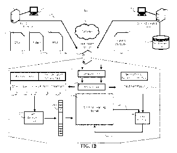
FIG. 1.B is a block diagram of a system that generates training data
structures for training a
machine learning model to predict effectiveness of a treatment for a disease
or disorder of a subject
having a particular set of biomaticers.
FIG. IC is a block diagram of a system for using a machine learning model that
has been
trained to predict effectiveness of a treatment for a disease or disorder of a
subject having a particular
set of biomarkers.
9
CA 03177323 2022- 10- 28
WO 2021/222867
PCT/US2021/030351
FIG. 1D is a flowchart of a process for generating training data for training
a machine
learning model to predict effectiveness of a treatment for a disease or
disorder of a subject having a
particular set of biomarkers.
FIG. 1.E is a flowchart of a process for using a machine learning model that
has been trained
to predict effectiveness of a treatment for a disease or disorder of a subject
having a particular set of
biomarkers.
FIG. IF is a block diagram of a system for predicting effectiveness of a
treatment for a
disease or disorder of a subject having a particular set of biornarkers by
using voting unit to interpret
output generated by multiple machine learning models.
FIG. 1G is a block diagram of system components that can be used to implement
systems of
FIGs. 2-3.
FIG. 1.11 illustrates a block diagram of an exemplary embodiment of a system
for deterniining
individualized medical intervention for cancer that utilizes molecular
profiling of a patient's
biological specimen.
FIGs. 2A-C are flowcharts of exemplary embodiments of (A) a method for
determining
individualized medical intervention for cancer that utilizes molecular
profiling of a patient's
biological specimen, (B) a method for identifying signatures or molecular
profiles that can be used to
predict benefit from therapy, and (C) an alternate version of (B).
FIG. 3 outlines an exemplary method of predicting a patient response to
immunotherapy.
FIG. 4 shows a survival plot for a biosignature to predict benefit or lack of
benefit from
immunotherapy in non-small cell lung cancer patients.
DETAILED DESCRIPTION
Described herein are methods and systems for characterizing various phenotypes
of biological
systems, on3anisms, cells, samples, or the like, by using molecular profiling,
including systems,
methods, apparatuses, and computer programs for training a machine learning
model and then using
the trained machine learning model to characterize such phenotypes. The term
"phenotype" as used
herein can mean any trait or characteristic that can be identified in part or
in whole by using the
systems and/or methods provided herein. In some implementations, the systems
can include one or
more computer programs on one or more computers in one or more locations,
e.g., configured for use
in a method described herein.
Phenotypes to be characterized can be any phenotype of interest, including
without limitation
a tissue, anatomical origin, medical condition, ailment, disease, disorder, or
useful combinations
thereof. A phenotype can be any observable characteristic or trait of, such as
a disease or condition, a
stage of a disease or condition, susceptibility to a disease or condition,
prognosis of a disease stage or
condition, a physiological state, or response / potential response (or lack
thereof) to interventions such
CA 03177323 2022- 10- 28
WO 2021/222867
PCT/US2021/030351
as therapeutics. A phenotype can result from a subject's genetic makeup as
well as the influence of
environmental factors and the interactions between the two, as well as from
epigenetic modifications
to nucleic acid sequences.
In various embodiments, a phenotype in a subject is characterized by obtaining
a biological
sample from a subject and analyzing the sample using the systems and/or
methods provided herein.
For example, characterizing a phenotype for a subject or individual can
include detecting a disease or
condition (including pre-symptomatic early stage detection), determining a
prognosis, diagnosis, or
theranosis of a disease or condition, or determining the stage or progression
of a disease or condition.
Characterizing a phenotype can include identifying appropriate treatments or
treatment efficacy for
specific diseases, conditions, disease stages and condition stages,
predictions and likelihood analysis
of disease progression, particularly disease recurrence, metastatic spread or
disease relapse. A
phenotype can also be a clinically distinct type or subtype of a condition or
disease, such as a cancer
or tumor. Phenotype determination can also be a determination of a
physiological condition, or an
assessment of organ distress or organ rejection, such as post-transplantation.
The compositions and
methods described herein allow assessment of a subject on an individual basis,
which can provide
benefits of more efficient and economical decisions in treatment.
Theranostics includes diagnostic testing that provides the ability to affect
therapy or treatment
of a medical condition such as a disease or disease state. Theranostics
testing provides a theranosis in
a similar manner that diagnostics or prognostic testing provides a diagnosis
or prognosis, respectively.
As used herein, theranosties encompasses any desired form of therapy related
testing, including
predictive medicine, personalized medicine, precision medicine, integrated
medicine,
pharmacodiagnostics and Dx/Rx partnering. Therapy related tests can be used to
predict and assess
drug response in individual subjects, thereby providing personalized medical
recommendations.
Predicting a likelihood of response can be determining whether a subject is a
likely responder or a
likely non-responder to a candidate therapeutic agent, e.g., before the
subject has been exposed or
otherwise treated with the treatment. Assessing a therapeutic response can be
monitoring a response to
a treatment, e.g., monitoring the subject's improvement or lack thereof over a
time course after
initiating the treatment. Therapy related tests are useful to select a subject
for treatment who is
particularly likely to benefit or lack benefit from the treatment Or to
provide an early and objective
indication of treatment efficacy in an individual subject. Characterization
using the systems and
methods provided herein may indicate that treatment should be altered to
select a more promising
treatment, thereby avoiding the expense of delaying beneficial treatment and
avoiding the financial
and morbidity costs of less efficacious or ineffective treatment(s).
In various embodiments, a theranosis comprises predicting a treatment efficacy
or lack
thereof, classifying a patient as a responder or non-responder to treatment. A
predicted "responder"
can refer to a patient likely to receive a benefit from a treatment whereas a
predicted "non-responder"
can be a patient unlikely to receive a benefit from the treatment. Unless
specified otherwise, a benefit
11
CA 03177323 2022- 10- 28
WO 2021/222867
PCT/US2021/030351
can be any clinical benefit of interest, including without limitation cure in
whole or in part, remission,
or any improvement, reduction or decline in progression of the condition or
symptoms. The theranosis
can be directed to any appropriate treatment, e.g., the treatment may comprise
at least one of
chemotherapy. immunotherapy, targeted cancer therapy, a monoclonal antibody,
small molecule, or
any useful combinations thereof.
The phenotype can comprise detecting the presence of or likelihood of
developing a tumor,
neoplasm., or cancer, or characterizing the tumor, neoplasm, or cancer (e.g.,
stage, grade,
aggressiveness, likelihood of metastasis or recurrence, etc). In some
embodiments, the cancer
comprises an acute myeloid leukemia (AML), breast carcinoma,
cholangiocarcinoma, colorectal
adenocarcinoma, extrahepatic bile duct adenocarcinoma, female genital tract
malignancy, gastric
adenocarcinoma, gastroesophageal adenocarcinoma, gastrointestinal stromal
tumors (GIST),
glioblastoma, head and neck squarnous carcinoma, leukemia, liver
hepatocellular carcinoma, low
grade glioma, lung bronchioloalveolar carcinoma (BAC), lung non-small cell
lung cancer (NSCLC),
lung small cell cancer (SCLC), lymphoma, male genital tract malignancy,
malignant solitary fibrous
tumor of the pleura (MSFI), melanoma, multiple myeloma, neuroendocrine tumor,
nodal diffuse large
B-cell lymphoma, non epithelial ovarian cancer (non-EOC), ovarian surface
epithelial carcinoma,
pancreatic adenocarcinoma, pituitary' carcinomas, oligodendroglioma, prostatic
adenocarcinoma,
retroperitoneal or peritoneal carcinoma, retroperitoneal or peritoneal
sarcoma, small intestinal
malignancy, soft tissue tumor, thymic carcinoma, thyroid carcinoma, or uveal
melanoma. The systems
and methods herein can be used to characterize these and other cancers. Thus,
characterizing a
phenotype can be providing a diagnosis, prognosis or theranosis of one of the
cancers disclosed
herein.
In various embodiments, the phenotype comprises a tissue or anatomical origin.
For example,
the tissue can. be muscle, epithelial, connective tissue, nervous tissue, or
any combination thereof. For
example, the anatomical origin can be the stomach, liver, small intestine,
laige intestine, rectum, anus,
lungs, nose, bronchi, kidneys, urinary bladder, urethra, pituitary gland,
pineal gland, adrenal gland,
thyroid, pancreas, parathyroid, prostate, heart, blood vessels, lymph node,
bone marrow, thymus,
spleen, skin, tongue, nose, eyes, cars, teeth, uterus, vagina, testis, penis,
ovaries, breast, mammary
glands, brain, spinal cord, nerve, bone, ligament, tendon, or any combination
thereof. Additional non-
limiting examples of phenotypes of interest include clinical characteristics,
such as a stage or grade of
a tumor, or the tumor's origin, e.g., the tissue origin.
In various embodiments, phenotypes are determined by analyzing a biological
sample
obtained from a subject. A subject (individual, patient, or the like) can
include, but is not limited to,
mammals such as bovine, avian, canine, equine, feline, ovine, porcine, or
primate animals (including
humans and non-human primates). In preferred embodiments, the subject is a
human subject. A
subject can also include a mammal of importance due to being endangered, such
as a Siberian tiger, or
economic importance, such as an animal raised on a farm for consumption by
humans, or an animal of
12
CA 03177323 2022- 10- 28
WO 2021/222867
PCT/US2021/030351
social importance to humans, such as an animal kept as a pet or in a zoo.
Examples of such animals
include, but are not limited to, carnivores such as cats and dogs; swine
including pigs, hogs and wild
boars; ruminants or ungulates such as cattle, oxen, sheep, giraffes, deer,
goats, bison, camels or
horses. Also included are birds that are endangered or kept in zoos, as well
as fowl and more
particularly domesticated fowl, e.g., poultry, such as turkeys and chickens,
ducks, geese; guinea fowl.
Also included are domesticated swine and horses (including race horses). In
addition, any animal
species connected to commercial activities are also included such as those
animals connected to
agriculture and aquaculture and other activities in which disease monitoring,
diagnosis, and therapy
selection are routine practice in husbandry for economic productivity and/or
safety of the food chain.
The subject can have a pre-existing disease or condition, including without
limitation cancer.
Alternatively, the subject may not have any known pre-existing condition. The
subject may also be
non-responsive to an existing or past treatment, such as a treatment for
cancer.
Data Analysis and Machine Learning
Aspects of the present disclosure are directed towards a system that generates
a set of one or
more training data structures that can be used to train a machine learning
model to provide various
classifications, such as characterizing a phenotype of a biological sample.
Characterizing a phenotype
can include providing a diagnosis, prognosis, theranosis or other relevant
classification. For example,
the classification can be predicting a disease state or effectiveness of a
treatment for a disease or
disorder of a subject having a particular set of biomarkers. Once trained, the
trained machine learning
model can then be used to process input data provided by the system and make
predictions based on
the processed input data. The input data may include a set of features related
to a subject such data
representing one or more subject biomarkers and data representing a disease or
disorder, In some
embodiments, the input data may further include features representing a
proposed treatment type and
make a prediction describing the subject's likely responsive to the treatment.
The prediction may
include data that is output by the machine learning model based on the machine
learning model's
processing of a specific set of features provided as an input to the machine
learning model. The data
may include data representing one or more subject biomarkers, data
representing a disease or disorder,
and data representing a proposed treatment type as desired.
Innovative aspects of the present disclosure include the extraction of
specific data from
incoming data streams for use in generating training data structures. Of
critical importance is the
selection of a specific set of one or more biomarkers for inclusions in the
training data structure. This
is because the presence, absence or state of particular biomarkers may be
indicative of the desired
classification. For example, certain biomarkers may be selected to determine
whether a treatment for a
disease or disorder will be effective or not effective. By way of example, in
the present disclosure,
the Applicant puts forth specific sets of biomarkers that, when used to train
a machine learning model,
13
CA 03177323 2022- 10- 28
WO 2021/222867
PCT/US2021/030351
result in a trained model that can more accurately predict treatment
efficiency than using a different
set of biomarkers. See, e.g., Example 2.
The system is configured to obtain output data generated by the trained
machine learning
model based on the machine learning model's processing of the data. In various
embodiments, the
data comprises biological data representing one or more biomarkers, data
representing a disease or
disorder, and data representing a treatment type. The system may then predict
effectiveness of a
treatment for a subject having a particular set of biom.arkers. In. some
implementations, the disease or
disorder may include a type of cancer and the treatment for the subject may
include one or more
therapeutic agents, e.g., small molecule drugs, biologics, and various
combinations thereof. In this
setting, output of the trained machine learning model that is generated based
on trained machine
learning model processing of the input data that includes the set of
biomarkers, the disease or disorder
and the treatment type includes data representing the level of responsiveness
that the subject will be
have to the treatment for the disease or disorder.
In some implementations, the output data generated by the trained machine
learning model
may include a probability of the desired classification. By way of
illustration, such probability may be
a probability that the subject will favorably respond to the treatment for the
disease or disorder. In
other implementations, the output data may include any output data generated
by the trained machine
learning model based on the trained machine learning model's processing of the
input data. In some
embodiments, the input data comprising set of biomarkers, data representing
the disease or disorder,
and data representing the treatment type.
In some implementations, the training data structures generated by the present
disclosure may
include a plurality of training data structures that each include fields
representing feature vector
corresponding to a particular training sample. The feature vector includes a
set of features derived
from, and representative of, a training sample. The training sample may
include, for example, one or
more biomarkers of a subject, a disease or disorder of the subject, and a
proposed treatment for the
disease or disorder. The training data structures are flexible because each
respective training data
structure may be assigned a weight representing each respective feature of the
feature vector. Thus,
each training data structure of the plurality of training data structures can
be particularly configured to
cause certain inferences to be made by a machine learning model during
training.
Consider a non-limiting example wherein the model is trained to make a
prediction of likely
benefit of a certain treatment for a disease or disorder. As a result, the
novel training data. structures
that are generated in accordance with this specification are designed to
improve the performance of a
machine learning model because they can be used to train a machine learning
model to predict
effectiveness of the treatment for a disease or disorder of a subject having a
particular set of
biomarkers. By way of example, a machine learning model that could not perform
predictions
regarding the effectiveness of a treatment for a disease or disorder of a
subject having a particular set
of biomarkers prior to being trained using the training data structures,
system, and operations
14
CA 03177323 2022- 10- 28
WO 2021/222867
PCT/US2021/030351
described by this disclosure can learn to make predictions regarding the
effectiveness of a treatment
for a disease or disorder of a subject by being trained using the training
data structures, systems and
operations described by the present disclosure. Accordingly, this process
takes an otherwise general
purpose machine learning model and changes the general purpose machine leaning
model into a
specific computer for perform a specific task of performing predicting the
effectiveness of a treatment
for a disease or disorder of a subject having a particular set of biomarkers.
FIG. IA is a block diagram of an example of a prior art system 100 for
training a machine
learning model 110. In some implementations, the machine learning model may
be, for example, a
support vector machine. Alternatively, the machine learning model may include
a neural network
model, a linear regression model, a random forest model, a logistic regression
model, a naive Bayes
model, a quadratic discriminant analysis model, a K-nearest neighbor model, a
support vector
machine, or the like. The machine learning model training system 100 may be
implemented as
computer programs on one or more computers in one or more locations, in which
the systems,
components, and techniques described below can be implemented. The machine
learning model
training system 100 trains the machine learning model 110 using training data
items from a database
(or data set) 120 of training data items. The training data items may include
a plurality of feature
vectors. Each training vector may include a plurality of values that each
correspond to a particular
feature of a training sample that the training vector represents. The training
features may be referred
to as independent variables. In addition, the system 100 maintains a
respective weight for each
feature that is included in the feature vectors.
The machine learning model 110 is configured to receive an input training data
item 122 and
to process the input training data item 122 to generate an output 118. The
input training data item
may include a plurality of features (or independent variables "X") and a
training label (or dependent
variable "Y"). The machine learning model may be trained using the training
items, and once trained,
is capable of predicting X =J(Y).
To enable machine learning model 110 to generate accurate outputs for received
data items,
the machine learning model training system 100 may train the machine learning
model 110 to adjust
the values of the parameters of the machine learning model 110, e.g., to
determine trained values of
the parameters from initial values. These parameters derived from the training
steps may include
weights that can be used during the prediction stage using the fully trained
machine learning model
110.
In training, the machine learning model 110, the machine learning model
training system 100
uses training data items stored in the database (data set) 120 of labeled
training data items. The
database 120 stores a set of multiple training data items, with each training
data item in the set of
multiple training items being associated with a respective label. Generally,
the label for the training
data item identifies a correct classification (or prediction) for the training
data item, i .c., the
classification that should be identified as the classification of the training
data item by the output
CA 03177323 2022- 10- 28
WO 2021/222867
PCT/US2021/030351
values generated by the machine learning model 110. With reference to FIG. 1A,
a training data item
122 may be associated with a training label 122a.
The machine learning model training system 100 trains the machine learning
model 110 to
optimize an objective function. Optimizing an objective function may include,
for example,
minimizing a loss function 130. Generally, the loss function 130 is a function
that depends on the (i)
output 118 generated by the machine learning model 110 by processing a given
training data item 122
and (ii) the label 122a for the training data item 122, i.e., the target
output that the machine learning
model 110 should have generated by processing the training data item 122.
Conventional machine learning model training system 100 can train the machine
learning
model 110 to minimize the (cumulative) loss function 130 by performing
multiple iterations of
conventional machine learning model training techniques on training data items
from the database
120, e.g., hinge loss, stochastic gradient methods, stochastic gradient
descent with backpropagation,
or the like, to iteratively adjust the values of the parameters of the machine
learning model 110. A
fully trained machine learning model 110 may then be deployed as a predicting
model that can be
used to make predictions based on input data that is not labeled.
FIG. 1B is a block diagram of a system 200 that generates training data
structures for training
a machine learning model to predict effectiveness of a treatment for a disease
or disorder of a subject
having a particular set of biomarkers.
The system 200 includes two or more distributed computers 210, 310, a network
230, and an
application server 240. The application server 240 includes an extraction unit
242, a memory unit
244, a vector generation unit 250, and a machine learning model 270. The
machine learning model
270 may include one or more of a vector support machine, a neural network
model, a linear regression
model, a random forest model, a logistic regression model, a naive Bayes
model, a quadratic
discriminant analysis, model, a K-nearest neighbor model, a support vector
machine, or the like. Each
distributed computer 210, 310 may include a smartphone, a tablet computer,
laptop computer, or a
desktop computer, or the like. Alternatively, the distributed computers 210,
310 may include server
computers that receive data input by one or more terminals 205, 305,
respectively. The terminal
computers 205, 305 may include any user device including a smartphonc, a
tablet computer, a laptop
computer, a desktop computer or the like. The network 230 may include one or
more networks 230
such as a LAN, a WAN, a wired Ethernet network, a wireless network, a cellular
network, the
Internet, or any combination thereof.
The application server 240 is configured to obtain, or otherwise receive, data
records 220,
222, 224, 320 provided by one or more distributed computers such as the first
distributed computer
210 and the second distributed computer 310 using the network 230. In some
implementations, each
respective distributed computer 210, 310 may provide different types of data
records 220, 222, 224,
320. For example, the first distributed computer 210 may provide biomarker
data records 220, 222,
16
CA 03177323 2022- 10- 28
WO 2021/222867
PCT/US2021/030351
224 representing biomarkers for a subject and the second distributed computer
310 may provide
outcome data 320 representing outcome data for a subject obtained from the
outcomes database 312.
The biomarker data records 220, 222, 224 may include any type of biomarker
data that
describes a biometric attributes of a subject. By way of example, the example
of FIG. I B shows the
biomarker data records as including data records representing DNA biomarkers
220, protein
biomarkers 222, and RNA data biomarkers 224. These biomarker data records may
each include data
structures having fields that structure information 220a, 222a, 224a
describing biomarkers of a subject
such as a subject's DNA biomarkers 220a, protein biomarkers 222a, or RNA
biomarkers 224a.
However, the present disclosure need not be so limited. For example, the
biomarker data records 220,
222, 224 may include next generation sequencing data such as DNA alterations.
Such next generation
sequencing data may include single variants, insertions and deletions,
substitution, translocation,
fusion, break, duplication, amplification, loss, copy number, repeat, total
mutational burden,
microsatellite instability, or the like. Alternatively, or in addition, the
biomarker data records 220,
222, 224 may also include in situ hybridization data such as DNA copies. Such
in situ hybridization
data may include gene copies, gene translocations, or the like. Alternatively,
or in addition, the
biomarker data records 220, 222, 224 may include RNA data such as gene
expression or acne fusion,
including without limitation whole transcriptome sequencing. Alternatively, or
in addition, the
biomarker data records 220, 222, 224 may include protein expression data such
as obtained using
immunohistochemistry (EHC) . Alternatively, or in addition, the biomarker data
records 220, 222, 224
may include ADAPT data such as complexes.
In some implementations, the set of one or more biomarkers include one or more
biomarkers
listed in any one of Tables 2-8. However, the present disclosure need not be
so limited, and other
types of biomarkers may be used instead. For example. the biom.arker data may
be obtained by whole
exome sequencing, whole transcriptome sequencing, or a combination thereof.
The outcome data records 320 may describe outcomes of a treatment for a
subject. For
example, the outcome data records 320 obtained from the outcome database 312
may include one or
more data structures having fields that structure data attributes of a subject
such as a disease or
disorder 320a, a treatment 320a the subject received for the disease or
disorder, a treatment results
320a, or a combination of both. In addition, the outcome data records 320 may
also include fields that
structure data attributes describing details of the treatment and a subject's
response to the treatment.
An example of a disease or disorder may include, for example, a type of
cancer. A type of treatment
may include, for example, a type of drug, biologic, or other treatment that
the subject has received for
the disease or disorder included in the outcome data records 320. A treatment
result may include data
representing a subject's outcome of a treatment regimen such as beneficial,
moderately beneficial, not
beneficial, or the like. In sonic implementations, the treatment result may
include descriptions of a
cancerous tumor at the end of treatment such as an amount that the tumor was
reduced, an overall size
of the tumor after treatment, or the like. Alternatively, or in addition, the
treatment result may include
17
CA 03177323 2022- 10- 28
WO 2021/222867
PCT/US2021/030351
a number or ratio of white blood cells, red blood cells, or the like. Details
of the treatment may
include dosage amounts such as an amount of drug taken, a drug regimen, number
of missed doses, or
the like. Accordingly, though the example of FIG. I B shows that outcome data
may include a disease
or disorder, a treatment, and a treatment result, the outcome data may include
other types of
information, as described herein. Moreover; there is no requirements that the
outcome data be limited
to human "patients." Instead, the outcome data records 220, 222, 224 and
biometric data records 320
may be associated with any desired subject including any non-human. organism.
In some implementations, each of the data records 220, 222, 224, 320 may
include keyed data
that enables the data records from each respective distributed computer to be
correlated by application
server 240. The keyed data may include, for example, data representing a
subject identifier. The
subject identifier may include any form of data that identifies a subject and
that can associate
biomarker for the subject with outcome data for the subject.
The first distributed computer 210 may provide 208 the biomarker data records
220, 222, 224
to the application server 240. The second distributed compute 310 may provide
210 the outcome data
records 320 to the application server 240. The application server 240 can
provide the biomarker data
records 220 and the outcome data records 220, 222, 224 to the extraction unit
242.
The extraction unit 242 can process the received biomarker data 220, 222, 224
and outcome
data records 320 in order to extract data 220a-1, 222a-1, 224a-1, 320a-1, 320a-
2, 320a-3 that can be
used to train the machine learning model. For example, the extraction unit 242
can obtain data
structured by fields of the data structures of the biometric data records 220,
222, 224, obtain data
structured by fields of the data structures of the outcome data records 320,
or a combination thereof.
The extraction unit 242 may perform one or more information extraction
algorithms such as keyed
data extraction. pattern matching, natural language processing, or the like to
identify and obtain data.
220a-1, 222a-1, 224a-I., 320a-1, 320a-2, 320a-3 from the biometric data
records 220, 222, 224 and
outcome data. records 320, respectively. The extraction unit 242 may provide
the extracted data to the
memory unit 244. The extracted data unit may, be stored in the memory unit 244
such as flash
memory (as opposed to a hard disk) to improve data access times and reduce
latency in accessing the
extracted data to improve system performance. In some implementations, the
extracted data may be
stored in the memory unit 244 as an in-memory data grid.
In more detail, the extraction unit 242 may be configured to filter a portion
of the biomarker
data records 220, 222, 224 and the outcome data records 320 that will be used
to generate an. input
data structure 260 for processing by the machine learning model 270 from the
portion of the outcome
data records 320 that will be used as a label for the generated input data
structure 260. Such filtering
includes the extraction unit 242 separating the biomarker data and a first
portion of the outcome data
that includes a disease or disorder, treatment, treatment details, or a
combination thereof, from the
treatment result. The application server 240 can then use the biomarker data
220a-1, 222a-1, 224a-1,
320a-1., 320a-2 and the first portion of the outcome data that includes the
disease or disorder 320a-1.,
18
CA 03177323 2022- 10- 28
WO 2021/222867
PCT/US2021/030351
treatment 320a-2, treatment details (not shown in FIG. I B), or a combination
thereof, to generate the
input data structure 260. In addition, the application server 240 can use the
second portion of the
outcome data describing the treatment result 320a-3 as the label for the
generated data structure.
The application server 240 may process the extracted data stored in the memory
unit 244
correlate the biomarker data 220a-1, 222a-1, 224a-1 extracted from biomarker
data records 220, 222,
224 with the first portion of the outcome data 320a-1, 320a-2. The purpose of
this correlation is to
cluster biomarker data with outcome data so that the outcome data for the
subject is clustered with the
biomarker data for the subject. In some implementations, the correlation of
the biomarker data and
the first portion of the outcome data may be based on keyed data associated
with each of the
biomarker data records 220, 222, 224 and the outcome data records 320. For
example, the keyed data
may include a subject identifier.
The application server 240 provides the extracted biomarker data 220a-1, 222a-
1, 224a-1 and
the extracted first portion of the outcome data 320a-1, 320a-2 as an input to
a vector generation unit
250. The vector generation unit 250 is used to generate a data structure based
on the extracted
biomarker data 220a-1, 222a-1, 224a-1 and the extracted first portion of the
outcome data 320a-1,
320a-2. The generated data structure is a feature vector 260 that includes a
plurality of values that
numerical represents the extracted biomarker data 220a-1, 222a-1, 224a-1 and
the extracted first
portion of the outcome data 320a-1, 320a-2. The feature vector 260 may include
a field for each type
of biomarker and each type of outcome data. For example, the feature vector
260 may include one or
more fields corresponding to (i) one or more types of next generation
sequencing data such as single
variants, insertions and deletions, substitution, translocation, fusion,
break, duplication, amplification,
loss, copy number, repeat, total mutational burden, microsatellite
instability, (ii) one or more types of
in situ hybridization data such as DNA copies, gene copies, gene
translocations, (iii) one or more
types of RNA data such as gene expression or gene fusion, (iv) one or more
types of protein data such
as obtained using immunohistochemistry, (v) one or more types of ADAPT data
such as complexes,
and (vi) one or more types of outcomes data such as disease or disorder,
treatment type, each type of
treatment details, or the like.
The vector generation unit 250 is configured to assign. a weight to each field
of the feature
vector 260 that indicates an extent to which the extracted biomarker data 220a-
1, 222a-1, 224a-1 and
the extracted .first portion of the outcome data 320a-1, 320a-2 includes the
data represented by each
field. In one implementation, for example, the vector generation unit 250 may
assign a '1' to each
field of the feature vector that corresponds to a feature found in the
extracted biomarker data 220a-1,
222a-1, 224a-1 and the extracted first portion of the outcome data 320a-1,
320a-2. In such
implementations, the vector generation unit 250 may, for example, also assign
a '0' to each field of
the feature vector that corresponds to a feature not found in the extracted
biornarker data 220a-1,
222a-1, 224a-1 and the extracted first portion of the outcome data 320a-1,
320a-2. The output of the
19
CA 03177323 2022- 10- 28
WO 2021/222867
PCT/US2021/030351
vector generation unit 250 may include a data structures such as a feature
vector 260 that can be used
to train the machine learning model 270.
The application server 240 can. label the training feature vector 260.
Specifically, the
application server can use the extracted second portion of the patient outcome
data 320a-3 to label the
generated feature vector 260 with a treatment result 320a-3. The label of the
training feature vector
260 generated based on the treatment result 320a-3 can provide an indication
of an effectiveness of
the treatment 320a-2 for a disease or disorder 320a-1 of a subject defined by
the specific set of
biomarkers 220a-1, 222a-1, 224a-1, each of which is described by described in
the training data
structure 260.
The application server 240 can train the machine learning model 270 by
providing the feature
vector 260 as an. input to the machine learning model 270. The machine
learning model 270 may
process the generated feature vector 260 and generate an output 272. The
application server 240 can
use a loss function 280 to determine the amount of error between the output
272 of the machine
learning model 280 and the value specified by the training label, which is
generated based on the
second portion of the extracted patient outcome data describing the treatment
result 320a-3. The
output 282 of the loss finiction 280 can be used to adjust the parameters of
the machine learning
model 282.
In some implementations, adjusting the parameters of the machine learning
model 270 may
include manually tuning of the machine learning model parameters model
parameters. Alternatively,
in some implementations, the parameters of the machine learning model 270 may
be automatically
tuned by one or more algorithms of executed by the application server 242.
The application server 240 may perform multiple iterations of the process
described above
with reference to FIG. I B for each outcome data record 320 stored in the
outcomes database that
correspond to a set of biomarker data for a subject. This may include hundreds
of iterations,
thousands of iterations, tens of thousands of iterations, hundreds of
thousands of iterations, millions of
iterations, or more, until each of the outcomes data records 320 stored in the
outcomes database 312
and having a corresponding set of biomarker data for a subject are exhausted,
until the machine
learning model 270 is trained to within a particular margin of error, or a
combination thereof. A
machine learning model 270 is trained within a particular margin of error
when, for example, the
machine learning model 270 is able to predict, based upon a set of unlabeled
biomarker data, disease
or disorder data, and treatment data, an effectiveness of the treatment for
the subject having the
biomarker data. The effectiveness may include, for example, a probability; a
general indication of the
treatment being successful or unsuccessful, or the like.
FIG. IC is a block diagram of a system for using a machine learning model that
has been
trained to predict effectiveness of a treatment for a disease or disorder of a
subject having a particular
set of biomarkers.
CA 03177323 2022- 10- 28
WO 2021/222867
PCT/US2021/030351
The machine learning model 370 includes a machine learning model that has been
trained
using the process described with reference to the system of FIG. 1B above. The
trained machine
learning model 370 is capable of predicting, based on an. input feature vector
representative of a set of
one or more biomarkers, a disease or disorder, and a treatment, a level of
effectiveness for the
treatment in treating the disease or disorder for the subject having the
biomarkers. In some
implementations, the "treatment" may include a drug, treatment details (e.g.,
dosage, regiment, missed
doses, etc), or any combination thereof.
The application server 240 hosting the machine learning model 370 is
configured to receive
unlabeled biomarker data records 320, 322, 324. The biomarker data records
320, 322, 324 include
one or more data structures that have fields structuring data that represents
one or more particular
biomarkers such as DNA biomarkers 320a, protein biomarkers 322a, RNA
biomarkers 324a, or any
combination thereof. As discussed above, the received biomarker data records
may include types of
biomarkers not depicted by FIG. IC such as (i) one or more types of next
generation sequencing data
such as single variants, insertions and deletions, substitution,
translocation, fusion, break, duplication,
amplification, loss, copy number, repeat, total mutational burden,
microsatellite instability, (ii) one or
more types of in situ hybridization data such as DNA copies, gene copies, gene
translocations, (iii)
one or more types of RNA data such as gene expression or gene fusion, (iv) one
or more types of
protein data such as obtained using immunohistochemistry, , or (v) one or more
types of ADAPT data
such as complexes.
The application server 240 hosting the machine learning model 370 is also
configured to
receive data representing a proposed treatment data 422a for a disease or
disorder described by the
disease or disorder data 420a of the subject having biomarkers represented by
the received biomarker
data records 320. 322, 324. The proposed treatment data 422a for the disease
or disorder 422a are
also unlabeled and merely a suggestion. for treating a subject having
biomarkers representing by
biomarker data records 320, 322, 324.
In some implementations, the disease or disorder data 420a and the proposed
treatment 422a
is provided 305 by a terminal 405 over the network 230 and the biomarker data
is obtained from a
second distributed computer 310. The biomarker data may be derived from
laboratory machinery
used to perform various assays. In other implementations, the disease or
disorder data 420a, the
proposed treatment 422a, and the biomarker data 320, 322, 324 may each be
received from the
terminal 405. For example, the terminal 405 may be user device of a doctor, an
employee or agent of
the doctor working at the doctor's office, or other human entity that inputs
data representing a disease
or disorder, data representing a proposed treatment, and a data representing
one or more biomarkers
for a subject having the disease or disorder. In some implementations, the
treatment data 422 may
include data structures structuring fields of data representing a proposed
treatment described by a drug
name. In. other implementations, the treatment data 422 may include data
structures structuring fields
21
CA 03177323 2022- 10- 28
WO 2021/222867
PCT/US2021/030351
of data representing more complex treatment data such as dosage amounts, a
drug regimen, number of
allowed missed doses, or the like.
The application server 240 receives the biomarker data records 320, 322, 324,
the disease or
disorder data 420, and the treatment data 422. The application server 240
provides the biomarker data
records 320, 322, 324, the disease or disorder data 420, and the treatment
data 422 to an extraction
unit 242 that is configured to extract (i) particular biomarker data such as
DNA biomarker data 320a-
1, protein expression data 322a-1, 324a-1, (ii) disease or disorder data 420a-
1, and (iii) proposed
treatment data 420a-1 from the fields of the biomarker data records 320, 322,
324 and the outcome
data records 420, 422. In some implementations, the extracted data is stored
in the memory unit 244
as a buffer, cache or the like, and then provided as an input to the vector
generation unit 250 when the
vector generation unit 250 has bandwidth to receive an input for processing.
In other
implementations, the extracted data is provided directly to a vector
generation unit 250 for processing.
For example, in some implementations, multiple vector generation units 250 may
be employed to
enable parallel processing of inputs to reduce latency.
The vector generation unit 250 can generate a data structure such as a feature
vector 360 that
includes a plurality of fields and includes one or more fields for each type
of biomarker data and one
or more fields for each type of outcome data. For example, each field of the
feature vector 360 may
correspond to (i) each type of extracted biomarker data that can be extracted
from the biomarker data
records 320, 322, 324 such as each type of next generation sequencing data,
each type of in situ
hybridization data, each type of RNA data, each type of immunohistochemistry
data, and each type of
ADAPT data and (ii) each type of outcome data that can be extracted from the
outcome data records
420, 422 such as each type of disease or disorder, each type of treatment, and
each type of treatment
details.
The vector generation unit 250 is configured to assign a weight to each field
of the feature
vector 360 that indicates an extent to which the extracted biomarker data 320a-
1, 322a-1, 324a-1, the
extracted disease or disorder 420a-1, and the extracted treatment 422a-1
includes the data represented
by each field. In one implementation, for example, the vector generation unit
250 may assign a '1' to
each field of the feature vector 360 that corresponds to a feature found in
the extracted biomarker data
320a-1, 322a-1, 324a-1, the extracted disease or disorder 420a-1, and the
extracted treatment 422a-1.
In such implementations, the vector generation unit 250 may, for example, also
assign a '0' to each
field of the feature vector that corresponds to a feature not found in the
extracted biomarker data
320a-1, 322a-1, 324a-1., the extracted disease or disorder 420a-1., and the
extracted treatment 422a-1.
The output of the vector generation unit 250 may include a data structure such
as a feature vector 360
that can be provided as an input to the trained machine learning model 370.
The trained machine learning model 370 process the generated feature vector
360 based on
the adjusted parameters that were determining during the training stage and
described with reference
to FIG. 1B. The output 272 of the trained machine learning model provides an
indication of the
22
CA 03177323 2022- 10- 28
WO 2021/222867
PCT/US2021/030351
effectiveness of the treatment 422a-1 of the disease or disorder 420a-1 for
the subject having
biomarkers 320a-1, 322a-1, 324a-1. In some implementations, the output 272 may
include a
probability that is indicative of the effectiveness of the treatment 422a-1 of
the disease or disorder
420a-i for the subject having biomarkers 320a-1, 322a-1, 324a-1. In such
implementations, the
output 272 may be provided 311 to the terminal 405 using the network 230. The
terminal 405 may
then generate output on a user interface 420 that indicates a predicted level
of effectiveness of a
treatment of the disease or disorder for a person having the biomarkers
represented by the feature
vector 360.
In other implementations, the output 272 may be provided to a prediction unit
380 that is
configured to decipher the meaning of the output 272. For example, the
prediction unit 380 can be
configured to map the output 272 to one or more categories of effectiveness.
Then, the output of the
prediction unit 328 can be used as part of message 390 that is provided 311 to
the terminal 305 using
the network 230 for review by the subject, a guardian of the subject, a nurse,
a doctor, or the like.
FIG. ID is a flowchart of a process 400 for generating training data for
training a machine
learning model to predict effectiveness of a treatment for a disease or
disorder of a subject having a
particular set of biomarkers. In one aspect, the process 400 may include
obtaining, from a first
distributed data source, a first data structure that includes fields
structuring data representing a set of
one or more biomarkers associated with a subject (410), storing the first data
structure in one or more
memory devices (420), obtaining from a second distributed data source, a
second data structure that
includes fields structuring data representing outcome data for the subject
having the one or more
biomarkers (430), storing the second data structure in the one or more memory
devices (440),
generating a labeled training data structure that includes (i) data
representing the one or more
biomarkers, (ii) a disease or disorder. (iii) a treatment, and (iv) an
effectiveness of treatment for the
disease or disorder based on the first data structure and the second data
structure (450), and training a
machine learning model using the generated labeled training data (460).
FIG. 1E is a flowchart of a process 500 for using a machine learning model
that has been
trained to predict effectiveness of a treatment for a disease or disorder of a
subject having a particular
set of biomarkers. In one aspect, the process 500 may include obtaining a
data. structure representing
a set of one or more biomarkers associated with a subject (510), obtaining
data representing a disease
or disorder type for the subject (520), obtaining data representing a
treatment type for the subject
(530), generating a data structure for input to a machine learning model that
represents (i) the one or
more biomarkers, (ii) the disease or disorder, and (iii) the treatment type
(540), providing the
generated data structure as an input to the machine learning model that has
been trained using labeled
training data representing one or more obtained biomarkers, one or more
treatment types, and one or
more diseases or disorders (550), and obtaining an output generated by the
machine learning model
based on the machine learning model processing of the provided data structure
(560), and determining
23
CA 03177323 2022- 10- 28
WO 2021/222867
PCT/US2021/030351
a predicted outcome for treatment of the disease or disorder for the subject
having the one or more
biomarkers based on the obtained output generated by the machine learning
model (570).
Provided herein are methods of employing multiple machine learning models to
improve
classification performance. Conventionally, a single model is chosen to
perform a desired
prediction/classification. For example, one may compare different model
parameters or types of
models, e.g., random forests, support vector machines, logistic regression, k-
nearest neighbors,
artificial neural network, naive Bayes, quadratic discriminant analysis, or
Gaussian processes models,
during the training stage in order to identify the model having the optimal
desired performance.
Applicant realized that selection of a single model may not provide optimal
performance in all
settings. Instead, multiple models can be trained to perform the
prediction/classification and the joint
predictions can be used to make the classification. In this scenario, each
model is allowed to "vote"
and the classification receiving the majority of the votes is deemed the
winner.
This voting scheme disclosed herein can be applied to any machine learning
classification,
including both model building (e.g., using training data) and application to
classify naive samples.
Such settings include without limitation data in the fields of biology,
finance, conununications, media
and entertainment. In some preferred embodiments, the data is highly
dimensional "big data." In some
embodiments, the data comprises biological data, including without limitation
biological data
obtained via molecular profiling such as described herein. See, e.g., Example
1. The molecular
profiling data can include without limitation highly dimensional next-
generation sequencing data, e.g.,
for particular biomarker panels (see, e.g., Example 1) or whole mom and/or
whole transcriptome
data. The classification can be any useful classification, e.g., to
characterize a phenotype. For
example, the classification may provide a diagnosis (e.g., disease or
healthy), prognosis (e.g., predict
a better or worse outcome) or theranosis (e.g., predict or monitor therapeutic
efficacy or lack thereof).
FIG. IF is a block diagram of a system 600 using a voting unit to interpret
output generated
by multiple machine learning models. The system 600 is similar to the system
300 of FIG. IC.
However, instead of a single machine learning model 370, the system 600
includes multiple machine
learning models 370-0, 370-1 ... 370-x, where x is any non-zero integer
greater than I. In addition, the
system 600 also include a voting unit 480. As a non-limiting example, system
600 can be used for
predicting effectiveness of a treatment for a disease or disorder of a subject
having a paiticular set of
biomarkers. See Examples 2-4.
Each machine learning model 370-0, 370-1, 370-x can include a machine learning
model that
has been trained to classify a particular type of input data 320-0, 320-1 ...
320-x, wherein x is any
non-zero integer greater than 1 and equal to the number x of machine learning
models. In some
implementations, each of the machine learning models 370-0, 370-1, 370-x can
be of the same type.
For example, each of the machine learning models 370-0, 370-1, 370-x can be a
random forest
classification algorithm, e.g., trained using differing parameters. In other
implementations, the
machine learning models 370-0, 370-1, 370-x can be of different types. For
example, there can be
24
CA 03177323 2022- 10- 28
WO 2021/222867
PCT/US2021/030351
one or more random forest classifiers, one or more neural networks, one or
more K-nearest neighbor
classifiers, other types of machine learning models, or any combination
thereof.
Input data such as input data-0 320-0, input data-1 320-1, input data-x 320-x
can be obtained
by the application server 240. In some implementations, the input data 320-0,
320-1, 320-x is
obtained across the network 230 from one or more distributed computers 310,
405. By way of
example, one or more of the input data items 320-0, 320-1, 320-x can be
generated by correlating data
from multiple different data sources 210, 405. In such an implementation, (i)
first data describing
biomarkers for a subject can be obtained from the first distributed computer
310 and (ii) second data
describing a disease or disorder and related treatment can be obtained from
the second computer 405.
The application server 240 can correlate the first data and the second data to
generate an input data
structure such as input data structure 320-0. This process is described in
more detail in FIG. IC. The
input data items 320-0, 320-1, 320-x can be provided as respective inputs one-
at-a-time, in series, for
example, to the vector generation unit. The vector generation unit can
generate input vectors 360-0,
360-1, 360-x that corresponding to each respective input data 320-0, 320-1,
320-x. While some
implementations may generate vectors 360-0, 360-1, 360-x serially, the present
disclosure need not be
so limited.
Instead, in some implementations, the vector generation unit 250 can be
configured to operate
multiple parallel vector generation units that can parallelize the vector
generation process. In such
implementations, the vector generation unit 250 can receive input data 320-0,
320-1, 320-x in parallel,
process the input data 320-0, 320-1, 320-x in parallel, and generate
respective vectors 360-0, 360-1,
360-x that each correspond to one of the input data 320-0, 320-1, 320-x in
parallel.
In some implementations, the vectors 360-0, 360-1, 360-x can each be generated
based on
corresponding input data such as input data 320-0, 320-1, 320-x, respectively.
That is, vector 360-0 is
generated based on, and represents, input data 320-0. Similarly, vector 360-1
is generated based on,
and represents, input data 320-1. Similarly, vector 360-x is generated based
on, and represents, input
data 320-x.
In some implementations, each input data structure 320-0, 320-1, 320-x can
include data
representing biomarkers of a subject, data describing a disease or disorder
associated with the subject,
data describing a proposed treatment for the subject, or any combination
thereof. The data
representing the biomarkers of a subject can include data describing a
specific subset or panel of
genes from a subject. Alternatively, in some implementations, the data
representing biomarkers of the
subject can include data representing complete set of known genes for a
subject. The complete set of
known genes for a subject can include all of the genes of the subject. In some
implementations, each
of the machine learning models 370-0, 370-1, 370-x are the same type machine
learning model such
as a neural network trained to classify the input data vectors as
corresponding to a subject that is
likely to be responsive or likely to be non-responsive to a treatment
identified associated by the vector
processed by the machine learning model. In such implementations, though each
of the machine
CA 03177323 2022- 10- 28
WO 2021/222867
PCT/US2021/030351
learning models 370-0, 370-1, 370-x is the same type of machine learning
model, each of the machine
learning models 370-0, 370-1, 370-x may be trained in different ways. The
machine learning models
370-1, 370-1, 370-x can generate output data 272-0, 272-1, 272-x,
respectively, representing whether
a subject associated with input vectors 360-0, 360-1, 360-x is likely to be
responsive or is likely to be
unresponsive to a treatment associated with the input vectors 360-0, 360-1,
360-x. In this example,
the input data sets, and their corresponding input vectors, are the same -
e.g., each set of input data
has the same biomarkers, same disease or disorder, same treatment, or any
combination. Nonetheless,
given the different training methods used to train each respective machine
learning model 370-0, 370-
1, 370-x may generate different outputs 272-0, 272-1, 272-x, respectively,
based on each machine
learning model 370-0, 370-1, 370-x processing the input vector 360-0, 361-1,
361-x, as shown in FIG.
IF.
Alternatively, each of the machine learning models 370-0, 370-1, 370-x can be
a different
type of machine learning model that has been trained, or otherwise configured,
to classify input data
as representing a subject that is likely to be responsive or is likely to be
non-responsive to a treatment
for a disease or disorder. For example, the first machine learning model 370-1
can include a neural
network, the =chine learning model 370-1 can include a random forest
classification algorithm, and
the machine learning model 370-x can include a K-nearest neighbor algorithm.
In this example, each
of these different types of machine learning models 370-0, 370-1, 370-x can be
trained, or otherwise
configured, to receive and process an input vector and determine whether the
input vector is
associated with a subject that is likely to be responsive or likely to be non-
responsive to a treatment
also associated with the input vector. In this example, the input data sets,
and their corresponding
input vectors, can be the same - e.g., each set of input data. has the same
bioinarkers, same disease or
disorder, same treatment, or any combination. Accordingly, the machine
learning model 370-0 can be
a neural network trained to process input vector 360-0 and generate output
data 272-0 indicating
whether the subject associated with the input vector 360-0 is likely to be
responsive or non-responsive
to the treatment also associated with input vector 360-0. In addition, the
machine learning model 370-
1 can be a random forest classification algorithm trained to process input
vector 360-1, which for
purposes of this example is the same as input vector 360-0, and generate
output data 272-1 indicating
whether the subject associated with the input vector 360-1 is likely to be
responsive or non-responsive
to the treatment also associated with the input vector 360-1. This method of
input vector analysis can
continue for each of the x inputs, x input vectors, and x machine learning
models. Continuing with
this example with reference to FIG. IF the machine learning model 370-x can be
a K-nearest neighbor
algorithm trained to process input vector 360-x, which for purposes of this
example is the same as
input vector 360-0 and 360-1, and generate output data 272-x indicating
whether the subject
associated with the input vector 360-x is likely to be responsive or non-
responsive to the treatment
also associated with the input vector 360-x.
26
CA 03177323 2022- 10- 28
WO 2021/222867
PCT/US2021/030351
Alternatively, each of the machine learning models 370-0, 370-1, 370-x can be
the same type
of machine learning models or different type of machine learning models that
are each configured to
receive different inputs. For example, the input to the first machine learning
model 370-0 can include
a vector 360-0 that includes data representing a first subset or first panel
of genes of a subject and then
predict, based on the machine learning models 370-0 processing of vector 360-0
whether the subject
is likely to be responsive or likely to be non-responsive to a treatment. In
addition, in this example,
an input to the second machine learning model 370-1 can include a vector 360-1
that includes data
representing a second subset or second panel of genes of a subject that is
different than the first subset
or first panel of genes. Then, the second machine learning model can generate
second output data
272-1 that is indicative of whether the subject associated with the input
vector 360-1 is likely to be
responsive or likely to be non-responsive to the treatment associated with the
input vector 360-2. This
method of input vector analysis can continue for each of the x inputs, x input
vectors, and x machine
learning models. The input to the xth machine learning model 370-x can include
a vector 360-x that
includes data representing an x-th subset or xth panel of genes of a subject
that is different than (i) at
least one, (i) two or more, or (iii) each of the other x-1 input data vectors
370-0 to 370-x-1. In some
implementations, at least one of the x input data vectors can include data
representing a complete set
of genes from a subject. Then, the xth machine learning model 370-x can
generate second output data
272-x, the second output data 272-x being indicative of whether the subject
associated with the input
vector 360-x is likely to be responsive or likely to be non-responsive to the
treatment associated with
the input vector 360-x.
Multiple implementations of system 400 described above are not intended to be
limiting, and
instead, are merely examples of configurations of the multiple machine
learning models 370-0, 370-1,
370-x, and their respective inputs, that can be employed using the present
disclosure. With reference
to these examples, the subject can be any human, non-human animal, plant, or
other subject. As
described above, the input feature vectors can be generated, based on the
input data, and represent the
input data. Accordingly, each input vector can represent data that includes
one or more biomarkers, a
disease or disorder, and a treatment, a level of effectiveness for the
treatment in treating the disease or
disorder for the subject having the biomarkers. The "treatment" can. include
data describing any
therapeutic agent, e.g., small molecule drugs or biologics, treatment details
(e.g., dosage, regiment,
missed doses, etc), or any combination thereof.
In the implementation of FIG. IF, the output data 272-0, 272-1, 272-x can be
analyzed using a
voting unit 480. For example, the output data 272-0. 272-1, 272-x can be input
into the vote unit 480.
In some implementations, the output data 272-0, 272-1, 272-x can be data
indicating whether the
subject associated with the input vector processed by the machine learning
model is likely to be
responsive or non-responsive to treatment associated with the vector processed
by the machine
learning model. Data indicating whether the subject associated with the input
vector, and generated
by each machine learning model, can include a "0" or a "1." A "0," produced by
a machine learning
27
CA 03177323 2022- 10- 28
WO 2021/222867
PCT/US2021/030351
model 370-0 based on the machine learning model's 370-0 processing of an input
vector 360-0, can
indicate that the subject associated with the input vector 360-0 is likely to
be non-responsive to the
treatment associated with input vector 360-0. Similarity, as "1," produced by
a machine learning
model 360-0 based on the machine learning models' 370-0 processing of an input
vector 360-0, can
indicate that the subject associated with the input vector 360-0 is likely to
be responsive to the
treatment associated with the input vector 360-0. Though the example uses "0"
as non-responsive and
"1" as responsive, the present disclosure is not so limited. Instead, any
value can. be generated as
output data to represent the "responsive" and "non-responsive classes. For
example, in some
implementations "1" can be used to represent the "non-responsive" class and
"0" to represent the
"responsive" class. In yet other implementations, the output data 272-0, 272-
1, 272-x can include
probabilities that indicate a likelihood that the subject associated with an.
input vector processed by a
machine learning model is associated with a "responsive" or "non-responsive"
class. In such
implementations, for example, the generated probability can be applied to a
threshold, and if the
threshold is satisfied, then the subject associated with an input vector
processed by the machine
learning model can be determined to be in a "responsive" class.
The voting unit 480 can evaluate the received output data 270-0, 272-1, 272-x
and determine
whether the subject associated with the processed input vectors 360-0, 360-1,
360-x is likely to be
responsive or unresponsive to a treatment associated with the processed input
vectors 360-0, 360-1,
360-x. The voting unit 480 can then determine, based on the set of received
output data 270-0, 272-1,
272-x, whether the subject associated with input vectors 360-0, 360-1, 360-x
is likely to be responsive
to the treatment associated with the input vectors 360-0, 360-2, 360-x. In
some implementations, the
voting unit 480 can apply a "majority rule." Applying a majority rule, the
voting unit 480 can tally
the outputs 272-0, 272-1, and 272-x indicating that the subject is responsive
and outputs 272-0, 272-1,
272-x indicating that the subject is non-responsive. Then, the class - i.e.,
responsive or non-
responsive - having the majority predictions or votes is selected as the
appropriate classification for
the subject associated with the input vector 360-0, 360-1, 360-x.
In some implementations, the voting unit 480 can complete a more nuanced
analysis. For
example, in some implementations, the voting unit 480 can store a confidence
score for each machine
learning model 370-0, 370-1, 370x. This confidence score, for each machine
learning model 370-0,
370-1, 370-x, can be initially set to a default value such as 0, 1, or the
like. Then, with each round of
processing of input vectors, the voting unit 480, or other module of the
application server 240, can
adjust the confidence score for the machine learning model 370-0, 370-1, 370-x
based on whether the
machine learning model accurately predicted the subject classification
selected by the voting unit 480
during a previous iteration. Accordingly, the stored confidence score, for
each machine learning
model, can provide an indication of the historical accuracy for each machine
learning model.
In the more nuanced approached, the voting unit 480 can adjust output data 272-
0, 272-0,
272-x produced by each machine learning model 370-0, 370-1, 370-x,
respectively, based on the
28
CA 03177323 2022- 10- 28
WO 2021/222867
PCT/US2021/030351
confidence score calculated for the machine learning model. Accordingly, a
confidence score
indicating that a machine learning mode is historically accurate can be used
to boost a value of output
data generated by the machine learning model. Similarly, a confidence score
indicating that a
machine learning model is historically inaccurate can be used to reduce a
value of output data
generated by the machine learning model. Such boosting or reducing of the
value of output data
generated by a machine learning model can be achieved, for example, by using
the confidence score
as a multiplier of less than one for reduction and more than 1 for boosting.
Other operations can. also
be used to adjust the value of output data such as subtracting a confidence
score from the value of the
output data to reduce the value of the output data or adding the confidence
score to the value of the
output data to boost the value of the output data. Use of confidence scores to
boost or reduce the
value of output data generated by the machine learning models is particularly
useful when the
machine learning models are configured to output probabilities that will be
applied to one or more
thresholds to determine whether a subject is responsive or non-responsive to a
treatment. This is
because using the confidence score to adjust the output of a machine learning
model can be used to
move a generated output value above or below a class threshold, thereby
altering a prediction by a
machine learning model based on its historical accuracy.
Use of the voting unit 480 to evaluate outputs of multiple machine learning
models can lead
to greater accuracy in prediction of the effectiveness of a treatment for a
particular set of subject
biomukers, as the consensus amongst multiple machine learning models can be
evaluated instead of
the output of only a single machine learning model.
FIG. 1G is a block diagram of system components that can be used to implement
a systems of
Ms. 2 and 3.
Computing device 600 is intended to represent various forms of digital
computers, such as
laptops, desktops, workstations, personal digital assistants, servers, blade
servers, mainframes, and
other appropriate computers. Computing device 650 is intended to represent
various forms of mobile
devices, such as personal digital assistants, cellular telephones,
smartphones, and other similar
computing devices. Additionally, computing device 600 or 650 can include
Universal Serial Bus
(USB) flash drives. The USB flash drives can store operating systems and other
applications. The
USB flash drives can include input/output components, such as a wireless
transmitter or USB
connector that can he inserted into a USB port of another computing device.
The components shown
here, their connections and relationships, and their functions, are meant to
be exemplary only, and are
not meant to limit implementations of the inventions described and/or claimed
in this document.
Computing device 600 includes a processor 602, memory 604, a storage device
608, a high-
speed interface 608 connecting to memory 604 and high-speed expansion ports
610, and a low speed
interface 612 connecting to low speed bus 614 and storage device 608. Each of
the components 602,
604, 608, 608, 610, and 612, arc interconnected using various busses, and can
be mounted on a
common motherboard or in other manners as appropriate. The processor 602 can
process instructions
29
CA 03177323 2022- 10- 28
WO 2021/222867
PCT/US2021/030351
for execution within the computing device 600, including instructions stored
in the memory 604 or on
the storage device 608 to display graphical information for a GUI on an
external input/output device,
such as display 616 coupled to high speed interface 608. In other
implementations, multiple
processors and/or multiple buses can be used, as appropriate, along with
multiple memories and types
of memoiy. Also, multiple computing devices 600 can be connected, with each
device providing
portions of the necessary operations, e.g., as a server bank, a group of blade
servers, or a multi-
processor system.
The memory 604 stores information within the computing device 600. In one
implementation, the memory 604 is a volatile memory unit or units. In another
implementation, the
memory 604 is a non-volatile memory unit or units. The memory 604 can also be
another form of
computer-readable medium, such as a magnetic or optical disk.
The storage device 608 is capable of providing mass storage for the computing
device 600. In
one implementation, the storage device 608 can be or contain a computer-
readable medium, such as a
floppy disk device, a hard disk device, an optical disk device, or a tape
device, a flash memory or
other similar solid state memory device, or an array of devices, including
devices in a storage area
network or other configurations. A. computer program product can be tangibly
embodied in an
information carrier. The computer program product can also contain
instructions that, when executed,
perform one or more methods, such as those described above. The information
carrier is a computer-
or machine-readable medium, such as the memory 604, the storage device 608, or
memory on
processor 602.
The high speed controller 608 manages bandwidth-intensive operations for the
computing
device 600, while the low speed controller 612 manages lower bandwidth
intensive operations. Such
allocation of functions is exemplary only. In one implementation, the high-
speed controller 608 is
coupled to memory 604, display 616, e.g., through a graphics processor or
accelerator, and to high-
speed expansion ports 610, which can accept various expansion cards (not
shown). In the
implementation, low-speed controller 612 is coupled to storage device 608 and
low-speed expansion
port 614. The low-speed expansion port, which can include various
communication ports, e.g., USB,
Bluctooth, Ethernet, wireless Ethernet can be coupled to one or more
input/output devices, such as a
keyboard, a pointing device, microphone/speaker pair, a scanner, or a
networking device such as a
switch or router, e.g., through a network adapter. The computing device 600
can be implemented in a
number of different forms, as shown in the figure. For example, it can be
implemented as a standard
server 620, or multiple times in a group of such servers. It can also be
implemented as part of a rack
server system 624. In addition, it can be implemented in a personal computer
such as a laptop
computer 622. Alternatively, components from computing device 600 can be
combined with other
components in a mobile device (not shown), such as device 650. Each of such
devices can contain
one or more of computing device 600, 650, and an entire system. can be made up
of multiple
computing devices 600, 650 communicating with. each other.
CA 03177323 2022- 10- 28
WO 2021/222867
PCT/US2021/030351
The computing device 600 can be implemented in a number of different forms, as
shown in
the figure. For example, it can be implemented as a standard server 620, or
multiple times in a group
of such servers. It can also be implemented as part of a rack server system
624. In addition, it can be
implemented in a personal computer such as a laptop computer 622.
Alternatively, components from
computing device 600 can be combined with other components in a mobile device
(not shown), such
as device 650. Each of such devices can contain one or more of computing
device 600, 650, and an
entire system can be made up of multiple computing devices 600, 650
communicating with each
other.
Computing device 650 includes a processor 652, memory 664, and an input/output
device
such as a display 654, a communication interface 666, and a transceiver 668,
among other
components. The device 650 can also be provided with a storage device, such as
a micro-drive or
other device, to provide additional storage. Each of the components 650, 652,
664, 654, 666, and 668,
are interconnected using various buses, and several of the components can be
mounted on a common
motherboard or in other manners as appropriate.
The processor 652 can execute instructions within the computing device 650,
including
instructions stored in the memory 664. The processor can be implemented as a
chipset of chips that
include separate and multiple analog and digital processors. Additionally, the
processor can be
implemented using any of a number of architectures. For example, the processor
610 can be a CISC
(Complex Instruction Set Computers) processor, a RISC (Reduced Instruction Set
Computer)
processor, or a M1SC (Minimal Instruction Set Computer) processor. The
processor can provide, for
example, for coordination of the other components of the device 650, such as
control of user
interfaces, applications run by device 650, and wireless communication by
device 650.
Processor 652 can communicate with a user through control interface 658 and
display
interface 656 coupled to a display 654. The display 654 can. be, for example,
a 'FFT (Thin-Film-
Transistor Liquid Crystal Display) display or an OI,ED (Organic Light Emitting
Diode) display, or
other appropriate display technology. The display interface 656 can comprise
appropriate circuitry for
driving the display 654 to present graphical and other information to a user.
The control interface 658
can receive commands from a user and convert them for submission to the
processor 652. In addition,
an external interface 662 can be provide in communication with processor 652,
so as to enable near
area communication of device 650 with other devices. External interface 662
can provide, for
example, for wired communication in some implementations, or for wireless
communication in other
implementations, and multiple interfaces can also be used.
The memory 664 stores information within the computing device 650. The memory
664 can
be implemented as one or more of a computer-readable medium or media, a
volatile memory unit or
units, or a non-volatile memory unit or units. Expansion memory 674 can also
be provided and
connected to device 650 through expansion interface 672, which can include,
for example, a S1MM
(Single In Line Memory Module) card interface. Such expansion memory 674 can
provide extra
31
CA 03177323 2022- 10- 28
WO 2021/222867
PCT/US2021/030351
storage space for device 650, or can also store applications or other
information for device 650.
Specifically, expansion memory 674 can include instructions to carry out or
supplement the processes
described above, and can include secure information. also. Thus, for example,
expansion memory 674
can be provide as a security module for device 650, and can be programmed with
instructions that
permit secure use of device 650. In addition, secure applications can be
provided via the SIMM
cards, along with additional information, such as placing identifying
information on the SIMM card in
a non-hackable manner.
The memory can include, for example, flash memory and/or NVRAM memory, as
discussed
below. In one implementation, a computer program product is tangibly embodied
in an information
carrier. The computer program product contains instructions that, when
executed, perform one or
more methods, such as those described above. The information carrier is a
computer- or machine-
readable medium, such as the memory 664, expansion memory 674, or memoiy on
processor 652 that
can be received, for example, over transceiver 668 or external interface 662.
Device 650 can communicate wirelessly through communication interface 666,
which can
include digital signal processing circuitry where necessary. Communication
interface 666 can provide
for communications under various modes or protocols, such as GSM voice calls,
SMS, EMS, or MMS
messaging, CDMA, TDMA, PDC, WCDMA, CDMA2000, or GPRS, among others. Such
communication can occur, for example, through radio-frequency transceiver 668.
In addition, short-
range communication can occur, such as using a Bluetooth, Wi-Fi, or other such
transceiver (not
shown). In addition, GPS (Global Positioning System) receiver module 670 can
provide additional
navigation- and location-related wireless data to device 650, which can be
used as appropriate by
applications running on device 650.
Device 650 can also communicate audibly using audio codec 660, which can.
receive spoken
information from a user and convert it to usable digital information. Audio
codec 660 can likewise
generate audible sound for a user, such as through a speaker, e.g., in a
handset of device 650. Such
sound can include sound from voice telephone calls, can include recorded
sound, e.g., voice
messages, music files, etc. and can also include sound generated by
applications opezating on device
650.
The computing device 650 can be implemented in a number of different forms, as
shown in
the figure. For example, it can be implemented as a cellular telephone 680. It
can also be
implemented as part of a smartphone 682, personal digital assistant, or other
similar mobile device.
Various implementations of the systems and methods described here can be
realized in digital
electronic circuitry, integrated circuitry, specially designed ASICs
(application specific integrated
circuits), computer hardware, firmware, software, and/or combinations of such
implementations.
These various implementations can include implementation in one or more
computer programs that
arc executable and/or interpretable on a programmable system including at
least one programmable
processor, which can be special or general purpose, coupled to receive data
and instructions from, and
32
CA 03177323 2022- 10- 28
WO 2021/222867
PCT/US2021/030351
to transmit data and instructions to, a storage system, at least one input
device, and at least one output
device.
These computer programs (also known as programs, software, software
applications or code)
include machine instructions for a programmable processor, and can be
implemented in a high-level
procedural and/or object-oriented programming language, and/or in
assembly/machine language. As
used herein, the terms "machine-readable medium" "computer-readable medium"
refers to any
computer program product, apparatus and/or device, e.g., magnetic discs,
optical disks, memory,
Programmable Logic Devices (PLDs), used to provide machine instructions and/or
data to a
programmable processor, including a machine-readable medium that receives
machine instructions as
a machine-readable signal. The term "machine-readable signal" refers to any
signal used to provide
machine instructions and/or data to a programmable processor.
To provide for interaction with a user, the systems and techniques described
here can be
implemented on a computer having a display device, e.g., a CRT (cathode ray
tube) or LCD (liquid
crystal display) monitor for displaying information to the user and a keyboard
and a pointing device,
e.g., a mouse or a trackball by which the user can provide input to the
computer. Other kinds of
devices can be used to provide for interaction with a user as well; for
example, feedback provided to
the user can be any form of sensory feedback, e.g., visual feedback, auditory
feedback, or tactile
feedback; and input from the user can be received in any fonn, including
acoustic, speech, or tactile
input.
The systems and techniques described here can be implemented in a computing
system that
includes a back end component, e.g., as a data server, or that includes a
middleware component, e.g.,
an application server, or that includes a front end component, e.g., a client
computer having a
graphical user interface or a Web browser through which a user can interact
with an implementation
of the systems and techniques described here, or any combination of such back
end, middleware, or
front end components. The components of the system can be interconnected by
any form or medium
of digital data communication, e.g., a communication network. Examples of
communication
networks include a local area network ("LAN"), a wide area network ("WAN"),
and the Internet.
The computing system can include clients and servers. A. client and server are
generally
remote from each other and typically interact through a communication network.
The relationship of
client and server arises by virtue of computer programs running on the
respective computers and
having a client-server relationship to each other.
Molecular Profiling
The molecular profiling approach provides a method for selecting a candidate
treatment for an
individual that could favorably change the clinical course for the individual
with a condition or
disease, such as cancer. The molecular profiling approach provides clinical
benefit for individuals,
such as identifying therapeutic regimens that provide a longer progression
free survival (PFS), longer
33
CA 03177323 2022- 10- 28
WO 2021/222867
PCT/US2021/030351
disease free survival (DFS), longer overall survival (OS) or extended
lifespan. Methods and systems
as described herein are directed to molecular profiling of cancer on an
individual basis that can
identify optimal therapeutic regimens. Molecular profiling provides a
personalized approach to
selecting candidate treatments that are likely to benefit a cancer. The
molecular profiling methods
described herein can be used to guide treatment in any desired setting,
including without limitation the
front-line / standard of care setting, or for patients with poor prognosis,
such as those with metastatic
disease or those whose cancer has progressed on standard front line therapies,
or whose cancer has
progressed on previous chemotherapeutic or hormonal regimens.
The systems and methods provided herein may be used to classify patients as
more or less
likely to benefit or respond to various treatments. Unless otherwise noted,
the terms "response" or
"non-response," as used herein, refer to any appropriate indication that a
treatment provides a benefit
to a patient (a "responder" or "benefiter") or has a lack of benefit to the
patient (a "non-responder" or
"non-benefiter"). Such an indication may be determined using accepted clinical
response criteria such
as the standard Response Evaluation Criteria in Solid Tumors (RECIS1)
criteria, or other useful
patient response criteria such as progression free survival (PFS), time to
progression (TIT), disease
free survival (DFS), time-to-next treatment (TNT, TINT), tumor shrinkage or
disappearance, or the
like. RECIST is a set of rules published by an international consortium that
define when tumors
improve ("respond"), stay the same ("stabilize"), or worsen ("progress")
during treatment of a cancer
patient. As used herein and unless otherwise noted, a patient "benefit" from a
treatment may refer to
any appropriate measure of improvement, including without limitation a RECAST
response or longer
PFS/TTP/DFS/TN. T/TTNT, whereas "lack of benefit" from a treatment may refer
to any appropriate
measure of worsening disease during treatment. Generally disease stabilization
is considered a
benefit. although in certain circumstances, if so noted herein, stabilization
may be considered a lack of
benefit. A predicted or indicated benefit may be described as "indeterminate"
if there is not an
acceptable level of prediction of benefit or lack of benefit. In some cases,
benefit is considered
indeterminate if it cannot be calculated, e.g., due to lack of necessary data.
Personalized medicine based on pharnaacogenetic insights, such as those
provided by
molecular profiling as described herein, is increasingly taken for granted by
some practitioners and
the lay press, but forms the basis of hope for improved cancer therapy.
However, molecular profiling
as taught herein represents a fundamental departure from the traditional
approach to oncologic therapy
where for the most part, patients are grouped together and treated with
approaches that are based on
findings from light microscopy and disease stage. Traditionally, differential
response to a particular
therapeutic strategy has only been determined after the treatment was given,
i.e., a posteriori. The
"standard" approach to disease treatment relies on what is generally true
about a given cancer
diagnosis and treatment response has been vetted by randomized phase III
clinical trials and forms the
"standard of care" in medical practice. Tlw results of these trials have been
codified in consensus
statements by guidelines organizations such as the National Comprehensive
Cancer Network and The
34
CA 03177323 2022- 10- 28
WO 2021/222867
PCT/US2021/030351
American Society of Clinical Oncology. The NCCN Compendium TM contains
authoritative,
scientifically derived information designed to support decision-making about
the appropriate use of
drugs and biologics in patients with cancer. The NCCN Compendium TM is
recognized by the Centers
for Medicare and Medicaid Services (CMS) and United Healthcare as an
authoritative reference for
oncology coverage policy. On-compendium treatments are those recommended by
such guides. The
biostatistical methods used to validate the results of clinical trials rely on
minimizing differences
between patients, and are based on declaring the likelihood of error that one
approach is better than
another for a patient group defined only by light microscopy and stage, not by
individual differences
in tumors. The molecular profiling methods described herein exploit such
individual differences. The
methods can provide candidate treatments that can be then selected by a
physician for treating a
patient.
Molecular profiling can be used to provide a comprehensive view of the
biological state of a
sample. In an embodiment, molecular profiling is used for whole tumor
profiling. Accordingly, a
number of molecular approaches are used to assess the state of a tumor. The
whole tumor profiling
can be used for selecting a candidate treatment for a tumor. Molecular
profiling can be used to select
candidate therapeutics on any sample for any stage of a disease. In
embodiment, the methods as
described herein are nused to profile a newly diagnosed cancer. The candidate
treatments indicated by
the molecular profiling can be used to select a therapy for treating the newly
diagnosed cancer. In
other embodiments, the methods as described herein are used to profile a
cancer that has already been
treated, e.g., with one or more standard-of-care therapy. In embodiments, the
cancer is refractory to
the prior treatment's. For example, the cancer may be refractory to the
standard of care treatments for
the cancer. The cancer can be a metastatic cancer or other recurrent cancer.
The treatments can be on-
compendium. or off-compendium. treatments.
Molecular profiling can be performed by any known means for detecting a
molecule in a
biological sample. Molecular profiling comprises methods that include but are
not limited to, nucleic
acid sequencing, such as a DNA sequencing or RNA sequencing;
inununohistochemistry (IHC); in
situ hybridization (ISH); fluorescent in situ hybridization (FISH);
chromogenic in situ hybridization
(CISH); PCR amplification (e.g., qPCR or R1-PCR); various types of microarray
(mRNA expression
arrays, low density arrays, protein arrays, etc); various types of sequencing
(Sanger, pyrosequencing,
etc); comparative genomic hybridization (CGH); high throughput or next
generation sequencing
(NGS); Northern blot; Southern blot; immunoassay; and any other appropriate
technique to assay the
presence or quantity of a biological molecule of interest. In various
embodiments, any one or more of
these methods can be used concurrently or subsequent to each other for
assessing target genes
disclosed herein.
Molecular profiling of individual samples is used to select one or more
candidate treatments
for a disorder in a subject, e.g., by identifying targets for drugs that may
be effective for a given
cancer. For example, the candidate treatment can be a treatment known to have
an effect on cells that
CA 03177323 2022- 10- 28
WO 2021/222867
PCT/US2021/030351
differentially express genes as identified by molecular profiling techniques,
an experimental drug, a
government or regulatory approved drug or any combination of such drugs, which
may have been
studied and approved for a particular indication that is the same as or
different from the indication of
the subject from whom a biological sample is obtain and molecularly profiled.
When multiple biomarker targets are revealed by assessing target genes by
molecular
profiling, one or more decision rules can be put in place to prioritize the
selection of certain
therapeutic agent for treatment of an individual on a personalized basis.
Rules as described herein aide
prioritizing treatment, e.g., direct results of molecular profiling,
anticipated efficacy of therapeutic
agent, prior history' with the same or other treatments, expected side
effects, availability of therapeutic
agent, cost of therapeutic agent, drug-drug interactions, and other factors
considered by a treating
physician. Based on the recommended and prioritized therapeutic agent targets,
a physician can
decide on the course of treatment for a particular individual. Accordingly;
molecular profiling
methods and systems as described herein can select candidate treatments based
on individual
characteristics of diseased cells, e.g., tumor cells, and other personalized
factors in a subject in need of
treatment, as opposed to relying on a traditional one-size fits all approach
that is conventionally used
to treat individuals suffering from a disease, especially cancer. In some
cases, the recommended
treatments are those not typically used to treat the disease or disorder
inflicting the subject. In some
cases, the recommended treatments are used after standard-of-care therapies
are no longer providing
adequate efficacy.
The treating physician can use the results of the molecular profiling methods
to optimize a
treatment regimen for a patient. The candidate treatment identified by the
methods as described herein
can be used to treat a patient; however, such treatment is not required of the
methods. Indeed, the
analysis of molecular profiling results and identification of candidate
treatments based on those results
can be automated and does not require physician involvement.
Biological Entities
Nucleic acids include deoxyribonucleotides or ribonucleotides and polymers
thereof in either
single- or double-stranded form, or complements thereof. Nucleic acids can
contain known nucleotide
analogs or modified backbone residues or linkages, which are synthetic,
naturally occurring, and non-
naturally occurring, which have similar binding properties as the reference
nucleic acid, and which are
metabolized in a manner similar to the reference nucleotides. Examples of such
analogs include,
without limitation, phosphorothioates, phosphoramidates, methyl phosphonates,
chiral-methyl
phosphonates, 2-0-methyl ribonucleotides, peptide-nucleic acids (PNAs).
Nucleic acid sequence can
encompass conservatively modified variants thereof (e.g., degenerate codon
substitutions) and
complementary sequences, as well as the sequence explicitly indicated.
Specifically, degenerate codon
substitutions may be achieved by generating sequences in which the third
position of one or more
selected (or all) codons is substituted with mixed-base and/or deoxyinosine
residues (Batzer et al.,
36
CA 03177323 2022- 10- 28
WO 2021/222867
PCT/US2021/030351
Nucleic Acid Res. 19:5081 (1991); Ohtsuka et al., J. Biol. Chem. 260:2605-2608
(1985); Rossolini et
al., Mol. Cell Probes 8:91-98 (1994)). The tem nucleic acid can be used
interchangeably with gene,
cDNA, mRNA, oligonucleotide, and polynucleotide.
A particular nucleic acid sequence may implicitly encompass the particular
sequence and
"splice variants" and nucleic acid sequences encoding truncated forms.
Similarly, a particular protein
encoded by a nucleic acid can encompass any protein encoded by a splice
variant or truncated form of
that nucleic acid. "Splice variants," as the name suggests, are products of
alternative splicing of a
gene. After transcription, an initial nucleic acid transcript may be spliced
such that different (alternate)
nucleic acid splice products encode different polypeptides. Mechanisms for the
production of splice
variants vary, but include alternate splicing of exons. Alternate polypeptides
derived from the same
nucleic acid by read-through transcription are also encompassed by this
definition. Any products of a
splicing reaction, including recombinant forms of the splice products, are
included in this definition.
Nucleic acids can be truncated at the 5' end or at the 3' end. Polypeptides
can be truncated at the N-
terminal end or the C-terminal end. Truncated versions of nucleic acid or
polypeptide sequences can
be naturally occurring or created using recombinant techniques.
The terms "genetic variant" and "nucleotide variant" are used herein
interchangeably to refer
to changes or alterations to the reference human gene or cDNA sequence at a
particular locus,
including, but not limited to, nucleotide base deletions, insertions,
inversions, and substitutions in the
coding and non-coding regions. Deletions may be of a single nucleotide base, a
portion or a region of
the nucleotide sequence of the gene, or of the entire gene sequence.
Insertions may be of one or more
nucleotide bases. The genetic variant or nucleotide variant may occur in
transcriptional regulatory
regions, untranslated regions of wiRN A, exons, introns, exon/intron
junctions, etc. The genetic variant
or nucleotide variant can potentially result in stop codons, frame shifts,
deletions of amino acids,
altered gene transcript splice forms or altered amino acid sequence.
An allele or gene allele comprises generally a naturally occurring gene having
a reference
sequence or a gene containing a specific nucleotide variant.
A haploty, pe refers to a combination of genetic (nucleotide) variants in a
region of an mRNA
or a genomic DNA on a chromosome found in an individual. Thus, a haplotype
includes a number of
genetically linked polymorphic variants which are typically inherited together
as a unit.
As used herein, the term "amino acid variant" is used to refer to an amino
acid change to a
reference human protein sequence resulting from genetic variants or nucleotide
variants to the
reference human gene encoding the reference protein. The term "amino acid
variant" is intended to
encompass not only single amino acid substitutions, but also amino acid
deletions, insertions, and
other significant changes of amino acid sequence in the reference protein.
The temi "genotype" as used herein means the nucleotide characters at a
particular nucleotide
variant marker (or locus) in either one allele or both alleles of a gene (or a
particular chromosome
region). With respect to a particular nucleotide position of a gene of
interest, the nucleotide(s) at that
37
CA 03177323 2022- 10- 28
WO 2021/222867
PCT/US2021/030351
locus or equivalent thereof in one or both alleles form the genotype of the
gene at that locus. A
genotype can be homozygous or heterozygous. Accordingly, "genotyping" means
determining the
genotype, that is, the nucleotide(s) at a particular gene locus. Genotyping
can also be done by
determining the amino acid variant at a particular position of a protein which
can be used to deduce
the corresponding nucleotide variant(s).
The term "locus" refers to a specific position or site in a gene sequence or
protein. Thus, there
may be one or more contiguous nucleotides in a particular gene locus, or one
or more amino acids at a
particular locus in a poly-peptide. Moreover, a locus may refer to a
particular position in a gene where
one or more nucleotides have been deleted, inserted, or inverted.
Unless specified otherwise or understood by one of skill in art, the terms
"polypeptide,"
"protein," and "peptide" are used interchangeably herein to refer to an amino
acid chain in which the
amino acid residues are linked by covalent peptide bonds. The amino acid chain
can be of any length
of at least two amino acids, including full-length proteins. Unless otherwise
specified, polypeptide,
protein, and peptide also encompass various modified forms thereof, including
but not limited to
glycosylated forms, phosphorylated forms, etc. A polypeptide, protein or
peptide can also be referred
to as a gene product.
Lists of gene and gene products that can be assayed by molecular profiling
techniques are
presented herein. Lists of genes may be presented in the context of molecular
profiling techniques that
detect a gene product (e.g., an mRNA or protein). One of skill will understand
that this implies
detection of the gene product of the listed genes. Similarly, lists of gene
products may be presented in
the context of molecular profiling techniques that detect a gene sequence or
copy number. One of skill
will understand that this implies detection of the gene corresponding to the
gene products, including
as an example DNA encoding the gene products. A.s will be appreciated by those
skilled in the art, a
"biomarker" or "marker" comprises a gene and/or gene product depending on the
context.
The terms "label" and "detectable label" can refer to any composition
detectable by
spectroscopic, photochemical, biochemical, inununochemical, electrical,
optical, chemical or similar
methods. Such labels include biotin for staining with labeled streptavidin
conjugate, magnetic beads
(e.g., DYNABEADS1m), fluorescent dyes (e.g., fluorescein, Texas red,
rhodaminc, green fluorescent
protein, and the like), radiolabels (e.g., 1-1, 1251, 35s, 14C, or 3211),
enzymes (e.g., horse radish
peroxidase, alkaline phosphatase and others commonly used in an EL1SA), and
calorimetric labels
such as colloidal gold or colored glass or plastic (e.g., polystyrene,
polypropylene, latex, etc) beads.
Patents teaching the use of such labels include U.S. Pat. Nos. 3,817,837;
3,850,752; 3,939,350;
3,996,345; 4,277,437; 4,275,149; and 4,366,241. Means of detecting such labels
are well known to
those of skill in the art. Thus, for example, mdiolabels may be detected using
photographic film or
scintillation counters, fluorescent markers may be detected using a
photodetector to detect emitted
light. Enzymatic labels arc typically detected by providing the enzyme with a
substrate and detecting
the reaction product produced by the action of the enzyme on the substrate,
and calorimetric labels are
38
CA 03177323 2022-10-28
WO 2021/222867
PCT/US2021/030351
detected by simply visualizing the colored label. Labels can include, e.g.,
ligands that bind to labeled
antibodies, fluorophores, chemiluminescent agents, enzymes, and antibodies
which can serve as
specific binding pair members for a labeled ligand. An introduction to labels,
labeling procedures and
detection of labels is found in Polak and Van Noorden introduction to
Immunocytochemistry, 2nd ed.,
Springer Verlag, NY (1997); and in Haugland Handbook of Fluorescent Probes and
Research
Chemicals, a combined handbook and catalogue Published by Molecular Probes,
Inc. (1996).
Detectable labels include, but are not limited to, nucleotides (labeled or
unlabelled),
compomers, sugars, peptides, proteins, antibodies, chemical compounds,
conducting polymers,
binding moieties such as biotin, mass tags, calorimetric agents, light
emitting agents,
chemiluminescent agents, light scattering agents, fluorescent tags,
radioactive tags, charge tags
(electrical or magnetic charge), volatile tags and hydrophobic tags,
biomolecules (e.g., members of a
binding pair antibody/antigen, antibody/antibody, antibody/antibody- fragment,
antibody/antibody
receptor, antibody/protein A or protein G. hapten/anti-hapten, biotin/avidin,
biotin/streptavidin, folic
acid/folate binding protein, vitamin B12/intrinsic factor, chemical reactive
group/complementary
chemical reactive group (e.g., sulthythyl/maleimide, sulthydryl/haloacetyl
derivative,
amine/isotriocyanate, amine/succinimidyl ester, and amine/sulfonyl halides)
and the like.
The terms "primer", "probe," and "oligonucleotide" are used herein
interchangeably to refer
to a relatively short nucleic acid fragment or sequence. They can comprise
DNA. RNA, or a hybrid
thereof, or chemically modified analog or derivatives thereof. Typically, they
are single-stranded.
However, they can also be double-stranded having two complementing strands
which can be
separated by denaturation. Normally, primers, probes and oligonucleotides have
a length of from
about 8 nucleotides to about 200 nucleotides, preferably .from about 12
nucleotides to about 100
nucleotides, and more preferably about 18 to about 50 nucleotides. They can be
labeled with
detectable markers or modified using conventional manners for various
molecular biological
applications.
The term "isolated" when used in reference to nucleic acids (e.g., genomic
DNAs, cDNAs,
mRNAs, or fragments thereof) is intended to mean that a nucleic acid molecule
is present in a form
that is substantially separated from other naturally occurring nucleic acids
that arc normally associated
with the molecule. Because a naturally existing chromosome (or a viral
equivalent thereof) includes a
long nucleic acid sequence, an isolated nucleic acid can be a nucleic acid
molecule having only a
portion of the nucleic acid sequence in the chromosome but not one or more
other portions present on
the same chromosome. More specifically, an isolated nucleic acid can include
naturally occurring
nucleic acid sequences that flank the nucleic acid in the naturally existing
chromosome (or a viral
equivalent thereof). An isolated nucleic acid can be substantially separated
from other naturally
occurring nucleic acids that are on a different chromosome of the same
organism. An isolated nucleic
acid can also be a composition in which th.e specified nucleic acid molecule
is significantly enriched
39
CA 03177323 2022- 10- 28
WO 2021/222867
PCT/US2021/030351
so as to constitute at least 10%, 20%, 30%, 40%, 50%, 60%, 70%, 80%, 90%, 95%,
or at least 99% of
the total nucleic acids in the composition.
An isolated nucleic acid can be a hybrid nucleic acid having the specified
nucleic acid
molecule covalendy linked to one or more nucleic acid molecules that are not
the nucleic acids
naturally flanking the specified nucleic acid. For example, an isolated
nucleic acid can be in a vector.
In addition, the specified nucleic acid may have a nucleotide sequence that is
identical to a naturally
occurring nucleic acid or a modified form or mutein thereof having one or more
mutations such as
nucleotide substitution, deletion/insertion, inversion, and the like.
An isolated nucleic acid can be prepared from a recombinant host cell (in
which the nucleic
acids have been recombinantly amplified and/or expressed), or can be a
chemically synthesized
nucleic acid having a naturally occurring nucleotide sequence or an
artificially modified form. thereof.
The term "high stringency hybridization conditions," when used in connection
with nucleic
acid hybridization, includes hybridization conducted overnight at 42 C in a
solution containing 50%
formamide, 5xSSC (750 mM NaCl, 75 mM sodium citrate), 50 mM sodium phosphate,
pH 7.6,
5xDenhardes solution, 10% dextran sulfate, and 20 microgram/ml denatured and
sheared salmon
sperm DNA, with hybridization filters washed in 0.1xSSC at about 65 C. The
term. "moderate
stringent hybridization conditions," when used in connection with nucleic acid
hybridization, includes
hybridization conducted overnight at 37 C in a solution containing 50%
fonnamide, 5 x SSC (750
mM NaC1, 75 mM sodium citrate), 50 mM sodium phosphate, pH 7.6, 5xDenhardt's
solution, 10%
dcxtran sulfate, and 20 microgram/ml denatured and sheared salmon sperm DNA,
with hybridization
filters washed in 1xSSC at about 50 C. It is noted that many other
hybridization methods, solutions
and temperatures can be used to achieve comparable stringent hybridization
conditions as will be
apparent to skilled artisans.
For the purpose of comparin.g two different nucleic acid or polypeptide
sequences, one
sequence (test sequence) may be described to be a specific percentage
identical to another sequence
(comparison sequence). The percentage identity can be determined by the
algorithm of Karlin and
Altschul, Proc. Natl. Acad. Sci. USA, 90:5873-5877 (1993), which is
incorporated into various
BLA.ST programs. The percentage identity can. be determined by the "BLA.ST 2
Sequences" tool,
which is available at the National Center for Biotechnology Information (NCBI)
website. See
Tatusova and Madden, FEMS Microbiol. Lett., 174(2):247-250 (1999). For
pairwise DNA-DNA
comparison, the BLASTN program is used with default parameters (e.g., Match:
1; Mismatch: -2;
Open gap: 5 penalties; extension gap: 2 penalties; gap x_dropoff: 50; expect:
10; and word size: 11,
with filter). For pairwise protein-protein sequence comparison, the BLASTP
program can be
employed using default parameters (e.g., Matrix: BLOSUM62; gap open: 11; gap
extension: I;
x_dropoff: 15; expect: 10.0; and wordsize: 3, with filter). Percent identity
of two sequences is
calculated by aligning a test sequence with a comparison sequence using BLAST,
determining the
number of amino acids or nucleotides in the aligned test sequence that are
identical to amino acids or
CA 03177323 2022- 10- 28
WO 2021/222867
PCT/US2021/030351
nucleotides in the same position of the comparison sequence, and dividing the
number of identical
amino acids or nucleotides by the number of amino acids or nucleotides in the
comparison sequence.
When BLAST is used to compare two sequences, it aligns the sequences and
yields the percent
identity over defined, aligned regions. If the two sequences are aligned
across their entire length, the
percent identity yielded by the BLAST is the percent identity of the two
sequences. If BLAST does
not align the two sequences over their entire length, then the number of
identical amino acids or
nucleotides in the unaligned regions of the test sequence and comparison
sequence is considered to be
zero and the percent identity is calculated by adding the number of identical
amino acids or
nucleotides in the aligned regions and dividing that number by the length of
the comparison sequence.
Various versions of the BLAST programs can be used to compare sequences, e.g..
BLAST 2.1.2 or
BLA.ST+ 2.2.22.
A subject or individual can be any animal which may benefit from the methods
described
herein, including, e.g.; humans and non-human mammals, such as primates,
rodents; horses, dogs and
cats. Subjects include without limitation a eukaryotic organisms, most
preferably a mammal such as a
primate, e.g., chimpanzee or human, cow; dog; cat; a rodent, e.g., guinea pig,
rat, mouse; rabbit; or a
bird; reptile; or fish. Subjects specifically intended for treatment using the
methods described herein
include humans. A subject may also be referred to herein as an individual or a
patient. In the present
methods the subject has colorectal cancer, e.g., has been diagnosed with
colorectal cancer. Methods
for identifying subjects with colorectal cancer are known in the art, e.g.,
using a biopsy. See, e.g.,
Fleming et al., J Gastrointest Oncol. 2012 Sep; 3(3): 153-173; Chang et al.,
Dis Colon Rectum. 2012;
55(8):831-43.
Treatment of a disease or individual according to the methods described herein
is an approach
for obtaining beneficial or desired medical results. including clinical
results, but not necessarily a
cure. For purposes of the methods described herein, beneficial or desired
clinical results include, but
are not limited to, alleviation or amelioration of one or more symptoms,
diminishment of extent of
disease, stabilized (i.e., not worsening) state of disease, preventing spread
of disease, delay or slowing
of disease progression, amelioration or palliation of the disease state, and
remission (whether partial
or total), whether detectable or undetectable. Treatment also includes
prolonging survival as compared
to expected survival if not receiving treatment or if receiving a different
treatment. A treatment can
include administration of immunotherapy and/or chemotherapy. A biomarker
refers generally to a
molecule, including without limitation a gene or product thereof, nucleic
acids (e.g., DNA, RNA),
protein/peptide/polypeptide, carbohydrate structure, lipid, glycolipid,
characteristics of which can be
detected in a tissue or cell to provide information that is predictive,
diagnostic, prognostic and/or
dieranostic for sensitivity or resistance to candidate treatment.
41
CA 03177323 2022- 10- 28
WO 2021/222867
PCT/US2021/030351
Biological Samples
A sample as used herein includes any relevant biological sample that can be
used for
molecular profiling, e.g., sections of tissues such as biopsy or tissue
removed during surgical or other
procedures, bodily fluids, autopsy samples, and frozen sections taken for
histological purposes. Such
samples include blood and blood fractions or products (e.g., serum, buffs'
coat, plasma, platelets; red
blood cells, and the like), sputum, malignant effusion, cheek cells tissue,
cultured cells (e.g., primary
cultures, explants, and transformed cells), stool, urine, other biological or
bodily fluids (e.g., prostatic
fluid, gastric fluid, intestinal fluid, renal fluid, lung fluid, cerebrospinal
fluid, and .the like), etc. The
sample can comprise biological material that is a fresh frozen & formalin
fixed paraffin embedded
(FFPE) block, formalin-fixed paraffin embedded, or is within an RNA
preservative + formalin
fixative. More than one sample of more than one type can be used for each
patient. In a preferred
embodiment, the sample comprises a fixed tumor sample.
The sample used in the systems and methods of the invention can be a formalin
fixed paraffin
embedded (FFPE) sample. The FFPE sample can be one or more of fixed tissue,
unstained slides,
bone marrow core or clot, core needle biopsy, malignant fluids and fine needle
aspirate (FNA). In an
embodiment, the fixed tissue comprises a tumor containing formalin fixed
paraffin embedded (FFPE)
block from a surgery or biopsy. In another embodiment, the unstained slides
comprise unstained,
charged, unbaked slides from a paraffin block. In another embodiment, bone
marrow core or clot
comprises a decalcified core. .A formalin fixed core and/or clot can be
paraffin-embedded. In still
another embodiment, the core needle biopsy comprises 1, 2, 3, 4, 5, 6, 7, 8,
9, 10 or more, e.g., 3-4,
paraffin embedded biopsy samples. An 18 gauge needle biopsy can be used. The
malignant fluid can
comprise a sufficient volume of fresh pleural/ascitic fluid to produce a
5x5x2mm cell pellet. The fluid
can be formalin fixed in a paraffin block. In an embodiment, the core needle
biopsy comprises 1, 2, 3.
4, 5, 6, 7, 8, 9, 10 or more, e.g., 4-6, paraffin embedded aspirates.
A sample may be processed according to techniques understood by those in the
art A sample
can be without limitation fresh, frozen or fixed cells or tissue. In some
embodiments, a sample
comprises formalin-fixed paraffin-embedded (FFPE) tissue, fresh tissue or
fresh frozen (FF) tissue. A
sample can comprise cultured cells, including primary or immortalized cell
lines derived from. a
subject sample. A sample can also refer to an extract from a sample from a
subject. For example; a
sample can comprise DNA, RNA or protein extracted from a tissue or a bodily
fluid. Many techniques
and commercial kits are available for such purposes. The fresh sample from the
individual can be
treated with an agent to preserve RNA prior to further processing, e.g., cell
lysis and extraction.
Samples can include frozen samples collected for other purposes. Samples can
be associated with
relevant information such as age, gender, and clinical symptoms present in the
subject; source of the
sample; and methods of collection and storage of the sample. A sample is
typically obtained from a
subject.
42
CA 03177323 2022- 10- 28
WO 2021/222867
PCT/US2021/030351
A biopsy comprises the process of removing a tissue sample for diagnostic or
prognostic
evaluation, and to the tissue specimen itself. Any biopsy technique known in
the art can be applied to
the molecular profiling methods of the present disclosure. The biopsy
technique applied can depend
on the tissue type to be evaluated (e.g., colon, prostate, kidney, bladder,
lymph node, liver, bone
marrow, blood cell, lung, breast, etc.), the size and type of the tumor (e.g.,
solid or suspended, blood
or ascites), among other factors. Representative biopsy techniques include,
but are not limited to,
excisional biopsy, in.cisional biopsy, needle biopsy, surgical biopsy, and
bone marrow biopsy. An
"excisional biopsy" refers to the removal of an entire tumor mass with a small
margin of normal tissue
surrounding it. An "incisional biopsy" refers to the removal of a wedge of
tissue that includes a cross-
sectional diameter of the tumor. Molecular profiling can use a "core-needle
biopsy" of the tumor
mass, or a "fine-needle aspiration biopsy" which generally obtains a
suspension of cells from within
the tumor mass. Biopsy techniques are discussed, for example, in Harrison's
Principles of Internal
Medicine, Kasper, et al., eds., 16th ed., 2005, Chapter 70, and throughout
Part V.
Unless otherwise noted, a "sample" as referred to herein for molecular
profiling of a patient
may comprise more than one physical specimen. As one non-limiting example, a
"sample" may
comprise multiple sections from. a tumor, e.g., multiple sections of an FFPE
block or multiple core-
needle biopsy sections. As another non-limiting example, a "sample" may
comprise multiple biopsy
specimens, e.g., one or more surgical biopsy specimen, one or more core-needle
biopsy specimen, one
or more fine-needle aspiration biopsy specimen, or any useful combination
thereof. As still another
non-limiting example, a molecular profile may be generated for a subject using
a "sample"
comprising a solid tumor specimen and a bodily fluid specimen. In some
embodiments, a sample is a
unitary sample, i.e., a single physical specimen.
Standard molecular biology techniques known in the art and not specifically
described are
generally followed as in Sambrook et al., Molecular Cloning: A Laboratory
Manual, Cold Spring
Harbor Laboratory Press, New York (1989); and as in Ausubel et al.; Current
Protocols in Molecular
Biology, John Wiley and Sons, Baltimore, Md. (1989) and as in Perbal; A
Practical Guide to
Molecular Cloning, john Wiley & Sons, New York (1988); and as in Watson et
al., Recombinant
DNA, Scientific American Books, New York and in Birren ct al. (cds) Genome
Analysis: A Laboratory
Manual Series, Vols. 1-4 Cold Spring Harbor Laboratory Press, New York (1998)
and methodology as
set forth in U.S. Pat. Nos. 4;666,828; 4;683;202; 4,801,531; 5,192,659 and
5,272;057 and incorporated
herein by reference. Polymerase chain reaction (PCR) can be carried out
generally as in PCR
Protocols: A Guide to Methods and Applications, Academic Press, San Diego,
Calif. (1990).
Vesicles
The sample can comprise vesicles. Methods as described herein can include
assessing one or
more vesicles, including assessing vesicle populations. A vesicle, as used
herein, is a membrane
vesicle that is shed from cells. Vesicles or membrane vesicles include without
limitation: circulating
microvesicles (cMVs), microvesicle, exosome, nanovesicle, dexosome, bleb,
blebby, pmstasome,
43
CA 03177323 2022- 10- 28
WO 2021/222867
PCT/US2021/030351
microparticle, intralumenal vesicle, membrane fragment, intralumenal endosomal
vesicle, endosomal-
like vesicle, exocytosis vehicle, endosome vesicle, endosomal vesicle,
apoptotic body, multivesicular
body, secretory vesicle, phospholipid vesicle, liposomal vesicle, argosome,
texasome, secresome,
tolerosome, melamosome, oncosome, or exocytosed vehicle. Furthermore, although
vesicles may be
produced by different cellular processes, the methods as described herein are
not limited to or reliant
on any one mechanism, insofar as such vesicles are present in a biological
sample and are capable of
being characterized by the methods disclosed herein. Unless otherwise
specified, methods that make
use of a species of vesicle can be applied to other types of vesicles.
Vesicles comprise spherical
structures with a lipid bilayer similar to cell membranes which surrounds an
inner compartment which
can contain soluble components, sometimes referred to as the payload. In some
embodiments, the
methods as described herein make use of exosomes, which are small secreted
vesicles of about 40-
100 nm in diameter. For a review of membrane vesicles, including types and
characterizations, see
Thery et al., Nat Rev Immunol. 2009 Aug; 9(8):58I -93. Some properties of
different types of vesicles
include those in Table 1:
Table 1: Vesicle Properties
Feature Exosomes Micro- Ectosomes Mem- Exosome- Apoptotic
vesicles brane like
vesicles
________________________________________________________ particles vesicles
Size 50-100 nm 100-1.000 50-200 run 50-80 nm
20-50 nm 50-500 run
nm
Density in 1.13-1.19g/ml 1.04-1.07 1.1
g/m1 1.16-1.28
sucrose g/rtil
g/rril
EM Cup shape Irregular Bilamellar Round
Irregular Hetero-
appearance shape, round shape
geneous
electron structures
________________________________ dense
Scdimcn- 100,000 g 10,000 g 160.000- 100,000- 175,000
g -- 1,200 g,
tation 200,000 g 200,000 a
10,000 g,
100,000g
Lipid corn- Enriched in Expose PPS Enriched in No lipid
position cholesterol, cholesterol rafts
sphingomyelin and
and cerainide: diacylglycero
contains lipid I; expose PPS
rafts; expose
PPS
Major Tetraspanins lntegrins. C RI and
CD133; no TNIFRI Histones
protein (e.g., CD63, selectins and proteolytic CD63
markers CD9), Alix, CD40 ligand enzymes; no
TSG101 CD63
Intl-a-cellular Internal Plasma Plasma Plasma
origin compartments membrane membrane membrane
------------------- _Sendosomes) ..
Abbreviations: phosphatidylscrine (PPS); electron microscopy (EM)
44
CA 03177323 2022-10-28
WO 2021/222867
PCT/US2021/030351
Vesicles include shed membrane bound particles, or "microparticles," that are
derived from
either the plasma membrane or an internal membrane. Vesicles can be released
into the extracellular
environment from cells. Cells releasing vesicles include without limitation
cells that originate from, or
are derived from, the ectoderm, endoderm, or mesodenn. The cells may have
undergone genetic,
environmental, and/or any other variations or alterations. For example, the
cell can be tumor cells. A
vesicle can reflect any changes in the source cell, and thereby reflect
changes in the originating cells,
e.g., cells having various genetic mutations. In one mechanism, a vesicle is
generated intracellularly
when a segment of the cell membrane spontaneously invaginates and is
ultimately exocytosed (see for
example, Keller et al.. Immunol. Lett. 107(2): 102-8 (2006)). Vesicles also
include cell-derived
structures bounded by a lipid bilayer membrane arising from both herniated
evagination (blebbing)
separation and sealing of portions of the plasma membrane or from the export
of any intracellular
membrane-bounded vesicular structure containing various membrane-associated
proteins of tumor
origin, including surface-bound molecules derived from the host circulation
that bind selectively to
the tumor-derived proteins together with molecules contained in the vesicle
lumen, including but not
limited to tumor-derived microRNAs or intracellular proteins. Blebs and
blebbing are further
described in Charras et al., Nature Reviews Molecular and Cell Biology, Vol.
9, No. 11, p. 730-736
(2008). A vesicle shed into circulation or bodily fluids from tumor cells may
be referred to as a
"circulating tumor-derived vesicle." When such vesicle is an exosome, it may
be referred to as a
circulating-tumor derived exosome (CTE). In some instances, a vesicle can be
derived from a specific
cell of origin. CTE, as with a cell-of-origin specific. vesicle, typically
have one or more unique
biomarkers that permit isolation of the CTE or cell-of-origin specific
vesicle, e.g., from a bodily fluid
and sometimes in a specific manner. For example, a cell or tissue specific
markers are used to identify.
the cell of origin. Examples of such cell or tissue specific markers are
disclosed herein and can further
be accessed in the Tissue-specific Gene Expression and Regulation (TiGER)
Database, available at
bioinfo.wilmerjhu.edu/tiger/; Liu et al. (2008) TiGER: a database for tissue-
specific gene expression
and regulation. BMC Bioinformatics. 9:271; TissueDistributionDBs, available at
genome.dkfz-
heidelberg.de/menu/tissue_db/index.html.
A vesicle can have a diameter of greater than about 10 nm, 20 nm, or 30 urn. A
vesicle can
have a diameter of greater than 40 rim, 50 nm, 100 nm, 200 rim, 500 nm, 1000
nm or greater than
10,000 nm. A vesicle can have a diameter of about 30-1000 nm, about 30-800 nm,
about 30-200 nm,
or about 30-100 nm. In some embodiments, the vesicle has a diameter of less
than 10,000 nm, 1000
um, 800 nm, 500 nm, 200 nm, 100 nm, 50 nm, 40 nm. 30 urn, 20 nm or less than
10 nm. As used
herein the term "about" in reference to a numerical value means that
variations of 10% above or
below the numerical value are within the range ascribed to the specified
value. Typical sizes for
various types of vesicles are shown in Table I. Vesicles can be assessed to
measure the diameter of a
single vesicle or any number of vesicles. For example, the range of diameters
of a vesicle population
or an average diameter of a vesicle population can be determined. Vesicle
diameter can be assessed
CA 03177323 2022- 10- 28
WO 2021/222867
PCT/US2021/030351
using methods known in the art, e.g., imaging technologies such as electron
microscopy. In an
embodiment, a diameter of one or more vesicles is determined using optical
particle detection. See,
e.g., U.S. Patent 7,751,053, entitled "Optical Detection and Analysis of
Particles" and issued July 6,
2010; and U.S. Patent 7,399,600, entitled "Optical Detection and Analysis of
Particles" and issued
July 15, 2010.
In some embodiments, vesicles are directly assayed from a biological sample
without prior
isolation, purification, or concentration from the biological sample. For
example, the amount of
vesicles in the sample can by itself provide a biosignature that provides a
diagnostic, prognostic or
theranostic determination. Alternatively, the vesicle in the sample may be
isolated, captured, purified,
or concentrated from a sample prior to analysis. As noted, isolation, capture
or purification as used
herein comprises partial isolation, partial capture or partial purification
apart from other components
in the sample. Vesicle isolation can be performed using various techniques as
described herein or
known in the art, including without limitation size exclusion chromatography,
density gradient
centrifugation, differential centrifugation, nanomembrane ultrafiltration,
immunoabsorbent capture,
affinity purification, affinity capture. immunoassay, immunoprecipitation,
microfluidic separation,
flow cytometry or combinations thereof.
Vesicles can be assessed to provide a phenotypic characterization by comparing
vesicle
characteristics to a reference. In some embodiments, surface antigens on a
vesicle are assessed. A
vesicle or vesicle population carrying a specific marker can be referred to as
a positive (biomarker+)
vesicle or vesicle population. For example, a DLL44- population refers to a
vesicle population
associated with DLL4. Conversely, a DLL4- population would not be associated
with DLL4. The
surface antigens can provide an indication of the anatomical origin and/or
cellular of the vesicles and
other phenotypic information, e.g., tumor status. For example. vesicles found
in a patient sample can
be assessed for surface antigens indicative of colorectal origin and the
presence of cancer, thereby
identifying vesicles associated with colorectal cancer cells. The surface
antigens may comprise any
informative biological entity that can be detected on the vesicle membrane
surface, including without
limitation surface proteins, lipids, carbohydrates, and other membrane
components. For example,
positive detection of colon derived vesicles expressing tumor antigens can
indicate that the patient has
colorectal cancer. As such, methods as described herein can be used to
characterize any disease or
condition associated with an anatomical or cellular origin, by assessing, for
example, disease-specific
and cell-specific biomarkers of one or more vesicles obtained from a subject.
In embodiments. one or more vesicle payloads are assessed to provide a
phenotypic
characterization. The payload with a vesicle comprises any informative
biological entity that can be
detected as encapsulated within the vesicle, including without limitation
proteins and nucleic acids,
e.g., genomic or cDNA, MRNA, or functional fragments thereof, as well as
microRNAs (miRs). In
addition, methods as described herein are directed to detecting vesicle
surface antigens (in addition or
exclusive to vesicle payload) to provide a phenotypic characterization. For
example, vesicles can be
46
CA 03177323 2022- 10- 28
WO 2021/222867
PCT/US2021/030351
characterized by using binding agents (e.g., antibodies or aptamers) that are
specific to vesicle surface
antigens, and the bound vesicles can be further assessed to identify' one or
more payload components
disclosed therein. A.s described herein, the levels of vesicles with surface
antigens of interest or with
payload of interest can be compared to a reference to characterize a
phenotype. For example,
overexpression in a sample of cancer-related surface antigens or vesicle
payload, e.g., a tumor
associated mRNA or microRNA, as compared to a reference, can indicate the
presence of cancer in
the sample. The biomarkers assessed can be present or absent, increased or
reduced based on the
selection of the desired target sample and comparison of the target sample to
the desired reference
sample. Non-limiting examples of target samples include: disease; treated/not-
treated; different time
points, such as a in a longitudinal study; and non-limiting examples of
reference sample: non-disease;
normal; different time points; and sensitive or resistant to candidate
treatment(s).
In an embodiment, molecular profiling as described herein comprises analysis
of
microvesicles, such as circulating microvesicles.
MicroRNA
Various biomarker molecules can be assessed in biological samples or vesicles
obtained from
such biological samples. MicroRNAs comprise one class biomarkers assessed via
methods as
described herein. MicroRNAs, also referred to herein as miRNAs or miRs, are
short RNA strands
approximately 21-23 nucleotides in length. MiRNAs are encoded by genes that
are transcribed from
DNA. but are not translated into protein and thus comprise non-coding RNA. The
mats are processed
from primary transcripts known as pri-miRNA to short stem-loop structures
called pre-mi RNA and
finally to the resulting single strand miRNA. The pre-miRNA typically forms a
structure that folds
back on itself in self-complementary regions. These structures are then
processed by the nuclease
Dicer in animals or DOA in plants. Mature miRNA molecules are partially
complementary to one or
more messenger RNA. (mRNA) molecules and can function to regulate translation
of proteins.
Identified sequences of miRNA can be accessed at publicly available databases,
such as
www.microRNA.org, www.mirbase.org, or www.mirz.unibas.chicgi/miRNA.cgi.
miRNAs are generally assigned a number according to the naming convention "
nair-
[number]." The number of a miRNA is assigned according to its order of
discovery relative to
previously identified miRNA species. For example, if the last published miRNA
was mir-121, the next
discovered miRNA will be named mir-122, etc. When a miRNA is discovered that
is homologous to a
known miRNA from a different organism, the name can be given an optional
organism identifier, of
the form [organism identified- mir-[number]. Identifiers include hsa for Homo
sapiens and mmu for
Mus Musculus. For example, a human homolog to mir-I21 might be referred to as
hsa-mir-121
whereas the mouse homolog can be referred to as mmu-mir-121.
Mature microRNA is commonly designated with the prefix "miR" whereas the gene
or
precursor miRNA is designated with the prefix "mir." For example, mir-121 is a
precursor for miR-
121. When differing miRNA genes or precursors are processed into identical
mature miRNA.s, the
47
CA 03177323 2022- 10- 28
WO 2021/222867
PCT/US2021/030351
genes/precursors can be delineated by a numbered suffix. For example; mir-121-
1 and mir-121-2 can
refer to distinct genes or precursors that are processed into miR-121.
Lettered suffixes are used to
indicate closely related mature sequences. For example, mir-121a and mir-121b
can be processed to
closely related miRNAs miR-121a and miR-121b, respectively. In the context of
the present
disclosure, any microRNA (miRNA or miR) designated herein with the prefix mir-
* or miR-* is
understood to encompass both the precursor and/or mature species, unless
otherwise explicitly stated
otherwise.
Sometimes it is observed that two mature miRNA sequences originate from the
same
precursor. When one of the sequences is more abundant that the other, a "*"
suffix can be used to
designate the less common variant. For example, miR-121 would be the
predominant product whereas
miR-121* is the less common variant found on the opposite arm of th.e
precursor. If the predominant
variant is not identified, the miRs can be distinguished by the suffix "5p"
for the variant from the 5'
arm of the precursor and the suffix "3p" for the variant from the 3' arm. For
example, miR-121-5p
originates from the 5' arm of the precursor whereas miR-121-3p originates from
the 3 arm. Less
commonly, the 5p and 3p variants are referred to as the sense ("s") and anti-
sense ("as") forms,
respectively. For example, miR.-121-5p may be referred to as miR-121-s whereas
miR-121-3p may be
referred to as miR-121-as.
The above naming conventions have evolved over time and are general guidelines
rather than
absolute rules. For example, the let- and lin- families of miRNAs continue to
be referred to by these
monikers. The mir/miR convention for precursor/mature forms is also a
guideline and context should
be taken into account to determine which form is referred to. Further details
of miR naming can be
found at www.mirbase.org or Ambros et al., A uniform system for microRNA
annotation, RNA 9:277-
279 (2003).
Plant miRNAs follow a different naming convention as described in Meyers et
al., Plant Cell.
2008 20(12):3186-3190.
A number of miRNAs are involved in gene regulation, and miRNAs are part of a
growing
class of non-coding RNAs that is now recognized as a major tier of gene
control. In some cases,
miRNAs can interrupt translation by binding to regulatory sites embedded in
the 3'45n-1s of their
target niRNAs, leading to the repression of translation. Target recognition
involves complementary
base pairing of the target site with the mi RNA's seed region (positions 2--8
at the miRNA's 5' end),
although the exact extent of seed complementarity is not precisely determined
and can be modified by
3' pairing. In other cases, miRNAs function like small interfering RNAs
(siRNA) and bind to
perfectly complementary mRNA sequences to destroy the target transcript.
Characterization of a number of miRNAs indicates that they influence a variety
of processes,
including early development, cell proliferation and cell death, apoptosis and
fat metabolism. For
example, some miRNAs, such as lin-4, let-7, mir-14, mir-23, and bantam., have
been shown to play
48
CA 03177323 2022- 10- 28
WO 2021/222867
PCT/US2021/030351
critical roles in cell differentiation and tissue development. Others are
believed to have similarly
important roles because of their differential spatial and temporal expression
patterns.
The miRNA database available at miRBase (www.mirbase.org) comprises a
searchable
database of published miRNA sequences and annotation. Further information
about miRBase can be
found in the following articles, each of which is incorporated by reference in
its entirety herein:
Griffiths-Jones et al., miRBase: tools for microRNA genomics. NAR 2008
36(Database Issue): D154-
D158; Griffiths-Jones et al., miRBase: microRNA sequences, targets and gene
nomenclature. NA.R.
2006 34(Database issue):D140-D144; and Griffiths-Jones, S. The microRNA
Registry. NAR 2004
32(Database Issue):D109-D111. Representative miRNAs contained in Release 16 of
miRBase, made
available September 2010.
As described herein, microRNAs are known to be involved in cancer and other
diseases and
can be assessed in order to characterize a phenotype in a sample. See, e.g.,
Ferracin et al.,
Micromarkers: miRNAs in cancer diagnosis and prognosis, Exp Rev Mol Dia.g, Apr
2010, Vol. 10,
No. 3, Pages 297-308; Fabbri, miRNAs as molecular biomarkers of cancer, Exp
Rev Mol Diag, May
2010, Vol. 10, No. 4, Pages 435-444.
In an embodiment, molecular profiling as described herein comprises analysis
of microRNA.
Techniques to isolate and characterize vesicles and miRs are known to those of
skill in the art.
In addition to the methodology presented herein, additional methods can be
found in U.S. Patent Nos.
7,888,035, entitled "METHODS FOR.A.SSESSING RNA. PATTERNS" and issued Febniary
15, 2011;
and 7,897,356, entitled "METHODS AND SYSTEMS OF USING EXOSOM.ES FOR
DETERMINING PHENOTYPES" and issued March 1, 2011; and International Patent
Publication
Nos. WO/2011/066589, entitled "METHODS AND SYSTEMS FOR ISOLATING, STORING, AND
ANALYZING VESICLES" and filed November 30, 2010; WO/20.11/088226, entitled
"DETECTION
OF GASTROINTESTINAL DISORDERS" and filed January 13, 2011; WO/201.1/109440,
entitled
"BIOMARKERS FOR. THERANOSTICS" and filed March 1, 2011; and WO/2011/127219,
entitled
"CIRCULATING BIOMARKERS FOR DISEASE" and filed April 6,2011, each of which
applications are incorporated by reference herein in their entirety.
Circulating Mom arkers
Circulating biomarkers include biotnarkers that are detectable in body fluids,
such as blood,
plasma, serum. Examples of circulating cancer biomarkers include cardiac
troponin T (cTnT), prostate
specific antigen (PSA) for prostate cancer and CA125 for ovarian cancer.
Circulating biomarkers
according to the present disclosure include any appropriate biomarker that can
be detected in bodily
fluid, including without limitation protein, nucleic acids, e.g., DNA, mRNA
and microRNA, lipids,
carbohydrates and metabolites. Circulating biomarkers can include biomarkers
that are not associated
with cells, such as biomarkers that are membrane associated, embedded in
membrane fragments, part
of a biological complex, or free in solution. In some embodiments, circulating
biomarkers arc
biomarkers that are associated with one or more vesicles present in the
biological fluid of a subject.
49
CA 03177323 2022- 10- 28
WO 2021/222867
PCT/US2021/030351
Circulating biomarkers have been identified for use in characterization of
various phenotypes,
such as detection of a cancer. See, e.g., Ahmed N, et al., Proteomic-based
identification of
haptoglobin-1 precursor as a novel circulating biomarker of ovarian cancer.
Br. J. Cancer 2004;
Mathelin et al., Circulating proteinic biomarkers and breast cancer, Gynecol
Obstet Fertil. 2006 Jul-
Aug,34(7-8):638-46. Epub 2006 Jul 28; Ye et al., Recent technical strategies
to identify diagnostic
biomarkers for ovarian cancer. Expert Rev Proteomics. 2007 Feb,4(1):121-31;
Carney, Circulating
oncoproteins HER2/n.eu, F,GFR. and CAIX (MN) as novel cancer biomarkers.
Expert Rev Mol Diagn.
2007 May;7(3):309-19; Gagnon; Discovery and application of protein biomarkers
for ovarian cancer,
Curr Opin Obstet Gynecol. 2008 Feb;20(1):9-13; Pasterkamp et al., Immune
regulatory cells:
circulating biomarker factories in cardiovascular disease. Clin Sci (Lond).
2008 Aug;115(4):129-31;
Fabbri, miRNAs as molecular biomarkers of cancer, Exp Rev Mol Diag, May 2010,
Vol. 10, No. 4,
Pages 435-444; PCT Patent Publication WO/2007/088537; U.S. Patents 7,745,150
and 7,655,479;
U.S. Patent Publications 20110008808, 20100330683, 20100248290, 20100222230,
20100203566;
20100173788, 20090291932, 20090239246, 20090226937, 20090111121, 20090004687,
20080261258, 20080213907, 20060003465, 20050124071, and 20040096915, each of
which
publication is incorporated herein by reference in its entirety. In an
embodiment, molecular profiling
as described herein comprises analysis of circulating biomarkers.
Gene Expression Profiling
The methods and systems as described herein comprise expression profiling,
which includes
assessing differential expression of one or more target genes disclosed
herein. Differential expression
can include overexpression and/or underexpression of a biological product,
e.g.; a gene, mR.NA or
protein, compared to a control (or a reference). The control can include
similar cells to the sample but
without the disease (e.g., expression profiles obtained from samples from.
healthy individuals). A
control can be a previously determined level that is indicative of a drug
target efficacy associated with
the particular disease and the particular drug target. The control can be
derived from the same patient,
e.g., a normal adjacent portion of the same organ as the diseased cells, the
control can be derived from
healthy tissues from other patients, or previously determined thresholds that
are indicative of a disease
responding or not-responding to a particular drug target. The control can also
be a control found in the
same sample, e.g. a housekeeping gene or a product thereof (e.g., mRNA or
protein). For example, a
control nucleic acid can be one which is known not to differ depending on the
cancerous or non-
cancerous state of the cell. The expression level of a control nucleic acid
can be used to normalize
signal levels in the test and reference populations. Illustrative control
genes include, but are not
limited to, e.g., 0-actin, glyceraldehyde 3-phosphate dehydrogenase and
ribosomal protein Pl.
Multiple controls or types of controls can be used. The source of differential
expression can vary. For
example; a gene copy number may be increased in a cell, thereby resulting in
increased expression of
the gene. Alternately, transcription of the gene may be modified, e.g., by
chromatin remodeling,
differential metbylation, differential expression or activity of transcription
factors, etc. Translation
CA 03177323 2022- 10- 28
WO 2021/222867
PCT/US2021/030351
may also be modified, e.g., by differential expression of factors that degrade
mRNA, translate mRNA,
or silence translation, e.g., microRNAs or siRNAs. In some embodiments,
differential expression
comprises differential activity. For example, a protein may carry a mutation
that increases the activity
of the protein, such as constitutive activation, thereby contributing to a
diseased state. Molecular
profiling that reveals changes in activity can be used to guide treatment
selection.
Methods of gene expression profiling include methods based on hybridization
analysis of
polynucleotides, and methods based on sequencing of poly-nucleotides. Commonly
used methods
known in the art for the quantification of mRNA expression in a sample include
northern blotting and
in situ hybridization (Parker & Barnes (1999) Methods in Molecular Biology
106:247-283); RNAse
protection assays (Hod (1992) Biotechniques 13:852-854); and reverse
transcription polymerase chain
reaction (RT-PCR) (Weis et al. (1992) Trends in Genetics 8:263-264).
Alternatively, antibodies may
be employed that can recognize specific duplexes, including DNA duplexes, RNA
duplexes, and
DNA-RNA hybrid duplexes or DNA-protein duplexes. Representative methods for
sequencing-based
gene expression analysis include Serial Analysis of Gene Expression (SAGE),
gene expression
analysis by massively parallel signature sequencing (MPSS) and/or next
generation sequencing.
RT-PCR
Reverse transcription polymerase chain reaction (RT-PCR) is a variant of
polymerase chain
reaction (PCR). According to this technique, a RNA strand is reverse
transcribed into its DNA
complement (i.e., complementary DNA, or cDNA) using the enzyme reverse
transcriptase, and the
resulting cDNA is amplified using PCR. Real-time polymerase chain reaction is
another PCR variant,
which is also referred to as quantitative PCR, Q-PCR, qRT-PCR, or sometimes as
RT-PCR. Either the
reverse transcription PCR method or the real-time PCR method can be used for
molecular profiling
according to the present disclosure, and RT-PCR can refer to either unless
otherwise specified or as
understood by one of skill in the art.
RT-PCR can be used to determine RNA levels, e.g., mRNA or miRNA levels, of the
biomarkers as described herein. RT-PCR can be used to compare such RNA levels
of the biomarkers
as described herein in different sample populations, in normal and tumor
tissues, with or without drug
treatment, to characterize patterns of gene expression, to discriminate
between closely related RNAs,
and to analyze RNA structure.
The first step is the isolation of RNA, e.g., mRNA, from a sample. The
starting material can
be total RNA isolated from human tumors or tumor cell lines, and corresponding
normal tissues or
cell lines, respectively. Thus RNA can be isolated from a sample, e.g.. tumor
cells or tumor cell lines,
and compared with pooled DNA from healthy donors. If the source of mRNA is a
primary tumor,
mRNA can be extracted, for example, from frozen or archived paraffin-embedded
and fixed (e.g.
formalin-fixed) tissue samples.
General methods for mRN A extraction arc well known in the art and are
disclosed in standard
textbooks of molecular biology, including Ausubel et al. (1997) Current
Protocols of Molecular
51
CA 03177323 2022- 10- 28
WO 2021/222867
PCT/US2021/030351
Biology, John Wiley and Sons. Methods for RNA extraction from paraffin
embedded tissues are
disclosed, for example, in Rupp & Locker (1987) Lab invest. 56:A67, and De
Andres et al.,
BioTechniques 18:42044 (1995). In particular, RNA isolation can be performed
using purification kit,
buffer set and protease from commercial manufacturers, such as Qiagen,
according to the
manufacturer's instructions (QIAGEN Inc., Valencia, CA). For example, total
RNA from cells in
culture can be isolated using Qiagen RNeasy mini-columns. Numerous RNA
isolation kits are
commercially available and can be used in the methods as described herein.
In the alternative, the first step is the isolation of miRNA from a target
sample. The starting
material is typically total RNA isolated from human tumors or tumor cell
lines, and corresponding
normal tissues or cell lines, respectively. Thus RNA can be isolated from a
variety of primary tumors
or tumor cell lines, with pooled DNA from healthy donors. If the source of
miRNA is a primary
tumor, miRNA can be extracted, for example, from frozen or archived paraffin-
embedded and fixed
(e.g. formalin-fixed) tissue samples.
General methods for miRNA extraction are well known in the art and are
disclosed in
standard textbooks of molecular biology, including Ausubel et al. (1997)
Current Protocols of
Molecular Biology, John Wiley and Sons. Methods for RNA extraction from
paraffin embedded
tissues are disclosed, for example, in Rupp & Locker (1987) Lab Invest.
56:A67, and De Andres et al.,
BioTechniques 18:42044 (1995). In particular, RNA isolation can be performed
using purification kit,
buffer set and protease from commercial manufacturers, such as Qiagen,
according to the
manufacturer's instructions. For example, total RNA from cells in culture can
be isolated using
Qiagen RNeasy mini-columns. Numerous miRNA isolation kits are commercially
available and can
be used in the methods as described herein.
Whether the RNA comprises inRNA, miRNA or other types of RNA, gene expression
profiling by RT-PCR. can include reverse transcription of the RNA template
into cDNA, followed by
amplification in a PCR. reaction. Commonly used reverse transcriptases
include, but are not limited to,
avilo myeloblastosis virus reverse transcriptase (AMV-RT) and Moloney murine
leukemia virus
reverse transcriptase (mmuv-RT). l'he reverse transcription step is typically
primed using specific
primers, random hexamers, or oligo-dl.' primers, depending on the
circumstances and the goal of
expression profiling. For example, extracted RNA can be reverse-transcribed
using a GeneAmp RNA
PCR kit (Perkin Elmer, Calif., USA), following the manufacturer's
instructions. The derived cDNA
can then be used as a template in the subsequent PCR reaction.
Although the PCR step can use a variety of thermostable DNA-dependent DNA
polymerases,
it typically employs the Taq DNA polymerase, which has a 5'-3' nuclease
activity but lacks a
proof-reading endonuclease activity. TaqMan PCR typically uses the 5'-nuclease
activity of Taq or Tth
polymerase to hydrolyze a hybridization probe bound to its target amplicon,
but any enzyme with
equivalent 5' nuclease activity can be used. Two oligonueleotide primers arc
used to generate an
amplicon typical of a PCR reaction. A third oligonucleotide, or probe, is
designed to detect nucleotide
52
CA 03177323 2022- 10- 28
WO 2021/222867
PCT/US2021/030351
sequence located between the two PCR primers. The probe is non-extendible by
Taq DNA polymerase
enzyme, and is labeled with a reporter fluorescent dye and a quencher
fluorescent dye. Any laser-
induced emission from. the reporter dye is quenched by the quenching dye when
the two dyes are
located close together as they are on the probe. During the amplification
reaction, the Taq DNA
polymerase enzyme cleaves the probe in a template-dependent manner. The
resultant probe fragments
disassociate in solution, and signal from the released reporter dye is free
from the quenching effect of
the second fluorophore. One molecule of reporter dye is liberated for each new
molecule synthesized,
and detection of the unquenched reporter dye provides the basis for
quantitative interpretation of the
data.
TaqManTm.RT-PCR can be performed using commercially available equipment, such
as, for
example, ABI PRISM 7700Tm Sequence Detection System Tm (Perkin-Elmer-Applied
Biosystems,
Foster City, Calif., USA), or LightCycler (Roche Molecular Biochemicals,
Mannheim, Germany). hl
one specific embodiment, the 5' nuclease procedure is run on a real-time
quantitative PCR device
such as the ABI PRISM 7700 Sequence Detection System. The system consists of a
thermocycler,
laser, charge-coupled device (CCD), camera and computer. The system amplifies
samples in a 96-well
format on a thermocycler. During amplification, laser-induced fluorescent
signal is collected in real-
time through fiber optic cables for all 96 wells, and detected at the CCD. The
system includes
software for running the instrument and for analyzing the data.
TaqMan data are initially expressed as Ct, or the threshold cycle. A.s
discussed above,
fluorescence values are recorded during every cycle and represent the amount
of product amplified to
that point in the amplification reaction. The point when the fluorescent
signal is first recorded as
statistically significant is the threshold cycle (Ct).
To minimize errors and the effect of sample-to-sample variation, RT-PCR is
usually
performed using an internal standard. The ideal internal standard is expressed
at a constant level
among different tissues, and is unaffected by the experimental treatment. RNAs
most frequently used
to normalize patterns of gene expression are mRNAs for the housekeeping genes
glyceraldehyde-3-
phosphate-dehydrogenase (GAPDH) and 13-actin.
Real time quantitative PCR (also quantitative real time polymerase chain
reaction, QR11-PCR
or Q-PCR) is a more recent variation of the RT-PCR technique. Q-PCR can
measure PCR product
accumulation through a dual-labeled fluorigenic probe (i.e., TaqMan probe).
Real time PCR is
compatible both with quantitative competitive PCR, where internal competitor
for each target
sequence is used for normalization, and with quantitative comparative PCR.
using a normalization
gene contained within the sample, or a housekeeping gene for RT-PCR. See, e.g.
Held et al. (1996)
Genome Research 6:986-994.
Protein-based detection techniques are also useful for molecular profiling,
especially when
the nucleotide variant causes amino acid substitutions or deletions or
insertions or frame shift that
affect the protein primary, secondary or tertiary structure. To detect the
amino acid variations, protein
53
CA 03177323 2022- 10- 28
WO 2021/222867
PCT/US2021/030351
sequencing techniques may be used. For example, a protein or fragment thereof
corresponding to a
gene can be synthesized by recombinant expression using a DNA fragment
isolated from an
individual to be tested. Preferably, a cDNA fragment of no more than 100 to
150 base pairs
encompassing the polymorphic locus to be determined is used. The amino acid
sequence of the
peptide can then be determined by conventional protein sequencing methods.
Alternatively, the
HPLC-microscopy tandem mass spectrometry technique can be used for determining
the amino acid
sequence variations. In this technique, proteolytic digestion is performed on
a protein, and the
resulting peptide mixture is separated by reversed-phase chromatographic
separation. Tandem mass
spectrometry is then performed and the data collected is analyzed. See Gatlin
et al., Anal. Chem.,
72:757-763 (2000).
Microcuray
The biomarkers as described herein can also be identified, confirmed, and/or
measured using
the microarray technique. Thus, the expression profile biomarkers can be
measured in cancer samples
using microarray technology. In this method, polynucleotide sequences of
interest are plated, or
arrayed, on a microchip substrate. The arrayed sequences are then hybridized
with specific DNA
probes from cells or tissues of interest. The source of mRNA can be total RNA
isolated from a
sample, e.g., human tumors or tumor cell lines and corresponding normal
tissues or cell lines. Thus
RNA can be isolated from a variety of primary tumors or tumor cell lines. If
the source of mRNA is a
primary tumor, mRNA can be extracted, for example, from frozen or archived
paraffin-embedded and
fixed (e.g. formalin-fixed) tissue samples, which arc routinely prepared and
preserved in everyday
clinical practice.
The expression profile of biomarkers can be measured in either fresh or
paraffin-embedded
tumor tissue, or body fluids using microarray technology. In this method,
polyn.ucleotide sequences of
interest are plated, or arrayed, on a microchip substrate. The arrayed
sequences are then hybridized
with specific DNA probes from cells or tissues of interest. As with the RT-PCR
method, the source of
miRNA typically is total RNA isolated from human tumors or tumor cell lines,
including body fluids,
such as serum, urine, tears, and exosomes and corresponding normal tissues or
cell lines. Thus RNA
can be isolated from a variety of sources. If the source of miRNA is a primary
tumor, miRNA can. be
extracted, for example, from frozen tissue samples, which are routinely
prepared and preserved in
everyday clinical practice.
Also known as biochip, DNA chip, or gene array, cDNA microarray technology
allows for
identification of gene expression levels in a biologic sample. cDNAs or
oligonucleotides, each
representing a given gene, are immobilized on a substrate, e.g., a small chip,
bead or nylon membrane,
tagged, and serve as probes that will indicate whether they are expressed in
biologic samples of
interest. The simultaneous expression of thousands of genes can be monitored
simultaneously.
In a specific embodiment of the microarray technique, PCR amplified inserts of
eDNA clones
are applied to a substrate in a dense array. In one aspect, at least 100, 200,
300, 400, 500, 600, 700,
54
CA 03177323 2022- 10- 28
WO 2021/222867
PCT/US2021/030351
800, 900, 1,000, 1,500, 2,000, 3000, 4000, 5000, 6000, 7000, 8000, 9000,
10,000, 15,000, 20,000,
25,000, 30,000, 35,000, 40,000, 45,000 or at least 50,000 nucleotide sequences
are applied to the
substrate. Each sequence can. correspond to a different gene, or multiple
sequences can be arrayed per
gene. The microarrayed genes, immobilized on the microchip, are suitable for
hybridization under
stringent conditions. Fluorescently labeled cDNA probes may be generated
through incorporation of
fluorescent nucleotides by reverse transcription of RNA extracted from tissues
of interest. Labeled
cDNA. probes applied to the chip hybridize with. specificity to each spot of
DNA. on the array. After
stringent washing to remove non-specifically bound probes, the chip is scanned
by confocal laser
microscopy or by another detection method, such as a CCD camera. Quantitation
of hybridization of
each arrayed element allows for assessment of corresponding mRNA abundance.
With dual color
fluorescence, separately labeled cDNA probes generated from two sources of RNA
are hybridized
paiiwise to the array. The relative abundance of the transcripts from the two
sources corresponding to
each specified gene is thus determined simultaneously. The miniaturized scale
of the hybridization
affords a convenient and rapid evaluation of the expression pattern for large
numbers of genes. Such
methods have been shown to have the sensitivity required to detect rare
transcripts, which are
expressed at a few copies per cell, and to reproducibly detect at least
approximately two-fold
differences in the expression levels (Schena et al. (1996) Proc. Natl. Acad.
Sci. USA 93(2):106-149).
Microarray analysis can be performed by commercially available equipment
following manufacturer's
protocols, including without limitation the Affymetrix GeneChip technology
(Affymetrix, Santa
Clara, CA), Agilent (Agilent Technologies, Inc., Santa Clara, CA), or Illumina
(Illumina, Inc., San
Diego, CA) microarray technology.
The development of microarray methods for large-scale analysis of gene
expression makes it
possible to search systematically for molecular markers of cancer
classification and outcome
prediction in a variety of tumor types.
In some embodiments, the Agilent Whole Human Genome Microarray Kit (Agilent
Technologies, Inc., Santa Clara, CA). The system can analyze more than 41,000
unique human genes
and transcripts represented, all with public domain annotations. The system is
used according to the
m.anufacturer's instructions.
In some embodiments, the Illumina Whole Genome DASL assay (Illumina Inc., San
Diego,
CA) is used. The system offers a method to simultaneously profile over 24,000
transcripts from
minimal RNA input, from both fresh frozen (FT) and formalin-fixed paraffin
embedded (FFPE) tissue
sources, in a high throughput fashion.
Microarray expression analysis comprises identifying whether a gene or gene
product is up-
regulated or down-regulated relative to a reference. The identification can be
performed using a
statistical test to determine statistical significance of any differential
expression observed. In some
embodiments, statistical significance is determined using a parametric
statistical test. The parametric
statistical test can comprise, for example, a fractional factorial design,
analysis of variance (ANOVA),
CA 03177323 2022- 10- 28
WO 2021/222867
PCT/US2021/030351
a t-test, least squares, a Pearson correlation, simple linear regression,
nonlinear regression, multiple
linear regression, or multiple nonlinear regression. Alternatively, the
parametric statistical test can
comprise a one-way analysis of variance, two-way analysis of variance, or
repeated measures analysis
of variance. In other embodiments, statistical significance is determined
using a nonparametric
statistical test. Examples include, but are not limited to, a Wilcoxon signed-
rank test, a Mann-Whitney
test, a Kruskal-Wallis test, a Friedman test, a Spearman ranked order
correlation coefficient, a Kendall
Tau analysis, and a nonparametric regression test. In some embodiments,
statistical significance is
determined at a p-value of less than about 0.05, 0.01, 0.005, 0.001, 0.0005,
or 0.0001. Although the
microarray systems used in the methods as described herein may assay thousands
of transcripts, data
analysis need only be performed on the transcripts of interest, thereby
reducing the problem of
multiple comparisons inherent in performing multiple statistical tests. The p-
values can also be
corrected for multiple comparisons, e.g., using a Bonferroni correction, a
modification thereof, or
other technique known to those in the art, e.g., the Hochberg correction, Holm-
Bonferroni correction,
gidak correction, or Dunnett's correction. The degree of differential
expression can also be taken into
account. For example, a gene can be considered as differentially expressed
when the fold-change in
expression compared to control level is at least 1.2, 1.3, 1.4, 1.5, 1.6, 1.7,
1.8, 1.9, 2.0, 2.2, 2.5, 2.7,
3.0, 4, 5, 6, 7, 8, 9 or 10-fold different in the sample versus the control.
The differential expression
takes into account both overexpression and underexpression. A gene or gene
product can be
considered up or down-regulated if the differential expression meets a
statistical threshold, a fold-
change threshold, or both. For example, the criteria for identifying
differential expression can
comprise both a p-value of 0.001 and fold change of at least 1.5-fold (up or
down). One of skill will
understand that such statistical and threshold measures can be adapted to
determine differential
expression by any molecular profiling technique disclosed herein.
Various methods as described herein make use of many types of microarrays that
detect the
presence and potentially the amount of biological entities in a sample. Arrays
typically contain
addressable moieties that can detect the presence of the entity in the sample,
e.g., via a binding event.
Microarrays include without limitation DNA microarrays, such as cDNA
microarrays, oligonucleotide
microarrays and SNP microarrays, microRNA arrays, protein microarrays,
antibody microarrays,
tissue microarrays, cellular microarrays (also called transfection
microarrays), chemical compound
microarrays, and carbohydrate arrays (glycoarrays). DNA arrays typically
comprise addressable
nucleotide sequences that can bind to sequences present in a sample. MicroRNA
arrays, e.g., the
MMChips army from the University of Louisville or commercial systems from
Agilent, can be used to
detect microRNAs. Protein microarrays can be used to identify protein¨protein
interactions, including
without limitation identifying substrates of protein kinases, transcription
factor protein-activation, or
to identify the targets of biologically active small molecules. Protein arrays
may comprise an array of
different protein molecules, commonly antibodies, or nucleotide sequences that
bind to proteins of
interest. Antibody microarrays comprise antibodies spotted onto the protein
chip that are used as
56
CA 03177323 2022- 10- 28
WO 2021/222867
PCT/US2021/030351
capture molecules to detect proteins or other biological materials from a
sample, e.g., from cell or
tissue lysate solutions. For example, antibody arrays can be used to detect
biomarkers from bodily
fluids, e.g., serum or urine, for diagnostic applications. Tissue microarrays
comprise separate tissue
cores assembled in array fashion to allow multiplex histological analysis.
Cellular microarrays, also
called transfection microarrays; comprise various capture agents, such as
antibodies, proteins, or
lipids, which can interact with cells to facilitate their capture on
addressable locations. Chemical
compound microarrays comprise arrays of chemical compounds and can be used to
detect protein or
other biological materials that bind the compounds. Carbohydrate arrays
(glycoarrays) comprise
arrays of carbohydrates and can detect, e.g., protein that bind sugar
moieties. One of skill will
appreciate that similar technologies or improvements can be used according to
the methods as
described herein.
Certain embodiments of the current methods comprise a multi-well reaction
vessel, including
without limitation, a multi-well plate or a multi-chambered microfiuidic
device, in which a
multiplicity of amplification reactions and, in some embodiments, detection
are performed, typically
in parallel. In certain embodiments, one or more multiplex reactions for
generating amplicons are
performed in the sam.e reaction vessel, including without limitation, a multi-
well plate, such as a 96-
well, a 384-well, a 1536-well plate, and so forth; or a microfluidic device,
for example but not limited
to, a TaqManTm Low Density Array (Applied Biosystems, Foster City, CA). In
some embodiments, a
massively parallel amplifying step comprises a multi-well reaction vessel,
including a plate
comprising multiple reaction wells, for example but not limited to, a 24-well
plate, a 96-well plate, a
384-well plate, or a 1536-well plate; or a multi-chamber microfluidics device,
for example but not
limited to a low density array wherein each chamber or well comprises an
appropriate primer(s),
primer set(s), and/or reporter probe(s), as appropriate. Typically such
amplification steps occur in a
series of parallel single-plex, two-plex, three-plex, four-plex, five-plex, or
six-plex reactions, although
higher levels of parallel multiplexing are also within the intended scope of
the current teachings.
These methods can comprise PCR methodology, such as RT-PCR, in each of the
wells or chambers to
amplify and/or detect nucleic acid molecules of interest.
Low density arrays can include arrays that detect lOs or 100s of molecules as
opposed to
1000s of molecules. These arrays can be more sensitive than high density
arrays. In embodiments, a
low density array such as a TaqManTm Low Density Array is used to detect one
or more gene or gene
product in any of Tables 5-12 of W02018175501. For example, the low density
array can be used to
detect at least 1,2, 3,4, 5,6. 7, 8, 9, 10, 15, 20, 25, 30, 40. 50, 60, 70,
80, 90 or 100 genes or gene
products selected from any of Tables 5-12 of W02018175501.
In some embodiments, the disclosed methods comprise a microfluidics device,
"lab on a
chip," or micrototal analytical system (pTAS). In some embodiments, sample
preparation is
performed using a microfluidics device. In some embodiments, an amplification
reaction is performed
using a microfluidics device. In some embodiments, a sequencing or PCR
reaction is performed using
57
CA 03177323 2022- 10- 28
WO 2021/222867
PCT/US2021/030351
a microfluidic device. In some embodiments, the nucleotide sequence of at
least a part of an amplified
product is obtained using a microfluidics device. in some embodiments,
detecting comprises a
microfluidic device, including without limitation, a low density array, such
as a TaqManTm Low
Density Army. Descriptions of exemplary microfluidic devices can be found in,
among other places,
Published PCT Application Nos. WO/0185341 and WO 04/011666; Kartalov and
Quake, Nucl. Acids
Res. 32:2873-79, 2004; and Fiorini and Chiu, Bio Techniques 38:429-46, 2005.
Any appropriate microfluidic device can be used in the methods as described
herein.
Examples of microfluidic devices that may be used, or adapted for use with
molecular profiling,
include but are not limited to those described in U.S. Pat. Nos. 7,591,936,
7,581,429, 7,579,136,
7,575,722, 7,568,399, 7,552,741, 7,544,506, 7,541,578, 7,518,726, 7,488,596,
7,485,214, 7,467,928,
7,452,713, 7,452,509, 7,449,096, 7,431,887, 7,422,725, 7,422,669, 7,419,822,
7,419,639, 7,413,709,
7,411,184, 7,402,229, 7,390,463, 7,381,471, 7,357,864, 7,351,592, 7,351,380,
7,338,637, 7,329,391,
7,323,140, 7,261,824, 7,258,837, 7,253,003, 7,238,324, 7,238,255, 7,233,865,
7,229,538, 7,201,881,
7,195,986,7.189,581. 7,189,580, 7,189,368, 7,141,978, 7,138,062. 7,135,147,
7,125,711, 7,118,910,
7,118,661, 7,640,947, 7,666,361, 7,704,735; U.S. Patent Application
Publication 20060035243; and
International Patent Publication WO 2010/072410; each of which patents or
applications are
incorporated herein by reference in their entirety. Another example for use
with methods disclosed
herein is described in Chen et al., "Microfluidic isolation and transcriptome
analysis ofserum
vesicles," Lab on a Chip, Dec. 8, 20091)01: 10_10.39;016199f
Gene Expression Analysis by Massively Parallel Signature Sequencing (MPSS)
This method, described by Brenner et al. (2000) Nature Biotechnology 18:630-
634, is a
sequencing approach that combines non-gel-based signature sequencing with in
vitro cloning of
millions of templates on separate microbeads. First, a microbead library of
DNA templates is
constructed by in vitro cloning. This is followed by the assembly of a planar
array of the template-
containing microbcads in a flow cell at a high density. The free ends of thc
cloned templates on each
microbead are analyzed simultaneously, using a fluorescence-based signature
sequencing method that
does not require DNA fragment separation. This method has been shown to
simultaneously and
accurately provide, in a single operation, hundreds of thousands of gene
signature sequences from a
cDNA library.
MPSS data has many uses. The expression levels of nearly all transcripts can
be quantitatively
determined; the abundance of signatures is representative of the expression
level of the gene in the
analyzed tissue. Quantitative methods for the analysis of tag frequencies and
detection of differences
among libraries have been published and incorporated into public databases for
SAGETm data and are
applicable to MPSS data. The availability of complete genome sequences permits
the direct
33 comparison of signatures to genomic sequences and &ober extends the
utility of MPSS data. Because
the tenets for MPSS analysis are not pre-selected (like on a microarray), MPSS
data can characterize
58
CA 03177323 2022- 10- 28
WO 2021/222867
PCT/US2021/030351
the full complexity of transcriptomes. This is analogous to sequencing
millions of ESTs at once, and
genomic sequence data can be used so that the source of the MPSS signature can
be readily identified
by computational means.
Serial Analysis of Gene Expression (SAGE)
Serial analysis of gene expression (SAGE) is a method that allows the
simultaneous and
quantitative analysis of a large number of gene transcripts, without the need
of providing an individual
hybridization probe for each transcript. First, a short sequence tag (e.g.,
about 10-14 bp) is generated
that contains sufficient information to uniquely identify a transcript,
provided that the tag is obtained
from a unique position within each transcript. Then, many transcripts are
linked together to form long
serial molecules, that can be sequenced, revealing the identity of the
multiple tags simultaneously. The
expression pattern of any population of transcripts can be quantitatively
evaluated by determining the
abundance of individual tags, and identifying the gene corresponding to each
tag. See, e.g. Velculescu
et at. (1995) Science 270:484-487; and Velculescu et al. (1997) Cell 88:243-
51.
DNA Copy Number Profiling
Any method capable of determining a DNA copy number profile of a particular
sample can be
used for molecular profiling according to the methods described herein as long
as the resolution is
sufficient to identify a copy number variation in the biomarkers as described
herein. The skilled
artisan is aware of and capable of using a number of different platforms for
assessing whole genome
copy number changes at a resolution sufficient to identify the copy number of
the one or more
biomarkers of the methods described herein. Some of the platforms and
techniques are described in
the embodiments below. In some embodiments as described herein, next
generation sequencing or
ISH techniques as described herein or known in the art are used for
determining copy number / gene
amplification.
In some embodiments, the copy number profile analysis involves amplification
of whole
genome DNA by a whole genome amplification method. The whole genome
amplification method can
use a strand displacing polymera,se and random primers.
In some aspects of these embodiments, the copy number profile analysis
involves
hybridization of whole genome amplified DNA with a high density array. In a
more specific aspect,
the high density array has 5,000 or more different probes. In another specific
aspect, the high density
array has 5,000, 10,000, 20,000, 50,000, 100,000, 200,000, 300,000, 400,000,
500,000, 600,000,
700,000, 800,000, 900,000, or 1,000,000 or more different probes. In another
specific aspect, each of
the different probes on the array is an oligomicleotide having from about 15
to 200 bases in length. hi
another specific aspect, each of the different probes on the array is an
oligonueleotide having from
about 15 to 200, 15 to 150, 15 to 100, 15 to 75, 15 to 60, or 20 to 55 bases
in length.
59
CA 03177323 2022- 10- 28
WO 2021/222867
PCT/US2021/030351
In some embodiments, a microarray is employed to aid in determining the copy
number
profile for a sample, e.g., cells from a ttunor. Microarrays typically
comprise a plurality of oligomers
(e.g., DNA. or RNA polynucleotides or oligonucleotides, or other polymers),
synthesized or deposited
on a substrate (e.g., glass support) in an array pattern. The support-bound
oligomers are "probes",
which function to hybridize or bind with a sample material (e.g., nucleic
acids prepared or obtained
from the tumor samples), in hybridization experiments. The reverse situation
can also be applied: the
sample can be bound to the microarray substrate and the oligomer probes are in
solution for the
hybridization. In use, the array surface is contacted with one or more targets
under conditions that
promote specific, high-affinity binding of the target to one or more of the
probes. In some
configurations, the sample nucleic acid is labeled with a detectable label,
such as a fluorescent tag. so
that the hybridized sample and probes are detectable with scanning equipment.
DNA array technology
offers the potential of using a multitude (e.g., hundreds of thousands) of
different oligonucleotides to
analyze DNA copy number profiles. In some embodiments, the substrates used for
arrays are surface-
derivatized glass or silica, or polymer membrane surfaces (see e.g., in Z.
Otto, et al., Nucleic Acids
Res, 22, 5456-65 (1994); U. Maskos, E. M. Southern, Nucleic Acids Res, 20,
1679-84 (1992), and E.
M. Southern, et al., Nucleic Acids Res, 22, 1368-73 (1994), each incorporated
by reference herein).
Modification of surfaces of array substrates can be accomplished by many
techniques. For example,
siliceous or metal oxide surfaces can be derivatized with bifunctional
silanes, i.e., silanes having a
first functional group enabling covalent binding to the surface (e.g., Si-
halogen or Si-alkoxy group, as
in --SiC1.3 or --Si(OCE13)3, respectively) and a second fanctional group that
can impart the desired
chemical and/or physical modifications to the surface to covalently or non-
covalently attach ligands
and/or the polymers or monomers for the biological probe array. Silylated
derivatizations and other
surface derivatiz.ations that are known in the art (see for example U.S. Pat.
No. 5,624,711 to Sundberg,
U.S. Pat. No. 5,266,222 to Willis, and U.S. Pat. No. 5,137,765 to Farnsworth,
each incorporated by
reference herein). Other processes for preparing arrays are described in U.S.
Pat. No. 6,649,348, to
Bass et. al., assigned to Agilent Corp., which disclose DNA arrays created by
in situ synthesis
methods.
Polymer array synthesis is also described extensively in the literature
including in the
following: WO 00/58516, U.S. Pat. Nos. 5,143,854, 5,242,974, 5,252,743,
5,324,633, 5,384,261,
5,405,783, 5,424,186, 5,451,683, 5,482,867, 5,491,074, 5,527,681, 5,550,215,
5,571,639, 5,578,832,
5,593,839, 5,599,695, 5,624,711, 5,631,734, 5,795,716, 5,831,070, 5,837,832,
5,856,101, 5,858,659,
5,936,324, 5,968.740. 5,974,164, 5,981,185, 5,981,956, 6,025.601. 6,033,860,
6,040,193, 6,090,555.
6,136,269, 6,269,846 and 6,428,752, 5,412,087, 6,147,205, 6,262,216,
6,310,189, 5,889,165, and
5,959,098 in PCT Applications Nos. PCT/US99/00730 (International Publication
No. WO 99/36760)
and PCT/US01/04285 (International Publication No. WO 01/58593), which are all
incorporated
herein by reference in their entirety for all purposes.
CA 03177323 2022- 10- 28
WO 2021/222867
PCT/US2021/030351
Nucleic acid arrays that are useful in the present disclosure include, but are
not limited to,
those that are commercially available from Affymetrix (Santa Clara, Calif.)
under the brand name
GeneChipTm. Example arrays are shown. on the website at affy-nnetrix.com.
Another microarray
supplier is Mumina, Inc., of San Diego, Calif. with example arrays shown on
their website at
illumina.com.
In some embodiments, the inventive methods provide for sample preparation.
Depending on
the microarray and experiment to be performed, sample nucleic acid can be
prepared in a number of
ways by methods known to the skilled artisan. In some aspects as described
herein, prior to or
concurrent with genotyping (analysis of copy number profiles), the sample may
be amplified any
number of mechanisms. The most common amplification procedure used involves
PCR. See, for
example, PCR. Technology: Principles and Applications for DNA Amplification
(Ed. H. A. Erlich,
Freeman Press, NY, N.Y., 1992); PCR Protocols: A Guide to Methods and
Applications (Eds. Innis, et
al., Academic Press; San Diego, Calif, 1990); Manila et al., Nucleic Acids
Res. 19, 4967 (1991);
Eckert et al., PCR Methods and Applications 1, 17 (1991); PCR (Eds. McPherson
et al., 111.L Press,
Oxford); and U.S. Pat. Nos. 4,683,202, 4,683,195, 4,800,159 4,965,188, and
5,333,675, and each of
which is incorporated herein by reference in their entireties for all
purposes. In some embodiments,
the sample may be amplified on the array (e.g., U.S. Pat. No. 6,300,070 which
is incorporated herein
by reference).
Other suitable amplification methods include the ligase chain reaction (LCR)
(for example,
Wu and Wallace, Genomics 4, 560 (1989), Landegren et al., Science 241, 1077
(1988) and Barringer
et al. Gene 89:117 (1990)), transcription amplification (Kwoh et al., Proc.
Natl. Acad. Sci. USA 86,
1173 (1989) and W088/10315), self-sustained sequence replication (Guatelli et
al., Proc. Nat. Acad.
Sci. USA, 87, 1874 (1990) and W090/06995), selective amplification of target
polynucleotide
sequences (U.S. Pat. No. 6,410,276), consensus sequence primed poly-merase
chain reaction (CP-
PCR) (U.S. Pat. No. 4,437,975), arbitrarily primed poly-merase chain reaction
(AP-PCR) (U.S. Pat.
Nos. 5,413,909, 5,861,245) and nucleic acid based sequence amplification
(NABSA). (See, U.S. Pat.
Nos. 5,409,818, 5,554,517, and 6,063,603, each of which is incorporated herein
by reference). Other
amplification methods that may be used arc described in, U.S. Pat. Nos.
5,242,794, 5,494,810,
4;988;617 and in U.S. Ser. No. 09/854,317, each of which is incorporated
herein by reference.
Additional methods of sample preparation and techniques for reducing the
complexity of a
nucleic sample are described in Dong et al., Genome Research 11, 1418 (2001),
in U.S. Pat. Nos.
6,361,947, 6,391.592 and U.S. Ser. Nos. 09/916,135, 09/920,491 (U.S. Patent
Application Publication
20030096235), 09/910,292 (U.S. Patent Application Publication 20030082543),
and 10/013,598.
Methods for conducting polynucleotide hybridization assays are well developed
in the art.
Hybridization assay procedures and conditions used in the methods as described
herein will vary
depending on the application and are selected in accordance with the general
binding methods known
including those referred to in: Maniatis et al. Molecular Cloning: A
Laboratory Manual (2<sup>nd</sup> Ed.
61
CA 03177323 2022- 10- 28
WO 2021/222867
PCT/US2021/030351
Cold Spring Fiarbor, N.Y., 1989); Berger and Kimmel Methods in Enzymology,
Vol. 152, Guide to
Molecular Cloning Techniques (Academic Press, Inc., San Diego, Calif., 1987);
Young and Davism,
P.N..A.S, 80: 1194 (1983). Methods and apparatus for carrying out repeated and
controlled
hybridization reactions have been described in U.S. Pat. Nos. 5,871,928,
5,874,219, 6,045,996 and
6,386,749, 6,391,623 each of which are incorporated herein by reference.
The methods as described herein may also involve signal detection of
hybridization between
ligands in after (and/or during) hybridization. See U.S. Pat. Nos. 5,143,854,
5,578,832; 5,631,734;
5,834,758; 5,936,324; 5,981,956; 6,025,601; 6,141,096; 6,185,030; 6,201,639;
6,218,803; and
6,225,625, in U.S. Ser. No. 10/389,194 and in PCT Application PCT/US99/06097
(published as
W099/47964), each of which also is hereby incorporated by reference in its
entirety for all purposes.
Methods and apparatus for signal detection and processing of intensity data
are disclosed in,
for example, U.S. Pat. Nos. 5,143,854, 5,547,839, 5,578,832, 5,631,734,
5,800,992, 5,834,758;
5,856,092, 5,902,723, 5,936,324, 5,981,956, 6,025,601, 6,090,555, 6,141,096,
6,185,030, 6,201,639;
6,218,803; and 6,225,625, in U.S. Ser. Nos. 10/389,194, 60/493,495 and in PCT
Application
PCT/US99/06097 (published as W099/47964), each of which also is hereby
incorporated by
reference in its entirety for all purposes.
Immuno-based Assays
Protein-based detection molecular profiling techniques include immunoaffinity
assays based
on antibodies selectively immunoreactive with mutant gene encoded protein
according to the present
methods. These techniques include without limitation immunoprecipitation,
Western blot analysis,
molecular binding assays, enzyme-linked immunosorbent assay (ELISA), enzym.e-
linked
irnmunofiltration assay (ELIFA), fluorescence activated cell sorting (FACS)
and the like. For
example, an optional method of detecting the expression of a biomarker in a
sample comprises
contacting the sample with an antibody against the biomarker, or an
immunoreactive fragment of the
antibody thereof, or a recombinant protein containing an antigen binding
region of an antibody against
the biomarker; and then detecting the binding of the biomarker in the sample.
Methods for producing
such antibodies are known in the art. Antibodies can be used to
immunoprecipitate specific proteins
from solution samples or to inununoblot proteins separated by, e.g.,
polyacrylamide gels.
Imnumocytochemical methods can also be used in detecting specific protein
polymorphisms in tissues
or cells. Other well-known antibody-based techniques can also be used
including, e.g., ELISA,
radioimmunoassay (R1A), immtmomdiometric assays (IRMA) and immunoenzyrnatic
assays (IEMA),
including sandwich assays using monoclonal or polyclonal antibodies. See,
e.g., U.S. Pat. Nos.
4,376,110 and 4,486,530, both of which are incorporated herein by reference.
In alternative methods, the sample may be contacted with an antibody specific
for a
biomarker under conditions sufficient for an antibody-biomarker complex to
form, and then detecting
said complex. The presence of the biomarker may be detected in a number of
ways, such as by
62
CA 03177323 2022- 10- 28
WO 2021/222867
PCT/US2021/030351
Western blotting and ELISA procedures for assaying a wide variety of tissues
and samples, including
plasma or serum. A wide range of immunoassay techniques using such an assay
format are available,
see, e.g., U.S. Pat, Nos. 4,016,043, 4,424,279 and 4,018,653. These include
both single-site and two-
site or "sandwich" assays of the non-competitive types, as well as in the
traditional competitive
binding assays. These assays also include direct binding of a labelled
antibody to a target biomarker.
A number of variations of the sandwich assay technique exist, and all are
intended to be
encompassed by the present methods. Briefly, in a typical forward assay, an
unlabelled antibody is
immobilized on a solid substrate, and the sample to be tested brought into
contact with the bound
molecule. After a suitable period of incubation, for a period of time
sufficient to allow formation of an
antibody-antigen complex, a second antibody specific to the antigen, labelled
with a reporter molecule
capable of producing a detectable signal is then added and incubated, allowing
time sufficient for th.c
formation of another complex of antibody-antigen-labelled antibody. Any-
unreacted material, is
washed away, and the presence of the antigen is determined by observation of a
signal produced by
the reporter molecule. The results may either be qualitative, by simple
observation of the visible
signal, or may be quantitated by comparing with a control sample containing
known amounts of
biomarker.
Variations on the forward assay include a simultaneous assay, in which both
sample and
labelled antibody are added simultaneously to the bound antibody. These
techniques are well known
to those skilled in the art, including any minor variations as will be readily
apparent. In a typical
forward sandwich assay, a first antibody having specificity for the biomarker
is either covalently or
passively bound to a solid surface. The solid surface is typically glass or a
polymer, the most
commonly used polymers being cellulose, polyacrylamide, nylon, polystyrene,
polyvinyl chloride or
polypropylene. The solid supports may be in the form of tubes, beads, discs of
microplates, or any
other surface suitable for conducting an immunoassay. The binding processes
are well-known in the
art and generally consist of cross-linking covalently binding or physically
adsorbing, the polymer-
antibody complex is washed in preparation for the test sample. An aliquot of
the sample to be tested is
then added to the solid phase complex and incubated for a period of time
sufficient (e.g. 2-40 minutes
or overnight if more convenient) and under suitable conditions (e.g. from room
temperature to 40 C
such as between 25 C and 32 C inclusive) to allow binding of any subunit
present in the antibody.
Following the incubation period, the antibody subunit solid phase is washed
and dried and incubated
with a second antibody specific for a portion of the biomarker. The second
antibody is linked to a
reporter molecule which is used to indicate the binding of the second antibody
to the molecular
marker.
An alternative method involves immobilizing the target biomarkers in the
sample and then
exposing the immobilized target to specific antibody which may or may not be
labelled with a reporter
molecule. Depending on the amount of target and the strength of the reporter
molecule signal, a bound
target may be detectable by direct labelling with the antibody. Alternatively,
a second labelled
63
CA 03177323 2022- 10- 28
WO 2021/222867
PCT/US2021/030351
antibody, specific to the first antibody is exposed to the target-first
antibody complex to form a target-
first antibody-second antibody tertiary complex. The complex is detected by
the signal emitted by the
reporter molecule. By "reporter molecule", as used in the present
specification, is meant a molecule
which, by its chemical nature, provides an analytically identifiable signal
which allows the detection
of antigen-bound antibody. The most commonly used reporter molecules in this
type of assay are
either enzymes, fluorophores or radionuclide containing molecules (i.e.
radioisotopes) and
chemiluminescent molecules.
In the case of an enzyme immunoassay, an enzyme is conjugated to the second
antibody,
generally by means of glutaraldehyde or periodate. As will be readily
recognized, however, a wide
variety of different conjugation techniques exist, which are readily available
to the skilled artisan.
Commonly used enzymes include horseradish peroxidase, glucose oxidase, 0-
galactosidase and
alkaline phosphatase, amongst others. The substrates to be used with the
specific enzymes are
generally chosen for the production, upon hydrolysis by the corresponding
enzyme, of a detectable
color change. Examples of suitable enzymes include alkaline phosphatase and
peroxidase. It is also
possible to employ fluorogenic substrates, which yield a fluorescent product
rather than the
chromogenic substrates noted above. in all cases, the enzyme-labelled antibody
is added to the first
antibody-molecular marker complex, allowed to bind, and then the excess
reagent is washed away. A
solution containing the appropriate substrate is then added to the complex of
antibody-antigen-
antibody. The substrate will react with the enzyme linked to the second
antibody, giving a qualitative
visual signal, which may be further quantitated, usually
spcctrophotometrically, to give an indication
of the amount of biomarker which was present in the sample. Alternately,
fluorescent compounds,
such as fluorescein and rhodamine, may be chemically coupled to antibodies
without altering their
binding capacity. When activated by illumination with light of a particular
wavelength, the
fluorochrome-labelled antibody adsorbs the light energy, inducing a state to
excitability in the
molecule, followed by emission of the light at a characteristic color visually
detectable with a light
microscope. As in the EU, the fluorescent labelled antibody is allowed to bind
to the first antibody-
molecular marker complex. After washing off the unbound reagent, the remaining
tertiary complex is
then exposed to the light of the appropriate wavelength, the fluorescence
observed indicates the
presence of the molecular marker of interest. Immunofluorescence and EIA
techniques are both very
well established in the art. However, other reporter molecules, such as
radioisotope, chemiluminescent
or bioluminescent molecules, may also be employed.
Immunohistochernistry (IHC)
IHC is a process of localizing antigens (e.g., proteins) in cells of a tissue
binding antibodies
specifically to antigens in the tissues. The antigen-binding antibody can be
conjugated or fused to a
tag that allows its detection, e.g., via visualization. In some embodiments,
the tag is an enzyme that
can catalyze a color-producing reaction, such as alkaline phosphatase or
horseradish peroxidase. The
64
CA 03177323 2022- 10- 28
WO 2021/222867
PCT/US2021/030351
enzyme can be fused to the antibody or non-covalently bound, e.g., using a
biotin-avadin system.
Alternatively, the antibody can be tagged with a fluorophore, such as
fluorescein, rhodamine, DyLight
Fluor or Alexa Fluor. The antigen-binding antibody can be directly tagged or
it can itself be
recognized by a detection antibody that carries the tag. Using IFIC, one or
more proteins may be
detected. The expression of a gene product can be related to its staining
intensity compared to control
levels. In some embodiments, the gene product is considered differentially
expressed if its staining
varies at least 1.2, 1.3, 1.4, 1.5, 1.6, 1.7, 1.8, 1.9, 2.0, 2.2, 2.5, 2.7,
3.0, 4, 5, 6, 7, 8,9 or 10-fold in the
sample versus the control.
II-IC comprises the application of antigen-antibody interactions to
histochemical techniques.
In an illustrative example, a tissue section is mounted on a slide and is
incubated with antibodies
(polyclonal or monoclonal) specific to the antigen (primary reaction). The
antigen-antibody signal is
then amplified using a second antibody conjugated to a complex of peroxidase
antiperoxidase (PAP),
avidin-biotin-peroxidase (ABC) or avidin-biotin alkaline phosphatase. In the
presence of substrate and
chromogen, the enzyme forms a colored deposit at the sites of antibody-antigen
binding.
Immunofluorescence is an alternate approach to visualize antigens. In this
technique, the primary
antigen-antibody signal is amplified using a second antibody conjugated to a
fluorochrome. On UV
light absorption, the fluorochrome emits its own light at a longer wavelength
(fluorescence), thus
allowing localization of antibody-antigen complexes.
Epigenetic Status
Molecular profiling methods according to the present disclosure also comprise
measuring
epigenetic change, i.e., modification in a gene caused by an epigenetic
mechanism, such as a change
in methylation status or histone acetylation. Frequently, the epigenetic
change will result in an
alteration in the levels of expression of the gene which may be detected (at
the RNA or protein level
as appropriate) as an indication of the epigenetic change. Often the
epigenetic change results in
silencing or down regulation of the gene, referred to as "epigenetic
silencing." The most frequently
investigated epigenetic change in the methods as described herein involves
determining the DNA
methylation status of a gene, where an increased level of methylation is
typically associated with the
relevant cancer (since it may cause down regulation of gene expression).
Aberrant methylation, which
may be referred to as hypermethylation, of the gene or genes can be detected.
Typically, the
methylation status is determined in suitable CpG islands which are often found
in the promoter region
of the gene(s). The term "methylation," "methylation state" or "methylation
status" may refers to the
presence or absence of 5-methylcytosine at one or a plurality of CpG
dinucleotides within a DNA
sequence. CpG dinucleotides are typically concentrated in the promoter regions
and exons of human
genes.
Diminished gene expression can be assessed in terms of DNA methylation status
or in terms
of expression levels as determined by the methylation status of the gene. One
method to detect
CA 03177323 2022- 10- 28
WO 2021/222867
PCT/US2021/030351
epigenetic silencing is to determine that a gene which is expressed in normal
cells is less expressed or
not expressed in tumor cells. Accordingly, the present disclosure provides for
a method of molecular
profiling comprising detecting epigenetic silencing.
Various assay procedures to directly detect methylation are known in the art,
and can be used
in conjunction with the present methods. These assays rely onto two distinct
approaches: bisulphite
conversion based approaches and non-bisulphite based approaches. Non-
bisulphite based methods for
analysis of DNA methylation rely on the inability of methylation-sensitive
enzymes to cleave
methylation cytosines in their restriction. The bisulphite conversion relies
on treatment of DNA
samples with sodium bisulphite which converts umnethylated cytosine to uracil,
while methylated
cytosines are maintained (Furuichi Y, Wataya Y, Hayatsu H, Ukita T. Biochem
Biophys Res Commun.
1970 Dec 9;41(5):1185-91). This conversion results in a change in the sequence
of the original DNA.
Methods to detect such changes include MS AP-PCR (Methy-lation-Sensitive
Arbitrarily-Primed
Polymera.se Chain Reaction), a technology that allows for a global scan of the
genome using CG-rich
primers to focus on the regions most likely to contain CpG dinucleotides, and
described by Gonzalgo
et al., Cancer Research 57:594-599, 1997; MethyLightml, which refers to the
art-recognized
fluorescence-based real-time PCR technique described by Eals et al., Cancer
Res. 59:2302-2306,
1999; the HeavyMethylTmassay, in the embodiment thereof implemented herein, is
an assay, wherein
methylation specific blocking probes (also referred to herein as blockers)
covering CpG positions
between, or covered by the amplification primers enable methylation-specific
selective amplification
of a nucleic acid sample; Heavy,IVIethylimMethyLightrm is a variation of the
MethyLight" assay
wherein the MethyLightm4 assay is combined with methylation specific blocking
probes covering
CpG positions between the amplification primers; Ms-SNuPE (Methylation-
sensitive Single
Nucleotide Primer Extension) is an assay described by Gonzalgo & Jones,
Nucleic Acids Res.
25:2529-2531, 1997; MSP (Methylation-specific PCR) is a methylation assay
described by Herman et
al. Proc. Natl. Acad. Sci. USA 93:9821-9826, 1996, and by U.S. Pat. No.
5,786,146; COBRA
(Combined Bisulfite Restriction Analysis) is a methylation assay described by
Xiong & Laird,
Nucleic Acids Res. 25:2532-2534, 1997; MCA (Methylated CpG Island
Amplification) is a
methylation assay described by Toyota ct at., Cancer Res. 59:2307-12, 1999,
and in WO 00/26401A1.
Other techniques for DNA methylation analysis include sequencing, methylation-
specific
PCR (MS-PCR), melting curve methylation-specific PCR (McMS-PCR), MLPA with or
without
bisulfite treatment, QAMA, MSRE-PCR, MethyLight, ConLight-MSP, bisulfite
conversion-specific
methylation-specific PCR (BS-MSP), COBRA (which relies upon use of restriction
enzymes to reveal
methylation dependent sequence differences in PCR products of sodium bisulfite-
treated DNA),
methylation-sensitive single-nucleotide primer extension conformation (MS-
SNuPE), methylation-
sensitive single-strand conformation analysis (MS-SSCA), Melting curve
combined bisulfite
restriction analysis (McCOBRA), PyroM.ctliA, .Heavy.Methyl, MALD1-TOF,
MassARRAY,
Quantitative analysis of methylated alleles (QAMA), enzymatic regional
methylation assay (ERMA),
66
CA 03177323 2022- 10- 28
WO 2021/222867
PCT/US2021/030351
QBSUPT, MethylQuant, Quantitative PCR sequencing and oligonucleotide-based
microarray systems,
Pyrosequencing, Meth-DOP-PCR. A review of some useful techniques is provided
in Nucleic acids
research, 1998, Vol. 26, No. 10, 2255-2264; Nature Reviews, 2003, Vol.3, 253-
266; Oral Oncology,
2006, Vol. 42, 5-13, which references are incorporated herein in their
entirety. Any of these techniques
may be used in accordance with the present methods, as appropriate. Other
techniques are described
in U.S. Patent Publications 20100144836; and 20100184027, which applications
are incorporated
herein by reference in their entirety.
Through the activity of various acetylases and deacetylylases the DNA binding
function of
histone proteins is tightly regulated. Furthermore, histone acetylation and
histone deactelyation have
been linked with malignant progression. See Nature, 429: 457-63, 2004. Methods
to analyze histone
acetylation are described in U.S. Patent Publications 20100144543 and
20100151468, which
applications are incorporated herein by reference in their entirety.
Sequence Analysis
Molecular profiling according to the present disclosure comprises methods for
genotyping
one or more biomarkers by determining whether an individual has one or more
nucleotide variants (or
amino acid variants) in one or more of the genes or gene products. Genotyping
one or more genes
according to the methods as described herein in some embodiments, can provide
more evidence for
selecting a treatment.
The biomarkers as described herein can be analyzed by any method useful for
determining
alterations in nucleic acids or the proteins they encode. According to one
embodiment, the ordinary
skilled artisan can. analyze the one or more genes for mutations including
deletion mutants, insertion
mutants, frame shift mutants, nonsense mutants, missense mutant, and splice
mutants.
Nucleic acid used for analysis of the one or more genes can be isolated from
cells in the
sample according to standard methodologies (Sambrook et al., 1989). The
nucleic acid, for example,
may be gcnomic DNA or fractionated or whole cell RNA, or miRNA acquired from
exosomes or cell
surfaces. Where RNA is used, it may be desired to convert the RNA to a
complementary DNA. In
some embodiments, the RNA is whole cell RNA; in another, it is poly-A RNA; in
another, it is
exosomal RNA. Normally, the nucleic acid is amplified. Depending on the format
of the assay for
analyzing the one or more genes, the specific nucleic acid of interest is
identified in the sample
directly using amplification or with a second, known nucleic acid following
amplification. Next, the
identified product is detected. In certain applications, the detection may be
performed by visual means
(e.g., ediidium bromide staining of a gel). Alternatively, the detection may
involve indirect
identification of the product via chemiluminescence, radioactive scintigraphy
of radiolabel or
fluorescent label or even via a system using electrical or thermal impulse
signals (Affymax
Technology; Bellus, 1994).
67
CA 03177323 2022- 10- 28
WO 2021/222867
PCT/US2021/030351
Various types of defects are known to occur in the biomarkers as described
herein. Alterations
include without limitation deletions, insertions, point mutations, and
duplications. Point mutations can
be silent or can result in stop codons, frame shift mutations or amino acid
substitutions. Mutations in
and outside the coding region of the one or more genes may occur and can be
analyzed according to
the methods as described herein. The target site of a nucleic acid of interest
can include the region
wherein the sequence varies. Examples include, but are not limited to,
polymorphisms which exist in
different forms such as single nucleotide variations. nucleotide repeats,
multibase deletion (more than
one nucleotide deleted from the consensus sequence), multibase insertion (more
than one nucleotide
inserted from the consensus sequence), microsatellite repeats (small numbers
of nucleotide repeats
with a typical 5-1000 repeat units), di-nucleotide repeats, tri-nucleotide
repeats, sequence
rearrangements (including translocation and duplication), chimeric sequence
(two sequences from.
different gene origins are fused together), and the like. Among sequence
polymorphisms, the most
frequent polymorphisms in the human genome are single-base variations, also
called single-nucleotide
polymorphisms (SNPs). SNPs are abundant, stable and widely distributed across
the genome.
Molecular profiling includes methods for haplotyping one or more genes. The
haplotype is a
set of genetic determinants located on a single chromosome and it typically
contains a particular
combination of alleles (all the alternative sequences of a gene) in a region
of a chromosome. In other
words, the haplotype is phased sequence information on individual chromosomes.
Very often, phased
SNPs on a chromosome define a haplotype. A combination of haplotypes on
chromosomes can
determine a genetic profile of a cell. It is the haplotype that determines a
linkage between a specific
genetic marker and a disease mutation. Haplotyping can be done by any methods
known in the art.
Common methods of scoring SNPs include hybridization microarray or direct gel
sequencing,
reviewed in Landgren et at., Genome Research, 8:769-776, 1998. For example,
only one copy of one
or more genes can be isolated from an individual and the nucleotide at each of
the variant positions is
determined. Alternatively, an allele specific PCR. or a similar method can be
used to amplify only one
copy of the one or more genes in an individual, and SNPs at the variant
positions of the present
disclosure are determined. The Clark method known in the art can also be
employed for haplotyping.
A high throughput molecular haplotyping method is also disclosed in Tost et
al., Nucleic Acids Res.,
30(19):e96 (2002), which is incorporated herein by reference.
Thus, additional variant(s) that are in linkage disequilibrium with the
variants and/or
haplotypes of the present disclosure can be identified by a haplotyping method
known in the art, as
will be apparent to a skilled artisan in the field of genetics and
haplotyping. The additional variants
that are in linkage disequilibrium with a variant or haplotype of the present
disclosure can also be
useful in the various applications as described below.
For purposes of genotyping and haplotyping, both genomic DNA and mRNA/cDNA can
be
used, and both are herein, referred to generically as "gene."
68
CA 03177323 2022- 10- 28
WO 2021/222867
PCT/US2021/030351
Numerous techniques for detecting nucleotide variants are known in the art and
can all be
used for the method of this disclosure. The techniques can be protein-based or
nucleic acid-based. In
either case, the techniques used must be sufficiently sensitive so as to
accurately detect the small
nucleotide or amino acid variations. Very often, a probe is used which is
labeled with a detectable
marker. Unless otherwise specified in a particular technique described below,
any suitable marker
known in the art can be used, including but not limited to, radioactive
isotopes, fluorescent
compounds, biotin which is detectable using streptavidin, enzymes (e.g..
alkaline phosphatase),
substrates of an enzyme, ligands and antibodies, etc. See Jablonski et al.,
Nucleic Acids Res.,
14:6115-6128 (1986); Nguyen et al., Biotechniques, 13:116-123 (1992); Rigby et
al., J. Mol. Biol.,
113:237-251 (1977).
In a nucleic acid-based detection method, target DNA sample, i.e., a sample
containing
genomic DNA, eDNA, mRNA and/or miRNA, corresponding to the one or more genes
must be
obtained from the individual to be tested. Any tissue or cell sample
containing the genomic DNA,
miRNA, mRNA, and/or cDNA (or a portion thereof) corresponding to the one or
more genes can be
used. For this purpose, a tissue sample containing cell nucleus and thus
genomic DNA can be
obtained from the individual. Blood samples can also be useful except that
only white blood cells and
other lymphocytes have cell nucleus, while red blood cells are without a
nucleus and contain only
mRNA or miRNA. Nevertheless, miRNA and mRNA are also useful as either can be
analyzed for the
presence of nucleotide variants in its sequence or serve as template for cDNA
synthesis. The tissue or
cell samples can be analyzed directly without much processing. Alternatively,
nucleic acids including
the target sequence can be extracted, purified, and/or amplified before they
are subject to the various
detecting procedures discussed below. Other than tissue or cell samples, cDNAs
or genomic DNAs
from a cDNA or genomic DNA library constructed using a tissue or cell sample
obtained from the
individual to be tested are also useful.
To determine the presence or absence of a particular nucleotide variant,
sequencing of the
target genomic DNA or cDNA, particularly the region encompassing the
nucleotide variant locus to
be detected. Various sequencing techniques are generally known and widely used
in the art including
the Sanger method and Gilbert chemical method. The pyroscquencing method
monitors DNA
synthesis in real time using a luminometric detection system. Pyrosequencing
has been shown to be
effective in analyzing genetic polyinorphisms such as single-nucleotide
polymorphisms and can also
be used in the present methods. See Nordstrom et al., Biotechnol. Appl.
Biochem., 31(2):107-112
(2000); Ahmadian et al., Anal. Biochem., 280:103-110 (2000).
Nucleic acid variants can be detected by a suitable detection process. Non
limiting examples
of methods of detection, quantification, sequencing and the like are; mass
detection of mass modified
amplicons (e.g., matrix-assisted laser desorption ionization (MALDI) mass
spectrometry and
clectrospray (ES) mass spectrometry), a primer extension method (e.g.,
iPLEX.Tm; Sequcnom, Inc.),
microsequencing methods (e.g., a modification of primer extension
methodology), ligase sequence
69
CA 03177323 2022- 10- 28
WO 2021/222867
PCT/US2021/030351
determination methods (e.g., U.S. Pat. Nos. 5,679,524 and 5,952,174, and WO
01/27326), mismatch
sequence determination methods (e.g., U.S. Pat. Nos. 5,851,770; 5,958,692;
6,110,684; and
6,183,958), direct DNA sequencing, fragment analysis (FA), restriction
fragment length
polymorphism (RFLP analysis), allele specific oligonucleotide (ASO) analysis,
methylation-specific
PCR (MSPCR), pyrosequencing analysis, acycloprime analysis, Reverse dot blot,
GeneChip
microarrays, Dynamic allele-specific hybridization (DASH), Peptide nucleic
acid (PNA) and locked
nucleic acids (LNA) probes, TaqMan, Molecular Beacons, Intercalating dye, FRET
primers,
AlphaScreen, SNPstream, genetic bit analysis (GBA), Multiplex minisequencing,
SNaPshot, GOOD
assay, Microarray miniseq, arrayed primer extension (APEX), Microarray primer
extension (e.g.,
microarray sequence determination methods), Tag arrays, Coded microspheres,
Template-directed
incorporation (TDI), fluorescence polarization, Colorimetric oligonucleotide
ligation assay (OLA),
Sequence-coded OLA, Microarray ligation, Ligase chain reaction, Padlock
probes, Invader assay,
hybridization methods (e.g., hybridization using at least one probe,
hybridization using at least one
fluorescently labeled probe, and the like), conventional dot blot analyses,
single strand conformational
polymorphism analysis (SSCP, e.g., U.S. Pat. Nos. 5,891,625 and 6,013,499;
Orita et al., Proc. Natl.
Acad. Sci. U.S.A.86: 27776-2770 (1989)), denaturing gradient gel
electrophoresis (DGGE),
heteroduplex analysis, mismatch cleavage detection, and techniques described
in Sheffield et al., Proc.
Natl. Acad. Sci. USA 49: 699-706 (1991), White et al., Genomics 12: 301-306
(1992), Grompe et al.,
Proc. Natl. Acad. Sci. USA. 86: 5855-5892 (1989), and Grompe, Nature Genetics
5: 111-117 (1993),
cloning and sequencing, electrophoresis, the use of hybridization probes and
quantitative real time
polymemse chain reaction (QRT-PCR), digital PCR, nanopore sequencing, chips
and combinations
thereof. The detection and quantification of alleles or paralogs can be
carried out using the "closed-
tube" methods described in U.S. patent application Ser. No. 11/950,395, filed
on Dec. 4, 2007. In
some embodiments the amount of a nucleic acid species is determined by mass
spectrometry, primer
extension, sequencing (e.g., any suitable method, for example nanopore or
pyrosequencing),
Quantitative PCR (Q-PCR or QRT-PCR), digital PCR, combinations thereof, and
the like.
The term "sequence analysis" as used herein refers to determining a nucleotide
sequence, e.g.,
that of an amplification product. The entire sequence or a partial sequence of
a poly-nucleotide, e.g.,
DNA or mRNA, can be deteimined, and the determined nucleotide sequence can be
referred to as a
"read" or "sequence read." For example, linear amplification products may be
analyzed directly
without further amplification in some embodiments (e.g., by using single-
molecule sequencing
methodology). In certain embodiments, linear amplification products may be
subject to further
amplification and then analyzed (e.g., using sequencing by ligation or
pyrosequencing methodology).
Reads may be subject to different types of sequence analysis. Any suitable
sequencing method can be
used to detect, and determine the amount of, nucleotide sequence species,
amplified nucleic acid
species, or detectable products generated from the foregoing. Examples of
certain sequencing
methods are described hereafter.
CA 03177323 2022- 10- 28
WO 2021/222867
PCT/US2021/030351
A sequence analysis apparatus or sequence analysis component(s) includes an
apparatus, and
one or more components used in conjunction with such apparatus, that can be
used by a person of
ordinary skill to determine a nucleotide sequence resulting from processes
described herein (e.g.,
linear and/or exponential amplification products). Examples of sequencing
platforms include, without
limitation, the 454 platform (Roche) (Margulies, M. et al. 2005 Nature 437,
376-380), 11lumina
Genomic Analyzer (or Solexa platform) or SOLID System (Applied Biosystems; see
PCT patent
application publications WO 06/084132 entitled "Reagents, Methods, and
Libraries For Bead-Based
Sequencing" and W007/121,489 entitled "Reagents, Methods, and Libraries for
Gel-Free Bead-Based
Sequencing"), the Helicos True Single Molecule DNA sequencing technology
(Harris TD et al. 2008
Science, 320, 106-109), the single molecule, real-time (SMRTrm) technology of
Pacific Biosciences,
and nanopore sequencing (Soni 0 V and Meller A. 2007 Clin Chem 53: 1996-2001),
Ion
semiconductor sequencing (Ion Torrent Systems, Inc, San Francisco, CA), or DNA
nanoball
sequencing (Complete Genomics, Mountain View, CA), VisiGen Biotechnologies
approach
(Invitrogen) and polony sequencing. Such platforms allow sequencing of many
nucleic acid molecules
isolated from a specimen at high orders of multiplexing in a parallel manner
(Dear Brief Funct
Genomic Proteomic 2003; 1: 397-416; Haimovich, Methods, challenges, and
promise of next-
generation sequencing in cancer biology. Yale I Biol Med. 2011 Dec;84(4):439-
46). These non-
Sanger-based sequencing technologies are sometimes referred to as NextGen
sequencing, NGS, next-
generation sequencing, next generation sequencing, and variations thereof.
Typically they allow much
higher throughput than the traditional Sanger approach. See Schuster, Next-
generation sequencing
transforms today's biology, Nature Methods 5:16-18 (2008); Metzker, Sequencing
technologies - the
next generation. Nat Rev Genet. 2010 Ian; I 1(1):31-46; Levy and Myers,
Advancements in Next-
Generation Sequencing. Annu Rev Genomics Hum Genet. 2016 Aug 31;17:95-115.
These platforms
can allow sequencing of clonally expanded or non-amplified single molecules of
nucleic acid
fragments. Certain platforms involve, for example, sequencing by ligation of
dye-modified probes
(including cyclic ligation and cleavage), pyrosequencing, and single-molecule
sequencing. Nucleotide
sequence species, amplification nucleic acid species and detectable products
generated there from can
be analyzed by such sequence analysis platforms. Next-generation sequencing
can be used in the
methods as described herein, e.g., to determine mutations, copy number, or
expression levels, as
appropriate. The methods can be used to perform whole genome sequencing or
sequencing of specific
sequences of interest, such as a gene of interest or a fragment thereof.
Sequencing by ligation is a nucleic acid sequencing method that relies on the
sensitivity of
DNA ligase to base-pairing mismatch. DNA ligase joins together ends of DNA
that are correctly base
paired. Combining the ability of DNA ligase to join together only correctly
base paired DNA ends,
with mixed pools of fluorescently labeled oligonucleotides or primers, enables
sequence
determination by fluorescence detection. Longer sequence reads may be obtained
by including
primers containing cleavable linkages that can be cleaved after label
identification. Cleavage at the
71
CA 03177323 2022- 10- 28
WO 2021/222867
PCT/US2021/030351
linker removes the label and regenerates the 5' phosphate on the end of the
ligated primer, preparing
the primer for another round of ligation. In some embodiments primers may be
labeled with more than
one fluorescent label, e.g., at least 1, 2, 3, 4, or 5 fluorescent labels.
Sequencing by ligation generally involves the following steps. Clonal bead
populations can be
prepared in emulsion microreactors containing target nucleic acid template
sequences, amplification
reaction components, beads and primers. After amplification, templates are
denatured and bead
enrichment is performed to separate beads with extended templates from
undesired beads (e.g., beads
with no extended templates). The template on the selected beads undergoes a 3'
modification to allow
covalent bonding to the slide, and modified beads can be deposited onto a
glass slide. Deposition
chambers offer the ability to segment a slide into one, four or eight chambers
during the bead loading
process. For sequence analysis, primers hybridize to th.e adapter sequence. A.
set of four color dye-
labeled probes competes for ligation to the sequencing primer. Specificity of
probe ligation is
achieved by interrogating every 4th and 5th base during the ligation series.
Five to seven rounds of
ligation, detection and cleavage record the color at every 5th position with
the number of rounds
determined by the type of library used. Following each round of ligation, a
new complimentary primer
offset by one base in the 5- direction is laid down for another series of
ligations. Primer reset and
ligation rounds (5-7 ligation cycles per round) are repeated sequentially live
times to generate 25-35
base pairs of sequence for a single tag. With mate-paired sequencing, this
process is repeated for a
second tag.
Pyrosequencing is a nucleic acid sequencing method based on sequencing by
synthesis, which
relies on detection of a pyrophosphate released on nucleotide incorporation.
Generally, sequencing by
synthesis involves synthesizing, one nucleotide at a time, a DNA strand
complimentary to the strand
whose sequence is being sought. Target nucleic acids may be immobilized to a
solid support,
hybridized with a sequencing primer., incubated with DN.A polymerase, ATP
sulfurylase, luciferase,
apyrase, adenosine 5' phosphosulfate and luciferin. Nucleotide solutions are
sequentially added and
removed. Correct incorporation of a nucleotide releases a pyrophosphate, which
interacts with ATP
sulfurylase and produces AIP in the presence of adenosine 5' phosphosulfate,
fueling the luciferin
reaction, which produces a chemiluminescent signal. allowing sequence
detemiination. The amount of
light generated is proportional to the number of bases added. Accordingly, the
sequence downstream
of the sequencing primer can be determined. An illustrative system for
pyrosequencing involves the
following steps: ligating an adaptor nucleic acid to a nucleic acid under
investigation and hybridizing
the resulting nucleic acid to a bead; amplifying a nucleotide sequence in an
emulsion; sorting beads
using a picoliter multiwell solid support; and sequencing amplified nucleotide
sequences by
pyrosequencing methodology (e.g., Nakano et al., "Single-molecule PCR using
water-in-oil
emulsion;" Journal of Biotechnology 102: 117-124 (2003)).
Certain single-molecule sequencing embodiments arc based on the principal of
sequencing by
synthesis, and use single-pair Fluorescence Resonance Energy Transfer (single
pair FRED as a
72
CA 03177323 2022- 10- 28
WO 2021/222867
PCT/US2021/030351
mechanism by which photons are emitted as a result of successful nucleotide
incorporation. The
emitted photons often are detected using intensified or high sensitivity
cooled charge-couple-devices
in conjunction with total internal reflection microscopy (TIRM). Photons are
only emitted when the
introduced reaction solution contains the correct nucleotide for incorporation
into the growing nucleic
acid chain that is synthesized as a result of the sequencing process. In FRET
based single-molecule
sequencing, energy is transferred between two fluorescent dyes, sometimes
polvmethine cyanine dyes
Cy3 and Cy5, through. long-range dipole interactions. The donor is excited at
its specific excitation
wavelength and the excited state energy is transferred, non-radiatively to the
acceptor dye, which in
turn becomes excited. The acceptor dye eventually returns to the ground state
by radiative emission of
a photon. The two dyes used in the energy transfer process represent the
"single pair" in single pair
FRET. Cy3 often is used as the donor fluorophore and often is incorporated as
the first labeled
nucleotide. Cy5 often is used as the acceptor fluorophore and is used as the
nucleotide label for
successive nucleotide additions after incorporation of a first Cy3 labeled
nucleotide. The fluorophores
generally are within 10 nanometers of each for energy transfer to occur
successfully.
An example of a system that can be used based on single-molecule sequencing
generally
involves hybridizing a primer to a target nucleic acid sequence to generate a
complex; associating the
complex with a solid phase; iteratively extending the primer by a nucleotide
tagged with a fluorescent
molecule., and capturing an image of fluorescence resonance energy transfer
signals after each
iteration (e.g., U.S. Pat. No. 7,169,314; Braslaysky et al., PNA.S 100(7):
3960-3964 (2003)). Such a
system can be used to directly sequence amplification products (linearly or
exponentially amplified
products) generated by processes described herein. In some embodiments the
amplification products
can be hybridized to a primer that contains sequences complementary to
immobilized capture
sequences present on a solid support, a bead or glass slide for example.
Hybridization of the primer-
amplification product complexes with the immobilized capture sequences,
immobilizes amplification
products to solid supports for single pair FRET based sequencing by synthesis.
The primer often is
fluorescent, so that an initial reference image of the surface of the slide
with immobilized nucleic
acids can be generated. The initial reference image is useful for determining
locations at which true
nucleotide incorporation is occurring. Fluorescence signals detected in array
locations not initially
identified in the "primer only" reference image are discarded as non-specific
fluorescence. Following
immobilization of the primer-amplification product complexes, the bound
nucleic acids often are
sequenced in parallel by the iterative steps of, a) polymerase extension in
the presence of one
fluorescently labeled nucleotide, b) detection of fluorescence using
appropriate microscopy, TIRM for
example, c) removal of fluorescent nucleotide, and d) return to step a with a
different fluorescently
labeled nucleotide.
in some embodiments, nucleotide sequencing may be by solid phase single
nucleotide
sequencing methods and processes. Solid phase single nucleotide sequencing
methods involve
contacting target nucleic acid and solid support under conditions in which a
single molecule of sample
73
CA 03177323 2022- 10- 28
WO 2021/222867
PCT/US2021/030351
nucleic acid hybridizes to a single molecule of a solid support. Such
conditions can include providing
the solid support molecules and a single molecule of qrget nucleic acid in a
"microreactor." Such
conditions also can include providing a mixture in which the taiget nucleic
acid molecule can
hybridize to solid phase nucleic acid on the solid support. Single nucleotide
sequencing methods
useful in the embodiments described herein are described in U.S. Provisional
Patent Application Ser.
No. 61/021,871 filed Jan. 17, 2008.
In certain embodiments, nanopore sequencing detection methods include (a)
contacting a
target nucleic acid for sequencing ("base nucleic acid," e.g., linked probe
molecule) with sequence-
specific detectors, under conditions in which the detectors specifically
hybridize to substantially
complementary subsequences of the base nucleic acid; (b) detecting signals
from the detectors and (c)
determining the sequence of the base nucleic acid according to the signals
detected. In certain
embodiments, the detectors hybridized to the base nucleic acid are
disassociated from the base nucleic
acid (e.g., sequentially dissociated) when the detectors interfere with a
nanopore structure as the base
nucleic acid passes through a pore, and the detectors disassociated from the
base sequence are
detected. In some embodiments, a detector disassociated from a base nucleic
acid emits a detectable
signal, and the detector hybridized to the base nucleic acid emits a different
detectable signal or no
detectable signal. In certain embodiments, nucleotides in a nucleic acid
(e.g., linked probe molecule)
are substituted with specific nucleotide sequences corresponding to specific
nucleotides ("nucleotide
representatives"), thereby giving rise to an expanded nucleic acid (e.g., U.S.
Pat. No. 6,723,513), and
the detectors hybridize to the nucleotide representatives in the expanded
nucleic acid, which serves as
a base nucleic acid. In such embodiments, nucleotide representatives may be
arranged in a binary or
higher order arrangement (e.g., Soni and Metier, Clinical Chemistry. 53(11):
1996-2001 (2007)). In
some embodiments, a nucleic acid is not expanded, does not give rise to an
expanded nucleic acid,
and directly serves a base nucleic acid (e.g., a linked probe molecule serves
as a non-expanded base
nucleic acid), and detectors are directly contacted with the base nucleic
acid. For example, a first
detector may hybridize to a first subsequence and a second detector may
hybridize to a second
subsequence, where the first detector and second detector each have detectable
labels that can be
distinguished from one another, and where the signals from the first detector
and second detector can
be distinguished from one another when the detectors are disassociated from
the base nucleic acid. In
certain embodiments, detectors include a region that hybridizes to the base
nucleic acid (e.g., two
regions), which can be about 3 to about 100 nucleotides in length (e.g., about
4, 5, 6, 7, 8, 9, 10, 11,
12. 13, 14, 15, 16, 17. 18, 19, 20, 25, 30, 35, 40. 50, 55, 60, 65, 70. 75,
80, 85, 90, or 95 nucleotides in
length). A detector also may include one or more regions of nucleotides that
do not hybridize to the
base nucleic acid. In some embodiments, a detector is a molecular beacon. A
detector often comprises
one or more detectable labels independently selected from those described
herein. Each detectable
label can be detected by any convenient detection process capable of detecting
a signal generated by
74
CA 03177323 2022- 10- 28
WO 2021/222867
PCT/US2021/030351
each label (e.g., magnetic, electric, chemical, optical and the like). For
example, a CD camera can be
used to detect signals from one or more distinguishable quantum dots linked to
a detector.
In certain sequence analysis embodiments, reads may be used to construct a
larger nucleotide
sequence, which can be facilitated by identifying overlapping sequences in
different reads and by
using identification sequences in the reads. Such sequence analysis methods
and software for
constructing larger sequences from reads are known to the person of ordinary
skill (e.g.; Venter et al.,
Science 291: 1304-1351 (2001)). Specific reads, partial nucleotide sequence
constructs, and full
nucleotide sequence constructs may be compared between nucleotide sequences
within a sample
nucleic acid (i.e., internal comparison) or may be compared with a reference
sequence (i.e., reference
comparison) in certain sequence analysis embodiments. Internal comparisons can
be performed in
situations where a sample nucleic acid is prepared from multiple samples or
from a single sample
source that contains sequence variations. Reference comparisons sometimes are
performed when a
reference nucleotide sequence is known and an objective is to determine
whether a sample nucleic
acid contains a nucleotide sequence that is substantially similar or the same,
or different, than a
reference nucleotide sequence. Sequence analysis can be facilitated by the use
of sequence analysis
apparatus and components described above.
Primer extension polymorphism detection methods, also referred to herein as
"microsequencing" methods, typically are carried out by hybridizing a
complementaty
oligonucleotide to a nucleic acid carrying the polymorphic site. In these
methods, the oligonucleotide
typically hybridizes adjacent to the polymorphic site. The term "adjacent" as
used in reference to
"microsequencing" methods, refers to the 3' end of the extension
oligonucleotide being sometimes 1
nucleotide from the 5 end of the polymorphic site, often 2 or 3; and at times
4, 5, 6, 7; 8, 9, or 10
nucleotides from the 5' end of the polymorphic site, in the nucleic acid when
the extension
oligonucleotide is hybridized to the nucleic acid. The extension
oligonucleotide then is extended by
one or more nucleotides, often 1, 2, or 3 nucleotides, and the number and/or
type of nucleotides that
are added to the extension oligonucleotide determine which polymorphic variant
or variants are
present. Oligonucleotide extension methods are disclosed, for example, in U.S.
Pat. Nos. 4,656,127;
4,851,331; 5,679,524; 5,834,189; 5,876,934; 5,908,755; 5,912,118; 5,976,802;
5,981,186; 6,004,744;
6;013;431; 6;017,702; 6,046,005; 6,087,095; 6,210,891; and WO 01/20039. The
extension products
can be detected in any manner; such as by fluorescence methods (see, e.g.,
Chen & Kwok, Nucleic
Acids Research 25: 347-353 (1997) and Chen et al., Proc. Natl. Acad. Sci. USA
94/20: 10756-10761
(1997)) or by mass spectrometric methods (e.g., MALDI-TOF mass spectrometry)
and other methods
described herein. Oligonucleotide extension methods using mass spectrometry
are described, for
example, in U.S. Pat. Nos. 5,547,835; 5,605,798; 5,691,141; 5,849,542;
5,869,242; 5,928,906;
6,043,031; 6,194,144; and 6,258,538.
Microsequencing detection methods often incorporate an amplification process
that proceeds
the extension step. The amplification process typically amplifies a region
from a nucleic acid sample
CA 03177323 2022- 10- 28
WO 2021/222867
PCT/US2021/030351
that comprises the polymorphic site. Amplification can be carried out using
methods described above,
or for example using a pair of oligonucleotide primers in a polymerase chain
reaction (PCR), in which
one oligonucleotide primer typically is complementary to a region 3' of the
polymorphism and the
other typically is complementary to a region 5' of the polymorphism. A PCR
primer pair may be used
in methods disclosed in U.S. Pat. Nos. 4,683,195; 4,683,202; 4,965,188;
5,656,493; 5,998,143;
6;140;054; WO 01/27327; and WO 01/27329 for example. PCR primer pairs may also
be used in any
commercially available machines that perfonri PCR, such as any of the
GeneAmpTM Systems
available from Applied Biosystems.
Other appropriate sequencing methods include multiplex polony sequencing (as
described in
Shendure et al., Accurate Multiplex Polony Sequencing of an Evolved Bacterial
Genome,
Sciencexpress, Aug. 4, 2005, pg 1 available at www.sciencexpress.org/4 Aug.
2005/Page1/10.1126/scienee.1117389, incorporated herein by reference), which
employs immobilized
microbeads, and sequencing in microfabricated picoliter reactors (as described
in Margulies et al.,
Genome Sequencing in Microfabricated High-Density Picolitre Reactors, Nature,
August 2005,
available at www.nature.com/nature (published online 31 Jul. 2005,
doi:10.1038/nature03959,
incorporated herein by reference).
Whole genome sequencing may also be used for discriminating alleles of RNA
transcripts, in
some embodiments. Examples of whole genome sequencing methods include, but are
not limited to,
nanopore-based sequencing methods, sequencing by synthesis and sequencing by
ligation, as
described above.
Nucleic acid variants can also be detected using standard electrophoretic
techniques.
Although the detection step can sometimes be preceded by an amplification
step, amplification is not
required in the embodiments described herein. Examples of methods for
detection and quantification
of a nucleic acid using electrophoretic techniques can be found in the art. A
non-limiting example
comprises running a sample (e.g., mixed nucleic acid sample isolated from
maternal serum, or
amplification nucleic acid species, for example) in an agarose or
polyacrylamide gel. The gel may be
labeled (e.g., stained) with ethidium bromide (see, Sambrook and Russell,
Molecular Cloning: A
Laboratory Manual 3d cd., 2001). The presence of a band of the same size as
the standard control is
an indication of the presence of a target nucleic acid sequence, the amount of
which may then be
compared to the control based on the intensity of the band; thus detecting and
quantifying the target
sequence of interest. in some embodiments, restriction enzymes capable of
distinguishing between
maternal and paternal alleles may be used to detect and quantify target
nucleic acid species. In certain
embodiments, oligonucleotide probes specific to a sequence of interest are
used to detect the presence
of the target sequence of interest. The oligonucleotides can also be used to
indicate the amount of the
target nucleic acid molecules in comparison to the standard control, based on
the intensity of signal
imparted by the probe.
76
CA 03177323 2022- 10- 28
WO 2021/222867
PCT/US2021/030351
Sequence-specific probe hybridization can be used to detect a particular
nucleic acid in a
mixture or mixed population comprising other species of nucleic acids. Under
sufficiently stringent
hybridization conditions, the probes hybridize specifically only to
substantially complementary
sequences. The stringency of the hybridization conditions can be relaxed to
tolerate varying amounts
of sequence mismatch. A number of hybridization formats are known in the art,
which include but are
not limited to, solution phase, solid phase, or mixed phase hybridization
assays. The following articles
provide an overview of the various hybridization assay formats: Singer et al.,
Bioteclizfiques 4:230,
1986; Haase et al., Methods in Virology, pp. 189-226, 1984; Wilkinson, in situ
Hybridization,
Wilkinson ed., IRL Press, Oxford University Press, Oxford; and Hames and
Higgins eds., Nucleic
Acid Hybridization: A Practical Approach, IRL Press, 1987.
Hybridization complexes can be detected by techniques known in the art.
Nucleic acid probes
capable of specifically hybridizing to a target nucleic acid (e.g., rriRNA or
DNA) can be labeled by
any suitable method, and the labeled probe used to detect the presence of
hybridized nucleic acids.
One commonly used method of detection is autoradiography, using probes labeled
with 311, 1251,35s,
32P, "P, or the like. The choice of radioactive isotope depends on research
preferences due to ease
of synthesis, stability, and half-lives of the selected isotopes. Other labels
include compounds (e.g.,
biotin and digoxigenin), which bind to antiligands or antibodies labeled with
fluorophores,
chemiluminescent agents, and enzymes. In some embodiments, probes can be
conjugated directly
with labels such as fluorophores, chemiluminescent agents or enzymes. The
choice of label depends
on sensitivity required, case of conjugation with the probe, stability
requirements, and available
instrumentation.
In embodiments, fragment analysis (referred to herein as -FA") methods are
used for
molecular profiling. Fragment analysis (FA) includes techniques such as
restriction fragment length
polymorphism (RFLP) and/or (amplified fragment length polymorphism). If a
nucleotide variant in
the target DNA corresponding to the one or more genes results in the
elimination or creation of a
restriction enzyme recognition site, then digestion of the target DNA with
that particular restriction
enzyme will generate an altered restriction fragment length pattern. Thus, a
detected RFLP or AFLP
will indicate the presence of a particular nucleotide variant.
Terminal restriction fragment length polymorphism (TRFLP) works by PCR
amplification of
DNA using primer pairs that have been labeled with fluorescent tags. The PCR
products are digested
using RFLP enzymes and the resulting patterns are visualized using a DNA
sequencer. The results are
analyzed either by counting and comparing bands or peaks in the TRFLP profile,
or by comparing
bands .from one or more TRFLP runs in a database.
The sequence changes directly involved with an RFLP can also be analyzed more
quickly by
PCR. Amplification can be directed across the altered restriction site, and
the products digested with
the restriction enzyme. This method has been called Cleaved Amplified
Polymorphic Sequence
77
CA 03177323 2022-10-28
WO 2021/222867
PCT/US2021/030351
(CAPS). Alternatively, the amplified segment can be analyzed by Allele
specific oligonucleotide
(ASO) probes, a process that is sometimes assessed using a Dot blot.
A variation on Anil is cDNA-AFLP, which can be used to quantify differences in
gene
expression levels.
Another useful approach is the single-stranded conformation polymorphism assay
(SSCA),
which is based on the altered mobility of a single-stranded target DNA
spanning the nucleotide variant
of interest. A single nucleotide change in the target sequence can result in
different intramolectdar
base pairing pattern, and thus different secondary structure of the single-
stranded DNA, which can be
detected in a non-denaturing gel. See Orita et al., Proc. Natl. Acad. Sci.
USA, 86:2776-2770 (1989).
Denaturing gel-based techniques such as clamped denaturing gel electrophoresis
(CDGE) and
denaturing gradient gel electrophoresis (DOGE) detect differences in migration
rates of mutant
sequences as compared to wild-type sequences in denaturing gel. See Miller et
al., Biotechniques,
5:1016-24 (1999); Sheffield et al., Am. J. Hum, Genet., 49:699-706 (1991);
Wartell et al., Nucleic
Acids Res., 18:2699-2705 (1990); and Sheffield et al., Proc. Natl. Acad. Sci.
USA, 86:232-236
(1989). In addition, the double-strand conformation analysis (DSCA) can also
be useful in the present
methods. See Arguello et al., Nat. Genet., 18:192-194 (1998).
The presence or absence of a nucleotide variant at a particular locus in the
one or more genes
of an individual can also be detected using the amplification refractory
mutation system (ARMS)
technique. See e.g., European Patent No. 0,332,435; Newton et al., Nucleic
Acids Res., 17:2503-2515
(1989); Fox et al., Br. J. Cancer, 77:1267-1274 (1998); Robertson et al., Eur.
Respir. J., 12:477-482
(1998). In the ARMS method, a primer is synthesized matching the nucleotide
sequence immediately
5' upstream from the locus being tested except that the 3'-end nucleotide
which corresponds to the
nucleotide at the locus is a predetermined nucleotide. For example, the 3'-end
nucleotide can be the
same as that in the mutated locus. The primer can be of any suitable length so
long as it hybridizes to
the target DNA under stringent conditions only when its 3'-end nucleotide
matches the nucleotide at
the locus being tested. Preferably the primer has at least 12 nucleotides,
more preferably from about
18 to 50 nucleotides. If the individual tested has a mutation at the locus and
the nucleotide therein
matches the 3'-end nucleotide of the primer, then the primer can be further
extended upon hybridizing
to the target DNA template, and the primer can initiate a PCR amplification
reaction in conjunction
with another suitable PCR primer. In contrast, if the nucleotide at the locus
is of wild type, then
primer extension cannot be achieved. Various forms of ARMS techniques
developed in the past few
years can be used. See e.g., Gibson et al., Clin. Chem. 43:1.336-1341 (1997).
Similar to the ARMS technique is the mini sequencing or single nucleotide
primer extension
method, which is based on the incorporation of a single nucleotide. An
oligonucleotide primer
matching the nucleotide sequence immediately 5' to the locus being tested is
hybridized to the target
DNA., mRN A or miRNA in the presence of labeled dideoxyribonucleotides. A
labeled nucleotide is
incorporated or linked to the primer only when the dideoxyribonucleotides
matches the nucleotide at
78
CA 03177323 2022- 10- 28
WO 2021/222867
PCT/US2021/030351
the variant locus being detected. Thus, the identity of the nucleotide at the
variant locus can be
revealed based on the detection label attached to the incorporated
dideoxyribonucleotides. See
Syvanen et al.. Genomics, 8:684-692 (1990); Shtunaker et al., Hum. Mutat.,
7:346-354 (1996); Chen
et al., Genome Res., 10:549-547 (2000).
Another set of techniques useful in the present methods is the so-called
"oligonucleotide
ligation assay" (OLA) in which differentiation between a wild-type locus and a
mutation is based on
the ability of two oligonucleotides to anneal adjacent to each other on the
target DNA molecule
allowing the two oligonucleotides joined together by a DNA ligase. See
Landergren et al., Science,
241:1077-1080 (1988); Chen et al, Genome Res., 8:549-556 (1998); lannone et
al., Cytometry,
39:131-140 (2000). Thus, for example, to detect a single-nucleotide mutation
at a particular locus in
the one or more genes, two oligonucleotides can be synthesized, one having the
sequence just 5'
upstream from the locus with its 3' end nucleotide being identical to the
nucleotide in the variant
locus of the particular gene, the other having a nucleotide sequence matching
the sequence
immediately 3' downstream from the locus in the gene. The oligonucleotides can
be labeled for the
purpose of detection. Upon hybridizing to the target gene under a stringent
condition, the two
oligonucleotides are subject to ligation in the presence of a suitable ligase.
The ligation of the two
oligonucleotides would indicate that the target DNA has a nucleotide variant
at the locus being
detected.
Detection of small genetic variations can also be accomplished by a variety of
hybridization-
based approaches. Allele-specific oligonucleotides are most useful. Sec Conner
et al., Proc. Natl.
Acad. Sci. USA, 80:278-282 (1983); Saiki et al, Proc. Natl. Acad. Sci. USA,
86:6230-6234 (1989).
Oligonucleotide probes (allele-specific) hybridizing specifically to a gene
allele having a particular
gene variant at a particular locus but not to other alleles can be designed by
methods known in the art.
The probes can have a length. of, e.g., from 10 to about 50 nucleotide bases.
The target DNA and the
oligonucleotide probe can be contacted with each other under conditions
sufficiently stringent such
that the nucleotide variant can be distinguished from the wild-type gene based
on the presence or
absence of hybridization. The probe can be labeled to provide detection
signals. Alternatively, the
allele-specific oligonucleotide probe can be used as a PCR. amplification
primer in an "allele-specific
PCR" and the presence or absence of a PCR product of the expected length would
indicate the
presence or absence of a particular nucleotide variant.
Other useful hybridization-based techniques allow two single-stranded nucleic
acids annealed
together even in the presence of mismatch due to nucleotide substitution.
insertion or deletion. The
mismatch can then be detected using various techniques. For example, the
annealed duplexes can be
subject to electrophoresis. The mismatched duplexes can be detected based on
their electrophoretic
mobility that is different from the perfectly matched duplexes. See Cariello,
Human Genetics, 42:726
(1988). Alternatively, in an RNasc protection assay, a RNA probe can be
prepared spanning the
nucleotide variant site to be detected and having a detection marker. See
Giunta et al., Diagn. Mol.
79
CA 03177323 2022- 10- 28
WO 2021/222867
PCT/US2021/030351
Path., 5:265-270 (1996); Finkelstein et al., Genomics, 7:167-172 (1990);
Kinszler et al.. Science
251:1366-1370 (1991). The RNA probe can be hybridized to the target DNA or
mRNA forming a
heteroduplex that is then subject to the ribonuclease RNase A digestion. RNase
A digests the RNA
probe in the heteroduplex only at the site of mismatch. The digestion can be
determined on a
denaturing electrophoresis gel based on size variations. In addition,
mismatches can also be detected
by chemical cleavage methods known in the art. See e.g., Roberts et al.,
Nucleic Acids Res., 25:3377-
3378 (1997).
In the mutS assay, a probe can be prepared matching the gene sequence
surrounding the locus
at which the presence or absence of a mutation is to be detected, except that
a predetermined
nucleotide is used at the variant locus. Upon annealing the probe to the
target DNA to form a duplex,
the E. coli mutS protein is contacted with the duplex. Since the mutS protein
binds only to
heteroduplex sequences containing a nucleotide mismatch, the binding of the
mutS protein will be
indicative of the presence of a mutation. See Modrich et al., Ann. Rev.
Genet., 25:229-253 (1991).
A great variety of improvements and variations have been developed in the art
on the basis of
the above-described basic techniques which can be useful in detecting
mutations or nucleotide
variants in the present methods. For example, the "sunrise probes" or
"molecular beacons" use the
fluorescence resonance energy transfer (FRET) property and give rise to high
sensitivity. See Wolf et
al., Proc. Nat. Acad. Sci. USA, 85:8790-8794 (1988). Typically, a probe
spanning the nucleotide locus
to be detected are designed into a hairpin-shaped structure and labeled with a
quenching fluorophore
at one end and a reporter fluorophore at the other end. In its natural state,
the fluorescence from the
reporter fluorophore is quenched by the quenching fluorophore due to the
proximity of one
fluorophore to the other. Upon hybridization of the probe to the target DNA,
the 5' end is separated
apart from the 3'-end and thus fluorescence signal is regenerated. See
Naz.arenko et al., Nucleic Acids
Res., 25:2516-2521 (1997); Rychlik et al., Nucleic Acids Res., 17:8543-
8551(1989); Sharkey et al.,
Bio/Technology 12:506-509 (1994); Tyagi et al., Nat. Biotechnol., 14:303-308
(1996); Tyagi et al.,
Nat. Biotechnol., 16:49-53 (1998). The homo-tag assisted non-dimer system
(HANDS) can be used in
combination with the molecular beacon methods to suppress primer-dimer
accumulation. See Brownie
et al., Nucleic Acids Res., 25:3235-3241 (1997).
Dye-labeled oligonucleotide ligation assay is a FRET-based method, which
combines the
OLA assay and PCR. See Chen et al., Genome Res. 8:549-556 (1998). TaqMan is
another FRET-
based method for detecting nucleotide variants. A TaqMan probe can be
oligonucleotides designed to
have the nucleotide sequence of the gene spanning the variant locus of
interest and to differentially
hybridize with different alleles. The two ends of the probe are labeled with a
quenching fluorophore
and a reporter fluorophore, respectively. The TaqMan probe is incorporated
into a PCR reaction for
the amplification of a target gene region containing the locus of interest
using Taq polymerase. As Taq
polymcrase exhibits 5'-3' exonuclease activity but has no 3'-5' exonucleasc
activity, if the TaqMan
probe is annealed to the target DNA template, the 5'-end of the TaqMan probe
will be degraded by
CA 03177323 2022- 10- 28
WO 2021/222867
PCT/US2021/030351
Taq polymerase during the PCR reaction thus separating the reporting
.fluorophore from the quenching
fluorophore and releasing fluorescence signals. See Holland et al., Proc.
Natl. Mad. Sci. USA,
88:7276-7280 (1991); Kalinina et al., Nucleic Acids Res., 25:1999-2004 (1997);
Whitcombe et al.,
Clin. Chem., 44:918-923 (1998).
In addition, the detection in the present methods can also employ a
chemiluminescence-based
technique. For example; an oligonucleotide probe can be designed to hybridize
to either the wild-type
or a variant gene locus but not both. The probe is labeled with a highly
chemiluminescent acfidinium
ester. Hydrolysis of the acridiniurn ester destroys chemilurninescence. The
hybridization of the probe
to the tarLet DNA prevents the hydrolysis of the acridinium ester. Therefore,
the presence or absence
of a particular mutation in the target DNA is determined by measuring
chemihuninescence changes.
See Nelson et al., Nucleic Acids Res., 24:4998-5003 (1996).
The detection of genetic variation in the gene in accordance with the present
methods can also
be based on the "base excision sequence scanning" (BESS) technique. The BESS
method is a PCR-
based mutation scanning method. BESS T-Scan and BESS G-Tracker are generated
which are
analogous to T and G ladders of dideoxy sequencing. Mutations are detected by
comparing the
sequence of normal and mutant DNA. See, e.g., Hawkins et al., Electrophoresis,
20:1171-1176 (1999).
Mass spectrometry can be used for molecular profiling according to the present
methods. See
Graber et al., CUrr. Opin. Biotechnol., 9:14-18 (1998). For example, in the
primer oligo base extension
(PROBE-1'm) method, a target nucleic acid is immobilized to a solid-phase
support. A primer is
annealed to the target immediately 5' upstream from the locus to be analyzed.
Primer ex-tension is
carried out in the presence of a selected mixture of deoxyribonucleotides and
dideoxyribonucleotides.
The resulting mixture of newly extended primers is then analyzed by MALDT-TOF.
See e.g.,
Monforte et al., Nat. Med., 3:360-362 (1997).
In addition, the microchip or microarray technologies are also applicable to
the detection
method of the present methods. Essentially, in microchips, a large number of
different oligonucleotide
probes are immobilized in an array on a substrate or carrier, e.g., a silicon
chip or glass slide. Target
nucleic acid sequences to be analyzed can be contacted with the immobilized
oligonucleotide probes
on the microchip. Sec Lipsh.utz et al., Biotechniques, 19:442-447 (1995);
Cb.ce ct al., Science,
274:610-614 (1996); Kozal et al., Nat. Med. 2:753-759 (1996); Hada et al.,
Nat. Genet.; 14:441-447
(1996); Saiki et al., Proc. Natl. Acad. Sci. USA, 86:6230-6234(1989);
Cringeras et al., Genome Res.,
8:435448 (1998). Alternatively, the multiple target nucleic acid sequences to
be studied are fixed onto
a substrate and an array of probes is contacted with the immobilized target
sequences. See Drmanac et
al., Nat. Biotechnol., 16:54-58 (1998). Numerous microchip technologies have
been developed
incorporating one or more of the above described techniques for detecting
mutations. The microchip
technologies combined with computerized analysis tools allow fast screening in
a large scale. The
adaptation of the microchip technologies to the present methods will be
apparent to a person of skill in
the art apprised of the present disclosure. See, e.g., U.S. Pat. No. 5,925,525
to Fodor et al; Wilgenbus
81
CA 03177323 2022- 10- 28
WO 2021/222867
PCT/US2021/030351
et al., J. Mol. Med.; 77:761-786 (1999); Graber et al., Curr. Opin.
Biotechnol., 9:14-18 (1998); Hacia
et al., Nat. Genet., 14:441-447 (1996); Shoemaker et al., Nat. Genet., 14:450-
456 (1996); DeRisi et
at., Nat. Genet., 14:457460 (1996); Chee et al., Nat. Genet., 14:610-614
(1996); Lockhart et at., Nat.
Genet., 14:675-680 (1996); Drobyshev et al., Gene, 188:45-52 (1997).
As is apparent from the above survey of the suitable detection techniques, it
may or may not
be necessary to amplify the target DNA; i.e., the gene, cDNA, mRNA, miRNA, or
a portion thereof to
increase the number of target DNA molecule, depending on the detection
techniques used. For
example, most PCR-based techniques combine the amplification of a portion of
the target and the
detection of the mutations. PCR amplification is well known in the art and is
disclosed in U.S. Pat.
Nos. 4,683,195 and 4,800,159, both which are incorporated herein by reference.
For non-PCR-based
detection techniques, if necessary, the amplification can be achieved by,
e.g., in vivo plasmid
multiplication, or by purifying the target DNA from a large amount of tissue
or cell samples. See
generally; Sambrook et al., Molecular Cloning: A Laboratory Manual, 2 ed.,
Cold Spring Harbor
Laboratory, Cold Spring Harbor, N.Y., 1989. However, even with scarce samples,
many sensitive
techniques have been developed in which small genetic variations such as
single-nucleotide
substitutions can be detected without having to amplify the target DNA in. the
sample. For example,
techniques have been developed that amplify the signal as opposed to the
target DNA by, e.g.,
employing branched DNA or dendrimers that can hybridize to the target DNA. The
branched or
dendrimer DNA.s provide multiple hybridization sites for hybridization probes
to attach thereto thus
amplifying the detection signals. See Detmer et at., J. Clin..Microbiol.,
34:901-907 (1996); Collins et
al., Nucleic Acids Res., 25:2979-2984 (1997); Horn et at., Nucleic Acids Res.,
25:4835-4841 (1997);
Horn et al., Nucleic Acids Res.; 25:4842-4849 (1997); Nilsen et al., j. Theor.
Biol., 187:273-284
(1997).
The invadcrTM assay is another technique for detecting single nucleotide
variations that can. be
used for molecular profiling according to the methods. The invaderTM assay
uses a novel linear signal
amplification technology that improves upon the long turnaround times required
of the typical PCR
DNA sequenced-based analysis. See Cooksey et at., Antimicrobial Agents and
Chemotherapy
44:1296-1301 (2000). This assay is based on cleavage of a unique secondary
structure formed
between two overlapping oligonucleotides that hybridize to the target sequence
of interest to form a
"flap." Each "flap" then generates thousands of signals per hour. Thus, the
results of this technique
can be easily read, and the methods do not require exponential amplification
of the DNA target. The
Inva.derrm system uses two short DNA probes, which are hybridized to a DNA
target. The structure
formed by the hybridization event is recognized by a special cleavase enzyme
that cuts one of the
probes to release a short DNA "flap." Each released "flap" then binds to a
fluorescently-labeled probe
to form another cleavage structure. When the cleavase enzyme cuts the labeled
probe, the probe emits
a detectable fluorescence signal. See e.g. Lyamichev et al., Nat. Biotechnol.,
17:292-296 (1999).
82
CA 03177323 2022- 10- 28
WO 2021/222867
PCT/US2021/030351
The rolling circle method is another method that avoids exponential
amplification. Lizardi et
al., Nature Genetics, 19:225-232 (1998) (which is incorpomted herein by
reference). For example,
Sniper, a commercial embodiment of this method, is a sensitive, high-
throughput SNP scoring
system designed for the accurate fluorescent detection of specific variants.
For each nucleotide
variant two linear, allele-specific probes are designed. The two allele-
specific probes are identical
with the exception of the 3'-base, vvhich is varied to complement the variant
site. In the first stage of
the assay, target DNA is denatured and then hybridized with a pair of single,
allele-specific, open-
circle oligonucleotide probes. When the 3'-base exactly complements the target
DNA, ligation of the
probe will preferentially occur. Subsequent detection of the circularized
oligonucleotide probes is by
rolling circle amplification, whereupon the amplified probe products are
detected by fluorescence. See
Clark and Pickering, Life Science News 6, 2000, Amersham Pharmacia Biotech
(2000).
A number of other techniques that avoid amplification all together include,
e.g., surface-
enhanced resonance Raman scattering (SERRS), fluorescence correlation
spectroscopy, and single-
molecule electrophoresis. In SERRS, a chromophore-nucleic acid conjugate is
absorbed onto colloidal
silver and is irradiated with laser light at a resonant frequency of the
chromophore. See Graham et al.,
Anal. Chem., 69:4703-4707 (1997). The fluorescence correlation spectroscopy is
based on the spatio-
temporal correlations among fluctuating light signals and trapping single
molecules in an electric
field. See Eigen et al., Proc. Natl. Acad. Sci. USA, 91:5740-5747 (1994). in
single-molecule
electrophoresis, the electrophoretic velocity of a fluorescently tagged
nucleic acid is determined by
measuring the time required for the molecule to travel a predetermined
distance between two laser
beams. See Castro et al., Anal. Chem., 67:3181-3186 (1995).
In addition, the allele-specific oligonucleotides (A SO) can also be used in
in situ
hybridization using tissues or cells as samples. The oligonucleotide probes
which can hybridize
differentially with the wild-type gene sequence or the gene sequence harboring
a mutation may be
labeled with radioactive isotopes, fluorescence, or other detectable markers.
In situ hybridization
techniques are well known in the art and their adaptation to the present
methods for detecting the
presence or absence of a nucleotide variant in the one or more gene of a
particular individual should
be apparent to a skilled artisan apprised of this disclosure.
Accordingly, the presence or absence of one or more genes nucleotide variant
or amino acid
variant in an individual can be determined using any of the detection methods
described above.
Typically, once the presence or absence of one or more gene nucleotide
variants or amino acid
variants is determined, physicians or genetic counselors or patients or other
researchers may be
informed of the result. Specifically the result can be cast in a transmittable
form that can be
communicated or transmitted to other researchers or physicians or genetic
counselors or patients.
Such a form can vary and can be tangible or intangible. The result with regard
to the presence or
absence of a nucleotide variant of the present methods in the individual
tested can be embodied in
descriptive statements, diagrams, photographs, charts, images or any other
visual forms. For example,
83
CA 03177323 2022- 10- 28
WO 2021/222867
PCT/US2021/030351
images of gel electrophoresis of PCR products can be used in explaining the
results. Diagrams
showing where a variant occurs in an individual's gene are also useful in
indicating the testing results.
The statements and visual forms can be recorded on a tangible media such as
papers, computer
readable media such as floppy disks, compact disks, etc., or on an intangible
media, e.g., an electronic
media in the form of email or website on intemet or intranet. In addition, the
result with regard to the
presence or absence of a nucleotide variant or amino acid variant in the
individual tested can also be
recorded in a sound form and transmitted through any suitable media, e.g.,
analog or digital cable
lines, fiber optic cables, etc., via telephone, facsimile, wireless mobile
phone, internet phone and the
like.
Thus, the information and data on a test result can be produced anywhere in
the world and
transmitted to a different location. For example, when a genotyping assay is
conducted offshore, the
information and data on a test result may be generated and cast in a
transmittable form as described
above. The test result in a transmittable form thus can be imported into the
U.S. Accordingly, the
present methods also encompasses a method for producing a transmittable form
of information on the
genotype of the two or more suspected cancer samples from an individual. The
method comprises the
steps of (I) determining the genotype of the DNA from the samples according to
methods of the
present methods; and (2) embodying the result of the determining step in a
transmittable form. The
transmittable form is the product of the production method.
In Situ Hybridization
in situ hybridization assays are well known and are generally described in
Angerer et al.,
Methods En _zy, mol. 152:649-660 (1987). In an. in situ hybridization assay,
cells, e.g., from a biopsy,
are fixed to a solid support, typically a glass slide. If DNA is to he probed,
the cells are denatured with
heat or alkali. The cells are then contacted with a hybridization solution at
a moderate temperature to
permit annealing of specific probes that are labeled. The probes are
preferably labeled; e.g.; with
radioisotopes or fluorescent reporters, or enzymatically. FISH (fluorescence
in situ hybridization) uses
fluorescent probes that bind to only those parts of a sequence with which they
show a high degree of
sequence similarity. CISFT. (chromogenic in situ hybridization) uses
conventional peroxidase or
alkaline phosphatase reactions visualized under a standard bright-field
microscope.
In situ hybridization can be used to detect specific gene sequences in tissue
sections or cell
preparations by hybridizing the complementary strand of a nucleotide probe to
the sequence of
interest. Fluorescent in situ hybridization (FISH) uses a fluorescent probe to
increase the sensitivity of
in situ hybridization.
FISH is a cytogenetic technique used to detect and localize specific
polynueleotide sequences
in cells. For example, FISH can be used to detect DNA sequences on
chromosomes. FISH can also be
used to detect and localize specific RNAs, e.g., mRNAs, within tissue samples.
In FISH uses
fluorescent probes that bind to specific nucleotide sequences to which they
show a high degree of
84
CA 03177323 2022- 10- 28
WO 2021/222867
PCT/US2021/030351
sequence similarity. Fluorescence microscopy can be used to find out whether
and where the
fluorescent probes are bound. In addition to detecting specific nucleotide
sequences, e.g.,
translocations, fusion, breaks, duplications and other chromosomal
abnormalities, FISH can help
define the spatial-temporal patterns of specific gene copy number and/or gene
expression within cells
and tissues.
Various types of FISH probes can be used to detect chromosome translocations.
Dual color,
single fusion probes can be useful in detecting cells possessing a specific
chromosomal translocation.
The DNA probe hybridization taigets are located on one side of each of the two
genetic breakpoints.
"Extra signal" probes can reduce the frequency of normal cells exhibiting an
abnormal FISH pattern
due to the random co-localization of probe signals in a normal nucleus. One
large probe spans one
breakpoint, while the other probe flanks the breakpoint on the other gene.
Dual color, break apart
probes are useful in cases where there may be multiple translocation partners
associated with a known
genetic breakpoint. This labeling scheme features two differently colored
probes that hybridize to
targets on opposite sides of a breakpoint in one gene. Dual color, dual fusion
probes can reduce the
number of normal nuclei exhibiting abnormal signal patterns. The probe offers
advantages in
detecting low levels of nuclei possessing a simple balanced translocation.
Large probes span two
breakpoints on different chromosomes. Such probes are available as Vysis
probes from Abbott
Laboratories, Abbott Park, IL.
CISH, or chromogenic in situ hybridization, is a process in which a labeled
complementary
DNA or RNA strand is used to localize a specific DNA or RNA sequence in a
tissue specimen. CISH
methodology can be used to evaluate gene amplification; gene deletion,
chromosome translocation,
and chromosome number. CISH can use conventional enzymatic detection
methodology, e.g.,
horseradish peroxidase or alkaline phosphatase reactions. visualized under a
standard bright-field
microscope. In a common embodiment, a probe that recognizes the sequence of
interest is contacted
with a sample. An antibody or other binding agent that recognizes the probe,
e.g., via a label carried
by the probe, can be used to target an enzymatic detection system to the site
of the probe. In some
systems, the antibody can recognize the label of a FISH probe, thereby
allowing a sample to be
analyzed using both FISH. and CISH detection. CISH can be used to evaluate
nucleic acids in multiple
settings, e.g., formalin-fixed; paraffin-embedded (FFPE) tissue, blood or bone
marrow smear;
metaphase chromosome spread, and/or fixed cells. in an embodiment, CISH is
performed following
the methodology in the SPoT-Light HER2 CISH Kit available from Life
Technologies (Carlsbad,
CA) or similar CISH products available from Life Technologies. The SPoT-Lightt
HER2 CISH Kit
itself is FDA approved for in vitro diagnostics and can be used for molecular
profiling of HER2.
CISH can be used in similar applications as FISH. Thus, one of skill will
appreciate that reference to
molecular profiling using FISH herein can be performed using CISH, unless
otherwise specified.
CA 03177323 2022- 10- 28
WO 2021/222867
PCT/US2021/030351
Silver-enhanced in situ hybridization (SISH) is similar to CISH, but with SISH
the signal
appears as a black coloration due to silver precipitation instead of the
chromogen precipitates of
CISTI.
Modifications of the in situ hybridization techniques can be used for
molecular profiling
according to the methods. Such modifications comprise simultaneous detection
of multiple targets,
e.g., Dual ISH, Dual color C1SH, bright field double in situ hybridization
(BDISH). See e.g., the FDA
approved INFORM HER2 Dual ISH DNA Probe Cocktail kit from Ventana Medical
Systems, Inc.
(Tucson, AZ); DuoCISHTM, a dual color MIT kit developed by Dako Denmark A/S
(Denmark).
Comparative Genomic Hybridization (CGH) comprises a molecular cytogenetic
method of
screening tumor samples for genetic changes showing characteristic patterns
for copy number charms
at chromosomal and subchromosomal levels. Alterations in patterns can be
classified as DNA. gains
and losses. CGI-I employs the kinetics of in situ hybridization to compare the
copy numbers of
different DNA or RNA sequences from a sample, or the copy numbers of different
DNA or RNA
sequences in one sample to the copy numbers of the substantially identical
sequences in another
sample. In many useful applications of CGH, the DNA or RNA is isolated from a
subject cell or cell
population. The comparisons can be qualitative or quantitative. Procedures are
described that permit
determination of the absolute copy numbers of DNA sequences throughout the
genome of a cell or
cell population if the absolute copy number is known or determined for one or
several sequences. The
different sequences are discriminated from each other by the different
locations of their binding sites
when hybridized to a reference genome, usually metaphase chromosomes but in
certain cases
interphase nuclei. The copy number information originates from comparisons of
the intensities of the
hybridization signals among the different locations on the reference genome.
The methods, techniques
and applications of CGH are known, such as described in U.S. Pat. No.
6,335,167, and in U.S. App.
Ser. No. 60/804,818, the relevant parts of which are herein incorporated by
reference.
In an embodiment, CGT-I used to compare nucleic acids between diseased and
healthy tissues.
The method comprises isolating DNA from disease tissues (e.g., tumors) and
reference tissues (e.g.,
healthy tissue) and labeling each with a different "color" or fluor. The two
samples are mixed and
hybridized to normal metaphase chromosomes. In the case of array or matrix CGI-
1, the hybridization
mixing is done on a slide with thousands of DNA probes. A variety of detection
system can be used
that basically determine the color ratio along the chromosomes to determine
DNA regions that might
be gained or lost in the diseased samples as compared to the reference.
Molecular Profiling Methods
FIG. 111 illustrates a block diagram of an illustrative embodiment of a system
10 for
determining individualized medical intervention for a particular disease state
that uses molecular
profiling of a patient's biological specimen. System 10 includes a user
interface 12, a host server 14
including a processor 16 for processing data, a memory 18 coupled to the
processor, an application
86
CA 03177323 2022- 10- 28
WO 2021/222867
PCT/US2021/030351
program 20 stored in the memory 18 and accessible by the processor 16 for
directing processing of the
data by the processor 16, a plurality of internal databases 22 and external
databases 24, and an
interface with a wired or wireless communications network 26 (such as the
Internet, for example).
System 10 may also include an input digitizer 28 coupled to the processor 16
for inputting digital data
from data that is received from user interface 12.
User interface 12 includes an input device 30 and a display 32 for inputting
data into system
and for displaying information derived from the data processed by processor
16. User interface 12
may also include a printer 34 for printing the information derived from the
data processed by the
processor 16 such as patient reports that may include test results for targets
and proposed drug
10 therapies based on the test results.
Internal databases 22 may include, but are not limited to, patient biological
sample/specimen
information and tracking, clinical data, patient data, patient tracking, file
management, study
protocols, patient test results from molecular profiling, and billing
information and tracking. External
databases 24 nay include, but are not limited to, drug libraries, gene
libraries, disease libraries, and
public and private databases such as UniGene, OMIM, GO, T1GR, GenBank, KEGG
and Biocarta.
Various methods may be used in accordance with system 10. FIG. 2 shows a
flowchart of an
illustrative embodiment of a method for determining individualized medical
intervention for a
particular disease state that uses molecular profiling of a patient's
biological specimen that is non
disease specific. In order to determine a medical intervention for a
particular disease state using
molecular profiling that is independent of disease lineage diagnosis (i.e. not
single disease restricted),
at least one molecular test is performed on the biological sample of a
diseased patient. Biological
samples are obtained from diseased patients by taking a biopsy of a tumor,
conducting minimally
invasive surgery if no recent tumor is available, obtaining a sample of the
patient's blood, or a sample
of any other biological, fluid including, but not limited to, cell extracts,
nuclear extracts, cell lysates or
biological products or substances of biological origin such as excretions,
blood, sera, plasma, urine,
sputum, tears, feces, saliva, membrane extracts, and the like.
A target is defined as any molecular finding that may be obtained from
molecular testing. For
example, a target may include one or more genes or proteins. For example, the
presence of a copy
number variation of a gene can be determined. As shown in Fig. 2, tests for
finding such targets can
include, but are not limited to, NGS, fluorescent in-situ hybridization
(FISH), in-situ
hybridization (ISH), and other molecular tests known to those skilled in the
art.
Furthermore, the methods disclosed herein also including profiling more than
one target. For
example, the copy number, or presence of a CNV, of a plurality of genes can be
identified.
Furthermore, identification of a plurality of targets in a sample can be by
one method or by various
means. For example, the presence of a CNV of a first gene can be determined by
one method, e.g..
NGS, and the presence of a CN V of a second gene determined by a different
method, e.g., fragment
87
CA 03177323 2022- 10- 28
WO 2021/222867
PCT/US2021/030351
analysis. Alternatively, the same method can be used to detect the presence of
a (:NV in both the first
and second gene, e.g., NGS.
The test results are then compiled to determine the individual characteristics
of the cancer.
After determining the characteristics of the cancer, a therapeutic regimen is
identified.
Finally, a patient profile report may be provided which includes the patient's
test results for
various targets and any proposed therapies based on those results.
The systems as described herein can. be used to automate the steps of
identifying a molecular
profile to assess a cancer. In an aspect, the present methods can be used for
generating a report
comprising a molecular profile. The methods can comprise: performing molecular
profiling on a
sample from a subject to assess the copy number or presence of a CNV of each
of the plurality of
cancer biomarkers, and compiling a report comprising the assessed
characteristics into a list, thereby
generating a report that identifies a molecular profile for the sample. The
report can further comprise a
list describing the expected benefit of the plurality of treatment options
based on the assessed copy
number, thereby identifying candidate treatment options for the subject.
Molecular Profiling for Treatment Selection
The methods as described herein provide a candidate treatment selection for a
subject in need
thereof. Molecular profiling can be used to identify one or more candidate
therapeutic agents for an
individual suffering from a condition in which one or more of the biomarkers
disclosed herein are
targets for treatment. For example, the method can identify one or more
chemotherapy treatments for
a cancer. In an aspect, the methods provides a method comprising: performing
at least one molecular
profiling technique on at least one biomarker. Any relevant biomarker can. be
assessed using one or
more of the molecular profiling techniques described herein or known in the
art. The marker need
only have some direct or indirect association with a treatment to be useful.
Any relevant molecular
profiling technique can be performed, such as those disclosed here. These can
include without
limitation, protein and nucleic acid analysis techniques. Protein analysis
techniques include, by way
of non-limiting examples, immun.oassays, immunohistochemistry, and mass
spectrometry. Nucleic
acid analysis techniques include, by way of non-limiting examples,
amplification, polymerase chain
amplification, hybridization, microarrays, in situ hybridization, sequencing,
dye-terminator
sequencing, next generation sequencing, pyrosequencing, and restriction
fragment analysis.
Molecular profiling may comprise the profiling of at least one gene (or gene
product) for each
assay technique that is performed. Different numbers of genes can be assayed
with different
techniques. Any marker disclosed herein that is associated directly or
indirectly with a target
therapeutic can be assessed. For example, any "druggable target" comprising a
target that can be
modulated with. a therapeutic agent such as a small molecule or binding agent
such as an antibody, is a
candidate for inclusion in the molecular profiling methods as described
herein. The target can also be
indirectly drug associated, such as a component of a biological pathway that
is affected by the
88
CA 03177323 2022- 10- 28
WO 2021/222867
PCT/US2021/030351
associated drug. The molecular profiling can be based on either the gene,
e.g., DNA sequence, and/or
gene product, e.g., mRNA or protein. Such nucleic acid and/or polypeptide can
be profiled as
applicable as to presence or absence, level or amount, activity, mutation,
sequence, haplotype,
rearrangement, copy number, or other measurable characteristic. In some
embodiments, a single gene
and/or one or more corresponding gene products is assayed by more than one
molecular profiling
technique. A gene or gene product (also referred to herein as "marker" or
"biomarker"), e.g., an
mRNA or protein, is assessed using applicable techniques (e.g., to assess DNA,
RNA, protein),
including without limitation IST-T, gene expression, IT-IC, sequencing or
immunoassay. Therefore, any
of the markers disclosed herein can be assayed by a single molecular profiling
technique or by
multiple methods disclosed herein (e.g., a single marker is profiled by one or
more of IHC, ISH,
sequencing, microarray, etc.). In some embodiments, at least about 1, 2, 3,4,
5, 6, 7, 8, 9, 10, 11., 12,
13, 14, 15, 16, 17, 18, 19, 20, 21, 22, 23, 24, 25, 26, 27, 28, 29, 30, 35,
40, 45, 50, 55, 60, 65, 70, 75,
80, 85, 90, 95 or at least about 100 genes or gene products are profiled by at
least one technique, a
plurality of techniques, or using any desired combination of ISH, 1HC, gene
expression, gene copy,
and sequencing. In some embodiments, at least about 100, 200, 300, 400, 500,
600, 700, 800, 900,
1000, 2000, 3000, 4000, 5000, 6000, 7000, 8000, 9000, 10,000, 11,000, 12,000,
13,000, 14,000,
15,000, 16,000, 17,000, 18,000, 19,000, 20,000, 21,000, 22,000, 23,000,
24,000, 25,000, 26,000,
27,000, 28,000, 29,000, 30,000, 31,000, 32,000, 33,000, 34,000, 35,000,
36,000, 37,000, 38,000,
39,000, 40,000, 41,000, 42,000, 43,000, 44,000, 45,000, 46,000, 47,000,
48,000, 49,000, or at least
50,000 genes or gene products are profiled using various techniques. The
number of markers assayed
can depend on the technique used. For example, microarray and massively
parallel sequencing lend
themselves to high throughput analysis. Because molecular profiling queries
molecular characteristics
of the tumor itself, this approach provides information on therapies that
might not otherwise be
considered based on the lineage of the tumor.
in some embodiments, a sample from a subject in need thereof is profiled using
methods
which include but are not limited to RIC analysis, gene expression analysis,
ISH analysis, and/or
sequencing analysis (such as by PCR, RT-PCR, pyrosequencing, NGS) for one or
more of the
following: ABCC 1, ABCG2, A.CE2, ADA, ADH1C, AD114, AG!', AR, AREG, A SN S,
BC1.2, BCRP,
BDCA1, beta HI tubulin, BIRC5, B-RAF, BRCA I, BRCA2, CA2, caveolin, CD20,
CD25, CD33,
CD52, CDA, CDKN2A, CDKN1A, CDKN1B, CDK2, CDW52, CES2, CK 14, CK 17, CK 5/6, c-
KIT, c-Met, c-Myc, COX-2, Cyclin DI, DCK, DHER_, DNMT I, DNMT3A, DNMT3B, E-
Cadherin.,
ECGF1, EGFR, EML4-ALK fusion, EPHA2, Epiregulin, ER, ERBR2. ERCC1, ERCC3,
EREG,
ESR I., FLT1, folate receptor, FOLR1, FOLR2, FSHB, FSHPRHI, FSHR, TYN, GART,
GNA 11,
GNAQ, GNRH I, GNRH.R1, GSTP1, HCK, HDAC I, hENT-1, Her2/Neu,IIGF, HIF IA,
RIG!,
HSP90, HSP9OAA1, HSPCA, IGF-11t, IGFRBP, IGFRBP3, IGFRBP4, IGERBP5, IL13RA1,
IL2RA,
KDR, Ki67, Krr, K-RAS, LCK,
Lymphotoxin Beta Receptor, LYN, MET, MGMT. MLH1,
MMR, MRPI., MS4A I., MSET2, MSI-1.5, Myc, NFKB I, NFKB2, NFKBIA, NRAS, ODC1,
OGFR,
89
CA 03177323 2022- 10- 28
WO 2021/222867
PCT/US2021/030351
p16, p21, p27, p53, p95, PARP-1, PDGFC, PDGFR, PDGFRA, PDGFRB, POP, PGR, PI3K,
POLA;
POLA1, PPARG, PPARGC1, PR, PTEN, PTGS2, PTIPN12, RAF1, RARA, ROS1, RRM1, RRM2,
RRM2B, RXRB, RXRG, SIK2, SPARC, SRC, SSTR.I, SSTR2, SSTR3, SSTR4, SSTR5,
SurvM.n,
TKI, TLE3, TM'', TOP!, TOP2A, TOP2B, TS, TUBB3, TXN, TXNRD I., T.YMS, VDR,
VEGF,
VEGFA, VEGFC, VHL, YES!, ZAP70.
As understood by those of skill in the art; genes and proteins have developed
a number of
alternative names in the scientific literature. Listing of gene aliases and
descriptions used herein can
be found using a variety of online databases, including GeneCards
(www.genecards.org), HUGO
Gene Nomenclature (www.genenames.org), Entrez Gene
(www.nebi.nlm.nih.gov/entrez/query.fcgi?db=gene), UniProtKB/Swiss-Prot
(www.uniprot.org),
UniProtK.B/TrEMBL (www.uniprotorg), OMIM
(www.nebi .nlin.nih.gov/entrez/querylcgi?db¨OMIM), GeneLoc
(genecards.weizinann.ae.iligeneloct),
and Ensembl (www.ensembl.org). For example, gene symbols and names used herein
can correspond
to those approved by HUGO, and protein names can be those recommended by
UniProtKB/Swiss-
Prot. In the specification, where a protein name indicates a precursor, the
mature protein is also
implied. Throughout the application, gene and protein symbols may be used
interchangeably and the
meaning can be derived from context, e.g., ISH or NGS can be used to analyze
nucleic acids whereas
IfIC is used to analyze protein.
The choice of genes and gene products to be assessed to provide molecular
profiles as
described herein can be updated over time as new treatments and new drug
targets are identified. For
example, once the expression or mutation of a biomarker is correlated with a
treatment option, it can
be assessed by molecular profiling. One of skill will appreciate that such
molecular profiling is not
limited to those techniques disclosed herein but comprises any methodology
conventional for
assessing nucleic acid or protein levels, sequence information, or both. The
methods as described
herein can also take advantage of any improvements to current methods or new
molecular profiling
techniques developed in the future. In some embodiments, a gene or gene
product is assessed by a
single molecular profiling technique. In other embodiments, a gene and/or gene
product is assessed by
multiple molecular profiling techniques. In a non-limiting example, a gene
sequence can be assayed
by one or more of NGS, ISH and pyrosequencing analysis, the mRNA gene product
can be assayed by
one or more of NGS, RT-PCR and microarray, and the protein gene product can be
assayed by one or
more of 1HC and immunoassay. One of skill will appreciate that any combination
of biomarkers and
molecular profiling techniques that will benefit disease treatment are
contemplated by the present
methods.
Genes and gene products that are known to play a role in cancer and can be
assayed by any of
the molecular profiling techniques as described herein include without
limitation those listed in any of
International Patent Publications WO/2007/137187 (Intl Appl. No.
PCT/US2007/069286), published
November 29, 2007; WO/2010/045318 (Intl Appl. No. PCT/U52009/060630),
published April 22,
CA 03177323 2022- 10- 28
WO 2021/222867
PCT/US2021/030351
2010; WO/2010/093465 (Int'l Appl. No. PCT/US2010/000407), published August 19,
2010;
WO/2012/170715 (Int'l App!. No. PCT/US2012/041393), published December 13,
2012;
WO/2014/089241 (Int'l Appl. No. PCT/US201.3/073184), published June 12, 2014;
W0/2011/056688
(Int'l Appl. No. PCTIUS2010/054366), published May 12, 2011; WO/2012/092336
(Int'l App!. No.
PCT/US2011/067527), published July 5, 2012; WO/2015/116868 (Intl Appl. No.
PCT/US2015/013618), published August 6, 2015; W.0/2017/053915 (Intl Appl. No.
PCT/US2016/053614), published March 30, 2017; WO/2016/141169 (Intl Appl. No.
PCTMS2016/020657), published September 9, 2016; and W02018175501 (Int'l Appl.
No.
PCT/US2018/023438), published September 27, 2018; each of which publications
is incorporated by
reference herein in its entirety.
Mutation profiling can be determined by sequencing, including Sanger
sequencing, array
sequencing, pyrosequencing, NextGen sequencing, etc. Sequence analysis may
reveal that genes
harbor activating mutations so that drugs that inhibit activity are indicated
for treatment. Alternately,
sequence analysis may reveal that genes harbor mutations that inhibit or
eliminate activity, thereby
indicating treatment for compensating therapies. in some embodiments, sequence
analysis comprises
that of exon 9 and 11 of c-KIT. Sequencing may also be performed on EGFR-
kinase domain exons 18,
19, 20, and 21. Mutations, amplifications or misregulations of EGFR or its
family members are
implicated in about 30% of all epithelial cancers. Sequencing can also be
performed on PI3K,
encoded by the PIK.3CA gene. This gene is a found mutated in many cancers.
Sequencing analysis can
also comprise assessing mutations in one or more ABCC I., ABCG2, ADA, AR,
ASNS, BC L2, MRCS,
BRCA1, BRCA2, CD33, CD52, CDA, CES2, DCK, DHFR, DNMT1, DNMT3A, DNMT3B, ECGF1,
EGER, EPHA2, ERBB2, ERCC I , ERCC3, ESRI, FI.T1, FOLR2, FYN, GA RI; GNRH1,
GSTP1,
HCK, HDAC I , HIF IA, HSP9OAAI, IGEBP3, IGEBP4, IGFBP5, IL2RA, KDR, KIT, LCK,
IXN,
MET, MGMT, MLH1, MS4A I., MSH2, NEK.B1, NEK132, NEKBIA, NRAS, ()GM, PARP I,
PDGFC,
PDGFRA, PDGFRB, PGP, PGR, POLA I PTEN, PTGS2, PTPN12, RAF1, RARA, RRM1, RRM2,
RRM2B, RXRB, RXRG, SIK2, SPARC, SRC, SSTR1, SSTR2, SSTR3, SSTR4, SSTR5, TK1,
TNF,
TOP I, TOP2A, TOP2B, TXNRD1, Tyms, VDR, VEGFA, VHL, YES1, and ZAP70. One or
more of
the following genes can also be assessed by sequence analysis: ALK, EML4,
h.ENT-1, IGF-1R,
HSP9OAAI, MMR, p16, p21, p27, PARP-1, PI3K and TLE3. The genes and/or gene
products used
for mutation or sequence analysis can be at least 1 2, 3, 4, 5, 6, 7, 8, 9,
10, 15, 20, 25, 30, 40, 50, 60,
70, 80, 90, 100, 200, 300, 400, 500 or all of the genes and/or gene products
listed in any of Tables 4-
12 of W02018175501, e.g., in any of Tables 5-10 of W02018175501, or in any of
Tables 7-10 of
W02018175501.
In embodiments, the methods as described herein are used detect gene fusions,
such as those
listed in any of International Patent Publications WO/2007/137187 (Int'l Appl.
No.
PCT/1JS2007/069286), published November 29, 2007; WO/2010/045318 (Intl Appl.
No.
PCT/US2009/060630), published April 22, 2010; WO/2010/093465 (Intl Appl. No.
91
CA 03177323 2022- 10- 28
WO 2021/222867
PCT/US2021/030351
PCT/US2010/000407), published August 19, 2010; WO/2012/170715 (Int'l Appl. No.
PCT/US2012/041393), published December 13, 2012; WO/2014/089241 (Intl Appl.
No.
PCT/US2013/073184), published June 12, 2014; WO/2011/056688 (Int'lAppl. No.
PCT/US2010/054366), published May 12, 2011; WO/2012/092336 (Intl Appl. No.
PCT/US2011/067527), published July 5, 2012; WO/2015/116868 (Intl Appl. No.
PCT/US2015/013618), published August 6, 2015; W.0/2017/053915 (Intl Appl. No.
PCT/US2016/053614), published March 30, 2017; WO/2016/141169 (Mel Appl. No.
PCTMS2016/020657), published September 9,2016; and WO/2018/175501 (Inel Appl.
No.
PCT/US2018/023438), published September 27, 2018; each of which publications
is incorporated by
reference herein in its entirety. A fusion gene is a hybrid gene created by
the juxtaposition of two
previously separate genes. This can occur by chromosomal translocation or
inversion, deletion or via
trans-splicing. The resulting fusion gene can cause abnormal temporal and
spatial expression of genes,
leading to abnormal expression of cell growth factors, angiogenesis factors,
tumor promoters or other
factors contributing to the neoplastic transformation of the cell and the
creation of a tumor. For
example, such fusion genes can be oncogenic due to the juxtaposition of: 1) a
strong promoter region
of one gene next to the coding region of a cell growth factor, tumor promoter
or other gene promoting
oncogenesis leading to elevated gene expression, or 2) due to the fusion of
coding regions of two
different genes, giving rise to a chimeric gene and thus a chimeric protein
with abnormal activity.
Fusion genes are characteristic of many cancers. Once a therapeutic
intervention is associated with a
fusion, the presence of that fusion in any type of cancer identifies the
therapeutic intervention as a
candidate therapy for treating the cancer.
The presence of fusion genes can be used to guide therapeutic selection. For
example, the
BCR-ABL gene fusion is a characteristic molecular aberration in -90% of
chronic myelogenous
leukemia (CML) and in a subset of acute leukemias (Kurzrock et al., Annals of
Internal Medicine
2003; 138:819-830). The BCR-ABI, results from a translocation between
chromosomes 9 and 22,
commonly referred to as the Philadelphia chromosome or Philadelphia
translocation. The
translocation brings together the 5' region of the BCR gene and the 3' region
of ABL1, generating a
chimeric BCR-ABL1 gene, which encodes a protein with. constitutively active
tyrosine kinase activity
(Mittleman et al., Nature Reviews Cancer 2007; 7:233-245). The aberrant
tyrosine kinase activity
leads to de-regulated cell signaling, cell growth and cell survival, apoptosis
resistance and growth
factor independence, all of which contribute to the pathophysiology of
leukemia (Kurzrock et al.,
Annals of Internal Medicine 2003; 1.38:819-830). Patients with the
Philadelphia chromosome are
treated with imatinib and other targeted therapies. Imatinib binds to the site
of the constitutive tyrosine
kinase activity of the fusion protein and prevents its activity. Imatinib
treatment has led to molecular
responses (disappearance of BCR-ABL+ blood cells) and improved progression-
free survival in BCR-
ABL+ CML patients (Kantarjian et al., Clinical Cancer Research 2007; 13:1089-
1097).
92
CA 03177323 2022- 10- 28
WO 2021/222867
PCT/US2021/030351
Another fusion gene, IGH-MYC, is a defining feature of -80% of Burkitt's
lymphoma (Ferry
et al. Oncologist 2006; 11:375-83). The causal event for this is a
translocation between chromosomes
8 and 14, bringing the c-Myc oncogene adjacent to the strong promoter of the
immunoglobulin heavy
chain gene, causing c-tnyc overexpression (Mittleman et al., Nature Reviews
Cancer 2007; 7:233-
245). The c-tnyc rearrangement is a pivotal event in Imphomagenesis as it
results in a perpetually
proliferative state. It has wide ranging effects on progression through the
cell cycle, cellular
differentiation, apoptosis, and cell adhesion (Ferry et at. Oncologist 2006;
11:375-83).
A number of recurrent fusion genes have been catalogued in the Mittleman
database
(cgap.nci.nih.goviChromosomes/Mitelman). The gene fusions can be used to
characterize neoplasms
and cancers and guide therapy using the subject methods described herein. For
example, TMPRSS2-
ERG. TMPRSS2-ETV and SLC45A3-ELK4 fusions can be detected to characterize
prostate cancer;
and ETV6-NTRK3 and ODZ4-NRG I can be used to characterize breast cancer. The
EML4-ALK,
RLF-MYCL1, TGF-ALK, or CD74-ROS1 fusions can be used to characterize a lung
cancer. The
ACSL3-ETV1, Cl 50RF21-ETV I, FLJ35294-ETV I, HERV-ETV I, TMPRSS2-ERG, TM1'RSS2-
ETV1/4/5,11VIPRSS2-ETV4/5, SLC5A3-ERG, SLC5A3-ETV1, SLC5A3-ETV5 or KLK2-ETV4
fusions can be used to characterize a prostate cancer. The GOPC-ROS1 fusion
can be used to
characterize a brain cancer. The CHCEID7-PLAG1, CTNNBT-PLAGI, FHIT-HMGA2,
HMGA2-
NFIB, LIFR-PLAG1, or TCEAT-PLAG1 fusions can be used to characterize a head
and neck cancer.
The ALPHA-TFEB, NONO-TFE3, PRCC-TFE3, SFPQ-TFE3, CLTC-TFE3, or MALATI-TFEB
fusions can be used to characterize a renal cell carcinoma (RCC). The AKA P9-
.BRAF, CCDC6-RET,
ERC1-RETM, GOLGA5-RET, HOOK3-RET, HRH4-RET, KTN1-RET, NCOA4-RET, PCMI-RET,
PR.KARA1A-R.ET, RFG-REV, RFG9-REr, Ria-RET, TGENTRK1, TPM3-NTRK I IPM3-TPR,
TPR-MET, TPR-NTRK I, TRIM24-RET, TRIM27-RET or TRIM33-RET fusions can be used
to
characterize a thyroid cancer and/or papillary thyroid carcinoma; and the PAX8-
PPARy fusion can be
analyzed to characterize a follicular thyroid cancer. Fusions that are
associated with hematological
malignancies include without limitation TTL-ETV6, CDK6-MLL, CDK6-TLX3, ETV6-
FLT3, ETV6-
RUNX I, ETV6-TTL, MLL-AFF I, MLL-AFF3, MLL-AFF4, MLL-GAS7, TCBA1-ETV6, TCF3-
PBX1 or TCF3-T.FPT, which arc characteristic of acute lymphocytic leukemia
(ALL); BC1,1113-
TLX3, IL2-INFRFS 17, NUP2 I4-ABL I, NUP98-CCDC28A, TAL I -STIL, or ETV6-ABL2,
which
are characteristic of T-cell acute lymphocytic leukemia (T-ALL); ATIC-ALK, KRA
I 618-A LK,
MSN-ALK, MYH9-ALK, NPMI-ALK, TGF-ALK or TPM3-ALK, which are characteristic of
anaplastic large cell lymphoma (ALCL); BCR-ABLI, BCR-JAK2, ETV6-EVI1, ETV6-MN
I or
ETV6-TCBA I , characteristic of chronic myelogenous leukemia (CML); CBFB-MYT-
II 1, CHIC2-
ETV6, ETV6-ABL1, ETV6-ABL2, ETV6-ARNT, ETV6-CDX2, ETV6-HLXB9, ETV6-PERI,
MEF2D-DAZAP I, AML-AFF1, MLL-ARHGAP26, MLL-ARHGEF12, MLL-CASC5, MLL-
CBIõMLL-C.REBBP, MLL-DAB21 P. MLL-ELL, MLL-EP300, MLL-EPS15, MLL-FNBPI, MLL-
FOX03A, MLL-GMPS, MLL-GPFIN, MLL-MLLT1, MLL-MLLTII, MLL-MLLT3, MLL-MLLT6,
93
CA 03177323 2022- 10- 28
WO 2021/222867
PCT/US2021/030351
MLL-MY01F, MLL-PICALM, MLL-SEPT2, MLL-SEPT6, MLL-SORBS2, MYST3-SORBS2,
MYST-CREBBP, NPM1-MLF1, NUP98-HOXA13, PRDM16-EV11, RABEPI-PDGFRB, RUNX1-
EVII, RUNX1-MDS I , R.UNX I -RPL22, RUNX I -RUNX I T I, RUNXI-SH3D19, RUNX1. -
USP42,
RUNXI-YTHDF2, RUNX I.-ZNF687, or TAF15-ZNF-384, which are characteristic of
acute myeloid
leukemia (AML); CCND1-FSTL3, which is characteristic of chronic lymphocytic
leukemia (CLL);
BCL3-MYC, MYC-BTGI, BCL7A-MYC, BRWD3-AR.HGAP20 or grol-MYC, which are
characteristic of B-cell chronic lymphocytic leukemia (B-CLL); CITTA-BCL6,
CLTC-ALK, IL21R-
BCL6, PIMI-BCL6, TFCR-BCL6, 1KZF1-BCL6 or SEC31A-ALK, which are characteristic
of
diffuse large B-cell lymphomas (DLBCL); FLIP1-PDGFRA, FLT3-ETV6, KIAA1509-
PDGFRA,
PDE4D1P-PDGFRB, N1N-PDGFRB, TP53BP1-PDGFRB, oriPM3-PDGFRB. which are
characteristic of hyper cosinophilia / chronic cosinophilia; and IGH-MYC or
LCPI-BCL6, which are
characteristic of Burkitt's lymphoma. One of skill will understand that
additional fusions, including
those yet to be identified to date, can be used to guide treatment once their
presence is associated with
a therapeutic intervention.
The fusion genes and gene products can be detected using one or more
techniques described
herein. In. some embodiments, the sequence of the gene or corresponding mRNA
is determined, e.g.,
using Sanger sequencing, NGS, pyrosequencing, DNA microarrays, etc.
Chromosomal abnormalities
can be assessed using ISH, NGS or PCR techniques, among others. For example, a
break apart probe
can be used for ISH detection of ALK fusions such as EML4-ALK. KIF5B-ALK
and/or TFG-ALK. As
an alternate, PCR can be used to amplify the fusion product, wherein
amplification or lack thereof
indicates the presence or absence of the fusion, respectively. mRNA can be
sequenced, e.g., using
NGS to detect such fusions. See, e.g., Table 9 or Table 12 of W02018175501. In
some embodiments,
the fusion protein fusion is detected. Appropriate methods for protein
analysis include without
limitation mass spectroscopy, electrophoresis (e.g., 2D gel electrophoresis or
SDS-PAGE) or antibody
related techniques, including immunoassay, protein array or
immunohistochemistry. The techniques
can be combined. As a non-limiting example, indication of an ALK fusion by NGS
can be confirmed
by ISH or ALK expression using IHC, or vice versa.
Molecular Profiling Targets for Treatment Selection
The systems and methods described herein allow identification of one or more
therapeutic
regimes with projected therapeutic efficacy, based on the molecular profiling.
Illustrative schemes for
using molecular profiling to identify a treatment regime are provided
throughout. Additional schemes
are described in International Patent Publications WO/2007/137187 (1nel Appl.
No.
PCT/US2007/069286), published November 29, 2007; WO/2010/045318 (Intl Appl.
No.
PCT/US2009/060630), published April 22, 2010; WO/2010/093465 (Intl Appl. No.
PCT/US2010/000407), published August 19, 2010; WO/2012/170715 (InelAppl. No.
PCT/US2012/041393), published December 13, 2012; WO/2014/089241 (hie! Appl.
No.
94
CA 03177323 2022- 10- 28
WO 2021/222867
PCT/US2021/030351
PCT/US2013/073184), published June 12, 2014; WO/2011/056688 (Intl Appl. No.
PCT/US2010/054366), published May 12, 2011; WO/2012/092336 (Intl Appl. No.
PCT/US2011/067527), published July 5, 2012; WO/2015/116868 (Int'l Appl. No.
PCT/US2015/01361.8), published August 6, 2015; WO/2017/053915 (Intl Appl. No.
PCT/US2016/053614), published March 30, 2017; WO/2016/141169 (Int'l Appl. No.
PCT/US2016/020657), published September 9, 2016; and W02018175501 (Intl Appl.
No.
PCT/US2018/023438), published September 27, 2018; each of which publications
is incorporated by
reference herein in its entirety.
The methods described herein comprise use of molecular profiling results to
suggest
associations with treatment benefit. In some embodiments, rules are used to
provide the suggested
chemotherapy treatments based on the molecular profiling test results. Simple
rules can. be
constructed in the format of "if biorriarker positive then treatment option
one, else treatment option
two." Treatment options comprise no treatment with a specific drug, or
treatment with a specific
regimen (e.g., inununotherapy and/or chemotherapy). In some embodiments, more
complex rules are
constructed that involve the interaction of two or more biomarkers. Finally a
report can be generated
that describes the association of the predicted benefit of a treatment and the
biom.arker and optionally
a summary statement of the best evidence supporting the treatments selected.
Intimately the treating
physician will decide on the best course of treatment.
The selection of a candidate treatment for an individual can be based on
molecular profiling
results from any one or more of the methods described.
As disclosed herein, molecular profiling can be performed to determine the
presence, level, or
state of one or more genes or gene products (e.g., mRNA and protein) present
in a sample. The
presence level or state can be used to select a regimen that is predicted to
be efficacious. The methods
can include detection of mutations, indels, fusions, copy numbers, tumor
mutation burden (TMB),
microsatellite instability (MSI), protein expression, and the like in other
genes and/or gene products,
e.g., as described in International Patent Publications WO/2007/137187 (Int'l
Appl. No.
PCT/US2007/069286), published November 29, 2007; WO/2010/045318 (Intl Appl.
No.
PCT/US2009/060630), published April 22, 2010; WO/2010/093465 (InflAppl. No.
PCT/US2010/000407), published August 19, 2010; WO/2012/170715 (Intl Appl. No.
PCT/US2012/041393), published December 13, 2012; W0/2014/089241 (Int'l Appl.
No.
PCT/US2013/073184), published June 12, 2014; WO/2011/056688 (Intl Appl. No.
PCT/US2010/054366), published May 12. 2011; WO/2012/092336 (Int'l Appl. No.
PCT/US2011/067527), published July 5, 2012; WO/2015/116868 (Int'l Appl. No.
PCT/U52015/013618), published August 6, 2015; WO/2017/053915 (Int'l Appl. No.
PCT/US2016/053614), published March 30, 2017; WO/2016/141169 (Int'l Appl. No.
PCT/US2016/020657), published September 9, 2016; and W02018175501 (Intl Appl.
No.
CA 03177323 2022- 10-28
WO 2021/222867
PCT/US2021/030351
PCT/1J52018/023438), published September 27, 2018; each of which publications
is incorporated by
reference herein in its entirety.
The methods described herein are used to prolong survival of a subject with
cancer by
providing personalized treatment. In some embodiments, the subject has been
previously treated with
one or more therapeutic agents to treat the cancer. The cancer may be
refractory to one of these
agents, e.g., by acquiring drug resistance mutations. In some embodiments, the
cancer is metastatic. In
some embodiments, the subject has not previously been treated with one or more
therapeutic agents
identified by the method. Using molecular profiling, candidate treatments can
be selected regardless
of the stage, anatomical location, or anatomical origin of the cancer cells.
The present disclosure provides methods and systems for analyzing diseased
tissue using
molecular profiling as previously described above. Because the methods rely on
analysis of the
characteristics of the tumor under analysis, the methods can be applied in for
any tumor or any stage
of disease, such an advanced stage of disease or a metastatic tumor of unknown
origin. As described
herein, a tumor or cancer sample can be analyzed for a presence, level or
state of one or more
biomarkers in order to predict or identify a candidate therapeutic treatment.
The present methods can. be used for selecting a treatment of various cancers
such as
described herein.
The biomarker patterns and/or biomarker signature sets can comprise
pluralities of
biomarkers. In yet other embodiments, the biomarker patterns or signature sets
can comprise at least
6, 7, 8, 9, or 10 biomarkers. In some embodiments, the biomarker signature
sets or biomarker patterns
can comprise at least 15, 20, 30, 40, 50, or 60 biomarkers. In some
embodiments, the biomarker
signature sets or biomarker patterns can comprise at least 70, 80, 90, 100, or
200, bioniarkers. In some
embodiments. the biom.arker signature sets or biomarker patterns can comprise
at least 100, 200, 300,
400, 500, 1000, 2000, 5000, 10000, or 20000 biomarkers. For example, next-
generation approaches
may assess all known genes in a single experiment. Analysis of the one or more
biomarkers can be by
one or more methods, e.g., as described herein.
As described herein, the molecular profiling of one or more targets can be
used to determine
or identify a therapeutic for an individual. As a non-limiting example, the
copy number or expression
level of one or more biomarkers can be used to determine or identify a
therapeutic for an individual.
The one or more biomarkers, such as those disclosed herein, can be used to
form a biomarker pattern
or biomarker signature set, which is used to identify a therapeutic for an
individual. In some
embodiments, the therapeutic identified is one that the individual has not
previously been treated with.
For example, a reference biomarker pattern has been established for a
particular therapeutic, such that
individuals with the reference biomarker pattern will be responsive to that
therapeutic. An individual
with a biomarker pattern that differs from the reference, for example the
expression of a gene in the
biomarker pattern is changed or different from. that of the reference, would
not be administered that
therapeutic. In another example, an individual exhibiting a biomarker pattern
that is the same or
96
CA 03177323 2022- 10- 28
WO 2021/222867
PCT/US2021/030351
substantially the same as the reference is advised to be treated with that
therapeutic. In some
embodiments, the individual has not previously been treated with that
therapeutic and thus a new
therapeutic has been identified for the individual.
The genes used for molecular profiling, e.g., by WIC, ISH, sequencing (e.g.,
NGS), and/or
PCR (e.g., qPCR), or other methods can be selected from those listed in any
described in any one of
International Patent Publications WO/2007/137187 (Intl Appl. No.
PCT/US2007/069286), published
November 29, 2007; WO/2010/045318 (Intl Appl. No. PCT/US2009/060630),
published April 22,
2010; VV0/2010/093465 (Tnel Appl. No. PCT/US2010/000407), published August 19,
2010;
WO/2012/170715 (InVIAppl. No. P(T/US2012/041393), published December 13, 2012;
WO/2014/089241 (Int'lAppl. No. PCT/US2013/073184), published June 12, 2014;
WO/2011/056688
Appl. No. PCT/1JS2010/054366), published May 12, 2011; WO/2012/092336
(InelAppl. No.
PCT/US2011/067527), published July 5, 2012; WO/2015/116868 (Intl Appl. No.
PCT/US2015/013618), published August 6, 2015; WO/2017/053915 (Intl Appl. No.
PCT/US2016/053614), published March 30, 2017; WO/2016/141169 (Inel Appl. No.
PCT/US2016/020657), published September 9, 2016; and W02018175501 (hit'! Appl.
No.
PCT/US2018/023438), published September 27, 2018; each of which publications
is incorporated by
reference herein in its entirety.
A cancer in a subject can be characterized by obtaining a biological sample,
e.g., a tumor or
blood sample, from a subject and analyzing one or more biomarkers from the
sample. For example,
characterizing a cancer for a subject or individual can include identifying
appropriate treatments or
treatment efficacy for specific diseases, conditions, disease stages and
condition stages, predictions
and likelihood analysis of disease progression, particularly disease
recurrence, metastatic spread or
disease relapse. The products and processes described herein allow assessment
of a subject on an.
individual basis, which can provide benefits of more efficient and economical
decisions in treatment.
In an aspect, characterizing a cancer includes predicting whether a subject is
likely to benefit
from a treatment for the cancer. Biomarkers can be analyzed in the subject and
compared to biomarker
profiles of previous subjects that were known to benefit or not from a
treatment. If the biomarker
profile in a subject more closely align.s with that of previous subjects that
were known to benefit from
the treatment, the subject can be characterized, or predicted, as a one who
benefits from the treatment.
Similarly, if the biomarker profile in the subject more closely aligns with
that of previous subjects that
did not benefit from the treatment, the subject can be characterized, or
predicted as one who does not
benefit from the treatment. The sample used for characterizing a cancer can be
any useful sample,
including without limitation those disclosed herein.
The methods can further include administering the selected treatment to the
subject. Various
iinmunotherapies, e.g., checkpoint inhibitor therapies such as ipilimumab,
nivoltunab,
pembrolizumab, atezolizumab, avelumab, and durvalumab, arc FDA approved an.d
others arc in
97
CA 03177323 2022- 10- 28
WO 2021/222867
PCT/US2021/030351
clinical trials or developmental stages. in embodiments, immunotherapy and/or
chemotherapy
regimens are administered.
The present disclosure describes the use of a machine learning approach to
analyze molecular
profiling data to discover clinically relevant biosignatures for predicting
benefit or lack of benefit
from immunotherapy and/or chemotherapy. Herein, we trained machine learning
classification models
on non-small cell lung cancer (NSCLC) samples to recognize responders to
immunotherapy whether
or not the patient also had chemotherapy. See Examples 2-3. Benefit is a
relative temi and indicates
that a treatment has a positive influence in treating a patient with cancer,
e.g., reduction or
stabilization in tumor burden or disease effects, and does not require
complete remission. A subject
that receives a benefit may be referred to as a benefiter, responder, or the
like. Likewise, a subject
unlikely to receive a benefit or that does not benefit may be referred to
herein as a non-benefitcr, non-
responder, or similar.
As described in the Examples, e.g., Example 2, provided herein are systems and
methods
comprising: obtaining a biological sample comprising cells from a cancer in a
subject; and performing
an assay to assess at least one biomarker in the biological sample, wherein
the biomarkers comprise at
least comprises at least 1, 2, 3, 4, 5, 6, or 7 of CD274, CD8A, PDCD1, CD28,
DDR2, STK 1 I.,
CDK12. These gene identifiers are those commonly accepted in the scientific
community at the time
of filing and can be used to look up the genes at various well-known databases
such as the HUGO
Gene Nomenclature Committee (HNGC; genenames.org), NCBI's Gene database
(www.ncbi.nlm.nih.gov/gene), GeneCards (genecards.org,), Ensembl
(ensembl.org), UniPmt
(uniprot.org), and others. The method may assess useful combination of the
biomarkers, e.g., such that
provide desired information about the subject.
The biological. sample can be any useful biological sample from the subject
such as described
herein, including without limitation form.alin-fixed paraffin-embedded (FFPE)
tissue, fixed tissue, a
core needle biopsy, a fine needle aspirate, unstained slides, fresh frozen
(FF) tissue, formalin samples,
tissue comprised in a solution that preserves nucleic acid or protein
molecules, a fresh sample, a
malignant fluid, a bodily fluid, a tumor sample, a tissue sample, or any
combination thereof. In
preferred embodiments, the biological, sample comprises cells from a solid
tumor. The biological
sample may be a bodily fluid, which bodily fluid may comprise circulating
tumor cells (CTCs). In
some embodiments, the bodily fluid comprises a malignant fluid, a pleural
fluid, a peritoneal fluid, or
any combination thereof. The bodily fluid can be any useful bodily fluid from
the subject, including
without limitation peripheral blood, sera, plasma, ascites, urine,
cerebrospinal fluid (CSF), sputum,
saliva, bone marrow, synovial fluid, aqueous humor, amniotic fluid, cerumen,
breast milk,
broncheoalveolar lavage fluid, semen, prostatic fluid, cowper's fluid, pre-
ejaculatory fluid, female
ejaculate, sweat, fecal matter, tears, cyst fluid, pleural fluid, peritoneal
fluid, pericardial fluid, lymph,
chyme, chyle, bile, interstitial fluid, menses, pus, sebum, vomit, vaginal,
secretions, mucosa' secretion,
stool water, pancreatic juice, lavage fluids from sinus cavities,
bronchopulmonary aspirates, bla.stocyst
98
CA 03177323 2022- 10- 28
WO 2021/222867
PCT/US2021/030351
cavity fluid, or umbilical cord blood. In preferred embodiments, the bodily
fluid comprises blood or a
blood derivative or fraction such as plasma or serum. Circulating tumor cells
or cell free biomarkers,
e.g., nucleic acids and/or protein, can. be extracted from such bodily fluids.
The assay used to assess the biomarkers can be chosen to provide the desired
level of
information about the biomarker in the biological sample and thus about the
subject. In some
embodiments, the assessment comprises determining a presence, level, or state
of a protein or nucleic
acid for each biomarker. The nucleic acid can be a deoxyribonucleic acid
(DNA), ribonucleic acid
(RNA), or a combination thereof. The presence, level or state of various
proteins can be determined
using methodology such as described herein, including without limitation
immunohistochemistry
(11-1C), flow cytometry, an immunoassay, an antibody or functional fragment
thereof, an aptamer, or
any combination thereof. Similarly, the presence, level or state of various
nucleic acids can be
determined using methodology such as described herein, including without
limitation polymerase
chain reaction (PCR), in situ hybridization, amplification, hybridization,
microarray, nucleic acid
sequencing, dye termination sequencing, pyrosequencing, next generation
sequencing (NGS; high-
throughput sequencing), or any combination thereof. The state of the nucleic
acid can be any relevant
state, including without limitation a sequence, mutation, polymorphism,
deletion, insertion,
substitution, translocation, fusion, break, duplication, amplification,
repeat, copy number, copy
number variation (CNV; copy number alteration; CNA), or any combination
thereof. The state may be
wild type or non-wild type. In some embodiments, next-generation sequencing
(NGS) is used to
assess the presence, level, or state in a single assay. NGS can be used to
assess panels of biomarkers
(see, e.g., Example 1), whole exome, whole transcriptome, or any combination
thereof.
Useful groups of biomarkers for predicting response or benefit of
immunotherapy were
identified according to the machine learning modeling disclosed herein. Such
groups were identified
as described in Example 2 by analyzing data collected from. cancer patients
using molecular profiling
data collected as described in Example 1. Such useful groups or biomarkers are
further detailed in
Table 9 herein. Unless otherwise noted, the machine learning algorithms chose
DNA copy number,
point mutations and tumor mutational burden (FMB), each as determined by NGS,
and/or protein
expression as determined by .11-IC, as the relevant state of the specified
biomarkers. Sec Example 2.
Cells are typically diploid with two copies of each gene. However, cancer may
lead to various
genoinic alterations which can alter copy number. In some instances, copies of
genes are amplified
(gained), whereas in other instances copies of genes are lost. Genomic
alterations can affect different
regions of a chromosome. For example, gain or loss may occur within a gene, at
the gene level, or
within groups of neighboring genes. Gain or loss may be observed at the level
of cytogenetic bands or
even larger portions of chromosomal arms. Thus, analysis of such proximate
regions to a gene may
provide similar or even identical information to the gene itself. Accordingly,
the methods provided
herein are not limited to determining copy number of the specified genes, but
also expressly
99
CA 03177323 2022- 10- 28
WO 2021/222867
PCT/US2021/030351
contemplate the analysis of proximate regions to the genes, wherein such
proximate regions provide
similar or the same level of information.
In some embodiments, the assessment comprises determining a presence, level,
or state of a
protein or nucleic acid for each biomarker. The nucleic acid can be
deoxyribonucleic acid (DNA),
ribonucleic acid (RNA), or a combination thereof. Any form of such nucleic
acids that yields the
desired information can be assessed, including vvithout limitation coding RNA,
non-coding RNA,
mRNA, microRNA, IncRNA, snoRNA, or other forms.
The presence, level or state of the biomarkers can be measured with any useful
technique. For
example, protein is assessed using itnmunohistochemistry (II-IC), flow
cytometry, an inununoassay,
an antibody or functional fragment thereof, an aptamer, or any combination
thereof. Additional useful
techniques for assessing proteins are disclosed herein or known to those of
skill in the art. As another
example, the presence, level or state of nucleic acids can determined using
polymerase chain reaction
(PCR), in situ hybridization, amplification, hybridization, microarray,
nucleic acid sequencing, dye
termination sequencing, pyrosequencing, next generation sequencing (NGS; high-
throughput
sequencing), whole exome sequencing, whole transcriptome sequencing, or any
combination thereof
Additional useful techniques for assessing nucleic acids are disclosed herein
or known to those of skill
in the art.
Any useful state of the biomarkers can be assessed. Unlimited examples of the
state of the
nucleic acid include a sequence, mutation, polymorphism, deletion, insertion,
substitution,
translocation, fusion, break, duplication, amplification, repeat, copy number,
copy number variation
(CNV; copy number alteration; CNA), or any combination thereof In various
embodiments, high
throughput sequencing techniques, e.g., next generation sequencing (NGS),
including whole exome
sequencing and/or whole transcriptome sequencing, can be used to assess some
or all of these
characteristics in a single assay. Additional useful states of nucleic acids
are disclosed herein or
known to those of skill in the art.
Copy number is one useful state of nucleic acids. Various genomic
abnormalities may be
observed in cancer cells, including without limitation gain or loss at certain
regions. Thus, copy
numbers can be detected at the level of various genes or proximate regions to
such genes. In some
embodiments, assessing the biomarkers provided herein comprises performing an
assay to determine a
copy number of at least one of CD274, CD8A, PDCD1, CD28, DDR2, STK 1 1, CDK1
2, or proximate
genomic regions thereto. The methods may further comprise comparing the copy
number of the
biomarkers to a reference copy number (e.g., diploid), and identifying
biomarkers that have a copy
number variation or copy number alteration (CNV).
Additional biomarkers may be assessed as desired. In some embodiments,
assessing the
biomarkers further comprises determining TMB. 'FMB can be determined using
various techniques,
including without limitation restriction fragment length polymorphism (RFI.,P)
analysis and/or next-
generation sequencing. The TMB may be compared to a reference level, for
example, a mutational
100
CA 03177323 2022- 10- 28
WO 2021/222867
PCT/US2021/030351
load in non-cancer cells or tissue. The methods may include identifying
whether the tumor is TMB
high or TMB low. In some embodiments, a presence or level of ERCC1 and/or PD-
L1 protein is also
determined. The presence or level of ERCC1 and/or PD-L1 protein can. be
determined using
immunohistochemistry (11-IC) or other technique disclosed herein or known to
those of skill. The level
of the protein or proteins can be compared to a reference level for the
protein or each of the proteins,
e.g., a level in non-cancer cells or tissue. In addition to copy number,
nucleic acids may be queried for
various attributes, such as described above. In some embodiments, assessing
the biomarkers further
comprises determining a nucleic acid sequence in at least one of CD274, CD8A,
PDCD I , CD28,
DDR2, STK I I, and CDK12. The sequences can be determined using various
techniques, including
without limitation next-generation sequencing (NGS) of genomic DNA. In some
embodiments, the
sequencing is used to look for mutations in each sequence.
The systems and methods provided herein, e.g., to characterize a cancer based
on machine
learning analysis of the presence, level or state of various biomarkers, can
be used to identify a
treatment of likely benefit or lack of benefit for a cancer patient. In
various embodiments, the
treatment comprises a regimen comprising inummotherapy, a treatment comprising
administration of
chemotherapy, or a treatment comprising administration of a combination of
imnumotherapy and
chemotherapy. The immtmotherapy may comprise an immune checkpoint therapy,
including without
limitation at least one of ipilimtunab, nivolumab, pembroliztunab,
atezolizumab, avelumab,
durvalumab, and any combination thereof. Without being bound by theory,
additional
immunotherapy, e.g., those in development, may operate on similar biological
underpinnings (e.g.,
inhibit immune checkpoint pathways and thus allow the immune system to attack
the cancer) and are
also contemplated within the scope of the systems and methods provided herein.
Immune checkpoint therapy is also typically prescribed upon indication from a
companion
diagnostic (e.g., to confirm expression of the target protein), but it is not
always efficacious. For
example, the response rate to pembrolizumab may be less than 50% even in
patients pm-selected for
expression of PD-L I on at least 50% of tumor cells. See, e.g., Reck, M., et
al., Pembrolizumab versus
Chemotherapy for PD-Li-Positive Non-Small-Cell Lung Cancer. N Engl I Med 2016;
375:1823-
1833. And in some cases, checkpoint inhibitor therapy may exacerbate
hyperprogressive disease
characterized by acceleration of tumor growth during treatment. See, e.g.,
Ferrara, R et al.,
Hyperprogressive Disease in Patients With Advanced Non-Small Cell Lung Cancer
Treated With PD-
1/PD-Li Inhibitors or With Single-Agent Chemotherapy. TAMA Oncol. 2018 Nov
I;4(11):1543-
1552. Combined with the high costs and potential for adverse reactions to
checkpoint inhibitor
therapy, there is a need to improve identification of those patients likely to
benefit or not from such
therapies.
To meet such need; the present disclosure provides a method for predicting
benefit of
immunothcrapy for a cancer in a first subject, the method comprising:
obtaining, by one or more
computers, molecular data corresponding to a plurality of biomarkers selected
from the group
101
CA 03177323 2022- 10- 28
WO 2021/222867
PCT/US2021/030351
consisting of CD274, CD8A, PDCD1, CD28, DDR2, STK11, and CDK12, wherein the
obtained
molecular data was generated by assaying a biological sample from the first
subject; generating, by
the one or more computers, input data that includes a set of features
extracted from the obtained
molecular data; providing, by the one or more computers, the generated input
data as input to a
predictive model, the predictive model comprising at least one machine
learning model, wherein each
particular machine learning model of the at least one machine learning model
is trained to generate
output data that indicates wheth.er a subject is likely to benefit from an
imnumotherapy based on the
particular machine learning model processing of a set of features extracted
from molecular data
corresponding to the plurality of biomarkers (i.e., the plurality of
biomarkers selected from the group
consisting of CD274, CD8A, PDCD1, CD28, DDR2, STK11, and CDK12); processing,
by one or
more computers the generated input data through the at least one machine
learning model, to generate
first data indicating whether th.e first subject is likely to benefit from the
immunotherapy; determining,
by the one or more computers and based on the generated first data, a
likelihood that the first subject
is to benefit from the immunotherapy; based on the determined likelihood,
generating, by the one or
more computers, rendering data that, when rendered by a user device, causes a
user device to display
data that identifies the determined likelihood; and providing, by one or more
computers, the rendering
data to the user device.
In some embodiments, the rendering data is displayed by the user device, based
on one or
more threshold, as: i) likely benefit from the immunotherapy; ii) likely lack
benefit from the
immunotherapy; and/or iii) indeterminate benefit from the immunotherapy. The
threshold for such
characterization can be make based on a desired criteria, such as a confidence
value. In a non-limiting
example, the rendering data. may display as likely benefit from the
immunotherapy when there is high
confidence in such determination. Similarly, the rendering data may display as
likely lack of benefit
from the immunotherapy when there is high confidence in likely lack of
benefit, or alternately when
there is lack of confidence in the determined likelihood of benefit. An
indeterminate call may be made
when there is insufficient confidence in either likely benefit or likely lack
of benefit. In some
embodiments, determining, by the one or more computers and based on the
generated first data, a
likelihood that the first subject is to benefit from the immunotherapy
includes calculating a
probability.
The rendering data can be rendered in various fbmiats as desired. in some
embodiments, the
method further comprises: determining, by the one or more computers, whether
the first data satisfies
one or more thresholds: and based on a determination that the first data
satisfies one of the one or
more thresholds, determining that the first subject is likely to benefit from
the immunotherapy:
wherein generating, by the one or more computers, rendering data that, when
rendered by the user
device, causes the user device to display data that identifies the determined
likelihood comprises:
generating, by the one or more computers, rendering data that, when rendered,
causes the user device
to display data that indicates that the first subject is likely to benefit
from the immunotherapy. In some
102
CA 03177323 2022- 10- 28
WO 2021/222867
PCT/US2021/030351
embodiments, the method thither comprises: determining, by the one or more
computers, whether the
first data satisfies one or more thresholds; and based on a determination that
the first data does not
satisfy one of the one or more thresholds, determining that the first subject
is not likely to benefit from
the immunotherapy; wherein generating, by the one or more computers, rendering
data that, when
rendered by the user device, causes the user device to display data that
identifies the determined
likelihood comprises: generating, by the one or more computers, rendering data
that, when rendered,
causes the user device to display data that indicates that the first subject
is not likely to benefit from
the immunotherapy. in some embodiments, the method further comprises:
determining, by the one or
more computers, whether the first data satisfies one or more thresholds; and
based on a determination
that the first data is (i) equal to one of the one or more thresholds or (ii)
satisfies two of the one or
more thresholds, determining that the first subject is likely to have an
indeterminate benefit from the
immunotherapy; wherein generating, by the one or more computers, rendering
data that, when
rendered by the user device, causes the user device to display data that
identifies the determined
likelihood comprises: generating, by the one or more computers, rendering data
that, when rendered,
causes the user device to display data that indicates that the first subject
is likely to have an
indeterminate benefit from the immunotherapy. Accordingly the user display can
indicate that the first
subject is likely to benefit or likely to lack benefit from the immunotherapy,
and in some cases, the
likely benefit is determined to be indeterminate such as when there is
insufficient confidence in either
the prediction of likely benefit or likely lack of benefit.
Any useful selection of the biomarkers can be used. In some embodiments, the
plurality of
biomarkers comprises at least 2, 3, 4, 5, 6, or all 7 of CD274, CD8A, PDCD1,
CD28, DDR2, STK.11,
and CDK12, or any useful combination thereof. In some embodiments, the
plurality of biomarkers
comprises CD274. In some embodiments, the plurality of biomarkers comprises
CD8A. In some
embodiments, the plurality of biom.arkers comprises PDCD1. In some
embodiments, the plurality of
biomarkers comprises CD28. In some embodiments, the plurality of biomarkers
comprises DDR2. In
some embodiments, the plurality of biomarkers comprises STK11. In some
embodiments, the
plurality of biomarkers comprises CDK12. In some embodiments, the plurality of
biomarkers
comprises two of CD274, CD8A, PDCD1, CD28, DDR2, STK.11, and C.DK12. In some
embodiments, the plurality of biomarkers comprises three of CD274, CD8A,
PDCD1, CD28, DDR2,
STK11, and CDK 12. in some embodiments, the plurality of biomarkers comprises
four of CO274,
CD8A, PDCD1, CD28, DDR2, STK11, and CDK12. In some embodiments, the plurality
of
biomarkers comprises five of CD274, CD8A. PDCD1. CD28, DDR2, STKI1, and CDK12.
In some
embodiments, the plurality of biomarkers comprises six of CD274, CD8A, PDCD1,
CD28, DDR2,
STKII, and CDK12. In some embodiments, the plurality of biomarkers comprises
CD274. CD8A,
PDCD1, CD28, DDR2, S'IK11, and CDK12. In some embodiments, the plurality of
biomarkers
consists of 1, 2, 3, 4, 5, 6, or 7 of CD274, CD8A, PDCD1, CD28, DDR2, STK 11,
and CDK12. In
some embodiments, the plurality of biomarkers consists of at least 1, 2, 3, 4,
5, 6, or all 7 of CD274,
103
CA 03177323 2022- 10- 28
WO 2021/222867
PCT/US2021/030351
CD8A, PDCD1, CD28, DDR2, STK11, and CDK12. For example, comprehensive
molecular
profiling can be performed on a sample from the first subject, such as
described in Example 1, and
the predictive model using the plurality of biomarkers, which may be subset of
all biomarkers
assessed by the molecular profiling, is applied in order to predict benefit or
not from immunotherapy.
As described herein, such molecular profiling may also provide insight into
other therapies that are
more or less likely to benefit the patient, including without limitation
chemotherapies such as
platinum compounds. Further details regarding CD274, CD8A, PDCDI, CD28, DDR2,
STKII, and
CDK12 can be found in Example 2, see, e.g., Table 9 and accompanying text.
Any useful one or more biological sample from the first subject can be
assayed. In some
embodiments, the biological sample comprises fortnalin-fixed paraffin-embedded
(FFPE) tissue, fixed
tissue, a core needle biopsy, a fine needle aspirate, unstained slides, fresh
frozen (FF) tissue, formalin
samples, tissue comprised in a solution that preserves nucleic acid or protein
molecules, a fresh
sample, a malignant fluid, a bodily fluid, a tumor sample, a tissue sample, or
any combination thereof.
In some embodiments, the biological sample comprises cells from a solid tumor.
In some
embodiments, the biological sample comprises a bodily fluid. In some
embodiments, the bodily fluid
comprises a malignant fluid, a pleural fluid, a peritoneal fluid, or any
combination thereof. In some
embodiments, the bodily fluid comprises peripheral blood, sera, plasma,
ascites, urine, cerebrospinal
fluid (CSF), sputum, saliva, bone marrow, synovial fluid, aqueous humor,
amniotic fluid, cerumen,
breast milk, broncheoalveolru- lavage fluid, semen, prostatic fluid, cowper's
fluid, pre-ejaculatory
fluid, female ejaculate, sweat, fecal matter, tears, cyst fluid, pleural
fluid, peritoneal fluid, pericardial
fluid, lymph, chyme, chyle, bile, interstitial fluid, menses, pus, sebum,
vomit, vaginal secretions,
inucosal secretion, stool water, pancreatic juice, lavage fluids from sinus
cavities, broncliopulmonary
aspirates, blastocyst cavity fluid, or umbilical cord blood. Reference to the
biological sample can be
understood to apply to multiple samples. Non-limiting examples include
multiple biopsies from
different parts of a tumor, multiple biopsies from multiple tumors, multiple
sections of a tumor block,
or multiple types of sample such as lymph node and tumor or tumor and bodily
fluid. Any such useful
combination is envisioned within the scope of the invention.
The plurality of molecular data comprising output data generated by assaying
the biological
sample can be any useful data obtained by molecular profiling of the sample as
described herein. See,
e.g., Example 1 and throughout. The molecular data is obtained by performing
any useful assay,
including without limitation those assays described herein. In some
embodiments, assaying the
biological sample comprises determining a presence. level, or state of a
protein or nucleic acid for
each biomaticer, and the molecular data can comprise such presence, level, or
state determined for the
biomarkers which are assayed. In some embodiments, the nucleic acid comprises
deoxyribonucleic
acid (DNA), ribonucleic acid (RNA), or a combination thereof. The nucleic acid
may be, in whole or
in part, cell free nucleic acid, e.g., cell free total nucleic acid (ell-NA),
cell free deoxyribonucleic acid
(cfDNA), or cell free ribonucleic acid (cfRNA). The presence, level or state
of the nucleic acid can be
104
CA 03177323 2022- 10- 28
WO 2021/222867
PCT/US2021/030351
determined using any useful technique, including without limitation polymerase
chain reaction (PCR),
in situ hybridization, amplification, hybridization, microarmy, nucleic acid
sequencing, dye
termination sequencing, pyrosequencine, next generation sequencing (NGS; high-
throughput
sequencing), whole exome sequencing, whole transcriptome sequencing, whole
genome sequencing,
or any combination thereof The state of the nucleic acid can be any useful
state determined by
assaying the biological sample, including without limitation a sequence,
mutation, polymorphism,
deletion, insertion, substitution, translocation, fusion, break, duplication,
amplification, repeat, copy
number (may be referred to as copy number variation; CNV; copy number
alteration; CNA),
transcript level (may be referred to as transcript expression level, mRNA
transcript level, or the like),
or any combination thereof. In some embodiments, the level or state of the
nucleic acid comprises a
transcript level for at least one member of the plurality of biomarkers, e.g.,
at least 1, 2, 3, 4, 5, 6, of 7
of CD274, CD8A, PDCD I, CD28, DDR2, STK.11, and CDK1.2. In some embodiments,
the level or
state of the nucleic acid comprises a transcript level for CD274. In some
embodiments, the level or
state of the nucleic acid comprises a transcript level for CD8A. In some
embodiments, the level or
state of the nucleic acid comprises a transcript level for PDCDI. In some
embodiments, the level or
state of the nucleic acid comprises a transcript level for CD28. In som.e
embodiments, the level or
state of the nucleic acid comprises a transcript level for DDR2. In some
embodiments, the level or
state of the nucleic acid comprises a transcript level for STK11. In some
embodiments, the level or
state of the nucleic acid comprises a transcript level for CDK12. in some
embodiments, the level or
state of the nucleic acid comprises a transcript level for two of CD274, CD8A,
.PDCD1, CD28,
DDR2, STK11, and CDK12. In some embodiments, the level or state of the nucleic
acid comprises a
transcript level for three of CD274, CD8A, PDCD1, CD28, DDR2, STK11, and
CDK12. In some
embodiments. the level or state of the nucleic acid comprises a transcript
level for four of CD274,
CD8A., PDCD1, CD28, DDR2, STK11, and CDK12. In some embodiments, the level or
state of the
nucleic acid comprises a transcript level for five of CD274, CD8A, PDCD1,
CD28, DDR2, STK11,
and CDK12. In some embodiments, the level or state of the nucleic acid
comprises a transcript level
for six of CD274, CD8A, PDCD1, CD28, DDR2, STK I I, and CDK12. In some
embodiments, the
plurality of biomarkers comprises CD274, CD8A., PDCD1, CD28, DDR2, STK II, and
C.DK12. In
some embodiments, the plurality of biomarkers consists of!, 2, 3, 4, 5, 6, or
7 of CD274, CD8A,
PDCD1, CD28, DDR2, STK11, and CDK12. In some embodiments, the plurality of
biomarkers
consists of at least 1, 2, 3, 4, 5, 6, or all 7 of CD274, CD8A, PDCD1, CD28,
DDR2, STK11, and
CDK12. In some embodiments, the state of the nucleic acid comprises a
transcript level for all
members of the plurality of biomarkers. Any desired combination that provides
the desired confidence
in the prediction of benefit or not from immunotherapy can be chosen. In some
embodiments,
assaying the biological sample comprises performing WTS and the molecular data
comprises a
transcript level for at least one member of the plurality of biomarkers
obtained via the WTS,
optionally wherein the molecular data comprises a transcript level for all
members of the plurality of
105
CA 03177323 2022- 10- 28
WO 2021/222867
PCT/US2021/030351
biomarkers obtained via the WTS. WTS can be used to simultaneously obtain
transcript levels for the
members of the plurality of biomarkers such as described above. The presence,
level or state of the
protein can be determined using any useful technique, including without
limitation
immunohistochemistry (11-IC), flow cytometry, an immunoassay, an antibody or
functional fragment
thereof, an aptamer, or any combination thereof. In a non-limiting example, 11-
1C can be used to
determine a protein expression level and/or patterns in a tissue section. As
desired, this approach can
be used to query the presence or level of any one or more members of the
plurality of biomarkers
within the tumor microenvironment.
The likely benefit, or likely lack of benefit, to the patient from treatment
with immunotherapy
can be determined for various therapies. Without being bound by theory, such
various therapies may
operate under a similar mechanism of action. The PD-i receptor on activated T-
cells binds to ligands
PD-Ll or PD-L2 on other cells, and deactivates a potential T-cell-mediated
immune response against
normal cells. However, many cancers make proteins such as PD-L1 that bind to
PD-1, and thereby
inhibiting the immune response against the cancer. Both nivolumab and
pembrolizumab comprise
humanized antibodies that bind to and block PD-1 located on lymphocytes,
whereas atezolizumab,
avelumab, and durvalumab comprise human or humanized antibodies which bind to
PD-LI . These
drugs inhibit the immune checkpoint interactions such as between PD-1 and PD-
L1, and thus allow
the immune system to target and destroy cancer cells. See, e.g., Pardoll, D.,
The blockade of immune
checkpoints in cancer immunotherapy, Nat Rev Cancer. 2012 Apr; 12(4): 252-264.
In some
embodiments, the immunotherapy comprises an immune checkpoint therapy,
optionally wherein the
immune checkpoint therapy comprises at least one of ipilimumab, nivolumab,
pembrolizumab,
atezoliz.uniabõ avelumab, durvaluinab, and any combination thereof. In some
embodiments, the
immunotherapy comprises nivolumab and/or pembrolizumab. In some embodiments,
the
immunotherapy consists of nivolumab and/or pembrolizumab.
In some embodiments, the first subject has not previously been treated with
the
immunotherapy. In some embodiments, the cancer comprises a metastatic cancer,
a recurrent cancer,
or a combination thereof. In some embodiments, the first subject has not
previously been treated for
the cancer.
In some embodiments, the method further comprises administering the
immunotherapy to the
subject. In some embodiments, progression free survival (PFS), disease free
survival (DFS), or
lifespan is extended by the administration.
The immunotherapy may be beneficial across cancer types. For example, in 2017
pembrolizumab became the first drug for which the FDA approved marketing based
only on the
presence of a genetic mutation, with no limitation on the site of the cancer
or the kind of tissue in
which it originated. Pembrolizumab was so approved for use in any unresectable
or metastatic solid
tumor with. DNA mismatch repair deficiencies or a microsatellite instability-
high state, or, in the case
of colon cancer, tumors that have progressed following chemotherapy. Thus, the
methods provided
106
CA 03177323 2022- 10- 28
WO 2021/222867
PCT/US2021/030351
herein may be applied in various settings. In some embodiments, the cancer
comprises an acute
lymphoblastic leukemia; acute myeloid leukemia; adrenocortical carcinoma; AIDS-
related cancer;
AIDS-related lymphoma; anal cancer; appendix cancer, astrocytomas; atypical
teratoid/rhabdoid
tumor; basal cell carcinoma; bladder cancer; brain stem glioma; brain tumor,
brain stem glioma,
central nervous system atypical teratoid/rhabdoid tumor, central nervous
system embryonal tumors,
astrocytomas, craniopharyngioma, ependymoblastoma, ependymoma,
medulloblastoma,
medulloepithelioma, pineal parenchymal tumors of intennediate differentiation,
supratentorial
primitive neuroectoderrnal tumors and pineoblastoma; breast cancer; bronchial
tumors; Burkitt
lymphoma; cancer of unknown primary site (CUP); carcinoid tumor; carcinoma of
unknown primary
site; central nervous system atypical teratoid/rhabdoid tumor; central nervous
system embryonal
tumors; cervical cancer; childhood cancers; chordoma; chronic lymphocytic
leukemia; chronic
myelogenous leukemia; chronic myeloprolifemtive disorders; colon cancer;
colorectal cancer;
craniopharyngioma; cutaneous T-cell lymphoma; endocrine pancreas islet cell
tumors; endometrial
cancer; ependymoblastoma; ependymoma; esophageal cancer;
esthesioneuroblastoma; Ewing
sarcoma; extracranial germ cell tumor; extragonadal germ cell tumor;
extrahepatic bile duct cancer;
gallbladder cancer; gastric (stomach) cancer; gastrointestinal carcinoid
tumor; gastrointestinal stromal
cell tumor; gastrointestinal stromal tumor (GIST); gestational trophoblastic
tumor; glioma; hairy cell
leukemia; head and neck cancer; heart cancer; Hodgkin lymphoma; hypopharyngeal
cancer;
intraocular melanoma; islet cell tumors; Kaposi sarcoma: kidney cancer;
Langerhans cell
histiocytosis; laryngeal cancer; lip cancer; liver cancer; malignant fibrous
histiocytoma bone cancer;
medulloblastoma; medulloepithelioma; melanoma; Merkel cell carcinoma; Merkel
cell skin
carcinoma; mesothelioma; metastatic squamous neck cancer with occult primary;
mouth cancer;
multiple endocrine neoplasia syndromes; multiple myeloma; multiple
myeloma/plasma cell neoplasm;
mycosis fimgoides; myelodysplastic syndromes; myeloproliferative neoplasms;
nasal cavity cancer;
nasopharyrigeal cancer; neuroblastoma; Non-Hodgkin lymphoma; nonmelanoma skin
cancer; non-
small cell lung cancer; oral cancer; oral cavity cancer; oropharyngeal cancer;
osteosarcoma; other
brain and spinal cord tumors; ovarian cancer; ovarian epithelial cancer;
ovarian germ cell tumor;
ovarian low malignant potential tumor; pancreatic cancer; papillornatosis;
paranasal sinus cancer;
parathyroid cancer; pelvic cancer; penile cancer; pharyngeal cancer; pineal
parenchymal tumors of
intermediate differentiation; pineoblastoma; pituitary tumor; plasma cell
neoplasm/multiple myeloma;
pleuropulmonary blastom.a; primary central nervous system (CNS) lymphoma;
primary hepatocellular
liver cancer; prostate cancer; rectal cancer; renal cancer; renal cell
(kidney) cancer: renal cell cancer;
respiratory tract cancer; retinoblastoma; rhabdomyosarcoma; salivary gland
cancer; Sezary syndrome;
small cell lung cancer; small intestine cancer; soft tissue sarcoma; squamous
cell carcinoma;
squamous neck cancer; stomach (gastric) cancer; supratentorial primitive
neuroectodermal tumors; T-
cell lymphoma; testicular cancer; throat cancer; thymic carcinoma; thymoma;
thyroid cancer,
transitional cell cancer; transitional cell cancer of the renal pelvis and
ureter; trophoblastic tumor;
107
CA 03177323 2022- 10- 28
WO 2021/222867
PCT/US2021/030351
ureter cancer; urethral cancer; uterine cancer; uterine sarcoma; vaginal
cancer; vulvar cancer;
Waldenstrom macroglobulinemia; or Wilm's tumor. In some embodiments, the
cancer comprises an
acute myeloid leukemia (AML), breast carcinoma, cholangiocarcinoma, colorectal
adenocarcinoma,
extrahepatic bile duct adenocarcinoma, female genital tract malignancy,
gastric adenocarcinoma,
gastroesophageal adenocarcinoma, gastrointestinal strotnal tumor (GIST),
glioblastoma, head and
neck squamous carcinoma, leukemia, liver hepatocellular carcinoma, low grade
glioma, lung
bronchioloalveolar carcinoma (BAC), non-small cell lung cancer (NSCLC), lung
small cell cancer
(SCLC), lymphoma, male genital tract malignancy, malignant solitary fibrous
tumor of the pleura
(MSFT), melanoma, multiple myeloma, neuroendocrine tumor, nodal diffuse large
B-cell lymphoma,
non epithelial ovarian cancer (non-EOC), ovarian surface epithelial carcinoma,
pancreatic
adenocarcinoma, pituitary carcinomas, oligodendroszlioma, prostatic
adenocarcinoma, retroperitoncal
or peritoneal carcinoma, retroperitoneal or peritoneal sarcoma, small
intestinal malignancy, soft tissue
tumor, thymic carcinoma, thyroid carcinoma, or uveal melanoma. In some
embodiments, the cancer
comprises a lung cancer, optionally wherein the lung cancer comprises a non-
small cell lung cancer
(NSCLC). See, e.g., Example 2 herein.
Various types of statistical and machine learning models can be used to
construct classifiers,
such as described herein. In some embodiments, the at least one machine
learning model comprises
one or more of a random forest, support vector machine (SVM), logistic
regression, K-nearest
neighbor, artificial neural network, naïve Bayes, quadratic discriminant
analysis, Gaussian processes
models, decision tree, or a combination thereof. In some embodiments, the at
least one machine
learning model comprises an boosted tree, e.g., gradient boosting algorithm
such as supplied by
XGBoost (see github.com/dinlc/xgboost). In some embodiments, the at least one
machine learning
model comprises a support vector machine (SVM). In some embodim.ents,
determining, by the one or
more computers and based on the first data, whether the at least one machine
learning model indicates
that the first subject is likely to benefit from the immunotherapy, comprises
allowing each of a
plurality of machine learning models to vote whether the first subject is
likely to benefit. See, e.g.,
FIG. IF and related discussion herein. In some embodiments, each of the
plurality of machine
learning models has an equal vote, such as a majority rules. In some
embodiments, each of the
plurality of machine learning models has a weighted vote, i.e., such that the
vote from each of the
models can be differently weighted when making the prediction. In some
embodiments the weighted
voting is determined by providing, by the one or more computers, the obtained
votes of each of the at
least one machine learning model, as input into another machine learning
model, which may be called
the "voting model," which then determines whether the first subject is likely
to benefit from the
treatment. In such case, the voting model may be trained using output of the
at least one machine
learning models in order to make the final prediction regarding likelihood of
benefit or lack of benefit.
It will be appreciated that the embodiments described herein may be combined
in any useful
manner. In one non-limiting example, in sonic embodiments, the plurality of
biomarkers consists of
1 OH
CA 03177323 2022- 10- 28
WO 2021/222867
PCT/US2021/030351
CD274, CD8A, PDCD I CD28, DDR2, STK11, and CDK12; the biological sample
comprises cancer
cells, such as a tumor sample, and/or cell free biomarkers, such as nucleic
acids or proteins released
from cancer cells; assaying the biological sample comprises performing WTS and
th.e plurality of
molecular data comprises mRNA transcript levels; and/or the at least one
machine learning model
consists of a support vector machine. See, e.g., Example 2.
As noted, the method provides a prediction of likely benefit and also likely
lack of benefit. In
some embodiments, the method provided herein further comprises: obtaining, by
the one or more
computers, a second plurality of molecular data comprising output data
generated by assaying a
biological sample comprising cancer cells or circulating biomarkers from a
second subject, wherein
the second plurality of molecular data comprises molecular data for the
plurality of biomarkers;
providing, by the one or more computers, the obtained second plurality of
molecular data as input to a
second predictive model, the second predictive model comprising at least one
machine learning
model, wherein members of the at least one machine learning model are
configured to process the
obtained second plurality of molecular data for the plurality of biomarkers;
processing, by the one or
more computers, the second plurality of molecular data through the second
predictive model to
generate second data indicating whether the second subject is likely to
benefit from. the
immunotherapy; determining, by the one or more computers and based on the
second data, whether
the second predictive model indicates that the second subject is likely to
benefit from the
immunotherapy; based on a determination that the second predicti ve model
indicates that the second
subject is not likely to benefit from the immunotherapy, identifying, by the
one or more computers,
data that identifies likely lack of benefit of the immunotherapy; and
providing, by the one or more
computers; output that identifies the likely lack of benefit of the
immunotherapy. In some
embodiments. the plurality of biom.arkers consists of CD274, CD8A, PDCD I.,
CD28, DDR2, STI(1.1,
and CDK.12; the biological sample comprises cancer cells or cell free nucleic
acid released from
cancer cells; assaying the biological sample comprises performing WTS and the
plurality of molecular
data comprises transcript levels; the at least one machine learning model
consists of a support vector
machine; and/or the second predictive model is the same model as the
predictive model.
In some embodiments, the user device comprises a computer, e.g., a server,
desktop
computer, workstation or laptop. In some embodiments; the user device
comprises a mobile device,
e.g., a tablet or smartphone. in some embodiments, the one or more computers
comprises the user
device.
In some embodiments, the method further comprises generating a report
displaying the
rendering data that identifies the likely benefit, lack of benefit of
treatment, or indeterminate benefit
of the immunotherapy. Such report can comprise various information and
sections useful for a treating
physician when determining a course of action for the cancer patient, such as
described below in the
section with heading "Report" As desired, the display for displaying the
output can be a printout, a
file, a computer display, and any combination thereof.
109
CA 03177323 2022- 10- 28
WO 2021/222867
PCT/US2021/030351
In some embodiments, the method further comprises administering the
immunotherapy to the
subject based on the identified likelihood. In some embodiments, the
administering is based on the
likely benefit, likely lack of benefit and/or indeterminate benefit. See,
e.g., Example 3. In some
embodiments, the immunotherapy is administered to the subject if the provided
rendering data
identifies likely benefit of treatment with the immunotherapy. In some
embodiments, the
immunotherapy is administered to the subject if the provided rendering data
identifies indeterminate
benefit of treatment with the imnumotherapy. For example, the treating
physician may make such
determination to administer. In some embodiments, chemotherapy is administered
to the subject if the
provided output identifies the likely lack of benefit of treatment with the
immunotherapy, or
indeterminate benefit. In such scenarios, the immunotherapy can be
administered in addition to the
chemotherapy, such as at the discretion of the treating physician.
In a related aspect, the present disclosure provides a non-transitory computer-
readable
medium storing software comprising instructions executable by one or more
computers which, upon
such execution, cause the one or more computers to perform the operations as
described above.
In another related aspect, the present disclosure provides a system comprising
one or more
computers and one or more storage media storing instructions that, when
executed by the one or more
computers, cause the one or more computers to perform each of the operations
described above. In
some embodiments, the system further comprises laboratory equipment for
assaying the biological
sample. In some embodiments, the laboratory equipment comprises next-
generation sequencing
equipment. As desired, such equipment can perform whole exorne sequencing,
whole gcnome
sequencing, whole transcriptome sequencing, or any useful combination thereof
In the alternative or
in addition, such equipment can perform sequencing of targeted sets of nucleic
acids, such as by using
targeted amplification and/or hybrid capture of desired regions.
Report
In some embodiments, the methods as described herein comprise generating a
molecular
profile report. The report can be delivered to the treating physician or other
caregiver of the subject
whose cancer has been profiled. The report can comprise multiple sections of
relevant information,
including without limitation: I) a list of the genes in the molecular profile;
2) a description of the
molecular profile of the genes and/or gene products as determined for the
subject; 3) a treatment
associated with the molecular profile; and/or 4) and an indication whether one
or more treatment is
likely to benefit the patient, not benefit the patient, or has indeterminate
benefit. The list of the genes
in the molecular profile can be those presented herein. The description of the
molecular profile of the
genes as determined for the subject may include such information as the
laboratory technique used to
assess each biomarker (e.g., RT-PCR, FISH/CISH, PCR, FA/RELY, NGS, etc) as
well as the result and
criteria used to score each technique. By way of example, the criteria for
scoring a copy number
alteration may be a presence (i.e., a copy number that is greater or lower
than the "normal" copy
110
CA 03177323 2022- 10- 28
WO 2021/222867
PCT/US2021/030351
number present in a subject who does not have cancel; or statistically
identified as present in the
general population, typically diploid) or absence (i.e., a copy number that is
considered the same as
the "normal" copy number present in a subject who does not have cancer, or
statistically identified as
present in the general population, typically diploid). The treatment
associated with one or more of the
genes and/or gene products in the molecular profile may be determined using a
biomarker-treatment
association rule set such as in any of International Patent Publications
WO/2007/137187 (Intl Appl.
No. PCT/US2007/069286), published November 29, 2007; W0/2010/045318 (Int'l
Appl. No.
PCTMS2009/060630), published April 22, 2010; WO/2010/093465 (Intl Appl. No.
PCT/1JS2010/000407), published August 19, 2010; WO/2012/170715 (Int'l App!.
No.
PCT/US2012/041393), published December 13, 2012; WO/2014/089241 (Intl Appl.
No.
PCT/US2013/073184), published June 12, 2014; WO/2011/056688 (Int'l Appl. No.
PCT/US2010/054366), published May 12, 2011; WO/2012/092336 (Intl Appl. No.
PCT/US2011/067527), published July 5, 2012; WO/2015/116868 (Intl Appl. No.
PCT/US2015/013618), published August 6, 2015; WO/2017/053915 (Int'l Appl. No.
PCT/US2016/053614), published March 30, 2017; WO/2016/141169 (Int'l Appl. No.
PCT/US2016/020657), published September 9,2016; and W02018175501 (Int'l Appl.
No.
PCT/US2018/023438), published September 27, 2018; each of which publications
is incorporated by
reference herein in its entirety. Such rules can be updated as new information
becomes available
regarding various biomarkers, treatments, and the relationships thereof The
indication whether each
treatment is likely to benefit the patient, not benefit the patient, or has
indeterminate benefit may be
weighted. For example, a potential benefit may be a strong potential benefit
or a lesser potential
benefit. Such weighting can be based on any appropriate criteria, e.g., the
strength of the evidence of
the biomarker-treatment association, or the results of the profiling, e.g., a
degree of over- or
underexpression. As the treating physician is ultimately responsible for
treating their patient, such
physician may use the report to assist in guiding their treatment
recommendations.
Various additional components can be added to the report as desired. In some
embodiments,
the report comprises a list having an indication of whether one or more
biomarkers in the molecular
profile is associated with an ongoing clinical trial. The report may include
identifiers for any such
trials, e.g., to facilitate the treating physician's investigation of
potential enrollment of the subject in
the trial. In some embodiments, the report provides a list of evidence
supporting the association of the
biomarker in the molecular profile with the reported treatment. The list can
contain citations to the
evidentiary literature and/or an indication of the strength of the evidence
for the particular biomarker-
treatment association. In some embodiments, the report comprises a description
of various biomarkers
in the molecular profile. The description of the biomarkers in the molecular
profile can comprise
without limitation the biological function and/or various treatment
associations.
The molecular profiling report can be delivered to the caregiver for the
subject, e.g., the
oncologist or other treating physician. The caregiver can use the results of
the report to guide a
1 1 1
CA 03177323 2022- 10- 28
WO 2021/222867
PCT/US2021/030351
treatment regimen for the subject. For example, the caregiver may use one or
more treatments
indicated as likely benefit in the report to treat the patient. Similarly, the
caregiver may avoid treating
the patient with one or more treatments indicated as likely lack of benefit in
the report. Such decisions
are made by the caregiver with guidance from the report.
In some embodiments of the method of identifying at least one therapy of
potential benefit,
the subject has not previously been treated with the at least one therapy of
potential benefit. The
cancer may comprise a metastatic cancer, a recurrent cancer, or any
combination thereof. In some
cases, the cancer is refractory to a prior therapy, including without
limitation front-line or standard of
care therapy for the cancer. In some embodiments, the cancer is refractory to
all known standard of
care therapies. In other embodiments, the subject has not previously been
treated for the cancer. The
method may further comprise administering the at least one therapy of
potential benefit to the
individual. Progression free survival (PFS), disease free survival (DFS), or
lifespan can be extended
by the administration.
The report can be computer generated, and can be a printed report, a computer
file or both.
The report can be made accessible via a secure web portal. The report may be
displayed using any
desired medium. In some embodiments, the display is a printout, a computer
file, including without
limitation a pdf file, or may be displayed via an application on a computer
display such as a computer
monitor, laptop display, tablet, smartphone, or other mobile device.
In an aspect, the disclosure provides use of a reagent in carrying out the
methods as described
herein. In a related aspect, the disclosure provides of a reagent in the
manufacture of a reagent or kit
for carrying out the methods as described herein. In still another related
aspect, the disclosure
provides a kit comprising a reagent for carrying out the methods as described
herein. The reagent can
be any useful and desired reagent. In preferred embodiments, the reagent
comprises at least one of a
reagent for extracting nucleic acid from a sample, and a reagent for
performing next-generation
sequencing.
In an aspect, the disclosure provides a system for identifying at least one
therapy associated
with a cancer in an individual, comprising: (a) at least one host server; (b)
at least one user interface
for accessing the at least one host server to access and input data; (c) at
least one processor for
processing the inputted data; (d) at least one memory coupled to the processor
for storing the
processed data and instructions for: i) accessing a biomarker status (e.g.,
copy number or
presence/absence of a CNV, TMB, gene mutation, gene or protein expression,
etc) determined by a
method described herein; and ii) identifying, based on the biomarker status,
at least one therapy with
potential benefit or lack of benefit for treatment of the cancer; and (e) at
least one display for
displaying the identified therapy with potential benefit or lack of benefit
for treatment of the cancer. In
some embodiments, the system further comprises at least one memory coupled to
the processor for
storing the processed data and instructions for identifying, based on the
generated molecular profile
according to the methods above, at least one therapy with potential benefit
for treatment of the cancer;
112
CA 03177323 2022- 10- 28
WO 2021/222867
PCT/US2021/030351
and at least one display for display thereof. The system may further comprise
at least one database
comprising references for various biomarker states, data for drug/biomarkgr
associations, or both.
The at least one display can be a report provided by the present disclosure.
FIG. 3 outlines an exemplary method 300 of predicting a patient response to
immunotherapy-.
The method 300 is described herein as being performed by a system of one or
more computers such as
the system of FIG. 1B, IC, IF, 1G, or 1H.
The system can begin execution of the process 300 by using one or more
computers to obtain
310 molecular data corresponding to a plurality of biomarkers selected from
the group consisting of
CD274, CD8A, PDCD1, CD28, DDR2, STK1I, and CDK12. The obtained molecular data
can
include molecular data that is generated by assaying a biological sample from
a first subject such as
the patient.
The system can continue execution of the process 300 by using one or more
computers to
generate 320 input data that includes a set of features extracted from the
obtained molecular data. The
set of features can include data that describes any property, attribute, or
feature of the obtained
molecular data. In some implementations, the set of features can be numerical
represented as a
numerical vector. The numerical vector can include a numerical value for each
field of vector. Each
field of the vector can correspond to a particular property, attribute, or
feature of the molecular data.
Then, the numerical value in each field can indicate a level of expression of
the property, attribute, or
feature of the molecular data that is associated with the field. This is just
one example of a set of
features that can be generated based on the obtained molecular data for input
to one or more machine
learning models. Other sets of features or even other input data types can be
used. For example, in
some implementations, the obtained molecular data or a subset thereof may be
provided as an input to
one or more machine learning models at, e.g., stage 330.
The system can continue execution of the process 300 by using one or more
computers to
provide 330 the generated input data as input to a predictive model, the
predictive model comprising
at least one machine learning model, wherein each particular machine learning
model of the at least
one machine learning model is trained to generate output data that indicates
whether a subject is likely
to benefit from. an immunotherapy based on the particular machine learning
model processing of a set
of features extracted from molecular data corresponding to the plurality of
biomarkers selected from
the group consisting of CD274, CD8A, PDCD1, CD28, DDR2, STK11, and CDK12.
The at least one machine learning model can be trained in a number of
different ways. In one
implementation, for example, the at least one machine learning models can
include one machine
learning model. In such implementations, the machine learning model can be
trained using labeled
training data items. Each labeled training data item can correspond to a set
of features of molecular
data corresponding to the plurality of biomarkers selected from the group
consisting of CD274,
CD8A, PDCD I., CD28, .DDR2, STK I.1, and CDK12. In addition, each such
training data item. can
include a label. The label can indicate whether the set of features of
molecular data correspond to a
113
CA 03177323 2022- 10- 28
WO 2021/222867
PCT/US2021/030351
historical subject that benefitted from immunotherapy, a historical subject
that did not benefit from
immunotherapy, or a historical subject that had an indeterminate response to
immunotherapy.
Such labels need not be represented using the aforementioned textual words.
Instead, such
labels can be implemented using a single word or phrase (e.g., benefit, no
benefit, indeterminate). In
yet other examples, the label can be a numerical representation of the
aforementioned textual words or
phrases. Such numerical representations can include a binary representation of
the words or phrases.
In yet other implementations, a coded label can be used that can be decoded
with a key for the label to
be understood. For example, in some implementations, a "00" could be used for
indeterminate, a
"01" could be used for benefit, and a "10" could be used for no benefit. These
are just examples.
Indeed, any type of data can be used to create the aforementioned labels.
In addition, there is no requirement that three different labels arc used. In
other
implementations, labels can be merely benefit or no benefit (or a numerical or
coded representation
thereof). In other implementations, the labels may be labels indicating a
varying degree of the benefit
of lack thereof. For example, labels can be used that indicate no benefit, low
benefit, moderate
benefit, moderately high benefit, or high benefit. Then, techniques such as
thresholding can be used
to pigeon hole the output generated by the trained machine learning model at
run time.
In implementations where there is more than one machine learning model, each
machine
learning may be trained in the general manner describe above. However, in some
implementations,
each machine learning model can be trained to give more weight to particular
features of the
molecular data. In such implementations, each machine learning model can
generate weighted
outputs based on processing of the input data. Then, the machine learning
model can combine the
outputs into a single output or resolve the multiple outputs using the voting
techniques described
herein.
The system can continue execution of the process 300 by using one or more
computers to
process 340 the input data generated at stage 320 through the at least one
machine learning model.
The at least one machine learning model can generate, based on processing of
the input data generated
at stage 320, first data indicating whether the first subject is likely to
benefit from the immunotherapy.
In some implementations, the first data can include a probability. In the same
or other
implementations, the first data may be indicative of a confidence value
indicating a level of
confidence that the subject is likely to benefit from immunotherapy. In other
implementations, the
first data can include an output vector that requires further processing to
determine whether a subject
likely to benefit from. immunotherapy. For example, in some implementations,
the output vector can
include a plurality of fields that each correspond to a vote from each machine
learning model of a
plurality of machine learning models. The vote can be binary vote, non-binary
vote, weight
confidence score vote, or any type of vote.
The system can continue execution of the process 300 by using one or more
computers to
determine 350, by the one or more computers and based on the generated first
data, a likelihood that
114
CA 03177323 2022- 10- 28
WO 2021/222867
PCT/US2021/030351
the first subject is to benefit from the immunotherapy. This can include
processing the .first data
generated by the at least one machine learning model at stage 340 to determine
a likelihood that the
first subject is to benefit from the immunotherapy. In some implementations,
this can include the
process of obtaining the probability generated by the machine learning model
at stage 320. In other
implementations, the determining a likelihood that the first subject is to
benefit from the
immunotherapy can include processing the first data in order to translate the
first data a number,
probability, or other value that is indicative of a likelihood that the first
subject is to benefit from the
immunotherapy. In some implementations, for example, the first data can be
mapped to a value on a
scale of -5 to +5, with the value from -5 to +5 being indicative of a
likelihood that the first subject is
to benefit from the immunotherapy. In such implementations, for example, the -
5 may indicate that
the first subject would not benefit from immunotherapy and +5 can indicate
that tb.c first subject
would benefit from immunotherapy, with the values in between -5 to +5 (e.g., -
4, -3, -2, , 0, +1, +2,
+3, +4) being different varying degrees of likely benefit.
In some implementations, determining a likelihood that the first subject is to
benefit from
immunotherapy can further include using one or more computers to determine
whether the first data
satisfies one or more thresholds. In some implementations, in response to a
determination that the
first data satisfies one of the one or more thresholds, the system can
continue performance of the
process 300 by determining that the first subject is likely to benefit from
the immunotherapy.
Alternatively, in response to a determination that the first data does not
satisfy one of the one or more
thresholds, the system can continue performance of the process 300 by
determining that the first
subject is not likely to benefit from the immunotherapy. Alternatively, in
response to a determination
that the first data is (i) equal to one of the one or more thresholds or (ii)
satisfies two of the one or
more thresholds, the system. 300 can continue performance of the process 300
by determining that the
first subject is likely to have an indeterminate benefit from the
immunotherapy. However, the process
is not so limited. For example, in some implementations, the determining a
likelihood that the first
subject is to benefit from immunotherapy may include obtaining probability
data from a memory
location, receiving the probability from the at least one machine learning
model, or the like.
Based on the determined likelihood, the system. can continue the process 300
by using one or
more computers to generate 360 rendering data that, when rendered by a user
device, causes a user
device to display data that identifies the determined likelihood. In some
implementation, the data that
identifies the determined likelihood can include probability data. In other
implementations, the data
that identifies the determined likelihood can be data describing a class of
patient such as likely to
benefit from immunotherapy, not likely to benefit from immunotherapy, or
likely to be indeterminate
to immunotherapy. In yet other implementations, any type of data can be used
to provide an
indication, in any way, of the likelihood that the first subject will likely
benefit from immunotherapy.
The system can. continue execution of the process 400 by using one or more
computers to
provide 370 the rendering data to the user device. In some implementations,
the one or more
115
CA 03177323 2022- 10- 28
WO 2021/222867
PCT/US2021/030351
computers can include the user device. In other implementations, the one or
more computers can
transmit the rendering data the user device using one or more networks.
EXAMPLES
The invention is further described in the following examples, which do not
limit the scope as
described herein described in the claims.
Example 1: Next-Generation Profiling
Comprehensive molecular profiling provides a wealth of data concerning the
molecular status
of patient samples. We have performed such profiling on well over 100,000
tumor patients from
practically all cancer lineages using various profiling technologies as
described herein. To date, we
have tracked the benefit or lack of benefit from treatments in over 20,000 of
these patients. Our
molecular profiling data can thus be compared to patient benefit to treatments
to identify additional
biomarker signatures that predict the benefit to various treatments in
additional cancer patients. We
have applied this "next generation profiling" (NGP) approach to identify
biotnarker signatures that
correlate with patient benefit (including positive, negative, or indeterminate
benefit) to various cancer
therapeutics.
The general approach to NGP is as follows. Over several years we have
performed
comprehensive molecular profiling of tens of thousands of patients using
various molecular profiling
techniques. As further outlined in FIG. 2C, these techniques include without
limitation next
generation sequencing (NGS) of DNA to assess various attributes 2301., gene
expression and gene
fusion analysis of RNA 2302, 1FIC analysis of protein expression 2303, and ISH
to assess gene copy
number and chromosomal aberrations such as translocations 2304. We currently
have matched patient
clinical outcomes data for over 20,000 patients of various cancer lineages
2305. We use cognitive
computing approaches 2306 to correlate the comprehensive molecular profiling
results against the
actual patient outcomes data for various treatments as desired. Clinical
outcome may be determined
using the surrogate endpoint time-on-treatment (TOT) or time-to-next4reatment
(TTNT or TNT). See,
e.g., Roewr L (2016) Endpoints in Clinical Trials: Advantages and Limitations.
Evidence Based
Medicine and Practice 1: ell 1. doi:10.4172/ebmp.1000e111. The results provide
a biosignature
comprising a panel of biomarkers 2307, wherein the biosignature is indicative
of benefit or lack of
benefit from the treatment under investigation. The biosignature can be
applied to molecular profiling
results for new patients in order to predict benefit from the applicable
treatment and thus guide
treatment decisions. Such personalized guidance can improve the selection of
efficacious treatments
and also avoid treatments with lesser clinical benefit, if any.
Table 2 lists numerous biomarkers we have profiled over the past several
years. As relevant
molecular profiling and patient outcomes are available, any or all of these
biomarkers can serve as
features to input into the cognitive computing environment to develop a
biosignature of interest. The
116
CA 03177323 2022- 10- 28
WO 2021/222867
PCT/US2021/030351
table shows molecular profiling techniques and various biomarkers assessed
using those techniques.
The listing is non-exhaustive, and data for all of the listed biomarkers will
not be available for every
patient. It will further be appreciated that various biom.arker have been
profiled using multiple
methods. As a non-limiting example, consider the EGFR gene expressing the
Epidermal Growth
Factor Receptor (EGFR) protein. As shown in Table 2, expression of EGFR
protein has been detected
using IHC; EGFR gene amplification, gene rearrangements, mutations and
alterations have been
detected with ISH, Sanger sequencing, NGS, fragment analysis, and PCR such as
qPCR.; and EGFR
RNA expression has been detected using PCR techniques, e.g., qPCR, and DNA
microarray. As a
further non-limiting example, molecular profiling results for the presence of
the EGFR variant III
(EGFRvIII) transcript has been collected using fragment analysis (e.g., RFLP)
and sequencing (e.g.,
NOS).
Table 3 shows exemplary molecular profiles for various tumor lineages. Data
from these
molecular profiles may be used as the input for NGP in order to identify one
or more biosignatures of
interest. In the table, the cancer lineage is shown in the column "Tumor
Type." The remaining
columns show various biomarkers that can be assessed using the indicated
methodology (i.e.,
immunohistochemistry (IHC), in situ hybridization (ISH), or other techniques).
As explained above,
the biomarkers are identified using symbols known to those of skill in the
art. Under the 11-IC column,
"MMR" refers to the mismatch repair proteins MLH1, MSH2, MSH6, and PMS2, which
are each
individually assessed using IHC. Under the NGS column "DNA," "CNA" refers to
copy number
alteration, which is also referred to herein as copy number variation (CNV).
Whole transcriptome
sequencing (WTS) is used to assess all RNA transcripts in the specimen. One of
skill will appreciate
that molecular profiling technologies may be substituted as desired and/or
interchangeable. For
example, other suitable protein analysis methods can be used instead of IHC
(e.g., alternate
immunoassay formats), other suitable nucleic acid analysis methods can be used
instead of ISH (e.g.,
that assess copy number and/or rearrangements, translocations and the like),
and other suitable nucleic
acid analysis methods can be used instead of fragment analysis. Similarly,
FISH and CISH are
generally interchangeable and the choice may be made based upon probe
availability and the like.
Tables 4-6 present panels of genomic analysis and genes that have been
assessed using Next
Generation Sequencing (NGS) analysis of DNA such as genomic DNA. One of skill
will appreciate
that other nucleic acid analysis methods can be used instead of NGS analysis,
e.g., other sequencing
(e.g., Sanger), hybridization (e.g., microarray, Nanostring) and/or
amplification (e.g., PCR based)
methods. The biomarkers listed in Tables 7-8 can be assessed by RNA
sequencing, such as WTS.
Using WTS, any fusions, splice variants, or the like can be detected. Tables 7-
8 list biomarkers with
commonly detected alterations in cancer.
Nucleic acid analysis may be performed to assess various aspects of a gene.
For example,
nucleic acid analysis can include, but is not limited to, mutational analysis,
fusion analysis, variant
analysis, splice variants. SNP analysis and gene copy number/amplification.
Such analysis can be
117
CA 03177323 2022- 10- 28
WO 2021/222867
PCT/US2021/030351
performed using any number of techniques described herein or known in the art,
including without
limitation sequencing (e.g., Sanger, Next Generation, pyrosequencing), PCR,
variants of PCR such as
RT-PCR, fragment analysis, and the like. NGS techniques may be used to detect
mutations, fusions,
variants and copy number of multiple genes in a single assay. Unless otherwise
stated or obvious in
context, a "mutation" as used herein may comprise any change in a gene or
genome as compared to
wild type, including without limitation a mutation, polymorphism, deletion,
insertion, indels (i.e.,
insertions or deletions), substitution, transloeation, fusion, break,
duplication, amplification, repeat, or
copy number variation. Different analyses may be available for different
genomic alterations and/or
sets of genes. For example, Table 4 lists attributes of genomic stability that
can be measured with
NGS, Table 5 lists various genes that may be assessed for point mutations and
indels. Table 6 lists
various genes that may be assessed for point mutations, indels and copy number
variations. Table 7
lists various genes that may be assessed for gene fusions via RNA analysis,
e.g., via WTS, and
similarly Table 8 lists genes that can be assessed for transcript variants via
RNA. Molecular profiling
results for additional genes can be used to identify an NGP biosignature as
such data is available.
Table 2- Molecular Profiling Biomarkers
Technique Biomarkers
IHC ABL1, ACPP (PAP), Actin (ACTA), ADA, AFP, AKT1,
ALK, ALPP
(PLAP-1), APC, AR, ASNS, ATM, BAP1, BCL2, BCRP, BRAF,
BRCA.I, BRCA2, CA.I9-9, CALCA, CCND I (BCI.,1), CCR7, CD1.9,
CD276, CD3, CD33, CD52, CD80, CD86, CD8A, CDH1 (ECAD),
CDW52, CEACAM5 (CEA; CD66e), CES2, CHGA (CGA), CK 14, CK
17, CK 5/6, CK1, CK10, CK14, CKI5, CK16, CK19, CK2, CK3, CK4,
CK5, CK6, CK7, CK8, COX2, CSF1R, CTL4A, CTLA4, CTNNBI,
Cytokeratin, DCK., DES, DNMT1, EGFR, EGFR H-score, ERBB2
(FIER2), ERBB4 (HER4), ERCC I, ERCC3, ESR1 (ER), F8 (FACTORS),
FBXW7, FGFRI, FGFR2, FLT3, FOLR2, GART, GNA 11, GNAQ,
GNAS, Granzyme A, Granzytne B, GSTP1, HDAC1, HIFI& HNF1A,
HPL, HRAS, HSP9OAA1 (HSPCA), IDH1, 11)01, TI,2,11.,2RA (CD25),
JAK2, JAK3, KDR (VEGFR2), KI67, KIT (cKIT), KLK3 (PSA), KRAS,
KRT20 (CK20), KRT7 (CK7), KRTS (CYK8), LAG-3, MAGE-A, MAP
ICINASE PROTEIN (MAPK1/3), MDM2, MET (cMET), MGMT,
MPL, MRPI, MS4A I (CD20), MSH2, MSH4, MSH6, MSI,
MTAP, MUC I, MUC1.6, NFKB1, NFKB1A, NFKB2, NGF, NOTCH',
NPM1, NRAS, NY-ESO-I, DC]. (ODC), OGFR, p16, p95, PARP-1,
PD-1, PDGF, PDGFC, PDGFR, PDGFRA, PDGFRA
(PDGFR2), PDGFRB (PDGFR I ), PD-Li, PD-L2, PGR (PR), PIK3CA,
115
CA 03177323 2022-10-28
WO 2021/222867
PCT/US2021/030351
PIP, PMEIõ PMS2, POLA1 (POLA.), PR, PTEN, PTGS2 (COX2),
PTPN11, RAF1, RARA (RAR), RBI, RET, RHOH, ROS I, RRM1, RXR..
RXRB, SI0013, SETD2, SMAD4, SMARCB I, SMO, SPARC, SST,
SSTRI, STK I I, SYP, TAG-72, TIM-3, TK I, TLE3, TNF, TOP I
(TOP01), TOP2A (TOP2), TOP2B (TOPO2B), TP, TP53 (p53),
TRKA/B/C, TS, TUBB3, TXNRD1, TY.M.P (PDECGF), TYMS (TS),
VDR, VEGFA (VEGF), VHL, XDH, ZAP70
"SIT (CISI-T/FISI-T) Ipl9q, ALK, EML4-ALK, EGFR, ERCCL HER2, I-TPV (human
papilloma virus), MDM2, MET, MYC, PIK3CA, ROS I, TOP2A,
chromosome 17, chromosome 12
Pyrosequencing MGMT.' promoter methylation
Sanger sequencing BRAF, EGFR, GNA1.1, GNAQ, HRAS, IDH2, KIT, KRAS, NRAS,
PIK3CA
NGS See genes and types of testing in Tables 3-8, MST,
TMB
Fragment Analysis ALK, EML4-ALK, EGFR Variant III, HER2 exon 20, ROS1, MS1
PCR ALK, AREG, BRAF, BRCA I, EGFR, EML4, ERBB3, ERCC
I, EREG,
hENT-1, HSP9OAA1, IGF-1R, KIkAS, MMR, pI6, p2I, p27, PARP-1,
PGP (MDR-I), PIK3CA, RRM I, TLE3, TOPO I, TOPO2A, TS, TUBB3
Microarray ABCC I, ABCG2, ADA, AR, ASNS, BCL2, BIRC5, BRCA I,
BRCA2,
CD33, CD52, CDA, CES2, DCK, DHER, DNMTI, DNMT3A,
DNMT3B, ECGF I, EGFR, EPT-TA2, ERBB2, ERCC1, ERCC3, ESR I ,
FLT1, FOLR2, FYN, GART, GNRH1, GSTP1, HCK, HDAC I , HIFI A,
HSP9OAA I (HSPCA), IL2RAõ HSP9OAA1, KDR, KIT, LCK, LYN,
MGMT, MLHI, MS4A1, MSH2, NFKB I, NFKB2, OGFR_, PDGFC,
PDGFRA, PDGFRB, PGR, POLA I, PTEN, PTGS2, RAFT, RARA,
RRN41, RRM2, RRM2B, RXRB, RXRG, SPARC, SRC, SSTR1, SSTR2,
SSTR3, SSTR4, SSTR5, TK I, TNF, TOP 1, TOP2A, TOP213, TXNRD I,
TIMS, VDR., VEGFA, VHL, YES!. ZAP70
Table 3- Molecular Profiles
Whole
Next-Generation Transcriptome
Sequencing (NGS) Sequencing
Tumor Type IHC (WTS)
Other
Genomic
DNA Signatures RNA
(DNA)
Bladder MMR, PD-L1 Mutation, MSI, TMB Fusion Analysis
CNA
119
CA 03177323 2022- 10- 28
WO 2021/222867
PCT/US2021/030351
Breast AR, ER, Mutation, MST, TMB Fusion Analysis
Her2, TOP2A
Her2/Neu, MMR, CNA (CISH)
PD-L I PR, PTEN
Cancer of Unknown MMRõ PD-LI Mutation, MST, TMB Fusion Analysis
Primary CNA
Cervical ER, MMR, PD-Li, Mutation, MST, TMB
PR TRKA/B/C CNA
Cholangiocarcinoma/ Her2/Neu, MMR, Mutation, MSI, TMB Fusion Analysis Her2
(CIS.H)
Hepatobiliary PD-Li CNA
Colorectal and Small Her2/Neu. MMR, Mutation, MSI, TMB Fusion Analysis
Intestinal PD-L1, PTEN CNA
Endometrial ER, MMR, PD-L1, Mutation, MSI, TMB Fusion Analysis
PR, PTEN CNA
Esophageal Her2/Neu, MMR, Mutation, MST, TMB
PD-L1, CNA
......................... TRKAJB/C
Gastric/GET Her2/Neu, MMR, Mutation, MSI, TMB Her2
(CISH)
PD-L1, CNA
TRKAJB/C
GIST MMR, PD-Li, Mutation, MSI, TMB
PTEN, TRKA/B/C CNA
Glioma MMR, PD-Li Mutation, MSI, TMB Fusion Analysis
mGmT
CNA
Mcthylation
(Pyroseauencing)
Head & Neck MMR, p16, PD- Mutation, MSI, TMB HP'V
(CISH),
Li. TRKA/B/C CNA reflex
to confirm
p16 result
Kidney MMR, PD-LI, Mutation, MSI, TMB
TRKA/B/C CNA
Melanoma MMR, PD-Li, Mutation, MSI, TMB
TRKA/B/C CNA
Merkel Cell MMR, PD-Li, Mutation, MSI, 'FMB
TRKAJB/C CNA
Neuroendocrine/Small MMR, PD-Li, Mutation, MST, TMB
Cell Lung IRKA/B/C CNA
Non-Small Cell Lung AI-K, N1MR, PD- Mutation, MST, TMB Fusion Analysis
T.: I = PTEN CNA
Ovarian ER, MMR, PD-L I , Mutation, MST, TMB
PR, TRKA/B/C CNA
Pancreatic MMR, PD-Li Mutation, MSI, TMB Fusion Analysis
CNA
Prostate AR, MMR, PD-Li Mutation, MSI, TMB Fusion Analysis
CNA
Salivary Gland AR, Her2/Neu, Mutation, MSI, TMB Fusion Analysis
MMR, PD-Li CNA
Sarcoma 'MMR, PD-Li Mutation, MSI, TmB Fusion Analysis
CNA
Thyroid MMR, PD-LI Mutation, MST, TMB Fusion Analysis
CNA
Uterine Serous ER, Her2/Neu, Mutation, MST, TMB Her2
(CISH)
MMR, PD-T.:1, PR, CNA
PTEN, TR K A/B/C
120
CA 03177323 2022-10-28
WO 2021/222867
PCT/US2021/030351
Vul var Cancer (SCC) ER, MMR, PD-L I Mutation, MST, TMB
(22c3), PR, TRK. CNA
A/B/C
Other Tumors MMR, PD-L1, Mutation, MST, TMB
_________________________ TRKA/13/C CNA
Table 4¨ Genomic Stability Testing (DNA)
Microsatellite Instability (MSI) Tumor Mutational Burden (TMB)
Table 5¨ Point Mutations and Indels (DNA)
ABI I CRLF2 HOXC I I INUCI RHOH
A BL1 DD132 H0XCI3 M MY El R.NF2I3
AC KR3 DDIT3 HOXD11 MY CI- (MVO., I ) R.PL
I o
A.KT I DNM2 HOXD13 NBN SEPT5
AlVIER1 DNMT3A HRAS NDRG1 SEPT6
(FAM123B)
_________________ ............_, ___________________ . ____________________
AR ElF4A2 11(13KE NKX2-1 SFPQ
ARAI? ELF4 INHBA NONO SLC45A3
ATP2B.3 ET.N TRS2 NOTCH I SMARCA4
ATR.X ERCC I JUN NRA.S SOCS I
BCL1113 Fry 4 KAT6A NUM.A1 SOX2
(MY ST3)
BCL2 FAM46C KAT6B NUTM2B SPOP
BCL2L2 FANCF KCNJ5 OLTG2 SRC
BCOR FEV KDM5C OMD SSX1.
BCORL1 FOXL2 KDM6A P2RY 8 STAG2
BRD3 FOX03 KDSR PAFAIT 1 B2 TALI
BRD4 FOX04 KLF4 PA K3 TA L2
BTG I FSTL3 KLK2 PATZ I TBL I
XRI
BTK GATA I LASP1 PAX8 TCEA I
C15orf65 GATA2 LMO1 PDE4DIP TCL I A
CBLC GNA11 LMO2 PITF6 TERT
CD79B GPC3 MA FE3 PHOX2B TFE3
CDH1 HEY! MAX P1K3CG 'LETT
CDK12 T-TISTII-I3B MECOM PLAG 1 THRAP3
CDKN2B HISTIFT4T MEDI. 2 PMS1 TLX3
,
1 2 1
CA 03177323 2022- 10- 28
WO 2021/222867
PCT/US2021/030351
CDKN2C HLF MK1..1 POU5F I. TMPRSS2
.......,
CEBPA HMGN2P46 MLLT I I PPP2R I A UBR5
CHCHD7 FINE IA MNI PRFI VIIL
CNOT3 HOXA 1 I MPL PRKDC WAS
COL1 A 1 HOXA 13 MSN RA D21 ZBTB16
COX6C HOXA9 MTCP I RECQL4 ZRSR2
Table 6¨ Point Mutations, Indels and Copy Number Variations (DNA)
ABL2 CREW FUS MYC RUNX I
ACSL3 CREB3L1 GAS7 MYCN RUNX1T
I
_..._
ACSL6 CREB3L2 GATA3 M'Y'D88 SBDS
ADGRA2 CREBBP G1D4 (C17orf39) MYH I 1 SDC4
A FDN CRKL GMPS MYII9
SDI.I.AF2
A FF I CRTC I GNAI3 NACA SDHB
AFF3 CR-tC3 GNAQ NCKIPSD SDHC
AFF4 CSF IR ON AS .NCOAI SDHD
AKAP9 CSF3R GOLGA5 NCOA2 SEPT9
AKT2 CTCF GOPC NCOA4 SET
_
AKT3 CTLA4 GPHN NFI SETBP1
AL:DH2 CTNN.A1 GRIN2A. NF2 SETD2
ALK CTNNB I GSK3B NFE2I,2 SF3B1
APC CY LD H3F3A NFIB SH2B3
ARFRP I CYP2D6 ' H3F3B NFIC132 SH3GL1
ARTIGAP26 DAXX HERPUD I NFKBIA SLC34A2
....._
ARH ....... GEF12 DDR2 HGF NIN
SMAD2
ARID1A DDX 10 1-11PI NOTCH2 SMAD4
A RID2 DDX5 HN1G A I NPM1 SMARCBI
A R NT DDX6 HMGA2 NSD I SMARCE
I
L.......... ____
ASPSCR I DEK HNRNPA213 I NSD2 SMO
ASXL1 DICER! HOOK3 NSD3 SNX29
ATV I DOTIL IISP9OAA I NT5C2 SOX I 0
ATIC FRIT I HSP90AB I NTRK I SPECC I
.......
ATM ECT2L IDH I NTRK2 SPEN
_______________________________________________________________________________
____ ....
ATPIA1 EGFR IDH2 NTRK3 SRGAP3
ATR ELK4 IGF1R NUP214 SRSF2
:
122
CA 03177323 2022- 10- 28
WO 2021/222867
PCT/US2021/030351
rAURKA Ell 1KZE I NU P93 SR.SF3
1 A URKB EML4 1L2 N U P98 SS18
A )(NI EMSY IL2IR NUTM1 SS18L1
A XL EP300 IL6ST PALB2 STAT3
BAP1 EPHA3 II.,7R PA.X3 STAT4
BARD1 EPHA5 IRF4 PAX5 STAT5 B
BCLIO EPHB1 ITK PA X 7 STIL
BCL I IA EPS 15 JAK I PBRM I STK 1.1
BCL2L11 ERBB2 JAK2 PBX I SUFU
(HER2/NEU)
BCL3 ERBB3 (HER3) JAK3 PCM1 SUZI2
BCL6 ERBB4 (HER4) JAZF I PCSK7 SYK
BC L7A ERC I KDM5A PDC DI (PD 1) TAF15
BC L9 ERCC2 KDR (VEGFR2) PDCD I LG2 TCF 12
(PDL2)
SCR ERCC3 KEAP1 PDGFB TC F 3
BIRC 3 ERCC4 KIAA1549 PDGFRA TC F 7 L
2
BLM ERCC 5 KI1F5B PDGFRB TET I
BMPRI A ERG KIT PDK I TET2
BR A F ESR I K LHL6 PER 1 TFEB
BRCA1 ETV I KMI2A (MLL) PiCALM TFG
BRCA2 ETV5 KMT2C (MLL3) PIK3CA TFRC;
BRIP I ETV6 KIVIT2D (MLL2) PIK.3R.1. TGEBR2
BUB1B EWSR I KNL I PIK3R2 TLX I
CACN A I D EXT I KRA S PIM1 TNFAIP3
CALR EXT2 KTN I PML TNFR
SF14
CAMTA1 EZH2 LCK PMS2 TNERSF17
CANT I EZR LCP I POLE TOP I
CARD]] FANCA LORS POT 1 TP53
CARS FANCC LHFPL6 POU2AF I TPM3
CASP8 FANCD2 L1FR PPARG TPM4
CBFA2T3 FANCE LPP PRCC TPR
CBFB FANCG LRIG 3 PROM I TRAF7
CB L FANCL LRP I B PRDM I 6 TRI.M26
CBLB FAS LYL1 PRKAR I A TRIM27
123
CA 03177323 2022- 10- 28
WO 2021/222867
PCT/US2021/030351
CCDC6 FBXO I I MA F PRRX 1 TRIM33
CCNB I IP I FBXN,V7 MALT! PS1P I TRIP! I
CCND I FCRL4 MAML2 PTCH I TRRAP
CCND2 FGFIO MAP2K I PTEN TSC I
(MEK1)
CCND3 FGFI4 MAP2K2 PTPN I. I TSC 2
(MEK2)
CCNE1 FGF 19 MAP2K4 PTPRC TSHR
CD274 (PDL1) FGF23 1MAP3K1 RABEPI TTL
CD74 FGF3 MCL1 RAC I 1..T2AFI
CD79A FGF4 MDM2 RAD50 USP6
CDC:73 FGF6 MDM4 RAD5 I VEGFA
____________________________________________________ ,
_________________________
CDH I I FGFR1 M D S2 RAD51B VEGFB
CDK4 FGFR1OP M EF2B RAF1 VTIIA
CDK 6 FGFR2 ME:NI, RALGDS WDCP
CDK 8 FG FR 3 MET RANBP.17 WW .I
CDKN1B KW R4 MITE RAP IGDS1 WISP3
CDKN2A FT-I MLF I RARA W RN
CDX2 FHIT MI,H1 RB 1 WTI
CHEK I FIP I Li mwri R.BMI 5 ww-rR. I
CHEK2 FLCN MLLTI 0 REL XPA
CHIC2 FL!! MLLT3 RET XPC
CHN I FLTI. MLLT6 RICTOR X PO I
CIC FLT3 MN XI RI1412 YWHAE
C II-FA .F LT4 MRE I I RN F43 ZMYM2
CLP I FNBP I MSH2 ROS I ZNF2 17
CLTC FOXA1 MSH6 RPL22 ZNF331
CLTCL I FOX01 MS12 RPL5 ZN F384
CNBP FOXP I MTOR , RPM ZNE52 I
CNTRL FUBP I MYB RPTOR ZNF703
COPB I
Table 7¨ Gene Fusions (RNA)
AKT3 ETV4 MA ST2 NU NIBI., REcr
____________________________________________________ _ ---
ALK ETV5 MET N Lj TM1 ROS1
ARHGAP26 ETV6 MS MB PDGFRA RSPO2
124
CA 03177323 2022- 10- 28
WO 2021/222867
PCT/US2021/030351
AXI, EWSR1 MUSK PDGFRB RSPO3
BRAF FGFR I MYB PIK3CA TERT
BRD3 FGFR2 NOTCH1 PKN I TFE3
BRD4 FGFR3 NOTCH2 PPARG TFEB
EGFR FOR. NRG I PRKCA THADA
ERG INSR NTRK I PRKCB TMPRSS2
ESR1 MAMIõ2 NTRK2 RA Fl
ETV1 MASTI NTRK3 RELA
Table 8 ¨ Variant Transcripts
I AR-V7 J EGFR MET Exon 14 Skipping
I
Abbreviations used in this Example and throughout the specification, e.g., 11-
IC:
iinrnunohistochemistiy; ISH: in situ hybridization; CISH: colorimetric in situ
hybridization; FISH:
fluorescent in situ hybridization; NGS: next generation sequencing; PCR:
polymerase chain reaction;
CNA: copy number alteration; CNV: copy number variation; MSI: microsatellite
instability; TMB:
tumor mutational burden.
Example 2: Molecular Profiling Analysis for Prediction of Benefit of
lmmunotherapy
In this Example, state of the art machine learning algorithms as described
here (e.g., FIGs.
IA-IG and related description) were applied to comprehensive molecular
profiling data (see, e.g.,
Example 1 above; Tables 5-12 of WO/2018/175501 (based on international
Application No.
PCT/US2018/023438 filed 20.03.2018), as well as WO/2015/116868 (based on
International
Application No. PCT/US2015/013618, filed 29.01.2015), WO/2017/053915 (based on
International
Application No. PCT/US2016/053614, filed 24.09.2016), and WO/2016/141169
(based on
International Application No. PCT/US2016/020657, filed 03.03.2016)) to
identify a biosignature for
predicting benefit or lack of benefit from immunotherapy for treatment of
cancer.
The patient cohort comprised non-small cell lung cancer patients whose tumors
we profiled
for RNA expression levels using whole transcriptoine sequencing (WTS). The
patients had been
treated with immunotherapy (either peinbrolizumab or nivoluinab) and about 85%
had also received
chemotherapy of some form. We identified 95 such patients for use in model
building, each with the
requisite molecular profiling, immunotherapy treatment, and available outcomes
data. Benefit or lack
of benefit was modeled using time-to-next-treatment (TINT) with a cut-point of
130 days. Patients
treated with immunotherapy for less than 130 days were considered non-
responders (also referred to
as non-benefiters or the like) and patients treated with immunotherapy for 130
days or more were
considered as responders to such treatment (or benefiters, etc).
125
CA 03177323 2022- 10- 28
WO 2021/222867
PCT/US2021/030351
As noted, patient molecular profiling data was obtained using WTS, which
includes data for
over 22,000 genes. To avoid overfitting, we used a selection of transcript
features believed to be
involved in response to immunotherapy, which features are shown in Table 9.
The table lists common
gene symbols, name, and Gene ID from the Entrez gene browser made available by
the National
Center for Biotechnology Information, U.S. National Library of Medicine, U.S.
National Institute of
Health (see ncbi.nlm.nih.govigene).
Table 9 ¨ Immunotherapy response predictor features
Symbol/s Name Entrez Gene
ID
CD274, PD-L1, PDL I, B7H1 CD274 Antigen, Programmed 29126
Cell Death 1 Ligand 1
CD8A, CD8 CD8a molecule 925
PDCD1, PD- PD!, CD279 Programmed Cell Death 5133
CD28 CD28 molecule 940
DDR2 discoidin domain receptor 4921
tyrosine kinase 2
STK1 I serine/threonine kinase 11 6794
CDK12 cyclin dependent kinase 12 51755
Methods
The features were input into multiple models consisting of Random Forests,
Support Vector
Machines, Logistic Regression, K-Nearest Neighbors, Artificial Neural Network,
Naive Bayes,
Quadratic Discriminant Analysis, and Gaussian Processes models. Training data
consisting of the
biomarker values for each patient is assembled and labeled as either Benefiter
or Non-Benefiter
according to the patient's TNTT. Each model in the ensemble takes as input
this training data during
the training process, producing a final trained model capable of making
predictions of previously
unseen test cases. Novel test cases not in the training data are then fed
through each of the trained
models in the ensemble, with each model outputting a prediction of benefit or
lack of benefit for each
patient in the test set.
Descriptive statistics for each model include Hazard Ratio (HR), a measure of
difference in
risk between two populations. The farther the HR is from 1.0, the greater the
risk one population
experiences, relative to the other. Results are presented using the well-known
Kaplan¨Meier estimator
plots. See Kaplan, E. L.; Meier, P. (1958). "Nonparametric estimation from
incomplete observations."
J. Amer. Statist. Assoc. 53 (282): 457-481.
Results
FIG. 4 shows a Kaplan Meier survival plot with overall survival plotted
against time in
months. The model uses WTS data for transcripts of the genes in Table 9 and is
processed using a
126
CA 03177323 2022- 10- 28
WO 2021/222867
PCT/US2021/030351
support vector machine (SVM). The plot shows a split of 71 responders and 24
non-responders. The
HR is 0.415 with a significant p-value of 0.006. Thus the signature is capable
of identifying lung
cancer patients that response to immunotherapy without regard to history of
chemotherapy.
The standard methods for identifying those who may benefit from immunotherapy
includes
analysis of certain genes. For example, either pembrolizumab or nivolumab may
be indicated for front
line treatment of NSCLC tumors determined to express PD-L1 (CD274, see Table
9) among other
indications (e.g., not abnormal EFGR or ALK), or when front line chemotherapy
has failed. We
identified 2622 NSCLC patients we have tested for PD-Li protein expression by
114C and with
requisite outcomes data. Using a TTNT with a cut-point of 130 days to
determine response (i.e., the
same parameters used to build the model above), we did not find a significant
correlation between
PD-L1+ tumors and response to pembrolizumab or nivolumab (i.e., p-value >
0.05). Similarly, we did
not find a significant correlation between PD-i positivity, as determined by
1FIC of tumor samples,
and response to pembrolizumab or nivolumab. Thus the model out-performs
protein analysis of check
point markers in identifying responders to check point inhibitor
immunotherapy.
Example 3: Selecting Treatment for a Cancer Patient
An oncologist treating a NSCLC patient desires to determine whether to treat
the patient with
checkpoint inhibitor immunotherapy. A biological sample comprising tumor cells
from the patient is
collected. A molecular profile is generated for the sample. The model
described in Example 2 is used
to classify the molecular profile as indicative of likely response (benefit)
or non-response (lack of
benefit) to the immunotherapy. The classification is included in a report that
also describes the
molecular profiling that was performed. The report is provided to the
oncologist. The oncologist uses
the classification in the report to assist in determining a treatment regimen
for the patient. If the
classification is responder/benefitter, the oncologist treats that patient
with checkpoint inhibitor
immunotherapy. If the classification is non-responder, the oncologist treats
the patient with alternate
therapy, which may, at the discretion of the oncologist, comprise chemotherapy
or a combination of
immunotherapy and chemotherapy.
OTHER EMBODIMENTS
It is to be understood that while the invention has been described in
conjunction with. the
detailed description thereof, the foregoing description is intended to
illustrate and not limit the scope
as described herein, which is defined by the scope of the appended claims.
Other aspects, advantages,
and modifications are within the scope of the following claims.
127
CA 03177323 2022- 10- 28