Note : Les descriptions sont présentées dans la langue officielle dans laquelle elles ont été soumises.
WO 2021/252564
PCT/US2021/036515
METHOD AND ANALYZER TO CORRECT FOR UNKNOWN INTERFERENCES IN A PATIENT
BLOOD SAMPLE
[0001] This application claims benefit under 35 USC 119(e) of US Provisional
Application
No. 63/037,767, filed June 11, 2020. The entire contents of the above-
referenced patent
application(s) are hereby expressly incorporated herein by reference.
TECHNICAL FIELD
[0002] The present disclosure relates to an improved method and sample
analyzer that
corrects for unknown extracellular interferents, such as in blood samples,
without advanced
knowledge of a spectral profile signature of the extracellular interferent.
BACKGROUND
[0003] Various types of tests related to patient diagnosis and therapy can be
performed by
analysis of a sample, such as a patient's bodily fluids. These tests typically
use automated
sample analyzers onto which vials (for example, cuvettes, syringes,
vacutainers, capillary
tubes, etc.) containing samples have been loaded. The sample analyzer extracts
the samples
from the vials and combines the samples with various reagents in reaction
vessels. Frequently,
the samples are incubated or otherwise processed before being analyzed. Such
sample
analyzers obtain measurements from the sample in order to determine the
presence and/or
amount of analyte of interest. Although various known clinical analyzers for
chemical,
innnnunochennical and biological testing of samples are available, analytical
clinical technology
is challenged by increasing needs for improved levels of analysis. The
improvement of
analytical sensitivity continues to be a challenge.
[0004] Typical sample analyzers use an optical system during the test
procedure to obtain
readings from the sample. A typical optical system has an aligned light source
and a detector
(e.g., spectrophotometer). The sample vessel contains the sample and a reagent
and is
positioned between the light source and detector along an optical axis
centerline of the light
source. The light source emits broadband light into the input region into the
sample-reagent
combination inside the vessel. A chemical reaction of the sample-reagent
combination
produces chronnophores absorbing light at specific wavelengths proportional to
the
concentration of the analyte being measured. Light emitted from the
illuminated sample-
reagent combination inside the vessel exits the output region and is detected
by the detector.
- 1 -
CA 03181510 2022- 12- 5
WO 2021/252564
PCT/US2021/036515
[0005] The detector obtains an absorbance measurement of the emitted light
signal at
specific wavelengths following the Beer-Lambert law: Absorbance = ¨ log10
cx4), where T
is the transmission of light through the sample of interest in the vessel, and
To is the
transmission of light through a spectrally inert solution often referred to as
the "blank" term.
In addition to absorbance readings, other readings may be obtained, such as
turbidinnetric,
fluoronnetric, and like readings. The obtained readings are used to determine
the
concentration of one or more analytes in the sample using well-known
calibration techniques.
[0006] Whole blood is the natural state of blood consisting of virtually
transparent plasma
within which red blood cells are suspended (that contain the absorbing
pigment, hemoglobin,
of interest), lipids, white blood cells, platelets, and a host of other
constituents. Several
considerations must therefore be addressed in order to design an instrument
that will provide
an accurate measure of the hemoglobin content.
[0007] One consideration is light scatter that traditionally has been
minimized by breaking
the red blood cells to form a homogenous mixture with the plasma called lysed
blood. Some
devices lyse the red blood cells using ultrasound. Some point-of-care testing
devices use
spectrophotometric optical absorption measurement for the determination of the
oximetry
parameters on a whole blood sample. These devices are fluidic systems that
typically position
the patient blood sample in a slide cell sample chamber for testing the blood
sample. For
example, one system described in U.S. Patent No. 9,097,701 ("Apparatus for
Hennolyzing a
Blood Sample and for Measuring at Least One Parameter Thereof", issued August
4, 2015)
uses two piezo elements, with two balanced resonant elements, surrounding a
sample
chamber symmetrically, to lyse the red blood cells using acoustophoretic
forces. However,
these devices are difficult and expensive to manufacture, including requiring
a highly precise
symmetry with specially made resonant elements.
[0008] Improved lysis devices and methods, which may also be used for plasma
separation,
are described in the provisional application entitled "ACOUSTOPHORETIC LYSIS
DEVICES AND
METHODS", application serial number 63/036,537, filed on April 28, 2020, which
is hereby
incorporated in its entirety herein.
[0009] While scatter is not completely eliminated with lysed blood approaches
(lipids, cell
debris, and other large particles are still present), the nonlinearities
induced by the residual
- 2 -
CA 03181510 2022- 12- 5
WO 2021/252564
PCT/US2021/036515
scatter are small enough to be neglected or treated with simple corrections to
account for
their effects.
[0010] Other considerations are the sample vessel dimensions and optical
design to ensure
that the measurement takes place within an optimal absorption spectrum
(adequate signal
to noise) and at a fixed, known path length of the apparatus, respectively.
Absorption can be
adjusted by known dilution of the blood or by appropriate choice of sample
vessel dimensions
(for example, path length). The optical design may ensure that the incident
source light is
adequately collimated to create a unique path length and that enough
transmitted light is
collected to satisfy signal strength requirements.
[0011] Human blood, in particular, is composed of cellular components and
plasma. The
cellular components are red blood cells (RBC), white blood cells (WBC) and
platelets
comprising about 45% of the total volume. The remaining plasma components
comprise
about 55%, of which water is about 90%, and the solids are about 10% (ionic
components
Na+, K, Mg+2, Cl-, etc., and organic components lipids, glucose, vitamins,
hormones, amino
acids, urea, drug therapies, etc.). The proportion of cells to plasma is not
always 55% to 45%,
the proportion is dependent on a patient's physiological condition. This has
been thoroughly
studied and reported over decades and is approximately 45% to 52% for men, 37%
to 48% for
women.
[0012] A patient blood sample's total hemoglobin tHb is of particular interest
in
determining a patient's physiological health. Total hemoglobin concentration
is presented as
gicIL. Total hemoglobin tHb is the sum of the constituent fraction
concentrations contained
in the red blood cells: Oxyhennoglobin 02Hb, Deoxyhemoglobin HHb,
Carboxyhemoglobin
COHb and Methennoglobin MetHb. These constituent concentrations are of
particular interest
and known as hemoglobin forms. The measurement method for the hemoglobin forms
is
known in the industry as Oxinnetry, or CO-Oxinnetry which may be referred to
herein as
"Coox".
[0013] Each of these hemoglobin forms has a unique spectral profile signature,
in the
wavelength range of 450 nnn to 680 nnn. These spectral profile signatures
coefficients are
generated by measuring carefully formulated reference samples, applying a
rigorous process
and statistics using high resolution laboratory equipment. In general,
spectral profiles may be
quantified as numbers with the following processes: spectrometers deploy a
linear
photodiode array having N number of pixels, and a diffraction grating that
separates out N
- 3 -
CA 03181510 2022- 12- 5
WO 2021/252564
PCT/US2021/036515
discrete wavelengths within the wavelength range; each pixel receives light
from the grating
at a single dominant wavelength, and produces an electrical charge signal
proportional to the
amount of incident light at this wavelength; and, each pixel signal is
digitized by an analog to
digital converter. Hence a spectral profile contains N wavelength values.
Higher wavelength
resolution is achieved by interpolating between pixels resulting in more than
N wavelengths.
[0014] There are specific constituents contained in the red blood cells
(intracellular), and
specific constituents contained in the blood plasma (extracellular) that are
not of interest in
determining the hemoglobin forms (discussed below). In general, conventional
blood gas Co-
Oxinneter analyzers obtain a total absorbance measurement for a lysed blood
sample over the
wavelength range of 450 nm to 680 nnn by measuring the transmittance through
the lysed
blood sample (%TspecinnenLysedBlood), and measuring the transmittance through
a clear
solution such as deionized water or clear calibration solution (%TblankCAL);
and then
calculating the total absorbance using the following formula:
(%TspecimenLysedBlood
CooxAbsorbance = ¨log10 __________________________________________
010Tb1ankCAL
[0015] Then, a known "extinction coefficient" for each hemoglobin form or
other analyte
of interest is mathematically applied to the total Coox Absorbance measurement
to
determine a concentration for each hemoglobin form (or other analyte of
interest) from the
total absorbance measurement. An exemplary formula for calculating the
spectral coefficient
vector of dimension A, to determine a hemoglobin form concentration is set
forth below:
Absorbance(2t) = AnalyteConcentration(A,)* ExtictionCoe ficient(Ai) path
length
[0016] where Absorbance(Ai) is the absorption at discrete wavelength
AnalyteConcentration(Ai) is the analyte concentration to be determined at Ai,
ExtinctionCoefficient(2L0 is the known absorbance at Ai of the specific
analyte of interest, and
pathlength is the thickness of the sample vessel containing the blood sample
(a constant).
[0017] Specific constituents, however, that are not of interest within the
lysed blood
sample, or other sample, are referred to in the art as "interferents" or
"interfering
substances". These interfering substances cause errors in the calculated
concentrations of the
hemoglobin forms, and must be detected and corrected for. There are two types
of
interferents, i.e., intracellular interferents and extracellular interferents.
Intracellular
interferents originate from the red blood cells. Extracellular interferents
originate from the
- 4 -
CA 03181510 2022- 12- 5
WO 2021/252564
PCT/US2021/036515
plasma. Commonly known extracellular interferents include: lipids, methylene
dies used in
cyanosis therapy, and hydroxocobalannin used in vitamin B12 therapy. With
respect to known
interfering substances, each has a unique spectral profile within the 450 nnn
to 680 nnn range
where the hemoglobin forms of interest are measured. The spectral profiles of
the known
interfering substances are measured and programmed into the blood gas
analyzers so that
the blood gas analyzers can recognize and correct for the known interfering
substances.
[0018] Unknown interferents, however, can occur unexpectedly in blood samples,
such as
when new drug therapies are introduced. In prior art systems, when unknown
interferents
are detected, the analyzer result accuracy may be compromised and marked as
not useable.
Often a field notice is issued. This problem often requires the manufacturer
of the analyzer
to take corrective action, conduct sample studies, conduct verification and
validation studies,
and then issue a software release to include the spectral signature of the
newly detected
interferant.
[0019] It is desirable to produce a blood gas analyzer that corrects for new
and unknown
interferents without requiring field notices, sample studies, verification
studies, validation
studies and new software releases. The present disclosure is directed to an
improved blood
gas analyzer method that corrects for unknown extracellular interferents
without advanced
knowledge of the spectral profile signature of the unknown extracellular
interferent and by
measuring the samples in real time without requiring mathematical correction
for the
unknown extracellular interferents.
SUMMARY
[0020] The problem of measuring absorbance in samples having unknown
interferences is
addressed through analyzers and methods of use of analyzers to determine and
account for
unknown interferences in the samples utilizing absorbance and/or transmittance
measurements from a detector.
[0021]
In one aspect of the disclosure, a blood analyzer may comprise a housing
assembly
that defines an internal space; a light source mounted to the housing assembly
in the internal
space, the light source configured to generate an optical signal having
wavelengths spanning
a range from 450nnn to 680nnn, the optical signal being transmitted through a
path; a detector
within the path of the optical signal, the detector configured to generate
data indicative of
intensity of the optical signal at wavelengths within the range; a transparent
sample vessel
positioned within the path between the light source and the detector such that
the optical
- 5 -
CA 03181510 2022- 12- 5
WO 2021/252564
PCT/US2021/036515
signal passes through the transparent sample vessel prior to being received by
the detector;
a dispensing device adapted to pass a first portion of the blood sample into
the transparent
sample vessel at a first instance of time, the first portion being whole blood
or lysed blood,
and to pass the plasma sample into the sample vessel at a second instance of
time; a controller
having a processor executing logic that when executed by the processor causes
the processor
to obtain first data generated by the detector indicative of the optical
signal passing through
the first portion of the blood sample, and second data generated by the
detector indicative
of the optical signal passing through a plasma sample, the logic causing the
processor to
calculate a total absorbance spectrum in which the first data is adjusted by
the second data.
[0022]
In one aspect of the present disclosure, the analyzer may comprise a
plasma
separator to separate plasma from a blood sample to create a plasma sample. In
one aspect
of the present disclosure, the analyzer may comprise a lysis device, and the
first portion of
the blood sample may be lysed with the lysis device.
[0023]
In one aspect of the present disclosure, the first data may have first
values
indicative of the absorbance of the first portion of the blood sample at
various wavelengths,
and the second data may have second values indicative of absorbance of the
plasma sample
at various wavelengths, and wherein the first data is divided by the second
data.
[0024]
In one aspect of the present disclosure, a computerized method may be
performed
by a processor executing computer executable code stored on a computer
readable medium,
comprising: actuating a plasma separator to separate a plasma sample from a
whole blood
sample; actuating a detection unit to obtain first data indicative of a first
spectrophotometer
measurement of the plasma sample; actuating a lysis device to obtain a lysed
blood sample
from the whole blood sample; actuating the detection unit to obtain second
data indicative
of a second spectrophotometer measurement of the lysed blood sample;
determining a total
absorbance spectrum for the whole blood sample in which the first data is
adjusted by the
second data to remove effects in the first spectrophotometer measurement of
the plasma
sample of one or more unknown extracellular interferents in the whole blood
sample.
[0025]
In one aspect of the present disclosure, the computerized method may
comprise
determining in real time the total absorbance spectrum for the whole blood
sample with the
removed effects of the one or more unknown extracellular interferents.
- 6 -
CA 03181510 2022- 12- 5
WO 2021/252564
PCT/US2021/036515
BRIEF DESCRIPTION OF THE DRAWINGS
[0026] The foregoing summary, as well as the following detailed description of
the
illustrative embodiments of the present application, will be better understood
when read in
conjunction with the appended drawings. For the purposes of illustrating the
present
application, there is shown in the drawings illustrative embodiments of the
disclosure. It
should be understood, however, that the application is not limited to the
precise
arrangements and instrumentalities shown. The drawings are not intended to be
drawn to
scale, and certain features and certain views of the figures may be shown
exaggerated, to
scale or in schematic in the interest of clarity and conciseness. Not every
component may be
labeled in every drawing. Like reference numerals in the figures may represent
and refer to
the same or similar element or function. In the drawings:
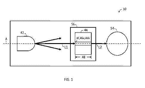
[0027] FIG. 1 is a schematic plan view of an exemplary spectrophotometer
system in
accordance with the present disclosure.
[0028] FIG. 2 is a schematic plan view of a sample analyzer according to an
embodiment of
the present disclosure;
[0029]
FIG. 3 is a flow chart of an exemplary method in accordance with the
present
disclosure.
[0030] FIG. 4 is a graph of exemplary Transmission (%T) normalized
spectrophotometer
measurements in accordance with the present disclosure;
[0031] FIG. 5 is a graph of exemplary Absorbance measurements and method
correction
with analyte results in accordance with the present disclosure; and
[0032] FIG. 6 is a graph of exemplary Extinction coefficients in accordance
with the present
disclosure.
DETAILED DESCRIPTION
[0033] The present disclosure provides a sample analyzer that can make
accurate
determination of component concentrations without interference from unknown
extracellular interferents. The sample analyzer may employ a dispensing device
to pass a first
portion of a blood sample into a transparent sample vessel at a first instance
of time, employ
a plasma separator to separate plasma from the blood sample, employ the
dispensing device
to pass a portion of the plasma of the blood sample into the transparent
sample vessel at a
second instance of time, obtain first data generated by a detector indicative
of an optical
signal passing through the first portion of the blood sample, and second data
generated by
- 7 -
CA 03181510 2022- 12- 5
WO 2021/252564
PCT/US2021/036515
the detector indicative of the optical signal passing through the portion of
the plasma of the
blood sample, the logic causing the processor to calculate a total absorbance
spectrum in
which the first data is adjusted by the second data. Because the first data
and the second data
both contain information indicative of the unknown extracellular interferent,
the information
with respect to the unknown extracellular interferent cancels out and is
effectively removed
from the total absorbance spectrum. In some embodiments, the analyzer may
employ a lysis
device to lyse red blood cells in the blood sample, after employing the
dispensing device to
pass a first portion of the blood sample into a transparent sample vessel at a
first instance of
time, to produce a lysed portion. The lysed portion may be used to measure
forms of
hemoglobin and bilirubin with the detector.
[0034] Before explaining at least one embodiment of the present disclosure in
detail, it is
to be understood that embodiments of the present disclosure are not limited in
their
application to the details of construction and the arrangement of the
components or steps or
methodologies set forth in the following description or illustrated in the
drawings. The
inventive concepts in the present disclosure are capable of other embodiments
or of being
practiced or carried out in various ways. Also, it is to be understood that
the phraseology and
terminology employed herein is for the purpose of description and should not
be regarded as
limiting.
[0035] In this detailed description of embodiments of the inventive concepts,
numerous
specific details are set forth in order to provide a more thorough
understanding of the
inventive concepts. However, it will be apparent to one of ordinary skill in
the art that the
inventive concepts disclosed and claimed herein may be practiced without these
specific
details. In other instances, well-known features have not been described in
detail to avoid
unnecessarily complicating the instant disclosure.
[0036] As used herein, language such as "including," "comprising," "having,"
"containing,"
or "involving," and variations thereof, is intended to be broad and encompass
the subject
matter listed thereafter, equivalents, and additional subject matter not
recited or inherently
present therein.
[0037] As used herein, the terms "first", "second" and the like are used to
specifically
identify items and are not intended, by themselves, to imply any particular
order.
[0038] Unless expressly stated to the contrary, or refers to an inclusive or
and not to an
exclusive or. For example, a condition A or B is satisfied by anyone of the
following: A is true
- 8 -
CA 03181510 2022- 12- 5
WO 2021/252564
PCT/US2021/036515
(or present) and B is false (or not present), A is false (or not present) and
B is true (or present),
and both A and B are true (or present).
[0039] In addition, use of the "a" or an are employed to describe elements and
components of the embodiments herein. This is done merely for convenience and
to give a
general sense of the inventive concepts. This description should be read to
include one or at
least one and the singular also includes the plural unless it is obvious that
it is meant
otherwise.
[0040] Throughout this disclosure and the claims, the terms "about,"
"approximately," and
"substantially" are intended to signify that the item being qualified is not
limited to the exact
value specified, but includes slight variations or deviations therefrom,
caused by measuring
error, manufacturing tolerances, stress exerted on various parts, wear and
tear, or
combinations thereof, for example.
[0041] The use of the term "at least one" will be understood to include one
and any quantity
more than one, including but not limited to each of, 2, 3, 4, 5, 10, 15, 20,
30, 40, 50, 100, and
all integers therebetween. The term "at least one" may extend up to 100 or
1000 or more,
depending on the term to which it is attached; in addition, the quantities of
100/1000 are not
to be considered limiting, as higher limits may also produce satisfactory
results. Singular terms
shall include pluralities and plural terms shall include the singular unless
indicated otherwise.
[0042]
The term "or combinations thereof" as used herein refers to all
permutations
and/or combinations of the listed items preceding the term. For example, "A,
B, C, or
combinations thereof" is intended to include at least one of: A, B, C, AB, AC,
BC, or ABC, and
if order is important in a particular context, also BA, CA, CB, CBA, BCA, ACB,
BAC, or CAB.
Continuing with this example, expressly included are combinations that contain
repeats of
one or more item or term, such as BB, AAA, AAB, BBC, AAABCCCC, CBBAAA, CABABB,
and so
forth. The skilled artisan will understand that typically there is no limit on
the number of items
or terms in any combination, unless otherwise apparent from the context.
[0043] In accordance with the present disclosure, certain components of the
sample
analysis system include circuitry. Circuitry, as used herein, could be analog
and/or digital
components, or one or more suitably programmed microprocessors and associated
hardware
and software, or hardwired logic. Also, certain portions of the
implementations may be
described as "circuitry" that perform one or more functions. The term
"circuitry," may include
hardware, such as a processor, an application specific integrated circuit
(ASIC), or a field
- 9 -
CA 03181510 2022- 12- 5
WO 2021/252564
PCT/US2021/036515
programmable gate array (FPGA), or a combination of hardware and software.
Software
includes one or more computer executable instructions that when executed by
one or more
component causes hardware to perform a specified function. It should be
understood that
the algorithms described herein are stored on one or more non-transitory
memory.
Exemplary non-transitory memory includes random access memory, read only
memory, flash
memory or the like. Such non-transitory memory can be electrically based or
optically based.
[0044] Finally, as used herein any reference to one embodiment" or "an
embodiment"
means that a particular element, feature, structure, or characteristic
described in the
embodiment is included in at least one embodiment. The appearances of the
phrase in one
embodiment" in various places in the specification are not necessarily
referring to the same
embodiment, although the inventive concepts disclosed herein are intended to
encompass
all combinations and permutations including one or more features of the
embodiments
described.
[0045] Referring now to the drawings, and in particular to FIG. 1, a
spectrophotometer
system 10 is shown. In one embodiment, the spectrophotometer system 10 may
comprise
one or more light source 42, a sample vessel 44 configured to hold a specimen
sample 46, and
a detector 54. The one or more light source 42 may emit light L1 into the
sample 46 in the
sample vessel 44 and the detector 54 may detect the luminescence L2 that exits
the sample
vessel 44 (that is, the luminescence of the sample 46). The sample vessel 44
may have a
thickness which may be the path length 48 the light travels through the sample
vessel 44.
[0046] In one embodiment, the detector 54 may be a spectrophotometer as is
known in the
art. The detector 54 may be referred to as the spectrophotometer 54 herein.
[0047] The sample vessel 44 may be configured to hold the sample 46 for
analysis. The
sample 46 may be any type of specimen, such as any type of liquid. For
example, the sample
46 can be a biological sample or body fluid, such as blood, plasma, urine, or
other fluids
obtained from a patient. Furthermore, the sample 46 may also include non-
biological sample
liquids. The sample 46 is not limited strictly to liquids obtained from the
patient. The following
description describes the analysis of a whole blood sample 46, a plasma sample
46a from the
whole blood sample 46, and a lysed blood sample 46b from the whole blood
sample 46.
However, it will be understood that other sample types may be used.
[0048] The one or more light source 42 is configured to emit the light L1 as
one or more
light signal along one or more axis A. The one or more light source 42 may be
a light emitting
- 10 -
CA 03181510 2022- 12- 5
WO 2021/252564
PCT/US2021/036515
diode and/or a neon lamp. In one example, the one or more light source 42 is
adapted to emit
an optical signal including light L1 of specified wavelength into the sample
46 contained in the
sample vessel 44. For instance, the one or more light source 42 may have a
light signal with a
broadband white light 450 nnn to 680 nnn.
[0049] In some embodiments for oxinnetry absorbance spectra measurement, for
example,
for analysis of blood 02Hb, HHb, COHb, MetHb, SulfHb, Fetal Hb and Bilirubin,
the one or
more light source 42 may be a broadband white light source of approximately
450 nnn to 680
nnn to illuminate the lysed blood sample 46b and the plasma sample 46a, at
separate
instances of time, for the spectrophotometer 54 to perform the absorbance
measurements.
While halogen lamps have typically been used to serve this purpose in the
past, white light
emitting diodes (LED) have been employed more recently. The one or more light
source 42
may produce contiguous radiation across the spectrum because many wavelengths
are used.
In addition to this, a precision spectral line light source may be used to
calibrate the
spectrophotometer 54. A neon gas lamp may be used to produce a number of
precision
spectral lines (one of the lines at 585.2488 nm is particularly useful in the
midrange of the
total spectrum), and to produce strong intensity relative to the many other
lines, allowing
shorter integration times to be used. In one embodiment, the neon line
calibration light
source may be turned on and off to periodically calibrate the
spectrophotometer 54 before a
measurement is made, which improves measurement precision.
[0050] In one embodiment, the spectrophotometer system 10 may include a sample
vessel
holder 56 that is configured to hold the sample vessel 44. The sample vessel
holder 56 may
be located adjacent to the detector 54 on the optical axis A. However, the
sample vessel
holder 56 and detector 54 may be arranged in configurations other than those
specifically
shown in the drawings.
[0051] In one embodiment, the spectrophotometer system 10 may include one or
more of
the following: a reflector, a lens, a filter, a light sensor (to monitor the
intensity of the light
L1), and/or a polarizer.
[0052] In one embodiment, as shown in FIG. 2, the spectrophotometer system 10
may be
part of a sample analyzer 12. The sample analyzer 12 may have a housing 60
having an internal
space, and one or more components of the sample analyzer 12 may be positioned
on or in
the housing.
- 11 -
CA 03181510 2022- 12- 5
WO 2021/252564
PCT/US2021/036515
[0053] In one embodiment, the sample analyzer 12 may include a controller 20
that
controls operation of the spectrophotometer system 10. The sample analyzer 12
may
comprise a dispensing device 24 to dispense the sample(s) 46 from one or more
sample vial,
and/or reagent, into the sample vessel 44. The dispensing device 24 may
include a motor 26
that powers the dispensing device 24, a pump 28, and a valve 30, such as a lee
valve. The
controller 20 may also control the motor 26 that powers the dispensing device
24, the pump
28 and the valve 30. The pump 28 may provide a plurality of liquids into the
sample vessel 44,
such as a wash solution, a clear blank calibration (CAL) solution, and one or
more portions of
the sample 46.
[0054] For example, when the sample 46 is blood, the controller 20 may actuate
the pump
28 to provide the wash solution into the sample vessel 44 to clean the sample
vessel 44,
followed by distinct portions of the blood sample 46. The distinct portions
may include plasma
as the plasma sample 46a, whole blood as the whole blood sample 46, or lysed
blood as the
lysed blood sample 46b. The controller 20 may actuate the pump 28 to clean the
sample
vessel 44 with the wash solution in between the distinct portions of the blood
being provided
by the pump 28 into the sample vessel 44. In some embodiments, the sample
analyzer 12 may
include more than one pump 28 with each pump supplying a particular type of
solution into
the sample vessel 44.
[0055] In certain embodiments, such as for sample analyzers adapted to analyze
blood
and/or plasma samples, the sample analyzer 12 may further comprise a plasma
separator 32
to separate plasma from the blood sample 46 for analysis. In some embodiments,
the sample
analyzer 12 may further comprise a lysis device 33 for lysing red blood cells
in the blood
sample 46. The plasma separator 32 may utilize active or passive methods to
separate a
portion of the plasma from the whole blood, prior to lysing red blood cells in
the blood sample
46. Exemplary active methods include magnetic, dielectrophoretic, centrifugal,
or acoustic
separation methods. In this case, the plasma separator 32 would be constructed
based upon
the requirements of at least one of the active methods. For example, to
utilize acoustic
separation methods, the plasma separator 32 may include a piezoelectric
element connected
to a glass slide supporting the blood sample, and a driver that provides an
electric signal to
the piezoelectric element with a sufficient frequency and voltage to separate
the red blood
cells from the plasma, without lysing or otherwise damaging the red blood
cells. In this case,
the plasma can be directed to a predetermined location on the glass slide so
that an
- 12 -
CA 03181510 2022- 12- 5
WO 2021/252564
PCT/US2021/036515
absorption reading of the plasma (substantially devoid of red blood cells) can
be taken while
the plasma separator 32 is actuated to actively separate the red blood cells
from the plasma.
Exemplary passive methods include hydrodynamic, sedimentation, and filtration
methods. In
one embodiment, the lysis device 33 and the plasma separator 32 may be a
single device. In
one embodiment, the lysis device 33 may include a piezoelectric element
connected to a glass
slide supporting the blood sample, and a driver that provides an electric
signal to the
piezoelectric element with a sufficient frequency and voltage for lysing the
red blood cells.
[0056] In one embodiment, the lysis device 33 and/or the plasma separator 32
may be
configured as described in the provisional application entitled
"ACOUSTOPHORETIC LYSIS
DEVICES AND METHODS", application serial number 63/036,537, filed on April 28,
2020,
which is hereby incorporated in its entirety herein.
[0057] In one embodiment, the sample analyzer 12 may further comprise one or
more
position sensors 34 used to determine the position of the stage 16 and/or
spectrophotometer
system 10 with respect to the dispensing device 24. A vacuum port 36 may be
included to
control pressure in the housing 14.
[0058] The sample vessel 44 and/or the spectrophotometer system 10 may be
stationary
or movable so as to bring the sample 46 and/or portions of the sample 46 into
the path of an
optical signal used by the spectrophotometer system 10 to obtain a
transmittance reading of
the sample 46 and/or portion thereof.
[0059] It should also be appreciated that the sample analyzer 12 can be
adapted to analyze
multiple samples 46. In one example, the sample analyzer 12 may include a
cartridge adapted
to hold a plurality of sample vessels 44. In yet another example, the sample
analyzer 12 may
be an automated analyzer that includes a moveable carousel for holding
multiple sample
vessels 44. Such an analyzer may include multiple detectors 54 testing for
different analytes
of interest. An exemplary automated analyzer is disclosed in U.S. Patent App.
Pub. No.
2010/0150779, incorporated herein by reference in its entirety. Other
exemplary sample
analyzers include the ADVIA and DIMENSION analyzers produced by Siemens
Healthcare
Diagnostics Inc.
[0060] The controller 20 may include circuitry configured to embody and/or
execute the
logic of the processes described herein. Logic embodied in the form of
software instructions
and/or firmware may be executed on a dedicated computer system or computer
systems, on
distributed processing computer systems, and/or the like. In some embodiments,
the logic
- 13 -
CA 03181510 2022- 12- 5
WO 2021/252564
PCT/US2021/036515
may be implemented in a stand-alone environment operating on a single computer
system
and/or logic may be implemented in a networked environment such as a
distributed
computer system using multiple computers and/or processors. For example, one
or more
microprocessors may work together or independently to execute processor
executable code
using one or more memories.
[0061] The spectrophotometer system 10 incorporated with the sample analyzer
12
illustrated in FIGS. 1 and 2 represent an exemplary sample analyzer that
illustrates inventive
concepts set forth in the present disclosure. However, the sample analyzer 12
as described
herein can be configured in other manners adapted to make measurements of the
sample 46
illuminated in the sample vessel 44. In one embodiment, the sample analyzer
may be
configured as a "RAPIDPoint 405" analyzer or a "RAPIDPoint 500" analyzer, both
manufactured by Siemens Medical Solutions, Malvern, Pennsylvania.
[0062] Referring now to FIG. 3, a flow chart of an exemplary method 100 of use
of the
sample analyzer 12 to determine a Coox absorbance measurement in blood samples
in
accordance with the present disclosure is disclosed. To address the role of
the unknown
interferent(s) in the detection of the presence and amount of the principal
forms of
hemoglobin, the present disclosure adjusts the transmittance measurement of
the whole
blood sample 46 and/or the lysed blood sample 46b with the transmittance
measurement of
the plasma sample 46a to reduce any interference in the measurement of the
principal forms
of hemoglobin caused by the unknown interferent(s). This adjustment can be
made without
having an extinction coefficient stored in the non-transitory memory of the
controller 20 (or
even accessed by a processor of the controller 20) and without having an
extinction
coefficient associated with the specific spectral signature of the unknown
interferent. Rather
than mathematically applying a known extinction coefficient for each
hemoglobin form or
other analyte of interest to the total Coox Absorbance measurement to
determine a
concentration for each hemoglobin form (or other analyte of interest) from the
total
absorbance measurement, as done in the prior art, the current method measures
in real time
the absorbance measurement without the unknown interferent(s).
[0063] In one embodiment, the controller 20 may be actuated to obtain an
absorbance
measurement for specific forms of hemoglobin. The controller 20 may actuate
the pump 28
to wash the transparent sample vessel 44. A clear blank CAL solution may be
delivered into
- 14 -
CA 03181510 2022- 12- 5
WO 2021/252564
PCT/US2021/036515
the vessel 44. Then, the controller 20 may actuate the spectrophotometer
system 10 to
measure the transmittance of the CAL solution in step 102, as further
explained below.
[0064] In one embodiment, the controller 20 may actuate the spectrophotometer
system
to measure the transmittance of the whole blood sample 46.
[0065] Next, the controller 20 may actuate the plasma separator 32 to separate
the plasma
46a from the whole blood sample 46, as indicated by a step 104, to generate
the plasma
sample 46a. In step 106, the controller 20 may actuate the spectrophotometer
system 10 to
measure the transmittance of the plasma sample 46a.
[0066] In one embodiment, the controller 20 may actuate the lysis device 33 to
lyse at least
a portion of the whole blood sample 46 at a step 108. The controller 20 may
actuate the pump
28 to place a portion of the lysed blood sample 46b of the lysed blood within
the sample
vessel 44, and measure the transmittance of the lysed blood sample 46b at step
110.
[0067] The order of measuring the transmittance of the whole blood sample 46,
the plasma
sample 46a, and the lysed blood sample 46b may vary. When a single transparent
sample
vessel 44 is used, the controller 20 may actuate the pump 28 to wash the
transparent sample
vessel 44 between new samples being applied into the transparent sample vessel
44.
[0068] Once the transmittance of the plasma sample 46a and the transmittance
of the
whole blood sample 46 and/or the lysed blood sample 46b has been measured, the
controller
may calculate a total absorbance spectrum in a step 112. To calculate the
total absorbance
spectrum, the measurement of the whole blood sample 46 is adjusted by the
measurement
of the plasma sample 46a using EQUATION 1, explained below, for example. The
controller
20 can then calculate the presence and amount of the specific hemoglobin forms
in a step
114 utilizing the lysed blood sample 46b by using EQUATION 2, explained below,
for example.
[0069] The method 100 can be automated as a sequence of instructions that are
performed
for determining unknown interferent(s) in the sample 46, and such sequence can
be repeated
for conducting readings on a plurality of samples 46.
[0070] In one embodiment, the detection of unknown interferent(s) and/or the
adjustment
may be displayed to a user on a display.
[0071] Now, the algorithms that may be used in the method 100 will be
discussed.
[0072] Absorption spectroscopy uses data pretreatment by converting the
measured
sample transmittance into sample absorption, as is well known in the art. The
logarithmic
- 15 -
CA 03181510 2022- 12- 5
WO 2021/252564
PCT/US2021/036515
relationship of conventional sample absorption to sample transmittance is
given by the
following equations:
[0073] A = -log(T)
[0074] and
[0075] T =1/1.
[0076] where:
[0077] A is the calculated absorption,
[0078] T is the calculated transmittance,
[0079] I is the measured intensity due to the sample, and
[0080] I. is the measured intensity with a blank sample such as deionized
water.
[0081] In multivariate analysis, multiple measurements are made to permit the
estimate of
concentrations in the samples 46 with several components. In absorption
spectroscopy
measurements at multiple wavelengths of light are often used to provide the
spectral
information needed for an accurate analysis. Vectors and matrices are used to
simplify the
equations. The use of vectors in column or row format depends on the
preference of the
writer. In the description of this invention, vectors are assumed to be in
column format and
annotated with small, boldface letters. However, this notation and format does
not limit the
scope of the disclosure. Matrices will be denoted with capital, boldface
letters.
[0082] The measured spectrum of an ideal sample can be described as:
[0083] A = E*c
[0084] where:
[0085] a is the column vector for the sample's absorption spectrum with each
row element
corresponding to sample absorption at a particular wavelength of light,
[0086] E is a matrix of column vectors, each representing the absorption
spectrum
(extinction) of a component or factor at particular wavelengths, and
[0087] c is a column vector describing the concentrations of the components
and factors in
E (including the scatter terms).
[0088] In an analysis of lysed and non-turbid blood for concentrations of the
principal forms
of hemoglobin, Oxyhennoglobin (02Hb), Deoxyhennoglobin (HHb),
Carboxyhemoglobin
(COHb) and Methemoglobin (MetHb), total or neonatal Bilirubin (BILI), Cyan
Methemoglobin
(CN_METHb), Sulfhemoglobin (SulfHb) (intracellular interferent), Methylene
blue dye
(METH_BLUE) (extracellular interferent), the E matrix is formed by the eight
vectors
-16 -
CA 03181510 2022- 12- 5
WO 2021/252564
PCT/US2021/036515
representing the eight absorption spectra (extinction coefficients) of these
components as in
the following equation:
[0089] ELYSED = 1e02Hb eHHb eCOHb eMetHb eCN METHb eSULFHb eBILI
eMETH BLUE 1,
where eHb denotes the column vector for the corresponding hemoglobin.
Experimental results
illustrating these spectra are shown in FIG. 5.
[0090] This model of absorption is adequate for determining component
concentrations in
homogenous samples with low turbidity and known interferents in which each of
the known
interferents has an extinction coefficient.
[0091] When an unknown interferent exists within the lysed blood sample 46b,
the E matrix
will take on unknown absorption values due to the presence of the unknown
interferent
(designated in the equations by "UNKNOWN"). Unknown interferents are mainly
extracellular
and may result due to patients receiving new drug therapies. The unknown
interferents thus
occur mainly within the plasma and not within the red blood cells within the
blood sample
46. Thus, the E matrix for the lysed blood sample 46b having an unknown
interferent (likely
due to drug therapy) will have the following form:
[0092] ELYSED = 1e02Hb eHHb eCOHb eMetHb eCN_METHb eSULFHb eedu eMETH BLUE
eUNKNOWN1
[0093] And, the E matrix for the plasma sample 46a from the blood sample 46
will have the
following form:
[0094] EPLASMA = [eedu eMETH BLUE eedu eUNKNOWN]
[0095] Thus, in accordance with the present disclosure, any unknown
interferent in the
ELYSED matrix can be removed by adjusting the ELYSED matrix with the [PLASMA
matrix.
[0096] In one embodiment, unknown interference correction can be implemented
rationnetrically using the following equation:
VeTspecimenLysedBiood
CoaxAbsarbance = ¨loy10 ____________________________________________________
[0097] EQUATION 1: %TblankPlasma
[0098] The CooxAbsorbance is calculated using the measured percent
transmission blood
sample signals. The spectrophotometer 54 may measure the %TblankPlasnna (that
is, the
percentage transmittance through the plasma sample 46a) in the denominator
within the
parenthesis of Equation 1 before the sample 46 is lysed by the use of plasma
separation such
as acoustophoresis. During plasma separation, the red blood cells are intact
and move out of
- 17 -
CA 03181510 2022-12-5
WO 2021/252564
PCT/US2021/036515
the field of view of the spectrophotometer 54. The transmittance through the
plasma sample
46a contains all extracellular interferences.
[0099] The spectrophotometer 54 may measure the patient lysed blood sample 46b
%TspecinnenLysedBlood (that is, the percentage transmittance through the lysed
blood
sample 46b) after plasma separation and after the blood sample 46 is lysed.
The lysing action
breaks up the red blood cell casings releasing the henne in the cells mixing
the sample
thoroughly with the plasma.
[0100] A clear calibration (CAL) solution may be used in any blood sample Coox
system to
flush clean the blood sample vessel 44, ensuring residual substances such as
carryover from
a previous blood sample are removed, and the vessel 44 is optically clear
prior to measuring
a new patient blood sample. Additionally, the CAL solution provides a 100%
transmission
signal at all wavelengths, and may be used to normalize the light source
signal. The second
form of Equation 1, commonly used in the art of blood Co-Oxinnetry,
substitutes the
denominator term with the clear CAL, as shown in Equation 2 below:
%TspecimenLysedBlood
CooxAbsorbance = ¨log10 ___________________________________________________
okTblankCAL
[0101] EQUATION 2:
where %TspecinnenLysedBlood is the transmittance measurement for the lysed
blood sample
46b, and %TblankPlasnna is the transmittance measurement for the plasma sample
46a.
[0102] Then, the absorbance for a given wavelength (i) of the matrix is known
from the
measurement of the patient blood sample 46, the specific analyte of interest's
concentration
is calculated using the blood sample signal measurement by rearranging this
equation to solve
for the AnalyteConcentration as shown below in Equation 3:
[0103] EQUATION 3:
Absorbance(AD = AnalyteConcentration(Ai) ExtictionCoe I f icient(AL)
pathlength
where pathlength is the thickness of the sample vessel 44 containing the blood
sample 46.
[0104] Using the above equations with the measured blood sample transmission
signals,
and the with the analyte extinction coefficients for Oxyhennoglobin,
Deoxyhemoglobin
Carboxyhennoglobin, and Methennoglobin, the analyte concentrations are
calculated, the
unknown interference in the blood plasma sample 46a is eliminated by the
radiometric
method.
- 18 -
CA 03181510 2022- 12- 5
WO 2021/252564
PCT/US2021/036515
[0105] FIGS. 4-7 are plots of experimental results of the method 100 of use of
the sample
analyzer 12 in correcting for unknown interferences in representative patient
blood samples
46. For explanatory purposes, in the experimental analysis Hydroxocobalannin
is used as the
unknown extracellular interferant in plasma. However, it will be understood
that the
extracellular interferent may be any unknown interferent or interferents.
[0106] FIG. 4 illustrates experimental normalized spectrophotometer
measurements of
percent transmission (y-axis) versus wavelength (x-axis) of a blood sample 46
and a plasma
sample 46a. The first curve (Ti) is a measured spectral profile of
transmittance of
hydroxocobalamin (B12) in the plasma sample 46a as determined by the analyzer
12. The
third curve (13) is a measured spectral profile of transmittance of hemoglobin
with the
hydroxocobalamin in the blood sample 46 as determined by the analyzer 12. The
second curve
(T2) is a measured spectral profile of transmittance of hemoglobin without the
hydroxocobalamin in the blood sample 46 as determined by the analyzer 12
having removed
the unknown interferent. The analyzer 12 corrects for the presence of
hydroxocobalamin
(B12) in the blood sample 46 to measure the spectral profile of transmittance
of hemoglobin
in the blood sample 46, which produces the curve 12.
[0107] FIG. 5 illustrates experimental Absorbance measurements (y-axis) versus
wavelength (x-axis), showing method correction with analyte results. The chart
in FIG. 5
illustrates experimental measurements from the analyzer 12 in the absorbance
domain. The
line Al is the corrected absorbance result for the blood sample 46, that is,
the absorbance
result without the unknown interferent.
[0108]
FIG. 6 illustrates experimental extinction coefficients for multiple
interferents,
absorbance measurements from the analyzer 12, showing percent transmission (y-
axis)
versus wavelength (x-axis). The extinction coefficients are reference spectra
that show the
absorbance of a sample having 100% of the particular interferent. In one
embodiment, the
analyzer 12 may check the sample 46 against known interferents, using the
known
interference extinction coefficients, before determining the unknown
interferent(s). In one
embodiment, the analyzer 12 may check the sample 46 against other known
interferents (not
shown) using the known interference extinction coefficients before determining
the unknown
interferent(s).
[0109] It should be understood that other manners to correct the transmittance
of the lysed
blood sample 46b with the plasma sample 46a can be used, such as preparing the
blood
- 19 -
CA 03181510 2022- 12- 5
WO 2021/252564
PCT/US2021/036515
sample 46 before introducing it to the sample analyzer 12. Preparing the blood
sample 46
may involve precisely splitting the sample 46 into two aliquots, one that is
centrifuged to
harvest only the plasma sample 46a, and a second that is the original sample
46 to then be
lysed into the lysed blood sample 46b. However, splitting the blood sample 46
may require a
larger sample volume, specialized laboratory equipment, a skilled operator,
approved
protocols, and is time consuming, also reducing the accuracy of the
measurements.
[0110] The sample analyzer 12 described in the present disclosure is capable
of exploitation
in industry in accordance with how it can be made and/or used.
[0111] Those skilled in the art will also appreciate that the present
disclosure may be
applied to other applications and may be modified without departing from the
scope of the
present disclosure. Accordingly, the scope of the present disclosure is not
intended to be
limited to the exemplary embodiments described above, but only by the appended
claims.
- 20 -
CA 03181510 2022- 12- 5