Note : Les descriptions sont présentées dans la langue officielle dans laquelle elles ont été soumises.
WO 2022/011321
PCT/US2021/041189
POSTERIOR CHAMBER DELIVERY DEVICE FOR SUSTAINED RELEASE
IMPLANT
[0001] This application claims the benefit of priority to
U.S. Provisional
Application Serial No. 63/050,452, filed July 10, 2020, and U.S. Provisional
Application Serial
No. 63/219,440, filed July 8, 2021. The entire contents of these applications
are incorporated by
reference in their entirety.
BACKGROUND
[0002] Glaucoma is generally a progressive disease of the
eye characterized by
progressive optic neuropathy with associated visual field loss. Glaucoma may
be further
associated with increased intraocular pressure. Based on its etiology,
glaucoma has been
classified as primary or secondary. Primary glaucoma in adults may be either
open-angle
glaucoma or acute or chronic angle-closure glaucoma. Secondary glaucoma
results from pre-
existing ocular diseases such as uveitis, intraocular tumor or an enlarged
cataract.
[0003] The underlying causes of primary glaucoma are not
yet known. Risk factors
include high or elevated intraocular pressure, advanced age, and family
history. Increased or
elevated intraocular pressure is due to the obstruction of aqueous humor
outflow. In primary
open-angle glaucoma, the anterior chamber and its anatomic structures appear
normal, but
drainage of the aqueous humor is impeded. In acute or chronic angle-closure
glaucoma, the
anterior chamber is shallow, the filtration angle is narrowed, and the iris
may obstruct the
trabecular meshwork at the entrance of the canal of Schlemm. Dilation of the
pupil may push the
root of the iris forward against the angle, and may produce pupillary block
and thus precipitate
an acute attack. Eyes with narrow anterior chamber angles are predisposed to
acute angle-closure
glaucoma attacks of various degrees of severity.
[0004] Secondary glaucoma is caused by any interference
with the flow of aqueous
humor from the posterior chamber into the anterior chamber and subsequently,
into the canal of
Schlemm. Inflammatory disease of the anterior segment may prevent aqueous
escape by causing
complete posterior synechi ea in iris bombe and may obstruct movement of
aqueous humor
through the pupil leading to elevated intraocular pressure. Other common
causes are intraocular
1
CA 03184833 2023- 1- 3
WO 2022/011321
PCT/US2021/041189
tumors, enlarged cataracts, central retinal vein occlusion, trauma to the eye,
operative procedures
and intraocular hemorrhage. Considering all types together, glaucoma occurs in
about 2% of all
persons over the age of 40 and may be asymptomatic for years before
progressing to noticeable
peripheral visual loss followed by central vision loss.
[0005] Glaucoma is considered to potentially be both an
anterior and posterior
ocular condition because a clinical goal of glaucoma treatment can be to not
only reduce elevated
intraocular pressure because of obstructed aqueous humor outflow from the
anterior chamber,
but to also prevent the loss of, or reduce the occurrence of loss of, vision
due to damage to or
loss of retinal cells or optic nerve cells (i.e., ganglion cells) in the
posterior of the eye (i.e.
neuroprotection). Clinical trials have shown that reducing TOP can help retard
the progression of
glaucoma and consistent IOP reduction is associated with reduced risks of
developing and
progressing optic nerve damage.
[0006] Patient non-adherence to topical therapy is one of
the major challenges to
preventing vision loss due to glaucoma. Patients that take no medication are
at the highest risk of
vision loss from glaucoma; however, patients that intermittently take their
medications are also at
risk since TOP fluctuation has also been identified as possible risk factor
for progression in some
patients.
SUMMARY
[0007] In light of the above, new drug delivery devices,
systems, and methods
would be beneficial, particularly for delivering therapeutic agents to the
posterior chamber of the
eye. It would be particularly advantageous to provide improved reductions in
TOP while
mitigating risks of corneal damage.
[0008] In an aspect, provided is a system for reducing the
intraocular pressure of a
patient in need. The system includes a delivery device having a housing sized
to be held by an
operator and an actuator. The delivery device includes a cannula defining a
lumen and having a
proximal end coupled to the housing. The cannula extends along a longitudinal
axis from the
proximal end to a distal end. The distal end of the cannula has rounded inner
and outer edges
defining a blunt, non-beveled distal opening from the lumen. A retention plug
is adhered to the
cannula spanning the lumen near the distal opening. An intraocular implant is
positioned within
the lumen of the cannula proximal to the retention plug.
2
CA 03184833 2023- 1- 3
WO 2022/011321
PCT/US2021/041189
[0009] The cannula can have an exposed working length
between the proximal end
and the distal end that is between about 12 mm and about 18 mm. The cannula
can have an outer
dimension sized to extend through a self-sealing corneal incision or puncture.
The cannula can
be no larger than about 28 gauge. The cannula can have a wall thickness no
greater than about 50
p.m. An outside surface of the cannula can be siliconized and the lumen of the
cannula can be
substantially non-siliconized. The retention plug prevents inadvertent release
of the implant from
the lumen prior to actuation of the device. The retention plug can be formed
from a retainer
solution of hydroxypropyl methylcellulose (HPMC). The retainer solution can
have a viscosity
between 6,000 cP and 13,000 cP. The retainer solution can have a concentration
greater than
about 2.5% and less than about 4%, and preferably about 3%. The retainer
solution can be
dispensed within the lumen of the cannula as a dispensed mass of greater than
100 mg and less
than 300 pg. The retainer solution can be a 3% F4M-HPMC in water having an
apparent
viscosity of about 8,640 cP to about 12,760 cP and dispensed into the cannula
as a dispensed
mass between 125 pg - 200 pg, the cannula being 28 gauge.
[0010] The implant can have a length of no more than 3.0
mm and a maximum
width of no more than about 0.5 mm. The intraocular implant can include
bimatoprost or a salt
thereof present in an amount of about 20% by weight of the implant and a
biodegradable
polymer matrix comprising at least one biodegradable polymer. The intraocular
implant can be
DURYSTATm. The intraocular implant can be DURYSTATm in either a 6 g, 10 jig,
15 g, or
201ag dosage.
[0011] The actuator on the housing can move a push rod
through the lumen of the
cannula to push the implant out from the lumen via a linkage. The actuator can
be coupled to the
push rod through the linkage, the push rod being movable along the
longitudinal axis as the
linkage is gradually flattened as the actuator is depressed. The push rod can
have a length
relative to a length of the cannula sufficient for a distal end of the push
rod to advance past the
distal end of the cannula upon deployment of the implant using the actuator.
[0012] In an interrelated aspect, provided is a system for
reducing intraocular
pressure of a patient in need that includes a delivery device having a housing
sized to be held by
an operator and an actuator. The delivery device includes a push rod linked to
the actuator and a
cannula having a tubular wall extending along a longitudinal axis between a
proximal end
3
CA 03184833 2023- 1- 3
WO 2022/011321
PCT/US2021/041189
coupled to the housing and a distal end. The tubular wall defines a lumen
sized to slidably
receive the push rod. The distal end of the cannula has rounded inner and
outer edges defining a
blunt, non-beveled distal opening into the lumen. The tubular wall between the
proximal end and
the distal end is about 12 mm and about 18 mm long. The tubular wall is
siliconized on its
external surface and the lumen is non-siliconized. A retention plug is
contained within and
spanning the lumen near the distal opening. The retention plug is formed from
a dispensed mass
of 3% hydroxypropyl methylcellulose (I-1PMC) retainer solution. An intraocular
implant is
positioned within the lumen of the cannula proximal to the retention plug and
distal to the push
rod. The implant includes 20% by weight bimatoprost or a salt thereof and a
biodegradable
polymer matrix having at least one biodegradable polymer.
[0013] In an interrelated aspect, provided is a method for
improving the efficacy of
a bimatoprost-containing intraocular implant in reducing intraocular pressure
of a patient in need
thereof. The method includes positioning a single bimatoprost-containing
intraocular implant
into a posterior chamber of an eye of the patient. The single bimatoprost-
containing intraocular
implant causes a greater reduction in intraocular pressure compared to an
equivalent
bimatoprost-containing intraocular implant positioned into an anterior chamber
of the eye of the
patient closer to a trabecular meshwork of the eye. The bimatoprost-containing
intraocular
implant can include 6, 10, 15, or 20 lig of bimatoprost or a salt thereof that
elutes over a period
of up to about 6 months. The implant can be effective to reduce the
intraocular pressure of the
patient over a period of time between about 12 months and about 24 months or
longer. The
method can further include advancing a blunt-tipped cannula having a lumen
containing the
implant through the anterior chamber over at least a portion of the pupil and
under at least a
portion of the iris; pushing the implant through the lumen of the cannula past
a retention plug
attached to the cannula so as to span the lumen and out a distal opening
defined by rounded inner
and outer edges of the cannula; and releasing the implant within a region of
the posterior
chamber of the eye behind the iris. In some variations, one or more of the
following can
optionally be included in any feasible combination in the above compositions,
methods, devices,
and systems. More details of compositions, methods, devices, and systems are
set forth in the
accompanying drawings and the description below. Other features and advantages
will be
apparent from the description and drawings.
4
CA 03184833 2023- 1- 3
WO 2022/011321
PCT/US2021/041189
BRIEF DESCRIPTION OF THE DRAWINGS
[0014] These and other aspects will now be described in
detail with reference to the
following drawings. Generally speaking, the figures are not to scale in
absolute terms or
comparatively, but are intended to be illustrative. Also, relative placement
of features and
elements may be modified for the purpose of illustrative clarity.
[0015] FIG. 1A shows a cross-section of the mammalian eye.
[0016] FIG. 1B shows the mammalian eye visualized by a
high-definition optical
coherence tomography system (RD-OCT).
[0017] FIG. 1C shows the canonical schematic of the
aqueous humor secreted
from the posterior chamber of the eye by the ciliary body through the pupil
and into the anterior
chamber.
[0018] FIG. 1D shows the mammalian eye with an implant
positioned within the
posterior chamber visualized by a RD-OCT system.
[0019] FIG. IE shows forming an incision in a cornea using
a sharpened tool,
[0020] FIG. 1F shows an applicator having an implant
within the cannula being
inserted through the cornea via the incision shown in FIG. 1E.
[0021] FIG. 1G shows the cannula being positioned through
the pupil and behind
the iris to deploy the implant within the posterior chamber.
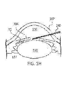
[0022] FIG. 111 shows the implant deployed from the
cannula of the applicator and
positioned within the ciliary sulcus.
[0023] FIG. 2A is a perspective view of an implant
delivery apparatus.
[0024] FIG. 2B is a partially exploded side view of the
housing, linkage, and
actuating lever of an implant delivery apparatus.
[0025] FIG. 2C is a cross-sectional view of an implant
delivery apparatus;
[0026] FIG. 2D is an enlarged side view of a nose cone and
cannula of the implant
delivery apparatus of FIG. 2A.
[0027] FIG. 2E is an enlarged view of the cannula of FIG.
2D.
CA 03184833 2023- 1- 3
WO 2022/011321
PCT/US2021/041189
[0028] FIG. 2F is a cross-sectional view of the cannula of
FIG. 2E showing the
presence of the retention plug.
[0029] FIG. 3 shows an enlarged perspective view of the
linkage of the implant
delivery apparatus shown in FIG. 2C.
[0030] FIG. 4A shows an enlarged perspective view of the
actuating lever of the
implant delivery apparatus shown in FIG. 2C.
[0031] FIG. 4B shows an enlarged perspective view of the
actuating lever of FIG.
4A.
[0032] FIG. 5 shows a cross-sectional, partial view of a
safety tab engaged with a
linkage.
[0033] FIG. 6 shows mean percentage change in IOP from
baseline in beagle dogs
over 3 months after placement of different doses of anterior chamber implants.
[0034] FIG. 7 shows TOP reduction with posterior chamber
implant containing
bimatoprost (solid bar) compared to control (hatched bar) after 1 week.
[0035] FIG. 8 shows central corneal endothelial cell
counts for implant containing
bimatoprost (solid bar) compared to control (hatched bar) are stable after
dosing with a posterior
chamber implant after 1 week.
[0036] It should be appreciated that the drawings are for
example only and are not
meant to be to scale. It is to be understood that devices described herein may
include features
not necessarily depicted in each figure.
DETAILED DESCRIPTION
[0037] Described herein is a new and improved method for
delivering
biodegradable intraocular implants into the posterior chamber of the eye. The
method provides
superior benefits over administering such implants into the anterior chamber,
including enhanced
therapeutic effects, decreased risk of vision loss, decreased risk of causing
damage to the corneal
endothelium, and/or decreased risk of corneal endothelial cell density loss.
Preferably, the
method may be used to deliver biodegradable implants that provide for the
extended release of
prostamides such as bimatoprost in an amount that is effective for treating an
ocular condition,
6
CA 03184833 2023- 1- 3
WO 2022/011321
PCT/US2021/041189
particularly glaucoma and ocular hypertension, and conditions associated with
glaucoma such as
elevated intraocular pressure.
[0038] Also described herein is a device for administering
biodegradable
intraocular implants into the posterior chamber of the eye. The device is
designed specifically to
deliver such implants into the posterior chamber, including the ciliary sulcus
and/or atop or
adjacent to the ciliary zonules, using a blunt-tipped cannula with rounded
edges having an
extended cannula length and an optionally siliconized outside cannula surface.
The cannula
includes a plug for retaining the implant within the lumen prior to ejection
into the posterior
chamber. The device permits reliable and safe delivery of each implant into
the posterior
chamber compared to other insertion devices while providing a significantly
decreased risk of
accidental trauma to the iris, lens capsule and corneal surfaces.
[0039] The implants are sized and configured for placement
in the posterior
chamber of the eye where the implant can deliver therapeutics such as
prostamide or other
therapeutic useful for treating glaucoma, to the tissues regulating the
production and outflow of
aqueous humor. The intraocular implants described here are designed to provide
a patient with
intraocular pressure-lowering levels of drug for a sustained period lasting
for 2 months or more.
[0040] Prostamides are potent ocular hypotensive agents
useful in the treatment of
a number of various ocular hypertensive conditions such as glaucoma, elevated
intraocular
pressure, and other ocular hypertensive episodes, including post-surgical and
post-laser ocular
hypertensive episodes. They belong to the family of prostaglandin F2Gt C-1
amides as discussed
in more detail in U.S. 9,492,316, which is incorporated by reference herein.
[0041] Commercially available prostamides include
bimatoprost, which exhibits no
meaningful interaction with prostaglandin (PG) sensitive receptors.
Nevertheless, bimatoprost is
a potent ocular anti-hypertensive agent and is highly effective for reducing
elevated intraocular
pressure in patients with open angle glaucoma or ocular hypertension.
Bimatoprost is typically
prescribed for use by patients in the form of an ophthalmic solution known by
the tradename
LUMIGANS. In the usual course of therapy, Patients apply one drop of LUMIGAN
solution
once daily to the surface of the affected eye(s) to reduce elevated
intraocular pressure.
[0042] Bimatoprost is believed to decrease intraocular
pressure (TOP) by increasing
aqueous humor outflow, either by enhancing the pressure-sensitive (presumed
trabecular)
7
CA 03184833 2023- 1- 3
WO 2022/011321
PCT/US2021/041189
outflow pathway or by increasing the pressure-insensitive (uveoscleral)
outflow without
significantly affecting the aqueous production rate (Lim el al. Ophthalmology.
2008 May;
115(5): 790-795.e4.).
[0043] Implants exist that are sized to fit within the
anterior chamber angle of the
eye to deliver directly to these uveoscleral outflow pathways (see, U.S.
9,492,316 and WO
2019/094652, which are each incorporated by reference herein). Release of a
drug from an
erodible polymer is the consequence of several mechanisms or combinations of
mechanisms.
Some of these mechanisms include desorption from the implant surface,
dissolution, diffusion
through porous channels of the hydrated polymer and erosion. The release of
the therapeutic
agent from the intraocular implant comprising a biodegradable polymer matrix
may include an
initial burst of release followed by a gradual increase in the amount of the
therapeutic agent
released, or the release may include an initial delay in release of the
therapeutic agent followed
by an increase in release. Fick's Second Law of Diffusion explains the
behavior of non-steady
state diffusion, i.e. diffusion that changes with time. Fick's Second Law is
useful to predict the
diffusion of a drug and the optimum implantation site of an implant containing
the drug as
discussed in U.S. 8,571,802, which is incorporated by reference herein. The
drug concentrations
in the ocular tissues closest to the implant would be highest and more
therapeutic compared with
drug concentrations further away from the implant. Thus, existing diffusion-
based implants for
enhancing uveoscleral outflow of aqueous from the anterior chamber were
preferably positioned
within the anterior chamber because based on Fick's Second Law the ideal
implant position is
nearest the outflow pathway (i.e., the trabecular meshwork) being treated.
Conventionally,
Fick's Second Law provided a basis for why implants for treating glaucoma are
positioned more
anteriorly (i.e. within the anterior chamber) and implants for treating
macular diseases are
positioned more posteriorly (i.e., within the vitreous). However, positioning
implants into the
posterior chamber has a number of advantages over the traditional anterior
placement as
described elsewhere herein.
[0044] FIGs. 1A-1D show cross-sections of an eye 100.
Particular regions of the
eye 100 include the cornea 102, iris 104, ciliary body 107, and ciliary sulcus
111. The cornea
102 and iris 104 surround the anterior chamber 106. Within the anterior
chamber is the anterior
chamber angle 112 and trabecular meshwork 114. Also shown are the corneal
epithelium 118,
8
CA 03184833 2023- 1- 3
WO 2022/011321
PCT/US2021/041189
sclera 116, vitreous 119, ciliary zonules 120, and ciliary process 121. The
vitreous chamber of
the eye is the rear two-thirds of the eyeball (behind the lens 110), and
includes the vitreous 119,
the retina, and the optic nerve. The posterior chamber 108 and lens 110 are
behind the iris 104.
[0045] The ciliary body 107 continuously forms aqueous
humor in the posterior
chamber 108 by secretion from the blood vessels. The aqueous humor flows
around the lens 110
and iris 104 into the anterior chamber 106 and exits the eye 10 through the
trabecular meshwork
114 situated at the iridocorneal angle 112 (see arrows of FIG. 1C). Some of
the aqueous humor
filters through the trabecular meshwork 114 near the iris root into Schlemm's
canal 113, a small
channel that drains into the ocular veins. A smaller portion rejoins the
venous circulation after
passing through the ciliary body 107 and eventually through the sclera 116
(the uveoscleral
route).
[0046] The posterior chamber 108 refers to the narrow,
fluid-filled space inside the
eye 100 that is posterior to the anterior chamber 106 and anterior to the
vitreous chamber 119.
The posterior chamber 108 is bordered by the anterior zonules 120, anterior
lens capsule, anterior
ciliary body 107, and the back of the iris 104. The posterior chamber 108
includes the space
posterior to the peripheral part of the iris 104 and anterior to the zonules
120, and includes the
ciliary sulcus 111. The volume of the ciliary sulcus 111 can vary from patient-
to-patient, but
generally includes the space of the posterior chamber 108 between the
posterior side of the iris
104 and the anterior side of the ciliary body 107. The deepest parts of the
ciliary sulcus 111 are
near the juncture of the posterior surface of the iris root 123 and the
anterior surface of the ciliary
body 107 and the shallow region of the ciliary sulcus 111 extending away from
this deep location
towards the tip of the ciliary processes 121.
[0047] Anterior chamber implants are typically inserted
through the cornea 102
using a sharpened, beveled cannula and ejected within the anterior chamber 106
such as between
the iris 104 and the innermost corneal surface, the corneal endothelium. The
anterior chamber
implants tend to settle inferiorly into the angle of the anterior chamber 106
(the junction between
the anterior surface of the iris 104 and the back surface of the cornea 102,
also called the
iridocorneal angle 112 (see FIG. 1A). The external surface of the cornea 102
is covered by the
corneal epithelium 118 and the internal surface of the cornea 102 is covered
by a thin, delicate
layer of endothelial cells. Anterior chamber implants are preferentially
positioned in contact
9
CA 03184833 2023- 1- 3
WO 2022/011321
PCT/US2021/041189
with the trabecular meshwork 114 and the anterior ciliary muscle tip in the
iridocorneal angle
112 of the anterior chamber 106 since those are the principal aqueous outflow
pathways to lower
TOP. However, the implant positioned in this location has the potential for
injuring the corneal
endothelium and obstructing vision. Corneal endothelial cell touch can
contribute to corneal
edema that leads to cloudiness of normally transparent cornea and may result
in vision loss if it
extends to the central cornea. Placement of implants within the posterior
chamber overcomes
these limitations. In addition, placement of implants into the posterior
chamber also provides the
attending physician with the further benefit of providing a viable alternative
mode of delivering
therapeutics useful for treating glaucoma for those patients that may be
particularly sensitive to
such therapeutics being placed in the more traditional anterior chamber
location ¨ potentially
permitting multiple implants to be delivered either concurrently or
consecutively ¨ due to the
enhanced safety profile of delivering an implant within the posterior chamber.
[0048] Implant delivery apparatus that ejects an
intracameral implant into the
anterior chamber must deliver the implant with a force that is sufficient to
drive the implant away
from the tip of the needle so that it does not adhere to the needle, which, as
the needle is
withdrawn from the eye, can damage endothelial cells causing corneal edema and
inflammation.
On the other hand, implants that are ejected too forcefully may strike the
iris or other side of the
anterior chamber, which can cause hemorrhages and also damage the endothelium.
Still further,
implants positioned in the anterior chamber can also damage the endothelium
due to contact
between the implant and cornea over the duration of drug delivery.
[0049] The implants described herein are positioned within
the posterior chamber,
including atop the zonules or within the deeper regions of the ciliary sulcus
111, offering
additional improvements relative to existing biodegradable intraocular
implants positioned
within the anterior chamber. FIGs. 1C and 1D are HD-OCT images of an eye. FIG.
1D shows
an implant 10 positioned within the posterior chamber. FIGs. 1E-1H show in
schematic an
example method of deploying an implant 10 within the posterior chamber, such
as within the
ciliary sulcus 111. Placement of the implants within the posterior chamber of
the eye releases a
therapeutically effective amount of the bimatoprost providing patients with
long-lasting relief
from ocular hypertension while also avoiding injury to the corneal
endothelium. Surprisingly,
the posterior chamber implants are more effective in reducing IOP than
equivalent implants
CA 03184833 2023- 1- 3
WO 2022/011321
PCT/US2021/041189
placed within the anterior chamber nearer to the aqueous outflow paths. Fick's
Second Law
would predict that an implant positioned within the posterior chamber 108 that
is positioned a
distance away from a target outflow pathway (i.e., the trabecular meshwork
114) would be less
effective in reducing TOP compared to an implant implanted nearer to the
target outflow pathway
due to a reduction in drug concentration at the target tissue. For example, an
implant positioned
within the posterior chamber 108 is a distance of approximately 3 mm to 5 mm
further away
from the trabecular meshwork 114 than an implant positioned within the
anterior chamber 106.
As will be discussed in more detail below, the drug concentration in the
target tissues calculated
using Fick's Second Law is reduced for the posterior chamber implant compared
to the anterior
chamber implant. However, despite these predictions, administration of the
implant into the
posterior chamber surprisingly resulted in reducing lower intraocular pressure
(TOP) when
compared to the TOP after anterior chamber administration of the same implant.
The single
Bimatoprost-containing intraocular implant caused a greater reduction in TOP
compared to an
equivalent Bimatoprost-containing intraocular implant positioned into the
anterior chamber of
the eye closer to the trabecular meshwork of the eye.
Definitions
[0050] The following definitions are included for the
purpose of understanding the
present subject matter and for constructing the appended patent claims.
Abbreviations used
herein have their conventional meaning within the chemical and biological
arts.
[0051] An "intraocular implant" refers to a solid or semi-
solid drug delivery system
or element that is sized and configured to be placed in an ocular region of
the eye, including, for
example, the anterior chamber. Other ocular regions of the eye into which an
intraocular implant
can be placed include the vitreous body, subconjunctival space, and subtenon
space. Intraocular
implants may be placed in an eye without significantly disrupting vision of
the eye. Examples of
an intraocular implant include extruded biodegradable filaments, such as a rod-
shaped implant
produced by a hot-melt extrusion process, comprising a biodegradable polymer
matrix and a
pharmaceutically active agent, associated with the polymer matrix, and cut to
a length suitable
for placement in an eye. Intraocular implants are biocompatible with the
physiological conditions
of an eye and do not cause adverse reactions in the eye. In certain forms of
the present invention,
an intraocular implant may be configured for placement in the anterior
chamber, posterior
11
CA 03184833 2023- 1- 3
WO 2022/011321
PCT/US2021/041189
chamber, subconjunctival space, or vitreous body of the eye. Intraocular
implants can be
biodegradable and may be configured in the form of a cylindrical or non-
cylindrical rod
produced by an extrusion process. According to some embodiments, the
intraocular implant may
comprise an active agent effective for treating a medical condition of the
eye.
[0052] An "intracameral- implant is an intraocular implant
that is sized and
configured for placement in the anterior chamber of the eye. The anterior
chamber refers to the
space inside the eye between the iris and the innermost corneal surface
(endothelium). An
intracameral implant is also an intraocular implant that can fit into the
anterior chamber angle
(iridocorneal angle) of the eye without contacting the corneal endothelium and
thereby without
causing corneal trauma, inflammation, or edema, or iris chaffing. One example
of an
intracameral implant is a hot-melt extruded, biodegradable, rod-shaped
filament comprising or
consisting of a biodegradable polymer matrix and an active agent associated
with the polymer
matrix and cut to a length suitable for placement in the anterior chamber of a
mammalian eye
(for example, a human eye). A rod-shaped intracameral implant can be 0.5 mm to
3 mm in length
and 0.05 mm to 0.5 mm in diameter or maximum width in the case of non-
cylindrical rods. An
intracameral implant is usually between 20 ug and 150 ug in total weight and
can fit into the
anterior chamber angle (iridocorneal angle) of the eye without contacting the
corneal
endothelium and thereby without causing corneal trauma, inflammation, or
edema, or iris
chaffing. For example, the intracameral implant delivered with the present
apparatus into the
anterior chamber of a mammalian eye, such as a human eye, can be 0.5 mm to 2.5
mm in length,
0.15 mm to 0.3 mm in diameter, and 20 ug to 120 ug in total weight. The
intracameral implant
can be DURYSTATm. The intracameral implant is preferably deliverable through a
27 gauge, 28
gauge, 29 gauge, or 30 gauge shaft. The inner diameter of the shaft may vary,
depending on
whether the shaft is a standard or ultra (or extra) thin-wall needle. The
diameter, width, or cross-
sectional area of the implant should be receivable in the lumen of the needle
so that the implant
can slidably translate through the lumen of the needle.
[0053] A "posterior chamber" implant is an intraocular
implant that is structured,
sized, or otherwise configured to be placed in the posterior chamber of an
eye. The posterior
chamber of the eye refers to the narrow, fluid-filled space inside the eye
that is posterior to the
anterior chamber and anterior to the vitreous chamber. The posterior chamber
includes the space
12
CA 03184833 2023- 1- 3
WO 2022/011321
PCT/US2021/041189
posterior to the peripheral part of the iris and anterior to the zonules of
the lens, and includes the
ciliary sulcus. A posterior chamber implant may be positioned within a region
of the ciliary
sulcus, within or around the zonules, the ciliary process, and the ciliary
muscle. Posterior
chamber implants are preferably no more than about 3 mm in length and no more
than about 0.5
mm in diameter or maximum width. A posterior chamber implant is usually less
than about 300
pg in total weight and can fit into the ciliary sulcus of the eye. Posterior
chamber implants can
fit within the ciliary sulcus of the eye without applying a tension on or a
significant force against
neighboring tissues. The posterior chamber implant resides within the
posterior chamber without
impacting the natural size or shape of the chamber. The posterior chamber
implant preferably
does not deform tissues within the posterior chamber significantly, but rather
passively resides
there in order to deliver drug. The posterior chamber implant can be
DURYSTATm. The
posterior chamber implant is preferably deliverable through a 27 gauge, 28
gauge, 29 gauge, or
30 gauge shaft. The inner diameter of the shaft may vary, depending upon
whether the shaft is a
standard or ultra (or extra) thin-wall needle. The diameter, width, or cross-
sectional area of the
implant should be receivable in the lumen of the shaft so that the implant can
slidably translate
through the lumen.
[0054] "Intraocular pressure" (TOP) refers to the fluid
pressure in the eye and is
determined by the difference in the rate of aqueous humor secretion and
outflow. Approximately
90% of the aqueous humor secreted exits through the trabecular meshwork in the
anterior
chamber. Resistance to outflow can lead to elevated intraocular pressure. Some
populations or
patient groups with normal tension (i.e., normotensive) glaucoma may have an
TOP of from
about 11 to 21 mm Hg. Some patient groups or patients with elevated
intraocular pressure or
ocular hypertension may have an IOP of greater than 20 or 21 mm Hg, as
measured with a
tonometer. Implants of the present disclosure are expected to be capable of
reducing intraocular
pressure in both normotensive and hypertensive glaucoma patients.
[0055] The terms "ocular condition" and "medical condition
of the eye" are
synonymous and used interchangeably herein and refer to a disease, ailment, or
condition which
affects or involves the eye or one of the parts or regions of the eye,
including the anterior or
posterior regions of the eye. The eye is the sense organ for sight. Broadly
speaking the eye
includes the eyeball and the tissues and fluids which constitute the eyeball,
the periocular
13
CA 03184833 2023- 1- 3
WO 2022/011321
PCT/US2021/041189
muscles (such as the oblique and rectus muscles) and the portion of the optic
nerve which is
within or adjacent to the eyeball. Non-limiting examples of a medical
condition of the eye (i.e.,
ocular condition) within the scope of the present disclosure include ocular
hypertension (or
elevated intraocular pressure), glaucoma, dry eye, and age-related macular
degeneration.
Glaucoma in a patient may be further classified as open-angle glaucoma or
angle-closure
glaucoma. In one possible method, the patient receiving a drug-containing
implant using an
apparatus according to this disclosure may have or be specifically diagnosed
with primary open-
angle glaucoma. A given patient having open-angle glaucoma may have low,
normal, or elevated
intraocular pressure. Other forms of glaucoma within the present disclosure
include
pseudoexfoliative glaucoma, developmental glaucoma, and pigmentary glaucoma.
[0056] "Associated with a biodegradable polymer matrix"
means mixed with,
dissolved and/or dispersed within, encapsulated by, surrounded and/or covered
by, or coupled to.
[0057] The term "biodegradable," as in "biodegradable
polymer" or
"biodegradable implant," refers to an element, implant, or a polymer or
polymers which degrade
in vivo, and wherein degradation of the implant, polymer or polymers over time
occurs
concurrent with or subsequent to release of the therapeutic agent. A
biodegradable polymer may
be a homopolymer, a copolymer, or a polymer comprising more than two different
structural
repeating units. The terms biodegradable and bioerodible are equivalent and
are used
interchangeably herein.
[0058] "Active agent," "drug," "therapeutic agent,"
"therapeutically active agent,"
and "pharmaceutically active agent" are used interchangeably herein to refer
to the chemical
compound, molecule, or substance that produces a therapeutic effect in the
patient (human or
non-human mammal in need of treatment) to which it is administered and that is
effective for
treating a medical condition of the eye.
[0059] The term "patient" can refer to a human or non-
human mammal in need of
treatment of a medical condition of the eye.
[0060] The term "treat", "treating", or "treatment" as
used herein, refers to
reduction, resolution, or prevention of an ocular condition, ocular injury or
damage, or to
promote healing of injured or damaged ocular tissue. A treatment is usually
effective to reduce at
14
CA 03184833 2023- 1- 3
WO 2022/011321
PCT/US2021/041189
least one sign or symptom of the ocular condition or risk factor associated
with an ocular
condition.
[0061] As used herein, "self-sealing" methods of
delivering intraocular implants
into the eye refers to methods of introducing implants through a needle and
into desired locations
of a patient's eye without the need for a suture, or other like closure means,
at the needle
puncture site. Such "self-sealing" methods do not require that the puncture
site (where the needle
penetrates the eye) completely seal immediately upon withdrawal of the needle,
but rather that
any initial leakage is minimum and dissipates in short order such that a
surgeon or another
equally skilled in the art, in his or her good clinical judgment, would not be
compelled to suture
or otherwise provide other like closure means to the puncture site. Generally,
insertion devices
that are no larger than about 25 gauge, about 26 gauge, about 27 gauge, about
28 gauge, about 29
gauge, or about 30 gauge or small, are considered self-sealing in this
context. Generally,
insertion devices that are larger than 25 gauge are not self-sealing unless
their use is
accompanied by a therapeutic or other agent, such as a gelling agent or
filler, which acts to
minimize leakage.
[0062] The word "about" means a range of values including
the specified value,
which a person of ordinary skill in the art would consider reasonably similar
to the specified
value. In embodiments, about means within a standard deviation using
measurements generally
acceptable in the art. In embodiments, about means a range extending to +/-
10% of the specified
value. In embodiments, about includes the specified value.
[0063] As used in the present disclosure, whether in a
transitional phrase or in the
body of a claim, the terms "comprise(s)" and "comprising" are to be
interpreted as having an
open-ended meaning. That is, the terms are to be interpreted synonymously with
the phrases
"having at least" or "including at least." When used in the context of a
process the term
"comprising" means that the process includes at least the recited steps, but
may include
additional steps. When used in the context of a molecule, compound, or
composition, the term
"comprising" means that the compound or composition includes at least the
recited features or
components, but may also include additional features or components.
[0064] For the purposes of promoting an understanding of
the embodiments
described herein, reference made to preferred embodiments and specific
language are used to
CA 03184833 2023- 1- 3
WO 2022/011321
PCT/US2021/041189
describe the same. The terminology used herein is for the purpose of
describing particular
embodiments only, and is not intended to limit the scope of the present
invention. As used
throughout this disclosure, the singular forms "a," "an," and "the" include
plural reference unless
the context clearly dictates otherwise. Thus, for example, a reference to "a
composition" includes
a plurality of such compositions, as well as a single composition, and a
reference to "a
therapeutic agent" is a reference to one or more therapeutic and/or
pharmaceutical agents and
equivalents thereof known to those skilled in the art, and so forth. All
percentages and ratios used
herein, unless otherwise indicated, are by weight.
[0065] The term "more" as used in the present disclosure
does not include infinite
number of possibilities. The term "more- as used in the present disclosure is
used as a skilled
person in the art would understand in the context in which it is used. For
example, more than "36
months" implies, as a skilled artisan would understand, 37 months or the
number of months the
ocular insert can be or is used by a subject, which is greater than 36 months,
without loss of
efficacy of the therapeutic agent in the insert. Similarly, for example, more
than "24 months"
implies, as a skilled artisan would understand, 25 months or the number of
months the ocular
insert can be or is used by a subject, which is greater than 36 months,
without loss of efficacy of
the therapeutic agent in the insert
[0066] Unless otherwise defined, all technical and
scientific terms used herein have
the same meaning as commonly understood by one of ordinary skill in the art to
which this
invention belongs. In the case of conflict, the present specification will
control. In the
specification, the singular forms also include the plural unless the context
clearly dictates
otherwise. Although methods and materials similar or equivalent to those
described herein can be
used in the practice or testing of the present invention, suitable methods and
materials are
described below. All publications, patent applications, patents and other
references mentioned
herein are incorporated by reference. The references cited herein are not
admitted to be prior art
to the claimed invention. In the case of conflict, the present specification,
including definitions,
will control. In addition, the materials, methods and examples are
illustrative only and are not
intended to be limiting.
[0067] The presently disclosed intraocular implants may be
effective in treating an
ocular condition in an eye of a patient, including an ocular condition
associated with elevated
16
CA 03184833 2023- 1- 3
WO 2022/011321
PCT/US2021/041189
intraocular pressure, and more specifically in reducing at least one sign or
symptom of, or risk
factor for glaucoma. The method generally comprises placing a biodegradable
intraocular
implant in an ocular region of the eye(s) of the patient affected by the
ocular condition. One
embodiment is a method for reducing intraocular pressure in a patient
suffering from elevated
intraocular pressure, ocular hypertension, or glaucoma, comprising placing a
prostamide-
containing biodegradable intraocular implant in an eye of the patient to
thereby reduce
intraocular pressure in the eye. Controlled and sustained administration of a
prostamide such as
bimatoprost to the eye through the use of one or more of the intraocular
prostamide-containing
implants described here may improve glaucoma treatment by reducing intraocular
pressure in a
patient suffering from glaucoma or ocular hypertension for an extended period
of time, such as
for 4, 5, or 6 months or more following placement of the implant in the eye.
Implantation of one
or two implants of the present disclosure into an eye of a patient may
possibly reduce the diurnal
fluctuation in intraocular pressure (TOP) in the eye for about two months or
longer as compared
to the diurnal fluctuation in an eye treated with once daily topical
administration of bimatoprost
to an eye.
[0068] In some implementations, controlled and sustained
administration of
bimatoprost to the eye through the use of one or more of the intraocular
implants described here
occurs over a period of time that is shorter than the TOP-reducing effects
occur. For example, the
bimatoprost can be administered to the eye from the implants (e.g. implant
containing about 6,
10, 15, or 20 ug bimatoprost) for 1, 2, 3, 4, 5, or up to about 6 months
whereas the TOP-reducing
effects of the bimatoprost exist for at least 12 months, 18 months, 24 months,
or more. The TOP-
reducing effects of the bimatoprost can last longer than the bimatoprost
elutes from the implant.
[0069] As described above, the implants comprise or
consist of a prostamide and a
biodegradable polymer matrix that is formulated to release the prostamide over
an extended
period of time, such as 60 days or longer. A polyethylene glycol, such as PEG
3350, may
optionally be included in the implant. The prostamide may comprise a compound
having
Formula I.
17
CA 03184833 2023- 1- 3
WO 2022/011321
PCT/US2021/041189
R1
X
A - B
R2
wherein the dashed bonds represent a single or double bond which can be in the
cis or trans
configuration, A is an alkylene or alkenylene radical having from two to six
carbon atoms, which
radical may be interrupted by one or more oxide radicals and substituted with
one or more
hydroxy, oxo, alkyloxy or alkylcarboxy groups wherein said alkyl radical
comprises from one to
six carbon atoms; B is a cycloalkyl radical having from three to seven carbon
atoms, or an aryl
radical, selected from the group consisting of hydrocarbyl aryl and heteroaryl
radicals having
from four to ten carbon atoms wherein the heteroatom is selected from the
group consisting of
nitrogen, oxygen and sulfur atoms; X is
N(R4)2 wherein R4 is independently selected from the
group consisting of hydrogen and a lower alkyl radical having from one to six
carbon atoms; Z is
0; one of 121 and R2 is
¨OH or a ¨0(CO)R6 group, and the other one is ¨OH or ¨
0(C0)126, or Rl is =0 and R2 is H, wherein R6 is a saturated or unsaturated
acyclic hydrocarbon
group having from 1 to about 20 carbon atoms, or ¨(CH2)mR7 wherein m is 0 or
an integer of
from 1 to 10, and R7 is cycloalkyl radical, having from three to seven carbon
atoms, or a
hydrocarbyl aryl or heteroaryl radical, as defined above.
[0070]
In a more specific embodiment, the prostamide contained by the implant
is
bimatoprost, which has the following chemical structure:
18
CA 03184833 2023- 1- 3
WO 2022/011321
PCT/US2021/041189
0
N =
HO,
%.µ
H
Ho.
Bimatoprost
CAS Registry No. 155206-00-1
[0071] In other embodiments, the intraocular implants for
delivery into the
posterior chamber and/or ciliary sulcus may contain a prostaglandin or
prostaglandin analog
suitable for treatment of glaucoma as described herein. The prostaglandin
analog can include one
or more of latanoprost (XALATANR), bimatoprost (LUMIGAN or LATISSE0),
carboprost,
unoprostone, prostamide, travatan, travoprost, or tafluprost, for example, and
other therapeutic
agents such as prostaglandin precursors, including anti-glaucoma drugs. Anti-
glaucoma drugs
include beta-blockers, such as timolol, betaxolol, levobetaxolol, and
carteolol; miotics, such as
pilocarpine; carbonic anhydrase inhibitors, such as brinzolamide and
dorzolamide; seretonergics;
muscarinics; dopaminergic agonists; and adrenergic agonists, such as
apraclonidine and
brimonidine.
[0072] The intraocular implants are intended to provide a
therapeutically effective
amount of the prostamide into an ocular region of the eye, preferably the
posterior chamber, for
2-4 months or longer, such as 12 months, 18 months, 24 months, or longer.
Thus, with a single
administration of the implant, a therapeutically effective amount of a
prostamide will be made
available near the site where it is needed and the therapeutic effect will be
maintained for an
extended period of time, rather than subjecting the patient to repeated
injections or, in the case of
self-administered eye drops, the burden of daily dosing.
19
CA 03184833 2023- 1- 3
WO 2022/011321
PCT/US2021/041189
[0073] The implant may be monolithic, i.e. having the
active agent (for example
bimatoprost) homogenously distributed throughout the polymeric matrix.
Alternatively, the
active agent may be distributed in a non-homogenous pattern in the polymer
matrix. For
example, an implant may include a portion that has a greater concentration of
the prostamide
compound relative to a second portion of the implant.
[0074] The implant may be effective for maintaining
intraocular pressure in an eye
at a reduced level (relative to the intraocular pressure in the eye compared
to an implant
positioned in the anterior chamber) for at least 2 months, 3 months, 4 months,
5 months, 6
months up to about 24 months and in some cases longer. The percent relative
reduction in TOP
over baseline in an eye after receiving the implant in the posterior chamber
may vary, depending
on the size of the implant (and therefore the drug load) and on the patient,
but may be at least
10%, 20%, 25%, 30%, 35%, 40%, 45%, 50%, up to about 60% reduction in TOP from
baseline.
The reduction may be from 10-20%, 20-30%, or 10-50% below baseline TOP (the
intraocular
pressure in the eye before receiving the implant) and may, in some instances,
remain at 20-30%
below baseline TOP for at least 2 months, 2-3 months, 4-6 months or longer,
and in some
instances for up to 24 months or longer after implantation of a single
implant.
[0075] In general, an intraocular implant can include
bimatoprost as the active
agent, a biodegradable polymer matrix, and optionally a polyethylene glycol.
The bimatoprost
(or other prostamide) may be from 5% to 90% by weight of the implant, or from
5% to 30% by
weight of the implant, or from 18-22% by weight of the implant, but is
preferably 20% by weight
of the implant. The biodegradable polymer matrix will generally be a mixture
of at least three
different biodegradable polymers independently selected from the group
consisting of poly(D,L-
lactide) (PLA) polymers and poly(D,L-lactide-co-glycolide) (PLGA) polymers.
For example, the
biodegradable polymer matrix may comprise or consist of first, second, and
third biodegradable
polymers that differ one from the other by their repeating unit, inherent
viscosity, or end-group,
or any combination thereof. In some instances, the biodegradable polymer
matrix according to
the present disclosure may comprise first, second, third, and fourth
biodegradable polymers
independently selected from the group consisting of poly(D,L-lactide) (PLA)
polymers and
poly(D,L-lactide-co-glycolide) (PLGA) polymers, wherein the first, second,
third, and fourth
polymers differ one from the other by their repeating unit, inherent
viscosity, or end-group, or
CA 03184833 2023- 1- 3
WO 2022/011321
PCT/US2021/041189
any combination thereof. Depending on the chain terminating agent used during
the synthesis of
the polymer, a PLA or PLGA polymer may have a free carboxylic acid end group
or alkyl ester
end group, and may be referred to herein as an acid-end or ester-end (or ester-
capped) PLA or
PLGA polymer, respectively.
[0076] One example of an intraocular implant (i.e., drug
delivery system) is an
extruded biodegradable intraocular implant sized for implantation in the
posterior chamber of an
eye, the implant comprising or consisting of 18% to 22% (e.g., 20%) by weight
(w/w)
bimatoprost, 3.5% to 6.5% (e.g., 5%) by weight PEG 3350, 18% to 22% (e.g.,
20%) by weight
R203S, which is an ester-end poly(D,L-lactide) polymer having an inherent
viscosity of 0.25-
0.35 dl/g, 13.5% to 16.5% (e.g., 15%) by weight R202H, which is an acid-end
poly(D,L-lactide)
polymer having an inherent viscosity of 0.16-0.24 dl/g, and 36% to 44% (e.g.,
40%) by weight
RG752S, which is an ester-end poly(D,L-lactide-co-glycolide) polymer having a
D,L-
lactide:glycolide molar ratio of about 75:25 and an inherent viscosity of 0.16-
0.24 dl/g, wherein
the inherent viscosity of each polymer is measured for a 0.1% w/v solution in
chloroform at 25
C. The implant may sustain release of a therapeutically effective amount of
the bimatoprost into
an eye for a period of two months or longer. Table 1 below shows an exemplary
formulation.
[0077] Table 1
Quantity in a
Material Formulation Overage
Function (% w/w) (%) 40 grain
Batch
(g)
Bimatoprost Drug Substance 20 5 8.51
Resomer RG752 S
(PLGA) Excipient 40 0 16
Resomer R203 S
(PLA) Excipient 20 0 8
Resomer R202H
(PLA) Excipient 15 0 6
PEG 3350 Excipient 5 0 2
[0078] Tn some embodiments, the intraocular implant is
sized and formulated for
placement in the posterior chamber of the eye. An implant sized for placement
in the posterior
chamber of an eye and capable of delivering a therapeutically effective amount
of bimatoprost to
the mammalian eye for an extended period according to this disclosure is
generally from 20 lag to
200 jug in total weight, from 0.5 to about 3.0 mm in length, and from 0.1 to
0.5 mm in diameter
21
CA 03184833 2023- 1- 3
WO 2022/011321
PCT/US2021/041189
(or other smallest dimension as may be appropriate for non-cylindrical
implants). In some
embodiments, an implant sized for placement in the posterior chamber (a
posterior chamber
implant) may weigh (therefore have a total weight) from about 30 to about 150
iLig and contain
from about 6 pg to about 30 pg of bimatoprost or other prostamide. In a
preferred embodiment,
the posterior chamber implant has a total weight of from 30 to 150 p.g and is
150 p.m to 300 pm
in diameter and 0.5 mm to 2.5 mm in length. In a more preferred embodiment,
the biodegradable
posterior chamber implant according to this disclosure has a total weight of
30 pg to 100 pg and
is 150 pm to 300 pm in diameter and 0.5 mm to 2.5 mm in length. In some
embodiments, the
implant is about 150 to about 300 pm in diameter or width, about 1.0 mm to
about 2.5 mm in
length, and about 30 pg to about 100 pg in total weight. In some embodiments,
the implant is
150 to about 300 pm in diameter or width, 1.0 mm to 2.5 mm in length, and 30
mg to 75 pg, or 30
to 90 jig in total weight. The implant may be an extruded implant (i.e., the
implant may be
produced by an extrusion process). In some embodiments, the implant is formed
by an extrusion
process and is 150 to 300 pm in diameter or width, 0.50 to 2.5 mm in length,
and 30 to 100 jig in
total weight.
[0079] Thus, a posterior chamber implant according to this
disclosure may have a
total weight of from about 20-120 jig, 30-100 jig, 30-90 mg, 30-75 jig, or 30-
50 pg. Non-limiting
examples include extruded implants containing about 6 pg, 10 g, 15 lig, or 20
jig ( 5%)
bimatoprost and having a total weight of about 30 g, 50 lig, 75 jig, or 100
pg ( 5%),
respectively. In certain forms the extruded implant may have a diameter of
about 200 [im or 250
( 5%) (before placement in the eye or other liquid or fluid environment) and a
length of
about 2.3 mm, 1.5 mm, or 1.0 mm ( 5%),In one embodiment the posterior chamber
implant is
about 200 pm to about 300 pm in diameter, and about 1.0 to about 2.3 mm in
length. An implant
sized for placement in the posterior chamber of an eye according to this
disclosure and according
to any of the foregoing embodiments can comprise 20% (w/w) bimatoprost, 20%
(w/w) 82035,
15% (w/w) R202H, 40% (w/w) RG752S, and 5% (w/w) polyethylene glycol (PEG)
3350.
[0080] Implants described here have the advantage of
avoiding contact with the
corneal endothelium due to their placement behind the iris thereby reducing
the risk of corneal
endothelial cell loss compared to implants positioned within the anterior
chamber. Implants of
the particular size, for example, no more than 3.0 mm long and no more than
0.5 mm maximum
22
CA 03184833 2023- 1- 3
WO 2022/011321
PCT/US2021/041189
width, may have the additional advantage of fitting within the posterior
chamber of the eye
without significant contact with or chafing of the iris.
[0081] One embodiment is an extruded biodegradable
intraocular implant
according to this disclosure that is sized for placement in the posterior
chamber of the eye,
whereby the implant is 150 to 300 [im in diameter, 0.50 to 3 mm in length, and
25 to 100 ug in
total weight. Another embodiment is an extruded biodegradable intraocular
implant according to
this disclosure that is sized for placement in the posterior chamber of the
eye, whereby the
implant is 150 to 250 'Lim ( 5%) in diameter, 0.75 to 2 mm in length, and 50
to 75 jag in total
weight. The implant according to either embodiment will usually comprise 20%
by weight
bimatoprost as the active agent in association with a biodegradable polymer
matrix comprising
or consisting of i) an ester-end poly(D,L-lactide), ii) an acid-end poly(D,L-
lactide), and iii) an
ester-end poly(D,L-lactide-co-glycolide) having a D,L-lactide:glycolide ratio
of about 75:25 and
an inherent viscosity of 0.16-0.24 dl/g, wherein the inherent viscosity is
measured for a 0.1%
solution of the polymer in chloroform at 25 C. In a more specific embodiment,
the ester end
poly(D,L-lactide) has an inherent viscosity of 0.25-0.35 dl/g and the acid-end
poly(D,L-lactide)
has an inherent viscosity of 0.16-0.24 dl/g.
[0082] The size and geometry of the implant can also be
used to control the rate of
release, period of treatment, and drug concentration at the site of
implantation. Larger implants
will deliver a proportionately larger dose, but depending on the surface to
mass ratio, may have a
slower release rate. The particular size and shape of the implant are chosen
to suit the site of
implantation, and may also be consistent with the size of the needle used to
place the implant
into the eye.
[0083] The implants may be produced in a variety of
shapes, including as a rod,
sheet, film, wafer, or compressed tablet, but are preferably in the form of an
extruded rod. An
extruded rod may be cylindrical or non-cylindrical in shape. The implants may
be monolithic, i.e.
having the active agent or agents homogenously distributed through the
polymeric matrix.
[0084] An implant according to this disclosure may
desirably provide a
substantially constant rate of prostamide release from the implant over the
life of the implant For
example, it may be desirable for the prostamide to be released in an amount
between 0.01 ug and
2 jig per day until 80-100% of the drug load has been released. However, the
release rate may
23
CA 03184833 2023- 1- 3
WO 2022/011321
PCT/US2021/041189
change to either increase or decrease depending on the formulation of the
biodegradable polymer
matrix. In addition, the release profile of the prostamide component may
include one or more
linear portions.
[0085] A therapeutically effective amount of bimatoprost
for reducing intraocular
pressure in an eye of a patient may correspond to a bimatoprost release rate
in the eye of about
50 to 500 ng/day.
[0086] Release of the prostamide from a biodegradable
polymer matrix may be a
function of several processes, including diffusion out of the polymer,
degradation of the polymer
and/or erosion or degradation of the polymer. Some factors that influence the
release kinetics of
active agent from the implant can include the size and shape of the implant,
the size of the active
agent particles, the solubility of the active agent, the ratio of active agent
to polymer(s), the
method of manufacture, the surface area exposed, and the erosion rate of the
polymer(s). For
example, polymers may be degraded by hydrolysis (among other mechanisms), and
therefore,
any change in the composition of the implant that enhances water uptake by the
implant will
likely increase the rate of hydrolysis, thereby increasing the rate of polymer
degradation and
erosion, and thus, increasing the rate of active agent release. Equally
important to controlling the
biodegradation of the polymer and hence the extended release profile of the
implant is the
relative average molecular weight of the polymeric composition employed in the
implant.
Different molecular weights of the same or different polymers may be included
in an implant to
modulate the release profile.
[0087] The release kinetics of the implants described
herein can be dependent in
part on the surface area of the implants. A larger surface area may expose
more polymer and
active agent to ocular fluid, and may cause faster erosion of the polymer
matrix and dissolution
of the active agent particles in the fluid. Therefore, the size and shape of
the implant may also be
used to control the rate of release, period of treatment, and active agent
concentration at the site
of implantation. As discussed herein, the matrix of the intraocular implant
may degrade at a rate
effective to sustain release of an amount of bimatoprost or other prostamide
for two months after
implantation into an eye.
[0088] The release rate of an active agent, such as
bimatoprost, from an implant
may be empirically determined using a variety of methods. A USP approved
method for
24
CA 03184833 2023- 1- 3
WO 2022/011321
PCT/US2021/041189
dissolution or release test can be used to measure the rate of release (USP
23; NF 18 (1995) pp.
1790-1798). For example, using the infinite sink method, a weighed sample of
the drug delivery
system (e.g., implant) is added to a measured volume of a solution containing
0.9% NaCl in
water (or other appropriate release medium such as phosphate buffered saline),
where the
solution volume will be such that the drug concentration after release is less
than 20%, and
preferably less than 5%, of saturation. The mixture is maintained at 37 C and
stirred slowly to
ensure drug release. The amount of drug released in to the medium as a
function of time may be
quantified by various methods known in the art, such as
spectrophotometrically, by HPLC, mass
spectroscopy, etc.
[0089] The intraocular implants described here comprise a
mixture of at least three
different biodegradable polymers selected from the group consisting of
poly(D,L-lactide) (PLA)
polymers and poly(D,L-lactide-co-glycolide) (PLGA) polymers. Differences
between the three
polymers may be with regard to the end group, inherent viscosity, or repeating
unit, or any
combination thereof
[0090] Poly (D,L-lactide), or PLA, may be identified by
CAS Number 26680-10-4,
and may be represented by the formula:
0
0
*
0
0
[0091]
[0092] Poly(D,L-lactide-co-glycolide), or PLGA, may be
identified by CAS
Number 26780-50-7, and may be represented by the formula:
CA 03184833 2023- 1- 3
WO 2022/011321
PCT/US2021/041189
( 0
( 0
0
0
[0093]
[0094] Thus, poly(D,L-lactide-co-glycolide) comprises one
or more blocks of D,L-
lactide repeat units (x) and one or more blocks of glycolide repeat units (y),
where the size and
number of the respective blocks may vary. The molar percent of each repeat
unit in a
poly(lactide-co-glycolide) (PLGA) copolymer may be independently 0-100%, 50-
50%, about 15-
85%, about 25-75%, or about 35-65%. In some embodiments, the D,L-lactide may
be about 50%
to about 85% of the PLGA polymer on a molar basis. The balance of the polymer
may
essentially be the glycolide repeat units. For example, the glycolide may be
about 15% to about
50% of the PLGA polymer on a molar basis.
[0095] More specifically the at least three different
biodegradable polymers
included in an intraocular implant according to this disclosure are
independently selected from
the group consisting of:
[0096] a) a poly(D,L-lactide) having an acid end group and
an inherent viscosity of
0.16-0.24 dl/g, as measured for a 0.1% solution in chloroform at 25 C (such
as for example
R202H);
[0097] b) a poly(D,L-lactide) having an ester end group
and an inherent viscosity
of 0.25-0.35 dl/g, as measured for a 0.1% solution in chloroform at 25 C
(such as for example
R203S);
[0098] c) a poly(D,L-lactide-co-glycolide) having an acid
end group, an inherent
viscosity of 0.16-0.24 dl/g (as measured for a 0.1% solution in chloroform at
25 C), and a D,L-
lactide:glycolide molar ratio of about 50:50 (such as for example RG502H);
[0099] d) a poly(D,L-lactide-co-glycolide) having an ester
end group, an inherent
viscosity of 0.16-0.24 dl/g (as measured for a 0.1% solution in chloroform at
25 C), and a D,L-
lactide:glycolide molar ratio of about 50:50 (such as for example RG502);
26
CA 03184833 2023- 1- 3
WO 2022/011321
PCT/US2021/041189
[00100] e) a poly(D,L-lactide-co-glycolide) having an ester
end group, an inherent
viscosity of 0.16-0.24 dl/g (as measured for a 0.1% solution in chloroform at
25 C), and a D,L-
lactide:glycolide molar ratio of about 75:25 (such as for example RG752S);
[00101] f) a poly(D,L-lactide-co-glycolide) having an ester
end group, an inherent
viscosity of 0.50-0.70 dl/g (as measured for a 0.1% solution in chloroform at
25 C), and a D,L-
lactide:glycolide molar ratio of about 75:25 (such as for example RG755S); and
[00102] g) a poly(D,L-lactide-co-glycolide) having an ester
end group, an inherent
viscosity of 1.3-1.7 dl/g (as measured for a 0.1% solution in chloroform at 25
C), and a D,L-
lactide:glycolide molar ratio of about 85:15 (such as for example RG858S).
[00103] Unless otherwise specified, the inherent
viscosities of the PLA and PLGA
polymers referred to in this disclosure are determined for a 0.1% (w/v)
solution of the polymer in
chloroform (CHC13) at 25 C.
[00104] Biodegradable PLA and PLGA polymers, such as the
RESOMER
Biodegradable Polymers R203S, R202H, RG752S, RG755S, and RG858S, are available
commercially from sources such as Evonik Industries, AG, Germany (Evonik Rohm
Pharma
GmbH), and Sigma-Aldrich.
[00105] In addition to bimatoprost and the at least three
different biodegradable
polymers, some implants according to this disclosure further include a
polyethylene glycol
having a molecular weight of 300 Da to 20,000 Da. For example, an implant may
comprise
polyethylene glycol 3350 (PEG 3350), or alternatively polyethylene glycol
20,000 (PEG 20K).
[00106] The prostamide component of the implant may be in a
particulate or powder
form and it may be entrapped by, embedded within, or distributed uniformly or
non-uniformly
throughout the biodegradable polymer matrix. In the presently disclosed
implants, the
prostamide will usually comprise about 20% of the implant on a weight to
weight (w/w) basis. In
other words, the prostamide will constitute about 20% of the implant by
weight. More generally,
the prostamide can comprise (i.e., be present in an amount of or constitute)
18% and 22% of the
implant by weight.
[00107] In addition to bimatoprost or other prostamide, the
intraocular implants and
other drug delivery systems (e.g., microspheres) disclosed herein may
optionally include one or
27
CA 03184833 2023- 1- 3
WO 2022/011321
PCT/US2021/041189
more buffering agents, preservatives, antioxidants, or other excipients, or
combinations thereof.
Suitable water soluble buffering agents include, without limitation, alkali
and alkaline earth
carbonates, phosphates, bicarbonates, citrates, borates, acetates, succinates
and the like, such as
sodium phosphate, citrate, borate, acetate, bicarbonate, carbonate and the
like. These agents are
advantageously present in amounts sufficient to maintain a pH of the system of
between 2 to 9
and more preferably 4 to 8. Suitable water soluble preservatives include
sodium bisulfite, sodium
bisulfate, sodium thiosulfate, ascorbate, benzalkonium chloride,
chlorobutanol, thimerosal,
phenylmercuric acetate, phenylmercuric borate, phenylmercuric nitrate,
parabens,
methylparaben, polyvinyl alcohol, benzyl alcohol, phenylethanol and the like
and mixtures
thereof. These buffering agents, preservatives, antioxidants, and other
excipients may be present
in amounts of from 0.001 to 10% by weight of the implant.
[00108] Examples of antioxidant agents include ascorbate,
ascorbic acid, alpha-
tocopherol, mannitol, reduced glutathione, various carotenoids, cysteine, uric
acid, taurine,
tyrosine, superoxide dismutase, lutein, zeaxanthin, cryptoxanthin,
astaxanthin, lycopene, N-
acetyl-cysteine, carnosine, gamma-glutamylcysteine, quercitin, lactoferrin,
dihydrolipoic acid,
citrate, vitamins E or esters of vitamin E, and retinyl palmitate.
[00109] An implant may comprise a prostamide compound (for
example,
bimatoprost), prostaglandin analogs (for example, travoprost, latanoprost),
any nitric oxide
donating prostaglandin analog or prostamide as the sole active agent or may
comprise a
combination of two or more prostamides or prostaglandin analogs.
[00110] The biodegradable implants may be sterilized by
gamma or by electron-
beam radiation and inserted or placed into the anterior chamber or vitreous
body of an eye by a
variety of methods and devices, including needle-equipped delivery devices
capable of ejecting
the implant into the ocular region of the eye. An effective dose of radiation
for sterilization may
be about 20-30 kGy.
[00111] Because of their ability to release a
therapeutically effective amount of
bimatoprost for an extended period (e.g., 60 days or longer including 3
months, 4 months, 5
months up to about 6 months), implants in accordance with this disclosure are
expected to be
capable of reducing intraocular pressure in a patient for periods at least as
long as drug delivery
occurs without the need for frequent intraocular injections or regular
instillation of eye drops to
28
CA 03184833 2023- 1- 3
WO 2022/011321
PCT/US2021/041189
the ocular surface as may be necessary with topical therapy. Accordingly, in
some forms, the
implants described here are used as monotherapy (i.e. used alone to control
the TOP without the
use of adjunctive antihypertensive eye drops) to reduce intraocular pressure
in a patient and
thereby treat an ocular condition as described herein. Nevertheless, an
implant in accordance
with this disclosure can, if desired, be used in dual therapy in conjunction
with the same or
different therapeutic agent that is applied topically.
[00112] The implant will preferably deliver a
therapeutically effective dose of the
prostamide to the eye(s) for at least two months after placement in the eye,
and will reduce the
ocular condition, or at least one sign or symptom, or risk factor associated
with the ocular
condition, for at least 1 month, or for at least 2, or 4 months, and
preferably for at least 6, 12, 24
months or more, following placement of the implant in the posterior chamber of
the eye. If
desired, more than one implant can be placed in the eye. For example, two
implants may be
placed in the posterior chamber of the eye to deliver a larger dose of the
prostamide. For
example, in one method an eye may be dosed with 20 pg of bimatoprost, by
placing two 10-1.tg
implants (each containing 20% bimatoprost by weight) in the posterior chamber
of the eye
simultaneously rather than using a single 20-mg implant. In another example,
in a different
method an eye may be dosed with 12 mg of bimatoprost, by placing two 6-1.tg
implants (each
containing 20% bimatoprost by weight) in the posterior chamber of the eye
simultaneously rather
than using a single 12-pg implant. Using two smaller implants may possibly
improve the
tolerability of the implants in the eye. Some embodiments comprise placing two
implants in an
ocular region of the eye, such as, for example, the posterior chamber,
anterior chamber, and/or
vitreous body of the eye. Depending on the overall size of the implant within
the lumen of the
needle, the arrangement of delivery apparatus components can be adjusted to
accommodate the
presence of the multiple implants.
[00113] One embodiment is a method for reducing intraocular
pressure in an eye of
a mammal, the method comprising placing a biodegradable intraocular implant
according to this
disclosure in an eye of the mammal, whereby the implant provides a prostamide
to the eye in an
amount effective for reducing intraocular pressure in the eye. In some forms
of this method the
mammal is a human patient that has elevated intraocular pressure, ocular
hypertension, or
glaucoma, and the implant is placed in the posterior chamber of the affected
eye(s) of the patient.
29
CA 03184833 2023- 1- 3
WO 2022/011321
PCT/US2021/041189
According to some embodiments, the method is effective for the lowering of
intraocular pressure
(TOP) in patients with open angle glaucoma or ocular hypertension. In other
embodiments, the
methods are effective for the lowering of TOP in patients with open angle
glaucoma. In some
embodiments the methods are therapeutically effective for the lowering of TOP
in patients with
open angle glaucoma or ocular hypertension who are inadequately managed with
topical TOP-
lowering medication (e.g., due to intolerance or nonadherence) or are
unsuitable for topical
therapy. In some embodiments the methods are therapeutically effective for the
lowering of TOP
in patients with open angle glaucoma who are inadequately managed with topical
TOP-lowering
medication (e.g., due to intolerance or nonadherence) or are unsuitable for
topical therapy. The
implant may be effective for reducing intraocular pressure in the eye for at
least two months after
placement in the posterior chamber of the eye. In some instances, the implant
may reduce
intraocular pressure in the eye for greater than 12 months after placement of
the implant in the
eye. In some embodiments, a single implant may reduce intraocular pressure for
between 12 and
24 months (see, e.g., US 2019/01923414 filed November 8, 2018).
[00114] Anterior chamber delivery of implants can pose a
risk for contact between
the inner surface of the corneal covered by a delicate layer of endothelial
cells. Contact with the
corneal endothelium can reduce corneal endothelial cell density and onset of
corneal edema in
the eye. The implants described herein are sized for placement behind the iris
within the
posterior chamber of the eye, which mitigates the problem posed by contact
between the implant
and the corneal endothelium. Surprisingly, posterior chamber placement of the
implant also
enhanced TOP reduction achieved by the drug treatment compared to placement of
the equivalent
implant in the anterior chamber. The enhanced TOP reduction occurred even
though drug release
occurred further away from the aqueous outflow paths and having a reduction in
drug
concentration at the target tissue. Posterior chamber placement of bimatoprost
implants
therefore provides a safer and more effective IOP reduction compared to
conventional anterior
chamber placement of bimatoprost implants.
[00115] The present disclosure also provides for a method
for reducing or lowering
intraocular pressure in a patient, the method comprising placing a
biodegradable intraocular
implant in an eye of the patient, thereby reducing intraocular pressure in the
eye for an extended
period such as, for example, for at least one month, two months, or for at
least four months. In
CA 03184833 2023- 1- 3
WO 2022/011321
PCT/US2021/041189
some instances, the patient may have open-angle glaucoma, or more specifically
primary open-
angle glaucoma, and/or ocular hypertension. The implant used in the method can
be any of the
prostamide-containing implants described herein. In a preferred embodiment,
the method
comprises placing an extruded intraocular implant comprising bimatoprost in an
eye of the
patient. The implant can be placed in the posterior chamber of the eye, for
example, in the ciliary
sulcus of the eye, atop the zonules, etc.
[00116] An extended duration of TOP-lowering effect may
also be observed with
other dosing regimens of an intraocular implant as described herein to treat a
patient with open-
angle glaucoma or ocular hypertension. For example, a patient may receive 1 or
2 or 3 or 4 or 5
or 6 or 7 or 8 total implants over a treatment duration, with a single implant
injected into the
patient's posterior chamber once every 3 months (about 12 weeks) or 4 months
(about 16 weeks)
or 5 months (about 20 weeks) or 6 (about 24 weeks) or 7 months (about 28
weeks) or 8 months
(about 32 weeks) or 9 months (about 36 weeks) or 10 months (about 40 weeks) or
11 months
(about 44 weeks) or 12 months (about 48 weeks) and experience an increased
duration of TOP-
lowering effect and/or amount of time without the need for a rescue medication
for reduction of
TOP (e.g., prostaglandin analog or prostamide¨containing eye drops such as
latanoprost,
travoprost, or bimatoprost). The duration of TOP-lowing effect or amount of
time without the
need for a rescue medication after such dosing regimen may be 5 months, 6
months, 7 months, 8
months, 9 months, 10 months, 11 months, 12 months, 13 months, 14 months, 15
months, 16
months, 17 months, 18 months, 19 months, 20 months, 21 months, 22 months, 23
months, 24
months, or more than 24 months. In some embodiments, the duration of TOP-
lowing effect or
amount of time without the need for a rescue medication can be in the range of
12 months to 24
months, over 12 months to 15 months, 13 months to 24 months, over 12 months to
24 months,
over 12 months to 16 months, over 12 months to 20 months, over 16 months to 24
months, and
the like. In some embodiments, the duration of TOP-lowing effect or amount of
time without the
need for a rescue medication can be in the range of 12 months to 24 months,
over 12 months to
15 months, 13 months to 24 months, over 12 months to 24 months, over 12 months
to 16
months, over 12 months to 20 months, over 16 months to 24 months, after
receipt of the final
implant over the treatment duration.
[00117] Delivery Device
31
CA 03184833 2023- 1- 3
WO 2022/011321
PCT/US2021/041189
[00118] Described herein is an implant delivery apparatus
that is designed
specifically to access and deliver an implant into the posterior chamber,
preferably within the
ciliary sulcus. The delivery apparatus generally includes an appropriately
sized shaft to minimize
trauma to the eye (for example, a 25, 26, 27, 28, 29, or 30 gauge shafts). In
some embodiments,
the hand-held applicator comprises an 25 to 30 gauge stainless steel cannula,
a lever, an actuator,
and a push rod to promote ejection of the implant from the device into the
eye.
[00119] FIGs. 2A-2F illustrate an implementation of such an
implant delivery
apparatus 200. The delivery apparatus 200 can include a housing 220 with a
nose cone 230
attached to and extending from a distal end region of the housing 220. The
housing 220 is sized
to be held in a single hand by an operator. The housing 220 can include an
actuator such as an
ejector button 250 configured to eject an implant loaded within the cannula
240. A cannula 240
can extend distally from the nose cone 230 and have an inner lumen sized to
contain the implant.
Preferably, the cannula 240 incorporates a retention plug 280 attached to the
cannula so as to
span the lumen inside the cannula 240, for example, near the distal opening,
to prevent
inadvertent premature release of the implant from the cannula 240 (see FIG.
2F). The intraocular
implant can be positioned within the lumen of the cannula proximal to the
retention plug 280.
The cannula 240 has an outer dimension sized and an exposed working length
specifically for ab
intern deployment of the implant into the posterior chamber of a patient's
eye, preferably
through a self-sealing corneal incision or puncture. In some implementations,
the cannula 240 is
no larger than 28 gauge. The cannula 240 can extend along a longitudinal axis
between a
proximal end of the cannula 240 to a distal end of the cannula 240. The distal
end forms a blunt
tip 241 due to the inner and outer edges 238, 239 defining the blunt, non-
beveled, distal opening
from the lumen 225 of the cannula 240 being rounded. The blunt-tipped cannula
240 with
rounded edges 238, 239 permits safe delivery of each implant 10 into the
posterior chamber by
reducing the risk of accidental trauma to the iris and lens capsule. The
exposed working length
of the cannula 240 beyond the nose cone 230 is longer compared to other
delivery devices
permitting for deeper insertion through the anterior chamber and pupil so that
the distal end
region of the cannula 240 is capable of reaching the posterior side of the
iris. The cannula 240 is
sized to allow about 1 mm to 2 mm of the distal end of the cannula 240 to be
positioned behind
the iris fringe for deployment of the implant.
32
CA 03184833 2023- 1- 3
WO 2022/011321
PCT/US2021/041189
[00120] The cannula 240 can be substantially straight along
a longitudinal axis A of
the apparatus 200 between the nose cone 230 to the distal tip 241 without
bends or curves. The
cannula 240 can be formed of a substantially rigid material and in a variety
of small gauges,
preferably up to 28 gauge having a nominal outer diameter (depending on wall
thickness of the
cannula) of about 0.362 mm and a nominal inner diameter of about 0.184 mm. The
cannula 240
can be stainless steel or other medical grade material that does not interact
with the drug product
contained within the lumen of the cannula. The gauge is selected such that the
inner diameter of
the cannula lumen 225, or bore, will correspond to or accommodate the outer
diameter of the
chosen implant, with enough tolerance such that the implant can be received
into and
subsequently ejected from the cannula lumen 225. The cannula 240 can
incorporate a standard
surgical luer lock fitting on its hub, which can be received and secured to a
corresponding luer
fitting provided on the end of housing 220. Preferably, the cannula 240 is not
a sharpened needle
and instead has a blunt, atraumatic tip 241 that terminates at a distal
opening from the lumen
225. FIGs. 2D-2F illustrate the tip 241 of the cannula 240 showing the rounded
inner and outer
edges 238, 239 that define the distal opening from the lumen 225 of the
cannula 240 (see FIG.
2F). The rounded edges 238, 239 permit the cannula 240 to be inserted to the
posterior chamber
without jabbing into the anterior capsule or scraping the posterior surface of
the iris. The iris is a
fairly loose pigmented layer that is prone to sloughing with contact,
particularly against square
edges. The rounded edges 238, 239 also reduce snagging on the corneal stroma
when entering
through the anterior chamber.
[00121] The cannula can be substantially rigid and
inflexible having a column
strength that prevents the cannula 240 from flexing during advancement to the
posterior
chamber. The outside surface of the cannula 240 can be tumble polished,
electropolished, or the
equivalent for a smooth, atraumatic finish that does not catch or otherwise
snag on eye tissue
such as the corneal stroma or the lens capsule during advancement toward the
posterior chamber.
Conventional hypodermic needles can be coated with hydrophobic materials like
silicone oil to
improve glide through tissues. 'the outside surface of the cannula 240 can be
coated such as with
a silicone oil coating. This "siliconized" outside surface provides lubricity
and a very low
coefficient of friction for the surface of the cannula 240 that decreases risk
of unintended trauma
at the point of cannular insertion into the eye (e.g., the cornea). This, in
combination with the
rounded edges of the cannula tip, significantly improve the safety of implant
delivery to the
33
CA 03184833 2023- 1- 3
WO 2022/011321
PCT/US2021/041189
posterior chamber. The cannula 240 can be siliconized, preferably by spray-
coating or electro
spray coating, with silicone oil so that the external surface of the cannula
240 is preferentially
siliconized and the inside surface (i.e., the lumen) is substantially non-
siliconized, or siliconized
only within a region near the opening into the lumen in trace amounts. Spray-
coated siliconized
cannulas can include a small amount of silicone migrating from the distal tip
and entering the
lumen, but not so much that the silicone coats a surface of the lumen where
the retention plug
may be positioned. Dip-coating with silicone can coat the outside surface of
the cannula as well
as allow silicone to enter the bore of the cannula via capillary forces and
coat the inside surfaces
of the cannula and thus is generally not a recommended method of applying
silicone unless
sufficient efforts are taken to prevent silicone from entering the lumen. This
coating of the
inside surfaces can be particularly problematic with larger needle sizes
(e.g., 22G). Siliconizing
the lumen 225 of the cannula 240 can negatively impact the robustness of the
retention plug 280
within the cannula 240 and increase the likelihood of plug failure or failure
to retain the implant
within the lumen 225 prior to actuation. Spray-coating avoids substantially
siliconizing the
bore and focuses the silicone to the outside surfaces so that the silicone and
the retention plug
280 do not come into substantial contact with one another. The retention plug
280 has improved
adherence to the non-siliconized surface of the cannula bore 225 compared to
the surface which
has been coated with silicone. The implant retention plug 280 will be
described in more detail
below.
[00122] The silicone can cover the entire exposed working
length of the cannula 240
near where a proximal end of the cannula 240 extends outside the nose cone 230
to the distal tip
241 of the cannula 240. For example, the silicone can cover the entire exposed
outer surface of
the cannula (e.g., about 12 to about 18 mm). The silicone can cover an exposed
length of the
cannula that is less than the entire exposed outer surface of the cannula 240.
For example, the
silicone can cover the distal end region along a length including the distal-
most end of the
cannula 240 to about 1.0 mm - about 5 mm away from the distal-most end. The
silicone can coat
the distal-facing surface of the cannula including the rounded outside edge
239 and the rounded
inside edge 238 defining the opening (see FIG. 2D). Complete coverage of the
surfaces of the
cannula 240 that come into contact with eye tissue can improve insertion of
the cannula through
that tissue, for example, the blunt tip through the opening in the cornea,
without snagging. In
some implementations, the distal-facing surface of the cannula 240 and the
rounded outside edge
34
CA 03184833 2023- 1- 3
WO 2022/011321
PCT/US2021/041189
239 are coated with silicone, but the rounded inside edge 238 is either not
coated with silicone,
or has a level of silicone that has been deemed to be insignificant and shown
to not interfere with
the ability of the retention plug to retain the implant within the lumen.
Still further, the silicone
coating can exclude both the inside and outside edges 238, 239 as well as the
distal-facing
surface of the cannula 240 such that the coating begins a short distance
proximal to the distal-
most end of the cannula 240. It should also be appreciated that the cannula
240 described herein
can be free of any silicone coatings whether on the inside surfaces or the
outside surfaces.
[00123] It is desirable, although not necessary, to use a
cannula 240 that corresponds
in dimensions to a 21 or 22 gauge shaft or smaller. The cannula 240 can have a
gauge up to 28
G, preferably between 27G and 28G. Small cannula sizes (e.g., 27, 28, 30 g)
have the important
advantage that they can be received through a puncture or incision that
according to techniques
described herein are self-sealing and advanced into the posterior chamber of
the eye without
significant contact with or chafing of the iris. In the present application,
this becomes
advantageous in that the implant delivery into the eye can be accomplished
without the need for
suturing the puncture site, as would be necessary were a larger gauge used.
Using a 21 or 22
gauge cannula or smaller, preferably 27 gauge or smaller, the implant can be
placed and the
cannula 240 withdrawn without excessive fluid leakage from the eye, despite
the normal fluid
pressures within the eye, and stitching of the puncture site can be avoided.
Microimplants are
dimensioned to have outer diameters to be received within the needle cannulaes
with sufficient
tolerances to be readily pushed through the cannula. For example and without
being so limited,
microimplants with a diameter of 0.018 inches can be easily delivered through
a 22 gauge thin
wall needle, and a microimplant with a diameter of 0.015 inches is easily
deliverable through a
23 gauge thin wall needle. A microimplant with a diameter of 0.007 inches is
easily deliverable
through a 28 gauge cannula having a wall of 50 microns.
[00124] The wall thickness of the cannula 240 can be about
38 p.m up to about 50
mm. In some implementations, the cannula 240 has a wall thickness that is
generally not greater
than about 50 [tm. The blunt-tipped cannula 240 is generally inserted through
a pre-made
opening in the eye and thus, need not have as much column strength as a tool
designed to punch
through a region of the eye without buckling. Thus, the cannula 240 can
incorporate a thinner
wall (i.e., about 38 lam) without risk of buckling. A 28 gauge cannula 240
having a wall
CA 03184833 2023- 1- 3
WO 2022/011321
PCT/US2021/041189
thickness of about 50 provides sufficient column strength for a
posterior chamber approach
without bending.
[00125] The exposed length of the cannula 240 between the
nose cone 230 of the
housing 220 and the distal tip 241 can vary. The cannula 240 can be inserted
into the posterior
chamber to a desired depth that is between about 12 mm to about 15 mm measured
from the
distal-most tip of the cannula 240 to the corneal surface where the cannula
first penetrates the
eye. The depth of penetration can vary and can be at least about 10 mm, at
least about 11 mm, at
least about 12 mm, at least about 13 mm, up to at least about 18 mm. In some
implementations,
the cannula 240 exposed working length between the proximal end and the distal
end can be
approximately 12 mm to 18 mm. In some implementations, the length of the
cannula 240 is
about 0.50" (12.7 mm). In other implementations, the length of the cannula 240
is about 0.66"
(16.7 mm) or longer up to about 18 mm. The extended length of the cannula 240
compared to
other devices for implant delivery in combination with the rounded distal
edges and
siliconization of the outside surface of the cannula 240 provide a safer and
more reliable
implantation of the implant to the posterior chamber.
[00126] The distal opening of the cannula 240 is distanced
from the distal end of the
nose cone 230 to allow for the implant to be delivered into the posterior
chamber, preferably into
a deep part of the ciliary sulcus near the iris root. The length of the
cannula 240 is sufficient to
extend from the external surface of the cornea near the limbus, across the
anterior chamber, over
the iris, and through the pupil into the posterior chamber, preferably into
the ciliary sulcus.
Generally, the distal opening from the cannula 240 is positioned near where
the user desires the
implant to be deployed. The distal opening from the cannula 240 can be
positioned just past the
iris fringe (e.g., about 1 mm to 2 mm past). The implant can be ejected a
distance past the distal
opening of the opening that is at least about 5 mm and no more than about 15
mm. As such the
distal end of the cannula 240 in order to deploy the implant into the ciliary
sulcus need only
extend through the eye to a location that is no more than about 15 mm away
from the target
location of deployment, preferably posterior to at least a portion of the
iris. The distal end of the
cannula can slide just past the iris near a 6 o'clock position of the eye so
that upon urging the
implant out of the cannula the implant can float down over the zonules or into
the ciliary sulcus.
36
CA 03184833 2023- 1- 3
WO 2022/011321
PCT/US2021/041189
The distal end of the cannula need not be positioned within the ciliary sulcus
in order for the
implant to settle within the sulcus.
[00127] Again with respect to FIGs. 2A-2B, the apparatus
200 can be ergonomically
configured for easy gripping and manipulation, and have a general overall
shape similar to a
conventional pen or other writing instrument. The apparatus will typically be
grasped by the user
between the thumb and the middle finger and can incorporate one or more
features to improve
grip and user comfort. For example, the housing 220 can include tactile ridges
227 in selected
areas such as around the ejector button 250 where the thumb and middle finger
of the user are in
contact the apparatus, to provide a more secure grip and feel to the user.
Ejector button 250 itself
can be provided with tactile grooves 253 on the button surface where the
finger (or thumb
depending on preference) typically contacts the button, also providing for a
more secure grip and
feel for the user. The ridges 227 can be rubberized for comfort and to provide
non-slip surfaces
by which to firmly grip and hold the device. The ejector button 250 preferably
incorporates no
stored energy so that the user can fully control the speed and distance the
implant extends out of
the distal opening of the cannula. The slower the ejector button 250 is
pressed, the slower the
implant advances, and the more controlled the ejection of the implant from the
cannula.
[00128] The housing 220 can be formed of two half sections
221 and 222. These
sections are preferably configured to snap-fit together, although other known
methods of
attaching the two halves together are contemplated, including, e.g., gluing,
welding, fusing, etc.
Alternatively, the housing could be singularly molded. Label plate 223 can
also be provided,
which likewise can be snapped onto or otherwise secured to, the housing.
[00129] The distal nose cone 230 can be integral with the
housing 220 or can be
manufactured as a separate piece that is secured to the housing 220.
Specifically, nose cone 230
can be secured to collar 224 of housing 220. Nose cone 230 can receive cannula
assembly
including cannula 240 and cannula hub 244. The hub 244 is configured for
receipt and
securement within nose cone 230 with cannula 240 extending through nose cone
hole 232. The
cannula lumen 225 is in communication with an inner passageway of the hub,
such that implant
can be passed through the inner passageway of the hub 244 and loaded into the
cannula lumen
225. Because the implant 10 is pre-positioned within the lumen 225 no transfer
step is necessary
in order to deploy the implant 10 using the apparatus 200. The cannula lumen
225 can be sized to
37
CA 03184833 2023- 1- 3
WO 2022/011321
PCT/US2021/041189
receive at least a portion of a plunger 246. The plunger 246 can include a
push rod 248
extending distal to a linkage 260. The push rod 248 is configured for slidable
receipt within the
cannula lumen 225, and is of sufficient length to displace a loaded implant
retained with the
cannula lumen 225 and eject it from the cannula tip 241. The cannula 240 can
include a tubular
wall extending along a longitudinal axis between a proximal end of the tubular
wall that is
coupled to the housing such that it extends outside the nose cone and a distal
end of the tubular
wall. The tubular wall of the cannula defines the lumen, which is sized to
slideably receive the
push rod 248. The actuator on the housing can move the push rod through the
lumen of the
cannula to push the implant out from the lumen via the linkage. The actuator
is coupled to the
push rod through the linkage, the push rod being movable along the
longitudinal axis as the
linkage is gradually flattened as the actuator is depressed. The push rod can
have a length
relative to a length of the cannula sufficient for a distal end of the push
rod to advance past the
distal end of the cannula upon deployment of the implant using the actuator.
In some
implementations, the push rod 248 can project beyond the distal tip 241 to aid
in deploying the
implant from the blunt distal tip 241. The distal end of the push rod 248 can
project beyond the
distal tip 241 by about 0.5 mm to about 1.0 mm. For example, the push rod 248
can be 27.64 mm
so that upon actuation of the device and full extension of the push rod 248
through the shaft at
least 0.5 mm of the push rod 248 extends distal to the distal-most tip of the
cannula 240. The
length of the push rod 248 can vary and need not project beyond the distal tip
241 to deploy the
implant. In some implementations, the push rod 248 is shortened to slow an
ejection speed of
the implant, which will be described in more detail below.
[00130] FIGs. 2B-2C illustrate actuating lever 252 and
linkage 260, which can be
retained within housing 220. Actuating lever 252 can include elongate member
254 having one
or more pins 255 extending from the member 254 at one end and ejector button
250 extending
from the other end. Pins 255 can extend along an axis A' that is normal to the
longitudinal axis A
of the cannula 240. The pins 255 can be received in corresponding pivot holes
(not visible) of
the housing sections 221, 222, such that when assembled, the lever 252 can
pivot about the pins
255 in a restricted range of motion within the housing 220.
[00131] FIG. 2C and also FIG. 3 shows the linkage 260 can
include front and rear
blocks 261 and 262, with a plurality of joined segments 263 extending
therebetween. The
38
CA 03184833 2023- 1- 3
WO 2022/011321
PCT/US2021/041189
segments 263 are sequentially joined to one another. Flexible joints 264
connect the segments
263 to each other and to the front and rear blocks 261, 262. The linkage 260
is flexible yet
resilient, and preferably formed of a contiguous, moldable plastic piece.
Portions of the linkage
260 having a relatively thin cross-sectional area of material form flexible
joints 264, and
disposed between thicker, less flexible segments 263. This allows for flexure
of the linkage 260
at the joint locations when a force is applied to the linkage 260. Other known
materials are also
suitable for the linkage 260, including e.g. shape memory alloys, provided the
resultant linkage is
capable of lengthwise extension when a force normal to or perpendicular to the
length of the
linkage is applied. When assembled, rear block 262 is fixedly secured into the
housing 220.
Guide pins 265, 266 can extend from front block 261 and be received within a
guide track of the
housing 220. The linkage 260 interacts with the button 250 in a manner that
allows a user to
depress the button 250 slowly so that advancement of the push rod along the
longitudinal axis of
the cannula 240 is gradual with depression of the button 250 that slowly
flattens the linkage 260
to avoid shooting the implant out of the lumen 225 of the cannula 240 at a
high velocity.
[00132] The underside of button 250 of actuating lever 252
can be in contact with
the linkage 260 (see FIG. 2C). In operation, depression of button 250 by the
user transmits force
against the linkage 260 through underside of button 250 in a direction
generally normal to the
longitudinal axis A of the apparatus. This force is transmitted through the
linkage 260, and is
converted into a longitudinal force along the longitudinal axis A of the
apparatus 200, through
flexure of the linkage joints. Because the rear block end 262 of the linkage
260 remains fixed to
the housing 220, this action results in translational motion of the free,
front block end 261 of the
linkage 260 in the direction away from the fixed rear block 262 of the linkage
260. This
translational movement of the front block 261 of the linkage 260, in turn,
pushes push rod 248
through the lumen 225 of cannula 240. Where an implant is loaded and retained
within the
cannula lumen 225, the motion of the push rod 248 in turn ejects the implant
from the cannula tip
241.
[00133] FIGs. 4A-4B show the button 250 can also include a
tab 257 extending
below a plane of a lower surface of the button 250. The tab 257 can engage
with tab slot (not
shown) of the housing 220. The engagement between the tab 257 and the tab slot
can retain the
actuating lever 252 in a locked, depressed condition, after deployment of the
implant 10. The tab
39
CA 03184833 2023- 1- 3
WO 2022/011321
PCT/US2021/041189
257 can include a detent which, when engaged in slot will provide an audible
click, signaling the
user that the implant has been deployed from the apparatus 200. In other
implementations, the
actuation of the button 250 provides some tactile feedback to the user that is
inaudible or only
somewhat audible to the patient so as to avoid causing inadvertent movements
by the patient
during deployment of the implant. The geometry of the lower surface of the tab
257 can be
rounded and the tab 257 can be moveable so that it flexes inward as the tab
257 moves
downward through the housing back the tab slot. Once the tab 257 moves a
distance past the tab
slot, the tab can return to its natural position and engage with the tab slot.
The geometry and
motion of the tab 257 relative to the tab slot provides an engagement that is
dampened so as to
provide at least some audible and tactile feedback to a user that the push rod
248 is in a fully
deployed configuration relative to the cannula 240 without a startling snap.
[00134] The actuator 250 of the delivery apparatus 200 can
vary including a
plunger, slider, pushbutton, lever, or other actuator configured to cause the
implant to be
deployed within the eye. The actuator 250 can be a push button as shown in
FIG. 2C that can
activate a lever that drives a plunger or push rod 248 or other suitable means
within the cannula
240 forward. As the push rod 248 moves forward, it can push the implant out of
the lumen 225
of the cannula 240 into the target area. In other implementations, the
delivery apparatus 200 can
include a stopper that can remain in place and the cannula 240 is withdrawn
relative to the
stopper to deploy the implant out of the lumen 225. In still further
implementations, the delivery
apparatus 200 can incorporate a push-pull configuration in which both the
outer cannula 240 and
inner stopper are movable relative to one another.
[00135] The method of implantation and the devices used to
position the implant
can vary depending on whether the patient is phakic or pseudophakic. For
example,
pseudophakes may be implanted with an applicator having a needle tip that is
sharp, whereas it is
preferable, although not required, to use a blunt cannula tip for phakic
patients to avoid
inadvertent damage to the natural lens.
[00136] Implants that are compatible with loading and
ejection from apparatus
described herein can be formed by a number of known methods, including phase
separation
methods, interfacial methods, extrusion methods, compression methods, molding
methods,
injection molding methods, heat press methods and the like. Particular methods
used can be
CA 03184833 2023- 1- 3
WO 2022/011321
PCT/US2021/041189
chosen, and technique parameters varied, based on desired implant size and
drug release
characteristics. For microimplants described herein, which can be delivered
through cannulas
corresponding to a 22 gauge shaft or smaller extrusion methods are
particularly useful. Extrusion
methods, as well as injection molding, compression molding, and tableting
methods, can all
achieve the small cross-sectional diameters or areas required of
microimplants. Extrusion
methods also may result in more homogenous dispersion of drug within polymer,
which can be
important given the small dimensions of microimplants.
[00137] The apparatus 200 can be packaged to include a
safety cap 205 extending
over the cannula 240 and secured to the housing 220 (see FIG. 2B). This will
provide a measure
of safety during handling of the apparatus 200. The button 250 or other
depression mechanism of
the apparatus 200 can include a notch or slot 258 that receives the rim of the
safety cap 205. In
this configuration, the safety cap 205 will then also operate to guard against
unintentional
depression of the button 250 or other depression mechanism and ejection of the
implant 10.
[00138] The apparatus 200 can incorporate a safety tab 290
configured to prevent
inadvertent actuation of the button 250 (see FIG. 5). The safety tab 290 can
include an external
gripping portion 292 and an internal post 294. The internal post 294 is
configured to prevent
actuation of the linkage 260 by the button 250. In an implementation, the post
294 can engage
with a region of the front block 261. When the post 294 is engaged with the
front block 261, the
translational movement of the front block 261 is prevented. Prior to
injection, a user can grip the
external portion 292 pulling it outward away from the housing 220 disengaging
the post 294
from the block 261. The block 261 can then translate upon actuation of button
250. The safety
tab 290 can have a discernable color or marking that alerts a user to its
presence and that it
should be removed prior to actuation of the button 250. For example, the tab
290 can have a first
color and the housing 220 can have a second, different color. The tab 290 can
also be labeled
with words or symbols such as an arrow guiding a user in the direction the tab
290 should be
pulled away from the housing 220.
[00139] When the apparatus is assembled with the implant,
the implant be
positioned immediately proximal of the opening at the cannula tip. In this
fashion, the
introduction of air into the eye can be avoided when the implant is ejected,
as could otherwise
occur were the implant located further within the cannula lumen and an air
bubble or air pocket
41
CA 03184833 2023- 1- 3
WO 2022/011321
PCT/US2021/041189
allowed to exist between the cannula tip and the implant and ejection of the
implant were to
force the air bubble or air pocket into the eye. One method to accomplish this
is to load the
implant distally into the cannula 240 followed by the plunger 246, with the
push rod 248 length
designed to push the implant to the desired pre-actuation position. When the
cannula assembly is
then installed onto the housing 220, the push rod 248 and thus the implant is
advanced to the
desired position. The push rod 248 can have a length sufficient to extend
through the cannula
240 and make contact with the implant 10 to urge the implant 10 from the
cannula 240. In some
implementations, the push rod 248 can have a length sufficient to extend at
least partially beyond
the distal tip of the cannula 240 during actuation so that the implant 10 is
expelled entirely from
the bore and away from the cannula 240. The push rod 248 can extend out the
opening so that
the distal-most end of the push rod 248 projects beyond the distal-most end of
the cannula 240.
This allows the push rod 248 to drive the implant 10 well away from the tip
241 of the cannula
240 and into the fluid-filled medium of the eye (e.g., the posterior chamber,
anterior chamber, or
vitreous), the implant 10 is less likely to adhere to the end of the cannula
240 and thereby the
chance that an implant is dragged out of the eye or becomes lodged in the
tissues (e.g., cornea or
choroid) when the shaft is removed. Deposition of the implant in the corneal
endothelium, for
example, may result in adverse complications. Implants preferably separate
from the tip 241
immediately after ejection. The present device can provide for clean
separation of the implant
from the device since the push rod 248 may not only drive the implant through
the lumen of the
cannula but may also drive the implant away from the cannula tip.
[00140] The retention plug 280 can effectively retain the
implant within the cannula
240, without significantly increasing the ejection force to deploy the implant
and also without
changing an outer dimension of the cannula 240. Minimizing the outer diameter
of the cannula
240 means the opening into the eye is also minimized. The retention plug 280
can be robust
enough to retain the implant within the cannula 240 during routine handling of
the device, and
still allow the implant to be ejected from the device without damaging the
implant. The retention
plug 280 can be formed so that it is positioned immediately proximal of the
opening at the
cannula tip 241 and the implant positioned immediately proximal to the plug
280. As discussed
above, this prevents the presence of an air pocket existing between the distal
end of the plug 280
and the tip of the cannula. The retention plug 280 retains the implant prior
to ejection with
reduced risk of premature implant ejection.
42
CA 03184833 2023- 1- 3
WO 2022/011321
PCT/US2021/041189
[00141] The length of the push rod 248 in cannulas
incorporating a retention plug
280 can be shortened to accommodate the presence of the plug. The retention
plug 280 can be
positioned just proximal to the opening from the cannula 240 so the plug 280
is fully inside the
bore behind the opening and the implant 10 can be positioned proximal to the
plug 280. The
distance the push rod 248 extends through the bore may be shortened by the
length of the plug
280, for example, between about 1 mm ¨2 mm. In some implementations, the
implant 10 can be
about 7 mm in length and the push rod 248 can be long enough so that the push
rod 248 contacts
a proximal end of the implant 10 to urge the implant 10 out the opening from
the bore. In other
implementations, the plug 280 can be about 1 mm in length, and the push rod
248 can be about
27 mm allowing additional space for the plug 280. As used herein, a
"shortened" push rod is a
push rod that has a length less than a standard length push rod in order to
accommodate the
presence of the plug within the bore while also maintaining the relative
positions of the implant
and the push rod as in the applicator with a standard length push rod. Thus,
the shortened push
rod is shorter in length by an axial length of the plug spanning the lumen of
the cannula. The
degree by which the push rod is shortened is related to a dispensed mass of
the retainer solution
into the bore and the axial length of the resulting plug within the bore. The
shortened push rod in
an applicator incorporating a retention plug 280 can be shorter than a push
rod of a standard
applicator without a retention plug 280 by about 1 mm up to less than 2 mm.
[00142] The relative length of the push rod 248 to the
cannula 240 can be varied in
order to control the force and speed of the implant ejection thereby
minimizing potential tissue
damage. In some implementations, the push rod 248 has a length configured to
extend out the
distal end of the cannula 240 upon deployment of the implant so that the
distal end of the push
rod 248 is positioned past the rounded inner and outside edges of the cannula
240. In other
implementations, the push rod 248 has a length configured to extend near the
distal end of the
cannula 240 upon deployment of the implant, but does not extend past the
distal edges of the
cannula 240. The push rod 248 in some implementations upon full actuation
extends past the
distal-most tip of the cannula 240. In other implementations, the push rod 248
upon full
actuation stops short of the distal-most tip of the cannula 240 and does not
extend past the distal-
most tip of the cannula 240. For example, the push rod 248 can be a shortened
push rod 248 (by
less than about 2 mm down to about 1 mm shorter than a standard push rod) to
accommodate the
presence of a retention plug 280 within the bore. In this implementation, the
push rod 248 may
43
CA 03184833 2023- 1- 3
WO 2022/011321
PCT/US2021/041189
stop short of the distal-most tip upon complete actuation of the push rod 248.
Prior to
deployment, the push rod 248 can be separated a distance away from the implant
10 positioned
in the bore. The distal end of the push rod 248 can be separated from the
proximal end of the
implant by about 1 mm up to about 2 mm. The shorter push rod 248, the
increased distance
between the push rod 248 and the implant 10, and the presence of the plug 280
can improve
implant ejection performance compared to applicators incorporating longer push
rod 248 and no
plug 280. The length of the push rod 248 and/or the presence of the plug 280
can slow the
ejection speed of the implant while maintaining a sufficient ejection distance
in the eye to
provide a reliable and safe deployment of the implant within the eye. The push
rod can be
shortened to accommodate the plug 280 such that the ejection speed of the
implant is slowed to
avoid striking delicate tissues upon actuation from the device.
[00143] The retention plug 280 can be formed of a single
polymer or a mixture of
two or more polymers. The retention plug 280 can be formed from a retainer
solution containing
a water-soluble polymer. The water-soluble polymer can be a cellulose ether,
such as
hydroxypropyl methyl cellulose "HPMC". HPMC is commercially available, for
example, from
DuPont Pharma under the brand name METHOCEL , and WALOCELTM, as well as from
Ashland under the brand name BENECELTM. HPMC is also available in different
grades having
different viscosities based on the nominal methoxy and hydroxypropoxyl
substitutions. The
HPMC can be F4M grade HPMC having viscosity grade of 4,000 mPa and 27.0% -
30.0%
methyoxyl substitution (e.g., 29.0%) and 4.0% - 7.5% hydroxypropoxyl
substitution (e.g. 6.0%).
The F4M grade HPMC can have a viscosity at 2% in water at about 20 C that is
between 2,663
¨4,970 cP. The HPMC can be E4M grade having viscosity grade of 4,000 cP and
28.0% -
30.0% methoxl substitution. The E4M grade HPMC can have an apparent viscosity
at 2% in
water at about 20 C that is between 2,663 ¨ 4,970 cP. Other suitable grades
of HPMC include
A4M, E4M, F4M, K4M, and J4M, including any of the other grades within the A,
E, F, K, and J
series. The polymer of the retention plug may also be formed from any of a
variety of water-
soluble polymers including HPMC as described above, hydroxypropyl cellulose
(HPC),
polyvidone or povidone, hypromellose acetate succinate (EIPMCAS), copovidone,
crospovidone,
methyl cellulose (MC), methylhydroxyethylcellulose (MHEC), sodium
carboxymethyl cellulose
(CMC), ethylcellulose (EC), hydroxyethylcellulose (HEC), hydroxypropyl-y-
cyclodextrin,
hydroxypropyl-f3-cyclodextrin, native cyclodextrin, N-methyl-2-pyrrolidone
(NMP), hyaluronic
44
CA 03184833 2023- 1- 3
WO 2022/011321
PCT/US2021/041189
acid, polyethylene oxide, polypropylene oxide, chitosan, agarose, polypeptide,
ficoll, hydroxy
ethyl methyl cellulose (HEMC), Poly (ethylene glycol) (PEG), N-(2-
Hydroxypropyl)
methacrylamide (FIPMA), Polyoxazoline, Polyphosphates, Polyphosphazenes,
Xanthan Gum,
Pectins, Dextran, Albuminor natural and synthetic protein. The retainer
solution containing the
polymer for forming the retention plug 280 can form viscous solutions having
an apparent
viscosity preferably between about 6,000 cP and about 13,000 cP, having
reasonable adhesion to
non-siliconized metals, be water soluble, and biocompatible for use in the eye
without causing
adverse events. The specification sheets, data sheets, and testing data of
these polymers are
herein incorporated by reference in their entirety.
[00144] The retention plug can be formulated into a polymer
retainer solution or
polymer retainer gel before it is applied to a cannula. The polymer retainer
solution or polymer
retainer gel can contain the polymer as well as additional excipients for
suitable purposes. In an
embodiment, certain excipients may be added to the polymer retainer gel or the
polymer retainer
solution to modify the viscosity of the solution or gel, so that they can be
easily applied to the
implant administration device. According to an embodiment, an excipient
comprising, consisting
essentially of, or consisting of isopropyl alcohol and/or water and/or a
buffer can be added to the
polymer to form the polymer retainer solution or the polymer retainer gel.
When used, one or
more excipient can be present in the solution or gel in an amount in the range
of 0.2% to 10% by
weight of the solution or gel, based on the total weight of the solution or
gel, and preferably
about 3%.
[00145] Whether or not a plug forms in a needle bore can be
a function of the
concentration of the HPMC solution. In some implementations, the polymer
retainer solution is
an HPMC solution in water for injection (WFI) that creates different
concentrations of liquid
HPMC solutions between 2% w/w and 4% w/w, or between 2.5% w/w and 3.5% w/w, or
preferably around 3% w/w. Concentrations as low as 1% solution fail to fully
close off the
lumen in the cannula whereas the viscosity of high concentration solutions
(e.g., 6%) prevents
dispensing within preferred dispense time, dispense pressure, and dispensing
needle gauge
parameters. The polymer retainer solution is preferably a solution of HPMC
(e.g., E4M or F4M)
that has a concentration greater than 2_5% and less than about 4%.
CA 03184833 2023- 1- 3
WO 2022/011321
PCT/US2021/041189
[00146] The different concentrations of HPMC solution can
have different apparent
viscosities. Apparent viscosity of the solution can be determined by
rotational viscometry using
a Brookfield RVDV-II+ Pro Extra viscometer and a Brookfield spindle 147-RV
with a SC4-13RP
sample chamber. The measurements can be taken at 100 rpm at a controlled
temperature of 25.0
0.1 C. The tightly capped samples and standards (15 mL) can be placed in a
water bath or
oven/incubator and stabilized at temperature for at least 3 hours for
equilibration prior to testing.
In an implementation, a 2.58% F4M-HPMC solution has an apparent viscosity of
6,780
centipoise (cP), a 2.8% F4M-HPMC solution has an apparent viscosity of 7,880
cP, a 3% F4M-
HPMC solution has an apparent viscosity of 10,680 cP, 3.15% F4M-HPMC solution
has an
apparent viscosity of 12,780 cP. The FIPMC solution can have an apparent
viscosity greater than
or equal to about 6,240 cP and less than or equal to about 12,870 cP, greater
than or equal to
about 6,780 cP and less than or equal to about 12,760 cP, greater than or
equal to about 8,640 cP
and less than or equal to 10,080 cP prior to application to the cannula. In
some implementations,
the viscosity of the retainer solution is between about 7,000 cP and about
13,000 cP. In some
implementations, the retainer solution is 3% F4M-HPMC solution having an
apparent viscosity
of 8,640 cP ¨ 12,760 cP dispensed into a 28 gauge cannula as a dispensed mass
that is between
125 pig - 200 pig.
[00147] The HPMC solution can be applied to a cannula
having different gauges
including 22 gauge, 25 gauge, 27 gauge, 28 gauge, or 30 gauge. Generally, the
larger the bore,
the more EIPMC mass is dispensed to form the plug that is contained within and
spans the bore.
Cannulas that are 28G may receive a dispensed mass of 100 ¨ 300 pig,
preferably about 125 ¨
275 pig, whereas cannulas that are larger (e.g., 22G) may receive a larger
dispensed mass of 300
¨ 1000 pig, preferably about 450 ¨ 850 pig. It should be appreciated that
other water-soluble,
biocompatible polymers are considered herein for the polymer retainer solution
for forming the
retention plug. The concentration of polymer retainer solution can be selected
to have an
apparent viscosity in the ranges described herein and dispensed within the
bore at a dispensed
mass range described herein as being suitable for the particular cannula
gauge.
[00148] Actuation force can be measured to determine how
much force is required
to press the actuator button to achieve deployment of an implant from the
cannula. The actuation
force of an applicator can be assessed by arranging the applicator in the
horizontal position with
46
CA 03184833 2023- 1- 3
WO 2022/011321
PCT/US2021/041189
the button pointing upwards by a fixture under force measuring equipment
probe. The fixture
keeps the applicator from deforming during the test so that actuation force on
the button is
measured accurately. The equipment probe can be connected to a force gauge so
it pushes down
onto the button of the applicator. The force in pounds (lbf) required to cause
the button to travel
downward into the housing of the applicator can be recorded by the equipment.
The viscosity
ranges described above for the retainer solutions to form a plug within the
cannula resulted in
actuation force to eject the implant that are comparable between 0.9 lbf and
1.5 lbf. Generally, an
actuation force that is below 5.0 pounds force is preferred.
[00149] The polymer retainer can be applied to the cannula
as a solution, gel, or hot
melt either by dipping the cannula tip into solution or by direct application
of the polymer
retainer to the tip of cannula. In embodiments where the polymer retainer is
applied to the
cannula tip as a solution or gel, the solution or gel solvent can be removed
before the cannula
containing the polymer retainer is inserted into a patient. In embodiments
where the polymer
retainer is applied as a hot melt, the melt can be cooled after application to
the cannula, but
before the cannula containing the polymer retainer is inserted into a patient.
[00150] Preferably, the cannula is not coated with the
polymer retainer solution by
dipping, but rather an amount of the solution is dispensed within the cannula
bore by a
dispensing tip of a controlled dispensing system to achieve a dispensed mass.
The controlled
dispensing system can be an EFD Ultra 2400 Fluid Dispenser, for example. The
dispense tip ID
range can be between about 0.00350" and 0.0050". Generally, the dispensing tip
has a gauge
that is smaller than the gauge of the cannula being plugged. For example, the
cannula being
plugged can be 28 gauge and the dispensing tip can be 32 gauge. The cannula
being plugged can
be 22 gauge and the dispensing tip can be 27 gauge.
[00151] A dispensed mass of the retainer solution can be
deposited directly into the
distal end of the cannula using the dispense needle and according to
parameters such as polymer
concentration, retainer solution viscosity, dispense needle gauge, applicator
gauge, dispense
time, dispense angle, dispense pressure, etc. to achieve a desired dispensed
mass suitable for
achieving a retention plug the fully plugs the bore of the cannula. The
dispensed mass of the
retainer solution can vary depending upon the concentration and apparent
viscosity of the
retainer solution as well as the cannula gauge being plugged. In some
implementations, the
47
CA 03184833 2023- 1- 3
WO 2022/011321
PCT/US2021/041189
dispensed mass of the retainer solution having an apparent viscosity of
between 8,640 cP ¨
12,760 cP to plug a 28 gauge cannula can be greater than 100 pg and less than
300 itLg, and
preferably is in a range of about 125 ¨ 275 pg. The dispensed mass of the
retainer solution
having an apparent viscosity of about 10,080 cP to plug a 22 gauge cannula can
be greater than
300 pg and less than 1,000 pg, and preferably is in a range of about 450 pg
and 850 pg.
[00152] The dispense parameters can vary depending on the
cannula gauge being
plugged and the solution being dispensed. The dispense pressure used to
deliver the mass can be
between 30 psi and 75 psi, preferably between about 50 and 55 psi. The
dispense time can be
between 1.0 and 3.0 seconds, preferably between about 1.4 and 2.8 seconds. The
dispense angle
can be between about 5 degrees and about 45 degrees. For a blunt-tipped
cannula the dispense
angle can be relatively shallow (e.g., less than about 10 degrees horizontal)
whereas a needle
having a bevel can have a greater dispense angle (e.g., about 30 degrees). In
an implementation,
for a viscosity range of retainer solution that is between 6,780 cP ¨ 7,880 cP
and a dispensing tip
ID range that is 90¨ 100 pm, the pressure range can be between 47¨ 51 psi to
achieve a
dispensed mass of between 125 ¨ 275 pg within a 28 gauge cannula.
[00153] The solution following dispensing can be left to
dry (e.g., at room
temperature) to form a plug within the bore. Generally, the plug is formed
proximal to the distal
opening and fully inside the bore. The plugging parameters are selected to
ensure complete
plugs are formed that fully span the bore of the cannula as opposed to simply
coating the walls.
Preferably, the cannula is siliconized with electro sprayed-on silicone oil so
that the silicone
coats the outside surface and avoids siliconizing the inside surface of the
cannula where the
retention plug is to adhere or attach, as discussed elsewhere herein. The
retainer plug attaches
better to a non-siliconized surface. However, the retention plug can attach
too well and
negatively impact the functionality of the applicator (e.g., increasing
actuation force to deploy
the implant). Thus, the siliconization process allows for small amounts of
silicone oil to migrate
within the opening by capillary force. The spray-coating fully siliconizes the
outside surface of
the cannula and just a small amount of silicone oil settles on the opening of
the lumen. The
amount of silicone oil that migrates from the opening into the lumen in
cannulas that are spray-
coated is less than the amount of silicone oil that migrates in dip-coated
cannulas thereby
providing better adherence of the polymer retainer plug to the wall than dip-
coated cannulas, but
48
CA 03184833 2023- 1- 3
WO 2022/011321
PCT/US2021/041189
does not adhere so strongly that the plug impacts the usability of the device
like the fully non-
siliconized needles. The silicone coating can cover the entire exposed length
of the cannula
(about 6 mm along an outer surface) or length that is less than the exposed
length of the cannula
as described elsewhere herein.
[00154]
A retention plug that retains an implant inside the cannula can vary in
its
effect on actuation force. Relevant parameters that can impact the suitability
of a retention plug
as a retention system for an applicator and its impact on actuation force from
the cannula can
vary including molecular weight of the polymer, concentration and viscosity of
the retainer
solution as well as the dispensed mass of the deposited retainer solution,
which can vary
depending on the inner dimension, dispense pressure, and dispense time of the
controlled
dispensing system used. The dispensed mass can be impacted by the retainer
solution
concentration and/or viscosity. For example, a higher concentration retainer
solution may have a
viscosity that the dispense pressure, dispense time, and/or inner dimension of
the dispensing
needle may need to be modified to achieve the same dispensed mass of a lower
concentration
retainer solution within the same size cannula. Some retainer solutions are
too viscous for a
particular cannula size and can form a plug that is too hard and increases the
actuation force
needed to deploy the implant that the device is not suitably usable. Other
retainer solutions are
not viscous enough for the cannula size used and form a plug that only coats
the inner walls of
the cannula leaving the aperture at least partially open and/or that fails to
prevent the implant
from falling out during transportation and handling. The viscosity of the
retainer solution can be
adjusted to provide different functional characteristics to the resulting plug
that may be selected
depending on the size of the cannula to be plugged. The viscosity of the
retainer solution can be
lower for a smaller gauge cannula and still sufficient to plug the opening to
suitably retain the
implant without impacting actuation force, but the same viscosity retainer
solution may not be
sufficient to plug the opening of a larger gauge cannula and instead merely
coat the inner walls
of the cannula or fail to retain the implant. And still further, an HPMC
solution may have a
viscosity sufficient to fully close the opening of the cannula, but be so
viscous that an overly
rigid plug forms such that the actuation force needed to push the implant
through the plug is too
high.
49
CA 03184833 2023- 1- 3
WO 2022/011321
PCT/US2021/041189
[00155] Viscosity of the retainer solution can be adjusted
by changing the
concentration of the retainer solution and/or the molecular weight of the
polymer. For example,
an HPMC starting material having a higher molecular weight may be selected to
create an
FEE'MC solution that is more viscous and suitable for a larger size cannula,
but that may be too
viscous for a smaller size cannula. The viscosity of the retainer solution can
be lowered by
selecting a lower molecular weight HIPMC such that the same concentration
retainer solution is
suitable for the smaller size cannula, but that may not be suitable for the
larger cannula size.
Viscosity that is too low prevents the plug from effectively retaining the
implant within the
cannula, for example, during transportation and handling. The plug may also
fail to sufficiently
slow the implant as it is ejected from the cannula and enters the eye. An
implant that shoots out
of the cannula too quickly can travel too far into the eye causing tissue
damage. Thus, the plug
is preferably formed from a retainer solution that has a viscosity suitable
for a particular cannula
size in order to retain the implant during handling and also slows down the
implant under the
same ejection force as it is ejected from the cannula to avoid damaging the
implant and the eye.
A suitable amount of polymer retainer may be used to effectively plug the
cannula to contain the
implant, but still beneficially minimize the amount of actuation force
necessary to expel the
implant from the cannula. The polymer retainer plugs described herein can
provide suitable
retention and adherence with the bore, but do not significantly increase the
actuation force
required to deploy the implant from the cannula. The actuation force on the
button to deploy the
implant from the cannula can be in the range of about 0.5 pound force to 2.0
pound force, or in a
range of about 1.0 pound force to about 1.5 pound force, which is similar to
the actuation force
on the button to deploy the implant when there is no plug present in the bore.
[00156] The need for frequent intraocular injections in
patients means the injections
should be easy and convenient to administer. This goal is sometimes at odds
with the need to
make the injections safe and painless. The plug within the bore makes the
applicator more user-
friendly so that the applicator may be casually handled without fear of the
implant slipping out of
the bore prior to implantation. However, the plug within the bore can be too
effective at
preventing the implant for slipping out of the bore. The retainer solution of
the plugs can be
calibrated to the bore size of the cannula so that the plug that forms fully
spans the bore rather
than simply coating the walls to prevent the implant from inadvertently
falling out of the cannula
CA 03184833 2023- 1- 3
WO 2022/011321
PCT/US2021/041189
while still being sufficiently frangible that the implant can pass through the
bore at the time of
actuation and deployment within the eye.
[00157] EIPMC has been used in combination with polystyrene
microbeads to
achieve higher TOP elevations in mouse models of glaucoma (Liu and Ding
"Establishment of an
experimental glaucoma animal model: A comparison of microbead injection with
or without
hydroxypropyl methylcellulose" Experimental and Therap. Med. 14:1953-1960,
2017).
Pleasingly, the retention plug formed of 3% F4M-HPMC solution for retaining
the Bimatoprost
SR implants and Ozurdex implants did not cause any detectable adverse events
when used for
this purpose.
[00158] Methods of Implantation
[00159] The delivery devices described herein allow for the
implant(s) to be
deployed in a location in the eye that allows for multiple administrations
with decreased risk of
corneal adverse events, in particular, endothelial cell loss. Positioning the
implant behind the iris
within the posterior chamber, and preferably within the ciliary sulcus, can
decrease the incidence
of corneal adverse events and provides more effective drug delivery compared
to intracameral
placement described in more detail below. The method of implantation to
position the implant
can vary depending on whether the patient is phakic or pseudophakic. For
example,
pseudophakes may be implanted with a more conventional beveled needle tip that
is sharp,
whereas it is preferable, although not required, to use a cannula having a
blunt tip for phakic
patients to avoid inadvertent damage to the natural lens. The cannula can be
designed
specifically to deploy implant(s) into the posterior chamber of a patient's
eye due to its blunt,
atraumatic distal tip having rounded inner and outer edges and overall
flexibility that allows for
ab interno access to the posterior chamber and, preferably, the ciliary
sulcus. Implants deployed
within this location have the distinct advantage of lower risk of corneal
endothelial cell
disruption while providing long term reduction of intraocular pressure for a
patient suffering
from glaucoma.
[00160] The method of implantation may involve accessing
the target area within
the ocular region with a delivery apparatus 200. FIGs. 1E-1H illustrate in
schematic a method of
delivering an implant ab intern to the ciliary sulcus. A sharp tool having a
distal end
configured to penetrate the cornea may be used to create an opening in the eye
and through
51
CA 03184833 2023- 1- 3
WO 2022/011321
PCT/US2021/041189
which the blunt tipped cannula is inserted. In an implementation, a separate
first tool 305, such
as a hypodermic needle (approximately 35 gauge up to 28 gauge), knife-tipped
device, surgical
blade, or diamond knife, can be used to form a puncture or stab incision in
the cornea 102 to
initially enter the anterior chamber 106 (see FIG. 1E) and the cannula 240
inserted after through
the opening (FIG. 1F). The opening can be approximately 1 mm wide. The opening
can be a
self-sealing corneal incision, for example, an incision that is about 1 mm in
size and no greater
than about 2.85 mm. The opening into the anterior chamber 106 can be made in
the eye, such as
at the level of the limbus of the cornea 102 in the superior-nasal quadrant
with the aid of a
surgical microscope. The corneal opening can be made superiorly or temporally.
This access
site provides an appropriate angle for insertion of the cannula 240 into the
posterior chamber 108
on the opposite side. The implant 10 may be placed within the lower hemisphere
of the eye,
preferably at a 6 o'clock position. When placing the implant inferiorly, the
cannula 240 can be
inserted tangentially from a temporal approach toward the ciliary sulcus 111
with a widely
dilated pupil 105. The cannula 240 can be inserted through the corneal opening
into the anterior
chamber 106 and through the pupil 105 and under at least a portion of the iris
104. The pupil
105 can be dilated with mydriatics such as tropicamide 0.5% to 1%.
Phenylephrine 2.5% can be
additionally used as well as other agents known to dilate the pupil including
atropine,
cyclopentolate, homatropine. Dilating the pupil can improve access to the
ciliary sulcus region.
Viscoelastic can be used to deepen the anterior chamber 106 as well as the
posterior chamber
108, particularly the space between the back of the iris 104 and the front of
the lens 111 to
improve access as well.
[00161] The distal tip of the cannula 240 can be advanced
through the anterior
chamber 106 at least partially into the posterior chamber 108 (see FIG. 1G)
and deploy the
implant 10 using the push rod 248 from the cannula 240 within or near the
ciliary sulcus 111 (see
FIG. 1H). The tip of the cannula 240 can be directed about 1-2 mm past the
iris fringe within the
posterior chamber prior to ejection of the implant from the lumen. In other
implementations, the
tip of the cannula 240 can be directed deep into the posterior chamber 108
within the ciliary
sulcus Ill. In other implementations, the cannula 240 may be advanced
sufficiently so that the
implant 10 exiting the cannula 240 may also rest on or within the zonules or
another location of
the posterior chamber 108 behind the iris 104. The cannula 240 need not deploy
the implant 10
directly into the posterior chamber 108. For example, the distal tip of the
cannula 240 can be
52
CA 03184833 2023- 1- 3
WO 2022/011321
PCT/US2021/041189
advanced to a location over the lens 110 of the eye and released onto the lens
110. Thereafter, an
iris spatula or forceps, like a long-bladed Kelman McPherson's forceps, may be
used to aid in
manually placing the implant 10 to a desired location behind the iris 104 into
the posterior
chamber 108.
[00162] As discussed in detail above, the distal end of the
cannula 240 can be blunt
with rounded edges, particularly when implanting a device into the posterior
chamber 108 of
phakic patients, so as to avoid penetrating the lens capsule inadvertently as
the cannula 240 is
inserted through the pupil 105 and over the anterior surface of the lens 110.
Once the distal
opening of the cannula 240 is positioned behind the iris 104 within the
posterior chamber 108
near or within, for example, the ciliary sulcus 111, the actuator 250 can be
activated to deploy
the implant out from the lumen 225 of the cannula 240 into the ciliary sulcus
111 of the posterior
chamber 108. The actuator 250 engages a linkage 260 that can be gradually
depressed as the
user pushes against the actuator 250. This gradual depression allows for a
user to slowly
advance the push rod 248 through the lumen 225 of the cannula 240 to advance
the implant 10
distally towards the distal open. The implant 10 is urged past the retention
plug 280 such that the
plug 280 and the implant 10 are released together within the eye.
[00163] After placement of the implant 10 and withdrawal of
the cannula 240 from
the eye, the pupil 105 can be brought down (constricted) with a topical or
intracameral
muscarinic agonist such as pilocarpine or intracameral Miochol
(acetylcholamine) or Miostat
(carbachol). This helps secure or "capture" the implant within the posterior
chamber 108 and
reduces the chance that the implant migrates in front of the iris 104 and out
of the ciliary sulcus
111 region. Additionally, rotation of the eye in a slightly downward
direction, or with the head
positioned with the chin tilted downwards, also improves placement of the
implant inferiorly in
the immediate post-op period.
[00164] If the anterior chamber 106 loses aqueous or the
depth is shallow,
viscoelastic substances or OVDs (ophthalmic visco-surgical devices) can be
used to deepen or
maintain the anterior chamber depth to gain better access to the posterior
chamber 108.
Cohesive viscoelastics are preferred since they can be more easily removed.
BSS can be injected
to deepen the chamber following the corneal incision and before the cannula
240 is inserted to
maintain depth of the anterior chamber 106.
53
CA 03184833 2023- 1- 3
WO 2022/011321
PCT/US2021/041189
[00165] Multiple implants can be placed through one cannula
240 dependent on how
many implants were initially placed in the cannula 240. The viscoelastic
substance can be
removed from the eye at the end of the procedure using an
irrigation/aspiration procedure to
prevent an TOP spike, taking care not to displace the posterior chamber
implant. The incisional
area can be closed as necessary with sutures or using stromal hydration with
BSS.
Experimental
[00166] Example 1: Dose-Response in Reduction in
Intraocular Pressure (I0P)
with Anterior chamber implant
[00167] Sixty male normotensive beagle dogs implanted with
different implant sizes
and bimatoprost doses were evaluated: implant length/dose of 1 mm/8 jig, 1.5
mm/15 jig, 1
mm/30 lag, and 2 mm/60 jig. Implants were administered using a 25-gauge
applicator device.
The right eye of animals received a single administration of implant at doses
8 jig, 15 jig, 30 rig,
and 60 jig. The untreated left eye was used to evaluate within-animal
differences between
treated and fellow eyes. Both eyes were prepared for intracameral implant
administration by
applying topical ophthalmic anesthetic (1 or 2 drops of 0.5% proparacaine)
approximately 5
minutes before dose administration. Eyes were rinsed with a diluted iodine
solution for 2 to 3
minutes and the periorbital region cleaned with cotton-tipped applicators. The
eyes were then
irrigated with saline, and topical ophthalmic anesthetic instilled to each
eye. The implant was
placed into the anterior chamber using the sterile, preloaded applicator, and
a broad-spectrum
antibiotic was topically applied to the eye.
[00168] IOP measurements were taken in the treated and
untreated eye. A dose-
dependent TOP-lowering effect in terms of mean TOP change in the treated right
eyes from
baseline (corrected for untreated left eye) was evident following
administration of the implant,
with all dose strengths reducing TOP through at least 90 days post-dose. The
mean baseline TOP
values (combined mean TOP values on study days -5 and -7) for the right and
left eyes,
respectively, were 14.8 and 14.3 mm Hg (8 lag), 14.0 and 13.6 mm Hg (15 ig),
14.9 and 14.6
mm Hg (30 lag), and 13.0 and 12.3 mm Hg (60 lag), demonstrating comparable TOP
values at
baseline between the 2 eyes.
[00169] FIG. 6 shows the mean percentage change in
intraocular pressure (TOP)
from baseline in beagle dogs in the treated right eye over 3 months after
intracameral
54
CA 03184833 2023- 1- 3
WO 2022/011321
PCT/US2021/041189
administration of the implant. Error bars indicate the standard error of the
mean. The TOP was
lowered following placement of the anterior chamber implant and a clear dose
response was
evident. The mean percentage reduction in TOP from baseline was about 10% (8
rig), 19% (15
fig), 24% (30 fig), and 30% (60 fig).
[00170] Ophthalmic examinations were conducted using a slit-
lamp bio-microscope.
The Standardization of Uveitis Nomenclature scoring system was used to
evaluate anterior
chamber cells, anterior chamber flare, and conjunctival hyperemia. Corneal
thickness
(pachymetry) measurements were taken using AccuPach V (Accutome, Malvern, PA,
USA).
Noncontact specular microscopy (TOPCON SP-300; Topcon Corporation, Tokyo,
Japan) was
conducted in both eyes to assess corneal endothelium. Intracameral placement
of bimatoprost
implant at all doses was well tolerated. Generally, implantation-site findings
were minimal and
resolved by day 8 or 9 post-dose. Mild to moderate conjunctival hyperemia (+1
to +2) was
evident in bimatoprost treated eyes with a dose-depending trend. Aqueous flare
was sporadically
and infrequently seen in all groups during the first week and resolved in all
groups by day 8 or 9.
There were no clear and consistent differences, or clinically meaningful
abnormalities, in corneal
thickness measurements between left and right eyes. Pupil diameter
measurements indicated a
dose-associated miosis in the bimatoprost treated eye that diminished over the
course of the
study. Bimatoprost implants had no effect on macroscopic or microscopic
findings assessed at 4
or 6 months following implantation, other than minimal attenuation of the
corneal endothelium
in the bimatoprost implant at 60 lag treated eye at 4 months and another at 6
months, which was
considered to be related to the implant rather than to bimatoprost.
[00171] Example 2: Reduction in Intraocular Pressure (I0P)
with posterior
chamber implant compared to anterior chamber implant
[00172] A patient with glaucoma was treated with a 10 ug
bimatoprost (20% by
weight) implant of the formulation set out in Table 1. The patient's baseline
intraocular pressure
was 24.5 mm Hg. The implant was initially positioned in the anterior chamber
of the left eye and
allowed to settle within the iridocorneal angle. TOP was measured at hour 0 in
both eyes at
weeks 4, 6, 8, 12, and 16 following implantation. The TOP was 17 mm Hg at week
4. The
average TOP for the anterior chamber implant at weeks 4, 6, and 8 was 15 mm
Hg, which was a
38.8% reduction in TOP over baseline. The implant moved from the anterior
chamber to the
CA 03184833 2023- 1- 3
WO 2022/011321
PCT/US2021/041189
posterior chamber between week 8 and week 12. The TOP was 15 mm Hg at week 12
and 11.5
mm Hg at week 16. The posterior chamber implant had an average TOP at weeks 12
and 16 of
13.3 mm Hg or a 45.7% reduction in TOP over baseline. The posterior chamber
implant caused a
greater reduction in TOP compared to the anterior chamber implant.
[00173] The patient continued to have TOP control with the
posterior chamber
implant until the 18 month time-point when topical glaucoma medication was
initiated. Anterior
chamber implants typically reduce TOP for only 9 months on average. Thus, the
posterior
chamber implant had a greater reduction in TOP over baseline for a prolonged
period of time
compared to the anterior chamber implant.
[00174] Posterior chamber placement of an implant increased
the distance between
the source of drug and target tissues compared to the anterior chamber
placement. According to
Fick's Second Law of Diffusion, 3-5 mm distance between drug source and target
tissue ought to
have led to a reduction of the drug concentration in the target tissues by
about 5% (or from 7.5%
to 12.5%). One would not have expected the posterior chamber implant, which
was further away
from the target tissues and lower theoretical drug concentration, to have had
higher effectiveness
in TOP reduction compared with the anterior chamber implant, which was
positioned closer to
the target tissues and higher theoretical drug concentration. One would not
have expected the
posterior chamber implant to also reduce TOP for a longer period of time
compared to the
anterior chamber implant.
[00175] Example 3: Reduction in Intraocular Pressure (I0P)
with Posterior
chamber implant
[00176] The efficacy and ocular tolerability of posterior
chamber placement of a
sustained release bimatoprost implant was studied. TOP was measured in 3
normotensive beagle
dogs implanted with a 30 jug bimatoprost implant in the posterior chamber. The
30 lag implant
included 20% w/w bimatoprost, 45% w/w PLA (Resomer R203S), 20% w/w PLGA
(Resomer
RG7525), 10% w/w PLA (Resomer RO2H), 5% w/w PEG-350. The implant was 2.3 mm in
length and 2501,im in diameter. Implants were administered using a 25-gauge
applicator device.
The right eye of animals received a single administration of the implant and
the contralateral eye
was untreated and used to evaluate within-animal differences between treated
and fellow eyes.
56
CA 03184833 2023- 1- 3
WO 2022/011321
PCT/US2021/041189
[00177] Dogs were dosed with a pre-anesthetic subcutaneous
injection of atropine
(0.022 mg/kg; Med-Pharmex Inc., Pomona, CA) and then anesthetized with an
intravenous
cocktail of ketamine (6.25 mg/kg; Putney, Portland, ME), xylazine (0.625
mg/kg; Akorn, Inc,
Decatur, IL) and acepromazine (0.125 mg/kg; Phoenix, St. Joseph, MD). Lid and
periocular
areas were prepped with 5% povidone/iodine solution for 3 minutes then washed
out with an
irrigation of balanced salt solution (BSS). Mydriatics were instilled to open
the pupil in order to
provide a larger area of access for placement of an implant behind the iris.
The applicator was
partially inserted through the clear cornea in the superior-nasal quadrant
with the aid of a
surgical microscope. The implant was dosed into the anterior chamber and
placed on the lens.
With the aid of an iris spatula, the implant was placed behind the iris into
the posterior chamber.
Zymaxide (gatifloxacin ophthalmic solution, Allergan, Irvine CA) was applied
topically after
the procedure.
[00178] Intraocular pressure was measured with a hand-held
rebound tonometer
(TonoVet; ICare, Helsinki, Finland) using the dog setting. Measurements were
taken at the same
time in the morning. Two measurements were taken per eye (6 values averaged
for each
measurement). Pupil diameter was measured with a Medimeter ruler with pupil
gauge (Prestige
Medical, Northridge, CA). Non-contact specular microscopy was conducted in
both eyes to
assess corneal endothelium and to measure central cornea endothelial cell
density.
[00179] TOP measurements were taken in the treated and
untreated eyes before
implantation and week 1 after dosing. Mean baseline TOP were 15.2 and 16.2 mm
Hg in the
study and non-study eye, respectively. At I -week post dosing, TOP decreased
by 30% compared
to fellow eye with the 30 [ig bimatoprost implant (FIG. 7). There was no
evidence of adverse
events including anterior segment inflammation and no changes to endothelial
cell density. FIG.
8 shows the central cornea endothelial cell density at 1 week after dosing was
2377 cells/mm2
and 2299 cells/mm2 in the study and non-study eye, respectively. There also
was no pigment
dispersion observed.
[00180] The 301.tg bimatoprost implant dosed into the
anterior chamber as described
above in Example 1 decreased TOP by 26% at 1-week post-dosing with no adverse
events (i.e.,
adverse tolerability findings). The 30 lig bimatoprost implant dosed into the
posterior chamber
decreased TOP by 30% at week 1 post-dosing with no adverse events. TOP
lowering was greater
57
CA 03184833 2023- 1- 3
WO 2022/011321
PCT/US2021/041189
with the 30 lig bimatoprost implant positioned in the posterior chamber
compared to the 30 lig
bimatoprost implant positioned in the anterior chamber.
[00181] In various implementations, description is made
with reference to the
figures. However, certain implementations may be practiced without one or more
of these
specific details, or in combination with other known methods and
configurations. In the
description, numerous specific details are set forth, such as specific
configurations, dimensions,
and processes, in order to provide a thorough understanding of the
implementations. In other
instances, well-known processes and manufacturing techniques have not been
described in
particular detail in order to not unnecessarily obscure the description.
Reference throughout this
specification to "one embodiment,- "an embodiment,- "one implementation, "an
implementation," or the like, means that a particular feature, structure,
configuration, or
characteristic described is included in at least one embodiment or
implementation. Thus, the
appearance of the phrase -one embodiment," -an embodiment," -one
implementation, -an
implementation," or the like, in various places throughout this specification
are not necessarily
referring to the same embodiment or implementation. Furthermore, the
particular features,
structures, configurations, or characteristics may be combined in any suitable
manner in one or
more implementations.
[00182] While this specification contains many specifics,
these should not be
construed as limitations on the scope of what is claimed or of what may be
claimed, but rather as
descriptions of features specific to particular embodiments. Certain features
that are described in
this specification in the context of separate embodiments can also be
implemented in
combination in a single embodiment. Conversely, various features that are
described in the
context of a single embodiment can also be implemented in multiple embodiments
separately or
in any suitable sub-combination. Moreover, although features may be described
above as acting
in certain combinations and even initially claimed as such, one or more
features from a claimed
combination can in some cases be excised from the combination, and the claimed
combination
may be directed to a sub-combination or a variation of a sub-combination.
Similarly, while
operations are depicted in the drawings in a particular order, this should not
be understood as
requiring that such operations be performed in the particular order shown or
in sequential order,
or that all illustrated operations be performed, to achieve desirable results.
Only a few examples
58
CA 03184833 2023- 1- 3
WO 2022/011321
PCT/US2021/041189
and implementations are disclosed. Variations, modifications and enhancements
to the described
examples and implementations and other implementations may be made based on
what is
disclosed.
[00183] In the descriptions above and in the claims,
phrases such as "at least one of'
or "one or more of' may occur followed by a conjunctive list of elements or
features. The term
"and/or" may also occur in a list of two or more elements or features. Unless
otherwise
implicitly or explicitly contradicted by the context in which it is used, such
a phrase is intended
to mean any of the listed elements or features individually or any of the
recited elements or
features in combination with any of the other recited elements or features.
For example, the
phrases "at least one of A and B;- "one or more of A and B;- and "A and/or B-
are each
intended to mean "A alone, B alone, or A and B together." A similar
interpretation is also
intended for lists including three or more items. For example, the phrases "at
least one of A, B,
and C;" -one or more of A, B, and C;" and "A, B, and/or C" are each intended
to mean "A alone,
B alone, C alone, A and B together, A and C together, B and C together, or A
and B and C
together."
[00184] Use of the term "based on," above and in the claims
is intended to mean,
"based at least in part on," such that an unrecited feature or element is also
permissible.
[00185] All methods described herein can be performed in
any suitable order unless
otherwise indicated herein or otherwise clearly contradicted by context. The
use of any and all
examples, or exemplary language (e.g., "such as") provided herein is intended
merely to better
illuminate the invention and does not pose a limitation on the scope of any
claim. No language in
the specification should be construed as indicating any non-claimed element
essential to the
practice of the invention.
[00186] Groupings of alternative elements, embodiments, or
implementations
disclosed herein are not to be construed as limitations. Each group member may
be referred to
and claimed individually or in any combination with other members of the group
or other
elements found herein. It is anticipated that one or more members of a group
may be included in,
or deleted from, a group for reasons of convenience and/or patentability. When
any such
inclusion or deletion occurs, the specification is deemed to contain the group
as modified thus
fulfilling the written description of all Markush groups used in the appended
claims.
59
CA 03184833 2023- 1- 3
WO 2022/011321
PCT/US2021/041189
[00187] P Embodiments
[00188] P Embodiment 1. A method for improving the efficacy
of a prostamide-
containing implant in reducing intraocular pressure, the method comprising:
positioning the
prostamide-containing intraocular implant into a posterior chamber of an eye
of a patient in need
thereof, and wherein the prostamide-containing implant causes a greater
reduction in intraocular
pressure of the patient in need thereof compared to the same prostamide-
containing implant
positioned into an anterior chamber of the eye of the patient.
[00189] P Embodiment 2. The method of P embodiment 1,
wherein the patient
has glaucoma or ocular hypertension.
[00190] P Embodiment 3. The method of P embodiment 1 or
2, wherein the
prostamide-containing intraocular implant resides within the posterior chamber
releasing the
prostamide for a period that is at least about 12 weeks up to about 24 months.
[00191] P Embodiment 4. The method of any one of P
embodiments 1-3,
wherein the prostamide-containing intraocular implant comprises bimatoprost or
a salt thereof
present in an amount of about 20% by weight of the implant.
[00192] P Embodiment 5. The method of any one of P
embodiments 1-4,
wherein the prostamide-containing intraocular implant comprises 6 lig, 10 jag,
15 jag, or 20 lig of
bimatoprost or a salt thereof.
[00193] P Embodiment 6. The method of any one of P
embodiments 1-5,
wherein the prostamide-containing intraocular implant comprises a
biodegradable polymer
matrix comprising at least one biodegradable polymer.
[00194] P Embodiment 7. The method of any one of P
embodiments 1-6,
wherein the prostamide-containing intraocular implant comprises a
biodegradable polymer
matrix, polyethylene glycol 3350, and a prostamide as the active agent,
wherein the prostamide
and polyethylene glycol 3350 are associated with the biodegradable polymer
matrix, which
comprises:
[00195] a) an ester end poly(D,L-lactide),
[00196] b) an acid end poly(D,L-lactide), and
CA 03184833 2023- 1- 3
WO 2022/011321
PCT/US2021/041189
[00197] c) an ester end poly(D,L-lactide-co-glycolide)
having a D,L-lactide to
glycolide molar ratio of about 75:25;
[00198] wherein the bimatoprost or a salt thereof
constitutes 18 to 22% of the
implant by weight, the ester end poly(D,L-lactide) constitutes 18 to 22% of
the implant by
weight, the acid end poly(D,L-lactide) constitutes 13.5 to 16.5% of the
implant by weight, the
ester end poly(D,L-lactide-co-glycolide) constitutes 35 to 45% of the implant
by weight, and
wherein the polyethylene glycol 3350 constitutes 3.5 to 6.5% of the implant by
weight.
[00199] P Embodiment 8. The method of any one of P
embodiments 1-7,
wherein the prostamide-containing intraocular implant comprises a
biodegradable polymer
matrix, polyethylene glycol 3350, and a prostamide as the active agent,
wherein the prostamide
and polyethylene glycol 3350 are associated with the biodegradable polymer
matrix, which
comprises:
[00200] a) an ester end poly(D,L-lactide) having an
inherent viscosity of 0.25-0.35
dl/g,
[00201] b) an acid end poly(D,L-lactide) having an inherent
viscosity of 0.16-0.24
dl/g, and
[00202] c) an ester end poly(D,L-lactide-co-glycolide)
having an inherent viscosity
of 0.16-0.24 dl/g and a D,L-lactide to glycolide molar ratio of about 75:25;
[00203] wherein the bimatoprost or a salt thereof
constitutes 18 to 22% of the
implant by weight, the ester end poly(D,L-lactide) constitutes 18 to 22% of
the implant by
weight, the acid end poly(D,L-lactide) constitutes 13.5 to 16.5% of the
implant by weight, the
ester end poly(D,L-lactide-co-glycolide) constitutes 36 to 44% of the implant
by weight, and
wherein the polyethylene glycol 3350 constitutes 3.5 to 6.5% of the implant by
weight, wherein
the inherent viscosity of each of the poly(D,L-lactide) and poly(D,L-lactide-
co-glycolide)
polymers is determined for a 0.1% solution of the polymer in chloroform at 25
C.
[00204] P Embodiment 9. The method of any one of P
embodiments 1-8,
wherein the implant is placed in the eye using an intraocular delivery
apparatus, the apparatus
comprising an elongate housing and a cannula extending longitudinally from the
housing, the
cannula having a proximal end and a distal blunt end and having a lumen
extending
61
CA 03184833 2023- 1- 3
WO 2022/011321
PCT/US2021/041189
therethrough, the lumen having an inner diameter sufficient to receive the
implant and permit
translation of the implant through the lumen and into the eye.
[00205] P Embodiment 10. The method of any one of P
embodiments 1-9,
wherein the implant comprises a rod shape having a diameter of about 150 Rm to
300 Rm and a
length of about 0.5 mm to 2.5 mm.
[00206] P Embodiment 11. The method of any one of P
embodiments 1-10,
wherein the cannula has a gauge size selected from the group consisting of 25
gauge, 26 gauge,
27 gauge, 28 gauge, 29 gauge, 30 gauge, and 32 gauge.
[00207] P Embodiment 12. The method of any one of P
embodiments 1-11,
wherein the distal blunt end of the cannula is inserted through the pupil of
the eye and positioned
near a ciliary sulcus.
[00208] P Embodiment 13. A method for reducing
intraocular pressure (TOP) in
a patient in need thereof, the method comprising:
[00209] positioning one or more prostamide-containing
intraocular implants into a
posterior chamber of an eye of a patient in need thereof, and
[00210] wherein the one or more postamide-containing
intraocular implants has a
greater effect on reducing TOP of the patient in need thereof over a period of
time compared to
the same one or more intraocular implants positioned into an anterior chamber
of the patient.
[00211] P Embodiment 14. A method of treating an ocular
condition in a
patient in need thereof, the method comprising:
[00212] administering to the patient an intraocular implant
comprising bimatoprost
and a biodegradable polymer, wherein the intraocular implant is administered
within the
posterior chamber of at least one eye of the patient, and
[00213] wherein the intraocular implant reduces intraocular
pressure of the patient to
a greater extent than the same intraocular implant administered to the
anterior chamber of the eye
of the patient.
[00214] P Embodiment 15. A method for improving the
efficacy of a
prostamide-containing implant in reducing intraocular pressure (TOP), the
method comprising:
62
CA 03184833 2023- 1- 3
WO 2022/011321
PCT/US2021/041189
[00215] implanting the prostamide-containing intraocular
implant into a posterior
chamber of an eye of a patient in need thereof, and
[00216] wherein the prostamide-containing implant causes a
reduction in one or
more adverse events in the patient when compared to implantation of the
prostamide-containing
implant in an anterior chamber of the patient.
[00217] P Embodiment 16. The method of P embodiment 15,
wherein the one
or more adverse events comprises anterior segment inflammation, corneal
endothelial cell
density changes, or pigment dispersion.
[00218] P Embodiment 17. A method of implanting an
intraocular implant in a
patient in need thereof to treat an ocular condition, the method comprising:
[00219] forming an aperture in a cornea of an eye of the
patient;
[00220] passing a cannula of an applicator through the
aperture, the cannula having
a lumen and a distal tip;
[00221] advancing the distal tip of the cannula through the
anterior chamber towards
the pupil;
[00222] implanting an intraocular implant through the lumen
of the cannula; and
[00223] positioning the intraocular implant within a region
of the posterior chamber
of the eye behind the iris.
[00224] P Embodiment 18. The method of P embodiment 17,
wherein the
patient is phakic.
[00225] P Embodiment 19. The method of P embodiments 17
or 18, wherein
the distal tip of the cannula is blunt.
[00226] P Embodiment 20. The method of any one of P
embodiments 17-19,
wherein the patient is pseudophakic.
[00227] P Embodiment 21. The method of any one of P
embodiments 17-20,
wherein the distal tip of the cannula is sharp.
63
CA 03184833 2023- 1- 3
WO 2022/011321
PCT/US2021/041189
[00228] P Embodiment 22. The method of any one of P
embodiments 17-21,
wherein the distal tip of the cannula is between 27 gauge and 32 gauge, and
wherein the cannula
has a working length that is between 12 mm and 18 mm.
[00229] P Embodiment 23. The method of any one of P
embodiments 17-22,
wherein the aperture is formed using a needle or a surgical blade.
[00230] P Embodiment 24. The method of any one of P
embodiments 17-23,
wherein the aperture is formed within a lower hemisphere of the cornea.
[00231] P Embodiment 25. The method of any one of P
embodiments 17-24,
wherein the aperture is made superiorly or temporally.
[00232] P Embodiment 26. The method of any one of P
embodiments 17-25,
further comprising dilating the pupil of the eye by administering one or more
mydriatics to the
patient.
[00233] P Embodiment 27. The method of any one of P
embodiments 17-26,
wherein the one or more mydriatics is selected from the group consisting of
tropicamide and
phenylephrine.
[00234] P Embodiment 28. The method of any one of P
embodiments 17-27,
wherein the implant is positioned within the ciliary sulcus in the posterior
chamber of the eye.
[00235] P Embodiment 29. The method of any one of P
embodiments 17-28,
wherein the implant is positioned on or within the ciliary zonules in the
posterior chamber of the
eye.
[00236] P Embodiment 30. The method of any one of P
embodiments 17-29,
further comprising constricting the pupil by administering a muscarinic
agonist.
[00237] P Embodiment 31. The method of any one of P
embodiments 17-30,
wherein the muscarinic agonist is selected from the group consisting of
pilocarpine,
acetylcholamine, and cabachol.
[00238] P Embodiment 32. The method of any one of P
embodiments 17-31,
further comprising deepening or maintaining a depth of the anterior chamber by
injecting a
64
CA 03184833 2023- 1- 3
WO 2022/011321
PCT/US2021/041189
viscoelastic substance or balanced saline solution after forming the aperture
and before inserting
the cannula.
[00239] P Embodiment 33. The method of any one of P
embodiments 17-32,
wherein the intraocular implant is a prostamide-containing implant that causes
a reduction in
intraocular pressure.
[00240] P Embodiment 34. The method of any one of P
embodiments 17-33,
wherein the reduction in intraocular pressure is greater than when the same
prostamide-
containing implant is positioned into the anterior chamber of the patient.
[00241] P Embodiment 35. The method of any one of P
embodiments 17-34,
wherein the patient has glaucoma or ocular hypertension.
[00242] P Embodiment 36. The method of any one of P
embodiments 17-35,
wherein the prostamide-containing intraocular implant comprises bimatoprost or
a salt thereof.
[00243] P Embodiment 37. The method of any one of P
embodiments 17-36,
wherein the bimatoprost is present in an amount of about 20% by weight of the
implant.
[00244] P Embodiment 38. A method for improving the
efficacy of an implant
in reducing intraocular pressure (TOP), the method comprising:
[00245] positioning the implant into a posterior chamber of
an eye of a patient in
need thereof, wherein the implant causes a greater reduction in TOP of the
patient in need thereof
compared to the same implant positioned into an anterior chamber of the
patient.
[00246] P Embodiment 39. The method of P embodiment 38,
wherein the
implant delivers a prostamide or a prostaglandin analog to the eye.
[00247] P Embodiment 40. The method of P embodiment 38
or 39, wherein the
implant delivers a compound selected from the group consisting of bimatoprost,
latanoprost, or
travoprost to the eye.
[00248] P Embodiment 41. A prostamide-containing implant
for use in the
reduction of intraocular pressure in a patient in need thereof,
[00249] wherein the implant is configured to be positioned
in a posterior chamber of
an eye of the patient in need thereof, and
CA 03184833 2023- 1- 3
WO 2022/011321
PCT/US2021/041189
[00250] wherein the implant results in a greater reduction
in intraocular pressure
than the same implant positioned in an anterior chamber of the eye of the
patient.
[00251] P Embodiment 42. The implant of P embodiments
41, wherein the
prostamide-containing intraocular implant comprises bimatoprost or a salt
thereof.
[00252] P Embodiment 43. The implant of P embodiment 41
or 42, wherein the
prostamide-containing intraocular implant comprises bimatoprost or a salt
thereof present in an
amount of about 20% by weight of the implant.
[00253] P Embodiment 44. The implant of any one of P
embodiments 41-43,
wherein the prostamide-containing intraocular implant comprises 6 lag, 10 lag,
15 lag, or 20 jig of
bimatoprost or a salt thereof.
[00254] P Embodiment 45. The implant of any one of P
embodiments 41-44,
wherein the prostamide-containing intraocular implant comprises a
biodegradable polymer
matrix, polyethylene glycol 3350, and a prostamide as the active agent,
wherein the prostamide
and polyethylene glycol 3350 are associated with the biodegradable polymer
matrix, which
comprises:
[00255] a) an ester end poly(D,L-lactide),
[00256] b) an acid end poly(D,L-lactide), and
[00257] c) an ester end poly(D,L-lactide-co-glycolide)
having a D,L-lactide to
glycolide molar ratio of about 75:25;
[00258] wherein the bimatoprost or a salt thereof
constitutes 18 to 22% of the
implant by weight, the ester end poly(D,L-lactide) constitutes 18 to 22% of
the implant by
weight, the acid end poly(D,L-lactide) constitutes 13.5 to 16.5% of the
implant by weight, the
ester end poly(D,L-lactide-co-glycolide) constitutes 35 to 45% of the implant
by weight, and
wherein the polyethylene glycol 3350 constitutes 3.5 to 6.5% of the implant by
weight.
[00259] P Embodiment 46. The implant of any one of P
embodiments 41-45,
wherein the prostamide-containing intraocular implant comprises a
biodegradable polymer
matrix, polyethylene glycol 3350, and a prostamide as the active agent,
wherein the prostamide
66
CA 03184833 2023- 1- 3
WO 2022/011321
PCT/US2021/041189
and polyethylene glycol 3350 are associated with the biodegradable polymer
matrix, which
comprises:
[00260] a) an ester end poly(D,L-lactide) having an
inherent viscosity of 0.25-0.35
dl/g,
[00261] b) an acid end poly(D,L-lactide) having an inherent
viscosity of 0.16-0.24
dl/g, and
[00262] c) an ester end poly(D,L-lactide-co-glycolide)
having an inherent viscosity
of 0.16-0.24 dl/g and a D,L-lactide to glycolide molar ratio of about 75:25;
[00263] wherein the bimatoprost or a salt thereof
constitutes 18 to 22% of the
implant by weight, the ester end poly(D,L-lactide) constitutes 18 to 22% of
the implant by
weight, the acid end poly(D,L-lactide) constitutes 13.5 to 16.5% of the
implant by weight, the
ester end poly(D,L-lactide-co-glycolide) constitutes 36 to 449/0 of the
implant by weight, and
wherein the polyethylene glycol 3350 constitutes 3.5 to 6.5% of the implant by
weight, wherein
the inherent viscosity of each of the poly(D,L-lactide) and poly(D,L-lactide-
co-glycolide)
polymers is determined for a 0.1% solution of the polymer in chloroform at 25
C.
[00264] P Embodiment 47. An implant substantially as
described herein.
[00265] P Embodiment 48. A method substantially as
described herein.
67
CA 03184833 2023- 1- 3