Note : Les descriptions sont présentées dans la langue officielle dans laquelle elles ont été soumises.
W02022/060536
PCT/US2021/047081
1
AN AUTONOMOUS MICROFLUIDIC DEVICE FOR SAMPLE PREPARATION
Technical Field
The present invention generally relates to a device
and method for consistent and user-independent preparation of
particulate or elongate fiber-like samples, such as micro-
particles, nanoparticles and/or micro/nano-sized fibers, for
subsequent analysis using microscopy or other inspection
techniques. In particular this is useful for applications in
transmission electron microscopy (TEM)or scanning electron
microscopy (SEM).
Background and Summary of the Invention
A consistent, user-independent and repeatable
sample preparation method is necessary for objective analysis
of liquid samples of micro and nano-sized particles such as
virus particles, virus-like particles, proteins, protein
complexes, fibers, delivery vesicles, pharmaceuticals and
inorganic particles.
For example, modified virus vectors are commonly
used in gene therapy applications. Determining the ratio of
infectious to un-infectious particles and debris/other
CA 03184982 2023-1-4
WO 2022/060536
PCT/US2021/047081
material in the sample provides invaluable information about
the quality and efficacy of the final gene therapy product
and the upstream development processes.
SEM (Scanning Electron Microscopy) and nsTEM
(Negative Stain Transmission Electron Microscopy)
application are clinical diagnostic devices where SEM and
nsTEM are used to detect and analyze infectious agents,
such as viruses, for diagnostic purposes. Additionally,
SEM and nsTEM are widely used in the characterization of
biological and inorganic particles and materials in
research, development, quality control of vaccines,
pharmaceuticals and materials. The main advantage of
SEM/nsTEM over chemical and bio-chemical characterization
techniques is the possibility of directly visualizing the
sample of interest. This makes it possible to determine,
for example, the cell morphology or to identify the virus
family of a pathogenic organism. In nsTEM, the image
contrast is achieved through a heavy metal stain solution
(uranyl acetate, phosphotungstic acid, etc.) that embeds
and preserves the particles of interest.
The value of TEM as a first screening tool to
identify viral pathogens in infectious diseases was
CA 03184982 2023- 1-4
WO 2022/060536 PCT/US2021/047081
3
demonstrated during the SARS epidemic 2003 where diagnostic
TEM first indicated that the causative virus was a member of
the coronavirus family. Considering the ability of emerging
infectious agents, such as Ebola, Zika or SARS-00V2, to
spread rapidly on an intercontinental level as a result of
globalized trade and travel, and the risks of bioterrorist
attacks due to the instability of the global political scene,
it is clear that access to efficient TEN analysis is a vital
part of our emergency preparedness, management and civil
defence. This is in addition to TEM's routine clinical use
and its use in process design and quality control in
pharmaceutical development and production.
When imaged using TEN, the stain scatters more
electrons than the particles in the sample. This results
in an image where the particles appear bright on a dark
background with a resolution in the order of a few
nanometers. Conventionally, TEN grids are prepared by
following a manual preparation protocol. This involves
pipetting 3-5 pl of the sample liquid onto a TEN grid and
then letting it adsorb for about 10-60 seconds depending
on the specimen. Excess sample is then manually blotted
off the grid by using blotting paper. Immediately after
CA 03184982 2023- 1-4
WO 2022/060536 PCT/US2021/047081
4
blotting the sample, 3-5 pl of an aqueous stain solution
is added to the grid.
Excess stain is then blotted off ideally leaving
a uniform thin layer or thin film of stain liquid
covering the adsorbed specimen. This thin film is left
to dry. The film embeds the specimen for TEN imaging and
protects it from dehydration. The stain also increases
the contrast. One problem is that this manual procedure
is highly dependent on the skill of the operator which
affects the preparation consistency and leads to
unreliable results. Inconsistent timing of the manual
steps and the final blotting are often the cause for bad
TEN grid preparations.
Alternative methods for trying to obtain a
consistent nsTEM sample preparation employ contact pin-
printing techniques where pipetting robots automatically
dispense liquids onto the TEN grid. These approaches
have some advantages over the manual preparation such as
reduction of liquid volumes and the possibility for
automation. However, they require special
instrumentation and are significantly more complex and
time-consuming than the manual preparation protocol.
CA 03184982 2023- 1-4
WO 2022/060536 PCT/U52021/047081
Also, a microfluidic device for nsTEM grid
preparation has been described. The TEM grid is confined
in a microfluidic channel and the liquid handling for the
sample preparation is controlled by an external pressure
5 pump. While this improves the preparation consistency
over manual preparations, the approach requires
significantly more liquid volume than the manual
procedure. It also requires special equipment and
involves the user to control the timing of every
preparation step which makes the preparation method
unreliable and inconsistent.
There are several hurdles that must be overcome to
reach the feasibility of using electron microscopy in time
and resource limited situations such as the development and
quality control in the production of pharmaceuticals,
material synthesis and routine clinical diagnostics. As
indicated above, the expert task of preparing the sample for
analysis is associated with extreme complexity. This makes
the use of the TEM technology a craftsmanship limited to a
small number of experts. A sample preparation method can be
learnt in a month for a person that has basic laboratory
skills but to master it takes about 10 years while still
CA 03184982 2023- 1-4
WO 2022/060536 PCT/US2021/047081
6
generating a significant expert variability. This means that
even experts in the field cannot produce consistent results
without undesirable variability.
Sample preparation is normally performed according
to a standardized procedure. First the sample is supplied
onto a sample support (which in the case of TEM is a metal
grid that is about 3mm in diameter) and left to adhere to the
sample support. In the next step, excess sample solution is
removed, and a stain to protect the particles and/or increase
the contrast is instantly added. In the case of negative
stain TEM, this stain is a heavy metal salt solution. Excess
stain is then removed. The removal of excessive fluid/stain
is done by blotting with a filter paper. An additional
washing step subsequent to the removal of excessive fluid is
sometimes done after the sample addition and prior to adding
the stain. Alternatively, the addition of liquids can be
done by dipping the grid into droplets of the liquids. These
steps are typically carried out manually by the instrument
operator and hence the results strongly depend on the
operator's ability to consistently perform the correct
procedure.
CA 03184982 2023-1-4
WO 2022/060536
PCT/US2021/047081
7
Also, regardless of how consistent and skilled the
operator is, it is not possible to consistently control the
forces the different preparation steps induce on the
particles. This affects the quality of the prepared sample
and limits the reliability of subsequent analysis results.
As indicated above, some automatic or semi-
automated preparation methods have been suggested in the
past. They rely on robotic dispensers, microfluidics using
special equipment or special sample holders connected to a
pipetting device. The robotic dispensers require only minute
sample volume but instead rely on highly specialized
equipment. The microfluidic-based sample preparation
approach results in more consistent preparations but again
rely on special equipment (special grid holder and external
pressure pump) and require about 10-times larger sample
volumes compared to manual preparation. A method using a
special pipette tip with a pocket/slit holding the grid has
also been suggested. This mprep-based approach also requires
larger sample volumes and involves manual timing steps. In
addition, the liquids are flushed on both sides of the grid
that increase the risk for poor quality preparations.
CA 03184982 2023- 1-4
WO 2022/060536 PCT/US2021/047081
8
Hence, there is a need for a more reliable and
consistent nsTEM sample preparation method. The present
invention provides a solution to the above described
problems without having the user-bias and consistency
problems associated with manual preparation, and without
the drawbacks of quality, large sample volumes, expensive
and special equipment associated with conventional
automated approaches.
More particularly, the device and method of the
present invention provides a consistent objective (user-
independent) and reproducible preparation of samples of
sub-visible particles for subsequent imaging and
analysis. The method of the present invention is based
on microfluidic technology combined with dissolvable
films that act as delay valves and absorption membranes.
It is all built into a disposable sample preparation
device or card, and hence does not require any special
equipment or large sample volumes. The different liquids
flow over the grid in a sequential fashion with a certain
delay and speed that is defined by the dissolvable films
and design of the absorption membranes (filters).
This combination allows for a highly automated
CA 03184982 2023- 1-4
WO 2022/060536
PCT/US2021/047081
9
procedure where different sample preparation liquids are
automatically flushed over the sample grid in a controlled
and well-defined manner. Once the user has added the sample
liquid, the entire grid preparation process is self-driven,
self-contained or automatic because the various liquids are
automatically driven through the device of the present
invention, without requiring any additional input, by relying
on capillary forces and other surface tension effects. It
should be understood that the use of stain and sample liquids
are merely illustrative examples of suitable liquids to be
used in the device of the present invention. A wide variety
of other liquids may be used, as required. The user
interaction is reduced to just adding the sample liquid after
preloading the stain to specific positions of the device in a
non-time sensitive manner. The addition of the sample
triggers a sequence of flushing steps over the sample grid
with liquids (such as stains) which are either pre-added or
pre-stored in the card/device. When the automatic
preparation is completed, the operator then simply transfers
the correctly prepared sample grid into the TEM or SEM
microscope or even transfers the card itself into the SEM or
light microscope.
CA 03184982 2023- 1-4
WO 2022/060536
PCT/US2021/047081
The device of the present invention preferably
constitutes or is realized as a disposable paper-based kit,
consisting of containers for adding liquids and absorption
membranes and where the grid onto which the sample is loaded
5 is either pre-fitted or added by the user. In case of a pre-
fitted grid, the user-input consists of only pipetting the
stain (unless the stain is pre-loaded) and then the sample
liquid into different containers in the device. The addition
of the last liquid triggers the start of the autonomous
10 preparation process where microfluidic forces drives the flow
of the two liquids (i.e. the stain and the sample liquid)
over the grid and where dissolvable valves control the timing
of the process. The grid may be coated or covered with a
thin carbon layer onto which the particles in the sample
liquid are permitted to adsorb or adhere until the
dissolvable membrane in the draining or unit is dissolved, so
that the particles remain on the grid and are subsequently
embedded by the stain liquid, as explained in detail below.
More particularly, the autonomous microfluidic
device of the present invention is, preferably, for
microscopy sample preparation. It should be understood
that the use of laminates in the device is merely an
CA 03184982 2023- 1-4
WO 2022/060536 PCT/US2021/047081
11
illustrative example and the device of the present
invention is not limited to using laminates. Any other
fabrication method could be used such as molding.
The microfluidic device of the present invention
has a first reservoir that preferably includes a first
liquid or into which a first liquid is added. The first
liquid is being held by a capillary stop valve in the
first reservoir. A second reservoir is in fluid
communication with the first reservoir. The second
reservoir has a second liquid and a sample support
disposed therein. The second reservoir has an inlet
opening defined therein. A draining unit is adjacent to
the second reservoir. The draining unit is being in
fluid communication with the second reservoir. The
draining unit has a first absorption member disposed
therein.
In an alternative embodiment of the present
invention, the microfluidic device has a channel defined
therein and the first reservoir is in fluid communication
with the second reservoir via the channel.
CA 03184982 2023- 1-4
WO 2022/060536
PCT/US2021/047081
12
In yet an alternative embodiment of the present
invention, the channel extends to an edge at the second
reservoir.
In another alternative embodiment of the present
invention, the sample support has a first width and the
opening has a width that is substantially similar to the
first width.
In an alternative embodiment of the present
invention, the draining or blotting unit has a
dissolvable membrane disposed therein below the first
absorption member.
In another embodiment of the present invention,
the draining unit has a second absorption member located
below the dissolvable membrane so that the dissolvable
membrane is disposed between the first absorbing member
and the second absorbing member.
In yet another embodiment of the present
invention, the first reservoir is a preloaded stain
reservoir containing a stain liquid.
In an alternative embodiment of the present
invention, the first liquid in the capillary stop valve
CA 03184982 2023-1-4
WO 2022/060536
PCT/US2021/047081
13
extends between the edge and another surface edge of the
channel.
In another embodiment of the present invention,
the first absorption member is a first filter or paper
and the second absorption member is a second filter or
paper.
In yet another embodiment of the present
invention, the dissolvable membrane is a film based on
poly-vinyl-alcohol (PVA).
In another embodiment of the present invention,
the sample support is a grid for negative-stain
transmission electron microscopy preparation.
In an alternative embodiment of the device of
the present invention, the first liquid is a stain.
In yet another embodiment, the device has an
additional reservoir upstream of the first reservoir.
In another embodiment of the device of the
present invention, the draining unit has a second
dissolvable member below the second absorption member,
and a third absorption member below the second
dissolvable member.
CA 03184982 2023-1-4
WO 2022/060536
PCT/US2021/047081
14
In an alternative embodiment, the draining unit
has a vent opening defined therein.
In yet an alternative embodiment, the draining
unit has a second dissolvable member and a third
absorption member disposed below the first dissolvable
member.
The method of the present invention is for
preparing a sample in a microfluidic device. A microfluidic
device is provided having a first reservoir in fluid
communication with a second reservoir in fluid communication
with and adjacent to a draining unit having a first absorbing
member disposed therein. The first reservoir contains a
first liquid that is being held in the first reservoir by a
capillary stop valve. The second reservoir has a sample
support disposed therein. A second liquid, containing
substances, is added to the second reservoir. The second
liquid contacts the first liquid and the first absorbing
member. The first absorbing member absorbs the second liquid
and the first liquid. The substances adhering to the sample
support.
In an alternative embodiment, the draining unit is
provided with a dissolvable membrane upstream of the first
CA 03184982 2023- 1-4
WO 2022/060536
PCT/U52021/047081
absorbing member. The second liquid or the first liquid
dissolving the dissolvable membrane prior to the first
absorbing member absorbing the first and second liquids.
In another embodiment, the substances adhere to the
5 sample support while the second liquid or the first liquid
dissolving the dissolvable membrane.
In yet another embodiment, the capillary stop valve
holding the first liquid in the first reservoir preventing
the first liquid from flowing into the second reservoir prior
10 to adding the second liquid to the second reservoir.
In another embodiment, the first liquid embedding
the substances adhered to the sample support.
In yet another embodiment, the capillary stop valve
is provided with an edge that separates the first reservoir
15 from the second reservoir and the edge holding the first
liquid in the first reservoir.
In another embodiment, a dissolvable membrane is
provided downstream of the first absorption member and a
second absorption member downstream of the dissolvable
membrane and the first absorption member absorbing the second
liquid and permitting the second liquid to come into contact
with the dissolvable member.
CA 03184982 2023- 1-4
WO 2022/060536
PCT/US2021/047081
16
In yet another embodiment, the second absorption
member absorbing the first liquid and the second liquid after
the dissolvable membrane has been dissolved.
In another embodiment, the second liquid breaking a
surface tension of the first liquid upon contact with the
first liquid held in the capillary stop valve.
In yet another embodiment, a time period required
to dissolve the dissolvable membrane controlling a permitted
time period for the substances to adhere to the sample
support.
In another embodiment, the second liquid contacting
the absorbing member before the first liquid.
Brief Description of the Drawings
The present invention is now described, by way of
example, with reference to the accompanying drawings, in
which:
Fig. lA is an elevational cross-sectional side view
of the device of the present invention showing sample
addition;
Fig. 1B is an elevational cross-sectional side view
of the device of the present invention showing time-
CA 03184982 2023- 1-4
WO 2022/060536
PCT/US2021/047081
17
controlled sample adsorption;
Fig. 1C is an elevational cross-sectional side view
of the device of the present invention showing automatic
draining of excessive sample and stain;
Fig. 1D is an elevational cross-sectional side view
of the device of the present invention showing film drying
before grid removal;
Fig. 2A is a top view of the device shown in Fig.
1A;
Fig. 2B is a top view of the device shown in Fig.
1B;
Fig. 2C is a top view of the device shown in Fig.
1C;
Fig. 2D is a top view of the device shown in Fig.
1D;
Fig. 3 is a schematic cross-sectional view of the
device of the present invention;
Fig. 4 is a schematic top view of the device shown
in Fig. 3;
Fig. 5 is a top view of the device of the present
invention;
Fig. 6 is a schematic view showing microfluidic
CA 03184982 2023- 1-4
WO 2022/060536
PCT/US2021/047081
18
timing results for five different devices of the present
invention;
Fig. 7 is a schematic view illustrating measurement
showing the average dissolving time of components of the
present invention;
Fig. 8 is a schematic view illustrating five grids,
five grid squares per grid and nine images per grid square
results in 225 images of the present invention;
Fig. 9A is a magnified view of a TEN grid prepared
by using the device of the present invention;
Fig. 9B is a magnified view of a sample area of the
same size as the area marked in Fig. 9A;
Fig. 9C is a magnified view of a sample area of the
same size as the area marked in Fig. 9B;
Fig. 10A is an example of an image from a first
grid prepared by using the device of the present invention;
Fig. 10B is an example of an image from a second
grid prepared by using the devoice of the present invention;
Fig. 10C Is an example of an image from a third
grid prepared by using the device of the present invention;
Fig. 10D is an example of an image from a fourth
grid prepared by using the device of the present invention;
CA 03184982 2023-1-4
WO 2022/060536
PCT/US2021/047081
19
Fig. 10E is an example of an image from a fifth
grid prepared by using the device of the present invention;
Fig. 11 is a schematic illustration of the average
diameter of the particles and the number of particles on each
grid of the present invention;
Fig. 12 is a table showing results of a manual
subset testing with five images per grid and the ratio of
true and false positives;
Fig. 13 is a schematic cross-sectional view of the
device of the present invention;
Fig. 14 is a top view of the device shown in Fig.
13;
Fig. 15 is a cross-sectional side view of a first
alternative embodiment of the device of the present
invention;
Fig. 16 is a cross-sectional side view of a second
alternative embodiment of the device of the present
invention;
Fig. 17A is an image of proteasomes at a first
magnification (the length of 200 nm is shown);
Fig. 17B is an image of proteasomes shown in Fig.
17A at a second magnification (the length of 100 nm is
CA 03184982 2023- 1-4
WO 2022/060536
PCT/US2021/047081
shown) ;
Fig. 17C is an image of protein (WPI) fibrils at a
first magnification (the length of 1 pm is shown);
Fig. 17D is an image of the WPI fibrils at a second
5 magnification (the length of 200 nm is shown);
Fig. 18 is an elevational schematic cross-sectional
view of a fourth alternative embodiment of the device of the
present invention;
Fig. 19 is an elevational schematic cross-sectional
10 view of a fifth alternative embodiment of the device of the
present invention; and
Fig. 20 is a cross-sectional view of a sixth
alternative embodiment of the device of the present
invention.
Detailed Description
A capillary-driven microfluidic device of the
present invention is presented herein for sample
preparation that requires the same small liquid volumes
as the conventional manual procedure does, and which
requires minimal user-interaction. More particularly,
the sample support is preferably a grid, such as a TEM
CA 03184982 2023-1-4
WO 2022/060536 PCT/US2021/047081
21
grid. The user merely initiates the autonomous sample
preparation process, waits for about one minute and then
extracts the TEN grid that is ready for imaging in a TEN
or SEM microscope. The autonomous process of the present
invention typically requires a film, that is soluble by
the sample liquid, such as a PVA (polyvinyl alcohol)
film for a water-based sample liquid, that automatically
controls the time for sample adsorption and draining of
excess liquids. Microfluidic consistency for five
microfluidic devices is demonstrated below by comparing
the timing and duration of the microfluidic TEN grid
preparation events. Furthermore, the adjustability of
the time-delay is explained for 15 devices using three
different thicknesses of the water-soluble film (12 pm,
24 pm, 36 pm). Sample preparation consistency is
examined by imaging five autonomously prepared TEN grids,
with AAV (Adeno-associated virus) particles as sample and
Methylamine Vanadate as stain.
A particle detection script, extracting
morphological information such as the average particle
size, was run on 45 microscopy images per grid to
investigate whether the images are suitable for automated
CA 03184982 2023-1-4
WO 2022/060536
PCT/US2021/047081
22
image analysis. The device of the present invention may
also be used to prepare protein samples and fibers for
TEM investigations and other stains may be used.
The device of the present invention adapts the
sample preparation steps of the manual procedure and
replaces user-interactions with automated and capillary-
driven micro fluidic events. The device is preferably,
but not necessarily, designed for single-use and does not
require special instrumentation.
Fig. 1A-1D illustrate the conceptual sequence
of the autonomous TEM grid preparation events in the
device of the present invention. Fig. lA shows how the
step of adding the sample triggers the autonomous
preparation process.
More particularly, the device 200 has a stain
reservoir 202 adjacent to a sample reservoir or grid
chamber 204. The sample reservoir is adjacent to a
draining or blotting unit 206. The stain reservoir 202
holds or contains a stain liquid 208. Preferably, the
stain liquid 208 is preloaded prior to use. The sample
reservoir 204 contains a sample liquid 210 that includes
substances 214, such as objects, molecules or particles,
CA 03184982 2023- 1-4
WO 2022/060536
PCT/US2021/047081
23
to be analyzed. The particles could be virus or virus-
like particles or any other type of fibrous or
particulate biological or inorganic object.
When the sample liquid 210 is applied into the
sample reservoir 204, the liquid 210 covers a sample
support 216 such as a TEM grid and connects to the
preloaded stain 208 upstream of the sample support or
grid 216 and to a blotting paper or filter 218 in the
draining or blotting unit 206 that is located downstream
of the grid 216. The contact between the sample liquid
210 and the absorption units in the draining unit 206
starts the time-controlled sample adsorption step (as
shown in Fig. 1B). When the sample liquid 210 is
deposited or added into the sample reservoir 204, the
sample liquid 210 comes into contact with a first
absorption unit 218 (such as a first blotting/filter
paper) of the draining unit 206. The draining unit 206
has a dissolvable valve or membrane 220 located below the
first absorption unit 218. The sample liquid 210 covers
the TEM grid 216 while the dissolvable valve 220, that
separates the first blotting paper 218 from a second
absorption unit such as a second blotting/filter paper
CA 03184982 2023- 1-4
WO 2022/060536
PCT/US2021/047081
24
222, is closed. The valve 220 is closed until it has
been dissolved by the liquid absorbed by the first
absorption member 218. The time it takes to dissolve the
dissolvable valve or membrane 220 is a critical step of
the present invention because during this time, the
particles 214 in the sample liquid 210 are permitted to
adhere to or be adsorbed by the grid 216.
Once valve 220 is dissolved, excess amounts of
both the sample liquid 210 and the stain liquid 208 are
autonomously drained or blotted off by the two absorption
units, blotting/filter papers 218 and 222, as shown in
Fig. 1C. The particles 214 adhere to or are adsorbed by
the grid 216. A remaining thin stain film 224 covers or
embeds the particles 214 on the grid 216 and dries while
the film 224 embeds the sample particles 214 (as shown in
Fig. 1D). The grid 216 is then ready for imaging and can
easily be retrieved by peeling off a flap 226 and
extracting the grid 216 with, for example, a pair of
tweezers.
Figs. 2A-2D are top views of the device showing
the corresponding frames.
CA 03184982 2023- 1-4
WO 2022/060536
PCT/U52021/047081
The key components of the device 200 are
depicted in the cross-sectional illustration shown in
Fig. 3 and in the top view shown in Fig. 4. Fig. 5 shows
a top view of a fabricated device of the present
5 invention.
As indicated above, the microfluidic device 200
of the present invention consists of the liquid (stain)
reservoir 202, the second liquid (sample) reservoir or
grid chamber 204 and the draining unit 206. The key
10 function of the stain reservoir 202 is to contain the
stain liquid 208 until the user adds the sample liquid
210 that includes the particles 214 (best shown in Figs.
1A-1D) that eventually come into contact with the stain
liquid 208, as described in detail below.
15 A key enabling feature is a capillary stop valve or
liquid pinning mechanism such as a pinning edge 228, as
indicated in Fig. 3 that separates the stain reservoir 202
from the grid chamber 204. The capillary stop valve or
mechanism could be a hydrophobic surface area and/or
20 geometrical structure where surface tension prevents the
liquid from going beyond the hydrophobic area and/or the
geometrical structure. Preferably, the geometrical stop
valve is a sudden divergence of the channel cross-section
CA 03184982 2023- 1-4
WO 2022/060536
PCT/US2021/047081
26
(e.g. an edge) in the flow direction of the channel or part
of the channel. In the preferred embodiment, the capillary
stop-valve is located the edge of the channel and ends where
the second reservoir starts. It is not necessary to have a
channel as long as there is an edge at the second reservoir
that stops the first liquid from flowing into the second
reservoir. If the edge had been located away from the second
reservoir then there is a risk that an air bubble is formed
between the first liquid and the second liquid so that no
contact between the two liquids can be established. It is
very important that the first liquid is easy accessible for
the second liquid so that the two liquids can connect and the
surface tension of the first liquid is broken.obj
One purpose of the liquid pinning mechanism of the
present invention is to confine a liquid in one reservoir
which is connected (in fluid communication) with a second
reservoir. The stain liquid 208 is pinned at or held by the
pinning edge 228 and is held to a hydrophilic underside 230
of a first laminate portion 232 due to surface tension forces
between the stain liquid 208 and the underside 230.
Preferably, the pinning mechanism 228 is an edge,
more particularly a sharp edge such as a 90-degree edge,
formed between a horizontal bottom surface 231 and a vertical
CA 03184982 2023-1-4
WO 2022/060536
PCT/US2021/047081
27
side wall 233 of laminate 252 that extends towards a laminate
253 and below the adhesive tape 254 holding the grid 216 in
place. Surface tension at the surface 234 extending between
the pinning edge 228 and the underside 230 holds the stain
liquid 208 in place in the stain reservoir 202 and so that
the surface 234 and the first laminate portion 232 extend
over the pinning edge 228. The surface tension is caused by
intermolecular forces near the surface leading to the
apparent presence of a surface film and to capillarity on the
surface. The surface of the liquid tends to contract and has
properties resembling those of a stretched elastic membrane.
The combination of the pinning edge 228, the hydrophilic
underside 230 and the surface tension in the surface 234 thus
enables the autonomous sample preparation process to be
initiated by the addition of the sample liquid 210.
The grid chamber 204 has a sample inlet opening
236 defined between a forward edge 238 of a flap laminate
240 and a rearward edge 242 of the laminate portion 232.
The stain reservoir 202 has an inlet opening 203 defined
between a rearward edge 244 of a second laminate portion
246 and a forward edge 248 of the first laminate portion
232. Preferably, the first and second laminate portions
CA 03184982 2023- 1-4
WO 2022/060536
PCT/US2021/047081
28
232, 246 and the flap laminate 240 are part of the same
laminate to make the fabrication of the device 200
easier.
The grid chamber 204 contains the grid 216, such
as TEM grid, and is connected to and in fluid
communication with the stain reservoir 202 upstream of
the grid 216. The grid chamber 204 is also connected to
and in fluid communication with the draining unit 206
downstream of the grid 216. Preferably, the capillary
forces drive the liquid sideways towards and into the
draining unit 206. More particularly, the grid chamber
204 is in fluid communication with the stain reservoir
202 via a channel 250 defined between the laminate 232
and a bottom laminate 252 of the stain reservoir 202.
The grid 216 is fixated by a low-tack adhesive
laminate 254 at the backside grid perimeter so that the
grid 216 is removably held to the laminate 254. A cavity
256 is formed below the grid 216 to make sure that no
liquid reaches the backside or underside of the grid 216
which otherwise could lead to TEN imaging artifacts.
The top or opening in the grid chamber 204
serves as the sample inlet 236 and ensures fast drying of
CA 03184982 2023- 1-4
WO 2022/060536
PCT/US2021/047081
29
the thin stain film after draining (blotting off) of
excessive liquids. The opening 236 is slightly smaller
than the length of the TEM grid 216 because the first
laminate portion 232 and its rearward edge 242 extends
over the grid 216. Similarly, the flap portion 240 and
its forward edge 238 extend over the grid 216. This
leaves an overlap between the top hydrophilic layer or
laminate portions 232, 240 and the grid 216. The overlap
ensures that the sample liquid 210 reliably connects with
the preloaded stain liquid 208 and the draining unit 206.
The draining unit 206 is, preferably, formed by
a stack of two absorption units or filter paper units 218
and 222 paper units and a water-soluble valve/membrane
such as PVA film 220 that separates the two absorptions
units or members 218, 222 from one another. The first
and second absorption members could be any suitable
porous matrix that provides good absorption of liquids
such as paper i.e. cellulose but also cotton fibers,
nitrocellulose and glass fibers. One function of the
first or top absorption member 218 is to ensure a good
contact between the draining unit and the second liquid
in the second reservoir. One function of the second
CA 03184982 2023-1-4
WO 2022/060536
PCT/US2021/047081
absorption member 222 is to ensure proper flow of the
liquids from the first and second reservoir and into the
second absorption member when the dissolvable membrane
220 has dissolved. Preferably, the two liquids flow in a
5 sequence over the sample support or TM grid 216 so that
the sample liquid 210 flows over the sample support first
and come in contact with the first absorption member 218
followed by the stain liquid 208 so that a portion of the
stain liquid 208 remains on the sample support and embeds
10 the substances or objects of the sample liquid 210. This
principle applies to all the embodiments of the present
invention even if the device only has one absorption
member or the absorption member is located downstream of
a dissolvable membrane such as a PVA film. PVA is
15 especially suitable for the scope of the invention as
biological specimens are typically prepared in aqueous
solution. The top paper unit 218 provides a stable
connection between the grid chamber 204 and the PVA layer
220. The paper unit 218 is in fluid communication with
20 the sample reservoir or grid chamber 204. A vent 264,
located above the top paper unit 218, ensures that no air
is trapped which could block the draining process. The
CA 03184982 2023- 1-4
WO 2022/060536 PCT/US2021/047081
31
vent is an important feature when using a gas-tight
membrane.
An important aspect of the present invention is
that the dissolving time of the PVA layer 220 controls
the adsorption time of the sample liquid 210 that has
been deposited on the TEM grid 216. The draining or
blotting step is triggered when the PVA layer 220 is
dissolved by the liquid 210 of the sample and the liquid
reaches the second absorption (paper) unit 222. The high
capillary (draining) force of the second absorption
(paper) unit 222 leads to fast absorption of the liquid
volumes 210 and 208 contained in the device 200. After
the sample preparation in complete, the flap portion 240
can be peeled off to collect the grid 216. Besides grid
collection, the flap portion 240 could allow the user to
introduce a grid of choice before the preparation
procedure.
Fig. 5 is a top view of a fabricated version of
the device 200 of the present invention.
Fig. 6 is a schematic view 400 showing test
results of five different devices (device nos. 1-5) of
the present invention. The view shows the preloading of
CA 03184982 2023- 1-4
WO 2022/060536
PCT/US2021/047081
32
stain at different times relative to the addition of the
sample liquid containing the particles. More
particularly, it shows the microfluidic timing results
for five different devices and the times for stain
preloaded, i.e. the time period between stain and sample
addition, adsorption time, draining/blotting time and
drying time of each device. The autonomous TEN grid
preparation starts at time 0 with, and is triggered by,
the sample addition, as described in detail above. The
stain is added about 40 seconds before the sample
addition (such as sample liquid 210) in device no. 1.
The stain is added about 20 seconds before the sample
addition in device no. 2, about 60 seconds in device no.
3, about 40 seconds in device no. 4 and about 50 seconds
before the addition of the sample liquid in device no. 5.
Although the time periods 402, 404, 406, 408, 410 for
preloading the stain until the addition of the sample
liquid vary between 20 seconds to 60 seconds, the time
periods 412 to complete the adsorption, draining and
drying steps are about the same for all five devices.
The steps following the preloading step are all slightly
longer than 20 seconds in total. The adsorption time
CA 03184982 2023- 1-4
WO 2022/060536
PCT/US2021/047081
33
period is thus the same as the time it takes for the
sample liquid, absorbed in the first absorption media
(such as paper), to dissolve the dissolvable membrane
(PVA film). This means it is not time critical when the
sample liquid is added relative to the time the stain was
added or preloaded which makes the process easier for the
user who adds the sample liquid.
Fig. 7 is a schematic view 420 showing the time
required to dissolve three different dissolvable
membranes 220a, 220b, 220c having thicknesses 12pm, 24pm
and 36pm, respectively. Membrane 220a required about 15
seconds to dissolve, membrane 220b about 90 seconds and
membrane 220c required over 180 seconds to dissolve.
Fig. 8 is a schematic view 430 showing an
imaging scheme with five grids 432, 434, 436, 438 and 440
with five grid squares 442 per grid and nine images 444
per grid square 442 that results in a total of 225 images
446.
Fig. 9A is an example magnified view 600, including
a first portion 602, of a TEM grid prepared by the device of
the present invention. Fig. 9B is a view 604, of higher
magnification relative to view 600 in Fig ap, including a
CA 03184982 2023-1-4
WO 2022/060536
PCT/US2021/047081
34
second portion 606, of the same size as the portion 602 marks
in the view 600, shown in Fig. 9A. In the same way, Fig. 9C
is a view 608, of higher magnification relative to view 604
in Fig. 9B, of the same size as the portion 606 of the view
604, shown in Fig. 9B.
Fig. 10A is an example of an image 610 from a first
grid 432, 612 that has been prepared by using the device and
method of the present invention. The image 610 includes or
depicts particles 614 such as virus particles. Similar to
Fig. 10A, Fig. 10B is an example of an image 616 from a
second grid 434, 618 that has been prepared by using the
device and method of the present invention. The image 616
includes particles 620 such as virus particles. Fig. 10C is
an example of an image 622 from a third grid 436, 624 that
has been prepared by using the device and method of the
present invention. The image 622 includes particles 626 such
as virus particles. Fig. 10D is an example of an image 628
from a fourth grid 438, 630 that has been prepared by using
the device and method of the present invention. The image
628 includes or depicts particles 632. Fig. 10E is an
example of an image 634 from a fifth grid 440, 636 that has
been prepared by using the device and method of the present
CA 03184982 2023- 1-4
WO 2022/060536
PCT/U52021/047081
invention. The image 634 includes or depicts particles 638
such as virus particles or any other suitable particle.
Fig. 11 is a graph 640 of the average diameter of
the particles and the number of particles on each grid that
5 has been prepared by using the device and method of the
present invention;
Fig. 12 is a table 642 showing results of a manual
subset testing with five images per grid and the ratio of
true and false positives;
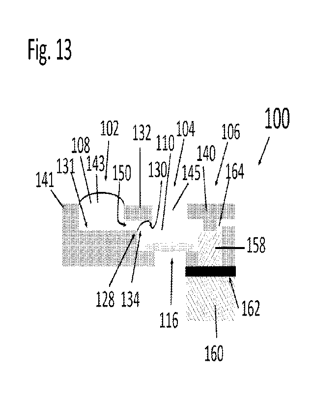
10 Fig. 13 is a cross-sectional view of the micro-
fluidic device 100 of the present invention. It should be
noted that the device 100 is not fabricated from laminates.
The device 100 is substantially similar to device 200 and
everything that applies to device 100 also applies to device
15 200 and vice versa. The device 100 has a microfluidic
platform with liquid reservoirs in fluid communication, and
absorption units and a dissolvable film that act as time-
controlled liquid drainage with a delay valve. The sample
support 116, here illustrated as a TEN grid, is positioned at
20 a bottom of the sample reservoir 104. The stain reservoir
102 has an inlet opening 143 defined between the front end of
a back section 141 of the device and the back end of a middle
CA 03184982 2023- 1-4
WO 2022/060536
PCT/US2021/047081
36
section 132 of the device. Preferably, if the device is
constructed using laminate technology, the sections 140, 132
and 141 are part of the same laminate which makes it easier
to fabricate the device 100
The sample reservoir 104 has an inlet opening 145
defined between the laminate or section 132 and the laminate
or section 140. The sample reservoir 104 is upstream (on one
side) connected to and in fluid communication with a stain
reservoir 102 via a microfluidic channel 150 that extends
between the sample reservoir 104 and the upstream stain
reservoir 102. It Is to be understood that the channel 150
may have a pinning edge or a discontinuity at only a portion
of the end of the channel 150 so that, for example, the
sidewalls do not have any edges. There may also be an edge
of the channel at the upper side of inner surface so that
there are two opposite edges at the end of the channel.
Preferably, the channel 150 is defined between a
hydrophilic underside 130 of a first laminate portion or
section 132 and a bottom surface 131 of the stain reservoir
102. The bottom surface 131 extends to a pinning edge 128.
Capillary forces between the liquid 108 and the underside 130
and the surface tension of the surface 134 hold the liquid
108 in the stain reservoir 102 and prevents the liquid 108
CA 03184982 2023-1-4
WO 2022/060536
PCT/US2021/047081
37
from flowing into the sample reservoir 104. In other words,
the pinning edge 128 prevents liquid 108, such as stain
liquid, added to the stain reservoir 102 from flowing into
the sample reservoir 104.
The sample reservoir 104 is downstream (on the
opposite side relative to the upstream connection to the
stain reservoir 102) connected to and in fluid communication
with a first filter or absorption media 158 which is
separated from a second filter or absorption media 160 by a
dissolvable film, membrane or valve 162. The first filter
158 is also connected to a vent 164. The vent 164 serves as
an emergency exit for potentially trapped air and gas which
would otherwise hinder the flow of the liquid 108, 110 into
and to be absorbed by the first and second filters 158, 160.
With reference to Fig. 14, the removable flap 126
is, upon completion of the grid preparation, removed from the
device 100 prior to removing the sample grid 116 from the
sample reservoir 102.
Fig. 15 shows a device 300 that is substantially
similar to the device 100 shown in Fig. 13 but includes an
additional reservoir 302 and an additional dissolvable film
or membrane or valve 320 and an additional absorption unit
CA 03184982 2023- 1-4
WO 2022/060536
PCT/US2021/047081
38
322. Only the main differences between device 100 and device
300 are here described. The device 300 is used when
additional liquids are to be flushed over the grid 116 in a
sequential and time-controlled manner. The additional liquid
reservoir 302 is placed upstream of the stain or second
reservoir 102 and in fluid communication with and connected
thereto via a channel 304 that is defined between a bottom
surface 306 of the reservoir 302 and a hydrophilic underside
308 of a laminate portion or section 310. Between the
reservoirs 302 and 102, there is a second pinning edge 312
that prevents liquid 314 in the upstream reservoir 302 from
flowing into the stain reservoir 102, 303. The liquid 314 is
held in place in the reservoir 302 in the same way as the
liquid 108 in the reservoir 102 i.e. by capillary forces to
the hydrophilic underside 308 and by surface tension in the
surface 316.
The order of the reservoirs corresponds to the
order in which the liquids flow over the grid 116. That is,
if the liquids are sample, wash, stain then the stain liquid
should be added to the upstream reservoir 302. The wash
liquid should be added to the middle or second reservoir 102,
which upon addition connects to the liquid in the upstream
CA 03184982 2023- 1-4
WO 2022/060536
PCT/US2021/047081
39
reservoirs 302. The sample liquid should be added to the
first reservoir 104 on top the grid 116, which upon addition
connects to the upstream liquid train of wash 305 and stain
314 and downstream connects to the draining unit 318.
The draining unit 318 has the absorption members
(filter papers) 158, 160 and dissolvable film 162 and is
located downstream of the sample reservoir 104. The draining
unit 318 has an additional dissolvable film 320 and another
filter or absorption member 322 to illustrate how the timing
of the additional liquid can be controlled. The thickness of
the first dissolvable film 158 decides how long the first
liquid 110 added to the sample reservoir 104 sits or stays on
top of the grid 116 i.e. how long the sample liquid 110 and
particles 114 are permitted to adhere to the grid 116. The
second filter 160 should be big enough to absorb and store
the amount of liquid corresponding to the volume of the
sample liquid 110. Once the liquid 110 reaches the second
dissolvable film or membrane 320, the flow of the liquid over
the grid 116 stops until the film 320 has been dissolved and
the last filter 322 in this setup with 3 liquids pulls the
second liquid 108, 305 and third liquid 314 over the grid 116
by absorbing all the volumes of all three liquids 110,
CA 03184982 2023- 1-4
WO 2022/060536
PCT/US2021/047081
108/305 and 314.
Fig. 16 shows a device 380 which includes
modifications of the devices shown in Figs. 13 and 15 and
illustrates how the shapes of the filter paper 382 in the
5 draining unit 384 can be modified in order to steer the flow-
speed of the liquids. Everything else in device 380 is
identical to the components of devices 100 and 300. A narrow
and thin filter paper or the filter paper 382 with a neck 386
slows down the flow speed over the grid 116 whereas a wide
10 and thick filter increases the flow speed.
Figs. 18-19 show alternative embodiments of the
devices 700, 800, respectively, that are virtually
identical to device 100 shown in Fig. 13 except that the
draining or blotting units 706, 806, respectively, are
15 different from draining or blotting unit 106.
Preferably, draining unit 706 has only one first
absorption member 758 but no dissolvable membrane or a
second absorption member, as shown in Fig. 13. The
operation of device 700 is substantially similar to that
20 of device 100 in that the liquids in the first and second
reservoirs are absorbed by the absorbing member 758
during a suitable time period so that there is enough
CA 03184982 2023-1-4
WO 2022/060536 PCT/US2021/047081
41
time for the particles in the sample liquid to adhere to
the grid 116, as explained in detail with reference to
devices 100, 200.
Similarly, device 800 is substantially similar
to that of device 100 except that the draining unit 806
has a dissolvable membrane 862 and a first absorption
member 858. The draining unit 806 does not have an
absorption member between the dissolvable membrane 862
and the second reservoir 104 so that the dissolvable
member 862 comes into direct contact with the second
liquid without the second liquid having to pass through
an absorption member before coming into contact with the
dissolvable member to start dissolving the dissolvable
membrane 862. Except for the differences of the draining
units 706, 806 compared to draining unit 106 all other
features and method steps of devices 700, 800 are the
same as devices 100, 200 described in detail above.
Fig. 20 is yet another embodiment of the device
900 that is substantially similar to the device 100 shown
in Fig. 13 except that it has a capillary channel 958 in
draining or blotting unit 906 instead of the first
absorption member 158. The capillary forces in the
CA 03184982 2023- 1-4
WO 2022/060536
PCT/US2021/047081
42
channel 958 urges the second liquid 110 from the second
reservoir 104 into the channel 958 so that the liquid 110
comes into contact with the dissolvable membrane 162 to
dissolve the membrane, as described in detail in
connection with devices 100 and 200.
In operation, the method of the present invention
comprises the steps of providing the stain reservoir 202
connected to and in fluid communication with the grid chamber
or sample liquid reservoir 204 that has a pre-mounted grid
216. The reservoir 204 is in turn connected to, and in fluid
communication with, the draining unit 206. The stain liquid
208 is added to the stain reservoir 202 which is contained in
the reservoir 202 until the user adds the sample liquid 210
including the particles 214 into the sample liquid reservoir
204.
This key feature is enabled through a capillary
stop valve or pinning mechanism, here in the form of an edge
228 located at the end of channel 250 that separates the
stain reservoir 202 from the grid chamber 204. The stain
liquid 208 is pinned at the pinning edge 228 due to capillary
forces so that the liquid 208 adheres to the underside 230
and extends over the pinning edge 228 and the surface tension
at the surface 234 prevents the liquid 208 from flowing into
CA 03184982 2023- 1-4
WO 2022/060536
PCT/US2021/047081
43
the grid chamber 204 although there is fluid communication
between the stain reservoir 202 and the sample reservoir or
grid chamber 204 via channel 250. The fact that the stain
liquid 208 is held inside the stain reservoir 202 in this way
enables the sample preparation process to be initiated by
adding the sample liquid 210 including the particles 214 into
the sample reservoir 204. The liquid 210 is added in such an
amount so that the liquid 210 comes into contact with surface
234 to break the surface tension of the liquid 208 between
the pinning edge 228 and the underside 230. When the surface
tension of the surface 234 is broken, the two liquids 210,
208 are connected with only minor mixing of the liquids at
the interface. When the sample liquid 210 including the
particles 214 are added to the grid chamber 204 via the
opening 236 from above the device 200, the liquid 210 also
flows into and connects with the draining unit 206 that is
downstream of the sample reservoir 204. The opening 236
through which the sample liquid 210 and particles 214 are
added is slightly smaller than the width of the grid 216 to
make the sample liquid 210 reliably connects to the stain
liquid 208 and the blotting unit 206. The cavity 257 located
below the grid 216 makes sure that no liquid flows and
attaches to the wrong side of the grid 216 and interferes
CA 03184982 2023-1-4
WO 2022/060536
PCT/US2021/047081
44
with the quality of the preparation.
The draining unit 206 has two absorption units
(such as filter papers) 218 and 222 and the soluble PVA film
220 located between the two filters 218, 222. The top filter
218 makes sure that the sample liquid 210 reliably connects
to the PVA film 220 by absorbing the liquid 210 so that the
liquid travels from a top side of the filter 218 to a bottom
of the filter 218 that is in contact with the dissolvable
film 220.
The vent 264 above the top filter serves as the
emergency exit for potentially trapped air, which could
otherwise block the connection between the sample liquid 210
and the draining unit 206. The sample liquid 210 flows
through the top filter 218 and upon contact with the PVA film
or layer 220 dissolves the PVA layer 220 so that the liquid
can flow into the filter 222 located below the filter 218.
The time is takes for the sample liquid 210 to dissolve the
PVA layer 220 is critical because it controls the time the
particles 214 in the sample liquid 210 are permitted to
adhere to and adsorb into the grid 216. Once the PVA layer
220 is dissolved, the liquid 210 followed by flow into and
connect to the bottom filter 222 which absorbs all the liquid
CA 03184982 2023- 1-4
WO 2022/060536
PCT/U52021/047081
210, 208 in the device 200. The filter or absorption member
222 first absorbs the sample liquid 210 and then the stain
liquid 208. The bottom filter 222 hence corresponds to the
manual blotting step. The opening 236 over the grid 216
5 through which the sample liquid 210 and the particles 214
were added, now ensures rapid drying of the thin stain film
224 that remains left after the draining/blotting by the two
absorption members or filters 218, 222 have absorbed all
excess liquid 210, 206. Finally, the flap 226 can be peeled
10 off the device 200 to provide easy access to the grid 216
that is easily extracted from the device 200 for subsequent
imaging in, for example, a ns-TEM device.
If more liquids need to be added in a sequential
manner, for example a washing liquid 305 in a washing step
15 before the stain liquid 314 is added to the sample liquid
110, another liquid reservoir 302 connected to and in fluid
communication with the middle reservoir 303 via the channel
304 and separated by the pinning edge 312 can be added, as
shown and described in connection with Fig. 15. The wash
20 liquid 305 should then be added to the middle reservoir 303
after the stain liquid 314 is added to the most upstream
reservoir 302. If the incubation time of the additional
CA 03184982 2023- 1-4
WO 2022/060536
PCT/US2021/047081
46
liquid (here the wash liquid 305) need to be controlled, an
additional layer of soluble film 320 and filter paper 322 can
be added to the draining unit 318.
The speed of the flow over the grid 116 can be
controlled by the shapes and thicknesses of the filter papers
in the draining unit. Less amount of available absorption
media (i.e. filter/paper) or lower capillarity (also known as
Wicking rate) of the filter results in a slower flow/drainage
and vice versa. For example: a thin, narrow and long filter
after the soluble film results in a slower liquid flow and
drainage pace.
Instead of adding a droplet of a pre-mixed stain
solution, the salt constituting the stain can be dried at the
bottom of the stain reservoir 102 and then only water or
another dissolvent/buffer is added to the reservoir 102 when
preparing the stain reservoir and the grid. The stain salt
is then dissolved when the dissolvent is added to create the
stain liquid 208.
Instead of adding a hydrophilized grid to the
preparation assembly kit, a hydrophilization liquid such as
Alcian-Blue can be flushed over the grid before the sample
liquid is added. In this way, the stain and sample liquids
CA 03184982 2023- 1-4
WO 2022/060536
PCT/US2021/047081
47
are loaded in two separate reservoirs upstream of the grid
chambers 102 and 302, (best shown in Fig. 15) connected via
microfluidic channels 304 and 150 but kept separate via
pinning edges 128 and 312. The grid hydrophilization liquid
is then added directly onto the grid 116 in the grid chamber
104 to start the sequence of liquids flowing over the grid
116, i.e., initiating the grid preparation process.
An alternative use of the method of the present
invention is to use it for controlled deposition of a matrix
on top of the grid 116. The same method as described above
applies with the exception that only one liquid, i.e. the
substrate, is used and it is added to the grid chamber 104.
For example, fibers, such as spider silk, may be permitted to
polymerize in the air-liquid interface on the droplet of the
sample (substrate) added onto the grid 116. When the soluble
layer 162 is dissolved., the spider silk gently falls down on
the EM grid while the filter paper 160 drains the device.
The fiber network (in this case the spider silk) disposed on
top of the grid then acts as a matrix forcing a. protein which
is later added to be placed in a random orientation on the
grid, before subsequent single particle reconstruction in
(cryo- or negative stain) TEM. Adding a protein directly to
CA 03184982 2023-1-4
WO 2022/060536
PCT/US2021/047081
48
the grid. 116 often results in that the protein orients itself
in a preferred orientation (i.e. laying down when elongated
and/or flat), which limits the resolution that can be
achieved in the reconstruction.
Experiments
As described above, the microfluidic device of
the present invention consists of several layers of
different materials, as particularly indicated in, for
example, Fig. 3 It was fabricated from hydrophilic sheets
(Type C laser printing transparency, Xerox, Elmstock,
UK), adhesive tape 1 (64620, Tesa, Norderstedt, Germany)
and adhesive tape 2 (300LSE, 3M, VWR, Spanga, Sweden).
Low-tack adhesive tape (Scotch 928, 3M, Amazon, Koblenz,
Germany) was used to fixate the 400 mesh TEN grids
(01754-F, Ted Pella Inc., Redding, CA) which are formvar
coated copper grids with a continuous carbon film.
Ahlstrom grade 238 and 222 (Ahlstrom Filtration LLC, Mt.
Holly Springs, RA) were used as absorption paper 1 and
absorption paper 2 in the draining or blotting unit,
respectively. The soluble film or membrane/valve was
fabricated from granular PVA (360627, Sigma-Aldrich, St.
CA 03184982 2023- 1-4
WO 2022/060536
PCT/US2021/047081
49
Louis, MO). AAV (adeno-associated virus) particles,
serotype 2 (AAV2) encapsulated with Cytomegalovirus (CMV)
promoter-driven expression of Green Fluorescent Protein
(GFP), with a stock concentration of 1x10¹³ gc/mL
(CV10004-50UL, AMS Biotechnology Ltd., Abingdon, UK) was
used as the sample.
The AAV sample was diluted with phosphate-
buffered saline (DPBS (-/-) 14190-094, Thermo- Fisher,
Uppsala, Sweden) to a concentration of lx1012 gc/mL. 26S
proteasome (#: E-350, BostonBiochem, Cambridge, MA) was
used as a test sample representing a large globular
protein complex. The sample with protein fibrils from
whey protein isolate (WPI)16, with an initial
concentration of 40 mg/ml, was a gift from the Division
of Applied Physical Chemistry at the Royal Institute of
Technology in Stockholm, Sweden.
NanoVanO, 2% Methylamine vanadate in solution,
(#2011-5ML, Nanoprobes, Yaphank, NY) and Uranyl Acetate
2% in solution (#2240-2, Electron Microscopy Sciences,
Hatfield, PA) were used as stain. Aqueous solutions of
food color dyes (EAN-codes: 5701073064665 and
CA 03184982 2023- 1-4
WO 2022/060536
PCT/U52021/047081
5701073064672, Dr.Oetker, Coop, Solna, Sweden) were used
as models for sample and stain.
Each device was fabricated using lamination
technology where the devices were formed by stacking
5 several layers of different materials, as described
previously. The cross section in Fig. 3 shows the
different layers. The denomination, brand name and
thicknesses of these layers were as:
Denomination Brand name Thickness
[pm]
Type C laser
Hydrophilic sheets printing 100
transparency, Xerox
Adhesive tape 1 64620, Tesa
170
Adhesive tape 2 300LSE, 3M
50
Low-tack adhesive Scotch 928, 3M 30
Paper 1 Ahlstrom grade 238
340
Paper 2 Ahlstrom grade 222
830
10 . The adhesives and the hydrophilic sheets were
structured using a cutting plotter (CE6000, Graphtec
America inc., Irvine, CA).
The PVA film or membrane was fabricated from an
aqueous solution of 20 wt% of granular PVA. Using a
15 thin-film applicator (4340, Elcometer, Manchester, UK)
the PVA films were uniformly transferred to laminating
pouches (3385694, Office Depot, LA Venlo, Netherlands)
CA 03184982 2023-1-4
WO 2022/060536 PCT/U52021/047081
51
and dried at room temperature. The final PVA film
thickness was measured using a thickness gauge with 1 pm
graduation (2109L Metric Dial Gauge, Mitutoyo, Upplands
Vasby, Sweden).
The PVA film was laminated to absorption paper 2
at 85 C using a laminator (Heat Seal Pro H600, GBC,
Northbrook, IL). The paper-PVA laminates were kept in a
humidity chamber at 80% relative humidity until 30
minutes before use.
The paper materials, including the paper-PVA
laminate, were cut by a laser cutter (VLS 2.30, Universal
Laser Systems, Vienna, Austria). After structuring, the
layers were assembled by using alignment pins and
laminated at room temperature. For improved particle
adhesion, the TEM grids were glow discharged in oxygen
plasma with a PELCO easiGlowTM (91000S-230, Ted Pella
Inc., Redding, CA) before fully assembling the
microfluidic device of the present invention. A fully
assembled fabricated device is shown in Fig. 5. The
dimensions of the device are 6 x 12 mm2. The devices
were used within one hour after glow discharging the TEM
grids.
CA 03184982 2023- 1-4
WO 2022/060536
PCT/U52021/047081
52
One important feature of the microfluidic device
of the present invention is that it is designed to
minimize user-interactions. To demonstrate the
autonomous device operation and microfluidic consistency
six devices were evaluated. Five devices were used with
AV particles as sample and NanoVan as stain. The grids
from these five devices were used to collect TEN images
for an automated image analysis on a total of 225 images.
To better visualize the individual preparation steps of
the autonomous device, one device was used with color dye
solutions. Blue dye solution and yellow dye solution
were used as models for sample and stain, respectively.
First, 5 41 of stain liquid was added via the stain inlet
into the stain reservoir. Then, the autonomous TEN grid
preparation mechanism was triggered by adding 5 41 of
sample to the sample inlet of the sample reservoir or
grid chamber.
The TEM grid preparation sequence of all the
devices was recorded with a camera with a frame rate of
50 frames per second. To analyze the device performance
and consistency of the autonomous preparation steps, the
time interval of each step was manually obtained. The
CA 03184982 2023- 1-4
WO 2022/060536
PCT/U52021/047081
53
time period between the addition of stain and the
addition of the sample liquid (including the particles)
was defined as the stain preloading time.
To demonstrate the robustness of the stain
reservoir, i.e. stain confinement without leakage, the
time between stain and sample addition was varied between
20-60 seconds wherein the stain liquid was held in place
by a surface extending between the pinning edge and an
underside of a hydrophilic surface i.e. capillary forces
and surface tension. As illustrated in Figs. 1A-1D, the
microfluidic TEM grid preparation steps after sample
addition includes sample adsorption, draining/blotting
and thin film drying. As critical aspect of the method
of the present invention is that the adsorption time of
the sample on the TEM grid corresponds to and is the same
as the dissolving time of the PVA film. It was defined
as the time between wetting of paper 1 and the start of
the blotting event. The PVA layer thickness was 10 pm.
The blotting time is the interval between the start and
the end of the draining/blotting event. The start of the
blotting event is defined as the moment when the liquid
first moves into the draining unit. The end of the
CA 03184982 2023- 1-4
WO 2022/060536
PCT/U52021/047081
54
blotting event is defined as the moment when the bulk of
liquid is drained by the draining unit leaving a thin
stain film on the TEN grid. After this, the drying
interval starts and lasts until the remaining thin film
of stain on the grid was visually dry.
In general, TEN imaging is a powerful
visualization technique for many different types of
samples. However, the required sample adsorption time
varies between different samples. The main reason for
this is that sample adsorption depends on the interaction
between sample and the carbon surface of the TEN grid.
Hence, devices with different adsorption times to account
for different sample requirements would be desirable.
Another key element of the microfluidic device
of the present invention is the dissolving time of the
water-soluble PVA film, that autonomously controls the
timing of the device, corresponds to the sample
adsorption time of the sample (i.e. film or layer of
particles embedded in stain) on the grid.
To demonstrate the adjustability of the
adsorption time of the sample on the grid, microfluidic
devices with three different thicknesses of the water-
CA 03184982 2023- 1-4
WO 2022/060536
PCT/US2021/047081
soluble film (12 pm, 24 pm and 36 pm) were fabricated and
investigated. Among the parameters that affect the
dissolving time (e.g. temperature, relative humidity),
the thickness of the dissolvable film is one of the
5 easiest parameters to tune and adjust. The PVA
thicknesses of 24 pm and 36 pm were achieved by stacking
multiple layers of 12 pm PVA sheets and laminating them
to paper 2 below the PVA sheets at 85 C with the
laminator. The paper-PVA laminates were kept in a
10 humidity chamber at 80% relative humidity until
30 minutes before use. The adsorption time was evaluated
of 15 devices, five devices per film thickness, using 5
pl of blue dye solution and 5 pl of yellow dye solution
as a model for sample and stain, respectively.
15 To assess the sample preparation quality, TEM
imaging was performed on the five autonomously prepared
TEM grids with AAV particles as sample and NanoVang as
stain. NanoVan(N was chosen because it is not
radioactive, unlike the commonly used Uranyl Acetate, and
20 can be handled in an ordinary laboratory. For all five
grids, it was investigated whether AAVs were successfully
adsorbed to the TEM grid and sufficiently embedded in
CA 03184982 2023- 1-4
WO 2022/060536
PCT/U52021/047081
56
stain. The AV particles on different magnification
levels were inspected, with a field of view (FOV) between
16 pm and 500 rim. The imaging was performed on MiniTEMTh
microscopes (Vironova AB, Stockholm, Sweden) with an
operating voltage of 25 kV.
To investigate whether the obtained TEM images
were useful for automated image analysis, a particle
detection script was applied to the TEM images of the
five autonomously prepared grids. A total of 225 images
were collected according to the imaging scheme shown in
Fig. 3. At low magnification, the user manually chose
five non-neighboring grid squares. Then, nine high
magnification images were acquired per grid square at a
FOV of 2 um, resulting in 45 images per grid. At this
magnification, where a pixel represents approximately
1 nm, a number of particles per image can usually be seen
and the morphology of the AAVs is typically visible.
Grid 1, grid 4 and grid 5 were imaged on the same
microscope, while grid 2 and grid 3 were imaged on a
second microscope. The particle detection script was
applied to all 225 images. AAVs have an icosahedral
capsid that appears round and has an expected diameter of
CA 03184982 2023- 1-4
WO 2022/060536
PCT/U52021/047081
57
20-25 nm. However, the script was designed to detect the
stain envelope around the AAV particles so that the
particles appear larger than the actual virus size.
Therefore, the particle detection script was set to
detect round objects within a diameter range of 24 nm to
32 nm. From the automated image analysis, the number of
detected particles per grid were obtained, where each
detected particle is characterized by its position and
size.
To quantify the particle detection results, a
manual particle detection was performed on a subset of 25
of the images, with five randomly chosen images per TEM
grid. The number of particles were manually counted and
compared with the results from the detection script.
This was done to find the ratio of true and false
positives, which both are important measures for the
performance of the detection script.
nsTEM is routinely used as a quality control
during the preparation of biological specimens, e.g.
protein complexes, for structural biology. To
investigate the potential use of the microfluidic device
for wider applications and with different stains,
CA 03184982 2023- 1-4
WO 2022/060536
PCT/US2021/047081
58
proteasomes were prepared and image, as a larger globular
protein complex, and protein fibrils from WPI, as a
filamentous protein. The PVA films in the used
microfluidic devices had a thickness of 15 pm,
corresponding to a dissolving time of around 35 seconds.
For the proteasomes and fibrils, stock solution of Uranyl
Acetate and NanoVan , was used, respectively.
As described in detail above, the TEN grid
preparation sequence is shown in Figs. 2A-2D. For
visibility, colored dye solutions were used instead of
sample and stain solutions. The first step shows how the
preloaded stain 208 (yellow dye solution) is contained in
the stain reservoir 202 and the sample 210, 214 (blue dye
solution) is added (best shown in Fig. 2A. In the second
step, the sample 210 including the particles, 214 cover
the TEN grid as long as the PVA film or valve is closed
(best shown in Fig. 2B. When the PVA film or valve has
dissolved, the stain and sample liquids are blotted (best
shown in Fig. 2C. Finally, the bulk of liquids is
contained in the draining media (blotting filter/paper)
and the stain film, including particles embedded therein,
dries (best shown in Fig. 2D). Compared to a previously
CA 03184982 2023- 1-4
WO 2022/060536
PCT/U52021/047081
59
reported microfluidic TEM grid preparation, the user
interactions were reduced by providing an autonomous
microfluidic operation that is controlled by the water-
soluble PVA film. Furthermore, a significantly lower
liquid consumption was demonstrated with liquid volumes
as small as in the manual preparation protocols.
To demonstrate microfluidic consistency, video
recordings were analyzed with respect to timing and
duration of the microfluidic events on the five devices
used with AAVs as sample and NanoVan as stain. Fig. 6
presents a bar chart with the time intervals for each of
the four sample preparation steps. The results show that
regardless of the length of the stain preloading time,
all the following steps including adsorption,
draining/blotting and drying, are close to identical for
the five devices. This demonstrates that the stain
reservoir reliably contains the stain until the sample is
added irrespective of the stain preloading time. The
average adsorption time for the five devices is
10.6 0.3 s, corresponding to a CV of 3%. This
indicates a highly consistent autonomous time-control of
the microfluidic device of the present invention. The
CA 03184982 2023- 1-4
WO 2022/060536
PCT/US2021/047081
average draining/blotting time is 0.8 0.1 s,
corresponding to a CV of 12.5%. While the CV seems high,
the absolute deviation is low and confirms the
microfluidic consistency of the device of the present
5 invention. The drying step does not end abruptly which
makes it difficult to measure the exact drying interval
by viewing videos. However, it was observed that all the
TEM grids were visually dry within one minute. The
reliable and fast drying is enabled by the grid area
10 sized top opening in the grid chamber.
The results show that the microfluidic device of
the present invention works as intended although the user
input was minimal. Irrespective of the stain preloading
time, the autonomous device operation after sample
15 addition is close to identical for the five devices which
demonstrates a high microfluidic consistency.
To demonstrate the adjustability of the sample
adsorption time, which corresponds to the PVA dissolving
time, three different thicknesses (12 pm, 24 pm and
20 36 pm) of the water-soluble PVA film were tested in the
microfluidic device of the present invention. Fig. 7
shows the measurement results of the adsorption time.
CA 03184982 2023- 1-4
WO 2022/060536 PCT/US2021/047081
61
The dissolving time of the PVA films increases with PVA
film thickness. For 12 pm, 24 pm and 36 pm thicknesses
of the PVA films, the average dissolving time is
14.4 0.9 s (n=5), 89.9 12.0 s (n=5) and
191.6 20.3 s (n=5), respectively. The results show
that it is possible to easily adjust the adsorption time
by changing the PVA film thickness. The variation of
dissolving time increases with increased PVA film
thickness. This could be due to small differences in the
PVA film thickness between different devices. However,
the variation is low enough to conclude that the
adsorption times can be controlled by the design of the
PVA layer of the present invention.
TEM =aging of the five autonomously prepared
TEM grids made it possible to assess the sample
preparation quality. Figs. 9A-9C show a magnification
series with three FOVs: 16 pm, 2 pm and 500 nm. In the
largest FOV (16 pm), the AV particles appear as dark
spots. The intermediate FOV (2 pm) shows a higher level
of detail. The particles are visible as bright, round
objects encircled by dark rings with a radially fading
stain gradient. The smallest FOV (500 nm) in this series
CA 03184982 2023- 1-4
WO 2022/060536
PCT/US2021/047081
62
has the highest level of detail and provides a close-up
view of the AAV particles.
Figs. 10A-10E show one exemplary image from each
of the five grids (FOV 2 pm). It was found that all five
TEM grids contain well embedded particles in the stain
film on the grid, visible as bright spots surrounded by
dark stain envelopes. Variations in the appearance of
the stain envelope might be due to local variations of
stain thickness. Also, thickness variations of the TEM
grid, e.g. caused by local inhomogeneities of the carbon
film, can result in variations of the image darkness.
Overall, the results from the five microfluidic devices
showed consistent preparation of TEM grids with well
embedded AAV particles.
To further demonstrate sample preparation
consistency, 225 TEM images were collected and an
automated particle detection was performed. The particle
detection script detected 5171 particles in all 225
images. Every grid, with 45 images, contained an average
of 1034 65 particles, corresponding to a CV of 6%.
This indicates a reproducible and consistent AAV particle
spreading over five independently prepared TEM grids.
CA 03184982 2023- 1-4
WO 2022/060536
PCT/US2021/047081
63
Using the results of the automated particle detection,
the detected size of the particles was extracted.
Fig. 11 is a graph 640 that shows the average
particle diameter for the detected particles in each
grid. Two different microscopes were used and even
though the calibrations might be slightly different, the
average particle size for each grid is well within the
error bars of the other samples. The average size of all
detected particles is 28 2 nm (n=5171), corresponding
to a CV of 7%. This low variation means that,
irrespective of the grid, all detected particles have a
similar detected size. The real size of AAV particles is
20-25 nm but appears larger when imaged in nsTEM due to
the stain envelope. The detection script is designed to
outline and measure particles at the stain layer, i.e.
outside the actual particle. Therefore, the detected
particle size is well within the expected size window.
The result of the manual particle detection in a
subset of 25 images allowed to quantify the automated
detection results. Fig. 12 summarizes the results of the
subset test. The manual count resulted in 605 particles
in the subset. The automated particle script found 557
CA 03184982 2023- 1-4
WO 2022/060536
PCT/US2021/047081
64
of these particles correctly (True positives), which
corresponds to a success rate of 92%. The script found
29 objects that were not correct (False positives), which
corresponds to 4.9%. With true positives above 90% and
false positives around 5%, it can be concluded that the
images and the autonomous sample preparation have
sufficient quality for simple automated image analysis.
To broaden the scope of applications proteasomes
and protein fibrils from WPI were prepared and imaged.
The results of those two samples 500, 502, presented in
Figs. 17A-17D, reveal an even spreading of the proteins
on the TEM grid with well-embedded areas suitable for TEM
investigations. Figs. 17A-17B show images 500 of 265
proteasomes at two different magnifications. A top view
504 and a side view 506 of the proteasomes can be
observed. Figs. 17C-17D show images of WPI fibrils 508
at two different magnifications. The analysis of the
proteasome specimen (best shown in Figs. 17A-17B) shows
that individual proteasomes can clearly be identified,
and structural features such as details of the subunits
can be distinguished. Different projections can be
observed on the images, with the top view appearing as a
CA 03184982 2023- 1-4
WO 2022/060536
PCT/U52021/047081
circular particle and the side view appearing
rectangular. The analysis of the WPI fibrils (best shown
in Figs. 17C-17D allows the observation and
characterization of well-defined individual fibrils of
5 various lengths. Overall, the morphological observations
are in line with reported data of similar samples
prepared with conventional manual nsTEM. The preparation
of these two protein samples did not require further
adjustments of the microfluidic device, hence
10 demonstrating the versatility and robustness of the
method.
Below is yet another possible application in the
field for the method of the present invention that would
require a modified device. The possible nsTEM sample
15 preparation application example is immunogold-labelling where
four liquids need to be flushed over the sample. The
sequence of preparation steps would be:
1)A grid, with the sample already attached thereto, is
added to the device;
20 2)A primary antibody is permitted to adhere to the grid
(so added directly onto the grid as the sample liquid in
the description above);
3)Once the binding has occurred, a blocking liquid is
CA 03184982 2023-1-4
WO 2022/060536
PCT/US2021/047081
66
flushed over the grid (e.g. BSA = bovine serum albumin,
or desiccated milk);
4)Then a second antibody connected to gold particles is
flushed over to bind to and hence mark the primary
antibody positions; and
5)Finally, non-bound gold particles are washed off with
the last washing step.
In summary, a capillary-driven single-use device
of the present invention for autonomous TEM sample
preparation has been presented. To avoid operator bias
and error-prone manual steps, the device of the present
invention is designed to minimize user-interactions. The
key design elements are the stain and sample reservoirs
combined with the water-soluble valve or PVA film and the
absorption membranes. These key elements enable the
starting of the autonomous TEM grid preparation with only
one non-critical user-interaction. The device
consistency both for the microfluidic performance and the
sample preparation quality have been demonstrated. The
consistency of the microfluidic performance was shown by
five microfluidic devices with close to identical TEM
grid preparation sequences. The sample preparation
consistency was demonstrated by five TEM grids that all
CA 03184982 2023-1-4
WO 2022/060536
PCT/US2021/047081
67
exhibit well embedded AV particles. This preparation
consistency was further highlighted by the results of the
automated particle detection. From a subset test with
true positives above 90% and false positives around 5%,
it was concluded that the images and the autonomous
sample preparation hold sufficient quality for image
analysis. The additional preparation of two protein
samples demonstrated the versatility of the microfluidic
device for a wider scope of applications. Furthermore,
the adjustability of timing of the microfluidic events
was demonstrated by changing the thickness of the water-
soluble valve or PVA film. This allows to account for
different sample adsorption requirements. To
account
for TEM sample preparation requiring different staining
times, the device of the present can be extended by a
second draining unit. In conclusion, the demonstrated
microfluidic device of the present invention presents a
promising, effective and reliable solution to alleviate
the problems associated with human inconsistency in
manual TEM grid preparations.
The evaporation arrangement of the present
invention is important and that the sample support is exposed
CA 03184982 2023- 1-4
WO 2022/060536
PCT/US2021/047081
68
to air for proper evaporation. Another aspect is that the
width of the opening and the width of the sample support
should be about the same. The humidity condition immediately
above the sample support is higher than the humidity outside
the opening i.e. above the device. At the liquid boundary of
the liquid sample on the sample support the humidity is 100%
while the relative humidity in the ambient air outside the
device is lower which promotes evaporation of the liquid in
the stain layer through the opening. It should be understood
that the first liquid can be held in the first reservoir
without using an edge. The two liquids can connect without
the use of the edge but, for example, putting pressure on one
of the liquid droplets. The edge, however, stops the first
liquid from flowing into the second reservoir. This is
because the edge is a discontinuity in the channel between
the two reservoirs and the travel of the capillary force
along the wall of the channel is stopped. The first liquid,
such as the stain liquid, could be held in the first
reservoir i.e. the stain reservoir without using an edge.
The pinning edge keeps the stain liquid in place i.e. stops
the stain liquid from flowing into the sample reservoir while
adding the liquid to the sample reservoir. The expansion
(bulging out) of the first liquid between the edge and the
CA 03184982 2023-1-4
WO 2022/060536
PCT/US2021/047081
69
hydrophilic upper surface is beneficial but not necessary.
It makes the device more robust.
It should also be understood that it is not
necessary to use capillary forces to hold the first liquid in
the first reservoir. The device preferably, but not
necessarily, has a channel going from the first reservoir to
the second reservoir. Preferably, the two reservoirs should
be in fluid communication and that the first liquid should be
held in the first reservoir. This confinement of the first
liquid in the first reservoir is preferably but not
necessarily based on capillary forces and/or surface tension
which are easily broken when the second liquid is added and
connects to the first liquid. It is not necessary that the
first reservoir is at a higher elevation compared to the
second reservoir and the blotting unit. All three units
could be located on a common surface at the same elevation.
Preferably, the dissolvable member decides the
timing of the adsorption of sample particles on the sample
support but this could also be adjusted by using different
filters that absorb fluids at different rates. For example,
small narrow filter slows down the absorption rate. The
drainage in micro channels could also acts as a delay
mechanism and drainage speed control. It may also be
CA 03184982 2023- 1-4
WO 2022/060536
PCT/US2021/047081
necessary to have a minimum speed or a certain delay time to
give enough time for the sample particles to adhere to the
sample support or grid. It may also be necessary to have a
high speed when draining the first liquid (stain) in order to
5 leave a stain layer. If the draining of the first liquid is
too slow, too much stain will be drained leaving the
particles unprotected. This results in a poor-quality
preparation. It should be noted that although the dissolvable
membrane or film delays the liquid flow and once the membrane
10 is dissolved the flow rate is quite rapid as opposed to a
very slow constant flow rate.
While the present invention has been described
in accordance with preferred compositions and
embodiments, it is to be understood that certain
15 substitutions and alterations may be made thereto without
departing from the spirit and scope of the following
claims.
CA 03184982 2023- 1-4