Note : Les descriptions sont présentées dans la langue officielle dans laquelle elles ont été soumises.
CA 03187298 2022-12-14
WO 2022/002834 PCT/EP2021/067653
1
COMPOUNDS FOR USE IN DIAGNOSIS AND/OR MONITORING OF FIBROSIS
TECHNICAL FIELD
The present disclosure concerns novel compounds comprising a non-cyclic
peptide, a
linker, a chelator and a nuclide such as a radionuclide. The compounds may be
used as
tracers such as radioactive tracers for use in the diagnosis and/or monitoring
of fibrosis
such as fibrosis occurring in the liver, kidney, heart, brain, pancreas, and
lungs of a
patient. The disclosure further relates to a method for preparing the
compounds, a
compound that may be used as an intermediate in the aforementioned method as
well as
a method for diagnosing and/monitoring of fibrosis in a patient.
BACKGROUND
Fibrosis is the formation of connective tissues that might occur in normal
physiology as a
response to injury, which is known as scarring. However, excess formation and
deposition
of connective tissue, which constitutes the pathological formation of
fibrosis, is an
important feature in many different tissues in disease, e.g., liver, kidney,
heart, brain,
pancreas, and lungs. The pathological formation of fibrosis is due to an
increase in the
production and deposition of collagens, especially collagen type I, which
results in loss of
tissue elasticity and progressive loss of organ function. It has been found
that fibrosis is
involved in a large number of prevalent and severe diseases involving organs
such as the
liver, kidney, heart, brain, pancreas and lungs.
Current treatments against fibrotic disease, i.e., fibrosis, mainly target the
inflammatory
cascade, but efforts to develop novel treatments have proven very challenging.
The
treatment objective is to slow down the fibrotic process. To date, there are
unfortunately
no drugs available that can reverse fibrosis. In addition to the challenge of
developing
drugs targeting the inflammatory system, fibrotic disease often lacks reliable
biomarkers.
Several pre-clinical disease models have been developed, but in many cases,
they suffer
in 'translatability' from mice to humans. Diagnosis of fibrotic disease may be
determined
from a biopsy sample when this is feasible. But methods to measure changes
precisely
and repeatedly in the fibrotic process as required in drug development are
largely lacking.
For fibrotic liver disease, Magnetic Resonance Elastography (MRE) is used as a
non-
invasive biomarker of liver stiffness, but for most fibrotic disease such non-
invasive
CA 03187298 2022-12-14
WO 2022/002834 PCT/EP2021/067653
2
methods are not yet available. Of course, non-invasive methods are more
desirable than
invasive methods, such as biopsies, since non-invasive methods are more
convenient,
can be performed repeatedly, and are associated with a lower risk of harming
the patient.
Therefore, further non-invasive diagnostic methods for detection of fibrosis
have been
proposed.
Nuclear Medicine and Biology, 41(2014) 728-736 discloses synthesis and
preclinical
evaluation of 68Ga-labeled collagelin analogs for imaging and quantification
of fibrosis by
positron emission tomography (PET). The analogs were prepared and intended for
binding to collagen overexpressed in fibrotic tissues, since collagen is a
biomarker that
can be targeted in molecular imaging of fibrosis providing direct
identification of the fibrotic
tissue. It is disclosed that the tracers displayed a pronounced washout
pattern from most
of the organs except for kidneys and bladder.
Sci. Trans. Med. 9, 2017, 1-11 discloses a type I collagen-targeted PET probe
for
pulmonary fibrosis detection and staging in preclinical models. The probe used
was 68Ga-
CBP8, which was found to have a specificity for type I collagen. It is stated
that 68Ga-
CBP8 provided significantly enhanced PET signal in the lungs of fibrotic mice
compared
with control mice, and that nonspecific uptake in the surrounding tissues was
similar and
low in both fibrotic and control mice but with high off-target accumulation in
the kidney.
WO 2018/053276 discloses polymer conjugates having utility in the treatment of
a subject
suffering from soft tissue conditions. The polymer conjugates comprise
sulfated
glycosaminoglycan chains which may be substituted with a collagen-binding
agent such
as a peptide with the sequence LRELHLNNN (IUPAC-IUB nomenclature).
Thus, collagens, especially collagen type I, is known as a biomarker for
fibrosis. Further,
for all organs but kidney the cyclic peptides of the above-mentioned
radioactive tracers
have been found to have affinity for collagen while exhibiting a low
background binding.
Importantly, to allow for accurate imaging of the fibrosis, the tracer such as
the radioactive
tracer should have a low non-specific binding to normal tissue, fast blood
clearance and
washout from healthy organs. Thus, there should be low or no binding to
tissues lacking
deposits of collagen such as collagen type I. In other words, the
biodistribution of the
CA 03187298 2022-12-14
WO 2022/002834 PCT/EP2021/067653
3
radiotracer should be selective so that binding mainly takes place to organs
involving
fibrotic tissue.
Radioactive tracers may exhibit retention in tissues for many different
reasons. Retention
of a collagen targeting radioactive tracer may be retained in tissues by e.g.,
non-specific
binding to cellular components, or by specific unintended targeting of
molecular entities
such as receptors. Radiolabeled peptides may additionally exhibit reabsorption
in the
renal tubules during urinary excretion, with subsequent intracellular trapping
of the
radionuclide in the kidney cortex. Regardless of the cause of such tissue
retention, it
precludes the measurement and diagnosis of the existence and/or progression of
fibrotic
lesions in said tissue.
Further, in order to detect the presence of fibrosis it is important that the
radioactive tracer
is able to thoroughly penetrate the organ to ensure that the entire organ is
investigated for
fibrosis. This may be more difficult in solid organs such as liver, kidney,
heart, brain,
pancreas, and lungs compared to non-solid organs.
There is a need for a tracer such as a radiotracer for fibrosis with a
suitable biodistribution
in all or most organs such as suitable biodistribution with respect to kidney.
Further, there
is a need for a tracer for fibrosis which is able to penetrate the entire
organ being
investigated for fibrosis.
It is an object of the present disclosure to alleviate at least one or more of
the problems
discussed above. Further, it is an object of the present disclosure to provide
advantages
and/or aspects not provided by hitherto known techniques.
SUMMARY
The above objects may be achieved with a composition in accordance with claims
1 and 2
or a compound in accordance with claim 19, and by using a method in accordance
with
claim 25. Further embodiments are set out in the dependent claims, the
description and in
the drawings.
The present disclosure provides a composition comprising:
(i) a compound of Formula I:
CA 03187298 2022-12-14
WO 2022/002834
PCT/EP2021/067653
4
C [ L 1 Q
P
Formula I ,
or a pharmaceutically acceptable salt thereof,
and
(ii) a nuclide M, or a pharmaceutically acceptable salt thereof,
wherein
C is a chelator selected from the group consisting of:
0
HOI
N
HOy=NI-
0
0
N-rNrrt!
0
HO ,
0
.*OH
iN-
0 0
HON\ NjN//
,
CA 03187298 2022-12-14
WO 2022/002834 PCT/EP2021/067653
Lni
N N
CCN p
HO-
0 0
?-0H ?-0H
1\1
1 N
0 OH NI 0 OH
OH 0
0
OH 0
5 0 0 OH
0
?µOH
0 0
HO NOH
CA 03187298 2022-12-14
WO 2022/002834 PCT/EP2021/067653
6
)
HO 4;4)
HO,
ISµ4) r)
'''s141.1
0 1.,1/4õ..\ N LI.
..011
;= 0
Hci
and a derivative of any one of the foregoing chelators,
L is a linker:
-
0
A
wherein
m is an integer within the range of from 1 to 20, and
X is NH or 0(0) and forms an amide bond, i.e. C(0)NH, with a 0(0) or NH moiety
of the
chelator,
p is 0 or 1,
Q is a peptide of
SEQ ID NO: 1, a peptide analogue of SEQ ID NO: 1 having at least 88.8%
identity to SEQ
ID NO: 1, and/or
a peptide of SEQ ID NO: 1, a peptide analogue of SEQ ID NO: 1 having at least
88.8%
identity to SEQ ID NO: 1 and in which the C-terminal COOH can be replaced with
CON H2,
CA 03187298 2022-12-14
WO 2022/002834 PCT/EP2021/067653
7
and
M is selected from the group consisting of 68Ga, 18F, 64ou, 44sc, 89zr, 111.
_,
in 67Ga, 99mTc, Mn
Gd, 177Lu.and 86199Y.
The present disclosure also provides a compound of Formula I as described
herein, or a
pharmaceutically acceptable salt thereof.
Further, the present disclosure provides a compound of Formula II:
_____________________ L _____
Formula II
or a pharmaceutically acceptable salt thereof,
said compound of Formula II being a combination of
(i) the compound of Formula I as defined in claim 1 and
(ii) the nuclide M as defined in claim 1,
wherein (i) and (ii) are provided in a ratio (i) / (ii) equal to one.
There is also provided
a composition as described herein,
or
a compound of Formula II as described herein, or a pharmaceutically acceptable
salt
thereof,
for use in diagnosing and/or monitoring of fibrosis.
There is also provided
a composition as described herein,
or
a compound of Formula II as described herein, or a pharmaceutically acceptable
salt
thereof,
for the manufacture of a preparation for the diagnosis and/or monitoring of
fibrosis.
CA 03187298 2022-12-14
WO 2022/002834 PCT/EP2021/067653
8
There is also provided a use of
a composition as described herein,
or
a compound of Formula II as described herein, or a pharmaceutically acceptable
salt
thereof,
for diagnosing and/or monitoring of fibrosis such as diagnosing and/or
monitoring fibrosis
in a patient suffering from, suspected to be suffering from and/or being
treated for,
fibrosis.
Further, there is provided a method for the diagnosis and/or monitoring of
fibrosis, said
method comprising the steps of:
a) administering an imaging agent selected from one or more of the
following:
a composition as described herein,
a compound of Formula II as described herein,
a pharmaceutically acceptable salt of a compound of Formula II as
described herein,
to a patient suffering from, suspected to be suffering from and/or being
treated for fibrosis;
b) subjecting the patient to a medical imaging technique, such as Positron
Emission Tomography (PET), Single-Photon Emission Computed
Tomography (SPECT) or Magnetic Resonance Imaging (MR1) imaging, and
recording signals from the imaging agent administered in step a);
c) determining and/or monitoring if the patient suffers from
fibrosis, and
d) optionally determining the extent of the fibrosis.
BRIEF DESCRIPTION OF THE DRAWINGS
Figure 1 shows the chemical structure of DOTA, i.e.1,4,7,10-
tetraazacyclododecane-
1,4,7,10-tetraacetic acid.
Figure 2 shows the chemical structure of NOTA, i.e. 1,4,7-triazacyclononane-
1,4,7-
triacetic acid.
Figure 3 shows the chemical structure of TETA, i.e. 1,4,8,11-
tetraazacyclotetradecane-
1,4,8,11-tetraacetic acid.
Figure 4 shows the chemical structure of DTPA, i.e.
diethylenetriaminepentaacetic acid.
CA 03187298 2022-12-14
WO 2022/002834
PCT/EP2021/067653
9
Figure 5 shows the chemical structure of DFO, i.e. desferrioxamine B.
Figure 6 shows the chemical structure of NOTAGA.
Figure 7 shows the chemical structure of DOTAGA.
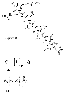
Figure 8 shows the chemical structure of compound 1.
Figure 9 shows the chemical structure of compound 14.
Figure 10a shows the total and non-specific binding of [68Ga]Ga-DOTA-NH-
(CH2CH20)2-
CH2-C(0)-LRELHLNNN-OH to hepatic tissue with induced fibrosis compared to non-
fibrotic liver.
Figure 10b shows the magnitude of binding of [68Ga]Ga-DOTA-NH-(CH2CH20)2-CH2-
C(0)-LRELHLNNN-OH to hepatic tissue and the correlation to the degree of
fibrosis.
Figure 11 shows the biodistribution of [68Ga]Ga-DOTA-NH-(CH2CH20)2-CH2-C(0)-
LRELHLNNN-OH in rats.
DESCRIPTION
The present disclosure provides a composition comprising or consisting of:
(i) a compound of Formula I:
[LI Q
Formula I , or a
pharmaceutically acceptable
salt thereof,
and
(ii) a nuclide M, or a pharmaceutically acceptable salt thereof,
wherein
C is a chelator selected from the group consisting of:
CA 03187298 2022-12-14
WO 2022/002834 PCT/EP2021/067653
0
HO/
N
HONI¨ 0
0 7N..crt.
N-7
0
HO ,
0
.*OH
0 iN-
0
N\ 7J-y.,
HO ,
H0,0 0 OH
N N
0 C ) 0
HO)-=N Nj-ti4
,
5
0 0
?-0H 10H
1\1
1 N
0 OH NI 0 OH
0
1 ,
CA 03187298 2022-12-14
WO 2022/002834
PCT/EP2021/067653
11
OH 0
H-\
0
OH 0
0 0 OH
0
rCH
0
0
HONOH
0
HOI
Hairp-,N1-
0
0 Lõ..\
OH
1-10
and a derivative of any one of the foregoing chelators,
CA 03187298 2022-12-14
WO 2022/002834 PCT/EP2021/067653
12
L is a linker:
-
A
wherein
m is an integer within the range of from 1 to 20, and
X is NH or 0(0) and forms an amide bond, i.e. C(0)NH, with a 0(0) or NH moiety
of the
chelator,
p is 0 or 1,
Q is a peptide of
SEQ ID NO: 1, a peptide analogue of SEQ ID NO: 1 having at least 88.8%
identity to SEQ
ID NO: 1, and/or
a peptide of SEQ ID NO: 1, a peptide analogue of SEQ ID NO: 1 having at least
88.8%
identity to SEQ ID NO: 1 and in which the C-terminal COOH can be replaced with
CON H2,
and
M is selected from the group consisting of 68Ga, 18F, 640u, 445c, 89zr, 1111n,
67Ga, 99mTc, Mn
Gd, 177Lu and 86199Y.
The composition described herein may comprise a compound of Formula II:
_____________________ L _____
Formula II
or a pharmaceutically acceptable salt thereof,
CA 03187298 2022-12-14
WO 2022/002834 PCT/EP2021/067653
13
said compound being a combination of
(i) the compound of Formula I as described herein, and
(ii) the nuclide M as described herein.
The ratio between the compound of Formula I and the nuclide M in the compound
of
Formula II, i.e. the ratio (i)/(ii), may be equal to one. Thus, there is
provided a composition
as described herein in which the ratio between the compound of Formula I and
the
nuclide M in the compound of Formula II, i.e. the ratio (i)/(ii), is equal to
one. However, it
may not always be possible to control the stoichiometry and therefore the
compound of
Formula I and the nuclide M may be combined in unequal amounts, such as
unequal
molar amounts, resulting in a composition comprising the aforementioned
compound of
Formula II, in which the ratio between the compound of Formula I and the
nuclide is one,
together with an additional amount of the compound of Formula I and/or nuclide
M.
While not wishing to be bound by any specific theory, it is believed that the
compounds
described herein such as the compound of Formula I or the compound of Formula
II act
by binding to collagen I. As a result, the aforementioned compounds or the
composition
comprising the aforementioned compounds may be used as an imaging agent for
fibrosis
such as fibrosis described herein.
The compounds described herein may comprise or consist of a chelator selected
from the
group consisting of: 1,4,7,10-tetraazacyclododecane-1,4,7,10-tetraacetic acid
(DOTA),
1,4,7-triazacyclononane-1,4,7-triacetic acid (NOTA), 1,4,8,11-
tetraazacyclotetradecane-
1,4,8,11-tetraacetic acid (TETA), diethylenetriaminepentaacetic acid (DTPA),
desferrioxamine B (DFO), 1,4,7-Triazacyclononane-1-glutaric acid-4,7-acetic
acid
(NOTAGA), 244,7,10-Tris(carboxymethyl)-1,4,7,10-tetraza-1-
cyclododecyl]glutaric acid
(DOTAGA) and a derivative thereof. The derivative may include exchange of one
or more
carboxylic acids into an amide or ester. In a further example, DOTAGA may be
used
instead of DOTA. When the chelator of the compounds described herein is based
on
DOTA, NOTA, TETA, DTPA, NOTAGA or DOTAGA a hydroxyl group of one of the
carboxylic acids is exchanged for NH through which binding to the linker takes
place.
When the chelator of the compounds described herein is DFO it binds via its
terminal
amino group to the linker's carbonyl group. As used herein, a carbonyl group
may be
denoted CO or 0(0).
CA 03187298 2022-12-14
WO 2022/002834 PCT/EP2021/067653
14
It will be appreciated that the value of the integer m of the compounds
disclosed herein
may be an integer within the above-mentioned range, i.e. from 1 to 20. In an
example, m
is 1, 2 or 3.
As described herein, the linker L comprises X which may be NH or C(0) forming
an amide
bond, i.e. C(0)NH, with a C(0) or NH moiety of the chelator. Thus, when X is
NH it binds
to a C(0) moiety, i.e. a carbonyl group, of the chelator. Further, when X is
C(0) it binds to
a NH moiety of the chelator.
Further, as described herein the linker L is:
- 0
Thus, the linker L may be drafted as -X-(CH2CH20),-CH2-C(0)-. It follows that
the
compound of Formula I may be drafted as Chelator-V-(CH2CH20),,-CH2-C(0)b-Q.
For
instance, when the chelator C is DOTA, X is NH, m is 2, p is 1 and Q is
LRELHLNNN the
compound of Formula I may be drafted DOTA-NH-(CH2CH20)2-CH2-C(0)-LRELHLNNN-
OH.
The peptide Q of the compounds described herein may comprise or consist of a
peptide
(i.e. an amino acid sequence) according to SEQ ID NO: 1 (LRELHLNNN) or an
analogue
of SEQ ID NO: 1 in which the C-terminal COOH is replaced with CON H2. When the
C-
terminal COOH is replaced by CON H2, the sequence is written e.g. -LRELHLNNN-
NH2
Alternatively, the peptide Q of the compounds described herein may comprise or
consist
of a peptide having at least 88.8% identity to SEQ ID NO: 1 or a sequence
having at least
88.8% identity to an analogue of SEQ ID NO: 1 in which the C-terminal COOH is
replaced
with CONH2. In the context of the present document, by a peptide having an
amino acid
sequence with at least 88.8% identity to an amino acid sequence of SEQ ID NO:
1 is
intended a peptide that is identical to SEQ ID NO: 1, except that the amino
acid sequence
of SEQ ID NO: 1 may include one amino acid change. The one amino acid change
may
involve a natural amino acid, i.e. an L amino acid, or a D amino acid. In
other words, to
obtain a peptide having an amino acid sequence at least 88.8% identical to SEQ
ID
NO: 1, one amino acid in SEQ ID NO: 1 may be deleted, extended, or substituted
with
CA 03187298 2022-12-14
WO 2022/002834 PCT/EP2021/067653
another amino acid, or one amino acid is inserted into SEQ ID NO: 1. The amino
acid
used for the substitution, extension or insertion may be a natural amino acid
or a D amino
acid. These amino acid changes of the SEQ ID NO: 1 may occur either at the
amino or
carboxy terminal position or anywhere between those terminal positions
interspersed
5 individually among amino acids in the SEQ ID NO: 1.
The letters in the peptide LRELHLNNN are the usual amino acid letters in which
each
amino acid is in L configuration, i.e. natural amino acids. Thus, LRELHLNNN
intends a
sequence Leu-Arg-Glu-Leu-His-Leu-Asn-Asn-Asn in which all amino acids are
natural
10 amino acids. In this document, Leu stands for leucine, Arg stands for
arginine, Glu stands
for glutamic acid, His stands for histidine and Asn stands for asparagine. The
peptide Q is
a non-cyclic peptide.
The percent identity between two amino acid or polynucleotide sequences is
determined
15 by dividing the number of matches by the length of the sequence set forth
in an identified
sequence followed by multiplying the resulting value by 100. The terms "%
identity", "%
identical", and the like, as used throughout this document, may for example be
calculated
as follows: The query sequence is aligned to the target sequence using the
CLUSTAL W
algorithm (Thompson et al., (1994) Nucleic Acids Research, 22: 4673-4680). A
comparison is made over the window corresponding to the shortest of the
aligned
sequences. The shortest of the aligned sequences may in some instances be the
target
sequence. In other instances, the query sequence may constitute the shortest
of the
aligned sequences. The amino acid residues at each position are compared and
the
percentage of positions in the query sequence that have identical
correspondences in the
target sequence is reported as % identity.
The amino acids of the peptide Q may be either in the L configuration, i.e.
natural amino
acids (denoted in uppercase letters), or in the D configuration. Amino acids
having a D
configuration are denoted with lowercase letters. Further examples of peptide
Q of the
compound of Formula I described herein are listed in Table I below.
The amino acids of Q may be described with one letter code as known in the art
so that
the Q may also be described as LRELHLNNN. It will be understood that in the
compounds
described herein are straight (i.e. non-cyclic) peptides, which are drafted so
that the N-
terminal is at the left-hand side and the C-terminal at the right hand side.
CA 03187298 2022-12-14
WO 2022/002834 PCT/EP2021/067653
16
The linker L may bind to any amino group on one of the amino acids of Q.
Either one of
the hydrogens of the N-terminal amino group may be replaced with a bond to the
linker L,
or, alternatively, the linker L may form a bond by replacing one of the
hydrogens of a side
chain amino group, e.g. in a Lysine situated in any position in Q,
There is also provided a composition as described herein, wherein the compound
of
Formula I is selected from the group consisting of a compound of Formula Ia,
Formula Ib,
Formula Ic, Formula Id, Formula Ie, Formula If or Formula Ig
0
HOI
N
HON I-
0 0
0 yNNO.)-c)
N `m
0
HO
Formula Ia
0
?µOH
N cl
HO 0
Formula Ib
CA 03187298 2022-12-14
WO 2022/002834
PCT/EP2021/067653
17
HO,.0 (:).,OH
Oil (N N) 0 0
HON)1\1)-NC)Q
Formula Ic
0 0
.\-OH
**OH 0
1\1
1 INOH
OON
y
HN0Q
-m 0
Formula Id
0 OH 0
H 1
Q)-0..1..rNNI.r.AR,
m - 11-\
O 0
OH 0
H
/
0 0 OH
Formula le
CA 03187298 2022-12-14
WO 2022/002834 PCT/EP2021/067653
18
HOH
0 0
HON\ _____________ /NOH
0
ON()Q
Formula If
OH
HON N,
OH
0 yNo
0
HO 0 N_
Formula Ig
or a derivative of any one of the foregoing compounds,
or a pharmaceutically acceptable salt of any one of the foregoing compounds or
a
derivative of any one of the foregoing compounds.
The present disclosure also provides a compound of Formula I as described
herein. Thus,
there is provided a compound of Formula I:
CA 03187298 2022-12-14
WO 2022/002834
PCT/EP2021/067653
19
__________________________ L ______
Formula I
or a pharmaceutically acceptable salt thereof,
wherein C, L, p, and Q are as described herein.
For example, when p is zero the structure of the compound of Formula I is C-Q,
i.e. no
linker is present When p is one the structure of the compound of Formula I is
C-L-Q.
Compounds of Formula I may have the following structures:
Compound 1
DOTA-NH-(CH2CH20)2-CH2-C(0)-L-R-E-L-H-L-N-N-N-OH (see Figure 8)
Compound 2
DOTA-NH-(CH2CH20)2-CH2-C(0)-L-R-E-L-H-L-N-A-N-OH
Compound 3
DOTA-NH-(CH2CH20)2-CH2-C(0)-L-R-E-L-H-L-N-N-N-N H2
Compound 4
DOTA-NH-(CH2CH20)2-CH2-C(0)-L-R-E-L-H-L-N-N-n-OH
Compound 5
DOTA-NH-(CH2CH20)2-CH2-C(0)-L-R-E-L-H-L-N-A-N-N H2
Compound 6
DOTA-NH-(CH2CH20)2-CH2-C(0)-L-R-E-L-H-V-N-N-N-OH
Compound 7
DOTA-NH-(CH2CH20)2-CH2-C(0)-L-R-E-I-H-L-N-N-N-OH
Compound 8
DOTA-NH-(CH2CH20)2-CH2-C(0)-L-R-E-L-H-L-N-N-OH
CA 03187298 2022-12-14
WO 2022/002834 PCT/EP2021/067653
Compound 9
DOTA-NH-(CH2CH20)2-CH2-C(0)-H-L-R-E-L-H-L-N-N-N-OH
Compound 10
5 DOTA-NH-(CH2CH20)2-CH2-C(0)-L-R-E-L-H-L-N-N-N-K-OH
Compound 11
DOTA-NH-(CH2CH20)3-CH2-C(0)-L-R-E-L-H-L-N-N-N-OH
10 Compound 12
DOTA-L-R-E-L-H-L-N-N-N-OH
Compound 13
H2N-L-R-E-L-H-L-N-N-N-K(DOTA-NH-(CH2CH20)2-CH2-C(0))-NH2
Compound 14
H2N-L-R-E-K(DOTA-NH-(CH2CH20)2-CH2-C(0))-H-L-N-N-N-OH (see Figure 9)
Compound 15
H2N-L-R-E-L-H-K(DOTA-NH-(CH2CH20)2-CH2-C(0))-N-N-N-OH
Compound 16
NOTA-NH-(CH2CH20)2-CH2-C(0)-L-R-E-L-H-L-N-N-N-OH
The chelators, linkers, peptides and peptide C-terminal of the compounds 1-16
are
summarized in Table I below:
Table I
Compound Chelator Linker
Peptide C-terminal
no.
1 DOTA- -NH-(CH2CH20)2-CH2-C(0)- -LRELHLNNN- -OH
2 DOTA- -NH-(CH2CH20)2-CH2-C(0)- - LRELHLNAN- -OH
3 DOTA- -NH-(CH2CH20)2-CH2-C(0)- - LRELHLNNN- -NH2
4 DOTA -NH-(CH2CH20)2-CH2-C(0)- -LRELHLNNn - -OH
CA 03187298 2022-12-14
WO 2022/002834 PCT/EP2021/067653
21
DOTA- -NH-(CH2CH20)2-CH2-C(0)- -LRELHLNAN- -NH2
6 DOTA- -NH-(CH2CH20)2-CH2-C(0)- LRELHVNNN -OH
7 DOTA- -NH-(CH2CH20)2-CH2-C(0)- -LREIHLNNN- -OH
8 DOTA- -NH-(CH2CH20)2-CH2-C(0)- -LRELHLNN-- -OH
9 DOTA- -NH-(CH2CH20)2-CH2-C(0)- -HLRELHLNNN- -OH
DOTA- -NH-(CH2CH20)2-CH2-C(0)- -LRELHLNNNK- -OH
11 DOTA- -NH-(CH2CH20)3-CH2-C(0)- -LRELHLNNN- -OH
12 DOTA- -LRELHLNNN- -OH
13 DOTA- -NH-(CH2CH20)2-CH2-C(0)*- LRELHLNNNK*- -NH2
14 DOTA- -NH-(CH2CH20)2-CH2-C(0)*- LREK*HLNNN- -OH
DOTA- -NH-(CH2CH20)2-CH2-C(0)*- LRELHK*NNN- -OH
16 NOTA- -NH-(CH2CH20)2-CH2-C(0)- -LRELHLNNN- -OH
For the compounds 13, 14 and 15 the linker is not attached to the N-terminal
of the
peptide but instead to the amino side chain of a lysine situated in different
positions in the
peptide. The linked parts are notated with an * both in the linker and in the
lysine that are
5 attached to each other. Parts marked in bold denotes changes in the compound
of
Formula I (i.e. changes in the chelator (C), the linker (L) or the peptide
sequence (Q) of
SEQ ID NO: 1) compared to Compound no. 1 in Table I.
There is also provided a compound of Formula II as described herein. Thus,
there is
10 provided a compound of Formula II:
C ___________________ L ______
Formula II
or a pharmaceutically acceptable salt thereof,
wherein C, L, p, Q and M are as described herein,
15 said compound of Formula II being a combination of
(i) the compound of Formula I as described herein, and
(ii) the nuclide M as described herein
In the compound of Formula II, the compound of Formula I and the nuclide M may
be
provided in a ratio equal to one, i.e. 1/1. Further, the compound of Formula
II may be
CA 03187298 2022-12-14
WO 2022/002834 PCT/EP2021/067653
22
provided in admixture with an additional amount of the compound of Formula I
and the
nuclide M.
The nuclide M of the compound of Formula II is believed to coordinate to one
or more of
the nitrogen atoms of the chelator and/or one or more oxygen of the carboxylic
acid
groups of the chelators. For instance, the nuclide M may coordinate to one or
more of the
nitrogen atoms of the cyclic structure and/or one or more of the carboxylic
acid groups
when the chelator is based on DOTA, NOTA, TETA, DTPA, NOTAGA or DOTAGA
The nuclide M may be as described herein. When M is a radionuclide, it may one
of the
following: 68Ga, 18F, 64Cu, 44sc, 89zr, 111. -,
in 67Ga, 99mTc, 177Lu, 88190Y. Further, the nuclide M
may be selected from the following groups:
(i) 68Ga, 18F, 64Cu, Ill.n-,
99mTc, Gd, 177Lu and 86190Y
(ii) 68Ga, or
(iii) 18F.
It will be appreciated that the nuclide M described herein may be provided as
a derivative
and/or complex. For instance, 18F may be provided as aluminum fluoride-18
(A118F).
The choice of the nuclide M may depend on the chelator C in the compound of
Formula I.
For instance, there is provided a compound as described herein wherein:
the chelator C is DOTA and the nuclide M is 68Ga, 64Cu, 1111n, Mn or Gd or
177Lu,
the chelator C is NOTA and the nuclide M is 86 Ga,
r 64Cu or 111In,
the chelator C is TETA and the nuclide M is 64Cu,
the chelator C is DFO and the nuclide M is 89Zr,
the chelator C is DTPA and the nuclide M is 1111n or 99mTc,
the chelator C is NOTAGA and the nuclide M is 68Ga, 64Cu or 1111n, and
the chelator C is DOTAGA and the nuclide M is 1111n, 177Lu, 86/90y.
The presence of the nuclide M in the compound of Formula II described herein
allows for
diagnosing and/or monitoring of fibrosis. Thus, the compound of Formula II may
be seen
as a tracer. If the nuclide is a radionuclide, i.e. an unstable atom that may
emit excess
energy such as in the form of ionizing radiation, the compound of Formula II
may be seen
as a radiotracer. The nuclide such as the radionuclide allows for tracing the
compound of
CA 03187298 2022-12-14
WO 2022/002834 PCT/EP2021/067653
23
Formula II when it binds to fibrotic tissue including collagen I. If the
tracer is a radiotracer
its radioactive decay may be used for the tracing.
The tracer described herein may be considered an imaging agent. Thus, the
compound of
Formula II or a pharmaceutically acceptable salt thereof may be an imaging
agent.
Further, the composition described herein may be an imaging agent.
The composition described herein may be a pharmaceutical composition
optionally further
comprising a pharmaceutically acceptable carrier, excipient and/ or diluent.
There is also provided
a composition as described herein,
or
a compound of Formula II as described herein, or a pharmaceutically acceptable
salt
thereof
for use in diagnosing and/or monitoring of fibrosis. The diagnosing and/or
monitoring may
take place in a patient suffering from, suspected to be suffering from and/or
being treated
for, fibrosis.
There is also provided
a composition as described herein,
or
a compound of Formula II as described herein, or a pharmaceutically acceptable
salt
thereof
for the manufacture of a preparation for the diagnosis and/or monitoring of
fibrosis.
There is also provided a use of
a composition as described herein,
or
a compound of Formula II as described herein, or a pharmaceutically acceptable
salt
thereof
for diagnosing and/or monitoring of fibrosis such as diagnosing and/or
monitoring fibrosis
in a patient suffering from, suspected to be suffering from and/or being
treated for,
fibrosis.
CA 03187298 2022-12-14
WO 2022/002834 PCT/EP2021/067653
24
Unexpectedly, the compositions and compounds described herein have been found
to
allow for diagnosing and/or monitoring of fibrosis. The diagnosing and/or
monitoring may
involve imaging. For instance, the imaging method may be one or more of the
following:
Positron Emission Tomography (PET),
Single-Photon Emission Computed Tomography (SPECT), or
Magnetic Resonance Imaging (MRI).
The imaging may take place ex vivo, and/or in vivo such as in a patient.
Further, it has surprisingly been found that the compositions and compounds
described
herein provide good biodistribution with respect to the organs affected by the
fibrosis.
Good biodistribution has in particular been found for kidney fibrosis.
The choice of imaging method will influence which nuclide M is used in the
compound of
Formula II described herein. For instance, when PET is used as imaging method
the
nuclide may be 68Ga, 18F, 64ou, 445-G, 89
Zr and 86Y. In a further example, when SPECT is
used as imaging method the nuclide M may be 111In, 67Ga, 99mTc, 99Y and 177Lu.
In still a
further example, when MR1 is used as imaging method the nuclide M may be Mn or
Gd.
The fibrosis described herein may be one or more of the following: liver
fibrosis, kidney
fibrosis, heart fibrosis, pancreas fibrosis, brain fibrosis, lung fibrosis.
For instance, the
fibrosis may be one or more of the following: liver fibrosis, kidney fibrosis,
heart fibrosis,
pancreas fibrosis, brain fibrosis, lung fibrosis such as idiopathic pulmonary
fibrosis. In an
example, the fibrosis may be kidney fibrosis. In particular, the fibrosis
described herein
may be fibrosis taking place in a solid organ such as the brain, heart,
kidney, liver, lungs
and pancreas. As used herein, a solid organ is an organ that has firm tissue
consistency
and is neither hollow nor liquid. It is also appreciated that the fibrosis
mentioned herein
may be fibrosis in the eye, i.e. ocular fibrosis.
Further, the diagnosing and/or monitoring of fibrosis may involve diagnosing
and/or
monitoring of the extent of fibrosis. For instance, the diagnosis and/or
monitoring of the
fibrosis my take place in conjunction with treatment of fibrosis in a patient.
In this way, the
usefulness of the treatment method and/or the extent of fibrosis may be
assessed.
Further, there is provided a method for the diagnosis and/or monitoring of
fibrosis, said
method comprising the steps of:
CA 03187298 2022-12-14
WO 2022/002834 PCT/EP2021/067653
a) administering an imaging agent selected from one or more of the
following:
a composition as described herein,
a compound of Formula II as described herein,
a pharmaceutically acceptable salt of a compound of Formula II as
5 described herein,
to a patient suffering from, suspected to be suffering from and/or being
treated for, fibrosis;
b) subjecting the patient to a medical imaging technique, such as Positron
Emission Tomography (PET), Single-Photon Emission Computed
10 Tomography (SPECT) or Magnetic Resonance Imaging (MR1) imaging, and
recording signals from the imaging agent administered in step a);
c) determining and/or monitoring if the patient suffers from fibrosis, and
d) optionally determining the extent of the fibrosis.
15 It will be appreciated that the monitoring described herein may involve
monitoring the
extent to which fibrosis has taken place. In this way, the progression of the
fibrosis may
be monitored and/or the extent of the fibrosis taking place in different
patients may be
monitored.
20 Additionally or alternatively, there is provided a method for the diagnosis
and/or
monitoring of fibrosis comprising the steps of:
a) subjecting a patient suffering from, suspected to be suffering from and/or
being treated
for, fibrosis, wherein said patient comprises a compound as described herein
such as a
compound of Formula II, to a medical imaging technique, such as Positron
Emission
25 Tomography (PET), Single-Photon Emission Computed Tomography (SPECT) or
Magnetic Resonance Imaging (MRI) imaging, and recording signals from the
radionuclide;
and
b) determining and/or monitoring if the patient suffers from fibrosis.
The fibrosis mentioned in the method for the diagnosis and/or monitoring of
fibrosis
described herein may be fibrosis as described herein.
Treatment methods of fibrosis
The diagnosing and/or monitoring of fibrosis described herein may be used in
conjunction
with a treatment method for fibrosis such as a treatment described herein. The
extent of
CA 03187298 2022-12-14
WO 2022/002834 PCT/EP2021/067653
26
fibrosis in a patient undergoing the treatment for fibrosis may then be
monitored using a
composition and/or compound as described herein.
Below is a listing of some of the major organs affected by fibrosis and the
treatment
options currently available.
Interstitial lung disease (ILD) ¨ includes a wide range of distinct disorders
in which
pulmonary inflammation and fibrosis are the final common pathways of
pathology.
Idiopathic pulmonary fibrosis is the most common type of ILD. ILD is usually
initially
treated with a corticosteroid (e.g. prednisone), sometimes in combination with
drugs that
supress the immune system.
Liver cirrhosis ¨viral hepatitis, schistosomiasis and chronic alcoholism are
the main
causes worldwide, but liver cirrhosis can also be developed from states of
fatty-liver
disease (NAFLD (non-alcoholic fatty liver disease) and NASH (non-alcoholic
steatohepatitis). Treatment mainly focuses on slowing down the cause of the
cirrhosis
(anti-virals, diet, exercise, better diabetes control). In severe cases, a
liver transplant may
be required.
Chronic Kidney Disease (CKD) ¨ is a not uncommon complication of diabetes
leading
to progressive loss of renal function. Untreated hypertensive diseases can
also contribute.
The disease is most often monitored by measuring GFR and albuminuria. Clinical
management involves blood-pressure management, ARB (angiotensin-receptor
blockade)
or ACE-I (angiotensin-converting enzyme inhibitor), reduced sodium intake,
good diabetes
control, smoke cessation etc.
Heart disease ¨ Myocardial fibrosis is a major determinant of diastolic
dysfunction or
failure. Diagnosis can in some cases be done by biopsy, but most often this is
not
feasible. Current non-invasive detection methods rely on cardiac magnetic
resonance
imaging and serum markers. Approved treatments include beta-blockers, ACE
inhibitors,
and aldosterone antagonists. Efforts to develop novel therapeutics are
ongoing, targeting
collagen synthesis and cross-linking.
Diseases of the eye ¨ macular degeneration and retinal and vitreal
retinopathy. Novel
treatment options include VEGF-inhibitors (i.e. inhibitors of vascular
endothelial growth
factor) to inhibit neovascularisation in the eye.
CA 03187298 2022-12-14
WO 2022/002834 PCT/EP2021/067653
27
Although the present disclosure is primarily aimed at improving the diagnosis
and/or
determining the extent of fibrotic disease, radiolabelling with a therapeutic
isotope could
potentially incur clinical benefit over currently available therapies.
The present disclosure also provides a method for the diagnosis and/or
monitoring of
fibrosis as described herein, wherein the patient undergoes treatment for
fibrosis such as
treatment involving one or more of the following: a corticosteroid, an
antiviral drug, a
diabetes drug, a blood pressure regulating drug, an angiotensin receptor
blockade drug,
an angiotensin-converting enzyme inhibitor, a beta blocker, an aldosterone
antagonist, a
vascular endothelial growth factor inhibitor.
Pharmaceutically Acceptable Salts
Compounds of the present disclosure may be provided in any form suitable for
the
intended administration. Suitable forms include pharmaceutically (i.e.
physiologically)
acceptable salts of a compound as disclosed herein. As used herein
"pharmaceutically
acceptable salt", where such salts are possible, includes salts prepared from
pharmaceutically acceptable non-toxic acids, i.e. pharmaceutically acceptable
acid
addition salts, or salts prepared from a base, i.e. pharmaceutically
acceptable base
addition salt.
Examples of pharmaceutically acceptable salts include, without limitation, non-
toxic
inorganic and organic acid addition salts such as hydrochloride, hydrobromide,
borate,
nitrate, perchlorate, phosphate, sulphate, formate, acetate, aconate,
ascorbate,
benzenesul phonate, benzoate, cinnamate, citrate, embonate, enantate,
fumarate,
glutamate, glycolate, lactate, maleate, malonate, mandelate,
methanesulphonate,
naphthalene-2-sulphonate, phthalate, propionate, salicylate, sorbate,
stearate, succinate,
tartrate, toluene-p-sulphonate, and the like. Hemisalts of acids may also be
formed, for
example, hemisulphate. Such salts may be formed by procedures well known and
described in the art.
Other acids such as oxalic acid and trifluoroacetic acid, which may not be
considered
pharmaceutically acceptable, may be useful in the preparation of salts useful
as
intermediates in obtaining a compound of the present disclosure and its
pharmaceutically
acceptable acid addition salt. Most peptides of Formula I are available as
trifluoroacetates. Precursors of the Formula I are heated with nuclide and
thereafter eluted
CA 03187298 2022-12-14
WO 2022/002834 PCT/EP2021/067653
28
from a column with HCI solution. Since tracer doses are quite low when
administered to a
subject, any residual trifluoroacetate remaining in the tracer composition
will not be
harmful, thus acceptable.
Further, the pharmaceutically acceptable salt may be a base addition salt. The
base
addition salt may be formed from a compound of Formula I and a metal, such as
an alkali
metal or an alkaline earth metal. The metal may be a metal ion such as Na, K+,
Mg2+ or
Ca2+. Alternatively, the salt may be formed from a compound of Formula I and
an amine
such as an organic amine. The amine may be ammonia, N,N"-
dibenzylethylenediamine,
chloroprocaine, choline, diethanolamine, ethylenediamine, N-methyl-D-glucamine
or
procaine.
Isomers
It will be appreciated by those skilled in the art that compounds disclosed
herein may exist
in stereoisomeric form(s) such as in the form of an enantiomer or a
diastereoisomer.
Compounds of the present disclosure include all such enantiomers, racemic
mixtures
thereof as well as mixtures in different proportions of the separate
enantiomers. For
example, there is provided a compound as disclosed herein in the form of a (-)-
enantiomer or in the form of a (+)-enantiomer.
Derivatives
The present disclosure also provides a derivative of the compounds disclosed
herein. The
derivative may be a compound as disclosed herein wherein the chelator has been
modified. For instance, one or more of the carboxylic acid groups of the
chelator may be
converted into e.g. an ester group or an amide group
Methods of preparation
The compound of Formula I as described herein may be prepared as follows.
Standard solid-phase peptide synthesis (SPPS) may be used to prepare the
peptide Q.
The resulting peptide Q may contain one or more protecting groups such as
Fmoc, Trt,
Pbf etc. which may be removed when appropriate. For instance, the N-terminal
amino
group of the peptide Q may be protected with e.g. a Fmoc group which may be
removed
prior to reaction with the chelator C or the linker L as described below.
CA 03187298 2022-12-14
WO 2022/002834 PCT/EP2021/067653
29
The N-terminal amino group of the peptide Q may be coupled to the chelator C
using a
coupling reagent such as PyBOP, H BTU, Oxyma, etc. resulting in the compound C-
Q.
Alternatively, the peptide Q may be coupled via its N-terminal group to the
linker L to
provide the compound L-Q, followed by further linking of L-Q to the chelator C
to provide
the compound C-L-Q. The coupling reactions may involve use of a coupling
reagent such
as PyBOP, HBTU, Oxyma, etc.
The compound C-Q or C-L-Q may subsequently be subjected to conditions allowing
for
removal of any protective groups present such as protective groups attached to
one or
more of the amino acids in the peptide Q.
The compound of Formula II may be obtained by combining the compound of
Formula I
with a nuclide M or a salt thereof as described herein. The compound of
Formula I may
then serve as an intermediate in the formation of the compound of Formula II.
Thus, there is provided a method for preparing a compound of Formula II as
described
herein, said method comprising the steps of:
a) preparing a compound of Formula I as described herein, and
b) combining the compound of Formula I with a nuclide M as described herein
thereby
providing the compound of Formula II.
The compound of Formula I and the nuclide M may be combined in equimolar
amounts to
provide a compound of Formula II in which the ratio between the compound of
Formula I
and the nuclide 1 is equal to one, i.e. 1/1. However, it may not always be
possible to
control the stoichiometry and therefore the compound of Formula I and the
nuclide M may
be combined in unequal amounts, such as unequal molar amounts, resulting in a
composition comprising the aforementioned compound of Formula II, in which the
ratio
between the compound of Formula I and the nuclide is one, and an additional
amount of
the compound of Formula I and/or nuclide M.
It will be appreciated that the nuclide M may be a radionuclide produced using
a
radionuclide generator or a cyclotron as known in the art.
CA 03187298 2022-12-14
WO 2022/002834
PCT/EP2021/067653
Abbreviations
ACE-I Angiotensin Converting Enzyme Inhibitor
ARB Angiotensin Receptor Blockade
BALB/c BALB/c is an albino, laboratory-bred strain of the house
mouse
5 BSA Bovine Serum Albumin
Bq Becquerel
CBP8 Collagen Binding Peptide 8
cc Cubic Centimeter
CKD Chronic Kidney Disease
10 DCM Dichloromethane
DFO Desferrioxamine B
DMF Dimethylformamide
DIEA N, N-Diisopropylethylamine
DOTA 1,4,7,10-Tetraazacyclododecane-1,4,7,10-tetraacetic acid
15 DOTAGA 2-[4,7,10-Tris(carboxymethyl)-1,4,7,10-tetraza-1-
cyclododecyl]glutaric acid
DTPA Diethylenetriaminepentaacetic acid
E Glutamic acid (Glu)
ESI Electrospray Ionization
20 Fmoc Fluorenylmethyloxycarbonyl protecting group
g gram(s)
GFR Glomerular Filtration Rate
H Histidine (His)
HBTU (2-(1H-benzotriazol-1-y1)-1,1,3,3-tetramethyluronium
25 hexafluorophosphate
HPLC High Performance Liquid Chromatography
ILD Interstitial lung disease
ivDde 4,4-dimethy1-2,6-dioxocyclohex-1-ylidene)isovaleryl
L Leucine (Leu)
30 MBq Mega Becquerel
min. minute(s)
MRE Magnetic Resonance Elastography
MRI Magnetic Resonance Imaging
MS Mass Spectroscopy
N Asparagine (Asn)
CA 03187298 2022-12-14
WO 2022/002834
PCT/EP2021/067653
31
NAFLD Non-Alcoholic Fatty Liver Disease
NASH Non-Alcoholic Steatohepatitis)
NNM N-Methylmorpholine
NOTAGA 1,4,7-Triazacyclononane-1-glutaric acid-4,7-acetic acid
NOTA 1,4,7-Triazacyclononane-1,4,7-triacetic acid
nM nanomolar
Pbf 2,2,4,6,7-Pentamethyldlhydrobenzofuran-5-sulfonyl
PBS Phosphate-buffered saline
PET Positron Emission Tomography
p.i. post injection
PyBOP Benzotriazol-1-yl-oxytripyrrolidinophosphonium
hexafluorophosphate
Arginine (Arg)
ROI Regions of interest
RP-HPLC Reversed phase high performance liquid chromatography
SPECT Single-Photon Emission Computed Tomography
SPPS Solid Phase Peptide Synthesis
SPR Surface Plasmon Resonance
SUV Standardized Uptake Value
t-Bu tert-Butyl
TES Triethylsilane
TETA 1,4,8,11-Tetraazacyclotetradecane-1,4,8,11-tetraacetic
acid
TFA Trifluoroacetic acid
Trt Trityl
UV Ultraviolet
VEGF Vascular Endothelial Growth Factor
Material and methods
Materials
The purchased chemicals were used without further purification: amino acids
(Novabiochem, Switzerland, Sigma-Aldrich, Sweden, Iris Biotech GmbH, Germany),
PyBOP (Novabiochem, Switzerland), 2CTCresin (Iris Biotech GmbH, Germany), Fmoc-
020c-OH (Iris Biotech GmbH, Germany), DOTA(tBu)3-0H and NOTA(tBu)2-0H
(CheMatech, France), piperidine (Sigma-Aldrich, Sweden), DMF (Fisher
Scientific, UK),
sodium acetate buffer (pH 4.6, 31048, Sigma-Aldrich, Stockholm, Sweden), 30%
HCI
CA 03187298 2022-12-14
WO 2022/002834 PCT/EP2021/067653
32
(Ultrapure, 1.00318.0250 Merck, Sigma-Aldrich) and trifluoroacetic acid (TFA,
Merck,
Darmstadt, Germany). Some of the compounds were prepared by external labs
(Vivitide/N EP)
Peptide Synthesis and coupling of the linker and chelator (Method A)
This method is here exemplified for synthesis of compounds 1 and 16 below.
Standard
solid-phase peptide synthesis (SPPS) was used to synthesize the precursor
peptides by
conjugating 2-(4,7,10-tris(2-(tert-butoxy)-2-oxoethyl)-1,4,7,10-
tetraazacyclododecan-1-
yl)acetic acid (DOTA(tBu)3) or 2-(4,7,10-tris(2-(tert-butoxy)-2-oxoethyl)-
1,4,7-
triazacyclononane-1,4,7-triacetic acid (NOTA(tBu)3) to the peptide sequence
LRELHLNNN via a linker (-X-(CH2CH20)2-CH2-C(0)-). All reactions were performed
at
room temperature unless otherwise noted.
Fmoc-Asn(Trt)-OH (238.7 mg, 0.40 mmol) and diisopropylethylamine (DIEA) in 6.0
mL dry
dichloromethane (DCM) was added to 2-chlorotrityl resin (375 mg, loading 1.6
mmol/g).
After 2 h 0.30 mL Me0H was added and reacted for 15 min. The resin was washed
with
DMF (2 x 5 mL) and DCM (2 x 5 mL), dried in vacuum to give 584.5 mg Fmoc-
Asn(Trt)
bound resin. New loading was calculated to 0.64 mmol/g and the side chain
protected
peptide LRELHLNNN was synthesized in a 4 mL disposable syringe equipped with a
porous polyethylene filter on a 374 pmol scale using SPPS and Fmoc/tert-butyl
(tBu)
protection. For the Fmoc protected amino acids the side chain protection were
as follows:
Asn(Trt), Arg(Pbf), Glu(Ot-Bu), His(Trt). 20% Piperidine in DMF (4 x 2 mL) was
used to
remove the Fmoc group after each coupling step and the amino acids were
coupled
overnight using PyBOP (540 pmol) in DMF (2 mL) in presence of DIEA (800 pmol).
After
completion of the coupling steps, the partially protected peptide on resin was
washed with
several portions of DMF, DCM and Me0H and dried in vacuum.
Part of the peptide on resin (approximately 30 pmol) was transferred to a 2 mL
disposable
syringe equipped with a porous polyethylene filter and after deprotection of
the Fmoc-
group coupled for 21 h with Fmoc-NH-(CH2CH20)2-CH2-C(0)-0H, 2 equivalents)
using
PyBOP (2 equivalents) and DIEA (3 equivalents) in 0.5 mL DMF. The Fmoc group
was
removed by treatment with 20% piperidine in DMF (2 mL for 1 min + 3 x 2 mL for
10 min).
After washing of the resin, DOTA(tBu)3-0H (2 equivalents) or NOTA(tBu)3-0H (2
equivalents) were coupled for 20 h using PyBOP, and DIEA DMF. The resins were
then
washed extensively with DMF and DCM and dried in vacuum.
CA 03187298 2022-12-14
WO 2022/002834 PCT/EP2021/067653
33
The resins were transferred to a centrifuge tube and treated with
triethylsilane (TES) and
95% aqueous TFA and the mixture was rotated for 2 h. The resins were removed
by
filtration and washed with TFA. The filtrates were partly evaporated under a
stream of
nitrogen and the crude products were precipitated by addition of diethyl
ether. The
precipitates were collected by centrifugation, washed with diethyl ether and
dried in
vacuum.
The crude, deprotected products were dissolved in 10% acetonitrile in water
and purified
with preparative reversed high-performance liquid chromatography (RP-HPLC).
The
preparative column used was a Nucleodur 018 HTec (21 x 125 mm, particle size 5
pm)
and eluent was a CH3CN/H20 gradient with 0.1% TFA at a flow rate of 10 mlimin
and
with UV detection at 220 nm. The pure fractions were lyophilized and the two
products
were obtained with more than 98 % purity determined from the 214 nm trace in a
HPLC
run.
Analytical RP-HPLC was performed on a Dionex UltiMate 3000 HPLC system using a
Penomenex Kinetex 018 column (50 x 3.0 mm, 2.6 pm particle size, 100 A pore
size). A
gradient of H20/CH3CN/0.05% HCOOH was used as eluent at a flow rate of 1.5
mL/min.
For detection UV and a Bruker amazon SL ion trap mass spectrometer with
electrospray
ionization (ESI) MS with positive mode scanning was used. The mass
spectrometry
analysis detected m/z =827.5 for [M+2H]2+, 551.8 for [M+3H]3+ and m/z=414.4
for
[M+4H]4+, with reconstituted molecular weight of 1652.85 for Compound 1,DOTA-
NH-
(CH2CH20)2-CH2-C(0)-LRELHLNNN-OH; and m/z = 776.8 for [M+2H]2+ and 518.3 for
[M+3H]3+, with reconstituted molecular weight of 1551.8 for Compound 16, NOTA-
NH-
(CH2CH20)2-CH2-C(0)-LRELHLNNN-OH.
Alternative Procedure for Peptide Synthesis and coupling of the linker and
chelator
(Method B):
The peptides of the invention can be synthesized using standard solid phase
peptide
chemistry with FMOC protected amino acids on resin using an automated
synthesizer
(e.g. AMS 422 Multiple Peptide Synthesizer or OEM Liberty Blue). Fmoc-
protected amino
acids are commercially available from sources as indicated above. For C-
terminal amides
RINK resins were used, e.g. Novabiochem Rink Amide AM Resin (200-400 mesh),
loading 0.64 mml/g, whereas for C-terminal acids preloaded Wang resins (100-
200
mesh), loading 0.50 mmol/g were used. Amino acid activation and couplings are
carried
out with HBTU (typically 6 equivalents) and NMM (N-methylmorpholine, typically
12
CA 03187298 2022-12-14
WO 2022/002834 PCT/EP2021/067653
34
equivalents). FMOC groups are removed using 20% piperidine in DMF. When the
linker ¨
chelator is attached to the N-terminus, the linker Fmoc-NH-(CH2CH20)2-CH2-C(0)-
OH (2
eq.) is coupled manually after removal of the Fmoc-group of the last amino-
acid (e.g.
leucine) of the peptide sequence using a standard activation procedure
(HBTU/2M DIEA
as activator/base) at 40 C for 3 h. To ensure complete coupling that step is
repeated.
Complete coupling can be monitored by applying the Kaiser test. The Fmoc-group
of the
linker is removed by using 20% piperidine in DMF. Finally, 2-(4,7,10-tris(2-
(tert-butoxy)-2-
oxoethyl)-1,4,7,10-tetraazacyclododecan-1-Aacetic acid (DOTA(tBu)3) is coupled
to the
free N-terminal amino group by a standard amino acid activation procedure
(double-
coupling, 2 equivalents of (DOTA(tBu)3, H BTU/ 2M DIEA as activator & base, 40
C , 3h) .
The resin-bound sequence is then cleaved using a cocktail of TFA / water /
thioanisole /
ethylmethylsulfide / ethanedithiol (20 ml: 1 ml: 1 ml: 1 ml: 1 ml).
Peptides are precipitated in ether / hexane and then isolated by
centrifugation. The dried
peptide pellets are reconstituted in a water and acetonitrile mixture and
lyophilized. The
lyophilized raw product is purified by preparative reverse phase HPLC (10 pm
C18
column, 25 x 250 mm) with acetonitrile-water buffers containing 0.1%TFA as
eluent.
Peptide containing fractions are analyzed and pure fractions are pooled and
lyophilized.
Analytical HPLC data is obtained on a 2.6 pm C18 analytical column with water-
acetonitrile gradients containing 0.1%TFA as eluent. Molecular weight is
confirmed by MS
analysis using a Bruker amaZon SL instrument. Compounds 2-6, 11, and 12 in
Example 2
below were synthesized according to Method B.
Alternative Procedure for Peptide Synthesis (Method C), used for cases when
the
linker and chelator are coupled to an amino functionality within an amino acid
side
chain:
In this case, the peptides of the invention can be synthesized using a
protocol very similar
to method B but using a special protected amino acid which allows selective
coupling to
the amino function of the amino acid side chain. Peptide assembly is
accomplished by
standard solid phase peptide chemistry with FMOC protected amino acids on
resin using
an automated synthesizer (e.g. AMS 422 Multiple Peptide Synthesizer or CEM
Liberty
Blue). Fmoc-protected amino acids are commercially available from sources as
indicated
above. For side chain modification Fmoc protected amino acids are used, in
which the
sidechain, e.g. the lysine, is protected by an orthogonally cleavable
protecting group such
as Fmoc-Lys(ivDde)-0H. For C-terminal amides RINK resins were used, e.g.
CA 03187298 2022-12-14
WO 2022/002834 PCT/EP2021/067653
Novabiochem Rink Amide AM Resin (200-400 mesh). Amino acid activation and
couplings are carried out with H BTU (typically 6 equivalents) and NMM (N-
methylmorpholine, typically 12 equivalents). FMOC groups are removed using 20%
piperidine in DMF. After assembly of the peptide on solid phase, the N-
terminal Fmoc
5 group is removed using 20% piperidine in DMF, and the N-terminus is
protected by using
Boc-anhydride. The ivDde protecting group on the amino acid to be
functionalized can
then be removed by using 2% hydrazine in DMF (2 x 30min). A test cleavage
confirms
ivDde removal. The linker, e.g. Fmoc-NH-(CH2CH20)2-CH2-C(0)-OH (2 eq.) is
coupled
manually using a standard activation procedure (HBTU/2M DIEA as
activator/base, 40 C,
10 3 h). To ensure complete coupling that step is repeated. Complete coupling
can be
monitored by applying the Kaiser test. The Fmoc-group of the linker is removed
by using
20% piperidine in DMF. Finally, 2-(4,7,10-tris(2-(tert-butoxy)-2-oxoethyl)-
1,4,7,10-
tetraazacyclododecan-1-Aacetic acid (DOTA(tBu)3) is coupled to the free amino
group
by a standard amino acid activation procedure (double-coupling, 2 equivalents
of
15 (DOTA(tBu)3, H BTU/ 2M DIEA as activator & base, 40 C, 3h). The resin-bound
sequence
is then cleaved using a cocktail of TFA / water / thioanisole /
ethylmethylsulfide /
ethanedithiol (20 ml: 1 ml: 1 ml: 1 ml: 1 ml). Peptides are precipitated in
ether/ hexane
and then isolated by centrifugation. The dried peptide pellets are
reconstituted in a water
and acetonitrile mixture and lyophilized. The lyophilized raw product is
purified by
20 preparative reverse phase HPLC (10 pm C18 column, 25 x 250 mm) with
acetonitrile-
water buffers containing 0.1%TFA as eluent. Peptide containing fractions are
analyzed
and pure fractions are pooled and lyophilized. Analytical HPLC data is
obtained on a 2.6
pm C18 analytical column with water-acetonitrile gradients containing 0.1%TFA
as eluent.
Molecular weight is confirmed by MS analysis using a Bruker amaZon SL
instrument. For
25 example, compound 13 in Example 2 below was synthesized according to Method
C.
Radiochemistry
Gallium-68 radiochemistry
A 68Ga/68Ga generator system with 68Ge attached to a column packed with
titanium
30 dioxide (1850 MBq, Eckert & Ziegler, Eurotope GmbH) was eluted with 0.1 M
HCI, in
order to obtain 68Ga (t% = 68 min, 13+ = 89% and EC = 11%). Second fraction of
1 ml
containing 70-80% of the generator radioactivity was buffered with 100 pl of
sodium
acetate buffer (pH 7) to ensure pH 4.2-4.6. After controlling the pH, 20
nanomoles (1 mM)
of Compound 1 or Compound 5 dissolved in deionized water was added, and the
mixture
35 was incubated in a heating block at 75 C for 15 minutes. Following
incubation, the crude
CA 03187298 2022-12-14
WO 2022/002834 PCT/EP2021/067653
36
product was left to cool down for two minutes and purified on solid phase
extraction
cartridge (HLB, Oasis) to obtain the pure product in 50% ethanol. Further, the
product was
analyzed by HPLC-UV-Radio system (VWR Hitachi Chromaster pump 5110, Knauer UV
detector 40D equipped with a remote UV flow cell, Bioscan Flow count equipped
with an
Eckert & Ziegler extended range module Model 106 and a Bioscan B-FC-3300
radioactivity probe and a VWR Hitachi Chromaster A/D Interface box).
Separation of the
analytes was accomplished using analytical column (Hichrom Vydac 214M5, 5 pm
C4, 50
x 4.6 mm). The conditions were as followed: A = 0.1 % TFA in H20; B = 0.1% TFA
in 70%
CH3CN, with UV-detection at 220 nm; linear gradient over 15 min, 5¨ 70%
solvent B
linear gradient over 15 minutes, flow rate was 1.0 mL/min. Data acquisition
and handling
were performed using Agilent OpenLAB Chromaster EZChrome Edition version
A.04.05.
Aluminum fluoride-18 radiochemistry
18F was produced by a Scanditronix MC-17 cyclotron by proton bombardment of
180
enriched water (>97%). Typically, 3-5 GBq of radioactivity was produced. The
radioactivity
was transferred to a hotcell and passed through a QMA SPE cartridge to retain
fluorine-
18. The cartridge was washed with water (1 mL) and then the radioactivity 200
pL NaCI
solution (0.9%). To a 1.5 mL vial was added 20 pL Compound 16 (40 nmol, 2 mM
solution
in Na0Ac pH 4.6), 10 pL of A1C13 (2 mM in Na0Ac pH 4.6), 50 pL Na0Ac (pH 4.6)
and
100 pL Et0H (99%). 50 pL of the saline solution containing 18F was added to
vial and then
it was heated to 100 C for 15 min. The reaction mixture was diluted with
water (3 mL)
and added to an HLB SPE cartridge which was then washed with water (3x1 mL).
The
product was eluted with 400 pL of Et0H (99%) and further diluted with 3.6 mL
PBS.
Quality control was performed in the same manner as with 88Ga using a gradient
of 10-
90% CH3CN in 50 mM ammonium formate (AMF, pH 3.5) over 8 minutes using a
Phenomenex LUNA C18. The activity yield was 0.3-0.8 GBq (10-20%, non-decay
corrected).
The present disclosure is further illustrated in the following non-limitative
examples.
Example 1:
Compounds 1 and 16 were synthesized according to Method A described above.
Compound 1: DOTA-NH-(CH2CH20)2-CH2-C(0)-LRELHLNNN-OH
CA 03187298 2022-12-14
WO 2022/002834 PCT/EP2021/067653
37
Purity: 95%; Mass detected m/z =827.5 for [M+2H]2+, 551.8 for [M+3H]3+ and
m/z=414.4
for [M+4H]4+, with reconstituted molecular weight of 1652.85 for DOTA-NH-
(CH2CH20)2-
CH2-C(0)-LRELHLNNN-OH
Compound 16: NOTA-NH-(CH2CH20)2-CH2-C(0)-LRELHLNNN-OH
Purity: 95%; Mass detected m/z = 776.8 for [M+2H]2+ and 518.3 for [M+3H]3+,
with
reconstituted molecular weight of 1551.8 for NOTA-NH-(CH2CH20)2-CH2-C(0)-
LRELHLNNN-OH.
Example 2:
Compounds 2-6, 11 and 12 were synthesized according to Method B or a variation
of it.
Compound 13 was synthesized in accordance with Method C.
Compound 2: DOTA-NH-(CH2CH20)2-CH2-C(0)-L-R-E-L-H-L-N-A-N-OH
Purity: 98.1 %; Mass detected m/z= 1610.83 (705.92 as M+2), (537.62 as M+3)
theoretical molecular weight: 1610.8
Compound 3: DOTA-NH-(CH2CH20)2-CH2-C(0)-L-R-E-L-H-L-N-N-N-N H2
Purity: 99.1 %; Mass detected m/z= 1653.01 (827.02 as M+2), (551.96 as M+3)
theoretical molecular weight: 1652.80
Compound 4: DOTA-NH-(CH2CH20)2-CH2-C(0)-L-R-E-L-H-L-N-N-n-OH
Purity:89.3% ; Mass detected m/z= 1654.58 (827.34 as M+2), (551.90 as M+3) ,
theoretical molecular weight: 1654.8
Compound 5: DOTA-NH-(CH2CH20)2-CH2-C(0)-L-R-E-L-H-L-N-A-N-NH2
Purity: 100%; Mass detected m/z= 1609.8 (805.45 as M+2), (537.25 as M+3),
theoretical
molecular weight:1609.8
Compound 6: DOTA-NH-(CH2CH20)2-CH2-C(0)-L-R-E-L-H-V-N-N-N-OH
Purity: 98.7 %; Mass detected m/z= 1640.76 (820.41 as M+2), (547.28 as M+3)
theoretical molecular weight: 1641.00
Compound 11: DOTA-NH-(CH2CH20)3-CH2-C(0)-L-R-E-L-H-L-N-N-N-OH
CA 03187298 2022-12-14
WO 2022/002834 PCT/EP2021/067653
38
Purity: 98.7 %; Mass detected m/z= 1698.77 (849.43 as M+2), (566.61 as M+3)
theoretical molecular weight: 1699.0
Compound 12: DOTA-L-R-E-L-H-L-N-N-N-OH:
Starting from Fmoc Asn(Trt)-Wang Resin (100-200 mesh), loading 0.50 mmol/g.
Purity: 99.3 %; Mass detected m/z= 1508.7 (754.88 as M+2), (503.58 as M+3)
theoretical
molecular weight: 1508.4
Compound 13: H2N-L-R-E-L-H-L-N-N-N-K(DOTA-NH-(CH2CH20)2-CH2-C(0))-NH2 was
synthesized according to Method C using Fmoc-Lys(ivdDe)-OH and Novabiochem
Rink
amide AM Resin LL (100-200 mesh)(loading 0.29 mmol/g). Standard amino acid
couplings were carried out in this case with 5 equivalents DIC/Oxyma.
Purity: 95.1 %; Mass detected m/z= 1781.9 (890.97 as M+2), (594.35 as M+3)
theoretical molecular weight: 1782.2
Likewise using Methods B or C the following peptides can be prepared:
Compound 7: DOTA-NH-(CH2CH20)2-CH2-C(0)-L-R-E-I-H-L-N-N-N-OH
Compound 8: DOTA-NH-(CH2CH20)2-CH2-C(0)-L-R-E-L-H-L-N-N-OH
Compound 9: DOTA-NH-(CH2CH20)2-CH2-C(0)-H-L-R-E-L-H-L-N-N-N-OH
Compound 10: DOTA-NH-(CH2CH20)2-CH2-C(0)-L-R-E-L-H-L-N-N-N-K-OH
Compound 14: H2N-L-R-E-K(DOTA-NH-(CH2CH20)2-CH2-C(0))-H-L-N-N-N-OH
Compound 15: H2N-L-R-E-L-H-K(DOTA-NH-(CH2CH20)2-CH2-C(0))-N-N-N-OH
Example 3:
Stability testing of peptides
Analytical high performance liquid chromatography (HPLC) was performed on a
Dionex
UltiMate 3000 HPLC system with a Bruker amazon SL ion trap mass spectrometer
and
detection by UV (diode array detector, 214, 254, and 280 nm) and electrospray
ionization
(ESI) MS using a Penomenex Kinetex C18 column (50 x 3.0 mm, 2.6 pm particle
size,
100 A pore size) and a Penomenex Kinetex Biphenyl column (50 x 4.6 mm, 2.6 pm
particle size, 100 A pore size). A gradient of H20/CH3CN/0.05% HCOOH was used
at a
flow rate of 1.5 mL/min.
Method A: detection at 214 nm
CA 03187298 2022-12-14
WO 2022/002834 PCT/EP2021/067653
39
column: Penomenex Kinetex 018 (50 x 3.0 mm, 2.6 pm particle size, 100 A pore
size)
solvent: H20+0.05c/o HCOOH: CH3CN+0.05c/o HCOOH (flow 1.5 ml/min)
gradient: 0-100% CH3CN+0.05c/o HCOOH (5 min)
volume: 1p1
mass analyzer: Bruker amaZon SL ion trap mass spectrometer, electrospray
positive ion
mode
Method B: detection at 214 nm
column: Penomenex Kinetex Biphenyl column (50 x 4.6 mm, 2.6 pm particle size,
100 A
pore size)
solvent: H20+0.05% HCOOH: CH3CN+0.05c/o HCOOH (flow 1.5 ml/min)
gradient: 0-100% CH3CN+0.05c/o HCOOH (5 min)
volume: 1p1
mass analyzer: Bruker amaZon SL ion trap mass spectrometer, electrospray
positive ion
mode
For stability testing, 500 pmol pure compound was dissolved in 1mL PBS buffer
(pH 7.4),
or 1mL sodium acetate buffer (pH 4.5, 100 mM). For the peptides stored in PBS,
the
solutions were stored for 14 days at 4 C and 23 C. At Oh, 1 day, 7 days and
14 days, the
solutions were analyzed by HPLC (analytical HPLC Method A and Method B).
For the peptides stored in sodium acetate buffer (pH 4.5, 100 mM) the
solutions were
analyzed by HPLC (analytical HPLC Method B) after Oh, and 1 day incubation at
4 C and
23 C.
The "%Purity" at each time point is defined by the %Relative purity at time
point "n" (n= 1
day, 7 days, 14 days in pH 7.4 PBS and 1 day at pH 4.5 NaAc) in relation to
the %Relative purity at to following the equation:
%Purity at tn= [(%Relative purity tn) x 100)] / %Relative purity to
The %Relative purity at to was calculated by dividing the peak area of the
peptide at to by
the sum of all peak areas at to following the equation:
%Relative purity to = [(peak area to) x 100] / sum of all peak areas to
CA 03187298 2022-12-14
WO 2022/002834
PCT/EP2021/067653
Similarly, the %relative purity tn was calculated by dividing the peak area of
the peptide at
tn by the sum of all peak areas at tn following the equation:
%Relative purity tn = [(peak area tn) x 1001 / sum of all peak areas tn
5
The results of the stability tests of the compound of the invention are given
below in
Table II, Table III and Table IV.
10 Table II shows the chemical stability of the peptides after incubation at
pH 7.4. Samples
were incubated up to 14 days at 23 C and 4 C and were analyzed using HPLC
Method A.
Table II
Stability at pH 7.4, HPLC Method A
Compound No Temp %Purity after
1 day 7 days 14
days
1 4 C >99 >99 >99
23 C >99 >98 >97
2 4 C >99 >99 >99
23 C >98 >98 >97
3 4 C 100 >99 >98
23 C >97 >97 >96
5 4 C >99 >99 >98
23 C >98 >97 >96
12 4 C >99 >99
23 C >99 >96
13 4 C 100 100
23 C 100 100
6 4 C >99 >99
23 C 100 >99
11 4 C >99 >97
23 C >99 >96
15 Table III shows the chemical stability of the peptides after incubation at
pH 7.4. Samples
were incubated up to 14 days at 23 C and 4 C and were analyzed using HPLC
Method B.
Table III
Stability at pH 7.4, HPLC Method B
Compound No Temp %Purity after
CA 03187298 2022-12-14
WO 2022/002834 PCT/EP2021/067653
41
1 day 7 days 14 days
1 4 C 100 >98 >96
23 C >99 >98 >97
2 4 C 100 >98 >99
23 C 100 >99 >98
3 4 C >99 >98 >96
23 C >99 >98 >98
4 C >99 >99 >98
23 C 100 >99 >98
12 4 C >99 >99
23 C >99 >95
13 4 C >99 >99
23 C >99 >99
6 4 C >99 >99
23 C >99 >99
11 4 C >99 >99
23 C >99 >99
Table IV shows the chemical stability of the peptides after incubation at pH
4.5. Samples
were incubated for 1 day at 23 C and 4 C and were analyzed using HPLC Method
B.
5 Table IV
Stability at pH 4.5, HPLC Method B
Compound No Temp %Purity after 1 day
1 4 C >95
23 C >96
2 4 C >99
23 C >99
3 4 C >99
23 C >99
5 4 C >99
23 C >99
12 4 C >98
23 C >98
13 4 C >99
23 C >99
6 4 C >99
23 C >99
CA 03187298 2022-12-14
WO 2022/002834 PCT/EP2021/067653
42
11 4 C >99
23 C >98
Example 4:
Radiolabelling
Compound 1 was labelled with 68Ga (n=7) and purified using a solid-phase
extraction
cartridge, resulting in a radiochemical purity of >97%.
Compound 16 was labelled with A118F (n=5) and purified using a solid-phase
extraction
cartridge, resulting in a radiochemical purity of >99%
Compound 5 was labelled with 68Ga (n=3) and purified using a solid-phase
extraction
cartridge, resulting in a radiochemical purity of >99%
Example 5:
In vitro autoradiography binding assay on tissue sections
Frozen liver from mice (female, Balb/c, Taconic) with various grade of
fibrosis (Treatment
with 0.5 mg CCI4/g body weight i.p.3 times per week for 3 weeks), as well as
control livers
(female, Balb/c, Taconic), were sectioned to 20 pm sections with a cryostat
microtome
(Micron HM560, Germany), mounted on Menzel Super Frost plus glass slides,
dried at
room temperature (RT) and stored at -20 C until used in the study. The
sections were
pre-incubated for 10 minutes at RT in PBS buffer containing 1% BSA (to reduce
tracer
binding to the glass surface). Further, the sections were incubated at 200 nM
(approximately at the expected Kd of 170 nM) concentration of [68Ga]Ga-DOTA-NH-
(CH2CH20)2-CH2-C(0)-LRELHLNNN-OH ([68Ga]Ga-1) for 40 minutes at RT in order to
determine the total binding of the tracer. To determine the non-specific
binding of the
tracer, section duplicates were incubated in the presence of 60 pM
unconjugated peptide,
i.e. LRELHLNNN. Following the incubation with the tracer, the sections were
washed one
minute in ice-cold PBS containing 1% BSA, and two times, one minute each in
ice-cold
PBS. Further, the sections were dried under a stream of warm air (37 C) for
10 min. As a
reference, 20 pl of the incubation solution was applied to a filter paper. The
sections
together with the reference were exposed to phosphor imaging plates for 2.5 h,
and
scanned by a Phosphorimager system (Cyclone Plus, Perkin Elmer). The sections
were
visualised and analysed using the software ImageJ (ImageJ 1.45S, NI H,
Bethesda, USA).
Regions of interest (ROls) were drawn on the liver tissues in the image, and
the mean
values of the tissue ROls were corrected for background uptake. Specific
binding was
CA 03187298 2022-12-14
WO 2022/002834 PCT/EP2021/067653
43
defined as the difference between total binding and non-displaceable binding,
and the
percentage of specific binding was defined as the ratio between the specific
binding and
the total binding multiplied by 100. Separate sections from the same biopsy
were stained
with Sirius Red to assess the grade of fibrosis.
The uptake of [68Ga]Ga-1 on the frozen sections of fibrotic mice liver was
inhibited using
60 pM of unconjugated LRELHLNNN peptide (Figure 9a). No detectable blocking
effect
was observed in healthy controls without fibrosis, as expected. [68Ga]Ga-1
demonstrated
a significant correlation (p<0.05) in binding (in the range of 1-80 fmol/mm3)
to grade 0-3
fibrotic liver tissue (n=10), with a correlation coefficient of 0.4 (Figure
9b). The binding to
healthy liver tissue controls (n=2) was in the range of 2-22 fmol/mm3.
Example 6:
Surface Plasmon Resonance assay was used to measure interactions with collagen
type 1
Surface plasmon resonance (SPR) binding analysis was performed with a Biacore
3000
instrument (Cytiva). 0.3 g/L Purecol bovine collagen type I in 10 mM sodium
acetate pH
4.2 was injected over an NHS/EDC activated CM5 sensor surface (Cytiva) to a
response
of nearly 10000 RU. In parallel, a blank surface was prepared by activation by
NHS/EDC.
Both surfaces were passivated using 1 M ethanolamine pH 8.5.
Serial dilutions of Q-peptides from 100 pM to 12.5 pM diluted in lx H BS-EP
(Cytiva) were
injected over the surfaces sequentially with blank injections of buffer
between every
distinct peptide, with 1 minute association and disassociation time for each
sample.
Example 7:
Ligand-Tracer technology was used to measure interactions with collagen type 1
Kinetics of tracer binding to and dissociation from collagen type 1 can also
be measured
in real-time using Ligand-Tracer Yellow instruments (Ridgeview Instruments AB)
at RT.
Corning CelIBIND cell culture dishes (100 mm) will be partially coated with
collagen
(50Oug/mL in 0.02M acetic acid). The dishes will be incubated overnight at 37
C, then
excess collagen will be removed, and the surface will be washed with 10mL 1%
BSA/PBS
solution. Uptake curves will be measured at increasing concentrations of 68 Ga-
labeled
peptides, then the medium will be replaced by fresh medium in order to follow
the
CA 03187298 2022-12-14
WO 2022/002834 PCT/EP2021/067653
44
dissociation. Association rate, dissociation rate and equilibrium dissociation
constant will
be calculated using TraceDrawer software (Ridgeview Instruments AB).
Example 8:
Ex vivo organ distribution in healthy rats
Sprague Dawley rats (obtained from Taconic, n=22, male, healthy, weight 287
25 g)
were used for ex vivo organ distribution assessment of biodistribution and
dosimetry.
Five MBq of [68Ga]Ga-1 (n=10) (corresponding to 5-10 pg) in phosphate-buffered
saline
(PBS, pH 7.4) was injected intravenously as a bolus to conscious rats. The
animals were
euthanized by a 002-02 mixture 10, 20, 40, 60 and 120 minutes post-injection.
The
radioactivity of the excised organs was measured in a gamma counter. Samples
from
blood, heart, lung, liver, spleen, adrenal glands, kidneys, intestines, with
or without
contents, muscle, testis, bone, brain, pancreas, urine bladder and bone marrow
were
collected. The remaining carcass was also measured in order to monitor the
radioactivity
elimination and recovery. The radioactivity readings were decay-corrected to
the time of
the injection, and the results were expressed as standardized uptake values
(SUV).
Experiment 9:
In vivo biodistribution in healthy rats by PET/MRI imaging
Biodistribution was measured by PET/MRI imaging in additional rats using a
small animal
PET-MRI system (nanoPET/MRI, 3T magnet, Mediso, Hungary). Anaesthetized
animals
were administered 5 MBq [68Ga]Ga-1 (n=5) or [68Ga]Ga-5 (n=3) via the tail
vein. Dynamic
whole-body PET scanning for up to 150 minutes was performed using multiple
whole-
body sweeps (3 beds per passage; 2 x 5min, 2 x 10min, 4x30min). Anatomical
axial and
corona! MR images were measured with T1-weighted (T1VV) spin echo sequences.
PET
images were reconstructed by the use of Maximum Likelihood Estimation
Maximized
(M LEM) algorithm (10 iterations). Maximum Intensity Projection (M IP) images
were
generated in Carimas 2.9 (Turku PET Center, Turku, Finland) to allow
quantitative
visualization of radiotracer uptake distribution in the entire body.
Example 10:
Extrapolation of the predicted human dosimetry from rat biodistribution data
CA 03187298 2022-12-14
WO 2022/002834 PCT/EP2021/067653
Data from dynamic biodistribution data on healthy rats was used to calculate
the human
predicted dosimetry. The residence times were calculated using trapezoidal
model
approximation of the organ uptake values (un-decay corrected) extrapolated to
a model of
human tissues weights. For dose assessment, OLINDA/EXM 1.1 software was used
to
5 compute the absorbed human doses in various organs on male phantoms.
Example 11:
Results of biodistribution and dosimetry calculations
Ex vivo organ distribution data from 19 organs was presented as decay-
corrected SUV
10 values. [68Ga]Ga-1 revealed fast blood clearance and washout from most of
the organs
with SUV values below one (Figure 10). The kidney SUV was at the level of four
at the 10-
minute time point, with a decrease to SUV.,-1 after 120 min p.i., indicating
on fast renal
excretion and low renal trapping. The pattern of the biodistribution was the
same as
assessed by dynamic PET (n=3). Red bone marrow exhibited the highest organ
absorbed
15 dose of 0.033 mSv/MBq thus being the critical organ. The total effective
dose for
[68Ga]Ga-1 was 13 pSv/MBq. The effective dose allows administration of up to
770 MBq
to humans annually; corresponding to at least three PET scans of 200 MBq.
Example 12:
20 Induction of lung fibrosis by bleomycin administration in rat
Bleomycin was administered intratracheally in lightly sedated rats (1500 units
in 200p1
saline). The health of the animals was followed for up to 2 weeks, when PET
examinations were performed.
25 Example 13:
In vivo binding in a model of bleomycin induced lung fibrosis
Compound 1 or Compound 5 labelled with Gallium-68 was administered to rats
with
bleomycin induced lung fibrosis or control rats without induced fibrosis. The
animals were
examined for binding in lung, as well as other tissues, by in vivo PET
scanning and/or ex
30 vivo organ distribution and measurement in a gamma counter. After gamma
counter
measurement, lung tissues were immediately frozen and embedded in OCT medium.
The
CA 03187298 2022-12-14
WO 2022/002834 PCT/EP2021/067653
46
embedded tissue was cryo-sectioned, and the sections exposed to a phosphor-
imager
plate to visualize the tissue binding distribution.
Separately, formalin tissue biopsies from the same animals were taken post-
mortem and
embedded in paraffin. The paraffin tissue blocks were sectioned and stained
for
morphology and presence of collagen deposits.
Example 14:
Comparison to known tracer compounds
A comparison of [68Ga]Ga-1 ([68Ga]Ga-DOTA-NH-(CH2CH20)2_0H2-C(0)-LRELH LN NN-
OH) was made with the following tracer compounds reported in the literature:
[68Ga]Ga-
CBP8 (Sci. Trans. Med. 9, 2017, 1-11), [68Ga]Ga-NOTA-Collaglin (Nuclear
Medicine and
Biology, 41(2014) 728-736) and [68Ga]Ga-NODAGA-Collaglin (Nuclear Medicine and
Biology, 41(2014) 728-736).
[68Ga]Ga-1 ([68Ga]Ga-NH-(CH2CH20)2-CH2-C(0)-LRELHLNNN-OH) was prepared as
described above in Example 4.
The biodistribution of putative collagen type I binding peptides [68Ga]Ga-DOTA-
CBP8,
[68Ga]Ga-NOTA-Collagelin and [68Ga]Ga-NODAGA-Collagelin was obtained from
published reports (references 1-2).
The biodistribution of [68Ga]Ga-1 ([68Ga]Ga-NH-(CH2CH20)2-CH2-C(0)-LRELHLNNN-
OH)
was carried out as described above.
Table V below shows the resulting SUV values for the tested compounds 60
minutes post
injection in kidney and liver, respectively.
Table V:
Tracer PET tracer Kidney Liver Animal
[68Ga]Ga-1 [68Ga]Ga-DOTA-NH- 1.7 0.7 Rat
(CH2CH20)2-CH2_C(0)-
LRELHLNNN-OH
Comparative Tracer 1 [68Ga]Ga-DOTA-CBP8 10 0.4 Mouse
Comparative Tracer 2 [68Ga]Ga-NOTA-Collagelin 6.2 0.5 Rat
CA 03187298 2022-12-14
WO 2022/002834 PCT/EP2021/067653
47
Comparative Tracer 3 [68Ga)Ga-NODAGA- 11.1 0.8 Rat
Collagelin
As seen from Figure 10, [68Ga]Ga-DOTA-NH-(CH2CH20)2-CH2-C(0)-LRELHLNNN-OH
([68Ga]Ga-1) demonstrated rapid clearance from most tissues (SUV<1 after 60
minutes) .
[68Ga]Ga-1 exhibited renal excretion, but importantly also unusually low re-
uptake into the
renal cortex. Almost all radiolabeled peptide exited the circulation into
urine. The kidney
background signal was therefore low (SUV 1) 2h after administration. A
comparison of
[68Ga]Ga-land Comparative Tracer 2 in Table V shows that replacement of the
cyclic
peptide CBP8 with the linear peptide LRELHLNNN lowered the SUV value for
kidney from
to 1.7 indicating lower non-specific renal retention. Further, the collagelin
tracers of
10 Comparative Tracers 3 and 4 had considerably higher SUV values for kidney
than the
[68Ga]Ga-1 tracer. For liver, the [68Ga]Ga-1 tracer 1 had a substantially
equal value or
somewhat higher SUV value than the Comparative Tracers 1-3. It was concluded
that the
compounds of the present disclosure, such as the compound of the [68Ga]Ga-1
tracer in
Table V above, are generally useful for tracing fibrosis. In particular, the
compounds of the
present disclosure were found to be useful for tracing fibrosis in kidney.
References
1. Nuclear Medicine and Biology, 41(2014) 728-736.
2. Sci. Trans. Med. 9, 2017, 1-11
3. W02018/053276