Note : Les descriptions sont présentées dans la langue officielle dans laquelle elles ont été soumises.
CA 03188659 2022-12-30
WO 2022/072095
PCT/US2021/048081
CANCER RADIOSENSITIZATION BY IN SITU FORMATION OF
GOLD NANOPARTICLES AND/OR GOLD NANOCLUSTERS
PRIORITY CLAIM
[001] This application claims the right of priority to US Provisional Patent
Application
63/046,611, filed June 30, 2020.
FIELD OF THE INVENTION
[002] The present invention relates generally to the field of cancer
treatment. More
particularly, it concerns the radiosensitization of cancer cells by in situ
formation of gold
nanoparticles or gold nanoclusters.
GOVERNMENT SUPPORT STATEMENT
[003] This invention was made with government support under grant number
CA252156,
awarded by the National Institutes of Health. The government has certain
rights in the
invention.
BACKGROUND OF THE INVENTION
[004] Radiation therapy (RT) is a long-established and effective component of
modem cancer
therapy for localized disease. However, the ultimate utility of radiation
therapy is limited by
the fact that some cancer cells are resistant to ionizing radiation.
Additionally, the delivery of
the ionizing radiation through healthy tissue or beyond the tumor margin
limits the radiation
dose and may result in unwanted side effects.
[005] In recent years, intravenously administered nanoparticles (NPs) have
shown great
promise as anti-cancer agents. One of their potential uses has been radiation
dose enhancement
by particles made of high atomic number (Z) elements such as gold. Several
studies have
demonstrated radiation dose enhancement in the presence of gold nanoparticles
(GNP)
resulting in substantial tumor regression and long-term survival in tumor-
bearing M1ce28,53-54
generating great excitement in the field of oncology. Unfortunately,
enthusiasm for clinical
translation of this strategy is dampened by (i) the high intratumoral GNP
concentrations (-1
mg/g tissue) needed, (ii) the strong dependence on the photon beam energy
(kilovoltage (kV)
x-rays), as predicted by Monte Carlo (MC) simulations, to achieve a
significant (>10%) dose
enhancement at a macroscopic scale, (iii) the requirement of almost
simultaneous
administration of GNPs and radiation, (iv) the lack of an understanding of
underlying
biological mechanisms driving the radiosensitization, and (v) the challenge of
gaining entry of
GNPs into tumor cells.
1
CA 03188659 2022-12-30
WO 2022/072095
PCT/US2021/048081
[006] Pancreatic ductal adenocarcinoma (pancreatic cancer, PDAC) is the
classic example of
a recalcitrant tumor that is extremely challenging to treat. It is one of the
most aggressive
human malignancies, with a yearly incidence that equals its mortality.' The
only real chance
for cure is surgical resection, but unfortunately only 15-20% individuals have
resectable
disease." Despite radical surgery, the overall survival rate for individuals
with localized
disease is approximately 20%. Administering systemic chemotherapy
intravenously is limited
by the hypovascularity and the dense stromal component (desmoplasia) of the
tumor
microenvironment. 19-24 These factors also contribute to a hostile
microenvironment (low pH,
low p02) as well as presenting a physical barrier, "fencing" off the tumor
from drugs or
radiosensitizing agents. Therapeutic strategies, which can bypass the
desmoplasia 'fortress'
and apply therapy in hypoxic microenvironments without significantly affecting
healthy cells
and tissues would address the critical issues inherently presented by PDAC
physiology.
[007] Localized therapies are a critical component of treatment and there is
renewed interest
in innovative ways to intensify RT. The increased toxicity and lack of
survival benefit from
elective irradiation of locoregional nodal basins has led to a shift in the
efforts towards focusing
dose-escalation on just the primary tumor.25 Stereotactic body radiation
therapy (SBRT)
complements this paradigm by allowing delivery of a highly conformal ablative
dose over a
relatively short period of time. In a recent phase II multi-institutional
trial of SBRT in
combination with single-agent gemcitabine showed overall survival (OS) of 13.9
months with
low rates of acute and late grade >2 toxicities.26 Other reports of
fractionated SBRT suggest
that OS of up to 15 months are achievable. SBRT has the advantage of requiring
just 5 fractions
of treatment and is usually performed by placing radiopaque fiducials in the
tumor, a procedure
that lends itself to co-opting for delivery of intratumorally injected agents
(i.e., gold ions). The
feasibility of intratumoral injection of a therapeutic agent before the first
fraction of RT each
week was established in a randomized study of TNFerade.27 Despite these
advances, there
remains a critical need to develop new methods to increase dose delivery to
cancer cells while
minimizing damage to normal tissue for the best outcomes.
[008] Ultimate utility of RT is limited by resistance of some cancer cells to
the treatment.
Attempts to improve outcomes of RT have largely focused on (i) increasing the
dose of
radiation delivered to the tumor, (ii) sensitizing the radioresistant fraction
of tumor cells to
conventional doses of RT, and (iii) targeting cancer cells specifically while
administering RT.
A novel approach to enhancing the radiation dose delivered to tumors is by
transiently
increasing the radiation-interaction probability of the target tissues using
high-Z materials. A
pioneering study showed a 66% increase in one-year survival for mammary tumor-
bearing
2
CA 03188659 2022-12-30
WO 2022/072095
PCT/US2021/048081
mice receiving radiotherapy after intravenous injections of 1.9 nm GNPs
compared to mice
without gold treatment.28 This is attributed to an increase in photoelectric
absorption
interactions due to the high Z of gold followed by the greater physical damage
to tumor cells
and endothelial cells by photoelectrons from GNPs. However, the extremely
large quantities
of gold in tumors (7 mg/g), the timing of radiation (2 min after injection)
and the radiation used
(single 26 Gy dose of 250 kVp x-rays) in this study was clinically
unappealing. Nonetheless,
this initial demonstration laid the foundation for more extensive evaluation
of a GNP-based
radiosensitization. Subsequent studies demonstrated the possibility of potent
radiosensitization
even when the concentration of gold within tumors (-0.0004 mg/g) is over a
thousand-fold
lower than that previously felt to be necessary.29' This improvement was
achieved by
increasing intracellular localization of GNPs using cancer cell specific
targeting.
[009] However, in the microenvironment of various types of cancers, including
pancreatic
cancer, even the smallest nanoparticles are diffusion limited by the
desmoplasia, which
prevents efficient delivery to cancer cells. Indeed, pancreatic cancer is
characterized by
hypovascularity in the setting of a dense stromal component with an exuberant
interstitial
matrix of glycosaminoglycans, collagen, and proteoglycans (desmoplasia) that
serves as a
physiological barrier to the delivery of drugs and nanoparticles. The
consequent hostile
microenvironment (low pH, low p02) of the tumor core harbors the most
aggressive tumor
cells with the greatest potential to regenerate if they survive cytotoxic
treatment. This problem
is further amplified by the presence of gastrointestinal mucosa immediately
adjacent to the
tumor that makes dose escalation difficult and often not readily achievable.
[0010] Thus, there is a need for new, more effective, radiosensitization
methods.
SUMMARY OF THE INVENTION
[0011] The following presents a simplified summary of the invention in order
to provide a
basic understanding of some aspects of the invention. This summary is not an
exhaustive
overview of the invention. It is not intended to identify key or critical
elements of the invention
or to delineate the scope of the invention. Its sole purpose is to present
some concepts in a
simplified form as a prelude to the more detailed description that is
discussed later.
[0012] In one embodiment, the present invention relates to a method,
comprising
administering, to a patient suffering from a cancer, a composition comprising
a compound
containing a gold atom; and administering, to a portion of the patient's body
in which the cancer
is present, radiation.
3
CA 03188659 2022-12-30
WO 2022/072095
PCT/US2021/048081
[0013] In one embodiment, the present invention relates to a kit, comprising a
composition
comprising a compound containing a gold atom; and instructions for use of the
composition in
a method comprising administering, to a patient suffering from a cancer, the
composition; and
administering, to a portion of the patient's body in which the cancer is
present, radiation.
BRIEF DESCRIPTION OF THE DRAWINGS
[0014] The following drawings form part of the present specification and are
included to
further demonstrate certain aspects of the present invention. The invention
may be better
understood by reference to one or more of these drawings in combination with
the detailed
description of specific embodiments presented herein.
[0015] Fig. 1 presents a flowchart of a method in accordance with embodiments
herein.
[0016] Fig. 2A depicts locally injected gold atoms uniformly distributed
throughout a
pancreatic cancer tumor due to their atomic size, in accordance with
embodiments herein.
[0017] Fig. 2B depicts cancer specific biosynthesis of gold nanoparticles with
nuclear
localization, in accordance with embodiments herein.
[0018] Fig. 2C depicts sensitization of cancer cells to local radiation
therapy with minimum
off-target damage to normal cells, in accordance with embodiments herein
[0019] Fig. 3A presents fluorescence images of control NIH3T3 cells (left) and
cells treated
with Au' (right). Both control and treated cells generated fluorescence with
nuclear staining
by Hoechst 33342. Fluorescent signal in the treated cells was associated with
intracellular
formation of gold nanoclusters (GNCs). Scale bars 20 p.m.
[0020] Fig. 3B reports cell viability after radiation relative to viability of
control cells that were
not exposed to gold at 0 Gy.
[0021] Fig. 4 presents fluorescence confocal images of non-cancerous (HPDE)
and cancerous
(MIAPaCa2) pancreatic cells treated with either 1 mM of Au' gold ions or
premade albumin-
GNCs at 1 mM Au for 24 hours. (From left to right) Cells: untreated; treated
with albumin-
GNCs; treated with Au' gold ions. The far-right image shows cells treated with
Au' at a
higher magnification. All cells showed blue fluorescence from Hoechst nuclear
stain. Red color
showing fluorescence signal from GNCs was ubiquitous in the MIAPaCa2 cells
treated with
Au' gold ions and was only sparsely seen in HPDE cells treated with Au' gold
ions or
albumin-GNCs. The strong red fluorescence in cancer cells indicated
intracellular synthesis of
GNCs from gold ions. Scale bars are 25 p.m.
[0022] Fig. 5 shows cross-sections of confocal fluorescence images of MIAPaCa
pancreatic
cancer cells treated with Au3+ showing localization of in situ-synthetized
GNCs (center,
4
CA 03188659 2022-12-30
WO 2022/072095
PCT/US2021/048081
"GNC") inside nuclei (right, visualized by "Hoechst" stain). Nuclear
boundaries are outlined
in the merged image (left, "Merge") for better visualization.
[0023] Fig. 6A shows live-cell confocal fluorescence images from x-ray
irradiation of
pancreatic cells, treated with either ionic gold (Au'), or without treatment.
Hoechst 33342
stain was used for nuclear contrast. Red channel was used for detection of
GNCs with 610 nm
emission and 561 nm laser excitation. Scale bars are 25 pm. We observed
significant
radiosensitization effect for treated cancerous cells (bottom right) and no
radiosensitization for
treated non-cancerous cells (top right) or untreated cells (top and bottom
left).
[0024] Fig. 6B shows clonogenic assay results from x-ray irradiation of
pancreatic cells,
treated with either ionic gold (Au'), or without treatment.
[0025] Fig. 7 shows fluorescence images of gold nanoclusters formed resulting
from 24 hr
treatments of 1.00 mM Au' (as chloroauric acid) in full cell media to PANC1
pancreatic cancer
cells. Cells are live during imaging. Scale bars are 10 pm.
[0026] Fig. 8A shows fluorescence images of gold nanoclusters formed resulting
from 24 hr.
treatments of either 1.00 mM Au' (as chloroauric acid), Au pre-fabricated
albumin coated
gold nanoclusters of similar fluorescent properties, or without treatment in
full cell media to
HPDE Pancreatic cells (top row), MIAPACA2 Pancreatic cancer cells (middle
row), and
PANC1 pancreatic cancer cells (bottom row). Hoechst nuclear stain is the only
imaging source
in the untreated column and the primary imaging source in the albumin-GNC
column and the
non-cancer Au' panel. Cells are live during imaging. Scale bars are 10 pm.
[0027] Fig. 8B quantifies GNC channel pixel intensity of the samples in each
of the rows of
Fig. 8A.
[0028] Fig. 9A shows fluorescence images of gold nanoclusters formed resulting
from 24 hr.
treatments of 1.00 mM Au' (as chloroauric acid) in full cell media to PANC1
pancreatic cancer
cells with Hoechst nuclear stain with cross sectional imaging demonstrating
the gold
nanocluster fluorescence is internal to the cell nuclei. Cells are live during
imaging. Scale bars
are 20 pm.
[0029] Fig. 9B shows transmission electron micrographs of PANC1 pancreatic
cancer cells
treated with 1.00 mM Au' (as chloroauric acid) in full cell media. Gold
nanoparticles within
the nucleolus can readily be seen in highest magnification view (right).
[0030] Fig. 10 presents fluorescence images of gold nanoclusters formed
resulting from 24 hr.
treatments of 1.00 mM Au3+ (as chloroauric acid) in full cell media to PANC1
pancreatic cancer
cells with Hoechst nuclear stain, under varied concentrations of fetal bovine
serum (FBS) in
the growth media. Cells are live during imaging. Scale bars are 20 pm
CA 03188659 2022-12-30
WO 2022/072095
PCT/US2021/048081
[0031] Fig. 11 shows fluorescence images of gold nanoclusters formed resulting
from 24 hr.
treatments of 1.00 mM Au' (as chloroauric acid) in full cell media to PANC1
pancreatic cancer
cells with Hoechst nuclear stain, under varied durations of time for cells to
condition the growth
media prior to treatment. Cells are live during imaging. Scale bars are 20 pm.
[0032] Fig. 12 presents fluorescence images of gold nanoclusters formed
resulting from
treatments of 1.00 mM Au3+ (as chloroauric acid) in full cell media to PANC1
pancreatic cancer
cells with Hoechst nuclear stain, under varied treatment duration times. Cells
are live during
imaging. Scale bars are 20 pm.
[0033] Fig. 13 shows fluorescence images of gold nanoclusters formed resulting
from 24 hr.
treatments of Au' (as chloroauric acid) in full cell media to PANC1 pancreatic
cancer cells
with Hoechst nuclear stain, under varied treatment Au' treatment
concentrations. Cells are
live during imaging. Scale bars are 20 pm.
[0034] Fig. 14 graphs fluorescent nanoparticle formation (ex560/ em610 nm)
with plasmonic
nanoparticle formation (A550 nm) as a function of Au" treatment concentration
made over 24
hours in full cell media to PANC1 pancreatic cancer.
[0035] Fig. 15 reports cell viability as a function of 24 hour Au3+ treatments
at varied
concentrations determined via AO/PI live-dead assay and in full cell media to
PANC1
pancreatic cancer.
[0036] Fig. 16 shows fluorescent nanoparticle formation (ex560/ em610 nm) as a
function of
Au' treatment concentration and cell density made over a 20 hour period in
full cell media to
PANC1 pancreatic cancer.
[0037] Fig. 17 shows plasmonic nanoparticle formation (A550 nm) as a function
of Au"
treatment concentration and cell density made over a 20 hour period in full
cell media to
PANC1 pancreatic cancer.
[0038] Fig. 18 shows longitudinal Pancl pancreatic cancer cell fluorescence
across a 20 hr
time period resulting from 0.20 mM treatment of Au' (as chloroauric acid) in
full cell media.
[0039] Fig. 19 reports cell viability as a function of 24 hour Au" treatments
at varied
concentrations determined via JC-1 mitochondrial depolarization assay and in
full cell media
to PANC1 pancreatic cancer.
[0040] Fig. 20 presents evidence of radiosensitization resulting from 24 hour
0.20 mM Au3+
treatments (lower plot) compared against non-treated (upper plot) determined
via clonogenic
survival assay and in full cell media to PANC1 pancreatic cancer.
[0041] Fig. 21 reports on a mechanistic study of radiosensitization
quantifying gamma H2AX
foci through fluorescent antibody staining measured at 0, 4, and 24 hours,
resulting from 24
6
CA 03188659 2022-12-30
WO 2022/072095
PCT/US2021/048081
hour 0.20 mM Au3+ treatments (lower three) compared against non-treated (upper
three)
combined with either 0 Gy or 8 Gy x-ray irradiation. Treatments are in full
cell media to
PANC1 pancreatic cancer.
[0042] Fig. 22 reports on a mechanistic study of radiosensitization
quantifying mitochondrial
depolarization through JC-1 assay measured at 0, 1, and 24 hours, resulting
from 24 hour, 0.20
mM Au3+ treatments (lower three) compared against non-treated (upper three)
combined with
either 0 Gy or 8 Gy x-ray irradiation. Treatments are in full cell media to
PANC1 pancreatic
cancer.
[0043] Fig. 23 reports on a mechanistic study of radiosensitization
quantifying total NADP
through NADP assay measured at 0, 1, and 24 hours, resulting from 24 hour,
0.20 mM Au3+
treatments (right four bars) compared against non-treated (left four bars)
combined with either
0 Gy or 8 Gy x-ray irradiation. Treatments are in full cell media to PANC1
pancreatic cancer.
[0044] Fig. 24 reports on a mechanistic study of radiosensitization
quantifying the ratio of
NADP+/NADPH through NADP assay measured at 0, 1, and 24 hours, resulting from
24 hour,
0.20 mM Au3+ treatments (right) compared against non-treated (left) combined
with either 0
Gy or 8 Gy x-ray irradiation. Treatments are in full cell media to PANC1
pancreatic cancer.
[0045] Fig. 25 reports on a mechanistic study of radiosensitization
quantifying the ratio of
peroxidation product formation resulting from X-ray damage through TBARS
assay, resulting
from 24 hour, 0.20 mM Au3+ treatments (right) compared against non-treated
(left) combined
with either 0 Gy or 8 Gy x-ray irradiation. Treatments are in full cell media
to PANC1
pancreatic cancer.
[0046] Fig. 26 presents evidence of radiosensitization, quantifying the cell
viability resulting
from X-ray damage through MTT assay measured 24 and 96 hours after x-ray
irradiation,
resulting from 24 hour, 0.20 mM Au3+ treatments (right bar in each dosage
pair) compared
against non-treated (left bar in each dosage pair) combined with either 0 Gy
or 8 Gy x-ray
irradiation. Treatments are in full cell media to PANC1 pancreatic cancer.
[0047] Fig. 27A. Fluorescence nanoparticle formation through IVIS imaging
(ex610/em660
nm) of nanoparticle formation in PANC1 xenografts in nu/nu mice 48 hours after
treatment
with 1.00 mM Au3+ (as chloroauric acid).
[0048] Fig. 27B. Fluorescence of extracted organs of treated mice shown in
Fig. 27A.
[0049] Fig. 28A shows transmission electron micrographs of nanoparticle
formation in
PANC1 xenografts in nu/nu mice 48 hours after treatment with 1.00 mM Au3+ (as
chloroauric
acid).
7
CA 03188659 2022-12-30
WO 2022/072095
PCT/US2021/048081
[0050] Fig. 28B quantifies particle diameters from the transmission electron
micrographs
shown in Fig. 28A.
[0051] Fig. 29 shows blood chemistry and hematology data following
nanoparticle formation
in PANC1 xenografts in nu/nu mice 48 hours after treatment with 1.00 mM Au'
(as
chloroauric acid) vs. controls.
[0052] Fig. 30 shows blood chemistry and hematology data following
nanoparticle formation
in PANC1 xenografts in nu/nu mice 48 hours after treatment with 1.00 mM Au"
(as
chloroauric acid) vs. controls.
[0053] Fig. 31 shows blood chemistry and hematology data following
nanoparticle formation
in PANC1 xenografts in nu/nu mice 48 hours after treatment with 1.00 mM Au'
(as
chloroauric acid) vs. controls.
[0054] Fig. 32 shows evidence of radiosensitization effect from nanoparticle
formation in
PANC1 xenografts in nu/nu mice 48 hours after treatment with 1.00 mM Au" (as
chloroauric
acid) (bottom and uppermost traces) compared to non-treated (middle two
traces) by tumor
volume measurements occurring after 10 Gy X-ray irradiation.
[0055] Fig. 33 shows fluorescence images of gold nanoclusters formed resulting
from 24 hr.
treatments of Au' (as chloroauric acid) in full cell media to 8505C thyroid
cancer cells and
Nthy-Ori-3-1 normal thyroid cells with Hoechst nuclear stain (blue), under
varied treatment
Au" treatment concentrations. Cells are live during imaging. Scale bars are 20
pin.
[0056] Fig. 34 shows fluorescence images of gold nanoclusters formed resulting
from 24 hr.
treatments of 1.00 mM Au' (as chloroauric acid) in full cell media to 8505C
thyroid cancer
cells with Hoechst nuclear stain with cross sectional imaging demonstrating
the gold
nanocluster fluorescence is internal to the cell nuclei. Cells are live during
imaging. Scale bars
are 20 pm.
[0057] Fig. 35A. Darkfield images of gold nanoparticle formation resulting
from 24 hr.
treatments of 1.00 mM Au3+ (as chloroauric acid) in full cell media to 8505C
thyroid cancer
and Nthy-Ori-3-1 normal thyroid cells with Hoechst nuclear stain. Cells are
fixed for imaging.
Scale bars are 20 pin.
[0058] Fig. 35B. Darkfield intensity areas under the curve (AUCs) for the
images shown in
Fig. 35A.
[0059] Fig. 36 shows cell viability as a function of 24 hour Au3+ treatments
at varied
concentrations determined via MTT assay and in full cell media to 8505C
thyroid cancer and
Nthy-Ori-3-1 normal thyroid cells.
8
CA 03188659 2022-12-30
WO 2022/072095
PCT/US2021/048081
[0060] Fig. 37 shows evidence of radiosensitization via induced double
stranded DNA breaks
in thyroid cancer quantifying gamma H2AX foci through fluorescent antibody
staining
measured at 24 hours after x-ray irradiation, resulting from 24 hour
treatments of 0.20 mM of
either Au' or Au prefabricated gold particles (GNPs) compared against non-
treated combined
with either 0 Gy or 8 Gy x-ray irradiation. Treatments are in full cell media.
[0061] While the subject matter disclosed herein is susceptible to various
modifications and
alternative forms, specific embodiments thereof have been shown by way of
example in the
drawings and are herein described in detail. It should be understood, however,
that the
description herein of specific embodiments is not intended to limit the
invention to the
particular forms disclosed, but on the contrary, the intention is to cover all
modifications,
equivalents, and alternatives falling within the spirit and scope of the
invention as defined by
the appended claims. Moreover, the stylized depictions illustrated in the
drawings are not
drawn to any absolute scale.
DESCRIPTION OF ILLUSTRATIVE EMBODIMENTS
[0062] Various illustrative embodiments of the invention are described below.
In the interest
of clarity, not all features of an actual implementation are described in this
specification. It will
of course be appreciated that in the development of any such actual
embodiment, numerous
implementation-specific decisions must be made to achieve the developers'
specific goals, such
as compliance with system-related, regulatory, and business-related
constraints, which will
vary from one implementation to another. Moreover, it will be appreciated that
such a
development effort might be complex and time-consuming but would nevertheless
be a routine
undertaking for those of ordinary skill in the art having the benefit of this
disclosure.
[0063] The present subject matter will now be described with reference to the
attached figures.
Various structures, systems, and devices are schematically depicted in the
drawings for
purposes of explanation only and so as to not obscure the present disclosure
with details that
are well known to those skilled in the art. Nevertheless, the attached
drawings are included to
describe and explain illustrative examples of the present disclosure. The
words and phrases
used herein should be understood and interpreted to have a meaning consistent
with the
understanding of those words and phrases by those skilled in the relevant art.
No special
definition of a term or phrase, i.e., a definition that is different from the
ordinary and customary
meaning as understood by those skilled in the art, is intended to be implied
by consistent usage
of the term or phrase herein. To the extent that a term or phrase is intended
to have a special
meaning, i.e., a meaning other than that understood by skilled artisans, such
a special definition
9
CA 03188659 2022-12-30
WO 2022/072095
PCT/US2021/048081
will be expressly set forth in the specification in a definitional manner that
directly and
unequivocally provides the special definition for the term or phrase.
[0064] As used herein the specification, "a" or "an" may mean one or more. As
used herein in
the claim(s), when used in conjunction with the word "comprising," the words
"a" or "an" may
mean one or more than one.
[0065] The use of the term "or" in the claims is used to mean "and/or" unless
explicitly
indicated to refer to alternatives only or the alternatives are mutually
exclusive, although the
disclosure supports a definition that refers to only alternatives and
"and/or." As used herein
"another" may mean at least a second or more.
[0066] Throughout this application, any given numerical value includes the
inherent variation
of error for the device, or the method being employed to determine the value,
or the variation
that exists between study subjects or healthcare practitioners.
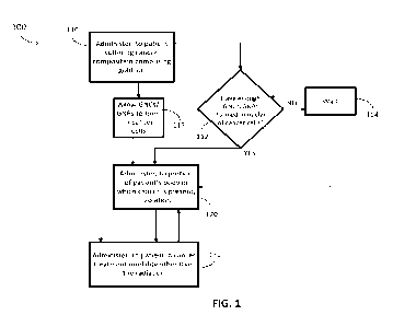
[0067] Fig. 1 presents a flowchart of a method 100 in accordance with
embodiments of the
present disclosure. The method 100 comprises administering 110, to a patient
suffering from a
cancer, a composition comprising a compound containing a gold atom; and
administering 120,
to a portion of the patient's body in which the cancer is present, radiation.
[0068] The patient may be any mammal suffering from the cancer. In one
embodiment, the
patient is a human being.
[0069] In embodiments, the present method may be performed in a veterinary
context. That is,
the patient may be any non-human mammal suffering from a cancer. The non-human
mammal
may be a research animal, a pet, livestock, a working animal, a racing animal
(e.g., a horse, a
dog, a camel, etc.), an animal at stud (e.g., a bull, a retired racing
stallion, etc.), or any other
non-human mammal for which it is desired to treat its cancer.
[0070] For convenience, the description will typically refer to human
patients. However, the
person of ordinary skill in the art having the benefit of the present
disclosure will readily be
able to adapt the teachings of the present disclosure to a veterinary context.
[0071] By "suffering from a cancer" is meant that the cancer is detectable in
the patient's body
using any diagnostic technique presently known or to be discovered.
"Suffering" does not
require the patient to be in pain from or have any naturally-perceptible
symptoms of the cancer.
Generally, as is known, the earlier a cancer can be treated, including before
the patient notices
pain or any other symptoms, the greater the chances of remission.
[0072] The present method may be used to treat any type of cancer. Desirably,
the cancer is
one that is known or reasonably expected, by the person of ordinary skill in
the art having the
benefit of the present disclosure, to be treatable by radiation after
radiosensitization by gold.
CA 03188659 2022-12-30
WO 2022/072095
PCT/US2021/048081
[0073] In one embodiment, the cancer is characterized by a desmoplastic
stroma. The stroma
is a biological structure containing one or more of connective tissue, blood
vessels, and
inflammatory cells in the cancer microenvironment. Desmoplastic stroma is
stroma that is
dense and fibrous. One comment characteristic of desmoplastic stroma is
limited delivery of
therapeutic molecules to tumor cells. In one embodiment, the desmoplastic
stroma may limit
diffusion of particles having a minimum dimension of 5 nm or greater to
malignant cells of the
cancer. By "limits diffusion" is meant that the rate of in vivo uptake of the
particles by the
malignant cells is reduced for the cells that are located further away from
the blood vessels or
injection site, i.e., the bigger the particle, the fewer particles reach
malignant cells.
Furthermore, the denser the stroma, the fewer particles diffuse inside the
tumor and the fewer
the particles delivered to malignant cells.
[0074] Not every presentation of the type of the cancer must feature a stroma
having this
diffusion-limiting parameter for the cancer to be "characterized by a
desmoplastic stroma."
[0075] By "minimum dimension" is meant the diameter, for spheres, or the
maximum width
of the smallest dimension, for oval or approximately rectangular or spherical
particles.
[0076] In one embodiment, the cancer is selected from the group consisting of
pancreatic
cancer, head-and-neck cancer, anaplastic thyroid cancer, brain cancer, liver
cancer, and breast
cancer. These cancers are well-recognized as being characterized by a dense
stroma However,
the method 100 may be performed on presentations of these cancers which are
not
characterized by a dense stroma.
[0077] In one particular embodiment, the cancer is pancreatic cancer.
[0078] In another embodiment, the cancer is head-and-neck cancer.
[0079] In yet another embodiment, the cancer is anaplastic thyroid cancer.
[0080] In an additional embodiment, the cancer is brain cancer.
[0081] In yet an additional embodiment, the cancer is liver cancer.
[0082] In an embodiment, the cancer is breast cancer.
[0083] The composition to be administered 110 comprises a compound containing
a gold atom.
By "compound containing a gold atom" is meant a compound containing gold in
any
oxidation/reduction state. The gold atom may be present as individual atoms,
soluble salts, or
as part of a molecule, polymer, or multiatom ion. The compound may contain one
or more
other atoms in any redox state that are one or more of covalently bound to a
gold atom, ionically
paired with a gold atom, or otherwise associated with a gold atom. In one
embodiment, the
compound may be an ionic compound containing an ion, typically an anion (a
negatively
charged ion) comprising gold in the Au' oxidation state, and a cationic
counterion (positively
11
CA 03188659 2022-12-30
WO 2022/072095
PCT/US2021/048081
charged ion), such as sodium, hydrogen, or another cation known for use in
pharmaceutical
salts and ionic compounds.
[0084] Use of the singular term "a compound" does not limit the composition to
comprising
only one compound containing a gold atom. The singular term "a gold atom" does
not limit the
compound(s) to comprising only one gold atom.
[0085] In one embodiment, the compound containing a gold atom is selected from
those
disclosed by C. Frank Shaw III, "Gold-Based Therapeutic Agents," Chem Rev
1999, hereby
incorporated herein by reference.
[0086] In one embodiment, the compound containing a gold atom is selected from
the group
consisting of
triethylphosphine(2,3,4,6-tetra-0-acety1-0-1-d-thiopyranosato-S)gold(I),
aurothioglucose salts, auranofin salts, aurothiomalate salts, chloroaurate
salts, buffered
chloroauric acid, (03PAu)2( DTE), (D3PAutTP, (D3PAu-thymidine, 03PAu(5-
fluorouridine),
03PAu(tegafur), ferrocene( -02PAuC1)2, Et3PAuCl, Et3PAuCN, Et3PAuCH3,
[(Et3P)2Au1C1,
Et3PAuSCN, Et3PAuSCH3, Et3PAuSG, Et3PAuSTg, Et3PAuSAtg (auranofin), Et3PAuS-a-
Atg
(epiauranofin), [AuSTm] [AuSTg] [AuSATgln, DPPE(AuC1)2, DPPE(AuSTg)2,
[Au(DPPE)21C1, [Au(R2P-Y-PR12)21X, Au(Streptonigrin),
[Me2AuC121[As041,
Me2Au(p.SCN)2AuMe2, Au(N-methylimidazole)C13, Au(2-methylbenzoxazole)C13,
Au(2,5-
dimethylbenzoxazole)C13, DPPE(AuC13)2,
[Au(damp)C12], [Au(damp)(SCN)21,
[Au(damp)(0Ac)21, [Au(damp)(malonate)1, iPr3PAuCN, Ph3PAuCN, Cy3PAuCN,
KAu(CN)2,
AuC14-' Au', Au', and mixtures thereof
[0087] In one embodiment, the compound containing a gold atom is selected from
the group
consisting of
triethylphosphine(2,3,4,6-tetra-0-acety1-0-1-d-thiopyranosato-S)gold(I),
aurothioglucose salts, auranofin salts, aurothiomalate salts, chloroaurate
salts, buffered
chloroauric acid, and mixtures thereof
[0088] In a particular embodiment, the compound containing a gold atom is
selected from
chloroaurate salts.
[0089] The concentration of the compound containing a gold atom may be varied
depending
on the route of administration, the presence or absence of other compounds in
the composition,
and other factors. The concentration may be selected as a routine matter by
the person of
ordinary skill in the art having the benefit of the present disclosure.
[0090] In one embodiment, the administering 110 the composition comprises
administering to
the patient an amount of gold from 0.0001 mg/g tumor cells to 10 mg/g tumor
cells. The mass
of tumor cells generally cannot be precisely weighed, but the person of
ordinary skill in the art
may generally
12
CA 03188659 2022-12-30
WO 2022/072095
PCT/US2021/048081
[0091] The composition may also comprise a solvent in which the compound
containing a gold
atom may be dissolved. Conveniently, the solvent may be water, although other
hydrophilic or
polar solvents that are pharmaceutically-acceptable may be used.
[0092] In one embodiment, the composition may further comprise one or more
other
pharmaceutically-acceptable compounds known for use in solution medicaments,
such as
buffers, preservatives, adjuvants, surfactants, diluents (e.g. saline or
dextrose) or the like. Such
particular other compounds may be routinely selected by the person of ordinary
skill in the art
having the benefit of the present disclosure.
[0093] Though not to be bound by theory, we have observed that compounds
containing a gold
atoms are generally preferentially taken up by cancer cells relative to normal
cells.
Accordingly, the composition generally lacks a need for targeting molecules or
moieties.
[0094] Though not to be by theory, we have observed that compound containing a
gold atoms,
after being taken up by cancer cells, tend to form gold nanoclusters (GNCs)
and/or gold
nanoparticles (GNPs) in situ. By "gold nanoclusters" is meant agglomerations
comprising gold.
By "gold nanoparticles" is meant gold nanoclusters that have a minimum
dimension of 1 nm
or greater. The gold nanoparticles and gold nanoclusters are not limited to
any particular shape
or structural motif Gold nanoparticles formed in situ may have a minimum
dimension of 5 nm
or greater, i.e., if pre-formed outside the cancer cell, would undergo limited
diffusion through
the stroma. Further, though again not to be bound by theory, we have observed
that in situ
GNC/GNP formation tends to occur in the cancer cell nucleus. From this, the
person of ordinary
skill in the art would expect that radiation dose enhancement arising from the
in situ
GNC/GNPs would inflict more damage on DNA and other structures in the cancer
cell nucleus
than in other structures of the cancer cell and would inflict more damage on
those other
structures of the cancer cell than on normal cells in the vicinity.
[0095] In one embodiment, the composition may comprise a micelle, liposome, a
mesoporous
silica particle, a polymersome, a polyethylene glycol (PEG) polymer cluster, a
tri-block
amphiphilic polymer, a di-block amphiphilic polymer, or two or more thereof In
a further
embodiment, the composition may additionally comprise a moiety which
preferentially
interacts with one or more tumor-related targets.
[0096] Alternatively, or in addition, the composition may comprise one or more
release
extension agents. For example, micelles, liposomes, mesoporous silica
particles,
polymersomes, PEG polymer clusters, and di- and tri-block amphiphilic
polymers, among
others, may allow extended release of gold atoms or ions. By the inclusion of
such agents, the
13
CA 03188659 2022-12-30
WO 2022/072095
PCT/US2021/048081
release from the composition of the compound containing a gold atom, or gold
atoms or ions
themselves, may proceed at a relatively steady rate for an extended period of
time.
[0097] The composition may be administered 110 to the patient by any route.
Such routes may
be characterized as systemic or local. Systemic routes include oral, nasal,
buccal, and
intravenous injection routes, among others. Local routes include subcutaneous,
intramuscular,
intraorganal, and intratumoral injection, and catheterized and endoscopic
routes, among others.
Generally, local routes in proximity to malignant cells of the cancer may be
desirable, in that
they are expected to require lower doses of the compound containing a gold
atom, may reduce
the risk of side effects, and may lead to more ready uptake of the compound
containing a gold
atom, or the gold atom itself, by the cancer cells.
[0098] In one embodiment, administering 110 the composition comprises
injection of the
composition in proximity to malignant cells of the cancer.
[0099] In the method 100, administering 110 the composition may be performed
in a single
dose or a plurality of doses. A plurality of doses may be desirable if the
total amount of gold
to be delivered would have toxic effects on healthy tissue if delivered in a
single dose. If a
plurality of doses is performed, the number of doses and the time between
doses can be selected
as a routine matter by the person of ordinary skill in the art having the
benefit of the present
disclosure.
[00100] The
method 100 also comprises administering 120, to a portion of the patient's
body in which the cancer is present, radiation.
[00101]
Radiation therapy is a well-known cancer therapy technique. Generally,
radiation comprising particles or photons that have sufficient energy or can
produce sufficient
energy via nuclear interactions is aimed at cancer cells to produce ionization
(i.e., loss of
electrons) in the cancer cells. This ionization generates reactive oxygen
species, which can
damage cellular structures directly, or may damage DNA, thereby disrupting
transcription and
translation and thereby disrupting cellular function. Exemplary ionizing
radiation types include
X-ray radiation and proton radiation. Apparatus and techniques for delivering
X-rays or protons
to a target tissue or cell are well known in the art.
[00102] The
amount of ionizing radiation needed in a given cell generally depends on
the nature of that cell. Means for determining an effective amount of
radiation are well known
in the art. For example, dosage ranges for X-rays range from daily doses of 50
to 200 cGy for
prolonged periods of time (3 to 8 weeks), to single or a small number (3-5)
doses of 500 to
2500 cGy. Common, but not limiting, X-ray treatment protocols involve five
doses, one each
on consecutive days or on alternating days.
14
CA 03188659 2022-12-30
WO 2022/072095
PCT/US2021/048081
[00103] In one
embodiment, the administering 120 the radiation comprises
administering X-rays or protons. In one particular embodiment, the
administering 120 the
radiation comprises administering X-rays. In another particular embodiment,
the administering
120 the radiation comprises administering protons.
[00104] After
administering 110 the composition, it may be desirable to allow time for
the gold atom or the compound containing a gold atom to penetrate the stroma,
be taken up by
the cancer cells, and form in situ GNC/GNPs. Accordingly, in one embodiment,
the method
100 further comprises allowing 115 gold nanoclusters (GNCs) and/or gold
nanoparticles
(GNPs) to form in the cancer cells. Because in situ GNC/GNP formation in
cancer cells,
especially pancreatic cancer cells, is spontaneous, no further action is
required. In one
embodiment, administering 120 the radiation is performed from 0 seconds to 14
days after
administering 110 the composition. In one embodiment, administering 120 the
radiation may
be performed from 30 minutes to 24 hours after administering 110 the
composition. In
embodiments wherein administering 110 the composition is performed in multiple
doses,
administering 120 the radiation is performed from 0 seconds to 14 days after
the final dose of
the composition. In particular embodiments, administering 120 the radiation
may be performed
from 30 minutes to 24 hours after administering 110 the final dose of the
composition.
[00105]
Generally, in situ formation of GNC/GNPs is expected after administering 110
the composition. However, depending on the cancer, the type of radiation, the
patient's
sensitivity to radiation, and/or other parameters, it may be desirable to
detect GNC/GNPs
formed in situ after administering 110 the composition. In one embodiment, the
method 100
may further comprise determining 112, after the administering the composition,
whether an
amount of GNC/GNPs, sufficient for radiation dose enhancement have formed in
the nuclei of
one or more malignant cells of the cancer. For example, determining 112 may
comprise
extracting malignant cells of the cancer from the patient's body and observing
GNC/GNP by
confocal fluorescence microscopy, flow cytometry, or other techniques that
will be known to
the person of ordinary skill in the art. Determining whether the amount of
GNC/GNPs is
sufficient for radiation dose enhancement will depend on one or more of the
total mass of gold
in the GNC/GNPs, the shape and structure of the GNC/GNPs, the proximity of the
GNC/GNPs
to the cancer cell nucleus, the type of cancer cell, or the nature and
intended dosage of the
radiation, among other parameters that will be apparent to the person of
ordinary skill in the
art having the benefit of the present disclosure.
[00106] If
determining 112 is performed, and the outcome is that an insufficient amount
of GNC/GNPs have formed, the method 100 flows to a wait 114. After the wait
114, flow may
CA 03188659 2022-12-30
WO 2022/072095
PCT/US2021/048081
return to determining at 112, or it may be presumed that enough GNC/GNPs have
formed, and
flow may pass to administering 120 the radiation.
[00107] The
method 100 may comprise additional events. In one embodiment, the
method 100 may further comprise administering 130, to the patient, a cancer
treatment
modality other than the radiation. Administering 130 the cancer treatment
modality other than
the radiation may be targeted against the same cancer as the radiation,
against metastases
thereof, against a primary tumor or metastases of a cancer other than cancer
targeted by the
radiation, or two or more thereof
[00108] A wide
variety of cancer treatment modalities other than radiation are known to
the person of ordinary skill in the art and need not be described in detail
here. By way of
example, in one embodiment, the cancer treatment modality other than the
radiation is selected
from the group consisting of surgical resection, chemotherapy, immunotherapy,
checkpoint
inhibitor therapy, oncolytic virus therapy, thermal therapy (e.g., RFA,
microwave ablation,
and/or cryotherapy), and two or more thereof
[00109]
Regardless of the particular cancer treatment modality other than radiation,
if
one or more is/are administered 130, the administering 130 may be performed
before, after, or
simultaneously with the administering 120 the radiation. Particular relative
and absolute timing
of administering 120 the radiation and administering 130 the other cancer
treatment modality
will be a routine matter for the person of ordinary skill in the art having
the benefit of the
present disclosure.
[00110] In one
embodiment, the present disclosure relates to a kit, comprising a
composition comprising a compound containing a gold atom; and instructions for
use of the
composition in a method comprising administering, to a patient suffering from
a cancer, the
composition; and administering, to a portion of the patient's body in which
the cancer is
present, radiation.
[00111] A "kit,"
as used herein, refers to a package containing the composition, and
instructions of any form that are provided in connection with the composition
in a manner such
that a clinical professional will clearly recognize that the instructions are
to be associated with
the composition.
[00112]
"Instructions" typically involve written text or graphics on or associated
with
packaging of compositions of the invention. Instructions also can include any
oral or electronic
instructions provided in any manner. Written text or graphics may include a
website URL or a
QR code encoding a website URL, where other instructions or supplemental
information may
be provided in electronic form.
16
CA 03188659 2022-12-30
WO 2022/072095
PCT/US2021/048081
[00113] The kit
may contain one or more containers, which can contain the composition
or a component thereof The kits also may contain instructions for mixing,
diluting, or
administering the composition. The kits also can include other containers with
one or more
solvents, surfactants, preservatives, and/or diluents (e.g., normal saline
(0.9% NaCl), or 5%
dextrose) as well as containers for mixing, diluting, or administering the
composition to the
patient in need of such treatment.
[00114] The
composition may be provided in any suitable form, for example, as a liquid
solution or as a dried material. When the composition provided is a dry
material, the material
may be reconstituted by the addition of solvent, which may also be provided by
the kit. In
embodiments where liquid forms of the composition are used, the liquid form
may be
concentrated or ready to use.
[00115] The kit,
in one embodiment, may comprise a carrier being compartmentalized
to receive in close confinement one or more containers such as vials, tubes,
and the like
[00116] The
composition is described above. In one embodiment, the compound
containing a gold atom is selected from the group consisting of
triethylphosphine(2,3,4,6-tetra-
0-acety1-0-1-d-thiopyranosato-S)gold(I), aurothioglucose salts,
auranofin salts,
aurothiomalate salts, chloroaurate salts, buffered chloroauric acid, and
mixtures thereof
[00117] The
method is described above. In one embodiment, the instructions comprise
instructions to administer the composition by injection of the composition in
proximity to
malignant cells of the cancer. Alternatively, or in addition, in one
embodiment, the instructions
comprise instructions to administer the radiation by administering X-rays or
protons. Again,
alternatively or in addition, in one embodiment, the instructions further
comprise instructions
to administer, to the patient, a cancer treatment modality other than the
radiation.
[00118] The
following examples are included to demonstrate preferred embodiments of
the invention. It should be appreciated by those of skill in the art that the
techniques disclosed
in the examples which follow represent techniques discovered by the inventor
to function well
in the practice of the invention, and thus can be considered to constitute
preferred modes for
its practice. However, those of skill in the art should, in light of the
present disclosure,
appreciate that many changes can be made in the specific embodiments which are
disclosed
and still obtain a like or similar result without departing from the spirit
and scope of the
invention.
17
CA 03188659 2022-12-30
WO 2022/072095
PCT/US2021/048081
Example 1
Specific Aims
[00119]
Radiation therapy (RT) is an integral component of modern therapy for locally
advanced unresectable pancreatic cancers. However, its ultimate utility is
severely limited by
the fact that some cancer cells are resistant to RT. Delivering higher doses
of RT to the gross
tumor to overcome radiation resistance has historically been challenging due
to the limited
radiation tolerance of the surrounding organs. Sequestering gold nanoparticles
(GNPs) within
tumors to amplify radiation-induced secondary electron showers has gained
traction in recent
years as a means to escalate radiation dose in the immediate vicinity of the
nanoparticle thus
confining the higher dose to the tumor and sparing surrounding tissues.
However, pancreatic
cancer is characterized by hypovascularity in the setting of a dense stromal
component with an
exuberant interstitial matrix of glycosaminoglycans, collagen, and proteogly
cans
(desmoplasia) that serves as a physiological barrier to the delivery of drugs
and nanoparticlesl-
3. The consequent hostile microenvironment (low pH, low p02) of the tumor core
harbors the
most aggressive tumor cells with the greatest potential to regenerate if they
survive cytotoxic
treatment.4 This problem is further amplified by the presence of
gastrointestinal mucosa
immediately adjacent to the tumor that makes dose escalation difficult and
often not readily
achievable.
[00120] Here we
propose to overcome problems with specific radiosensitization of
pancreatic cancer cells in the context of a dense stromal environment by
intratumoral delivery
of an aqueous solution of the compound containing gold atoms (i.e., buffered
chloroauric acid)
instead of gold nanoparticles (GNP) thus achieving the ultimate reduction in
size of a
therapeutic agent ¨ an atomic scale. Our hypothesis is that small compounds
containing a gold
atoms (i) will uniformly distribute throughout the tumor as their diffusion is
not likely to be
impeded by the stroma, and (ii) will be reduced to GNPs after specific uptake
by cancer cells
that (iii) will result in cancer cell radiosensitization to RT. This
hypothesis is based on our
compelling preliminary data demonstrating efficient synthesis of GNPs inside
pancreatic
cancer cells with a high nuclear localization that is critical for efficient
radiosensitization due
to a higher dose delivery to nuclei by the secondary Auger electrons.
Furthermore, normal
pancreatic cells did not significantly produce GNPs. In addition, recent
literature reports
demonstrated intracellular synthesis of GNPs from chloroauric acid' occurs
with higher
efficiency in cancerous versus non-cancerous cells6,8,9,11,12 with a
preferential nuclear
localization of the nanoparticles.' These studies further support our
hypothesis of cancer
specific intracellular synthesis of GNPs. Changing the current delivery
paradigm from pre-
18
CA 03188659 2022-12-30
WO 2022/072095
PCT/US2021/048081
made GNPs with sizes of 5-200 nm to delivery of ¨0.3 nm compounds containing
gold atoms
is associated with a staggering ¨16 to 1,400 size reduction of a gold
therapeutic agent that is of
paramount importance in penetrating desmoplastic tumors. Indeed, soluble
compounds
containing gold atoms are on the same size scale with similar transport
kinetics as physiological
salts (e.g., Ca2+, Na+, K+) which can diffuse even inside dense biological
environments.
Moreover, compounds containing gold atoms have decades-long history of a safe
clinical use
in treatment of rheumatoid arthritis15 providing a clear path towards clinical
translation.
[00121] We
envision clinical implementation of our approach as an added boost to
significantly increase efficacy of stereotactic body radiotherapy (SBRT) in
patients with a
pancreatic tumor. Recent clinical data from our group and others shows that
radiation dose
enhancement increases local control and overall survival of locally advanced
pancreatic cancer
patients16. However, the proximity of gastrointestinal mucosa to the tumor in
many instances
precludes this dose escalation in clinical practice. But, when high atomic
number (gold,
hafnium) nanoparticles are present within tumors, irradiation of the tumor
results in radiation
dose enhancement via an increase in the fluence of photo-/Auger electrons
ejected from
gold/hafnium. We expect that changing the current paradigm from delivery of
pre-made GNPs
to in situ synthesis of GNPs by cancer cells will overcame delivery barriers
in pancreatic tumors
and, thus, will result in a highly significant improvement of RT outcomes.
Here, we will test
our hypothesis via two Specific Aims.
[00122] Aim 1.
Optimization and characterization of intracellular synthesis of GNPs by
pancreatic cancer cells.
[00123] 1.1.
Optimize the dose of the compound containing gold atoms and the
timeframe for intracellular synthesis of GNPs. Compare efficiency of the GNP
synthesis by
normal and cancer cells.
[00124] 1.2.
Determine intracellular distribution of GNPs as a function of time. These
studies will provide insight into mechanisms of intracellular synthesis and of
intranuclear
accumulation of GNPs.
[00125] Aim 2:
Evaluate radiosensitization efficacy of in situ synthetized GNPs in
models of pancreatic cancer.
[00126] 2.1.
Compare RT of cancer and normal cells after treatment with compounds
containing gold atoms in vitro.
[00127] 2.2
Determine toxicity of administration of compounds containing gold atoms
in a murine model.
19
CA 03188659 2022-12-30
WO 2022/072095
PCT/US2021/048081
[00128] 2.3.
Determine in vivo biodistribution and cellular internalization of GNPs after
intratumoral delivery of compounds containing gold atoms.
[00129] 2.4.
Determine radiosensitization efficacy and tumor distribution of in situ
synthetized GNPs in an orthotopic human pancreatic patient derived xenograft
murine tumor
model.
[00130] These
studies will provide the framework for continued development of a
readily deployable radiosensitization strategy for pancreatic cancer. This
strategy is inherently
simplistic, with a single active component - gold atoms, but it takes
advantage of a complex
cell biology in order to produce therapeutic GNPs that localize to the
nucleus.
Innovation
[00131] The key
innovation of our approach is (1) a paradigm shift from delivery of pre-
made GNPs to an atomic size gold precursor for tumor radiosensitization; this
represents the
ultimate size reduction of a therapeutic agent outside of the radiation
therapy itself (i.e., x-rays,
protons, etc.). Our strategy is inherently simplistic in design, as it employs
a single, readily-
procurable component (e.g., chloroauric acid) (Fig. 2A-2C). However, it also
relies on a
complex cell biology that is behind in situ synthesis of GNPs which is still
poorly understood.
We appreciate that the specificity of RT with our approach is dependent on
differences in
synthesis efficiency and in intracellular localization of GNPs in normal and
cancer cells.
Therefore, the second innovative aspect of our project is (2) studies of
intracellular formation
of GNPs in different cell types with an emphasis on gaining further
understanding of cellular
uptake, intracellular reduction, and trafficking of gold atoms by cancerous
and normal cells.
This understanding will provide foundation for future clinical applications of
our strategy
wherein radiosensitization agents are generated within the pathological tissue
in a phenotype
dependent manner as a cell-level personalized therapy. (3) Here this new
concept will be
validated in the specific context of PDAC. A dense desmoplasia is a signature
of pancreatic
cancer forming a formidable therapy delivery challenge that we plan to
overcome with the
ultimate size reduction of radiosensitizing precursors to an atomic level.
Taken together these
innovations will provide a clinically translatable solution to three key
challenges in delivery of
radiosensitization agents to PDAC: (i) tumor penetration, (ii) cancer specific
cellular uptake
and (iii) nuclear localization for efficient tumor radiosensitization that
requires greatly reduced
gold amount and more clinically relevant radiation (megavoltage radiation)
than prior
approaches to radiosensitization with GNPs.
CA 03188659 2022-12-30
WO 2022/072095
PCT/US2021/048081
Preliminary Data
Feasibility of radiosensitization via intracellular GNP formation
[00132] Our
initial evaluation of radiosensitization via intracellular GNP formation was
performed with 3T3 mouse fibroblast cells. 3T3 cells were chosen because they
were
previously characterized for intracellular GNP synthesis," and fibroblasts are
considered "bad
players" and potential therapeutic targets in the pancreatic cancer
microenvironmental
niche.44'45 GNPs with sizes below 2nm ¨ gold nanoclusters (GNCs) - are known
to exhibit a
bright fluorescence in the visible region." Therefore, their intracellular
formation was verified
via confocal fluorescence imaging with 561 nm excitation and 610 nm emission
after cell
treatment with 1 mM Au3+ (i.e., chloroauric acid) in cell culture media for 24
hours (Fig. 3A-
3B). The images in Fig. 3A show uniform formation of GNCs in NIH3T3 cells.
Radiosensitization with intracellular GNC formation (i.e., delivery of Au')
was compared with
pre-fabricated albumin-coated GNCs (Albumin- GNC) prepared according to work
by me46
and control cells without treatment via MTS assay (Fig. 3B). Albumin-GNCs were
chosen for
comparison to see if a simple combination of extracellular gold nanoclusters
with the most
abundant serum protein (i.e., albumin) would produce a comparable
radiosensitization to the
intracellular synthetized GNCs.
[00133] 3T3
cells were first incubated with either 0.1 mM of sodium chloroaurate or
albumin-GNCs (0.1 mM Au ) in cell culture media for 10 hours. Then, the cells
including the
nontreated control were irradiated with X-rays at dosages of 0, 4 and 6 Gy in
the X-ray X-RAD
225 CX irradiator system (Precision). The MTS assay showed a significant
increase in
radiosensitization by in situ synthetized GNCs as compared to Albumin-GNC
control at both
the 4 and 6 Gy doses (Fig. 3B).
[00134] In our
future studies we will use a standard clonogenic survival assay for
quantitation of the radiosensitization effect. Note that no difference in cell
viability was
observed between Au' treated and untreated cells at 0 Gy indicating that the
incubation with
chloroauric acid is not cytotoxic.
[00135] In situ
synthesis of GNCs is greatly enhanced in pancreatic cancer cells as
compared to non-cancerous cells
[00136] We
compared in situ synthesis of GNCs by pancreatic cancer cells
(MIAPACA2) and pancreatic noncancerous cells (HPDE) (Fig. 4). Confocal
fluorescence
images were obtained using a Leica TCS SP8 confocal microscope with 561 nm
excitation and
610 nm emission optimized for detection of GNCs. Live cell nuclear stain
(Hoechst 33342 was
used to define location of nuclei. The fluorescence images reveal a striking
increase in GNC
21
CA 03188659 2022-12-30
WO 2022/072095
PCT/US2021/048081
formation in cancerous cells as compared to noncancerous cells after
incubation with sodium
chloroaurate (Au') (Fig. 4). Virtually no fluorescence was observed in either
cell type
incubated with pre-made albumin-GNCs indicating limited uptake of the
extracellularly
formed nanoclusters. In cancer cells (MIAPACA2), the fluorescence from GNCs
was
uniformly distributed throughout the intracellular space with a significant
fraction in nuclei as
revealed by optical sectioning (Fig. 5). Almost no background fluorescence was
observed in
the extracellular space (Fig. 4), indicating that GNCs are not synthetized in
the extracellular
environment.
[00137] In Situ
Synthesis of GNCs is more prevalent in cancer cells with greater
radiosensitization as compared to non-cancerous cells
[00138] Our
initial evaluation of intracellular gold nanocluster formation was performed
with pancreatic cancer cells (LTPA) and pancreatic non-cancerous cells (MS1)
(Fig. 6A-6B).
Intracellularly formed GNCs are known to exhibit a bright fluorescence in the
visible region."
Therefore, their intracellular formation was verified via confocal
fluorescence imaging using
Leica TCS SP8 confocal microscope with 561 nm excitation and 610 nm emission
optimized
for detection of GNCs. Live cell nuclear stain, Hoechst 33342, was used to
define the location
of nuclei.
[00139] The
fluorescence images revealed a striking increase in GNC formation in
cancerous cells as compared to non-cancerous cells after incubation with
buffered chloroauric
acid (Au') (Fig. 6A). Radiosensitization due to intracellular GNC formation
(i.e., delivery of
Au') was compared between cancerous and non-cancerous cells using a standard
clonal assay.
For radiosensitization, cells were incubated with 0.1 mM of buffered
chloroauric acid in cell
culture media for 24 hours. Then, the cells were lifted and plated in 30mm
culture dishes at
optimized cell densities for observation of colony formation following
irradiation with X-rays
at dosages ranging from 0 to 8 Gy (XRAD SmART). The clonogenic assay showed a
significantly greater radiosensitization effect from Au3+ treatment in cancer
cells as compared
to untreated control (Fig. 6B). Importantly, there was no significant
radiosensitization in non-
cancerous cells. The radiosensitization results correlated well with the
fluorescence images
showing greater production of GNCs by cancerous cells (Fig. 6A-6B). For LTPA
pancreatic
cancer cells the surviving fraction values decreased by a factor of 2.3x and
3.5x for radiation
of 4 Gy and 6 Gy, respectively, in cells treated with gold atoms compared to
untreated control
(Fig. 6B). This relative decrease in surviving fraction is similar or better
than previously
reported values observed in cells treated with pre-synthetized GNPs. We
believe that the
22
CA 03188659 2022-12-30
WO 2022/072095
PCT/US2021/048081
observed improvement in radiation efficiency in killing cancer cells might be
associated with
a strong nuclear localization of in situ synthetized GNCs.
Summary of preliminary data
[00140] Taken
together our preliminary data demonstrate that (i) intracellularly
synthetized GNCs can produce a radiosensitization effect; (ii) in situ
formation of GNCs is
significantly greater in cancer pancreatic cells as compared to non-cancerous
pancreatic cells;
and (iii) there is substantial localization of the GNCs inside cell nuclei.
These results provide
a foundation for development of a novel paradigm-shifting radiosensitization
strategy for
clinically translatable RT of pancreatic tumors. Here we evaluate and optimize
this strategy
using a rigorous research plan that culminates with validation studies in
clinically relevant
models of pancreatic cancer.
Future work
[00141] Aim 1.
Optimization and characterization of intracellular synthesis of GNCs
and GNPs by cancer cells. 1.1. Optimize the dose of gold atoms and the
timeframe for
intracellular synthesis of GNCs. These studies will be carried out in a panel
of human cancer
pancreatic cell lines from more radiation resistant PANC-1 and BxPC3 cells to
more sensitive
HPAC, MIAPaCa-2 and AsPC-1 cells as well as patient derived pancreatic cancer
cells. Non-
cancer pancreatic cell line (HPDE) will be used as a normal control. In
addition, we will
evaluate in situ synthesis of GNCs in cells that are associated with a tumor
microenvironment
- murine (J774A.1, ATCC) and human (MV-4-11, ATCC) macrophages; murine (3T3,
ATCC)
and human (HUF, ATCC) fibroblasts.
[00142] In a
typical experiment cells growing in sterile optical microplates will be
treated with buffered chloroauric acid (HAuC14, Sigma-Aldrich) at
concentrations ranging
from 0.01mM ¨ 10 mM for time periods up to 48 hours under standard cell
incubation
parameters (-95% humidity, 5% CO2, 37 C, normal pH) in the cell media with 0,
5, 10, 15
and 20% FBS; note that phenol red-free media will be use as this indicator dye
can interfere
with optical measurements. All samples will be prepared at least in
triplicate. Evaluation of
GNC and GNP formation will be carried out every two hours with a BioTek
Cytation 5 plate
reader using fluorescence (561 nm excitation/610 nm emission) and UV-Vis
absorbance
acquired from the whole sample (i.e., cells+media) and the cells and the media
alone; the
samples will be staggered to allow long breaks between measurements. After
media
replacement, cell viability will be determined by an MTS assay; note the
initial UV-Vis
measurements from the cells alone will be used to correct for background
absorbance at 490
nm. Then, cells and media from all samples will be analyzed for the total gold
content by
23
CA 03188659 2022-12-30
WO 2022/072095
PCT/US2021/048081
Inductively Coupled Plasma Mass Spectrometer (ICP-MS, Agilent). In a separate
set of
experiments longitudinal fluorescence and UV-Vis measurements will be carried
out with non-
cytotoxic doses of Au' (from the previous study) every hour up to the first 8
hours and then,
at 20h, 24h, 28h, 40h, 44h and 48h. Untreated cells will be used as controls
and treatments with
equimolar gold concentrations of albumin-GNCs and citrate-reduced 5 nm and
PEGylated 5
nm spherical GNPs for comparison.
[00143] In these
studies, fluorescence and UV-Vis data will provide kinetics of
GNC/GNP bio-synthesis and changes in their concentration over time inside
cells and in the
surrounding media. These two methods are complimentary because fluorescence is
sensitive
to formation of very small GNCs and its intensity diminishes with transition
to GNPs where
UV-Vis has much better sensitivity due to a pronounced absorbance associated
with plasmon
resonances of the particles. ICP-MS quantifies the total gold content
regardless of its physical
state that will determine kinetics of gold uptake by various cells. These
experiments will
determine the optimum conditions (i.e., dose and time) for formation of
intracellular GNCs and
GNPs without cytotoxicity to normal cells. They will also identify parameters
that provide the
highest difference in formation of GNCs/GNPs between cancerous and normal
cells. Final
characterization of GNCs formation will be carried out by Flow Cytometry that
will determine
heterogeneity of GNC biosynthesis in different cell populations and will
further quantify
differences between normal and cancerous cells.
[00144] In
addition, we will carry out initial evaluation of a potential role of cell-
excreted vesicles, peptides, and nucleic acids in biosynthesis of GNCs. Cells
will be grown for
various periods of time (i.e., 12, 24 and 48 hours); care will be taken to
make sure that the cells
do not grow beyond confluence by adjusting the number of seeded cells. At each
time point
the optimum dose of Au' (from the studies above) will be applied to the cells
and biosynthesis
of GNCs/GNPs will be monitored using the methodology described above. Note
that the cell
media will not be replaced to preserve all biological substances released by
the cells during
growth. These experiments will determine if the cell conditioned media results
in an
extracellular formation of GNCs/GNPs and/or influences gold uptake by cells.
Cells washed
with a fresh media before addition of gold atoms will be used as controls.
[00145] 1.2.
Intracellular Distribution of GNCs as a Function of Time will be determine
using confocal fluorescence (Leica TCS 5P8 Confocal Microscope). In addition,
samples at
various time points that are associated with changes in fluorescence intensity
and/or
intracellular distribution of fluorescent GNCs will be analyzed by
transmittance electron
microscopy (TEM, JEM 1010, JEOL). The major goal of these studies is to
understand
24
CA 03188659 2022-12-30
WO 2022/072095
PCT/US2021/048081
spatiotemporal progression of GNC biosynthesis by cells. This knowledge will
ultimately
allow optimization of timing and parameters of RT with intracellularly
synthetized GNCs. In
a highly synergistic to this proposal study we are collaborating with Dr. S.
H. Cho in the
Department of Radiation Physics at the M.D. Anderson Cancer Center on
development and
validation of Monte Carlo computational modeling of gold-mediated
radiosensitization that
can predict radiation dose enhancement based on distribution of
GNCs/GNPs.16'35'36 Further, a
number of reported studies proposed various mechanisms of in situ biosynthesis
of
GNCs/GNPs, including the potential role of various cellular compartments that
are rich in
biomolecules with sufficient reducing potential for gold atoms reduction
including (i) the
cytoplasmic cell membrane that contains reducing enzymes and glycosylated
moieties;6 (ii)
reactive oxygen species (ROS), glutathione (GSH) and glutathione disulfide
(GSH-GSSG),
nicotinamide adenosine dinucleotide phosphate hydrogenase enzyme (NAD(P)H) and
QOH-1
enzymes in the cytoplasm;8'14 (iii) and nucleotides in the nucleus.47 Our
studies can provide an
insight into which compartments and in what sequence are involved in synthesis
and trafficking
of GNCs.
[00146] In a
typical experiment, cells will be grown in a live cell imaging chamber under
normal conditions (-95% humidity, 5% CO2, 37 C, and normal pH) in a cell
culture media.
Confocal fluorescence images will be collected before and in 20 minute
intervals after
administration of gold atoms up to a 24 hour period. Initial cell localization
will be determined
using bright-field imaging. Cellular nuclei will be stained with the Hoechst
stain (live cells
nuclear stain). Cytoplasmic cell membranes will be labeled with Di0 membrane
tracer (484
nm excitation/501 nm emission, ThermoFisher) that does not overlap with
fluorescence of
GNCs based on our preliminary data; other lipophilic carbocyanine tracers can
be explored if
needed, e.g., DiR (750 nm exc./780 nm em.).
[00147] After
this longitudinal study, in a separate set of experiments we will collect
samples for TEM analyses to determine cellular distribution of GNCs in the
context of cellular
compartments and organelles with higher resolution. In addition, the total
amount of gold in
the cytoplasm (plus cytoplasmic membrane) and the nucleus will be determined
using ICP-
MS. To this end we will separate nuclei using mechanical lysis (Tip
sonication, QSonica)
followed by differential centrifugation (LK-90 Ultracentrifuge) as described
in detail by Lodish
and coworkers". Image analysis will be done in ImageJ or IMARIS (Bitplane).
Cell and
nucleus boundaries will be segmented using fluorescence of cytoplasmic
membrane labeled
with Di0 and Hoechst stain, respectively. Then intensity of GNC fluorescence
inside and
outside cells as well as in cytoplasm versus nuclei will be quantified.
Statistical analyses.
CA 03188659 2022-12-30
WO 2022/072095
PCT/US2021/048081
Student's t test will be used to compare Gaussian-distributed data, whereas
the nonparametric
Mann-Whitney test (two-group comparison) will be used to analyze non-normally
distributed
data. P values of less than 0.05 will be considered statistically significant.
[00148] Expected
outcomes: Significantly higher efficiency of GNC biosynthesis by
pancreatic cancer cells relative to pancreatic normal cells. Highly efficient
uptake of gold atoms
from the surrounding media by cancer cells with substantial accumulation of
GNCs inside cell
nuclei. Optimized dose of gold atoms that results in efficient synthesis of
GNCs by cancer cells
with minimum cytotoxicity to normal pancreatic cells.
[00149] Possible
obstacles: If we discover a high fraction of GNP formation inside cells,
two-photon luminescence can be used for their detection, since GNPs do not
exhibit
sufficiently high one-photon fluorescence cross section as opposed to GNCs.
Multiple dosing
of gold atoms can be implemented if a single dose exhibits substantial
cytotoxicity towards
normal cells.
[00150] Aim 2:
Evaluate radiosensitization efficacy of in situ synthetized GNCs in
models of pancreatic cancer.
[00151] 2.1.
Compare RT of cancer and normal cells after treatment with gold atoms in
vitro. A panel of normal and pancreatic human cells described above will be
treated under
optimum conditions (from Aim 1) with gold atoms. Then the ability of
biosynthesized
GNCs/GNPs will be evaluated in a clonogenic survival assay after irradiation
with 0, 2, 4, 6, 8
or 10 Gy of radiation. The cell survival will be monitored during the standard
period of ¨10-
16 days.' The data will be fit to the linear-quadratic model of cellular
response to radiation
and parameters such as surviving fraction at 2 Gy (SF2), a/0 ratio, and Do
will be used to
compare treatment efficiencies. These data will be compared with fluorescence
microscopy,
TEM and ICP-MS results (Aim 1) to correlate treatment response with cellular
uptake,
internalization, and intracellular distribution. Treatments with equimolar
gold concentrations
of albumin-GNCs and 5 nm spherical GNPs will be used for comparison.
[00152]
Evaluation of mechanism of radiosensitization in vitro. Gamma H2AX foci
(markers of unrepaired DNA strand breaks) will be quantified at multiple time
points following
radiation in the absence and presence of gold atom treatment. Activation of
DNA repair
pathways (ATM, ATR, Ku70, Ku80, DNAPKcs, chkl and chk2 Western blot analyses),
mitochondrial oxidative stress pathways (membrane potential JC-1 assay and
NADP/NADPH
ratio), and cell membrane lipid peroxidation (TBARS assay) will be evaluated
in treated cells.
Blocking studies with N-acetyl cysteine will determine whether oxidative
stress induced effects
are reversible.
26
CA 03188659 2022-12-30
WO 2022/072095
PCT/US2021/048081
[00153] The JC-1
assay measures the charge potential of the mitochondria of cells
through the fluorometric ratio of the JC-1 dye (ThermoFisher Scientific,
Waltham, MA). JC-1
is a cationic carbocyanine dye that accumulates in mitochondria. The dye
exists as a monomer
at low concentrations and yields green fluorescence, similar to fluorescein.
At higher
concentrations, the dye forms J-aggregates that exhibit a broad excitation
spectrum and an
emission maximum at ¨590 nm. These characteristics make JC-1 a sensitive
marker for
mitochondrial membrane potential. In other words, mitochondrial depolarization
is indicated
by a decrease in the red/green fluorescence intensity ratio. Mitochondrial
depolarization is an
indicator of reduced cell viability.
[00154] Expected
outcomes: Although not comprehensive, these studies will identify
the magnitude of and mechanisms of radiosensitization of cancer cells by
GNCs/GNPs
generated intracellularly via applications of ionic gold.
[00155] Possible
obstacles: While the emphasis of the mechanistic studies is on DNA
damage, the parallel investigation of mitochondrial and cell membrane
signaling alterations
after radiation will allow identification of non-DNA adaptive responses of
cells to radiation.
[00156] 2.2.
Toxicity study will be performed in C57BL6 mice without tumors. Eight
mice per group (4 male and 4 female) will be evaluated for toxicity of 2
administration routes
(i.v. and i.p.) at 3 dose levels and at 2 time points (1 week and 4 weeks).
Toxicity assessment
will include mouse weight, biochemistry panel (renal function, liver function
tests, and
electrolytes), hematology panel and histopathological evaluation of normal
organs (liver,
spleen, heart, lung, pancreas, and kidney) as described previously by us50
.
[00157] 2.3. In
vivo biodistribution, cellular internalization, and subcellular trafficking
will be determined in murine models of pancreatic cancer. Ionic gold will be
injected at 3 doses
into pancreatic tumors under ultrasound (US) guidance. If US is unable to
provide sufficient
contrast, the mouse abdomen will be opened under anesthesia and the treatment
will be made
under visual guidance. Eight animals (4 male and 4 female) will be sacrificed
at 3 time points
based on in vitro cell studies (Aim 1); in addition, we will monitor formation
of GNCs using
IVIS Spectrum system (Caliper). Distribution of in situ synthetized GNCs/GNPs
in the
pancreas, including uptake by cancer cells, will be determined in slices of
excised tumors using
a combination of confocal fluorescent microscopy, MALDI (Waters Synapt G2-Si),
and
histology with silver stain. These experiments will be used to fine tune the
dose of gold atoms
and timing of RT for in vivo radiosensitization study.
[00158] 2.4.
Evaluation of radiosensitization in vivo. A tumor regrowth delay
experiment will be used to determine radiosensitization in vivo. An orthotopic
human
27
CA 03188659 2022-12-30
WO 2022/072095
PCT/US2021/048081
pancreatic tumor model will be used in these experiments. This model closely
reproduces the
complex biology of pancreatic human cancer.51'52 Once tumors are a few mm in
diameter (as
determined by US imaging), they will be given intratumoral injections of gold
atoms in saline
(at the optimum dose from Aims 2.1 and 2.3). A pre-made nanomaterial (Albumin-
GNC or 5
nm spherical GNPs) with the best radiosensitization (Aim 2.1) will be used for
comparison.
Radiotherapy will be administered after a time delay determined in the Aim 2.3
and confirmed
by IVIS fluorescence, to allow diffusion of gold atoms throughout the tumor,
intracellular
nanoparticle reduction, and nuclear localization. A customized collimator will
be used to
administer a dose of 10 Gy using a small animal irradiator (XRAD255). Tumor
size by US and
mouse weight will be measured three times a week and mice will be euthanized
when they
experience a 20% weight loss from baseline. Tumor volume measurements (based
of US) will
be used to determine the time to tumor volume doubling in each treatment group
(control,
radiation, GNPs (or albumin- GNC), ionic gold, GNPs + radiation, and ionic
gold + radiation).
[00159]
Statistical analyses. The primary comparison will be between (i) radiation
alone
and (ii) radiation + ionic gold. We will use t test or Mann-Whitney test for
two-group
comparisons and ANOVA or Krustal-Wallis test for multiple-group comparisons.
For the
repeated measures (e.g., tumor size), we will use the linear mixed model.
Subgroup analysis
will be conducted for male and female mice. The sample size chosen for this
experiment is
based on estimates of a mean delay time of ¨7 days [standard deviation (SD) of
¨3 days] for
the control (radiation alone) group of tumors to double in volume. To detect a
mean difference
of 10 days for the test group [90% power, two-sided a of 5%1, we will need 8
animals per group
assuming similar SDs in the test group. If it turns out that the SD in the
control group is
considerably different from that in the test group (say 3 days vs. 6 days),
then we would need
animals per group. We will use equal numbers of male and female animals.
[00160] Expected
outcomes: These studies will evaluate the radiosensitization of
pancreatic tumors by intracellular GNC/GNP formation in vivo.
[00161] Possible
obstacles: Injection of ionic gold can also be monitored through
photoacoustic imaging that can provide higher needle and tumor contrast. In
case US is not
sensitive enough to monitor tumor growth, we will switch to a 7T MRI (Bruker).
Conclusion
[00162] Our
compelling preliminary data in pancreatic cells provide the scientific
premise of our strategy wherein gold atoms are used for radiosensitization of
PDAC that is
based on the following observations: (i) gold atoms have diffusion kinetics
similar to other
soluble salts and thus can penetrate throughout the tumor more readily than
molecules or
28
CA 03188659 2022-12-30
WO 2022/072095
PCT/US2021/048081
nanoparticles;37' (ii) intracellular GNP formation preferentially occurs via
interactions with
cancerous cells;6,8,9,11,12 (iii) biosynthesized GNPs innately localize within
the nucleus;5-7 and
(iv) GNPs have a high radiosensitization efficiency if located within the
nucleus of target
cells.28'' We see it as a highly innovative and exciting opportunity to
greatly improve
radiosensitization efficiency of cancer cells in situ.
Example 2
[00163] We followed up on the experiments described in Example 1, as
follows:
[00164] 2.1. Intracellular distribution and time dependence of gold
nanocluster (GNC)
in situ formation. We used a combination of TEM (Fig. 9B) and confocal
fluorescence
microscopy (Fig. 7) to demonstrate a high level of intranuclear localization
of intracellularly
formed GNCs. Then, we used longitudinal live cell confocal fluorescence
imaging to observe
time dependence of the intracellular distribution of GNCs formed through
biomineralization of
Au3+. We found that biosynthesis of GNCs occurred simultaneously throughout
the cells,
however, at different rates in different subcellular regions. The nucleolus
had the highest rate
of GNC formation (Fig. 18). This observation indicates that GNC biosynthesis
occurred
without additional intracellular transport and that Au' ions were able to
permeate throughout
the cell.
[00165] 2.2. Comparisons of efficiency of the GNC in situ synthesis by
cancer and
normal cells. We compared GNC biosynthesis between normal (HPDE) and cancerous
(Mia-
PaCa-2 and PANC1) pancreatic cells using fluorescent confocal microscopy (Fig.
8A). In
addition, we compared the efficiency of the intracellular synthesis with
extracellular uptake of
prefabricated albumin coated GNCs (Fig. 8A). We showed that cancerous
pancreatic cells
exhibited ¨2-fold greater fluorescence due to GNC biomineralization than non-
cancerous
HPDE (Fig. 8B). Further, there was 12.20- and 7.69-times greater fluorescence
due to in situ
synthesis relative to cellular uptake of prefabricated GNCs in PANC1 and Mia-
PaCa-2 cells,
respectively (Fig. 8B).
[00166] 2.3. Optimization of conditions for intracellular synthesis of
GNCs. We
determined optimal conditions for intracellular synthesis of fluorescent GNCs
by PANC1 cells
through variations in (1) how long cells condition media before addition of
Au' ions (Fig. 11),
(2) the concentration of FBS within the media (Fig. 10), (3) concentration of
added Au' ions
(Fig. 13), and (4) incubation time (Fig. 12). We used the following standard
conditions: 24 hr
cell conditioning time, 10% FBS, 1.00 mM Au', and 24 hr Au3+ treatment
duration. In the
optimization studies one of these parameters was varied at a time as shown in
Fig. 10-Fig. 13.
No significant changes were observed due to variations either in duration of
media conditioning
29
CA 03188659 2022-12-30
WO 2022/072095
PCT/US2021/048081
by cells up to 72-hr or changes in FBS concentration between 0-10% (v/v) in
the media. We
observed significant formation of GNCs at Au" concentrations between 0.20-0.75
mM, with
fluorescence reduced at concentrations greater than 1.00 mM, possibly, due to
formation of
larger non-fluorescent gold nanoparticles (GNPs). We observed fluorescence as
early as 30
minutes after addition of gold atoms with a significant increase up to 4
hours.
[00167] 2.4.
Characterize cytotoxicity of gold atom treatment. An MTS viability assay
was used with 0 - 2.0 mM Au" dose range. There were no impacts on viability to
PANC1 cells
for concentrations 0.0-0.20 mM Au", with a decrease in cell viability to ¨81%
at 0.75 mM.
MTS assay was not usable at concentrations > 0.75 mM Au' due to intracellular
formation of
larger GNPs and their absorbance interfering with the MTS results. Therefore,
at higher Au'
we switched to AO/PI live-dead staining (Fig. 15) and JC-1 mitochondrial
depolarization
assays (Fig. 19). JC-1 assay indicated cell viability ¨80% for concentrations
between 0.20-1.50
mM Au3+.
[00168] 2.5.
Evaluate radiosensitization efficacy and radiosensitization mechanisms for
in situ synthetized GNCs. Based on the studies described in Sections 2.3-2.4,
we selected the
following conditions for radiosensitization studies in cell culture: 24 hr
media conditioning by
cells, 10% FBS (v/v) within, and 0.20 mM Au3+ for 24 hrs.
[00169] A
standard clonogenic assay was used to evaluate radiosensitization efficacy of
intracellular synthetized GNC in PANC1 cells (Fig. 20). Intracellular formed
GNCs resulted
in a substantial radiosensitization of PANC1 cells as was measured by
differences between
cells pretreated with Au" and untreated control in surviving fraction at
radiation dosages of 2,
4, and 6 Gy with average respective surviving fractions of 64.9, 20.3, and
3.8% without ionic
gold vs. 47.3, 7.3, and 2.2% with ionic gold (p<0.0005) (Fig. 20). Dose
enhancement factor at
10% surviving fraction (DEF10%) was calculated at 1.317 indicative of a strong
radiosensitization in Au' treated cells.
[00170] Further,
we characterized the mechanisms of radiosensitization through (i)
quantification of double stranded DNA breaks via y-H2AX Foci (Fig. 21), (ii)
mitochondrial
polarization via JC-1 assay (Fig. 22), (iii)NADP total (Fig. 23) and
NADP:NADPH ratio (Fig.
24) via NADP(H) assay, and (iv) peroxidation production via TBARS assay (Fig.
25). In a
short-term study of double stranded DNA breaks 4 hrs after 8Gy irradiation, we
surprisingly
found 29% fewer DNA breaks in the Au' treated cells than in untreated control.
However, the
DNA breaks recovered to baseline at 24 hr. post-irradiation in the control
cells, while there was
no significant repair in the treated cells. Similarly, there was no recovery
in the mitochondrial
depolarization of pancreatic cancer cells pretreated with Au' ions while the
mitochondria
CA 03188659 2022-12-30
WO 2022/072095
PCT/US2021/048081
return to normal polarization state for control cells at 24hr post-irradiation
(Fig. 22). NADP(H)
assay showed that at 24 hr. post-irradiation the NADP total was ¨43% lower
(Fig. 23) and the
NADP:NADPH ratio was 181% larger (Fig. 24) for Au3+ treated cells than for the
non-treated
cells, respectively, that indicates significant dysregulation of cellular
metabolism in
radiosensitized cells. For TBARS assay showed ¨2x increase in peroxidation
product
formation in the Au3+ treated cells at 24 hr. post irradiation (Fig. 25).
[00171] Specific
Objectives of our studies were to: (1) determine intracellular
distribution and kinetics of intracellular synthesis of GNCs; (2) determine
and optimize
environmental factors that impact intracellular synthesis of GNCs; (3) compare
efficiency of
the GNC synthesis by normal and cancer cells; (4) compare efficiency of
intracellular synthesis
of GNCs with cellular uptake of prefabricated GNCs; (5) characterize
cytotoxicity of cell
treatment with Au' ions; (6) evaluate radiosensitization efficacy of
pancreatic cancer cells in
cell culture; (7) determine the underlying mechanism of the radiosensitization
effect.
[00172]
Significant Results were tightly connected to the Specific Objectives.
Specifically, we (i) observed high colocalization of intracellular GNCs in
nucleolus; (ii)
determined that intracellular GNC synthesis occurs at higher efficiency in
cancerous compared
to normal pancreatic cells; (iii) showed that intracellular GNC synthesis is
more efficient for
gold internalization than uptake of prefabricated GNCs; (iv) optimized
conditions for cell
treatment with Au3+ ions; (v) demonstrated efficient radiosensitization of
pancreatic cancer
cells; (vi) showed that radiosensitization leads to effective suppression of
cell repair
mechanisms post X-ray irradiation.
[00173] These
studies prove our key hypothesis that small gold atoms can yield GNCs
after specific uptake by cancer cells that results in cancer cell
radiosensitization to radiotherapy.
Further, normal pancreatic cells do not substantially produce GNCs.
[00174] In
addition to the figures referred to above, other figures presented herein
relate
to Example 2.
[00175] Fig. 9A
shows fluorescence images of gold nanoclusters formed resulting from
24 hr. treatments of 1.00 mM Au3+ (as chloroauric acid) in full cell media to
PANC1 pancreatic
cancer cells with Hoechst nuclear stain. The cross-sectional imaging
demonstrates the gold
nanocluster fluorescence is internal to the cell nuclei. Cells are live during
imaging. Scale bars
are 20 pm.
[00176] Fig. 14
graphs fluorescent nanoparticle formation (ex560/ em610 nm) with
plasmonic nanoparticle formation (A550 nm) as a function of Au' treatment
concentration
made over 24 hours in full cell media to PANC1 pancreatic cancer.
31
CA 03188659 2022-12-30
WO 2022/072095
PCT/US2021/048081
[00177] Fig. 16 shows fluorescent nanoparticle formation (emission at 610
nm) as a
function of Au3+ treatment concentration and cell density made over a 20 hour
period in full
cell media to PANC1 pancreatic cancer.
[00178] Fig. 17 shows plasmonic nanoparticle formation (A550 nm) as a
function of
Au' treatment concentration and cell density made over a 20 hour period in
full cell media to
PANC1 pancreatic cancer.
[00179] Fig. 18 shows longitudinal Pancl pancreatic cancer cell
fluorescence across a
20 hr time period resulting from 0.20 mM treatment of Au3+ (as chloroauric
acid) in full cell
media. Generally, GNC formation was greatest in the nucleolus, with lesser
amounts in the
nucleus and even lesser amounts in the cell outside of the nucleus.
[00180] Fig. 26 presents evidence of radiosensitization, quantifying the
cell viability
resulting from X-ray damage through MTT assay measured 24 and 96 hours after x-
ray
irradiation, resulting from 24 hour, 0.20 mM Au3+ treatments (right bar in
each dosage pair)
compared against non-treated (left bar in each dosage pair) combined with
either 0 Gy or 8 Gy
x-ray irradiation. Treatments are in full cell media to PANC1 pancreatic
cancer. As can be
seen, after 24 hours, the treatment resulted in significantly lower cell
viability than the
untreated controls at all radiation dosages greater than 0 Gy.
[00181] Fig. 27A. Fluorescence nanoparticle formation through IVIS imaging
(ex610/em660 nm) of nanoparticle formation in PANC1 xenografts in nu/nu mice
48 hours
after treatment with 1.00 mM Au' (as chloroauric acid).
[00182] Fig. 27B. Fluorescence of extracted organs of treated mice shown in
Fig. 27A.
[00183] Fig. 28A shows transmission electron micrographs of nanoparticle
formation in
PANC1 xenografts in nu/nu mice 48 hours after treatment with 1.00 mM Au' (as
chloroauric
acid).
[00184] Fig. 28B quantifies particle diameters from the transmission
electron
micrographs shown in Fig. 28A.
[00185] Fig. 29 shows blood chemistry and hematology data following
nanoparticle
formation in PANC1 xenografts in nu/nu mice 48 hours after treatment with 1.00
mM Au3+ (as
chloroauric acid) vs. controls.
[00186] Fig. 30 shows blood chemistry and hematology data following
nanoparticle
formation in PANC1 xenografts in nu/nu mice 48 hours after treatment with 1.00
mM Au' (as
chloroauric acid) vs. controls.
32
CA 03188659 2022-12-30
WO 2022/072095
PCT/US2021/048081
[00187] Fig. 31
shows blood chemistry and hematology data following nanoparticle
formation in PANC1 xenografts in nu/nu mice 48 hours after treatment with 1.00
mM Au3+ (as
chloroauric acid) vs. controls.
[00188] Fig. 32
shows evidence of radiosensitization effect following nanoparticle
formation in PANC1 xenografts in nu/nu mice 48 hours after treatment with 1.00
mM Au3+ (as
chloroauric acid) (bottom and uppermost traces) compared to non-treated
(middle two traces)
by tumor volume measurements occurring after 10 Gy X-ray irradiation.
[00189] Fig. 33
shows fluorescence images of gold nanoclusters formed resulting from
24 hr. treatments of Au' (as chloroauric acid) in full cell media to 8505C
thyroid cancer cells
and Nthy-Ori-3-1 normal thyroid cells with Hoechst nuclear stain (blue), under
varied
treatment Au' treatment concentrations. Cells are live during imaging. Scale
bars are 20 pm.
[00190] Fig. 34
shows fluorescence images of gold nanoclusters formed resulting from
24 hr. treatments of 1.00 mM Au3+ (as chloroauric acid) in full cell media to
8505C thyroid
cancer cells with Hoechst nuclear stain. Cross sectional imaging demonstrates
the gold
nanocluster fluorescence is internal to the cell nuclei. Cells are live during
imaging. Scale bars
are 20 pm.
[00191] Fig.
35A. Darkfield images of gold nanoparticle formation resulting from 24 hr.
treatments of 1.00 mM Au' (as chloroauric acid) in full cell media to 8505C
thyroid cancer
and Nthy-Ori-3-1 normal thyroid cells with Hoechst nuclear stain. Cells are
fixed for imaging.
Scale bars are 20 pm.
[00192] Fig.
35B. Darkfield intensity areas under the curve (AUCs) for the images
shown in Fig. 35A.
[00193] Fig. 36
shows cell viability as a function of 24 hour Au' treatments at varied
concentrations determined via MTT assay and in full cell media to 8505C
thyroid cancer and
Nthy-Ori-3-1 normal thyroid cells. Au' treatment gave a significantly lower
viability of cancer
cells than normal cells at all concentrations.
[00194] Fig. 37
shows evidence of radiosensitization via induced double stranded DNA
breaks in thyroid cancer quantifying gamma H2AX foci through fluorescent
antibody staining
measured at 24 hours after x-ray irradiation, resulting from 24 hour
treatments of 0.20 mM of
either Au' or Au prefabricated gold particles (GNPs) compared against non-
treated combined
with either 0 Gy or 8 Gy x-ray irradiation. Treatments are in full cell media.
33
CA 03188659 2022-12-30
WO 2022/072095
PCT/US2021/048081
REFERENCES
1. Jain, R.K. Delivery of molecular and cellular medicine to solid tumors. J
Control
Release 53, 49-67 (1998).
2. Huang, X.H., Jain, P.K., El-Sayed, I.H. & El-Sayed, M.A. Plasmonic
photothermal
therapy (PPTT) using gold nanoparticles. Lasers in Medical Science 23, 217-228
(2008).
3. Jain, R.K. & Stylianopoulos, T. Delivering nanomedicine to solid tumors.
Nat Rev
Clin Oncol 7, 653-664 (2010).
4. Wong, C., Stylianopoulos, T., Cui, J., Martin, J., Chauhan, V.P., Jiang,
W., Popovic,
Z., Jain, R.K., Bawendi, M.G. & Fukumura, D. Multistage nanoparticle delivery
system for
deep penetration into tumor tissue. Proc Natl Acad Sci USA 108, 2426-2431
(2011).
5. Anshup, Venkataraman, J.S., Subramaniam, C., Kumar, R.R., Priya, S., Kumar,
T.S.,
Omkumar, R., John, A. & Pradeep, T. Growth of gold nanoparticles in human
cells. Langmuir
21, 11562-11567 (2005).
6. Shamsaie, A., Jonczyk, M., Sturgis, J.D., Robinson, J.P. & Irudayaraj, J.
Intracellularly grown gold nanoparticles as potential surface-enhanced Raman
scattering
probes. Journal of biomedical optics 12, 020502 (2007).
7. Liu, Z., Hu, C., Li, S., Zhang, W. & Guo, Z. Rapid intracellular growth of
gold
nanostructures assisted by functionalized graphene oxide and its application
for surface-
enhanced Raman spectroscopy. Analytical chemistry 84, 10338-10344 (2012).
8. El-Said, W.A., Cho, H.Y., Yea, C.H. & Choi, J.W. Synthesis of metal
nanoparticles
inside living human cells based on the intracellular formation process.
Advanced materials 26,
910-918 (2014).
9. West, A.L., Schaeublin, N.M., Griep, M.H., Maurer-Gardner, El., Cole, D.P.,
Fakner, A.M., Hussain, S.M. & Kama, S.P. In situ synthesis of fluorescent gold
nanoclusters
by nontumorigenic microglial cells. ACS applied materials & interfaces 8,
21221-21227
(2016).
10. Zhao, C., Du, T., Rehman, F.U., Lai, L., Liu, X., Jiang, X., Li, X., Chen,
Y., Zhang,
H. & Sun, Y. Biosynthesized gold nanoclusters and iron complexes as scaffolds
for multimodal
cancer bioimaging. Small 12, 6255-6265 (2016).
11. Drescher, D., Traub, H., Buchner, T., Jakubowski, N. & Kneipp, J.
Properties of in
situ generated gold nanoparticles in the cellular context. Nanoscale 9, 11647-
11656 (2017).
12. Singh, A.V., Jahnke, T., Kishore, V., Park, B.-W., Batuwangala, M., Bill,
J. & Sitti,
M. Cancer cells biomineralize ionic gold into nanoparticles-microplates via
secreting defense
proteins with specific gold-binding peptides. Acta biomaterialia 71, 61-71
(2018).
34
CA 03188659 2022-12-30
WO 2022/072095
PCT/US2021/048081
13. Rehman, F.U., Du, T., Shaikh, S., Jiang, X., Chen, Y., Li, X., Yi, H.,
Hui, J., Chen,
B. & Selke, M. Nano in nano: Biosynthesized gold and iron nanoclusters cargo
neoplastic
exosomes for cancer status biomarking. Nanomedicine: Nanotechnology, Biology
and
Medicine 14, 2619-2631 (2018).
14. Wang, J., Zhang, G., Li, Q., Jiang, H., Liu, C., Amatore, C. & Wang, X. In
vivo
self-bio-imaging of tumors through in situ biosynthesized fluorescent gold
nanoclusters.
Scientific reports 3, 1157 (2013).
15. Forestier, J. Rheumatoid arthritis and its treatment by gold ions. The
lancet 224,
646-648 (1934).
16. Krishnan, S., Chadha, AS., Suh, Y., Chen, H.C., Rao, A., Das, P., Minsky,
B.D.,
Mahmood, U., Delclos, M.E., Sawakuchi, GO., Beddar, S., Katz, M.H., Fleming,
J.B., Javle,
M.M., Varadhachary, G.R., Wolff, R.A. & Crane, C.H. Focal Radiation Therapy
Dose
Escalation Improves Overall Survival in Locally Advanced Pancreatic Cancer
Patients
Receiving Induction Chemotherapy and Consolidative Chemoradiation. Int J
Radiat Oncol
Biol Phys 94, 755-765 (2016).
17. Jemal, A., Siegel, R., Ward, E., Murray, T., Xu, J., Smigal, C. & Thun,
M.J. Cancer
statistics, 2006. CA Cancer J Clin 56, 106-130 (2006).
18. Sener, S.F., Fremgen, A., Menck, H.R. & Winchester, D.P. Pancreatic
cancer: a
report of treatment and survival trends for 100,313 patients diagnosed from
1985-1995, using
the National Cancer Database. J Am Coll Surg 189, 1-7 (1999).
19. Cairns, R., Papandreou, I. & Denko, N. Overcoming physiologic barriers to
cancer
treatment by molecularly targeting the tumor microenvironment. Mol Cancer Res
4, 61-70
(2006).
20. Cai, W. & Chen, X. Anti-angiogenic cancer therapy based on integrin
alphavbeta3
antagonism. Anti-cancer agents in medicinal chemistry 6, 407-428 (2006).
21. Huang, Y.F., Chang, H.T. & Tan, W. Cancer cell targeting using multiple
aptamers
conjugated on nanorods. Anal Chem 80, 567-572 (2008).
22. Janssen, M.L., Oyen, W.J., Dijkgraaf, I., Massuger, L.F., Frielink, C.,
Edwards,
D.S., Raj opadhye, M., Boonstra, H., Corstens, F.H. & Boerman, O.C. Tumor
targeting with
radiolabeled alpha(v)beta(3) integrin binding peptides in a nude mouse model.
Cancer research
62, 6146-6151 (2002).
23. Lowery, A.R., Gobin, A.M., Day, ES., Halas, N.J. & West, J.L.
Immunonanoshells
for targeted photothermal ablation of tumor cells. International journal of
nanomedicine 1, 149-
154 (2006).
CA 03188659 2022-12-30
WO 2022/072095
PCT/US2021/048081
24. Xie, H., Diagaradjane, P., Deorukhkar, A.A., Goins, B., Bao, A., Phillips,
W.T.,
Wang, Z., Schwartz, J. & Krishnan, S. Integrin alphavbeta3-targeted gold
nanoshells augment
tumor vasculature-specific imaging and therapy. International journal of
nanomedicine 6, 259-
269 (2011).
25. Huguet, F., Goodman, K.A., Azria, D., Racadot, S. & Abrams, R.A.
Radiotherapy
technical considerations in the management of locally advanced pancreatic
cancer: American-
French consensus recommendations. Int J Radiat Oncol Biol Phys 83, 1355-1364
(2012).
26. Herman, J.M., Chang, D.T., Goodman, K.A., Dholakia, AS., Raman, S.P.,
Hacker-
Prietz, A., Iacobuzio-Donahue, C.A., Griffith, M.E., Pawlik, T.M., Pai, J. S.,
O'Reilly, E.,
Fisher, G.A., Wild, A.T., Rosati, L.M., Zheng, L., Wolfgang, C.L., Laheru,
D.A., Columbo,
L.A., Sugar, E.A. & Koong, A.C. Phase 2 multi-institutional trial evaluating
gemcitabine and
stereotactic body radiotherapy for patients with locally advanced unresectable
pancreatic
adenocarcinoma. Cancer 121, 1128-1137 (2015).
27. Herman, J.M., Wild, A.T., Wang, H., Tran, P.T., Chang, K.J., Taylor, G.E.,
Donehower, R.C., Pawlik, T.M., Ziegler, M.A., Cai, H., Savage, D.T., Canto,
MI., Klapman,
J., Reid, T., Shah, R.J., Hoffe, SE., Rosemurgy, A., Wolfgang, C.L. & Laheru,
D.A.
Randomized phase III multi-institutional study of TNFerade biologic with
fluorouracil and
radiotherapy for locally advanced pancreatic cancer: final results. J Clin
Oncol 31, 886-894
(2013).
28. Hainfeld, J.F., Slatkin, D.N. & Smilowitz, H.M. The use of gold
nanoparticles to
enhance radiotherapy in mice. Physics in Medicine & Biology 49, N309 (2004).
29. Lee, J., Chatterjee, D.K., Lee, M.H. & Krishnan, S. Gold nanoparticles in
breast
cancer treatment: Promise and potential pitfalls. Cancer Lett. 347, 46-53
(2014).
30. Leung, M.K.K., Chow, J.C.L., Chithrani, B.D., Lee, M.J.G., Oms, B. &
Jaffray,
D.A. Irradiation of gold nanoparticles by x-rays: Monte Carlo simulation of
dose enhancements
and the spatial properties of the secondary electrons production. Med Phys 38,
624-631(2011).
31. Lechtman, E., Mashouf, S., Chattopadhyay, N., Keller, B.M., Lai, P., Cai,
Z., Reilly,
R.M. & Pignol, J.P. A Monte Carlo-based model of gold nanoparticle
radiosensitization
accounting for increased radiobiological effectiveness. Phys Med Biol 58, 3075-
3087 (2013).
32. Berbeco, RI., Ngwa, W. & Makrigiorgos, G.M. Localized Dose Enhancement to
Tumor Blood Vessel Endothelial Cells Via Megavoltage X-Rays and Targeted Gold
Nanoparticles: New Potential for External Beam Radiotherapy. Int J Radiat
Oncol 81, 270-276
(2011).
36
CA 03188659 2022-12-30
WO 2022/072095
PCT/US2021/048081
33. Ngwa, W., Makrigiorgos, G.M. & Berbeco, R.I. Applying gold nanoparticles
as
tumor-vascular disrupting agents during brachytherapy: estimation of
endothelial dose
enhancement. Phys Med Biol 55, 6533-6548 (2010).
34. Zygmanski, P., Liu, B., Tsiamas, P., Cifter, F., Petersheim, M., Hesser,
J. & Sajo,
E. Dependence of Monte Carlo microdosimetric computations on the simulation
geometry of
gold nanoparticles. Phys Med Biol 58, 7961-7977 (2013).
35. Cho, S.H., Jones, B.L. & Krishnan, S. The dosimetric feasibility of gold
nanoparticle-aided radiation therapy (GNRT) via brachytherapy using low-energy
gamma-/x-
ray sources. Phys Med Biol 54, 4889-4905 (2009).
36. Jones, B.L., Krishnan, S. & Cho, S.H. Estimation of microscopic dose
enhancement
factor around gold nanoparticles by Monte Carlo calculations. Med Phys 37,
3809-3816 (2010).
37. Mangal, R., Srivastava, S., Narayanan, S. & Archer, L.A. Size-dependent
particle
dynamics in entangled polymer nanocomposites. Langmuir 32, 596-603 (2016).
38. Tammet, H. Size and mobility of nanometer particles, clusters and ions.
Journal of
Aerosol Science 26, 459-475 (1995).
39. Poling-Skutvik, R., Krishnamoorti, R. & Conrad, J.C. Size-dependent
dynamics of
nanoparticles in unentangled polyelectrolyte solutions. ACS Macro Letters 4,
1169-1173
(2015).
40. Lee, I.-C., Ko, J.-W., Park, S.-H., Lim, J.-0., Shin, I.-S., Moon, C.,
Kim, S.-H.,
Heo, J.-D. & Kim, J.-C. Comparative toxicity and biodistribution of copper
nanoparticles and
cupric ions in rats. International journal of nanomedicine 11,2883 (2016).
41. Jain, S., Butterworth, K., Prise, K., O'Sullivan, J. & Hirst, D. Gold
nanoparticles
cause radiosensitization in prostate cancer cell lines in hypoxic conditions.
International
Journal of Radiation Oncology. Biology. Physics 72, S715- S716 (2008).
42. Su, X.-Y., Liu, P.-D., Wu, H. & Gu, N. Enhancement of radiosensitization
by metal-
based nanoparticles in cancer radiation therapy. Cancer biology & medicine 11,
86 (2014).
43. Mesbahi, A. A review on gold nanoparticles radiosensitization effect in
radiation
therapy of cancer. Reports of Practical Oncology & Radiotherapy 15, 176-180
(2010).
44. Richards, K.E., Zeleniak, A.E., Fishel, M.L., Wu, J., Littlepage, L.E. &
Hill, R.
Cancer-associated fibroblast exosomes regulate survival and proliferation of
pancreatic cancer
cells. Oncogene 36, 1770 (2017).
45. Shan, T., Chen, S., Chen, X., Lin, W.R., Li, W., Ma, J., Wu, T., Cui, X.,
Ji, H. &
Li, Y. Cancer-associated fibroblasts enhance pancreatic cancer cell invasion
by remodeling the
metabolic conversion mechanism. Oncology reports 37, 1971-1979 (2017).
37
CA 03188659 2022-12-30
WO 2022/072095
PCT/US2021/048081
46. Xie, J., Zheng, Y. & Ying, J.Y. Protein-directed synthesis of highly
fluorescent gold
nanoclusters. Journal of the American Chemical Society 131, 888-889 (2009).
47. Rehman, F.U., Jiang, H., Selkec, M. & Wang, X. Mammalian cells: a unique
scaffold for in situ biosynthesis of metallic nanomaterials and biomedical
applications. J. of
Material Chemistry B 6, 6501-6514 (2018).
48. Lodish, H., Berk, A., Zipursky, S.L., Matsudaira, P., Baltimore, D. &
Darnell, J.
Purification of cells and their parts. in Molecular Cell Biology. 4th edition
(WH Freeman,
2000).
49. Wolfe, T., Chatterjee, D., Lee, J., Grant, J.D., Bhattarai, S., Tailor,
R., Goodrich,
G., Nicolucci, P. & Krishnan, S. Targeted gold nanoparticles enhance
sensitization of prostate
tumors to megavoltage radiation therapy in vivo. Nanomedicine 11, 1277-1283
(2015).
50. Bhattarai, SR., Derry, P.J., Aziz, K., Singh, P.K., Khoo, A.M., Chadha,
AS.,
Liopo, A., Zubarev, E.R. & Krishnan, S. Gold nanotriangles: scale up and X-ray
radiosensitization effects in mice. Nanoscale 9, 5085-5093 (2017).
51. Kim, M.P., Evans, D.B., Wang, H., Abbruzzese, J.L., Fleming, J.B. &
Gallick, G.E.
Generation of orthotopic and heterotopic human pancreatic cancer xenografts in
immunodeficient mice. Nat Protoc 4, 1670-1680 (2009).
52. Thomas, R.M., Truly, M.J., Kim, M.P., Kang, Y., Zhang, R., Chatterjee, D.,
Katz,
M.H. & Fleming, J.B. The growth of patient-derived tumorgrafts in mice
predicts a clinical
recurrence after surgical resection of pancreatic ductal adenocarcinoma. Ann
Surg Oncol 22,
1884-1892 (2015).
53. Hainfeld J F, Dilmanian FA, Slatkin D N, Smilowitz H M. Radiotherapy
enhancement with gold nanoparticles. J Pharm Pharmacal 2008; 60:977-85.
54. Joh D Y, Sun L, Stangl M, AT Zaki A, Murry S, Santoiemma P, Davis J J,
Baumann
B C, AlonsoBasanta M, Bhang D, Kao G D, Tsourkas A, Dorsey J F. Selective
targeting of
brain tumors with gold nanoparticle-induced radio sensitization. PLoS One.
2013 Apr. 30;
8(4): e62425.
[00195] All of
the methods disclosed and claimed herein can be made and executed
without undue experimentation in light of the present disclosure. While the
compositions and
methods of this invention have been described in terms of preferred
embodiments, it will be
apparent to those of skill in the art that variations may be applied to the
methods and in the
steps or in the sequence of steps of the method described herein without
departing from the
concept, spirit and scope of the invention. More specifically, it will be
apparent that certain
38
CA 03188659 2022-12-30
WO 2022/072095
PCT/US2021/048081
agents which are both chemically and physiologically related may be
substituted for the agents
described herein while the same or similar results would be achieved. All such
similar
substitutes and modifications apparent to those skilled in the art are deemed
to be within the
spirit, scope and concept of the invention as defined by the appended claims.
39