Note : Les descriptions sont présentées dans la langue officielle dans laquelle elles ont été soumises.
SZD-0036-CA
DETECTION METHOD FOR TUMOR-SPECIFIC T CELLS
FIELD OF THE INVENTION
[0001] The present invention belongs to the field of immunotherapy and
immunodetection, and in
particular relates to a method for detecting tumor-specific T cells based on
whole cells.
BACKGROUND OF THE INVENTION
[0002] In recent years, immune technology has developed very rapidly,
especially in the field of
cancer immunotherapy. With the increasing awareness of cancer, people have
found that the
human immune system and various immune cells play a key role in the process of
inhibiting the
occurrence and development of cancer. Recently, PD-1 antibody therapy, CAR-T
and other
therapies have been approved for clinical use, with good clinical effects.
However, cancer
immunotherapy with a cancer vaccine, a PD-1 antibody and the like is only
effective for some
patients. Therefore, how to judge the effectiveness of immunotherapy drugs and
the prognosis of
patients before or during drug use is very critical.
[0003] The technique herein discloses a detection particle that can be
effectively used for detecting
the content of tumor-specific T cells, a corresponding preparation method
thereof, a kit including
the detection particle, and a detection method using the detection particle
for detecting the content
of tumor-specific T cells. The detection particle is used to activate tumor-
specific T cells for the
detection of the content of tumor-specific T cells based on any one or more of
the cell secretions
secreted by activated tumor-specific T cells, the proliferation status of
activated tumor-specific T
cells, or the cell surface markers of activated tumor-specific T cells in the
sample to be tested.
I mmunotherapy relies on T cells activated by cancer specific/associated
antigens in the immune
system to kill tumor cells, so the content of cancer-specific T cells in
patients is closely related to
the efficacy of immunotherapy. However, there is a lack of effective means to
comprehensively
and accurately detect the content of cancer-specific T cells in patients'
peripheral blood.
SUMMARY OF THE INVENTION
Technical Issues
1
CA 03193344 2023- 3- 21
SZD-0036-CA
[0004] The present invention provides a method for detecting tumor-specific T
cells and their
contents in peripheral tissues, which can provide reference information for
the prognosis of cancer
patients. Tumor-specific T cells are activated after being co-incubated with
tumor cells, tumor
tissue whole cells, tumor cell lysate components, tumor tissue whole-cell
lysate components, or
nano/micron particles loaded with lysate components, and secrete or express
some specific
molecules. The content of tumor-specific T cells can be determined by
detecting these specific
molecules secreted or expressed. The key technology is the activation of T
cells.
Solutions to the problems
Technical Solutions
[0005] The present invention applies the following technical solution: A
method for detecting
tumor-specific T cells is provided, comprising the following steps: incubating
activators with
peripheral immune cells, and then detecting specific molecules of the tumor-
specific T cells, thus
achieving detection of the tumor-specific T cells.
[0006] A method for detecting the content of tumor-specific T cells is
provided, comprising the
following steps: incubating activators with peripheral immune cells, then
detecting specific
molecules of the tumor-specific T cells, and then obtaining the content of
tumor-specific T cells
according to the ratio of the number of tumor-specific T cells to the number
of peripheral immune
cells.
[0007] In the present invention, the activators include tumor cells, tumor
tissue whole cells, tumor
cell lysate components, and tumor tissue whole-cell lysate components, and may
also include
immunoadjuvants; among them, the lysate components in tumor cell lysate
components and tumor
tissue whole-cell lysate components can be either water-soluble or non-water-
soluble components
of lysates, preferably water-soluble and non-water-soluble lysate components.
[0008] In the present invention, the activators can be free cells or free
lysate components, or lysate
components loaded on nano/micron particles; the free lysate components or the
lysate components
loaded on nano/micron particles are preferred; and the lysates are loaded
inside and/or on the
surfaces of nano/micron particles. The ways in which the lysate components are
loaded inside
and/or on the surfaces of nano/micron particles include, but are not limited
to, non-covalent bond
adsorption, electrostatic interaction, hydrophobic interaction, hydrogen bond
interaction, covalent
bond, etc.. The present invention can simultaneously use nano/micron particles
loaded with water-
soluble components and nano/micron particles loaded with non-water-soluble
components, or
2
CA 03193344 2023- 3- 21
SZD-0036-CA
nano/micron particles together loaded with water-soluble and non-water-soluble
components, or
nano/micron particles loaded only with water-soluble components, or
nano/micron particles
loaded only with non-water-soluble components.
[0009] In the present invention, incubation is carried out under the
conditions that cells can survive,
such as 4 C-60 C, preferably 37 C; and the incubation time is 1-100 h, such as
5-70 h, preferably
10-50 h.
[0010] In the present invention, the nano/micron particles can be organic
materials, inorganic
materials or biological materials, such as synthetic polymer materials,
natural polymer materials
or inorganic materials. The nano/micron particles are nano particles or micron
particles, wherein
the particle size of nano particles is 1-1,000 nm, preferably 30-800 nm, and
further preferably 50-
600 nm; and the particle size of micron particles is 1-1,000 ttm, preferably 1-
100 tim, further
preferably 1-10 tim, most preferably 1-5 tim. The specific preparation methods
of nano/micron
particles are of the prior art, including a solvent evaporation method, a
dialysis method, an
extrusion method, a hot melt method, etc.. There is no limit to the shape of
nano/micron particles,
which can be spherical, ellipsoidal, barrel-shaped, polygonal, linear, worm-
shaped, square,
triangular, butterfly-shaped, disk-shaped, etc..
[0011] The method of loading cell lysate components onto nano/micron particles
in the present
invention is a solvent evaporation method such as a double emulsion method, or
other methods
that can load cell lysates onto nano/micron particles. Specifically, when the
activators are the cell
lysate components loaded on nano/micron particles, the preparation method is
as follows: adding
an aqueous phase solution to an organic phase solution of a nano/micron
particle material, then
performing ultrasonic treatment or stirring or homogenization treatment, then
adding the obtained
sample to a first emulsifier solution, then performing ultrasonic treatment or
stirring or
homogenization treatment, then adding the obtained sample to a second
emulsifier solution, and
then stirring to obtain nano/micron particles loaded with the cell lysate
components as activators.
For example, the following steps are included: (1) adding an aqueous phase
solution to an organic
phase solution of a polymer material, performing ultrasonic treatment or
stirring or
homogenization treatment, then adding the obtained sample to a first
emulsifier solution, then
performing ultrasonic treatment or stirring or homogenization treatment, then
adding the obtained
samples to a second emulsifier solution and stirring, followed by centrifuging
and then
resuspending the precipitate to obtain a residue, or ultrafiltering to obtain
a residue.
3
CA 03193344 2023- 3- 21
SZD-0036-CA
[0012] (2) freeze-drying the residue from step (1), and re-dispersing it in a
dispersion solution; or
dispersing the residue from step (1) in a dispersion solution, and adding an
aqueous phase solution
to mix and then stand to obtain nano/micron particles as activators.
[0013] As described above, after the addition to a second emulsifier solution,
stirring and then
centrifuging or ultrafiltering, nano/micron particles loaded inside with the
lysate components or
the lysate components/immunoadjuvants are obtained. Further, the lysate
components or the lysate
components/immunoadjuvants are loaded on the surfaces of nano/micron particles
loaded inside
with the lysate components or the lysate components/immunoadjuvants.
[0014] The aqueous phase solution is a lysate component solution, or a lysate
component/immunoadjuvant solution; the ultrasonic treatment is carried out by
the probe
ultrasonic treatment or any other ultrasonic method; the stirring is
mechanical stirring, magnetic
stirring, etc.; and the homogenization treatment is high-pressure
homogenization treatment or
high-shear homogenization treatment.
[0015] Preferably, when the aqueous phase solution is a lysate component
solution, the
concentration of protein and peptides are greater than 1 ng/mL, preferably 1-
100 mg/mL; when
the aqueous phase solution is a lysate component/immunoadjuvant solution, the
concentration of
protein and peptides are greater than 1 ng/mL, preferably 1-100 mg/mL, and the
concentration of
immunoadjuvant is greater than 0.01 ng/mL, preferably 0.01-20 mg/mL. In the
organic phase
solution of a polymer material, the solvent is DMSO, acetonitrile, ethanol,
chloroform, methanol,
DMF, isopropanol, dichloromethane, propanol, ethyl acetate, etc., preferably
dichloromethane;
and the concentration of the polymer material is 0.5-5,000 mg/mL, preferably
100 mg/mL. The
first emulsifier solution is preferably a polyvinyl alcohol aqueous solution
with a concentration of
10-50 mg/mL, preferably 20 mg/mL. The second emulsifier solution is preferably
a polyvinyl
alcohol aqueous solution with a concentration of 1-20 mg/mL, preferably 5
mg/mL. The dispersion
solution is a PBS buffer solution or normal saline or pure water.
[0016] Preferably, when the stirring is mechanical or magnetic stirring, the
stirring speed is greater
than 50 rpm, e.g. 50-1500 rpm, and the stirring time is greater than 1 min,
e.g. 0.5-5 h; during the
ultrasonic treatment, the ultrasonic power is 50-500 W, and the ultrasonic
time is greater than 0.1
s, e.g. 2-200 s; during the homogenization treatment, a high-
pressure/ultrahigh-pressure
homogenizer or a high-shear homogenizer shall be used, with the pressure
greater than 20 psi for
the high-pressure/ultrahigh-pressure homogenizer and the rotational speed
greater than 1,000 rpm
4
CA 03193344 2023- 3- 21
SZD-0036-CA
for the high-shear homogenizer. The ultrasonic treatment or stirring or
homogenization treatment
is performed for achieving a nanometer or micrometre size of particles, and
the size of the prepared
nanoparticles or microparticles can be controlled by controlling the
ultrasonic time or the stirring
speed or the pressure and time of homogenization treatment, too large or too
small of which will
make the particle size change.
[0017] In the present invention, the volume ratio of the aqueous phase
solution to the organic phase
solution of a polymer material is 1:(1.1-5,000), preferably 1:(1.5-500); the
volume ratio of the
organic phase solution of a polymer material to the first emulsifier solution
is 1:(1.1-1,000),
preferably 1:(1.5-500); the volume ratio of the first emulsifier solution to
the second emulsifier
solution is 1:(1.5-2,000), preferably 1:(2-500); and the volume ratio of the
dispersion solution to
the aqueous phase solution is (1:10,000)-(10,000:1), preferably (1:100)-
(100:1), most preferably
(1:30)-(30:1).
[0018] In the present invention, cancer cells or tumor tissue whole cells are
in a free state. The
tumors include blood tumors and solid tumors, such as endocrine system tumors,
nervous system
tumors, reproductive system tumors, digestive system tumors, respiratory
system tumors, blood
cancer, skin cancer, breast cancer, lung cancer, liver cancer, stomach cancer,
pancreatic cancer,
brain cancer, colon cancer, prostate cancer, rectal cancer, head and neck
cancer, kidney cancer,
bone cancer, nasal cancer, bladder cancer, thyroid cancer, esophageal cancer,
cervical cancer,
ovarian cancer, uterine cancer, pelvic cancer, testicular cancer, penis
cancer, lymphatic cancer,
tongue cancer, gingival cancer, retinoblastoma, and sarcoma.
[0019] A solubilizing solution of the present invention for dissolving the non-
water-soluble lysate
components is a urea aqueous solution, a guanidine hydrochloride aqueous
solution, a sodium
deoxycholate aqueous solution, an SDS aqueous solution, a glycerin aqueous
solution, an alkaline
solution, an acidic solution, a protein degrading enzyme aqueous solution, an
albumin aqueous
solution, a lecithin aqueous solution, an inorganic salt solution, a
polyethylene glycol octyl phenyl
ether (Triton) aqueous solution, dimethyl sulfoxide (DMSO), acetonitrile,
ethanol, methanol, N,N-
dimethyl formamide (DM F), propanol, isopropanol, Tween, acetic acid,
cholesterol, amino acid,
glycoside, and choline; and the non-water-soluble lysate components can be
dissolved in the
solubilizing solution, or an organic solvent such as DMSO, glycerin,
acetonitrile, ethanol,
methanol, DM F, isopropanol, dichloromethane, propanol, ethyl acetate, etc..
[0020] In the present invention, after being activated by the activators, T
cells secrete specific
5
CA 03193344 2023- 3- 21
SZD-0036-CA
molecules including proteins, peptides, nucleic acids, sugars, or lipids; the
specific molecules can
be located in the cell membrane, cytoplasm, organelle or nucleus after being
expressed; and tumor-
specific T cells can be identified or quantified by detection, the detection
methods comprising, but
not limited to, flow cytometry, enzyme-linked immunospot assay, enzyme-linked
immunosorbent
assay, colloidal gold immunochromatography, gene detection technology, and
multicytokine
detection technology.
[0021] The immunoadjuvant of the present invention is an immunopotentiator or
an
immunosuppressant; and the immunopotentiator or immunosuppressant can be
loaded only inside
the nano/micron particles, only on the surfaces of nano/micron particles, or
together inside and on
the surfaces of nano/micron particles. The immunopotentiator is used to
enhance the detection of
immune cells that can secrete or express IFN-y, IL-12 and other pro-
inflammatory cell markers;
and the immunosuppressant is used to enhance the detection of immune cells
that can secrete or
express IL-10 and other anti-inflammatory cell markers.
Beneficial Effects
[0022] The present invention provides a method for activating the cancer-
specific T cells in
peripheral tissues by using free whole-cell components or whole-cell lysate
components loaded on
particles, detects the content of activated cancer-specific T cells by
conventional detection
technology, and can provide information support for the efficacy of cancer
immunotherapy. When
polypeptide antigen is used to stimulate and activate cancer-specific T cells
in the prior art, the
inaccuracy of cancer-specific T cells activated and subsequently detected will
affect the scheme
design and treatment effect of the subsequent immunotherapy. The present
invention loads the free
whole-cell components of cancer cells or tissues or the whole-cell components
into nano/micron
particles to activate cancer-specific T cells and detect the content of
activated cancer-specific T
cells, making the detected content of cancer-specific T cells more extensive
and accurate.
BRIEF DESCRIPTION OF THE DRAWINGS
[0023] In order to make the examples of the present invention or the technical
solutions in the
prior art more clearly explained, the drawings needed in the examples or the
prior art will be briefly
introduced in the following description.
[0024] Fig. 1 is a schematic diagram of the preparation process of the
activator of the present
6
CA 03193344 2023- 3- 21
SZD-0036-CA
invention.
[0025] Fig. 2 is a structural diagram of nano/micron particles loaded with
water-soluble and non-
water-soluble cell components.
[0026] Fig. 3 is a structural diagram of nano/micron particles loaded with
water-soluble and non-
water-soluble cell components.
[0027] Fig. 4 is a structural diagram of nano/micron particles loaded with
water-soluble and non-
water-soluble cell components.
[0028] Fig. 5 is a structural diagram of nano/micron particles loaded with
water-soluble and non-
water-soluble cell components.
[0029] Fig. 6 shows the experimental results of melanoma in Example 1.
[0030] Fig. 7 shows the experimental results of breast cancer in Example 2.
[0031] Fig. 8 shows the experimental results of melanoma in Example 3.
[0032] Fig. 9 shows the experimental results of lung cancer in Example 4.
[0033] In the following experimental results, each data point in the tumor
growth inhibition
experimental graph is a mean standard error of mean (mean SEM), and other
experimental
data points are a mean standard deviation (mean SD); the significant
difference in the tumor
growth inhibition experiment is analyzed by an ANOVA method, and the
significant difference in
other experiments is analyzed by a t-test; * means that there is a significant
difference (P < 0.05)
between this group and the control group, ** means that there is a significant
difference (P <
0.01) between this group and the control group, and *** means that there is a
significant difference
(P < 0.0001) between this group and the control group.
DETAILED DESCRIPTION OF THE EMBODIMENTS
[0034] The present invention discloses a method for detecting the content of
tumor-specific T cells
in peripheral tissues to predict the prognosis of patients, which is helpful
for the diagnosis and
treatment of diseases. Those skilled in the art can learn from this article
and appropriately improve
the process parameters. In particular, it should be noted that all similar
replacements and
modifications are obvious to those skilled in the art and are considered to be
included in the present
invention. The methods and products of the present invention have been
described through
7
CA 03193344 2023- 3- 21
SZD-0036-CA
preferred examples. It is obvious that those skilled in the art can change or
appropriately modify
and combine the methods described herein without departing from the content,
spirit and scope of
the present invention, so as to realize and apply the technology of the
present invention.
[0035] The present invention discloses a technology for detecting tumor-
specific T cells and their
contents in peripheral tissues, which comprises the following steps:
incubating activators with
peripheral immune cells, and then detecting specific molecules of the tumor-
specific T cells, thus
achieving detection of the tumor-specific T cells; and incubating activators
with peripheral
immune cells, then detecting specific molecules of the tumor-specific T cells,
and then obtaining
the content of tumor-specific T cells according to the ratio of the number of
tumor-specific T cells
to the number of peripheral immune cells.
[0036] The present invention uses tumor tissue whole cells or tumor cells for
detection, which can
be divided into three steps: (1) collecting tumor cells or tumor tissues; (2)
co-incubating the tumor
cells or tumor tissue whole cells with samples of peripheral tissues
containing immune cells such
as T cells for more than 10 min, such as 16 h; and (3) using flow cytometry,
enzyme-linked
immunospot assay (ELISPOT), enzyme-linked immunosorbent assay ([LISA),
multicytokine
assay and the like to detect the specific molecules that mark the activation
of T cells. The specific
molecules can be secreted outside T cells, and can be expressed inside or on
the surfaces of T cells.
The specific molecules are proteins, nucleic acids, sugars, or lipids.
[0037] The present invention uses nano/micron particles loaded with cancer
cell lysate components
or tumor tissue lysate components for detection, which can be divided into
four steps: (1) collecting
tumor tissue whole cells or tumor cells; (2) preparing tumor cell lysate
components or tumor tissue
whole-cell lysate components, which can be further loaded on nano/micron
particles; (3) co-
incubating the nano/micron particles loaded with lysate components or lysate
components with
samples containing T cells and other immune cells in peripheral tissues for
more than 10 min, such
as 16 h; and (4) using flow cytometry, ELISPOT, ELISA, multicytokine assay and
the like to
detect the specific molecules that mark the activation of T cells. The
specific molecules can be
secreted outside T cells, and can be expressed inside or on the surfaces of T
cells. The specific
molecules are proteins, nucleic acids, sugars, or lipids.
[0038] In the present invention, tumor cells or tumor tissue whole cells can
be used after
inactivation or (and) denaturation treatment before or (and) after cell lysis,
or can be used directly
without any inactivation or (and) denaturation treatment before or (and) after
cell lysis. The
8
CA 03193344 2023- 3- 21
SZD-0036-CA
inactivation or (and) denaturation treatment methods can be ultraviolet
irradiation and high-
temperature heating. In the actual application process, the inactivation or
denaturation treatment
methods such as radiation irradiation, high pressure, freeze-drying and
formaldehyde can also be
used. Those skilled in the art can understand that they can make appropriate
adjustments in the
actual application process according to the specific situation.
[0039] Further, the activator includes an immunoadjuvant, which is an
immunosuppressant or an
immunopotentiator; and the activator can be loaded only inside the nano/micron
particles, only on
the surfaces of nano/micron particles, or simultaneously inside and on the
surfaces of nano/micron
particles.
[0040] Fig. 1 is a schematic diagram of the preparation process of the
activator of the present
invention; and Figs. 2-5 are a structural diagram of nano/micron particles
loaded with whole cells.
In the actual application process, only the nano/micron particles with a
specific structure can be
used, or the nano/micron particles with two or more different structures can
be used at the same
time. In Fig. 2, the immunoadjuvant is contained inside and on the surfaces of
nano/micron
particles; in Fig. 3, the immunoadjuvant is only distributed inside the
nano/micron particles; in Fig.
4, the immunoadjuvant is contained only on the surfaces of nano/micron
particles; in Fig. 5, the
immunoadjuvant is contained neither inside nor on the surfaces of nano/micron
particles; in 2a-2j
of Fig. 2, 6a-6j of Fig. 3, 10a-10j of Fig. 4, and 14a-14j of Fig. 5, when the
water-soluble or non-
water-soluble components in the cells or tissue components loaded on the
nano/micron particles
are distributed inside the nano/micron particles, no obvious core is formed;
in 3a-3j of Fig. 2, 7a-
7j of Fig. 3, 11a-11j of Fig. 4, and 15a-15j of Fig. 5, when the water-soluble
or non-water-soluble
components in the cells or tissue components loaded on the nano/micron
particles are distributed
inside the nano/micron particles, a core is formed during the preparation
process or by using
polymers or inorganic salts; in 4a-4j of Fig. 2, 8a-8j of Fig. 3, 12a-12j of
Fig. 4, and 16a-16j of
Fig. 5, when the water-soluble or non-water-soluble components in the cells or
tissue components
loaded on the nano/micron particles are distributed inside the nano/micron
particles, a plurality of
cores are formed during the preparation process or by using polymers or
inorganic salts; and in 5a-
5j of Fig. 2, 9a-9j of Fig. 3, 13a-13j of Fig. 4, and 17a-17j of Fig. 5, when
the water-soluble or
non-water-soluble components in the cells or tissue components loaded on the
nano/micron
particles are distributed inside the nano/micron particles, they are located
in the outer layer of the
formed core. In each figure, 1 represents the water-soluble components in the
cells or tissue
components; 2 represents the non-water-soluble components in the cells or
tissue components; 3
9
CA 03193344 2023- 3- 21
SZD-0036-CA
represents the immunoadjuvant; 4 represents the nano/micron particles; 5
represents the core of
nano/micron particles; a represents that what are loaded inside and on the
surfaces of nano/micron
particles are water-soluble components in the cells or tissue components; b
represents that what
are loaded inside and on the surfaces of nano/micron particles are non-water-
soluble components
in the cells or tissue components; c represents that what are loaded inside
the nano/micron particles
are non-water-soluble components in the cells or tissue components, and what
are loaded on the
surfaces of nano/micron particles are water-soluble components in the cells or
tissue components;
d represents that what are loaded inside the nano/micron particles are water-
soluble components
in the cells or tissue components, and what are loaded on the surfaces of
nano/micron particles are
non-water-soluble components in the cells or tissue components; e represents
that the water-
soluble and non-water-soluble components in the cells or tissue components are
simultaneously
loaded inside the nano/micron particles, and are simultaneously loaded on the
surfaces of
nano/micron particles; f represents that the water-soluble and non-water-
soluble components in
the cells or tissue components are simultaneously loaded inside the
nano/micron particles, while
only the water-soluble components in the cells or tissue components are loaded
on the surfaces of
nano/micron particles; g represents that the water-soluble and non-water-
soluble components in
the cells or tissue components are simultaneously loaded inside the
nano/micron particles, while
only the non-water-soluble components in the cells or tissue components are
loaded on the surfaces
of nano/micron particles; h represents that only the non-water-soluble
components in the cells or
tissue components are loaded inside the nano/micron particles, while the water-
soluble and non-
water-soluble components in the cells or tissue components are simultaneously
loaded on the
surfaces of nano/micron particles; and i represents that only the water-
soluble components in the
cells or tissue components are loaded inside the nano/micron particles, while
the water-soluble and
non-water-soluble components in the cells or tissue components are
simultaneously loaded on the
surfaces of nano/micron particles.
[0041] In some examples, first the cell lysate components can be loaded into
the nano/micron
particles, with the immunoadjuvant simultaneously loaded; and then the cell
lysate components
are loaded onto the surfaces of nano/micron particles, with the immunoadjuvant
simultaneously
loaded onto the surfaces of nano/micron particles. In practical applications,
it is possible to directly
lyse tumor cells or tumor tissue whole cells with a solubilizing solution
(such as an 8 M urea
aqueous solution or a 6 M guanidine hydrochloride aqueous solution), then
directly dissolve the
cell lysate components, and then load them onto the nano/micron particles.
CA 03193344 2023- 3- 21
SZD-0036-CA
[0042] The method of loading cell lysate components onto nano/micron particles
is a solvent
evaporation method, or any other methods that can load the cell lysate
components onto the
nano/micron particles. In some embodiments, the compound emulsion method in
the solvent
evaporation method is used to prepare the nanoparticles; the material used to
prepare the
nano/micron particles is a polymer material, such as the organic macromolecule
poly(lactic-co-
glycolic acid) (PLGA) with a molecular weight of 24-38 Kda; the PLGA material
is biodegradable
and has been approved by FDA as a drug dressing, suitable for preparing the
nano/micron particles;
and the immunoadjuvant used is poly(I:C) or CpG.
[0043] Preferably, "freeze-drying the residue from step (1)" is to resuspend
the residue from step
(1) in a freeze-drying protective agent aqueous solution and then freeze-dry
it; and the freeze-
drying protective agent is preferably trehalose or sucrose, with a
concentration of 2-8 wt%,
preferably 3-6 wt%.
[0044] In the present invention, the compound emulsion method is used for the
preparation of
nanoparticles, with any other commonly used nano/micron particle preparation
method also
allowed to be used in practical applications; PLGA is used as the material for
the preparation of
nano/micron particles, with any other material that can be used to prepare the
nano/micron particles
also allowed to be used in practical applications; nano particles are used in
some examples, and
micron particles are used in some other examples, with those skilled in the
art allowed to use
nano/micron particles in practical applications according to the actual
situation; flow cytometry is
used as the detection method in some examples, and ELISPOT or [LISA is used as
the detection
method in some other examples, with multicytokine assay and other detection
methods also
allowed to be used in practical applications according to the actual
situation; interferon-y (I FN-y)
is used as the specific molecule of tumor-specific T cells in some examples,
with any other specific
molecules, either being secretory or being membrane binding, including
proteins, nucleic acids,
sugars and lipids, also allowed to be used in practical applications; and the
specific cytokine
detected in this example is pro-inflammatory, with anti-inflammatory
cytokines, such as IL-10 and
TGF-I3, also allowed to be used in practical applications.
[0045] In the present invention, poly(I:C) and CpG are used as an
immunoadjuvant, with no
immunoadjuvant or any other immunoadjuvant with an immunopotentiating/
immunosuppressing
function such as the following also allowed to be used in practical
applications: a pattern
recognition receptor agonist, a BCG cell-wall skeleton, BCG methanol
extraction residue, BCG
cell-wall acyl dipeptide, mycobacterium phlei, polyantigen A, mineral oil, a
virus-like particle, an
11
CA 03193344 2023- 3- 21
SZD-0036-CA
immunopotentiating reconstituted influenza virosome, cholera enterotoxin, a
saponin and its
derivatives, BCG, Resiquimod, thymosin, newborn bovine liver active peptide,
imiquimod,
polysaccharide, curcumin, an immunoadjuvant poly ICLC, corynebacterium parvum
vaccine, a
hemolytic streptococcus preparation, a coenzyme Q10, levamisole, polycytidylic
acid, interleukin,
interferon, polyinosinic acid, polyadenylate, alum, aluminum phosphate,
lanolin, vegetable oil,
endotoxin, a liposome adjuvant, GM-CSF, M F59, a double stranded RNA, a double
stranded DNA,
aluminum hydroxide, CAF01, ginseng, and astragalus and other effective
ingredients of traditional
Chinese medicine. As for immunoadjuvants, they can be added or not added in
the present
invention. When added, the immunoadjuvants are at least one of immunoadjuvants
from
microorganisms, products of human or animal immune system, intrinsic immune
agonists,
adaptive immune agonists, chemical synthetic drugs, fungal polysaccharides,
and traditional
Chinese medicines.
[0046] In order to further understand the present invention, the technical
solutions in the examples
of the present invention will be described clearly and completely in
combination with the examples
of the present invention. Obviously, the described examples are only part, not
all, of the examples
of the present invention. Based on the examples, all the other examples
obtained by those skilled
in the art without making creative efforts shall fall within the scope of
protection of the present
invention.
[0047] Unless otherwise specified, the specific methods used in the examples
of the present
invention are conventional methods, and the materials and reagents used can be
obtained
commercially. The nano/micron particle structures, preparation methods,
methods of co-
incubation with T cells in peripheral tissues, strategies for detecting
activated T cells, etc. involved
in the examples of the present invention are only representative ones; and the
methods described
in the present invention can also be used for other nano/micron particle
structures, preparation
methods, methods of co-incubation with T cells in peripheral tissues, and
strategies for detecting
activated T cells. The examples only list the application of the present
invention in some cancers,
but the present invention can also be used in other cancers. For the specific
methods or materials
used in the examples, those skilled in the art can, on the basis of the
technical idea of the present
invention, make conventional replacement choices according to the existing
technologies, not
limited to the specific records of the examples of the present invention.
12
CA 03193344 2023- 3- 21
SZD-0036-CA
[0048] Example 1: Detection of tumor-specific T cells in peripheral tissues of
melanoma-
bearing mice
[0049] In this example, the mouse melanoma was used as a cancer model to
illustrate the use of
free cancer cell whole-cell lysates to detect the tumor-specific T cells in
peripheral tissues and the
content of tumor-specific T cells. Since the amount of peripheral blood of
mice is not much and
the number of peripheral immune cells in the peripheral blood is limited,
while the splenocytes is
rich and contains enough peripheral immune cells, so the peripheral immune
cells in the spleen of
mice were used for relevant detection in this example. The immune cells in the
spleen belong to
peripheral immune cells, and the immune cells in the peripheral blood also
belong to peripheral
immune cells. In clinical practice, the peripheral immune cells in human
peripheral blood can be
used for detection.
[0050] In this example, B16-F10 mouse melanoma cells were used as a tumor cell
model. First,
the B16-F10 cells were lysed to prepare the lysate components of B16-F10
cells; then, the free
tumor cell lysate components were co-incubated with the peripheral immune
cells overnight; and
finally, flow cytometry was used to analyze the specific molecules (interferon
y) of tumor-specific
T cells. The steps were specifically as follows: (1) Lysis of tumor cells and
collection of
components: collecting the B16-F10 cells, removing the culture medium and
freezing the cells at
-80 C, then adding ultrapure water and freezing and thawing the cells
repeatedly for three times,
and meanwhile performing 150 W ultrasonic treatment to destroy the lysed
cells; after the cell
lysis, centrifuging the lysates at a rotational speed of 12,000 rpm for 5 min,
and then taking the
supernatant as the water-soluble lysate components in the B16-F10 cells; and
the precipitate was
added with an 8 M urea aqueous solution to solubilize the precipitate, thus
obtaining the non-
water-soluble lysate components in the B16-F10 cells.
[0051] (2) Co-incubation of free tumor cell lysate components with peripheral
immune cells:
female C57BL/6 mice aged 6-8 weeks were selected to prepare melanoma-bearing
mice. On Day
0, inoculating 150,000 B16-F10 cells subcutaneously into the lower right back
of each mouse; on
Days 4, 7, 10, 15 and 20, subcutaneously injecting the mice with PBS or an
cancer nanovaccine
loaded with whole-cell components of corresponding melanoma cancer cells. In
the experiment,
the tumor volume of mice was recorded every three days from day 6. The tumor
volume was
calculated by the formula v = 0.52 x a x b2, where v being the tumor volume, a
being the tumor
length, and b being the tumor width. On day 18 or day 24, C57BL/6 mice in the
PBS group or in
the nanovaccine treatment group were sacrificed, and peripheral immune cells
in their spleens
13
CA 03193344 2023- 3- 21
SZD-0036-CA
were collected for subsequent parallel experiments.
[0052] The cells were resuspended in a DMEM culture medium containing 10% FBS
with a
concentration of 4 x 10⁶ cells/mL. And then 10% (by volume, based on the
culture medium) of
water-soluble lysate components (40 mg/mL) and 1% (by volume, based on the
culture medium)
of non-water-soluble lysate components (30 mg/mL) were added into the cells,
followed by
incubating at 37 C with 5% CO2 for 20 h. The mouse splenocytes were collected
by centrifuging
at 400 g centrifugation after incubations.
[0053] (3) Detection of activated tumor-specific T cells by flow cytometry:
first, the collected
mouse spleen cells were treated with Fc block to avoid non-specific loading.
And then, the anti-
CD3 antibody, anti-CD4 antibody and anti-CD8 antibody were applied to carry
out extracellular
staining on the mouse splenocytes. After that, the cells were fixed and
membranes of the cells were
broken, followed by using the I FN-y antibody to stain mouse splenocytes
intracellularly. Finally,
FACS AriaTM III system was utilized to detect the mouse spleen cells, the
Flow.] o 10 software
was applied to analyze the results. The ratio of the CD4+ T cells that could
secrete I FN-y after
being activated to all the CD4+ T cells and the ratio of the CD8+ T cells that
could secrete I FN-y
after being activated to all the CD8+ T cells were analyzed respectively.
[0054] (4) Experimental results: The above experimental results were shown in
Fig. 6. In Fig. 6, a
showed the inhibition effect of cancer vaccine treatment on the tumor growth
rate (II> 8), b showed
the ratio of the activated tumor-specific CD8+T cells in the peripheral immune
cells of peripheral
spleen after the incubation with the tumor tissue lysates to the CD8+ cells in
spleen analyzed by
flow cytometry, and c showed the ratio of the activated tumor-specific CD4+ T
cells in the
peripheral immune cells of peripheral spleen after the incubation with the
tumor tissue lysates to
the CD4+ cells in the peripheral immune cells of spleen analyzed by flow
cytometry. As shown in
Fig. 6, compared with the PBS blank control group, there were significantly
more activated T cells
after the peripheral immune cells of mice in the vaccine treatment group were
co-incubated with
the tumor cell lysates, which indicated that the content of tumor-specific T
cells in the peripheral
tissues of mice treated with the cancer vaccine increased significantly. It
could be seen that the
free whole cell of the present invention could be used to detect the content
of tumor-specific T
cells in the peripheral blood of cancer patients.
14
CA 03193344 2023- 3- 21
SZD-0036-CA
[0055] Example 2: Detection of cancer-specific T cells in peripheral tissues
of breast cancer-
bearing mice
[0056] In this example, the mouse breast cancer was used as a cancer model to
illustrate the use of
free tumor tissue whole-cell lysates to detect the tumor-specific T cells in
peripheral tissues and
the content of tumor-specific T cells. The peripheral blood of mice is not
much and the number of
peripheral immune cells in the peripheral blood is limited, while the spleen
is rich of blood flow
and contains enough peripheral immune cells, so the peripheral immune cells in
the spleen of mice
were used for relevant detection in this example. In clinical practice, the
peripheral immune cells
in human peripheral blood can be used for detection.
[0057] In this example, 4T1 mouse breast tumor cells were used as a tumor cell
model. First, the
tumor tissue whole cells were lysed to prepare water-soluble and non-water-
soluble components;
then, the free tumor whole-cell lysate components were co-incubated with the
peripheral immune
cells overnight; and finally, flow cytometry was used to analyze the specific
molecules (interferon
y) of tumor-specific T cells.
[0058] (1) Lysis of tumor tissues and collection of components: inoculating
400,000 4T1 breast
tumor cells subcutaneously into the back of each BALB/c mouse, and sacrificing
the mice when
the tumors inoculated in each mouse grew to a volume of 200-1500 mm3, followed
by collecting
the tumor tissues. The tumor tissues were cut into pieces and then grinded,
followed by going
through a cell filter screen, adding pure water, and then repeatedly freezing
and thawing for 5
times. After the cells in the tumor tissues were lysed, the cell lysates were
centrifuged at a
rotational speed of 3,000 g for 20 min, and then the supernatant was taken as
the water-soluble
components in the tumor tissue whole cells; an 8 M urea aqueous solution were
added to the
obtained precipitate to solubi I ize the precipitate, thus obtaining the non-
water-soluble components.
[0059] (2) Co-incubation of free whole cell components with peripheral immune
cells: selecting
female BALB/c mice aged 6-8 weeks to prepare breast tumor-bearing mice. On day
0, inoculating
400,000 B16-F10 cells subcutaneously into the lower right back of each mouse;
on days 4, 7, 10,
15 and 20, subcutaneously injecting the mice with PBS or an cancer nanovaccine
loaded with
whole-cell components of breast cancer cell. In the experiment, the tumor
volumes of mice were
recorded every three days from day 6. The tumor volume was calculated by the
formula v = 0.52
x a x b2, where v being the tumor volume, a being the tumor length, and b
being the tumor width.
On day 24, the mice were sacrificed in the vaccine treated group and the PBS
group, and the
CA 03193344 2023- 3- 21
SZD-0036-CA
immune cells in their spleens were collected.
[0060] The cells were resuspended in an RPM! 1640 culture medium containing
10% FBS with a
concentration of 4 x 10⁶ cells/mL. 5% (by volume, based on the culture medium)
of water-soluble
components (35 mg/mL) and 1% (by volume, based on the culture medium) of non-
water-soluble
components (35 mg/mL) dissolved in an 8 M urea solution were added into the
cells, followed by
incubating at 37 C with 5% CO2 for 18 h. Then, the incubated mouse splenocytes
were collected
after 400 g centrifugation.
[0061] (3) Detection of activated cancer-specific T cells by flow cytometry:
first, the mouse
splenocytes were treated with Fc block to avoid non-specific loading. And
then, the anti-CD3
antibody, anti-CD4 antibody and anti-CD8 antibody were applied to carry out
extracellular
staining on the mouse splenocytes. Then, the cells were and membranes of the
cells were broken,
followed by using the IFN-y antibody for intracellular staining of the mouse
splenocytes. Finally,
FACS AriaTM Ill system was utilized to detect the mouse splenocytes, and the
Flow.] o 10 software
was applied to analyze the results. The ratio of the CD4+ T cells that could
secrete I FN-y after
being activated to all the CD4+ T cells and the ratio of the CD8+ T cells that
could secrete I FN-y
after being activated to all the CD8+ T cells were analyzed respectively.
[0062] (4) Experimental results: The above experimental results of breast
cancer were shown in
Fig. 7. In Fig. 7, a showed the inhibition effect of cancer vaccine treatment
on the tumor growth
rate (n > 9), b showed the ratio of the activated cancer-specific CD8+ T cells
in the peripheral
immune cells of peripheral spleen after the incubation with the tumor tissue
lysates to the CD8+
cells in the peripheral immune cells of spleen, and c showed the ratio of the
activated cancer-
specific CD4+T cells in the peripheral immune cells of peripheral spleen after
the incubation with
the tumor tissue lysates to the CD4+ cells in the peripheral immune cells of
spleen.
[0063] As shown in Fig. 7, compared with the PBS blank control group, there
were significantly
more activated T cells after the peripheral immune cells of mice, in the
vaccine treatment group,
were co-incubated with the tumor tissue whole-cell lysates. This indicated
that the content of
cancer-specific T cells in the peripheral tissues of mice treated with the
cancer vaccine increased
significantly. It could be seen that the free whole cell of the present
invention could be used to
detect the content of cancer-specific T cells in the peripheral blood of
cancer patients.
[0064] Example 3: Activation of tumor-specific T cells in peripheral tissues
by nanoparticles
loaded with melanoma tumor tissue lysate components
16
CA 03193344 2023- 3- 21
SZD-0036-CA
[0065] In this example, a mouse melanoma model was used to explain how to
prepare
nanoparticles loaded with melanoma tumor tissue whole-cell components and how
to use the
nanoparticles to activate the tumor-specific T cells in peripheral tissues.
After T cells were
activated, the content of activated tumor-specific T cells was detected by
[LISA.
[0066] In this example, Enzyme-Linked I mmunoSorbent Assay (ELISA) was used to
detect I FN-
y secreted by the activated T cells. In practical applications, other methods
such as ELISPOT and
flow cytometry can also be used to detect other substances secreted by
activated T cells or
expressed on the surfaces of cell membranes.
[0067] In this example, mouse B16-F10 melanoma cells were inoculated into
C57BL/6 mice. Then
the tumor tissues were extracted, and the water-soluble components in the
tumor tissue lysate
components and the original non-water-soluble components solubilized with 8 M
urea solution
were simultaneously loaded onto the inside and surfaces of the nanoparticles.
PLGA was used as
the framework material of nanoparticles to prepare nanoparticles loaded with
water-soluble and
non-water-soluble components of tumor tissue lysates by the solvent
evaporation method, and such
nanoparticles were utilized to activate the tumor-specific T cells in the
peripheral tissues of mice.
[0068] (1) Lysis of tumor tissues and collection of different components:
150,000 B16-F10
melanoma cells were inoculated subcutaneously into the back of each C57BL/6
mouse, and the
mice were sacrificed followed by extracting the tumor tissues when the tumors
inoculated in each
mouse grew to a volume of 200-1500 mm3. Tumor tissues were cut into pieces and
grinded,
followed by going through a cell filter screen. The sample was then added with
pure water, and
then repeatedly lyophilized for 5 times, with ultrasonic treatment performed
at 150 W for 2 min at
each thawing. After the cells in the tumor tissues were lysed, the cell
lysates of tumor tissues were
centrifuged at 100 g for 5 min, and then the supernatant was taken as the
water-soluble components
soluble in pure water in the tumor tissues; an 8 M urea aqueous solution was
added to solubilized
the obtained precipitate, thus converting the original non-water-soluble
components insoluble in
pure water into components soluble in an 8 M urea aqueous solution. The water-
soluble
components of tumor tissue lysates obtained above and the original non-water-
soluble components
dissolved in an 8 M urea solution were utilized as the source of raw materials
to prepare
na no particles.
[0069] (2) Preparation of nanoparticles loaded with whole-cell components: In
this example, the
nanoparticles loaded with cell components and the blank control nanoparticles
were prepared by
17
CA 03193344 2023- 3- 21
SZD-0036-CA
the double emulsion method in the solvent evaporation method. The molecular
weight of the
material PLGA, used for preparing the nanoparticles, was 24-38 KDa, and the
preparation method
was as described previously. In addition, in this example, nanoparticles
together loaded with four
melanoma polypeptide antigens as follows were also prepared: Melan-A:26-35
(L27: GI LTV),
Melan-A: 51-73 (RR23: RNGYRALMDKSLHVGTQCALTRR), gp100:25-33 (EGSRNQDWL)
and gp100: 44-59 (WNRQLYPEWTEAQRLD). The concentration of each peptide was 5
mg/mL
in the preparation of nanoparticles. The steps were specifically as follows:
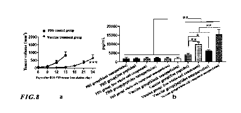
200 L of the above
water-soluble component solution (30 mg/mL) and 200 L of the non-water-
soluble component
solution (30 mg/mL) were added to 1 mL of PLGA (100 mg) dichloromethane
solution, and then
conducting the conventional ultrasonic treatment for 30 s. And then the sample
was mixed with
2.5 mL of polyvinyl alcohol aqueous solution (20 mg/mL), conducting the
conventional ultrasonic
treatment for 30 s. After that, the sample was mixed with 50 mL of polyvinyl
alcohol aqueous
solution (5 mg/mL), followed by stirring conventionally until the complete
volatilization of
organic solvent (dichloromethane). Subsequently, the sample was centrifuged at
12,000 rpm for
10 min. The supernatant was taking out and the precipitate was resuspended in
20 mL of trehalose
aqueous solution (4 wt%). The sample was freeze-drying at -80 C, and then the
sample was
resuspended in 10 mL of normal saline. And then mixing with 0.5 mL of the
above water-soluble
component solution (30 mg/mL) and 0.5 mL of the non-water-soluble component
solution (30
mg/mL), followed by standing for 60 s to obtain the activator of nanoparticles
loaded with the
lysate components inside and on the surfaces of the nanoparticles.
[0070] The preparation of empty nanoparticles and nanoparticles loaded with
the four polypeptide
antigens was the same as above, with the lysate components replaced or not
added.
[0071] The average particle size of nanoparticles before being loaded with the
lysate components
on the surfaces thereof was about 280 nm; the particle size of nanoparticles
after being loaded with
the lysate components on the surfaces thereof was about 300 nm; and 150 i_tg
of lysate components
were loaded on per mg of PLGA nanoparticles. The particle size of blank
nanoparticles was about
250 nm. The particle size of nanoparticles loaded with the polypeptide
antigens was about 290 nm,
and the total peptide loading capacity of 1 mg of PLGA nanoparticles was about
50 vg of
polypeptide antigens.
[0072] (3) Nanoparticles activating tumor-specific T cells: female C57BL/6
mice aged 6-8 weeks
were selected to prepare melanoma-bearing mice. On day 0, 150,000 B16-F10
melanoma cells
were inoculated subcutaneously into the lower right back of each mouse. On
days 4, 7, 10, 15 and
18
CA 03193344 2023- 3- 21
SZD-0036-CA
20, the mice were subcutaneously injected with PBS or cancer nanovaccines
loaded with whole-
cell components of cancer cells. In the experiment, the tumor volumes of mice
were recorded every
three days from Day 6 and the tumor volume was calculated by the formula v =
0.52 x a x b2,
where v being the tumor volume, a being the tumor length, and b being the
tumor width. On day
18 or day 24, the mice in the PBS group or in the vaccine treatment group were
sacrificed and the
peripheral immune cells in their spleens were collected.
[0073] The cells were resuspended in an RPM! 1640 culture medium containing
10% FBS with a
concentration of 5 x 10⁶ cells/mL. Then, the nanoparticles loaded with the
water-soluble and non-
water-soluble components with a final concentration of 300 g/mL were added
into the sample;
or the nanoparticles loaded with the polypeptide antigens with a final
concentration of 300 g/mL
were added into the sample; or the blank nanoparticles of the same amount were
added into the
sample; or the free whole-cell lysate components of the same amount were added
into the sample;
or the free peptide antigen of the same amount were added into the sample. The
sample were then
incubated in an incubator at 37 C (5% CO2) for 72 h and the samples were then
centrifugated at
400 g for 5 min, followed by collecting the supernatant and analyzing the
concentration of I FN-y
in the supernatant by the [LISA detection.
[0074] In the [LISA detection method, the tumor-specific T cells, after being
activated, would
secrete specific cell secretions such as I FN-y. The concentration of such
specific cell secretions
represented the content of activated tumor-specific T cells.
[0075] (4) Experimental results: The above experimental results of melanoma
were shown in Fig.
8. In fig. 8, a showed the inhibition effect of cancer vaccine treatment on
the tumor growth rate (n
> 8), and b showed the content of activated tumor-specific T cells in the
peripheral immune cells
of peripheral spleen after the incubation with free tumor tissue lysates or
nanoparticles loaded with
tumor tissue lysates analyzed by the [LISA detection.
[0076] As shown in Fig. 8, compared with the PBS blank control group and the
blank nanoparticle
group in the vaccine treatment group, the number of activated T cells was
significantly higher in
the vaccine treated group after the co-incubation of peripheral immune cells
of mice with free
whole-cell components of tumor tissue, or nanoparticles loaded with whole-cell
components of
tumor tissue, or free peptide antigens, or nanoparticles loaded with
polypeptide antigens, which
indicated that the content of tumor-specific T cells in the peripheral tissues
of mice treated with
the cancer vaccine increased significantly. It could be seen that the free
whole-cell components of
19
CA 03193344 2023- 3- 21
SZD-0036-CA
tumor tissue of the present invention could be used to detect the content of
tumor-specific T cells
in the peripheral blood of cancer patients. When free whole-cell components of
tumor tissue or
free polypeptide antigens were used to stimulate and activate T cells, the
free whole-cell
components could stimulate and activate more T cells; when nanoparticles
loaded with whole-cell
lysate components or nanoparticles loaded with polypeptide antigens were used
to stimulate and
activate T cells, the nanoparticles loaded with whole-cell lysate components
could stimulate and
activate more T cells; moreover, the nanoparticles loaded with whole-cell
lysate components
stimulated and activated more T cells than the free whole-cell lysate
components.
[0077] Example 4: Activation of tumor-specific T cells in peripheral tissues
by micron
particles loaded with lung cancer tumor tissue lysate components and
immunoadjuvants
[0078] This example described, based mouse lung cancer, the preparation of
micron particles
loaded with lung cancer tumor tissue lysate components and immunoadjuvants,
and the
preparation of micron particles loaded only with lung cancer tumor tissue
lysate components, so
as to activate tumor-specific T cells in peripheral tissues; and ELISPOT was
used to detect the
content of tumor-specific T cells. This example tested the effects of adding
with CpG or adding
with poly I:C as the immunoadjuvant or without any immunoadjuvant,
respectively.
[0079] This example used enzyme-linked immunospot assay (ELISPOT) to detect
the specific
molecule IFN-y of activated tumor-specific T cells; in practical applications,
flow cytometry,
ELISA and other methods can also be used to detect other specific molecules of
tumor-specific T
cells.
[0080] In this example, mouse LLC lung tumor cells were inoculated into
C57BL/6 mice and then
the tumor tissues were extracted. The water-soluble components in tumor tissue
lysate components
and the non-water-soluble components dissolved in a 6 M guanidine
hydrochloride solution were
then obtained. PLGA was utilized as the framework material to prepare micron
particles, loaded
with water-soluble components of tumor tissue lysates and non-water-soluble
components, by the
solvent evaporation method. The water-soluble and non-water-soluble components
in the tumor
tissue lysates were simultaneously loaded inside and on the surfaces of micron
particles; and in
the micron particles containing an immunoadjuvant, the immunoadjuvant was only
loaded inside
the micron particles. These micron particles were then used to detect the
tumor-specific T cells in
the peripheral tissues of mice.
[0081] (1) Lysis of tumor tissues and collection of components: 2,000,000 LLC
lung tumor cells
CA 03193344 2023- 3- 21
SZD-0036-CA
were inoculated subcutaneously into the back of each C57BL/6 mouse, and the
mice were
sacrificed when the tumors inoculated in each mouse grew to a volume of 200-
1500 mm3, followed
by collecting the tumor tissues. The tumor tissues were cut into pieces and
then grinded, followed
by going through a cell filter screen and adding pure water. And then tumor
tissue whole cells
were inactivated and denatured by conventional ultraviolet irradiating and
heating, followed by
repeatedly lyophilizing for 5 times, with ultrasonic treatment performed at
250 W for 1 min at each
thawing. After the tumor tissue whole cells were lysed, the cell lysates were
centrifuged at a
rotational speed of 5,000 rpm for 15 min. And then, the supernatant was taken
as the water-soluble
components in the tumor tissue whole cells; and a 6 M guanidine hydrochloride
aqueous solution
was added to the obtained precipitate to solubilize it, thus obtaining the non-
water-soluble
components.
[0082] (2) Preparation of micron particles loaded with whole-cell components:
In this example,
the micron particles loaded with cell lysates were prepared by the double
emulsion method in the
solvent evaporation method, and the molecular weight of the material PLGA used
for preparing
the micron particles was 24-38 KDa. The preparation method was same as
described previously.
The steps were specifically as follows: 1501uL of the above water-soluble
component solution (60
mg/mL) or 200 tit of the non-water-soluble component solution (10 mg/mL) was
added to 2 mL
of PLGA (50 mg) dichloromethane solution, and then the sample was stirred
conventionally for
150 s. Subsequently, the sample was mixed with 10 mL of polyvinyl alcohol
aqueous solution (15
mg/mL), and conducted the conventional ultrasonic treatment for 50 s, followed
by mixing with
300 mL of polyvinyl alcohol aqueous solution (8 mg/mL) and stirring
conventionally until the
complete volatilization of organic solvent (dichloromethane). And then, the
sample was
centrifuged at 10,000 rpm for 30 min and the supernatant was taken out. The
precipitate was
resuspended in 20 mL of sucrose aqueous solution (5 wt%), followed by freeze-
drying at -80 C.
And then, the sample was resuspended in 5 mL of normal saline and mixed with 3
mL of the above
water-soluble component solution (10 mg/mL) and 0.5 mL of the non-water-
soluble component
solution (40 mg/mL), followed by standing for 20 min to obtain the activator
micron particles
loaded with the lysate components inside and on the surfaces.
[0083] 1501uL of the above water-soluble component solution (60 mg/mL) or 200
tit of the non-
water-soluble component solution (10 mg/mL) was mixed with 100 ttL of
immunoadjuvant (CpG
or poly I:C) solution (0.25 mg/mL), and then the mixture was added to 2 mL of
PLGA (50 mg)
dichloromethane solution, followed by stirring conventionally for 150 s. The
sample was then
21
CA 03193344 2023- 3- 21
SZD-0036-CA
mixed with 10 mL of polyvinyl alcohol aqueous solution (15 mg/mL) and
conducted the
conventional ultrasonic treatment for 50 s. Subsequently, the sample was mixed
with 300 mL of
polyvinyl alcohol aqueous solution (8 mg/mL), and stirred conventionally until
the complete
volatilization of organic solvent (dichloromethane). The sample was
centrifuged at 10,000 rpm for
30 min and the supernatant was taken out. The sample was resuspended in 20 mL
of sucrose
aqueous solution (5 wt%), followed by freeze-drying at -80 C and resuspending
in 5 mL of normal
saline. And then, the sample was mixed with 2 mL of the above water-soluble
component solution
(10 mg/mL) and 0.5 mL of the non-water-soluble component solution (40 mg/mL),
followed by
standing for 20 min to obtain the activator micron particles loaded with
lysate components inside
and on the surfaces.
[0084] The average particle size of micron particles before being loaded with
the cell lysate
components on the surfaces thereof was about 2.0 tim; the particle size of
micron particles after
being loaded with the cell lysate components on the surfaces thereof was about
2.1 tim; and 160
lug of cell lysate components were loaded on per mg of PLGA micron particles.
[0085] (3) Activation of tumor-specific T cells by micron particles: female
C57BL/6 mice aged 6-
8 weeks were selected to prepare lung cancer-bearing mice. On day 0, 2,000,000
LLC lung tumor
cells were inoculated subcutaneously into the lower right back of each mouse.
On days 4, 7, 10,
15 and 20, the mice were subcutaneously injected with PBS or cancer
nanovaccines loaded with
whole-cell components of cancer cell. In the experiment, the tumor volumes of
mice were recorded
every three days from Day 6 and the tumor volume was calculated by the formula
v = 0.52 x a x
b2, where v being the tumor volume, a being the tumor length, and b being the
tumor width. On
day 24, the mice in the PBS group and in the vaccine treatment group were
sacrificed, followed
by collecting the immune cells in their spleens.
[0086] The cells were resuspended in an RPM! 1640 culture medium containing
10% FBS with a
concentration of 5 x 10⁶ cells/mL. 100 [IL of the above spleen cells were
added to a 96-well plate
that was pre-coated with an I FN-y antibody a (capture antibody) and then the
plate was sealed with
a culture medium for more than 1 h. 25 i_tg of micron particles loaded with
water-soluble
components and 25 i_tg of micron particles loaded with non-water-soluble
components were added
to the cells, and then the sample was incubated at 37 C with 5% CO2 for 72 h.
And then, the
mixture of cells and micron particles were discarded and the 96-well plate was
washed, followed
by adding an I FN-y antibody b (detection antibody) and then incubating in an
incubator at 37 C
22
CA 03193344 2023- 3- 21
SZD-0036-CA
(5% CO2) for more than 2 h. The solution containing the I FN-y antibody b was
discarded, the 96-
well plate was washed, followed by using a corresponding method to develop
color. Thus spots
formed on the surface of 96-well plate and the ELISPOT analyzer was applied to
read the data and
analyze the experimental results.
[0087] In the ELISPOT detection, the tumor-specific T cells were activated to
secrete cell
secretions such as I FN-y, which would bind to the antibody a loaded on the 96-
well plate; and after
the addition of the antibody b, a double-antibody sandwich structure would be
formed, and the
detection antibody was connected with an enzyme that could assist in color
development. When a
substrate was added for color development, a spot would be formed at the
location of each
activated cell. The formation of a spot represented an activated tumor-
specific T cell, so the
number of tumor-specific T cells in the tested sample could be known by
measuring the number
of spots formed by color development in each well of the 96-well plate.
[0088] (4) Experimental results: The above experimental results of lung cancer
were shown in Fig.
9. In fig. 9, a showed the inhibition effect of cancer vaccine treatment on
the tumor growth rate (n
> 9), and b showed the content of activated tumor-specific T cells in the
peripheral immune cells
of peripheral spleen after the incubation with micron particles loaded with
tumor tissue lysates
analyzed by the ELISPOT detection.
[0089] As shown in Fig. 9, compared with the PBS blank control group, there
were significantly
more activated T cells after the peripheral immune cells of mice in the
vaccine treatment group
were co-incubated with the micron particles loaded with tumor whole-cell
components or the
micron particles together loaded with tumor whole-cell components and
immunoadjuvants, which
indicated that the content of tumor-specific T cells in the peripheral tissues
of mice treated with
the cancer vaccine increased significantly. Moreover, whether CpG or Poly(I:C)
was used as an
immunoadjuvant, after the co-incubation with peripheral immune cells, the
micron particles loaded
with tumor whole-cell components and immunoadjuvants could activate more T
cells than the
micron particles loaded with tumor whole-cell components. The above results
indicated that
adding immunoadjuvants could activate more cancer antigen-specific T cells.
[0090] The content of tumor-specific T cells in peripheral blood and other
peripheral tissues of
cancer patients is positively correlated with the prognosis of patients. The
present invention
collects whole-cell components of tumor cells or tumor tissues, and then co-
incubates the free
whole-cell components or the cell lysate components loaded on nano/micron
particles with
23
CA 03193344 2023- 3- 21
SZD-0036-CA
peripheral immune cells. After the tumor-specific T cells are activated,
specific molecules of the
tumor-specific T cells are detected, so that the content of the tumor-specific
T cells in peripheral
tissues, such as peripheral blood, can be determined. The inventiveness of the
present invention
lies in using cancer cells, tumor tissue whole cells, cancer cell lysate
components or tumor tissue
whole cell lysate components as activators to detect tumor-specific T cells in
peripheral immune
cells, with all the involved particle loading, cell incubation, specific
secretion detection, etc. being
present technologies in the field.
24
CA 03193344 2023- 3- 21