Note : Les descriptions sont présentées dans la langue officielle dans laquelle elles ont été soumises.
WO 2022/093708
PCT/US2021/056483
DEEP MAGNETIC RESONANCE FINGERPRINTING
AUTO-SEGMENTATION
CROSS REFERENCE TO RELATED APPLICATIONS
100011 The present application claims priority to U.S.
Provisional Application No.
63/106,641, titled "Deep Magnetic Resonance Fingerprinting Auto-Segmentation,"
filed
October 28, 2020, which is incorporated herein in its entirety.
BACKGROUND
100021 A computing device may use computer vision techniques to
detect various
objects within an image. For certain images, it may be difficult to accurately
and reliable
segment such objects in an automated manner.
SUMMARY
100031 At least one aspect of the present disclosure is directed
to systems and
methods of training models to segment tomographic images. A computing system
may
identify a training dataset. The training dataset may have: a plurality of
sample tomographic
images derived from a section of a subject, a plurality of tissue parameters
associated with the
section of the subject corresponding to the plurality of sample tomographic
images, and an
annotation identifying at least one region on the section of the subject in at
least one of the
plurality of sample tomographic images. The computing system may train an
image
segmentation model using the training dataset. The image segmentation model
may include a
generator to determine a plurality of acquisition parameters using the
plurality of sample
tomographic images. The plurality of acquisition parameters may define an
acquisition of the
plurality of sample tomographic images from the section of the subject. The
image
segmentation model may include an image synthesizer to generate a plurality of
synthesized
tomographic images in accordance with the plurality of tissue parameters and
the plurality of
acquisition parameter. The image segmentation model may include a
discriminator to
determine a classification result indicating whether an input tomographic
image
corresponding to one of the plurality of sample tomographic image or the
plurality of
-1-
CA 03196850 2023- 4- 27
WO 2022/093708
PCT/US2021/056483
synthesized tomographic images is synthesized. The image segmentation model
may include
a segmentor to generate, using the input tomographic image, a segmented
tomographic image
identifying the at least one region on the section of the subject. The
computing system may
store the image segmentation model for use to identify one or more regions of
interest in the
tomographic images.
[0004] In some embodiments, the computing system may train the
image
segmentation model by determining a segmentation loss metric based on the
segmented
tomographic image and the annotation. In some embodiments, the computing
system may
train the image segmentation model by updating one or more parameters of at
least one of the
generator, the discriminator, and the segmentor of the image segmentation
model using the
segmentation loss metric.
[0005] In some embodiments, the computing system may train the
image
segmentation model by determining a matching loss metric based on the
plurality of sample
tomographic image and the corresponding plurality of synthesized tomographic
image In
some embodiments, the computing system may train the image segmentation model
by
updating one or more parameters of at least one of the generator, the
discriminator, and the
segmentor of the image segmentation model using the matching loss metric. In
some
embodiments, the computing system may train the image segmentation model by
updating
one or more parameters of at least one of the generator and the discriminator
using a loss
metric associated with the segmentor.
[0006] In some embodiments, the computing system may provide,
responsive to
training of the image segmentation model, the plurality of acquisition
parameters for
acquisition of the tomographic images. The plurality of acquisition parameters
may identify
at least one of a flip angle (FA), a repetition time (TR), or an echo time
(TE).
[0007] In some embodiments, the segmentor of the image
segmentation model may
include a plurality of residual layers corresponding to a plurality of
resolutions to generate the
segmented tomographic image. Each of the plurality of residual layers may have
one or more
-2-
CA 03196850 2023- 4- 27
WO 2022/093708
PCT/US2021/056483
residual connection units (RCUs) to process at least one feature map for a
corresponding
resolution of the plurality of resolutions.
[0008] In some embodiments, each of the plurality of sample
tomographic images
may be acquired from the section of the subject in vivo via magnetic resonance
imaging. The
plurality of tissue parameters may identify at least one of proton density
(PD), a longitudinal
relaxation time (Ti), or a transverse relaxation time (T2) for the acquisition
of the first
tomographic image.
[0009] At least one aspect of the present disclosure is directed
to systems and
methods of segmenting tomographic images. A computing system may identify a
plurality of
acquisition parameters derived from training of an image segmentation model
and defining an
acquisition of tomographic images. The computing system may receive a
plurality of
tomographic images of a section of a subject using the plurality of
acquisition parameters and
a plurality of tissue parameters. The plurality of tissue parameters may be
associated with the
section of the subject corresponding to the plurality of tomographic images.
The section of
the subject may have at least one region of interest. The computing system may
apply the
image segmentation model to the plurality of tomographic images to generate a
segmented
tomographic image. The computing system may apply store the segmented
tomographic
image identifying the at least one region of interest on the section of the
subject
10010] In some embodiments, the computing system may establish
the image
segmentation model comprising a generator to determine the plurality of
acquisition
parameters, an image synthesizer to generate at least one synthesized
tomographic image, a
discriminator to determine whether an input tomographic image is synthesized,
using a
training dataset. The training dataset may include a sample tomographic image
and an
annotation identifying at least one region of interest within the sample
tomographic image.
In some embodiments, the computing system may update one or more parameters of
the
generator, the discriminator, and the sewnentor using a loss metric. The loss
metric may
include at least one of a segmentation loss metric or a matching loss metric.
-3 -
CA 03196850 2023- 4- 27
WO 2022/093708
PCT/US2021/056483
100111 In some embodiments, the computing system may apply a
segmentor of the
image segmentation model to the tomographic image, without applying a
generator, an image
synthesizer, and a discriminator used to train the image segmentation model
based on a
training dataset. In some embodiments, the image segmentation model may
include a
segmentor. The segmentor may include a plurality of residual layers
corresponding to a
plurality of resolutions to generate the segmented tomographic image. Each of
the plurality
of residual layers may have one or more residual connection units (RCUs) to
process at least
one feature map for a corresponding resolution of the plurality of
resolutions.
[0012] In some embodiments, the computing system may provide, to
a magnetic
resonance imaging (MRI) device, the plurality of acquisition parameters for
the acquisition
the tomographic image. The plurality of acquisition parameters may identify at
least one a
flip angle (FA), a repetition time (TR), or an echo time (TE). The plurality
of tissue
parameters may identify at least one of a proton density (PD), a longitudinal
relaxation time
(Ti), or a transverse relaxation time (T2).
BRIEF DESCRIPTION OF THE DRAWINGS
[0013] FIG. 1 depicts a block diagram of a system for training
of deep magnetic
resonance (MR) fingerprinting auto-segmentation, in accordance with an
illustrative
embodiment;
[0014] FIG. 2A depicts a block diagram of a deep-learning
segmentation network in
the system for training deep-MR fingerprinting auto-segmentation, in
accordance with an
illustrative embodiment;
[0015] FIG. 2B depicts a block diagram of a residual connection
unit in the deep-
learning segmentation network, in accordance with an illustrative embodiment;
[0016] FIG. 3 depicts a set of segmented images produced from
MRI TI-weighted and
T2-weight contrast images using different approaches, in accordance with an
illustrative
embodiment;
-4-
CA 03196850 2023- 4- 27
WO 2022/093708
PCT/US2021/056483
100171 FIG. 4 depicts a block diagram of a system for segmenting
tomographic
images in accordance with an illustrative embodiment;
[0018] FIG. 5 depicts a block diagram of a tom ograph in the
system for segmenting
tomographic images in accordance with an illustrative embodiment;
[0019] FIG. 6 depicts a block diagram of an image segmentation
model in the system
for segmenting tomographic images in accordance with an illustrative
embodiment;
[0020] FIG. 7A depicts a block diagram of a generator in the
system for segmenting
tomographic images in accordance with an illustrative embodiment;
[0021] FIG. 7B depicts a block diagram of an encoder in the
generator in the system
for segmenting tomographic images in accordance with an illustrative
embodiment,
[0022] FIG. 7C depicts a block diagram of a convolution stack in
the encoder of the
generator in the system for segmenting tomographic images in accordance with
an illustrative
embodiment;
[0023] FIG. 7D depicts a block diagram of a decoder in the
generator in the system
for segmenting tomographic images in accordance with an illustrative
embodiment;
[0024] FIG. 7E depicts a block diagram of a deconvolution stack
of the decoder in the
generator in the system for segmenting tomographic images in accordance with
an illustrative
embodiment;
[0025] FIG. 8 depicts a block diagram of an image synthesizer in
the image
segmentation model in the system for segmenting tomographic images in
accordance with an
illustrative embodiment;
[0026] FIG. 9A depicts a block diagram of a discriminator in the
image segmentation
model in the system for segmenting tomographic images in accordance with an
illustrative
embodiment;
-5-
CA 03196850 2023- 4- 27
WO 2022/093708
PCT/US2021/056483
100271 FIG. 9B depicts a block diagram of a convolution stack in
the discriminator in
the image segmentation model in the system for segmenting tomographic images
in
accordance with an illustrative embodiment;
[0028] FIG. 10A depicts a block diagram of a segmentor in the
image segmentation
model in the system for segmenting tomographic images in accordance with an
illustrative
embodiment;
[0029] FIG. 10B depicts a block diagram of a residual connection
unit (RCU) block
in the segmentor in the image segmentation model in the system for segmenting
tomographic
images in accordance with an illustrative embodiment;
[0030] FIG. 10C depicts a block diagram of a residual connection
unit (RCU) in a
RCU block in the segmentor in the image segmentation model in the system for
segmenting
tomographic images in accordance with an illustrative embodiment;
[0031] FIG. 10D depicts a block diagram of a set of residual
layers in a segmentor in
the image segmentation model in the system for segmenting tomographic images
in
accordance with an illustrative embodiment;
[0032] FIG. 11 depicts a block diagram of configuring the
tomograph upon
establishment of the image segmentation model in the system for segmenting
tomographic
images in accordance with an illustrative embodiment;
[0033] FIG. 12A depicts a flow diagram of a method of training
models to segment
tomographic images in accordance with an illustrative embodiment;
[0034] FIG. 12B depicts a flow diagram of a method of applying
models to segment
tomographic images in accordance with an illustrative embodiment; and
[0035] FIG. 13 depicts a block diagram of a server system and a
client computer system
in accordance with an illustrative embodiment.
-6-
CA 03196850 2023- 4- 27
WO 2022/093708
PCT/US2021/056483
DETAILED DESCRIPTION
100361 Following below are more detailed descriptions of various
concepts related to,
and embodiments of, systems and methods for segmenting tomographic images It
should be
appreciated that various concepts introduced above and discussed in greater
detail below may
be implemented in any of numerous ways, as the disclosed concepts are not
limited to any
particular manner of implementation. Examples of specific implementations and
applications
are provided primarily for illustrative purposes.
[0037] Section A describes an approach for deep magnetic
resonance fingerprinting
auto-segmentation.
[0038] Section B describes systems and methods for systems and
methods for training
deep-learning models to segment tomographic images and segmenting tomographic
images
using deep-learning models.
[0039] Section C describes a network environment and computing
environment
which may be useful for practicing various computing related embodiments
described herein.
A. Deep Magnetic Resonance Fingerprinting Auto-Segmentation
[0040] Segmentation of tumors and organs at risk from MR images
is a part of
successful radiation treatment planning, which can include offline planning
(before the
treatment session) and online planning (during the treatment session using the
recently
introduced MRI-Linac systems). Manual segmentation by radiologists is time
consuming,
susceptible to errors and can be particularly inefficient for the case of
online treatment
planning. Conversely, despite the tremendous success in automated segmentation
techniques
using deep learning, the accuracies particularly for tumors fall short of the
acceptable levels
due in part to a lack of sufficient MRI contrast to distinguish structures
from background as
well as a lack of sufficiently large training sets.
[0041] The focus of the present disclosure rests on leveraging
MRI contrast. If the
MRI contrast between tumor or organ and the background is improved,
segmentation
-7-
CA 03196850 2023- 4- 27
WO 2022/093708
PCT/US2021/056483
becomes an easier task. In other words, if the contrast between tumor and
background is
easily discernable on MRI, the features for differentiating tumor from
background can be
more easily extracted despite training data size limitations. There is hence a
desire for a
robust automatic MR contrast enhanced automated segmentation technique of
tumors and
organs at risk.
[0042] Although various deep machine learning techniques have
enabled significant
accuracy improvements in segmentation compared to machine learning methods,
such
approaches may be impacted by scanner variations that may alter the contrast
of the
foreground and background voxels in the images, in particular for conventional
MRI. An
alternative to conventional MRI is MR fingerprinting (MRF). MRF acquires
quantitative
tissue parameters, which are not affected by scanner variations. The main
claim of this
disclosure is to combine deep learning and MRF to learn the underlying imaging
parameters
that produce the optimal MR contrast to maximize segmentation accuracy.
[0043] An additional benefit of the approach is its
computational tractability. Rather
than estimating the distribution of MRI signal intensities across an entire
image, as is done
with other approaches, the present method may use estimation of a fixed set of
scalar MRI
parameters, significantly simplifying the problem. Estimating the distribution
of MRI
intensities is challenging because some structures with distinct imaging
contrasts may not be
accurately modeled, leading to the so called "hallucination" problem and a
potential loss of
structures like tumors in the deep learning synthesis Other approaches may use
of specific
constraints on the segmented structures to ensure that the shape of the
structures (with some
average MR intensity for the whole structure) is preserved, or learn a
disentangled feature
representation to extract the same features irrespective of the differences in
image contrasts.
[0044] Presented herein the use of an architecture that combines
MR parameters
learning with segmentation. While other approaches have used deep learning to
synthesize
1VIR contrasts, none have been combined with automated segmentation for MR-
guided
radiation therapy. The systems and methods presented herein is an approach to
do a fully
integrated MR fingerprinted segmentation system for MR guided radiation
therapy
treatments.
-8-
CA 03196850 2023- 4- 27
WO 2022/093708
PCT/US2021/056483
100451 MR fingerprinting (MRF) is a novel method for rapid
acquisition of
quantitative tissue parameter maps. Unlike conventional MRI, the quantitative
maps reflect
the underlying physiological tissue parameters, which are not affected by
instrumental
variations. The image contrast in MRI is a result of the interplay between the
underlying
tissue parameters (Ti, T2, etc.) and the pulse sequence acquisition
parameters. The MRI
acquisition process is governed by a set of known equations - the Bloch
equations. If the
underlying tissue parameters are known, an MR acquisition can be simulated by
calculating
the effects of RF pulses and gradients on the tissue parameters. By modifying
the acquisition
parameters governing the pulse sequence (flip angle (FA), repetition time
(TR), echo time
(TE) etc.) any desired image contrast can be synthesized in post-processing
without requiring
additional scans. Moreover, judicious selection of the acquisition parameters
can enhance
contrast between different tissue types such as gray and white matter or
healthy and diseased
tissue.
[0046] Domain adaptation techniques in medical image analysis
and in computer
vision aim to solve the problem of learning to extract a common feature
representation that
can be applied to one or more imaging domains (such as varying imaging
contrasts) to
successfully segment on all domains. The focus of these works has typically
been to
successfully generalize to imaging modalities that differ slightly from the
modality on which
the model was trained to accommodate different Milt contrasts arising from
differences in
scanner parameters or X-rays, be scalable across images used in training and
testing (such as
simulated images used in training, and images from video cameras under
different
illumination conditions for testing)for modeling diverse imaging conditions
(e.g. daytime vs.
Nighttime vs. Raining etc).
[0047] Approaches in medical image analysis have also considered
and developed
solutions for using one imaging modality (such as computed tomography) to help
in the
segmentation of a different imaging modality (like MRI) when there are few or
no expert
labels available for training a deep learning model. Examples include new deep
learning
approaches using disentangled representations. These methods extract domain-
specific
attribute (e.g. textural, contrast, and edge appearances) features and domain-
agnostic content
-9-
CA 03196850 2023- 4- 27
WO 2022/093708
PCT/US2021/056483
(such as common high-level spatial organization of organs in all the
considered imaging
domains) features. These methods were developed for cardiac CT and MRI,
abdomen organs
CT and MRI and for tumor and normal organ segmentations for MR guided
radiotherapy.
None of the above methods consider the problem from the perspective of using
the deep
learning to extract the best possible MR contrast for individual structures to
generate a
consistent and accurate segmentation.
[0048] The systems and methods described herein may leverage the
power of MRF to
obtain rapid tissue maps to improve auto-segmentation of tumors and organs at
risk. The
premise of the method is that images are easier to segment when the signal
intensity
differences between the structure of interest and its surrounding background
are large. When
performing segmentations using clinically available MRI acquired using
specific imaging
protocols, the imaging variations may be appropriate for segmenting some of
the organs but
not all the organs at risk. Acquisition parameters are often optimized in
coordination with the
radiologists to produce the sequence that can be best interpreted visually by
the radiologists.
Finding the set of acquisition parameters that will yield MR images with
optimal contrast for
each segmented tumor and organ at risk structures is challenging because of
the large number
of parameters involved.
100491 The approach provides an automated way to extract the
different sets of
optimal acquisition parameters and facilitate segmentation of the tumor and
multiple organ at
risk structures These parameters will be modeled as latent vectors that will
be jointly learned
with a segmentation network. The implementation itself will combine generative
adversarial
network (GAN) with segmentation architecture framework with losses and
constraints to
regularize these networks. In practice, the losses and constraints using this
framework can be
applied to any specific GAN (such as Cycl eGAN, disentangled representations
using
variational auto-encoders), and segmentation networks such as commonly used U-
net, dense
fully convolutional networks, and deeper architectures like the proposed
multiple resolution
residual networks, among others. In other words, the focus is on the framework
(combining
generator-discriminator-segmentor), and the regularization constraints or
losses used for
MRF based segmentation.
-10-
CA 03196850 2023- 4- 27
WO 2022/093708
PCT/US2021/056483
100501 Referring now to FIG. 1, depicted is a block diagram of a
system for training
of deep magnetic resonance (MR) fingerprinting auto-segmentation. the present
framework
may be implemented using a variational auto-encoder using a disentangled
feature
representation to model the contrast parameters for producing organ/tumor
specific MR
contrast images, and a multiple resolution deep residual network (MRRN) for
segmentation.
10051] MR Fingerprinting: A set of N tumor patients is scanned
with the MRF
sequence and the underlying MR tissue parameters (PD, Ti, T2, etc.) extracted.
This data
will form the training set for the subsequent neural networks.
[0052] Generator: The GAN network may include a generator
comprised of a
sequence of convolutional layer encoders to extract the features from the
images, a decoder
that produces a set of MR parameters, which are matched by a variational auto-
encoder to a
latent code prior. The latent code prior is learned from the input data and is
optimized such
that the segmentation accuracies are maximized for all segmented organs. In
other words, the
generator samples the distribution of acquisition parameters. These parameters
are then used
to generate a contrast- weighted image by simulating an acquisition using the
Bloch
equations and the quantitative tissue parameters.
10053] Discriminator: The discriminator is trained in an
adversarial manner such that
its loss is minimized when the loss for the generator is maximized. In other
words, the
discriminator tries to determine if an MR contrast weighted image is real (as
seen in set of
standard MR contrast images like Ti weighted, T2 weighted, etc.) or are fake
(that is
produced by using the MR parameters extracted by the generator and combined
using the
Bloch equations). This is necessary to ensure that the produced images are
realistic and are
useful for diagnostic purposes as well as segmentation.
[0054] The discriminator may combine the segmentation results
with the generated
images as a joint distribution of image and the segmentation. Other approaches
have used
images and a scalar classification output or a scalar latent variable but
never a pair of images
as proposed in this invention. An approach may be implemented for performing
unsupervised cross-modality segmentation using CT labeled datasets to segment
MR image
-11 -
CA 03196850 2023- 4- 27
WO 2022/093708
PCT/US2021/056483
sets for lung tumor, abdomen organs, and head and neck parotid gland datasets.
This
approach shows more accurate results than other approaches.
[0055] Loss: We will use a jointly trained generator-di
scriminator-segmentor
network. All networks will be optimized using losses computed for image
generation,
discrimination, and segmentation. The generator and discriminator will be
trained in an
adversarial manner so that each network tries to improve at the cost of the
other network.
The losses used for training the two networks will include the distribution
matching losses
between multiple MR contrast weighted sequences available using standard
imaging
protocols and the generated contrasts, the joint distribution of segmentation
and image
matching losses to focus the contrast generation to improve figure- ground
segmentation,
prior distribution (of the variation auto-encoder) losses computed using
Kullback-Leibler
divergences (for each of the individual parameters that are assumed to follow
a Gaussian
prior including a mean and a standard deviation), and the structure
segmentation losses
computed by maximizing the overlap in the segmented regions between the
radiologist-
provided segmentation and the algorithm generated segmentation.
[0056] In detail, the distribution matching loss will use
mathematically robust
Wasserstein distance metrics to ensure stable network convergence. This loss
computes the
differences between the generated and the expected target (including multiple
MR contrast
sequences) by capturing the finer modes in the distribution. It is
particularly suited when the
MR sequence has multiple modes as opposed the standard Kulback-Leibler based
average
distribution matching loss that only matches the peak of the MR intensity
histogram. As the
goal is to approximate the contrast to any of the real MR contrast sequences,
the minimum
distance to those sequences will be used. The joint-distribution matching will
combine the
MR contrast image and the segmentation as a multi-channel image and the
matching will be
done to minimize the distance of this multi- channel image with respect to any
of the target
MRI contrast image and the corresponding segmentation produced using the
segmentation
network.
-12-
CA 03196850 2023- 4- 27
WO 2022/093708
PCT/US2021/056483
100571 The segmentation loss will be computed using cross-
entropy loss to minimize
the voxel-wise errors in the segmentation for multiple organs and tumor, as
well as boundary-
weighted losses to weight the losses in the boundary more than in the center
of the organs.
[0058] After training to convergence, the acquisition parameters
sampled by the
trained generator provide the optimal parameters for maximizing tumor
segmentation quality.
These parameters can then be used prospectively on new and unseen tumor
patient data.
Simultaneously, the trained discriminator provides the optimal segmentation
algorithm for
this data.
[0059] Referring now to FIGs. 2A and 2B, shown is a block
diagram of the MRRN
network that was used for producing normal organ at risk segmentation for
thoracic organ at
risk structures from CT image sets. The framework allows for any segmentation
architecture
to be used. A deep network architecture based on the multiple resolution
residual network
(MRRN) may be used in some implementations. This network may include multiple
residual
feature streams that carry image features computed at various image
resolutions. These
features are connected residually (by adding the features) to the
convolutional layer inputs in
each layer to incorporate additional information from the individual feature
streams. In
addition, each layer also receives a residual input from the input of the
previous layer. This
increases the set of connections and thereby the capacity of the network and
its ability to
ensure stable convergence by back-propagating losses from the output end to
the input end
without loss of gradients.
[0060] Referring now to FIG. 3, shown are representative
examples of abdomen
organ segmentation using the joint-density discriminator technique called
probabilistic
segmentation and image based generator adversarial network (PSIGAN) for
learning to
segment MRI images without any labeled examples from MRI. Comparisons to
multiple
state-of-the-art methods, together with the achieved Dice similarity
coefficient (DSC)
accuracy is shown. The average DSC computed over all the four organs is shown
for
conciseness.
-13 -
CA 03196850 2023- 4- 27
WO 2022/093708
PCT/US2021/056483
B. Systems and Methods for Training Deep-Learning Models to
Segment
Tomographic Images and Segmenting Tomographic Images Using Deep-Learning
Models
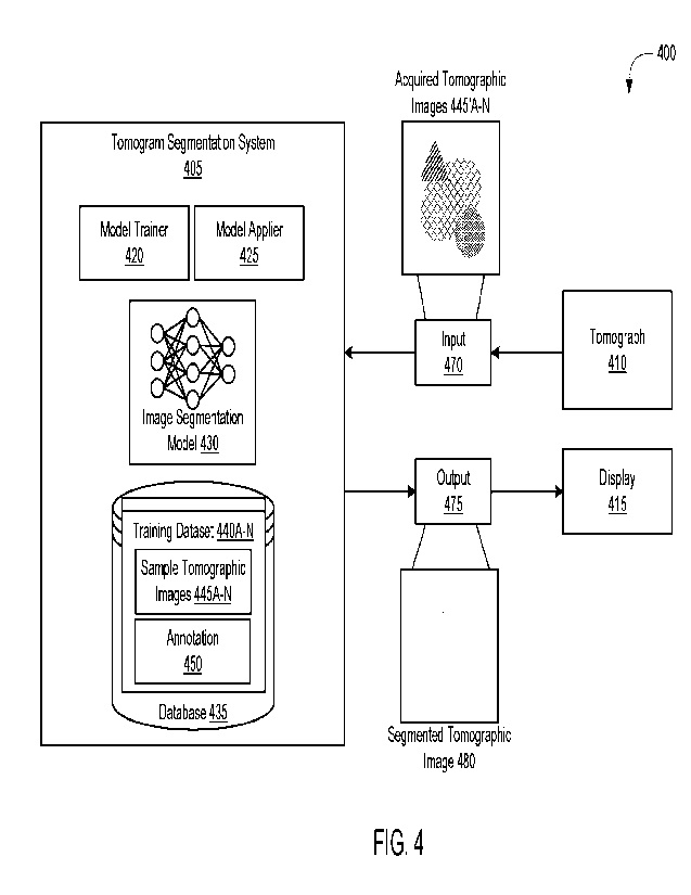
[0061] Referring now to FIG. 4, depicted is a block diagram of a
system 400 for
segmenting tomographic image. In overview, the system 400 may include at least
one
tomogram segmentation system 405, at least one tomograph 410, and at least one
display
415, among others. The tomogram segmentation system 405 may include at least
one model
trainer 420, at least one model applier 425, at least one image segmentation
model 430, and at
least one database 435, among others. The database 435 may include one or more
training
datasets 440A¨N (hereinafter generally referred to as training dataset 440).
Each training
dataset 440 may include a set of sample tomographic images 445A¨N (hereinafter
generally
referred to as tomographic images 445) and at least one annotation 450, among
others. In
some embodiments, the tomograph 410 and the display 415 may be separate from
or a part of
the tomograph segmentation system 405. Each of the components in the system
400 listed
above may be implemented using hardware (e.g., one or more processors coupled
with
memory) or a combination of hardware and software as detailed herein in
Section C. Each of
the components in the system 400 may implement or execute the functionalities
detailed
herein in Section A.
[0062] In further detail, the tomogram segmentation system 405
itself and the
components (such as the model trainer 420, the model applier 425, and the
image
segmentation model 430) may have a training mode and a runtime mode (sometimes
referred
herein as an evaluation or inference mode). Under the training mode, the
tomogram
segmentation system 405 may train or establish the image segmentation model
430 using the
training dataset 440. In particular, the model trainer 420 may initiate,
establish, and maintain
the image segmentation model 430 using the sample tomographic images 445 and
the
annotation 450 of the training dataset 440. Under runtime mode, the tomogram
segmentation
system 405 may identify, retrieve, or otherwise receive at least one set of
acquired
tomographic images 445'A¨N (hereinafter generally referred to as acquired
tomographic
images 445') in at least one input 470 from the tomograph 410. In addition,
the tomogram
-14-
CA 03196850 2023- 4- 27
WO 2022/093708
PCT/US2021/056483
segmentation system 405 may generate a segmented tomographic image 480 using
the set of
acquired tomographic images 445' to provide for presentation on the display
415. The
sample tomographic images 445 may be derived from the acquired tomographic
images 445'.
In discussing the system 400, both the sample tomographic images 445 and the
acquired
tomographic images 445' are referred to generally as tomographic images 445.
10063] Referring now to FIG. 5, depicted is a block diagram of
the tomograph 410
(also referred herein as an imaging device or an image acquisition device) in
the system 400.
The tomograph 410 may produce, output, or otherwise generate one or more
tomographic
images 445 (e.g., tomographic images 445A¨C as depicted) in accordance with a
tomographic imaging technique. The tomograph 410 may be, for example, a
magnetic
resonance imaging (MRI) scanner, a nuclear magnetic resonance (NIVIR) scanner,
X-ray
computed tomography (CT) scanner, an ultrasound imaging scanner, and a
positron emission
tomography (PET) scanner, and a photoacoustic spectroscopy scanner, among
others. The
tomographic imaging technique used by the tomograph 410 may include, for
example,
magnetic resonance imaging (MRI), nuclear magnetic resonance (NMR) imaging, X-
ray
computed tomography (CT), ultrasound imaging, positron emission tomography
(PET)
imaging, and photoacoustic spectroscopy, among others. The present disclosure
discusses the
tomograph 410 and the tomographic images 445 primarily in relation to MRI, but
the other
imaging modalities listed above may be supported by the tomogram segmentation
system
405.
10064] The tomograph 410 may image or scan at least one volume
505 of at least one
subject 500 in accordance with the tomographic imaging technique in generating
the
tomographic images 445. For example, to carry out MRI imaging, the tomograph
410 may
apply one or more magnetic fields (e.g., an external magnetic field) to the
subject 500 in vivo
and measure the radiofrequency (RF) signals emitted from the subject 500. The
subject 500
may include, for example, a human, an animal, a plant, or a cellular organism,
among others.
The volume 505 may correspond to a three-dimension portion of the subject 500.
For
example, the volume 505 may correspond to a head, a torso, an abdomen, a hip,
an arm, or a
leg portion of the subject 500, among others. Within the volume 505 under
scan, the subject
-15-
CA 03196850 2023- 4- 27
WO 2022/093708
PCT/US2021/056483
500 may have one or more objects, such as a tissue, an organ, blood vessels,
neurons, bone,
or other material.
[0065] By scanning, the tomograph 410 may acquire, obtain, or
otherwise receive one
or more sets of data points corresponding to the volume 505 of the subject
500. The sets of
data points acquired by the tomograph 410 may be three-dimensional or two-
dimensional.
The sets of data points may be acquired along at least one section 510 within
the volume 505
of the subject 500 (e.g., in vivo). Each section 510 may correspond to a two-
dimensional
cross-section (e.g., a front, a sagittal, a transverse, or an oblique plane)
within the volume 505
of the subject 500. By extension, each set of data points may correspond to a
respective
section 510 in the volume 505 of the subject 500. At least one section 510 may
include one
or more regions of interest (ROI) 515. Each ROT 515 may correspond to a
particular feature
within the volume 505 or section 510 of the subject 500 under scan. The
feature may
include, for example, a lesion, a hemorrhage, a tumor (benign or malignant),
infarction,
edema, fat, or bone, among others.
[0066] The tomograph 410 may be configured with, store, or
include a set of tissue
parameters 520A¨N (hereinafter referred generally as tissue parameters 520)
and a set of
acquisition parameters 525A¨N (hereinafter referred generally as acquisition
parameters
525). The tissue parameters 520 may be associated with one or more objects
within the
volume 505 or the section 510 of the subject 500. In particular, the tissue
parameters 520
may characterize, identify, or otherwise define properties of the object
within the volume 505
of the section 510 of the subject 500 in relation to tomographic imaging
technique. The
acquisition parameters 525 may be associated with the acquisition of the data
from the
subject 500 under the tomographic imaging technique. In particular, the
acquisition
parameters 525 may define the operational characteristics of the tomograph 410
in obtaining
the data from the subject 500.
[0067] The types of parameters in both the tissue parameters 520
and the acquisition
parameters 525 may depend on the tomographic imaging technique applied by the
tomograph
410. For MRI (e.g., MR fingerprinting), the tissue parameters 520 may include,
for example,
a proton density (PD), a longitudinal relaxation time (Ti) (sometimes referred
herein as a
-16-
CA 03196850 2023- 4- 27
WO 2022/093708
PCT/US2021/056483
spin-lattice time) and a transverse relaxation time (T2) (sometimes referred
herein as a spin-
spin time), among others. The proton density may identify a quantity of
hydrogen nuclei in
the volume 505 of tissue within the subject 500. The longitudinal relaxation
time may refer
to a time constant for spins of the nuclei aligned with the external magnetic
field. The
transverse relaxation time may refer to a time constant for loss of phase
coherence among the
spins of the nuclei oriented at a transverse angle to the external magnetic
field. In addition,
the acquisition parameters 525 may define the RF pulses applied to the subject
500 during
scanning of the volume 505. The acquisition parameters 525 may include, for
example, a flip
angle (FA), a repetition time (TR), and an echo time (TE), among others. The
flip angle may
indicate an amount of rotation of a macroscopic magnetization vector outputted
by the
applied RP pulse in relation to the magnetic field. The repetition time may
identify an
amount of time between a start of a pulse sequence and a start of a succeeding
pulse
sequence. The echo time may identify a time between an excitation RF pulse and
a peak of a
spin echo.
[0068] With the data acquired from the volume 505 of the subject
500, the tomograph
410 may generate one or more tomographic images 445 in accordance with the
tomographic
imaging technique. At least a portion of the tomographic image 445 may
correspond to the
ROT 515 within the volume 505 or section 510 of the subject 500. In some
embodiments,
each tomographic image 445 may be three-dimensional corresponding to the
volume 505 of
the subject 500 under scan. When three-dimensional, the tomographic image 445
may
include one or more slices corresponding to the scanned sections 510 within
the volume 505.
In some embodiments, each tomographic image 445 may be two-dimensional
corresponding
to a section 510 defined in the volume 505 under scan. In some embodiments,
the tomograph
410 may generate a set of tomographic images 445 two-dimensions derived from
the three-
dimensional data points acquired from scanning.
100691 Using the tissue parameters 520 and the acquisition
parameters 525, the
tomograph 410 may process the measured data points obtained from the scanning
of the
volume 505 of the subject 500 in generating the tomographic images 445. The
tomograph
410 may apply different weights of the tissue parameters 520 in generating the
tomographic
-17-
CA 03196850 2023- 4- 27
WO 2022/093708
PCT/US2021/056483
images 445. In the illustrated example, the tomograph 410 may generate a first
tomographic
image 445A weighted using a first tissue parameter 460A (e.g., proton density
(PD)), a
second tomographic image 445B weighted using a second tissue parameter 460B
(e.g.,
longitudinal relaxation time (Ti)), and a third tomographic image 445C
weighted using a
third tissue parameter 460C (e.g., transverse relaxation time (T2)).
10070] Upon generation, the tomograph 410 may send, transmit, or
otherwise provide
the tomographic images 445 to the tomogram segmentation system 405. In some
embodiments, the tomograph 410 may also provide the tissue parameters 520
associated with
the tomographic images 445 to the tomogram segmentation system 405. For
example, the
tomograph 410 may provide the weights of tissue parameters 520 for each of the
tomographic
images 445 acquired from the subject 500. In some embodiments, the tomograph
410 may
send, transmit, or otherwise provide the acquisition parameters 525 to the
tomogram
segmentation system 405. The acquisition parameters 525 may be common or
substantially
the same (e.g., less than 10% difference) among the set of tomographic images
445.
[0071] Referring now to FIG. 6, depicted is a block diagram of
the image
segmentation model 430 in the system 400. The image segmentation model 430 may
be
configured at least in part as a generative adversarial network (GAN), a
variational auto-
encoder, or other unsupervised or semi-supervised model, among others. As
depicted, the
image segmentation model 430 may include at least one generator 600, at least
one image
synthesizer 605, at least one discriminator 610, and at least one segmentor
615, among others.
The image segmentation model 430 may have at least one input and at least one
output. The
input of the image segmentation model 430 may include one or more of the
tomographic
images 445 (e.g., from the training dataset 440 or the tomograph 410). The
output of the
image segmentation model 430 may include at least one segmented tomographic
image 480.
The segmented tomographic image 480 may identify the one or more ROIs 515
within the
corresponding tomographic image 445. The segmented tomographic image 480 may
identify
the one or more ROIs 515 within the corresponding tomographic image 445.
[0072] The components of the image segmentation model 430 may be
inter-related to
one another in accordance to a defined architecture (also referred herein as a
structure). Each
-18-
CA 03196850 2023- 4- 27
WO 2022/093708
PCT/US2021/056483
of the components in the image segmentation model 430 may have at least one
input and at
least one output connected to one another. The input of the generator 600 may
include the
tomographic image 445 provided to the image segmentation model 430. The output
of the
generator 600 may include a set or reconstructed tomographic images 445"A¨N
(hereinafter
generally referred to as reconstructed tomographic images 445") and a set of
acquisition
parameters 525'A¨N (hereinafter generally referred to as acquisition
parameters 525'). The
tomographic images 445 (or reconstructed tomographic images 445") set of
acquisition
parameters 525' may be fed to the input of the image synthesizer 605. The
input of the image
synthesizer 605 may be connected to the output of the generator 600, and may
include the set
of acquisition parameters 525'. The output of the image synthesizer 605 may
include a set of
synthesized tomographic images 620A¨N (hereinafter generally referred to as
synthesized
tomographic images 625). The input of the discriminator 610 may include the
tomographic
image 445 provided to the image segmentation model 430 or the synthesized
tomographic
images 620 outputted by the image synthesizer 605. The output of the
discriminator 610 may
include at least one classification result 625. The input of the discriminator
610 may be fed
forward to the input of the segmentor 615. The input of the segmentor 615 may
include the
tomographic image 445 originally fed into the image segmentation model 430.
The output of
the segmentor 615 may correspond to the output of the image segmentation model
430, and
may include the segmented tomographic image 480.
100731 In each component of the image segmentation model 430,
the input and output
may be related to each other via a set of parameters to be applied to the
input to generate the
output. The parameters of the generator 600, the discriminator 610, and the
segmentor 615
may include weights, kernel parameters, and neural components, among others.
The
parameters in the generator 600, the discriminator 610, and the segmentor 615
may be
arranged in one or more transform layers. Each transform layer may identify,
specify, or
define a combination or a sequence of the application of the parameters to the
input values.
The parameters of the image synthesizer 605 may include a constants, factors,
or
components, among others. The parameters in the image synthesizer 605 may be
applied to
the input in accordance with a policy or equation. The parameters may be
arranged in
-19-
CA 03196850 2023- 4- 27
WO 2022/093708
PCT/US2021/056483
accordance with a machine learning algorithm or model in the respective
components of the
image segmentation model 430.
[0074] When in training mode, the model trainer 420 may initiate
or establish the
image segmentation model 430 (including the generator 600, the discriminator
610, and the
segmentor 615) using the training dataset 450. The initiation and the
establishment of the
image segmentation model 430 may be under the training mode, and the sample
tomographic
images 445 of the training dataset 450. In establishing the image segmentation
model 430,
the model trainer 420 may access the database 435 to retrieve, obtain, or
otherwise identify
the training dataset 440. From each training dataset 440, the model trainer
420 may extract,
retrieve, or otherwise identify the set of sample tomographic images 445. The
set of sample
tomographic images 445 may be derived or acquired from a previous scan of the
subject 500
(e.g., the volume 505 or the section 510). Each tomographic image 445 in the
set may
correspond to different weightings of the tissue parameters 520. In some
embodiments, the
training dataset 440 may identify or include the set of tissue parameters 520
for each of the
tomographic images 445. In some embodiments, the training dataset 440 may
identify or
include the set of acquisition parameters 525 used to generate the set of
tomographic images
445.
100751 From the training dataset 440, the model trainer 420 may
retrieve, extract, or
otherwise identify the annotation 450. For at least one of the tomographic
images 445 in the
set, the training dataset 440 may identify or include the annotation 450. In
some
embodiments, the training dataset 440 may include the annotation 450 for one
of the
tomographic images 445 with a particular weighting of the tissue parameters
520. For
example, the annotation 450 of the training dataset 440 may be associated with
the first
tomographic image 445A weighted using the first tissue parameter 520. The
annotation 450
may label or identify one or more ROIs 515 within the associated tomographic
image 445. In
some embodiments, the annotation 450 may identify the location of the ROIs 515
(e.g., using
pixel coordinates) within the tomographic image 445. In some embodiments, the
annotation
450 may identify an outline of the ROIs 515. The annotation 450 may have been
manually
created. For example, a clinician may manually have examined one of
tomographic images
-20-
CA 03196850 2023- 4- 27
WO 2022/093708
PCT/US2021/056483
445 for the one or more ROIs 515, and add the annotation 450 with an outline
of the ROIs
515 thereon. The annotation 450 may be maintained and stored separately from
the
tomographic images 445 (e.g., using different files).
[0076] With the identification, the model applier 425 may apply
the image
segmentation model 430 to each tomographic image 445 of the training dataset
440 during
training mode. In training mode, the model applier 425 may run and process the
tomographic
images 445 through the generator 600, the image synthesizer 605, the
discriminator 610, and
the segmentor 615 of the image segmentation model 430. In runtime mode, the
model
applier 425 may retrieve, identify, or otherwise receive the tomographic
images 445 from the
tomograph 410, and apply the image segmentation model 430 to the tomographic
images
445. Upon receipt, the model applier 425 may apply at least the segmentor 615
of the image
segmentation model 430 to the acquired tomographic images 445.
[0077] The application of the image segmentation model 430 to
the tomographic
images 445 may be in accordance with the structural of the image segmentation
model 430
(e.g., as depicted). In some embodiments, the model applier 425 may apply the
tomographic
images 445 individually (e.g., one after another). In applying the image
segmentation model
430, the model applier 425 may feed each tomographic image 445 into the input
of the image
segmentation model 430. The model applier 425 may process the input
tomographic image
445 in accordance with the components (e.g., the generator 600, the image
synthesizer 605,
the discriminator 610, and segmentor 615) and architecture of the image
segmentation model
430. The function and structure of the image segmentation model 430 in
processing the input
and updating the weights of the components is detailed herein below in
conjunction with
FIGs. 7A-11.
[0078] Referring now to FIG. 7A, depicted is a block diagram of
the generator 600
(sometimes referred herein as a generator network) of the image segmentation
model 430 in
the system 400. The generator 600 may include at least one encoder 700, at
least one decoder
705, and at least one sampler 710, among others. In some embodiments, the
components of
the generator 600 may be arranged in accordance with a variational auto-
encoder
architecture. In some embodiments, the generator 600 may lack the sampler 710.
The set of
-21 -
CA 03196850 2023- 4- 27
WO 2022/093708
PCT/US2021/056483
weights of the generator 600 may be arranged in accordance with the encoder
700 and the
decoder 705. Within generator 600, the encoder 700, the decoder 705, and the
sampler 710
may be connected in series, and each may have at least one input and at least
one output. The
input of the encoder 700 may correspond to the input of the generator 600, and
may include
the tomographic image 445. The output of the encoder 700 may include a set of
features 715
(also referred herein as latent features or feature map), and may be fed to
the sampler 710.
The output of the sampler 710 may be connected with the input of the decoder
705. The
output of the decoder 705 may correspond to the output of the generator 600,
and may
include at least one reconstructed tomographic image 445".
100791 In feeding into the image segmentation model 430, the
model applier 425 may
provide each tomographic image 445 as the input of the generator 600. The
model applier
425 may process the input tomographic image 445 in accordance with the weights
of the
encoder 700 to produce, output, or otherwise generate the set of features 715.
The set of
features 715 may correspond to a lower resolution or dimension representation
of the
tomographic image 445. For example, the set of features 715 may represent
latent attributes
of the tomographic image 445, and may be partially correlated with the tissue
parameters 520
or the acquisition parameters 525. The model applier 425 may feed the set of
features 715
forward in the generator 600 to the sampler 710.
[0080] Continuing on, the model applier 425 may use the sampler
710 to identify or
select one or more features from the set of features 715 as a sampled subset
of features 715'.
The selection of the features from the set of features 715 may be in
accordance with a
distribution function (e.g., a probability distribution such as a Gaussian or
normal
distribution). In some embodiments, the sampler 710 may select the subset of
features 715'
using the distribution function. In some embodiments, the sampler 710 may
identify one or
more distribution characteristics (e.g., mean, variance, and standard
deviation) of the values
within the set of feature 715. The distribution characteristics may be used by
the model
applier 425 to select the subset from the set of features 715. In some
embodiments, the
model applier 425 may use the sampler 710 to identify or select a set of
acquisition
parameters 525' from the set of features 715 or the subset of features 715'.
The subset of
-22-
CA 03196850 2023- 4- 27
WO 2022/093708
PCT/US2021/056483
features 715' selected by the sampler 710 may correspond or represent a
distribution of the
original tissue parameters 520 or the acquisition parameters 525 used to
generate the
tomographic image 445. In some embodiments, the model applier 425 may select
at least a
portion of the subset of features 515' as the set of acquisition parameters
525'.
[0081] Upon sampling, the model applier 425 may apply the
decoder 705 to the
sampled set of features 715'. Using the weights of the decoder 705, the model
applier 425
may process the subset of features 715' to produce, output, or generate the
reconstructed
tomographic image 445". The reconstructed tomographic image 445" may
correspond to the
original tomographic image 445 fed into the generator 600. The reconstructed
tomographic
image 445" may have a greater dimension than the set of features 715 generated
by the
encoder 700. The reconstructed tomographic image 445" may also have the same
dimensions
(or resolution) as the original tomographic image 445. The model applier 425
may feed the
reconstructed tomographic image 445 and the set of acquisition parameters 525
[0082] The model trainer 420 may establish and train the
generator 600 using the
training dataset 440. The model trainer 420 may calculate, generate, or
otherwise determine
at least one loss metric (also referred herein as a reconstruction loss). The
determination of
the loss metric may be based on a comparison between the reconstructed
tomographic image
445" and the original tomographic image 445 used to generate the reconstructed
tomographic
image 445". The loss metric may indicate or correspond to a degree of
deviation between the
original tomographic image 445 and the reconstructed tomographic image 445"
(e.g., a pixel-
by-pixel value comparison). The loss metric may be calculated in accordance
with a loss
function, such as a Kullback-Leibler (KL) divergence, a root mean squared
error, a relative
root mean squared error, and a weighted cross entropy, among others. In
general, when the
degree of deviation is greater, the loss metric may be also greater.
Conversely, when the
degree of deviation is lower, the loss metric may be lower.
[0083] Using the loss metric, the model trainer 420 may modify,
configure, or
otherwise update at least one of the weights in the generator 600, such as in
the encoder 700
or the decoder 750. The loss metric may be back-propagated to update the
weights in the
generator 600. The updating of weights may be in accordance with an
optimization function
-23 -
CA 03196850 2023- 4- 27
WO 2022/093708
PCT/US2021/056483
(or an objective function) for the generator 600. The optimization function
may define one or
more rates or parameters at which the weights of the generator 600 are to be
updated. For
example, the model trainer 420 may use the optimization function with a set
learning rate, a
momentum, and a weight decay for a number of iterations in training. The
updating of the
weights may be repeated until a convergence condition.
10084] Referring now to FIG. 7B, depicted is a block diagram of
the encoder 700 in
the generator 600 in the image segmentation model 430 of the system 400. The
encoder 700
may include a set of convolution stacks 720A¨N (hereinafter generally referred
to as
convolution stacks 720). The input and the output of the encoder 700 may be
related via a set
of parameters define within the set of convolution stacks 720. The set of
convolution stacks
720 can be arranged in series or parallel configuration, or in any
combination. In parallel
configuration, the input of one convolution stacks 720 may include the input
of the entire
encoder 700. In a series configuration (e.g., as depicted), the input of one
convolution stacks
720 may include the output of the previous convolution stacks 720. The input
of the first
convolution stack 720A may be the tomographic image 445. The output of the
last
convolution stack 720N may be the set of features 715.
[0085] Referring now to FIG. 7C, depicted is a block diagram of
the convolution
stack 720 in the encoder 700 of the generator 600 in the image segmentation
model 430 of
the system 400. Each convolution stack 720 may include a set of transform
layers 725A¨N
(hereinafter generally referred to as transform layers 725). The set of
transform layers 725
may be arranged in series or parallel configuration, or in any combination.
The transform
layers 725 may define or include the weights for the corresponding convolution
stack 720 in
the encoder 700. The set of transform layers 725 can include one or more
weights to modify
or otherwise process the input to produce or generate an output set of
features. For example,
the set of transform layers 725 may include at least one convolutional layer,
at least one
normalization layer, and at least one activation layer, among others. The set
of transform
layers 725 can be arranged in series, with an output of one transform layer
725 fed as an input
to a succeeding transform layer 725. Each transform layer 725 may have a non-
linear input-
to-output characteristic. In some embodiments, the set of transform layers 725
may be a
-24-
CA 03196850 2023- 4- 27
WO 2022/093708
PCT/US2021/056483
convolutional neural network (CNN). The convolutional layer, the normalization
layer, and
the activation layer (e.g., a rectified linear unit (ReLU)) may be arranged in
accordance with
the CNN architecture.
[0086] Referring now to FIG. 7D, depicted is a block diagram of
the decoder 705 and
the sampler 710 in the generator 600 in the image segmentation model 430 of
the system 400.
The decoder 705 may include a set of deconvolution stacks 730A¨N (hereinafter
generally
referred to as deconvolution stacks 730). The input and the output of the
decoder 705 may be
related via a set of parameters define within the set of deconvolution stacks
730. The set of
deconvolution stacks 730 can be arranged in series or parallel configuration,
or in any
combination. In parallel configuration, the input of one deconvolution stacks
730 may
include the input of the entire decoder 705. In a series configuration (e.g.,
as depicted), the
input of one deconvolution stacks 730 may include the output of the previous
deconvolution
stacks 730. The input of the first deconvolution stack 730A may be the set of
features 715
generated by the encoder 700 or the sampled set of features 715' (e.g., as
depicted) from the
set of features 715. The output of the last deconvolution stack 730N may be
the
reconstructed tomographic image 445".
[0087] Referring now to FIG. 7E, depicted is a block diagram of
each deconvolution
stack 730 of the decoder 705 in the generator 600 in the image segmentation
model 430 of
the system 400. The deconvolution stack 730 may include at least one sampler
735 and a set
of transform layers 740A¨N (hereinafter generally referred to transform layers
740). The up-
sampler 735 and the set of transform layers 740 can be arranged in series
(e.g., as depicted) or
parallel configuration, or in any combination. The up-sampler 735 may increase
the image
resolution of the input to increase a dimension (or resolution) to fit the set
of transform layers
740. In some implementations, the up-sampler 735 can apply an up-sampling
operation to
increase the dimension of the input. The up-sampling operation may include,
for example,
expansion and an interpolation filter, among others. In performing the up-
sampling
operation, the up-sampler 735 may insert null (or default) values into the
input to expand the
dimension. The insertion or null values may separate the pre-existing values.
The up-
sampler 735 may apply a filter (e.g., a low-pass frequency filter or another
smoothing
-25-
CA 03196850 2023- 4- 27
WO 2022/093708
PCT/US2021/056483
operation) to the expanded feature map. With the application, the up-sampler
735 may feed
the resultant input into the transform layers 740.
[0088] The set of transform layers 740 can include one or more
weights to modify or
otherwise process the input to produce or generate an output. For example, the
set of
transform layers 740 may include at least one convolutional layer, at least
one normalization
layer, and at least one activation layer, among others. The set of transform
layers 740 can be
arranged in series, with an output of one transform layer 740 fed as an input
to a succeeding
transform layer 740. Each transform layer 740 may have a non-linear input-to-
output
characteristic. In some embodiments, the set of transform layers 740 may be a
convolutional
neural network (CNN). The convolutional layer, the normalization layer, and
the activation
layer (e.g., a rectified linear unit (ReLU)) may be arranged in accordance
with the CNN
architecture.
[0089] Referring now to FIG. 8, depicted is a block diagram of
the image synthesizer
605 in the image segmentation model 430 of the system 400. The image
synthesizer 605 may
be configured with or may otherwise include at least one synthesis policy 800.
The synthesis
policy 800 may specify, identify, or otherwise define one or more relations
among the
parameters (e.g., the set of tissue parameters 520 and acquisition parameters
525 or 525')
with which to generate at least one synthesized tomographic image 620. For
example, the
synthesis policy 800 may be the Bloch equation as discussed herein in Section
A. In general,
the synthesis policy 800 may adjust or set a contrast of the synthesized
tomographic image
620 from the tomographic image 445.
[0090] According to the synthesis policy 800, the model applier
425 may generate the
synthesized tomographic image 620 using the tomographic image 445 (or the
reconstructed
tomographic image 445'), the tissue parameters 520, and the acquisition
parameters 525'. In
generating, the model applier 425 may identify the set of tissue parameters
520 associated
with the tomographic image 445. In some embodiments, the model applier 425 may
retrieve,
extract, or identify the set of tissue parameters 520 associated with the
tomographic image
445 from the training dataset 440. As discussed above, the tissue parameters
520 may be
included in the training dataset 440 along with the tomographic images 445. In
some
-26-
CA 03196850 2023- 4- 27
WO 2022/093708
PCT/US2021/056483
embodiments, the model applier 425 may retrieve, receive, or identify the set
of tissue
parameters 520 from the tomograph 40 that provided the tomographic image 445.
In
addition, the model applier 425 may retrieve, receive, or identify the set of
acquisition
parameters 525' from the generator 600. As discussed above, the sampler 710 of
the
generator 600 may provide the set of acquisition parameters 525' from the
distribution of
features 715.
[0091] With the identification, the model applier 425 may
process the tissue
parameters 520, the acquisition parameters 525', and the tomographic image 445
in
accordance with the one or more relations specified by the synthesis policy
800. In some
embodiments, the model applier 425 may feed the tissue parameters 520 and the
acquisition
parameters 525' to the relations (e.g., Bloch equation) as defined by the
synthesis policy 800
to generate a set of resultants. In some embodiments, the model applier 425
may use
different weights of the tissue parameters 520 in applying the synthesis
policy 800. The
model applier 425 may apply the resultants to the tomographic image 445 to
produce, output,
and generate the synthesized tomographic image 620. With the output, the model
applier 425
may feed the synthesized tomographic image 620to the discriminator 610.
[0092] Referring now to FIG. 9A, depicted a block diagram of the
discriminator 610
(sometimes referred herein as a discriminator network) in the image
segmentation model 430
in the system 400. The discriminator 610 may include a set of convolution
stacks 905A¨N
(hereinafter generally referred to as convolution stacks 905). The input and
the output of the
discriminator 610 may be related via a set of parameters define within the set
of convolution
stacks 905. The set of convolution stacks 905 can be arranged in series or
parallel
configuration, or in any combination. In parallel configuration, the input of
one convolution
stacks 905 may include the input of the entire discriminator 610. In a series
configuration
(e.g., as depicted), the input of one convolution stacks 905 may include the
output of the
previous convolution stacks 905. The input of the first convolution stack 905A
may include
at least one input tomographic image 900 corresponding to the original
tomographic image
445 or the synthesized tomographic image 620 from the image synthesizer 605.
The output
of the last convolution stack 905N may be the classification result 625.
-27-
CA 03196850 2023- 4- 27
WO 2022/093708
PCT/US2021/056483
100931 In training, the model applier 425 may identify or select
one of the
tomographic image 445 or the synthesized tomographic image 620 as the input
image 900 to
apply to the discriminator 610. The input image 900 may be associated with one
of the
weightings of the tissue parameters 520. With the identification, the model
applier 425 may
feed the input image 900 as the input to the discriminator 610 to determine
whether the input
image 900 is synthesized. The discriminator 610 may process the input image
900 in
accordance with the weights of the discriminator 610 (e.g., as defined in the
convolution
stacks 905) to produce, output, or generate the classification result 625. The
classification
result 625 may indicate whether the input image 900 is synthesized (e.g.,
processed by the
image synthesizer 605) or real (e.g., from the tomograph 410). For example,
the
classification result 625 may indicate the Boolean value of "false" when the
input image 900
is determined to be synthesized. Conversely, the classification result 625 may
indicate the
Boolean value of "true" when the input image 900 is determined to be
realistic. While
training, the accuracy of the determination regarding the source of the input
by the
discriminator 610 may increase.
10094] The model trainer 420 may establish and train the
discriminator 610 based on
the input image 900 and the classification result 625. To train, the model
trainer 420 may
compare the classification result 625 to the source for input image 900
provided to the
discriminator 610. When the classification result 625 indicates that the input
image 900 is
realistic and the input image 900 is the synthesized tomographic image 620,
the model trainer
420 determine that the classification result 625 is inaccurate. When the
classification result
625 indicates that the input image 900 is synthetic and the input image 900 is
the
tomographic image 445, the model trainer 420 may also determine that the
classification
result 625 is inaccurate. Conversely, when the classification result 625
indicates that the
input image 900 is realistic and the input image 900 is the tomographic image
445, the model
trainer 420 determine that the classification result 625 is inaccurate. When
the classification
result 625 indicates that the input image 900 is synthetic and the input image
900 is the
synthesized tomographic image 620, the model trainer 420 may also determine
that the
classification result 625 is accurate.
-28-
CA 03196850 2023- 4- 27
WO 2022/093708
PCT/US2021/056483
100951 Based on the comparisons, the model trainer 420 may
calculate, generate, or
otherwise determine a loss metric (also referred herein as a matching loss)
for the
discriminator 610. The determination of the loss metric may be based on
classification
results 625 generated for the set of input images 900 with different weights
of the tissue
parameters 520. The loss metric may indicate or correspond to a degree of
inaccuracy of the
discriminator 610 in producing the correct classification results 625. The
loss metric may be
calculated in accordance with a loss function, such as a Kullback-Leibler (KL)
divergence,
Wasserstein loss function, a root mean squared error, a relative root mean
squared error, and
a weighted cross entropy, among others. In some embodiments, the model trainer
420 can
calculate or determine the loss function by replicating a probability
distribution. In general,
when the inaccuracy is greater, the loss metric may be also greater.
Conversely, when the
inaccuracy is lower, the loss metric may also be lower.
[0096] Using the loss metric, the model trainer 420 may modify,
configure, or
otherwise update at least one of the weights in the generator 600 (e.g., the
encoder 700 and
the decoder 705) and the discriminator 610 (e.g., the set of convolution
stacks 905). The loss
metric may be back-propagated to update the weights in the generator 600 and
the
discriminator 610. The updating of weights may be in accordance with an
optimization
function (or an objective function) for the generator 600 and the
discriminator 610. The
optimization function may define one or more rates or parameters at which the
weights of the
generator 600 and the discriminator 610 are to be updated. For example, the
model trainer
420 may use the optimization function with a set learning rate, a momentum,
and a weight
decay for a number of iterations in training. The updating of the weights may
be repeated
until a convergence condition. In iteratively updating the weights, the
accuracy of the
classification result 625 and the set of acquisition parameters 525' to
contrast the
tomographic images 445 may be improved.
100971 Referring now to FIG. 9B, depicted is a block diagram of
the convolution
stack 905 in the discriminator 610 in the image segmentation model in the
system 400. Each
convolution stack 905 may include a set of transform layers 910A¨N
(hereinafter generally
referred to as transform layers 910). The set of transform layers 910 may be
arranged in
-29-
CA 03196850 2023- 4- 27
WO 2022/093708
PCT/US2021/056483
series or parallel configuration, or in any combination. The transform layers
910 may define
or include the weights for the corresponding convolution stack 905 in the
discriminator 610.
The set of transform layers 910 can include one or more weights to modify or
otherwise
process the input to produce or generate an output set of features. For
example, the set of
transform layers 910 may include at least one convolutional layer, at least
one normalization
layer, and at least one activation layer, among others. The set of transform
layers 910 can be
arranged in series, with an output of one transform layer 910 fed as an input
to a succeeding
transform layer 910. Each transform layer 910 may have a non-linear input-to-
output
characteristic. In some embodiments, the set of transform layers 910 may be a
convolutional
neural network (CNN). The convolutional layer, the normalization layer, and
the activation
layer (e.g., a rectified linear unit (ReLU)) may be arranged in accordance
with the CNN
architecture.
[0098] Referring now to FIG. 10A, depicted is a block diagram of
the segmentor 615
(also referred herein as a segmentor network) in the image segmentation model
430 in the
system 400. The segmentor 615 may include or maintain a set of residual layers
1000A¨N
(hereinafter generally referred to as residual layers 1000) arranged in a
defined architecture.
For example, the segmentor 615 may be configured as a multi-resolution
residual network
(MRRN) architecture (e.g., as discussed below). The input and the output of
the segmentor
615 may be related via a set of parameters defined within the set of residual
layers 1000.
Each residual layer 1000 may correspond to a magnification factor or a
resolution of features
to be processed. In some embodiments, successive residual layers 1000 may
correspond to
lower magnifications or resolutions than previous residual layers 1000. For
example, the first
residual layer 1000A may correspond to a magnification factor of 10x, while
the second
residual layers 1000B may correspond to a magnification factor of 5x. The set
of residual
layers 1000 can be arranged in series or parallel configuration, or in any
combination. The
input of one or more of the residual layers 1000 may include at least one
input tomographic
image 1005 corresponding to the original tomographic image 445 or the
synthesized
tomographic image 620 from the image synthesizer 605. The output of the
residual layers
1000 of the segmentor 615 may be the segmented tomographic image 480.
-30-
CA 03196850 2023- 4- 27
WO 2022/093708
PCT/US2021/056483
100991 In training, the model applier 425 may identify or select
one of the
tomographic image 445 or the synthesized tomographic image 620 as the input
image 1005 to
apply to the discriminator 610. The input image 1005 may be associated with
one of the
weightings of the tissue parameters 520. With the identification, the model
applier 425 may
apply the input image 1005 to the segmentor 615 in accordance with
architecture of the set of
residual layers 1000. The model applier 425 may process the input image 1005
using the
weights of the segmentor 615 (e.g., as defined using the set of residual
layers 1000) to
produce, output, or generate the segmented tomographic image 480. The
segmented
tomographic image 480 may identify the ROIs 515 corresponding to various
features in the
subject 500 under scan acquired in the input image 1005. In the illustrated
example, the
segmented tomographic image 480 may include an outline of the ROIs 515
corresponding to
the features in the input image 1005.
[0100] The model trainer 420 may establish and train the
segmentor 615 using the
segmented tomographic image 480 and the annotation 450 for the tomographic
image 445
used to generate the segmented tomographic image 480. As discussed above, the
annotation
450 may label or identify one or more ROIs 515 within the associated
tomographic image
445. In training, the model trainer 420 may compare the segmented tomographic
image 480
and the annotation 450. Based on the comparison, the model trainer 420 may
calculate,
generate, or otherwise determine a loss metric. The loss metric may indicate
or correspond a
degree of deviation between the ROIs 515 identified by the segmented
tomographic image
480 from the ROIs 515 identified by the annotation 450 (e.g., a pixel-by-pixel
comparison).
The loss metric may be determine accordance with a loss function, such as a
Kullback-
Leibler (KL) divergence, a root mean squared error, a relative root mean
squared error, and a
weighted cross entropy, among others. In general, when the degree of deviation
is greater,
the loss metric may be also greater. Conversely, when the degree of deviation
is lower, the
loss metric may be lower.
[0101] With the determination of the loss metric, the model
trainer 420 may modify,
configure, or otherwise update at least one of the weights in the generator
600 (e.g., the
encoder 700 and the decoder 705), the discriminator 610 (e.g., the set of
convolution stacks
-31 -
CA 03196850 2023- 4- 27
WO 2022/093708
PCT/US2021/056483
905), and the segmentor 615 (e.g., the set of residual layers 1000). The loss
metric may be
back-propagated to update the weights in the generator 600, the discriminator
610, and the
segmentor 615. The updating of weights may be in accordance with an
optimization function
(or an objective function) for the generator 600 and the discriminator 610.
The optimization
function may define one or more rates or parameters at which the weights of
the generator
600, the discriminator 610, and the segmentor 615 are to be updated. For
example, the model
trainer 420 may use the optimization function with a set learning rate, a
momentum, and a
weight decay for a number of iterations in training. The updating of the
weights may be
repeated until a convergence condition. In updating the weights, the
acquisition parameters
525' to contrast the tomographic images 445 and the accuracy of the
identification of ROIs
515 by the segmentor 615 may be improved.
[0102] Referring now to FIG. 10B, depicted is a block diagram of
a residual
connection unit (RCU) block 1010 in the segmentor 615 in the image
segmentation model
430 of the system 400. One or more instances of the RCU block 1010 may be
included into
at least one of the residual layers 1000 of the segmentor 615. In some
embodiments, the
RCU blocks 1010 may be located in the second and subsequent residual layers
1000B¨N and
may be omitted from the first residual layer 100A. Each RCU block 1010 may
include a set
of residual connection units (RCUs) 1015A¨N (hereinafter generally referred to
as RCUs
1015). The input and the output of the RCU block 1010 may be related via a set
of
parameters defined within the set of RCUs 1015. The set of RCUs 1015 may be
arranged in
series or parallel configuration, or in any combination. In a series
configuration (e.g., as
depicted), the input of one RCUs 1015 may include the output of the previous
RCUs 1015.
The RCU block 1010 may have at least one input and a set of outputs. The input
of RCU
block 1010 may include features from the same residual layer 1000 as the RCU
block 1010.
Each RCU 1015 of the RCU block 1010 may define the outputs for the RCU block
1010. At
least one output of the RCU block 1010 may be fed forward along the same
residual layer
1000. One or more outputs of the RCU block 1010 may be fed to another residual
layer 1000
(e.g., to a residual layer 1000 above as depicted).
-32-
CA 03196850 2023- 4- 27
WO 2022/093708
PCT/US2021/056483
101031 Referring now to FIG. 10C, depicted is a block diagram of
a residual
connection unit (RCU) 1015 in a RCU block 1010 in the segmentor 615 in the
image
segmentation model 430 in the system 400. The RCU 1015 may include a set of
transform
layers 1020A¨N (hereinafter generally referred to as transform layers 1020),
at least one
down-sampler 1025, at least one aggregation operator 1030, at least one
convolution unit
1040, and at least one up-sampler 1045. Each RCU 1015 of the RCU 1010 may
include one
or more inputs and one or more outputs. The inputs may include features from
the same
residual layer 1000 and a different residual layer 1000 (e.g., one of the
residual layers 1000
above the current residual layer 1000). The outputs may include features to be
fed along the
same residual layer 1000 and a different residual layer 1000 (e.g., one of the
residual layers
1000 above the current residual layer 1000).
[0104] The down-sampler 1025 may retrieve, receive, or identify
features from the
different residual layer 1000. The down-sampler 1025 can reduce the resolution
(or
dimensions) of the features received from the previous residual layer 1000 to
an resolution to
fit the set of transform layers 1020. In some implementations, the down-
sampler 1025 can
apply a pooling operation to reduce the image resolution of the received
features. The
pooling operation can include, for example, max-pooling to select the highest
value within
each subset of features or mean-pooling to determine an average value within
the subset in
the features. In some embodiments, the down-sampler 1025 can apply a down
sampling
operation to the features.
[0105] The aggregation operator 1030 may retrieve, receive, or
identify features from
the same residual layer 1000 and features outputted by the down-sampler 1025
from a
different residual layer 1000. The input from the same residual layer 1000 can
include, for
example, features. The features of the same residual layer 1000 can be of the
resolution
already fitting the set of transform layers 1020 in the RCU 1015. With receipt
of both inputs,
the input aggregation operator 1030 can combine the features from the down-
sampler 134
and features from the same residual layer 1000 to generate features to input
into the set of
transform layers 1020. The combination of the features may include
concatenation, weighted
summation, and addition, among others.
-33 -
CA 03196850 2023- 4- 27
WO 2022/093708
PCT/US2021/056483
101061 The set of transform layers 1020 of the RCU 1015 may be
arranged in series
or parallel configuration, or in any combination. The transform layers 1020
may define or
include the weights for the corresponding RCU 1015 in the RCU block 1010. The
set of
transform layers 1020 can include one or more weights to modify or otherwise
process the
input to produce or generate an output set of features. For example, the set
of transform
layers 1020 may include at least one convolutional layer, at least one
normalization layer, and
at least one activation layer, among others. The set of transform layers 1020
can be arranged
in series, with an output of one transform layer 1020 fed as an input to a
succeeding
transform layer 1020. Each transform layer 1020 may have a non-linear input-to-
output
characteristic. In some embodiments, the set of transform layers 1020 may be a
convolutional neural network (CNN). The convolutional layer, the normalization
layer, and
the activation layer (e.g., a rectified linear unit (ReLU)) may be arranged in
accordance with
the CNN architecture.
101071 The convolution unit 1040 of the RCU 1015 may receive the
set of features
processed by the set of transform layers 1020. The set of features processed
by the transform
layers 1020 may have a size or dimension incompatible for processing by the
previous
residual layer 1000. For example, the set of features may have a depth beyond
the
compatible number for the previous residual layer 1116. Upon receipt, the
convolution unit
1040 may apply a dimension reduction operator to the set of features from the
set of
transform layers 1020. The dimension reduction operator may include a 1 < 1
convolution, a
3 3 convolution, linear dimension reduction, and non-linear
dimension reduction, among
others. Application of the dimension reduction operator by the convolution
unit 1040 may
reduce the size or the reduction of the features. Upon application, the output
of the
convolution unit 1040 may be fed into the up-sampler 1045.
101081 The up-sampler 1045 of the RCU 1015 may increase the
image resolution of
the input to increase a dimension (or resolution) to match the resolution of
the features in the
other residual layer 1000. In some implementations, the up-sampler 1045 can
apply an up-
sampling operation to increase the dimension of the input. The up-sampling
operation may
include, for example, expansion and an interpolation filter, among others. In
performing the
-34-
CA 03196850 2023- 4- 27
WO 2022/093708
PCT/US2021/056483
up-sampling operation, the up-sampler 1045 may insert null (or default) values
into the input
to expand the dimension. The insertion or null values may separate the pre-
existing values.
The up-sampler 1045 may apply a filter (e.g., a low-pass frequency filter or
another
smoothing operation) to the expanded feature map. With the application, the up-
sampler
1045 may feed the resultant to the other residual layer 1000.
10109] Referring now to FIG. 10D, depicted is a block diagram of
the set of residual
layers 1000 in the segmentor 615 of the image segmentation model 430. In the
depiction, the
segmentor 615 may include four residual layers 1000A¨D, but the segmentor 615
may have
any number of residual layers 1000. The various components, such as the
residual layers
1000, the RU blocks 1010, the RCUs 1015, among others, may be connected with
one
another in the manner defined by the incremental MRRN architecture for the
segmentor 615
(e.g., as depicted). Each residual layer 1000 may have a corresponding
residual stream
1050A¨N (hereinafter generally referred to as residual streams 1050). Each
residual stream
1050 may carry or may be used to maintain a corresponding set of features
1055A¨N
(hereinafter generally referred to as features 1055). Each set of features
1055 may have the
resolution (or dimension) associated with the corresponding residual layer
1000 and by
extension the corresponding residual stream 1050.
101101 The components of the segmentor 615 may reside on or
defined relative to the
respective residual layer 1000 or the residual stream 1050. In the illustrated
example, each
residual layer 1000B¨D beneath the first residual layer 1000A may include one
or more RCU
blocks 1010. The input for each RCU block 1010 may be obtained from the same
residual
layer 1000. For instance, the RCU block 1010 in the second residual layer
1000B may be the
set of features 1055B carried along the same residual layer 1000B. The output
for one or
more of the RCU blocks 1010 may be fed along the same residual layer 1000. The
output for
the RCU blocks 1010 may be also fed to a different residual layer 1000. For
example, in the
fourth residual layer 1000D, the RCU block 1010 may provide the output
features to the other
residual layers 100B¨D.
[0111] In addition, the segmentor 615 may have one or more
pooling units 1060.
Each pooling unit 1060 may span between at least two of the residual layers
1000 and by
-35-
CA 03196850 2023- 4- 27
WO 2022/093708
PCT/US2021/056483
extension at least two of the residual streams 1050. Each pooling unit 1060
may retrieve,
receive, or otherwise identify features 1055 from one residual layer 1000 to
reduce in
resolution for processing by a succeeding residual layer 1000 (e.g., from the
first residual
layer 1000A to the second residual layer 1000B). The pooling unit 1060 may
apply a pooling
operation to the features 1055 identified from the residual layer 1000. The
pooling operation
can include, for example, max-pooling to select the highest value within each
set patch in the
feature map or mean-pooling to determine an average value within the set patch
in the feature
map. The features 1055 processed by the pooling unit 1060 may be of an
resolution less than
the resolution of the original features 1055 identified from the residual
layer 1000.
101121 The segmentor 615 may have one or more aggregation units
1065 along or
within the respective residual streams 1050 of the residual layers 1000. The
aggregator unit
1065 may receive input (e.g., the set of features 1055) from the residual
stream 1050 of the
same residual layer 1000 and the set of features 1055 from another residual
layer 1000. Upon
receipt of both inputs, the aggregator unit 1065 may combine features 1055 in
the residual
stream of the same residual layer 1000 and the features 1055 from the other
residual layer
1000. The combination by the aggregator unit 1065 may generate features 1055
for further
processing along the residual stream 1050. The combination of the features
1055 by the
aggregation unit 1065 may include concatenation, weighted summation, and
addition, among
others.
[0113] Referring now to FIG. 11, depicted is a block diagram of
configuring the
tomograph 410 upon establishment of the image segmentation model 430 in the
system 400.
With the completion of the training of the image segmentation model 430, the
model trainer
420 may send, transmit, or otherwise provide at least one configuration 1100
to the
tomograph 410. The configuration 1100 may identify or include the set of
acquisition
parameters 525' from the generator 600 of the image segmentation model 430. In
providing
the configuration 1100, the model trainer 420 may identify the set of
acquisition parameters
525' via the sampler 710 of the generator 600. The model trainer 420 may
retrieve or
identify a distribution of acquisition parameters 525' via the sampler 710
from the features
715 generated by the encoder 700 of the generator 600. Based on the
distribution, the model
-36-
CA 03196850 2023- 4- 27
WO 2022/093708
PCT/US2021/056483
trainer 420 may determine or generate the set of acquisition parameters 525'
to provide as the
configuration 1100 to the tomograph 410.
[0114] Upon receipt of the configuration 1100, the tomograph 410
may store and
maintain the set of acquisition parameters 525'. In storing, the tomograph 410
may replace
the previous set of acquisition parameters 525. In general, the new set of
acquisition
parameters 525' may yield tomographic images 445 with higher contrast relative
to
tomographic images 445 generated using the previous set of tomographic images
445. When
in runtime mode, the tomograph 410 may provide a new set of tomographic images
445 from
scanning a volume 505 of a subject 500. The model applier 425 may retrieve,
receive, or
identify the new set of tomographic images 445. The model applier 425 may
process each
tomographic image 445 as discussed above to produce, output, or generate the
segmented
tomographic image 480 for the respective tomographic image 445. The model
applier 425
may provide the segmented tomographic image 480 via the output 475 to the
display 415 for
presentation. In some embodiments, the output 475 may also identify or include
the original
tomographic image 445 associated with the segmented tomographic image 480. The
display
415 may be part of the tomogram segmentation system 405 or on another
computing device
that may be communicatively coupled to the tomogram segmentation system 405.
The
display 415 may present or render the output 475 upon receipt. For example,
the display 415
may render a graphical user interface that shows the selected segmented image
480 and the
acquired tomographic image 445.
[0115] Referring now to FIG. 12A, depicted is a flow diagram of
a method 1200 of
training models to segment tomographic images. The method 1200 may be
implemented
using or performed by any of the components described herein, for example, the
system 400
as described in conjunction with FIGs. 4-11 or the computing system as
detailed in
conjunction with FIG. 13. In overview, a computing system (e.g., the tomogram
segmentation system 405) may identify a training dataset (e.g., the training
dataset 440)
(1205). The computing system may apply a segmentation model (e.g., the image
segmentation model 430) (1210). The computing system may determine a loss
metric
(1215). The computing system may update the segmentation model (1220).
-37-
CA 03196850 2023- 4- 27
WO 2022/093708
PCT/US2021/056483
101161 Referring now to FIG. 12B, depicted is a flow diagram of
a method 1250 of
applying models to segment tomographic images. The method 1250 may be
implemented
using or performed by any of the components described herein, for example, the
system 400
as described in conjunction with FIGs. 4-11 or the computing system as
detailed in
conjunction with FIG. 13. In overview, a computing system (e.g., the tomogram
segmentation system 405) may identify a tomographic image (e.g., the
tomographic image
455) (1255). The computing system may apply a segmentation model (e.g., the
image
segmentation model 430) (1260). The computing system may provide a segmented
image
(e.g., the segmented tomographic image 480) (1265).
C. Computing and Network Environment
[0117] Various operations described herein can be implemented on
computer
systems. FIG. 13 shows a simplified block diagram of a representative server
system 1300,
client computer system 1314, and network 1326 usable to implement certain
embodiments of
the present disclosure. In various embodiments, server system 1300 or similar
systems can
implement services or servers described herein or portions thereof. Client
computer system
1314 or similar systems can implement clients described herein. The system 400
described
herein can be similar to the server system 1300. Server system 1300 can have a
modular
design that incorporates a number of modules 1302 (e.g., blades in a blade
server
embodiment); while two modules 1302 are shown, any number can be provided.
Each
module 1302 can include processing unit(s) 1304 and local storage 1306.
[0118] Processing unit(s) 1304 can include a single processor,
which can have one or
more cores, or multiple processors. In some embodiments, processing unit(s)
1304 can
include a general-purpose primary processor as well as one or more special-
purpose co-
processors such as graphics processors, digital signal processors, or the
like. In some
embodiments, some or all processing units 1304 can be implemented using
customized
circuits, such as application specific integrated circuits (ASICs) or field
programmable gate
arrays (FPGAs). In some embodiments, such integrated circuits execute
instructions that are
stored on the circuit itself. In other embodiments, processing unit(s) 1304
can execute
-38-
CA 03196850 2023- 4- 27
WO 2022/093708
PCT/US2021/056483
instructions stored in local storage 1306. Any type of processors in any
combination can be
included in processing unit(s) 1304.
[0119] Local storage 1306 can include volatile storage media
(e.g., DRAM, SRAM,
SDRAM, or the like) and/or non-volatile storage media (e.g., magnetic or
optical disk, flash
memory, or the like). Storage media incorporated in local storage 1306 can be
fixed,
removable or upgradeable as desired. Local storage 1306 can be physically or
logically
divided into various subunits such as a system memory, a read-only memory
(ROM), and a
permanent storage device. The system memory can be a read-and-write memory
device or a
volatile read-and-write memory, such as dynamic random-access memory. The
system
memory can store some or all of the instructions and data that processing
unit(s) 1304 for
runtime. The ROM can store static data and instructions that are to be
processed by
processing unit(s) 1304. The permanent storage device can be a non-volatile
read-and-write
memory device that can store instructions and data even when module 1302 is
powered
down. The term "storage medium" as used herein includes any medium in which
data can be
stored indefinitely (subject to overwriting, electrical disturbance, power
loss, or the like) and
does not include carrier waves and transitory electronic signals propagating
wirelessly or over
wired connections.
101201 In some embodiments, local storage 1306 can store one or
more software
programs to be executed by processing unit(s) 1304, such as an operating
system and/or
programs implementing various server functions such as functions of the system
400 of FIG.
4or any other system described herein, or any other server(s) associated with
system 400 or
any other system described herein.
[0121] "Software" refers generally to sequences of instructions
that, when executed
by processing unit(s) 1304 cause server system 1300 (or portions thereof) to
perform various
operations, thus defining one or more specific machine embodiments that
execute and
perform the operations of the software programs. The instructions can be
stored as firmware
residing in read-only memory and/or program code stored in non-volatile
storage media that
can be read into volatile working memory for execution by processing unit(s)
1304. Software
can be implemented as a single program or a collection of separate programs or
program
-39-
CA 03196850 2023- 4- 27
WO 2022/093708
PCT/US2021/056483
modules that interact as desired. From local storage 1306 (or non-local
storage described
below), processing unit(s) 1304 can retrieve program instructions to execute
and data to
process in order to execute various operations described above.
[0122] In some server systems 1300, multiple modules 1302 can be
interconnected
via a bus or other interconnect 1308, forming a local area network that
supports
communication between modules 1302 and other components of server system 1300.
Interconnect 1308 can be implemented using various technologies including
server racks,
hubs, routers, etc.
[0123] A wide area network (WAN) interface 1310 can provide data
communication
capability between the local area network (interconnect 1308) and the network
1326, such as
the Internet. Technologies can be used, including wired (e.g., Ethernet, IEEE
1302.3
standards) and/or wireless technologies (e.g., Wi-Fi, IEEE 1302.11 standards).
[0124] In some embodiments, local storage 1306 is intended to
provide working
memory for processing unit(s) 1304, providing fast access to programs and/or
data to be
processed while reducing traffic on interconnect 1308. Storage for larger
quantities of data
can be provided on the local area network by one or more mass storage
subsystems 13 12 that
can be connected to interconnect 1308. Mass storage subsystem 1312 can be
based on
magnetic, optical, semiconductor, or other data storage media. Direct attached
storage,
storage area networks, network-attached storage, and the like can be used. Any
data stores or
other collections of data described herein as being produced, consumed, or
maintained by a
service or server can be stored in mass storage subsystem 1312. In some
embodiments,
additional data storage resources may be accessible via WAN interface 1310
(potentially with
increased latency).
[0125] Server system 1300 can operate in response to requests
received via WAN
interface 1310. For example, one of modules 1302 can implement a supervisory
function and
assign discrete tasks to other modules 1302 in response to received requests.
Work allocation
techniques can be used. As requests are processed, results can be returned to
the requester
via WAN interface 1310. Such operation can generally be automated. Further, in
some
-40-
CA 03196850 2023- 4- 27
WO 2022/093708
PCT/US2021/056483
embodiments, WAN interface 1310 can connect multiple server systems 1300 to
each other,
providing scalable systems capable of managing high volumes of activity. Other
techniques
for managing server systems and server farms (collections of server systems
that cooperate)
can be used, including dynamic resource allocation and reallocation.
101261 Server system 1300 can interact with various user-owned
or user-operated
devices via a wide-area network such as the Internet. An example of a user-
operated device
is shown in FIG. 13 as client computing system 1314. Client computing system
1314 can be
implemented, for example, as a consumer device such as a smartphone, other
mobile phone,
tablet computer, wearable computing device (e.g., smart watch, eyeglasses),
desktop
computer, laptop computer, and so on.
[0127] For example, client computing system 1314 can communicate
via WAN
interface 1310. Client computing system 1314 can include computer components
such as
processing unit(s) 1316, storage device 1318, network interface 1320, user
input device 1322,
and user output device 1324. Client computing system 1314 can be a computing
device
implemented in a variety of form factors, such as a desktop computer, laptop
computer, tablet
computer, smartphone, other mobile computing device, wearable computing
device, or the
like.
[0128] Processor 1316 and storage device 1318 can be similar to
processing unit(s)
1304 and local storage 1306 described above. Suitable devices can be selected
based on the
demands to be placed on client computing system 1314; for example, client
computing
system 1314 can be implemented as a "thin" client with limited processing
capability or as a
high-powered computing device. Client computing system 1314 can be provisioned
with
program code executable by processing unit(s) 1316 to enable various
interactions with
server system 1300.
[0129] Network interface 1320 can provide a connection to the
network 1326, such as
a wide area network (e.g., the Internet) to which WAN interface 1310 of server
system 1300
is also connected. In various embodiments, network interface 1320 can include
a wired
interface (e.g., Ethernet) and/or a wireless interface implementing various RF
data
-41 -
CA 03196850 2023- 4- 27
WO 2022/093708
PCT/US2021/056483
communication standards such as Wi-Fi, Bluetooth, or cellular data network
standards (e.g.,
3G, 4G, LTE, etc.).
[0130]
User input device 1322 can include any device (or devices) via which a user
can provide signals to client computing system 1314; client computing system
1314 can
interpret the signals as indicative of particular user requests or
information. In various
embodiments, user input device 1322 can include any or all of a keyboard,
touch pad, touch
screen, mouse or other pointing device, scroll wheel, click wheel, dial,
button, switch,
keypad, microphone, and so on.
[0131]
User output device 1324 can include any device via which client computing
system 1314 can provide information to a user. For example, user output device
1324 can
include a display to display images generated by or delivered to client
computing system
1314. The display can incorporate various image generation technologies, e.g.,
a liquid
crystal display (LCD), light-emitting diode (LED) including organic light-
emitting diodes
(OLED), projection system, cathode ray tube (CRT), or the like, together with
supporting
electronics (e.g., digital-to-analog or analog-to-digital converters, signal
processors, or the
like). Some embodiments can include a device such as a touchscreen that
functions as both
input and output device. In some embodiments, other user output devices 1324
can be
provided in addition to or instead of a display. Examples include indicator
lights, speakers,
tactile "display" devices, printers, and so on.
[0132]
Some embodiments include electronic components, such as microprocessors,
storage and memory that store computer program instructions in a computer
readable storage
medium. Many of the features described in this specification can be
implemented as
processes that are specified as a set of program instructions encoded on a
computer readable
storage medium. When these program instructions are executed by one or more
processing
units, they cause the processing unit(s) to perform various operations
indicated in the
program instructions. Examples of program instructions or computer code
include machine
code, such as is produced by a compiler, and files including higher-level code
that are
executed by a computer, an electronic component, or a microprocessor using an
interpreter.
Through suitable programming, processing unit(s) 1304 and 1316 can provide
various
-42-
CA 03196850 2023- 4- 27
WO 2022/093708
PCT/US2021/056483
functionality for server system 1300 and client computing system 1314,
including any of the
functionality described herein as being performed by a server or client, or
other functionality.
[0133] It will be appreciated that server system 1300 and client
computing system
1314 are illustrative and that variations and modifications are possible.
Computer systems
used in connection with embodiments of the present disclosure can have other
capabilities not
specifically described here. Further, while server system 1300 and client
computing system
1314 are described with reference to particular blocks, it is to be understood
that these blocks
are defined for convenience of description and are not intended to imply a
particular physical
arrangement of component parts. For instance, different blocks can be but need
not be
located in the same facility, in the same server rack, or on the same
motherboard. Further,
the blocks need not correspond to physically distinct components. Blocks can
be configured
to perform various operations, e.g., by programming a processor or providing
appropriate
control circuitry, and various blocks might or might not be reconfigurable
depending on how
the initial configuration is obtained. Embodiments of the present disclosure
can be realized
in a variety of apparatus including electronic devices implemented using any
combination of
circuitry and software.
10134] While the disclosure has been described with respect to
specific embodiments,
one skilled in the art will recognize that numerous modifications are
possible. Embodiments
of the disclosure can be realized using a variety of computer systems and
communication
technologies including but not limited to specific examples described herein.
Embodiments
of the present disclosure can be realized using any combination of dedicated
components
and/or programmable processors and/or other programmable devices. The various
processes
described herein can be implemented on the same processor or different
processors in any
combination. Where components are described as being configured to perform
certain
operations, such configuration can be accomplished, e.g., by designing
electronic circuits to
perform the operation, by programming programmable electronic circuits (such
as
microprocessors) to perform the operation, or any combination thereof.
Further, while the
embodiments described above may make reference to specific hardware and
software
components, those skilled in the art will appreciate that different
combinations of hardware
-43 -
CA 03196850 2023- 4- 27
WO 2022/093708
PCT/US2021/056483
and/or software components may also be used and that particular operations
described as
being implemented in hardware might also be implemented in software or vice
versa
[0135] Computer programs incorporating various features of the
present disclosure
may be encoded and stored on various computer readable storage media; suitable
media
include magnetic disk or tape, optical storage media such as compact disk (CD)
or DVD
(digital versatile disk), flash memory, and other non-transitory media.
Computer readable
media encoded with the program code may be packaged with a compatible
electronic device,
or the program code may be provided separately from electronic devices (e.g.,
via Internet
download or as a separately packaged computer-readable storage medium).
[0136] Thus, although the disclosure has been described with
respect to specific
embodiments, it will be appreciated that the disclosure is intended to cover
all modifications
and equivalents within the scope of the following claims.
-44-
CA 03196850 2023- 4- 27