Note : Les descriptions sont présentées dans la langue officielle dans laquelle elles ont été soumises.
CA 03197045 2023-03-27
WO 2022/087336 PCT/US2021/056150
PROSTHETIC VALVE DOCKING DEVICE
CROSS-REFERENCE TO RELATED APPLICATIONS
[001] This application claims the benefit of U.S. Provisional Application No.
63/252,524,
filed October 5, 2021, U.S. Provisional Application No. 63/159,130, filed
March 10, 2021,
and U.S. Provisional Application No. 63/105,099, filed October 23, 2020, all
of which are
incorporated by reference herein.
FIELD
[002] The present disclosure concerns examples of a docking device configured
to secure a
prosthetic valve at a native heart valve, as well as methods of assembling
such devices.
BACKGROUND
[003] Prosthetic valves can be used to treat cardiac valvular disorders.
Native heart valves
(e.g., the aortic, pulmonary, tricuspid and mitral valves) function to prevent
backward flow or
regurgitation, while allowing forward flow. These heart valves can be rendered
less effective
by congenital, inflammatory, infectious conditions, etc. Such conditions can
eventually lead
to serious cardiovascular compromise or death. For many years, the doctors
attempted to
treat such disorders with surgical repair or replacement of the valve during
open heart
surgery.
[004] A transcatheter technique for introducing and implanting a prosthetic
heart valve
using a catheter in a manner that is less invasive than open heart surgery can
reduce
complications associated with open heart surgery. In this technique, a
prosthetic valve can be
mounted in a compressed state on the end portion of a catheter and advanced
through a blood
vessel of the patient until the valve reaches the implantation site. The valve
at the catheter tip
can then be expanded to its functional size at the site of the defective
native valve, such as by
inflating a balloon on which the valve is mounted or, for example, the valve
can have a
resilient, self-expanding stent or frame that expands the valve to its
functional size when it is
advanced from a delivery sheath at the distal end of the catheter. Optionally,
the valve can
have a balloon-expandable, self-expanding, mechanically expandable frame,
and/or a frame
expandable in multiple or a combination of ways.
- 1 -
CA 03197045 2023-03-27
WO 2022/087336 PCT/US2021/056150
[005] In some instances, a transcatheter heart valve (THV) may be
appropriately sized to be
placed inside a particular native valve (e.g., a native aortic valve). As
such, the THV may not
be suitable for implantation at another native valve (e.g., a native mitral
valve) and/or in a
patient with a larger native valve. Additionally or alternatively, the native
tissue at the
implantation site may not provide sufficient structure for the THV to be
secured in place
relative to the native tissue. Accordingly, improvements to THVs and the
associated
transcatheter delivery apparatus are desirable.
SUMMARY
[006] The present disclosure relates to methods and devices for treating
valvular
regurgitation and/or other valve issues. Specifically, the present disclosure
is directed to a
docking device configured to receive a prosthetic valve and the methods of
assembling the
docking device and implanting the docking device.
[007] Certain examples of the disclosure concern a docking device for securing
a prosthetic
valve at a native valve. The docking device can include a coil comprising a
proximal end and
a distal end, a first cover surrounding at least a portion of the coil, and a
guard member
surrounding at least a portion of the first cover. The guard member can
include an
expandable member and a second cover surrounding an outer surface of the
expandable
member. A distal end portion of the guard member can be fixedly attached to
the first cover.
A proximal end portion of the guard member can be movable relative to the
first cover and
the coil. The guard member can be movable between a radially compressed state
and a
radially expanded state. The proximal end portion of the guard member can be
disposed
closer to the proximal end of the coil when the guard member is in the
radially compressed
state than in the radially expanded state.
[008] According to certain examples, a docking device for securing a
prosthetic valve at a
native valve can include a coil comprising a plurality of helical turns when
deployed at the
native valve, an expandable member extending radially outwardly from the coil,
and a cover
member surrounding an outer surface of the expandable member. The expandable
member
can be movable between a radially-compressed/axially-elongated state and a
radially-
expanded/axially-foreshortened state. Both a distal end portion of the cover
member and a
distal end portion of the expandable member can be fixedly coupled to the coil
via a distal
suture. The distal suture can include a plurality of knots and a plurality of
wraps. A proximal
end portion of the expandable member can be fixedly coupled to a proximal end
portion of
- 2 -
CA 03197045 2023-03-27
WO 2022/087336 PCT/US2021/056150
the cover member. The proximal end portion of the expandable member and the
proximal
end portion of the cover member can be axially movable relative to the coil.
[009] Certain examples of the disclosure concern a cover assembly for a
docking device
configured to receive a prosthetic valve. The cover assembly can include a
first cover
configured to cover at least a portion of a helical coil of the docking
device, an expandable
member surrounding at least a portion of the first cover, and a second cover
surrounding an
outer surface of the expandable member. The expandable member can be
changeable
between a radially expanded state and a radially compressed state. A distal
end portion of the
second cover can be fixedly coupled to a distal end portion of the expandable
member and the
first cover. A proximal end portion of second cover can include a fold
covering a proximal
end of the expandable member. The proximal end of the expandable member can be
slidably
movable relative to the first cover and the helical coil of the docking
device.
[010] Certain examples of the disclosure also concern an implant assembly. The
implant
assembly can include a radially expandable and compressible prosthetic valve,
and a docking
device configured to receive the prosthetic valve. The docking device can
include a coil
configured to surround native tissue when deployed at an implant position. The
prosthetic
valve can be configured to be radially expandable within the coil. The docking
device can
also include a guard member covering at least a portion of the coil. The guard
member can
be configured to reduce paravalvular leakage. The guard member can include an
expandable
member and a cover member surrounding an outer surface of the expandable
member. The
expandable member can extend radially outwardly from the coil and is movable
between a
radially compressed state and a radially expanded state. A distal end portion
of the cover
member and a distal end portion of the expandable member can be fixedly
coupled to the coil
via a distal suture. A proximal end portion of the expandable member can be
fixedly coupled
to a proximal end portion of the cover member via a proximal suture. The
proximal end
portion of the expandable member can be axially movable relative to the coil.
[011] Certain examples of the disclosure also concern a method for assembling
a cover
assembly for a docking device configured to receive a prosthetic valve. The
method can
include positioning a cover member within a lumen of an expandable member,
securing a
proximal end portion of the cover member to a proximal end of the expandable
member,
removing a distal end portion of the cover member out of the expandable member
through the
proximal end of the expandable member, covering an outer surface of the
expandable
- 3 -
CA 03197045 2023-03-27
WO 2022/087336 PCT/US2021/056150
member with the cover member, securing the distal end portion of the
expandable member to
a tubular member of the docking device, and securing the distal end portion of
the cover
member to the expandable member and the tubular member. The tubular member can
be
configured to surround at least a portion of a docking device.
[012] According to certain examples, a method for assembling a cover assembly
for a
docking device configured to receive a prosthetic valve can include securing a
proximal end
portion of an outer cover to a proximal end of an expandable member, inverting
the outer
cover from inside the expandable member to outside the expandable member,
folding the
outer cover at the proximal end of the expandable member, and securing a
distal end portion
of the expandable member and a distal end portion of the outer cover to an
inner cover of the
docking device.
[013] According to certain examples, a method for assembling a docking device
configured
to receive a prosthetic valve can include securing a proximal end portion of a
cover member
to a proximal end of an expandable member, folding the proximal end portion of
the cover
member at the proximal end of the expandable member, covering an outer surface
of the
expandable member with the cover member, and securing a distal end portion of
the
expandable member and a distal end portion of the cover member to the docking
device.
[014] According to certain examples, a method for assembling a docking device
configured
to receive a prosthetic valve can include attaching a proximal end portion of
a cover member
to a proximal end of an expandable member, attaching a distal end portion of
the cover
member to a distal end portion of the expandable member, and securing the
distal end portion
of the cover member and the distal end portion of the expandable member to the
docking
device through a plurality of knots, a plurality of wraps, and one or more
locking stitches.
Each of the locking stitches can extend through the plurality of wraps and/or
the plurality of
knots.
[015] Certain examples of the disclosure also concern a method for implanting
a prosthetic
valve. The method can include deploying a docking device at a native valve,
wherein the
docking device can include a coil and a guard member, wherein the coil
deployed at the
native valve can include a stabilization turn and one or more functional turns
distal to the
stabilization turn, wherein the guard member can cover at least a portion of
the stabilization
turn; and deploying the prosthetic valve within the docking device. Deploying
the docking
device at the native valve can include wrapping around leaflets of the native
valve with the
- 4 -
CA 03197045 2023-03-27
WO 2022/087336 PCT/US2021/056150
one or more functional turns of the coil and resting the stabilization turn of
the coil against a
native wall around the native valve. Deploying the prosthetic valve can
include placing the
prosthetic valve in a radially compressed state within the one or more
functional turns of the
coil and radially expanding the prosthetic valve to a radially expanded state,
wherein radially
expanding the prosthetic valve can cause radial expansion of the one or more
functional turns
of the coil.
[016] According to certain examples, a docking device for securing a
prosthetic valve at a
native valve can include a coil configured to surround native tissue when
deployed at the
native valve and a tubular member surrounding at least a portion of the coil.
The tubular
member can include at least a first seating marker and a second seating
marker. The first
seating marker can be positioned proximal relative to the second seating
marker. The
docking device can also include a retention element surrounding at least a
portion of the
tubular member, and a guard member surrounding at least a portion of the
retention element
and configured to reduce paravalvular leakage. A proximal end of the guard
member can be
axially movable relative to the coil. When deployed at the native valve, the
proximal end of
the guard member can be positioned between the first seating marker and the
second seating
marker.
[017] According to certain examples, a docking device for securing a
prosthetic valve at a
native valve can include a coil configured to have a helical configuration
when deployed at
the native valve. The coil in the helical configuration includes a
stabilization turn, one or
more functional turns, and an ascending portion located between the
stabilization turn and the
one or more functional turns. The docking device can also include at least a
first seating
marker and a second seating marker disposed distal to the ascending portion.
The first
seating marker can be positioned proximal relative to the second seating
marker. The
docking device can further include a guard member surrounding at least a
portion of the coil
and configured to reduce paravalvular leakage. A proximal end of the guard
member can be
axially movable relative to the coil. When deployed at the native valve, the
proximal end of
the guard member can be positioned between the first seating marker and the
second seating
marker.
[018] According to certain examples, a docking device for securing a
prosthetic valve at a
native valve can include a coil configured to surround native tissue when
deployed at the
native valve, a radiopaque marker disposed at a predefined location on the
coil, and a guard
- 5 -
CA 03197045 2023-03-27
WO 2022/087336 PCT/US2021/056150
member surrounding at least a portion of the coil and configured to reduce
paravalvular
leakage. A proximal end of the guard member can be axially movable relative to
the coil.
When deployed at the native valve, the proximal end of the guard member can be
positioned
distal to the radiopaque marker.
[019] According to certain examples, a cover assembly for a docking device
configured to
receive a prosthetic valve can include a retention element configured to
surround at least a
portion of a coil of the docking device, and a guard member surrounding at
least a portion of
the retention element and configured to reduce paravalvular leakage. A
radiopaque marker
can be positioned at a proximal end portion of the retention element. A
proximal end of the
guard member can be configured to be axially movable relative to the retention
element.
When deployed at a native valve, the proximal end of the guard member can be
positioned
distal to the radiopaque marker.
[020] Certain examples of the disclosure also concern an implant assembly. The
implant
assembly can include a radially expandable and compressible prosthetic valve
and a docking
device configured to receive the prosthetic valve. The docking device can
include a coil
configured to surround native tissue when deployed at a native valve, a
radiopaque marker
positioned at a proximal portion of the coil, a retention element surrounding
at least a portion
of the coil, and a guard member surrounding at least a portion of the
retention element and
configured to reduce paravalvular leakage. A proximal end of the guard member
can be
axially movable relative to the retention element. When deployed at the native
valve, the
proximal end of the guard member can be positioned distally relative to the
radiopaque
marker.
[021] Certain examples of the disclosure also concern a method for assembling
a cover
assembly for a docking device configured to receive a prosthetic valve. The
method can
include inserting a tubular member through a lumen of a retention element,
securing a
retention element to the tubular member, and securing a distal end of a guard
member to a
distal portion of the tubular member. At least one radiopaque marker can be
disposed at a
proximal portion of the tubular member. A proximal end of the guard member can
be axially
movable relative to the retention element. The guard member can be movable
between a
radially expanded state and a radially compressed state. When the guard member
is in the
radially expanded state, a proximal end of the guard member can be positioned
distal to the
radiopaque marker.
- 6 -
CA 03197045 2023-03-27
WO 2022/087336 PCT/US2021/056150
[022] Certain examples of the disclosure also concern a method for implanting
a prosthetic
valve. The method can include deploying a docking device at a native valve,
wherein the
docking device deployed at the native valve comprises a coil and a guard
member
surrounding at least a portion of the coil; positioning a proximal end of the
guard member
distal to a predefined location on the coil; and deploying the prosthetic
valve within the
docking device.
[023] Also disclosed herein is another example of docking device for securing
a prosthetic
valve at a native valve. The docking device can include a coil configured to
surround native
tissue when deployed at the native valve, a retention element surrounding at
least a portion of
the coil, and a guard member surrounding at least a portion of the retention
element and
configured to reduce paravalvular leakage. A proximal end of the guard member
can be
axially movable relative to the retention element. An inner surface of the
guard member can
be configured to frictionally engage with the retention element so that the
retention element
frictionally impedes the proximal end of the guard member to move axially
relative to the
coil.
[024] According to certain examples, a docking device for securing a
prosthetic valve at a
native valve can include a coil configured to surround native tissue when
deployed at the
native valve and a sealing member having an inner edge coupled the coil and an
outer edge
that is movable between a folded position and an extended position. The outer
edge in the
folded position can extend along and adjacent to the coil. At least a segment
of the outer
edge in the extended position can be spaced apart from the coil.
[025] According to certain examples, a docking device for securing a
prosthetic valve at a
native valve can include a coil configured to surround native tissue when
deployed at the
native valve and a skirt coupled to the coil, wherein the skirt is movable
between a delivery
configuration and a deployed configuration. When the skirt is in the delivery
configuration,
an outer edge of the skirt can extend along and adjacent the coil. When the
skirt is in the
deployed configuration, at least a segment of the outer edge of the sealing
member can extend
radially away from the coil so as to reduce paravalvular leakage.
[026] According to certain examples, a docking device for securing a
prosthetic valve at a
native valve can include a coil configured to surround native tissue when
deployed at the
native valve and a skirt coupled to the coil. The skirt can be movable between
a first
configuration and a second configuration. The skirt can be folded along the
coil when the
- 7 -
CA 03197045 2023-03-27
WO 2022/087336 PCT/US2021/056150
skirt is in the first configuration. The skirt can be flat or substantially
flat and extend radially
from the coil when the skirt is in the second configuration.
[027] Certain examples of the disclosure concern a device for reducing
paravalvular leakage
between a prosthetic valve received in a docking device and native tissue
surrounding the
prosthetic valve. The device can include a first edge fixedly attached to a
coil of the docking
device and a second edge that is movable between a folded position and an
extended position.
The second edge in the folded position can extend along and adjacent to the
coil. The second
edge in the extended position can radially fan out from the coil.
[028] Certain examples of the disclosure concern a method for assembling a
docking device
configured to receive a prosthetic valve. The method can include attaching a
skirt to a coil.
The coil can be configured to surround native tissue when deployed at a native
valve. An
outer edge of the skirt can be movable between a folded position and an
extended position.
The outer edge in the folded position can extend along and adjacent to the
coil. The outer
edge in the extended position can radially fan out from the coil.
[029] Certain examples of the disclosure concern a method for implanting a
prosthetic
valve. The method can include deploying a docking device at a native valve and
deploying
the prosthetic valve within the docking device. The docking device deployed at
the native
valve can include a coil and a skirt extending radially outwardly from the
coil. The skirt can
have a planar or generally planar surface that forms an oblique angle relative
to a central
longitudinal axis of the docking device.
[030] Certain examples of the disclosure concern a medical assembly including
a docking
device described above and a radially expandable and compressible prosthetic
valve
configured to be received within the docking device.
[031] Certain examples of the disclosure concern a medical assembly including
a docking
device described above and a delivery apparatus configured to deliver the
docking device to a
target implantation site of a patient.
[032] Certain examples of the disclosure concern a medical assembly including
an
implantable device having a frame and a skirt coupled to a sealing segment of
the frame.
When the implantable device is deployed at a target implantation site, an
outer edge of the
skirt can radially fan out from the sealing segment of the frame, and the
sealing segment of
the frame can form a helical shape rotating about a central longitudinal axis
of the frame such
- 8 -
CA 03197045 2023-03-27
WO 2022/087336 PCT/US2021/056150
that a proximal end of the sealing segment has an offset relative to a distal
end of the sealing
segment along the central longitudinal axis of the frame.
[033] Certain examples of the disclosure concern a docking device for securing
a prosthetic
valve at a native valve. The docking device can include a coil configured to
surround native
tissue when deployed at the native valve and a paravalvular leakage guard
connected to the
coil. The paravalvular leakage guard can be movable between a delivery
configuration and a
deployed configuration. When the paravalvular leakage guard is in the delivery
configuration, an outer edge of the paravalvular leakage guard can extend
along and adjacent
the coil. When the paravalvular leakage guard is in the deployed
configuration, the outer
edge of the paravalvular leakage guard can form a helical shape rotating about
a central
longitudinal axis of the coil and at least a segment of the outer edge of
paravalvular leakage
guard can extend radially away from the coil.
[034] The foregoing and other objects, features, and advantages of the
disclosed technology
will become more apparent from the following detailed description, which
proceeds with
reference to the accompanying figures.
BRIEF DESCRIPTION OF THE DRAWINGS
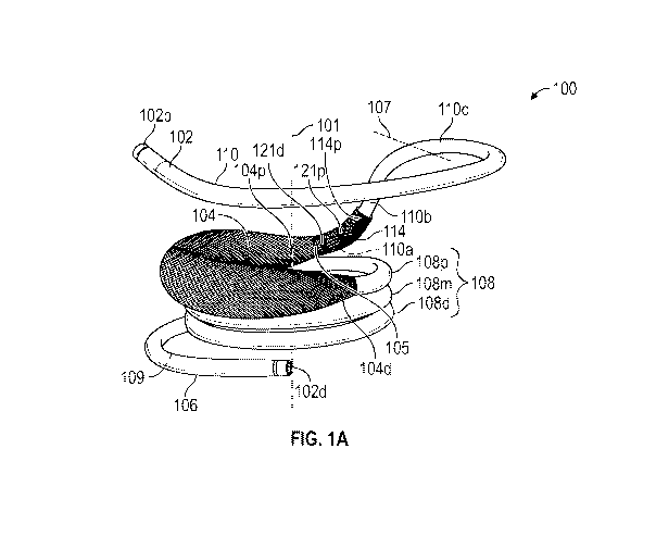
[035] FIG. lA is a side perspective view of a docking device in a helical
configuration,
according to one example.
[036] FIG. 1B is a top view of the docking device depicted in FIG. 1A.
[037] FIG. 1C is a cross-sectional view of the docking device taken along line
1C-1C
depicted in FIG. 1B, according to one example.
[038] FIG. 1D is a cross-sectional view of the docking device taken along the
same line as
in FIG. 1C, except in FIG. 1D, the docking device is in a substantially
straight delivery
configuration.
[039] FIG. lE is a cross-sectional view of the docking device taken along line
1C-1C
depicted in FIG. 1B, according to another example.
[040] FIG. 1F is a cross-sectional view of the docking device taken along the
same line as in
FIG. 1E, except in FIG. 1F, the docking device is in a substantially straight
delivery
configuration.
- 9 -
CA 03197045 2023-03-27
WO 2022/087336 PCT/US2021/056150
[041] FIG. 1G is a schematic diagram depicting the docking device in a
substantially
straight configuration.
[042] FIG. 2A is a perspective view a prosthetic valve, according to one
example.
[043] FIG. 2B is a perspective view of the prosthetic valve of FIG. 2A with an
outer cover,
according to one example.
[044] FIG. 3A is a perspective view of an exemplary prosthetic implant
assembly
comprising the docking device depicted in FIG. lA and the prosthetic valve of
FIG. 2B
retained within the docking device.
[045] FIG. 3B is a side elevation view of the prosthetic implant assembly of
FIG. 3.
[046] FIGS. 4-42 depict various detail views of the docking device,
illustrating an
exemplary method of attaching a paravalvular leakage guard to the docking
device, according
to one example.
[047] FIGS. 43-59 illustrate an exemplary method of attaching a retention
element to a
tubular member to form a part of a cover assembly, according to one example.
[048] FIG. 60 is a side view of a delivery assembly comprising a delivery
apparatus and the
docking device of FIG. 1A, according to one example.
[049] FIG. 61A is a side cross-sectional view of a sleeve shaft, according to
one example.
[050] FIG. 61B is a side cross-sectional view of a pusher shaft, according to
one example.
[051] FIG. 62A is a side cross-sectional view of an assembly comprising the
sleeve shaft of
FIG. 61A, the pusher shaft of FIG. 61B, and a delivery sheath, wherein the
sleeve shaft
covers a docking device.
[052] FIG. 62B is a side cross-sectional view of the same assembly of FIG.
62A, except the
docking device is uncovered by the sleeve shaft.
[053] FIG. 63 is a schematic cross-sectional view of a distal end portion of a
delivery
system, showing fluid flow through lumens within the delivery system.
[054] FIG. 64A illustrates a perspective view of an example of a sleeve shaft
covering a
docking device and extending outside of a delivery sheath of a delivery
system.
- 10 -
CA 03197045 2023-03-27
WO 2022/087336 PCT/US2021/056150
[055] FIG. 64B illustrates the sleeve shaft surrounding a pusher shaft after
deploying the
docking device from the delivery system of FIG. 64A and removing the sleeve
shaft from the
docking device.
[056] FIGS. 65-78 depict various portions of an exemplary implantation
procedure in which
the delivery apparatus of FIG. 60 is being used to implant the prosthetic
implant assembly of
FIG. 3A at a native mitral valve location using a transseptal delivery
approach.
[057] FIGS. 79A is a top perspective view of another docking device having a
foldable PVL
guard in a deployed configuration, according to one example.
[058] FIG. 79B is a top perspective view of the docking device depicted in
FIG. 79A.
[059] FIG. 79C is a bottom perspective view of the docking device depicted in
FIG. 79A.
[060] FIG. 79D is a cross-sectional view of a sealing member of the docking
device and
depicts an example measurement of flatness of the sealing member.
[061] FIG. 80A is a perspective view of an example sleeve shaft covering the
docking
device of FIG. 79A and a sealing member of the docking device being in a
delivery
configuration.
[062] FIG. 80B depicts the sleeve shaft is partially removed from the docking
device and a
part of the sealing member is exposed and radially expanded.
[063] FIG. 81 is a top view of a docking device, according to another example.
[064] FIG. 82A is a top view of a docking device, according to another
example.
[065] FIG. 82B is a cross-sectional view of the docking device of FIG. 82A.
[066] FIG. 83 is a top view of a docking device, according to another example.
[067] FIG. 84 is an atrial side view of a docking device implanted in the
mitral valve,
according to one example.
[068] FIG. 85 is an atrial side view of the docking device of FIG. 84 after a
prosthetic valve
is received within the docking device, according to one example.
DETAILED DESCRIPTION
General Considerations
[069] It should be understood that the disclosed examples can be adapted to
deliver and
implant prosthetic devices in any of the native annuluses of the heart (e.g.,
the pulmonary,
- 11-
CA 03197045 2023-03-27
WO 2022/087336 PCT/US2021/056150
mitral, and tricuspid annuluses), and can be used with any of various delivery
approaches
(e.g., retrograde, antegrade, transseptal, transventricular, transatrial,
etc.).
[070] For purposes of this description, certain aspects, advantages, and novel
features of
the examples of this disclosure are described herein. The disclosed methods,
apparatus, and
systems should not be construed as being limiting in any way. Instead, the
present disclosure
is directed toward all novel and nonobvious features and aspects of the
various disclosed
examples, alone and in various combinations and sub-combinations with one
another. The
methods, apparatus, and systems are not limited to any specific aspect or
feature or
combination thereof, nor do the disclosed examples require that any one or
more specific
advantages be present or problems be solved. The technologies from any example
can be
combined with the technologies described in any one or more of the other
examples. In view
of the many possible examples to which the principles of the disclosed
technology may be
applied, it should be recognized that the illustrated examples are only
preferred examples and
should not be taken as limiting the scope of the disclosed technology.
[071] Although the operations of some of the disclosed examples are described
in a
particular, sequential order for convenient presentation, it should be
understood that this
manner of description encompasses rearrangement, unless a particular ordering
is required by
specific language set forth below. For example, operations described
sequentially may in
some cases be rearranged or performed concurrently. Moreover, for the sake of
simplicity,
the attached figures may not show the various ways in which the disclosed
methods can be
used in conjunction with other methods. Additionally, the description
sometimes uses terms
like "provide" or "achieve" to describe the disclosed methods. These terms are
high-level
abstractions of the actual operations that are performed. The actual
operations that
correspond to these terms may vary depending on the particular implementation
and are
readily discernible by one of ordinary skill in the art.
[072] As used in this application and in the claims, the singular forms "a,"
"an," and "the"
include the plural forms unless the context clearly dictates otherwise.
Additionally, the term
"includes" means "comprises." Further, the terms "coupled" and "connected"
generally
mean electrically, electromagnetically, and/or physically (e.g., mechanically
or chemically)
coupled or linked and does not exclude the presence of intermediate elements
between the
coupled or associated items absent specific contrary language.
- 12 -
CA 03197045 2023-03-27
WO 2022/087336 PCT/US2021/056150
[073] As used herein, the term "proximal" refers to a position, direction, or
portion of a
device that is closer to the user and further away from the implantation site.
As used herein,
the term "distal" refers to a position, direction, or portion of a device that
is further away
from the user and closer to the implantation site. Thus, for example, proximal
motion of a
device is motion of the device away from the implantation site and toward the
user (e.g., out
of the patient's body), while distal motion of the device is motion of the
device away from
the user and toward the implantation site (e.g., into the patient's body). The
terms
"longitudinal" and "axial" refer to an axis extending in the proximal and
distal directions,
unless otherwise expressly defined.
[074] As used herein, the term "approximately" and "about" means the listed
value and any
value that is within 10% of the listed value. For example, "about 1 mm" means
any value
between about 0.9 mm and about 1.1 mm, inclusive.
[075] Directions and other relative references (e.g., inner, outer, upper,
lower, etc.) may be
used to facilitate discussion of the drawings and principles herein, but are
not intended to be
limiting. For example, certain terms may be used such as "inside," "outside,",
"top,"
"down," "interior," "exterior," and the like. Such terms are used, where
applicable, to
provide some clarity of description when dealing with relative relationships,
particularly with
respect to the illustrated examples. Such terms are not, however, intended to
imply absolute
relationships, positions, and/or orientations. For example, with respect to an
object, an
"upper" part can become a "lower" part simply by turning the object over.
Nevertheless, it is
still the same part and the object remains the same. As used herein, "and/or"
means "and" or
"or," as well as "and" and "or."
Introduction to the Disclosed Technology
[076] Disclosed herein are various systems, apparatuses, methods, etc.,
including anchoring
or docking devices, which can be used in conjunction with expandable
prosthetic valves at a
native valve annulus (e.g., a native mitral and/or tricuspid valve annulus),
in order to more
securely implant and hold the prosthetic valve at the implant site.
Anchoring/docking devices
according to examples of the disclosure can, for example, provide a stable
anchoring site,
landing zone, or implantation zone at the implant site in which prosthetic
valves can be
expanded or otherwise implanted. Many of the disclosed docking devices
comprise a circular
or cylindrically-shaped portion, which can (for example) allow a prosthetic
heart valve
comprising a circular or cylindrically-shaped valve frame or stent to be
expanded or otherwise
- 13 -
CA 03197045 2023-03-27
WO 2022/087336 PCT/US2021/056150
implanted into native locations with naturally circular cross-sectional
profiles and/or in native
locations with naturally with non-circular cross sections. In addition to
providing an anchoring
site for the prosthetic valve, the anchoring/docking devices can be sized and
shaped to cinch
or draw the native valve (e.g., mitral, tricuspid, etc.) anatomy radially
inwards. In this manner,
one of the main causes of valve regurgitation (e.g., functional mitral
regurgitation), specifically
enlargement of the heart (e.g., enlargement of the left ventricle, etc.)
and/or valve annulus, and
consequent stretching out of the native valve (e.g., mitral, etc.) annulus,
can be at least partially
offset or counteracted. Some examples of the anchoring or docking devices
further include
features which, for example, are shaped and/or modified to better hold a
position or shape of
the docking device during and/or after expansion of a prosthetic valve
therein. By providing
such anchoring or docking devices, replacement valves can be more securely
implanted and
held at various valve annuluses, including at the mitral valve annulus which
does not have a
naturally circular cross-section.
[077] In some instances, a docking device can comprise a paravalvular leakage
(PVL) guard
(also referred to herein as "a guard member"). The PVL guard can, for example,
help reduce
regurgitation and/or promote tissue ingrowth between the native tissue and the
docking device.
[078] The PVL guard can, in some examples, be movable between a delivery
configuration
and a deployed configuration. When the PVL guard is in the delivery
configuration, an outer
edge of the PVL guard can extend along and adjacent the coil. When the PVL
guard is in the
deployed configuration, the outer edge of the PVL guard can form a helical
shape rotating
about a central longitudinal axis of the coil and at least a segment of the
outer edge of PVL
guard can extend radially away from the coil.
[079] In certain examples, the PVL guard can cover or surround a portion of a
coil of the
docking device. As described more fully below, such PVL guard can move from a
radially
compressed (and axially elongated) state to a radially expanded (and axially
foreshortened)
state, and a proximal end portion of the PVL guard can be axially movable
relative to the coil.
[080] In other examples, the PVL guard can be foldable along a segment of a
coil of the
docking device. As described more fully below, such PVL guard can have an
inner edge
coupled the coil and an outer edge that is movable between a folded position
and an extended
position. The outer edge in the folded position can extend along and adjacent
to the coil, and
at least a segment of the outer edge in the extended position can be spaced
apart from the coil.
[081] Exemplary methods of attaching the PVL guard to the docking device and
example
methods of limiting axial movement of the PVL guard are also disclosed herein.
- 14 -
CA 03197045 2023-03-27
WO 2022/087336 PCT/US2021/056150
Exemplary Docking Devices
[082] FIGS. 1A-1G show a docking device 100, according to one example. The
docking
device 100 can, for example, be implanted within a native valve annulus (see,
e.g., FIG. 67).
As depicted in FIGS. 3A-3B and 78, the docking device can be configured to
receive and
secure a prosthetic valve within the docking device, thereby securing the
prosthetic valve at
the native valve annulus.
[083] Referring to FIGS. 1A-1G, the docking device 100 can comprise a coil 102
and a
guard member 104 covering at least a portion of the coil 102. In certain
examples, the coil
102 can include a shape memory material (e.g., nickel titanium alloy or
"Nitinol") such that
the docking device 100 (and the coil 102) can move from a substantially
straight
configuration (also referred to as "delivery configuration") when disposed
within a delivery
sheath of a delivery apparatus (as described more fully below) to a helical
configuration (also
referred to as "deployed configuration," as shown in FIGS. 1A-1B) after being
removed from
the delivery sheath.
[084] In certain examples, when the guard member 104 is in the deployed
configuration, the
guard member 104 can extend circumferentially relative to a central
longitudinal axis 101 of
the docking device 100 from 180 degrees to 400 degrees, or from 210 degrees to
330 degrees,
or from 250 degrees to 290 degrees, or from 260 degrees to 280 degrees. In one
particular
example, when the guard member 104 is in the deployed configuration, the guard
member
104 can extend circumferentially 270 degrees relative to the central
longitudinal axis 101. In
other words, the guard member 104 can extend circumferentially from about one
half of a
revolution (e.g., 180 degrees) around the central longitudinal axis 101 in
some examples to
more than a full revolution (e.g., 400 degrees) around the central
longitudinal axis 101 in
other examples, including various ranges in between. As used herein, a range
(e.g., 180-400
degrees, from 180 degrees to 400 degrees, and between 180 degrees and 400
degrees)
includes the endpoints of the range (e.g., 180 degrees and 400 degrees).
[085] In some examples, the docking device 100 can also include a retention
element 114
surrounding at least a portion of the coil 102 and at least being partially
covered by the guard
member 104. In some instances, the retention element 114 can comprise a
braided material.
In addition, the retention element 114 can provide a surface area that
encourages or promotes
tissue ingrowth and/or adherence, and/or reduce trauma to native tissue. For
example, in
certain instances, the retention element 114 can have a textured outer surface
configured to
- 15 -
CA 03197045 2023-03-27
WO 2022/087336 PCT/US2021/056150
promote tissue ingrowth. In certain instances, the retention element 114 can
be impregnated
with growth factors to stimulate or promote tissue ingrowth.
[086] In one example, as illustrated in FIGS. 1A-1B and 3A-3B, at least a
proximal end
portion of the retention element 114 can extend out of a proximal end of the
guard member
104. In another example, the retention element 114 can be completely covered
by the guard
member 104.
[087] As described further below, the retention element 114 can be designed to
interact with
the guard member 104 to limit or resist motion of the guard member 104
relative to the coil
102. For example, a proximal end 105 of the guard member 104 can have an inner
diameter
that is about the same as an outer diameter of the retention element 114. As
such, an inner
surface of the guard member 104 at the proximal end 105 can frictionally
interact or engage
with the retention element 114 so that axial movement of the proximal end 105
of the guard
member 104 relative to the coil 102 can be impeded by a frictional force
exerted by the
retention element 114.
[088] The coil 102 has a proximal end 102p and a distal end 102d (which also
respectively
define the proximal and distal ends of the docking device 100). When being
disposed within
the delivery sheath (e.g., during delivery of the docking device into the
vasculature of a
patient), a body of the coil 102 between the proximal end 102p and distal end
102d can form
a generally straight delivery configuration (i.e., without any coiled or
looped portions, but can
be flexed or bent) so as to maintain a small radial profile when moving
through a patient's
vasculature. After being removed from the delivery sheath and deployed at an
implant
position, the coil 102 can move from the delivery configuration to the helical
deployed
configuration and wrap around native tissue adjacent the implant position. For
example,
when implanting the docking device at the location of a native valve, the coil
102 can be
configured to surround native leaflets of the native valve (and the chordae
tendineae that
connects native leaflets to adjacent papillary muscles, if present), as
described further below.
[089] The docking device 100 can be releasably coupled to a delivery
apparatus. For
example, in certain examples, the docking device 100 can be coupled to a
delivery apparatus
(as described further below) via a release suture that can be configured to be
tied to the
docking device 100 and cut for removal. In one example, the release suture can
be tied to the
docking device 100 through an eyelet or eyehole 103 located adjacent the
proximal end 102p
- 16-
CA 03197045 2023-03-27
WO 2022/087336 PCT/US2021/056150
of the coil. In another example, the release suture can be tied around a
circumferential recess
that is located adjacent the proximal end 102p of the coil 102.
[090] In some examples, the docking device 100 in the deployed configuration
can be
configured to fit at the mitral valve position. In other examples, the docking
device can also
be shaped and/or adapted for implantation at other native valve positions as
well, such as at
the tricuspid valve. As described herein, the geometry of the docking device
100 can be
configured to engage the native anatomy, which can, for example, provide for
increased
stability and reduction of relative motion between the docking device 100, the
prosthetic
valve docked therein, and/or the native anatomy. Reduction of such relative
motion can,
among other things, prevent material degradation of components of the docking
device 100
and/or the prosthetic valve docked therein and/or prevent damage or trauma to
the native
tissue.
[091] As shown in FIGS. 1A-1B, the coil 102 in the deployed configuration can
include a
leading turn 106 (or "leading coil"), a central region 108, and a
stabilization turn 110 (or
"stabilization coil") around the central longitudinal axis 101. The central
region 108 can
possess one or more helical turns having substantially equal inner diameters.
The leading
turn 106 can extend from a distal end of the central region 108 and has a
diameter greater
than the diameter of the central region 108 (in one or more configurations).
The stabilization
turn 110 can extend from a proximal end of the central region 108 and has a
diameter greater
than the diameter of the central region 108 (in one or more configurations).
[092] In certain examples, the central region 108 can include a plurality of
helical turns,
such as a proximal turn 108p in connection with the stabilization turn 110, a
distal turn 108d
in connection with the leading turn 106, and one or more intermediate turns
108m disposed
between the proximal turn 108p and the distal turn 108d. In the example shown
in FIG. 1A,
there is only one intermediate turn 108m between the proximal turn 108p and
the distal turn
108d. In other examples, there are more than one intermediate turns 108m
between the
proximal turn 108p and the distal turn 108d. Some of the helical turns in the
central region
108 can be full turns (i.e., rotating 360 degrees). In some examples, the
proximal turn 108p
and/or the distal turn 108d can be partial turns (e.g., rotating less than 360
degrees, such as
180 degrees, 270 degrees, etc.).
[093] A size of the docking device 100 can be generally selected based on the
size of the
desired prosthetic valve to be implanted into the patient. In certain
examples, the central
- 17 -
CA 03197045 2023-03-27
WO 2022/087336 PCT/US2021/056150
region 108 can be configured to retain a radially expandable prosthetic valve
(as shown in
FIGS. 3A-3B and described further below). For example, the inner diameter of
the helical
turns in the central region 108 can be configured to be smaller than an outer
diameter of the
prosthetic valve when the prosthetic valve is radially expanded so that
additional radial force
can act between the central region 108 and the prosthetic valve to hold the
prosthetic valve in
place. As described herein, the helical turns (e.g., 108p, 108m, 108d) in the
central region
108 are also referred to herein as "functional turns."
[094] The stabilization turn 110 can be configured to help stabilize the
docking device 100
in the desired position. For example, the radial dimension of the
stabilization turn 110 can be
significantly larger than the radial dimension of the coil in the central
region 108, so that the
stabilization turn 110 can flare or extend sufficiently outwardly so as to
abut or push against
the walls of the circulatory system, thereby improving the ability of the
docking device 100 to
stay in its desired position prior to the implantation of the prosthetic
valve. In some
examples, the diameter of stabilization turn 110 is desirably larger than the
native annulus,
native valve plane, and/or native chamber for better stabilization. In some
examples, the
stabilization turn 110 can be a full turn (i.e., rotating about 360 degrees).
In some examples,
the stabilization turn 110 can be a partial turn (e.g., rotating between about
180 degrees and
about 270 degrees).
[095] In one particular example, when implanting the docking device 100 at the
native
mitral valve location, the functional turns in the central region 108 can be
disposed
substantially in the left ventricle and the stabilization turn 110 can be
disposed substantially
in the left atrium. The stabilization turn 110 can be configured to provide
one or more points
or regions of contact between the docking device 100 and the left atrial wall,
such as at least
three points of contact in the left atrium or complete contact on the left
atrial wall. In certain
examples, the points of contact between the docking device 100 and the left
atrial wall can
form a plane that is approximately parallel to a plane of the native mitral
valve.
[096] In some examples, the stabilization turn 110 can have an atrial portion
110a in
connection with the proximal turn 108p of the central region 108, a
stabilization portion 110c
adjacent to the proximal end 102p of the coil 102, and an ascending portion
110b located
between the atrial portion 110a and the stabilization portion 110c. Both the
atrial portion
110a and the stabilization portion 110c can be generally parallel to the
helical turns in the
central region 108, whereas the ascending portion 110b can be oriented to be
angular relative
- 18 -
CA 03197045 2023-03-27
WO 2022/087336 PCT/US2021/056150
to the atrial portion 110a and the stabilization portion 110c. For example, in
certain
examples, the ascending portion 110b and the stabilization portion 110c can
form an angle
from about 45 degrees to about 90 degrees (inclusive). In certain examples,
the stabilization
portion 110c can define a plane that is substantially parallel to a plane
defined by the atrial
portion 110a. A boundary 107 (marked by a dashed line in FIG. 1A) between the
ascending
portion 110b and the stabilization portion 110c can be determined as a
location where the
ascending portion 110b intersects the plane defined by the stabilization
portion 110c. The
curvature of the stabilization turn 110 can be configured so that the atrial
portion 110a and
the stabilization portion 110c are disposed on approximately opposite sides
when the docking
device 100 is fully expanded. When implanting the docking device 100 at the
native mitral
valve location, the atrial portion 110a can be configured to abut the
posterior wall of the left
atrium and the stabilization portion 110c can be configured to flare out and
press against the
anterior wall of the left atrium (see e.g., FIGS. 70-71 and 78).
[097] As noted above, the leading turn 106 can have a larger radial dimension
than the
helical turns in the central region 108. As described herein, the leading turn
106 can help
more easily guide the coil 102 around and/or through the chordae tendineae
and/or
adequately around all native leaflets of the native valve (e.g., the native
mitral valve, tricuspid
valve, etc.). For example, once the leading turn 106 is navigated around the
desired native
anatomy, the remaining coil (such as the functional turns) of the docking
device 100 can also
be guided around the same features. In some examples, the leading turn 106 can
be a full
turn (i.e., rotating about 360 degrees). In some examples, the leading turn
106 can be a
partial turn (e.g., rotating between about 180 degrees and about 270 degrees).
As described
further below in reference to FIG. 76, when a prosthetic valve is radially
expanded within the
central region 108 of the coil, the functional turns in the central region 108
can be further
radially expanded. As a result, the leading turn 106 can be pulled in the
proximal direction
and become a part of the functional turn in the central region 108.
[098] In certain examples, at least a portion of the coil 102 can be
surrounded by a first
cover 112. As shown in FIGS. 1C-1F, the first cover 112 can have a tubular
shape and thus
can also be referred to as a "tubular member." In certain examples, the
tubular member 112
can cover an entire length of the coil 102. In certain examples, the tubular
member 112
covers only selected portion(s) of the coil 102.
- 19-
CA 03197045 2023-03-27
WO 2022/087336 PCT/US2021/056150
[099] In certain examples, the tubular member 112 can be coated on and/or
bonded on the
coil 102. In certain examples, the tubular member 112 can be a cushioned,
padded-type layer
protecting the coil. The tubular member 112 can be constructed of various
native and/or
synthetic materials. In one particular example, the tubular member 112 can
include expanded
polytetrafluoroethylene (ePTFE). In certain examples, the tubular member 112
is configured
to be fixedly attached to the coil 102 (e.g., by means of textured surface
resistance, suture,
glue, thermal bonding, or any other means) so that relative axial movement
between the
tubular member 112 and the coil 102 is restricted or prohibited.
[0100] In some examples, as illustrated in FIGS. 1C-1D, at least a portion of
the tubular
member 112 can be surrounded by the retention element 114. In some examples,
the tubular
member 112 can extend through an entire length of the retention element 114.
Exemplary
methods of attaching the retention element 114 to the tubular member 112 are
described
further below.
[0101] In some examples, a distal end portion of the retention element 114 can
extent axially
beyond (i.e., positioned distal to) the distal end of the guard member 104,
and a proximal end
portion of the retention element 114 can extend axially beyond (i.e.,
positioned proximal to)
the proximal end 105 of the guard member 104 to aid retention of prosthetic
valve and tissue
ingrowth. In one example, a distal end of the retention element 114 can be
positioned
adjacent the leading turn 106 (e.g., near the location marked by the dashed
line 109 in FIG.
1A). In another example, the distal end of the retention element 114 can be
disposed at or
adjacent a distal end of the coil 102. In one example, a proximal end of the
retention element
114 can be disposed at or adjacent the ascending portion 110b of the coil 102.
In one
example, as illustrated in FIGS. 1E-1F, at least a portion of the tubular
member 112 may not
be surrounded by the retention element 114.
[0102] In certain examples, the docking device 100 can have one or more
seating markers.
For example, FIGS. 1A-1B show a proximal seating marker 121p and a distal
seating marker
121d, wherein the proximal seating marker 121p is positioned proximal relative
to the distal
seating marker 121d. Both the proximal and distal seating markers 121p, 121d
can have
predefined locations relative to the coil 102. As shown, both the proximal and
distal seating
markers 121p, 121d can be disposed distal to the ascending portion 110b, e.g.,
at the atrial
portion 110a, of the coil 102. In addition, a proximal end portion of the
retention element
114 can extend to, and/or positioned at, the ascending portion 110b.
- 20 -
CA 03197045 2023-03-27
WO 2022/087336 PCT/US2021/056150
[0103] In certain examples, both the proximal and distal seating markers 121p,
121d can
include a radiopaque material so that these seating markers can be visible
under fluoroscopy
such as during an implantation procedure. As described further below, the
seating markers
121p, 121d can be used to mark the proximal and distal boundaries of a segment
of the coil
102 where the proximal end 105 of the guard member 104 can be positioned when
deploying
the docking device 100.
[0104] In certain examples, the seating markers 121p, 121d can be disposed on
the tubular
member 112 and covered by the retention element 114. In some examples, the
seating
markers 121p, 121d can be disposed on the atrial portion 110a of the coil 102
and covered by
the tubular member 112. In particular examples, the seating markers 121p, 121d
can be
disposed directly on the retention element 114. In yet alternative examples,
the seating
markers 121p, 121d can be disposed on different layers relative to each other.
For example,
one of the seating markers (e.g., 121p) can be disposed outside the tubular
member 112 and
covered by the retention element 114, whereas another seating marker (e.g.,
121d) can be
disposed directly on the coil 102 and covered by the tubular member 112.
[0105] In certain examples, a segment of the coil 102 located between the
proximal seating
marker 121p and the distal seating marker 121d can have an axial length
between about 2 mm
and about 7 mm, or between about 3 mm and about 5 mm. In one specific example,
the axial
length of the coil segment between the proximal seating marker 121p and the
distal seating
marker 121d is about 4 mm.
[0106] In certain examples, an axial distance between the proximal seating
marker 121p and
a distal end of the ascending portion 110b is between about 10 mm and about 30
mm, or
between about 15 mm and about 25 mm. In one specific example, the axial
distance between
the proximal seating marker 121p and the distal end of the ascending portion
110b is about 20
mm.
[0107] Although two seating markers 121p, 121d are shown in FIGS. 1A-1B, it is
to be
understood that the number of seating markers can be more than two or less
than two. For
example, in one example, the docking device 100 can have only one seating
marker (e.g.,
121p). In another example, one or more additional seating markers can be
placed between
the proximal and distal seating markers 121p, 121d. As noted above, the
proximal end 105 of
the guard member can be positioned between the proximal and distal seating
markers 121p,
121d when deploying the docking device 100. As such, these additional seating
markers can
- 21 -
CA 03197045 2023-03-27
WO 2022/087336 PCT/US2021/056150
function as a scale to indicate a precise location of the proximal end 105 of
the guard member
104 relative to the coil 102.
[0108] As described herein, the guard member 104 can constitute a part of a
cover assembly
120 for the docking device 100. In some examples, the cover assembly 120 can
also include
the tubular member 112. In some examples, the cover assembly 120 can further
include the
retention element 114.
[0109] In some examples, as shown in FIGS. 1A-1B, when the docking device 100
is in the
deployed configuration, the guard member 104 can be configured to cover a
portion (e.g., the
atrial portion 110a) of the stabilization turn 110 of the coil 102. In certain
examples, the
guard member 104 can be configured to cover at least a portion of the central
region 108 of
the coil 102, such as a portion of the proximal turn 108p. In certain
examples, the guard
member 104 can extend over the entirety of the coil 102.
[0110] As described herein, the guard member 104 can radially expand so as to
help
preventing and/or reducing paravalvular leakage. Specifically, the guard
member 104 can be
configured to radially expand such that an improved seal is formed closer to
and/or against a
prosthetic valve deployed within the docking device 100. In some examples, the
guard
member 104 can be configured to prevent and/or inhibit leakage at the location
where the
docking device 100 crosses between leaflets of the native valve (e.g., at the
commissures of
the native leaflets). For example, without the guard member 104, the docking
device 100
may push the native leaflets apart at the point of crossing the native
leaflets and allow for
leakage at that point (e.g., along the docking device or to its sides).
However, the guard
member 104 can be configured to expand to cover and/or fill any opening at
that point and
inhibit leakage along the docking device 100.
[0111] In another example, when the docking device 100 is deployed at a native
atrioventricular valve, the guard member 104 covers predominantly a portion of
the
stabilization turn 110 and/or a portion of the central region 108. In one
example, the guard
member 104 can cover predominantly the atrial portion 110a of the
stabilization turn 110 that
is located distal to the ascending portion 110b. Thus, the guard member 104
does not extend
into the ascending portion 110b (or at least the guard member 104 can
terminate before the
anterolateral commissure 419 of the native valve, see e.g., FIGS. 70-71) when
the docking
device 100 is in the deployed configuration. In certain circumstances, the
guard member 104
can extend onto the ascending portion 110b. This may cause the guard member
104 to kink,
- 22 -
CA 03197045 2023-03-27
WO 2022/087336 PCT/US2021/056150
which (in some instances) may reduce the performance and/or durability of the
guard
member. Thus, the retention member 114 can, among other things, improve the
functionality
and/or longevity of the guard member 114 by preventing the guard member 104
from
extending into the ascending portion 110b of the coil 102.
[0112] Yet in alternative examples, the guard member 104 can cover not only
the atrial
portion 110a but can also extend over the ascending portion 110b of the
stabilization turn
110. This can occur, e.g., in circumstances when the docking device is
implanted in other
anatomical locations and/or the guard member 104 is reinforced to reduce the
risk of wire
break.
[0113] In various examples, the guard member 104 can help covering an atrial
side of an
atrioventricular valve to prevent and/or inhibit blood from leaking through
the native leaflets,
commissures, and/or around an outside of the prosthetic valve by blocking
blood in the
atrium from flowing in an atrial to ventricular direction (i.e., antegrade
blood flow)¨other
than through the prosthetic valve. Positioning the guard member 104 on the
atrial side of the
valve can additionally or alternatively help reduce blood in the ventricle
from flowing in a
ventricular to atrial direction (i.e., retrograde blood flow).
[0114] In some examples, the guard member 104 can be positioned on a
ventricular side of
an atrioventricular valve to prevent and/or inhibit blood from leaking through
the native
leaflets, commissures, and/or around an outside of the prosthetic valve by
blocking blood in
the ventricle from flowing in a ventricular to atrial direction (i.e.,
retrograde blood flow).
Positioning the guard member 104 on the ventricular side of the valve can
additionally or
alternatively help reduce blood in the atrium from flowing in the atrial
direction to ventricular
direction (i.e., antegrade blood flow)¨other than through the prosthetic
valve.
[0115] The guard member 104 can include an expandable member 116 and a cover
member
118 (also referred to as a "second cover" or an "outer cover") surrounding an
outer surface of
the expandable member 116. In certain examples, the expandable member 116
surrounds at
least a portion of the tubular member 112. In certain examples, the tubular
member 112 can
extend (completely or partially) through the expandable member 116.
[0116] The expandable member 116 can extend radially outwardly from the coil
102 (and the
tubular member 112) and is movable between a radially compressed (and axially
elongated)
state and a radially expanded (and axially foreshortened) state. That is, the
expandable
member 116 can axially foreshorten when it moves from the radially compressed
state to the
-23 -
CA 03197045 2023-03-27
WO 2022/087336 PCT/US2021/056150
radially expanded state and can axially elongate when it moves from the
radially expanded
state to the radially compressed state.
[0117] In certain examples, the expandable member 116 can include a braided
structure, such
as a braided wire mesh or lattice. In certain examples, the expandable member
116 can
include a shape memory material that is shape set and/or pre-configured to
expand to a
particular shape and/or size when unconstrained (e.g., when deployed at a
native valve
location). For example, the expandable member 116 can have a braided structure
containing
a metal alloy with shape memory properties, such as Nitinol or cobalt
chromium. The
number of wires (or fibers, strands, or the like) forming the braided
structure can be selected
to achieve a desired elasticity and/or strength of the expandable member 116.
In certain
examples, the number of wires used to braid the expanding member 116 can range
from 16 to
128 (e.g., 48 wires, 64 wires, 96 wires, etc.). In certain examples, the braid
density can range
from 20 picks per inch (PPI) to 70 PPI, or from 25 PPI to 65 PPI. In one
specific example,
the braid density is about 36 PPI. In another specific example, the braid
density is about 40
PPI. In certain examples, the diameter of the wires can range from about 0.002
inch to about
0.004 inch. In one particularly example, the diameter of the wires can be
about 0.003 inch.
In another example, the expandable member 116 can be a combination of braided
Nitinol
wire and textile (e.g., polyethylene terephthalate (PET),
polytetrafluoroethylene (PTFE), etc.)
yarns. In yet another example, the expandable member 116 can include a
polymeric material,
such as a thermoplastic material (e.g., PET, polyether ether ketone (PEEK),
thermoplastic
polyurethane (TPU), etc.).
[0118] In certain examples, the expandable member 116 can include a foam
structure. For
example, the expandable member can include an expandable memory foam which can
expand to a specific shape or specific pre-set shape upon removal of a
crimping pressure
(e.g., removal of the docking device 100 from the delivery sheath) prior to
delivery of the
docking device.
[0119] As described herein, the cover member 118 can be configured to be so
elastic that
when the expandable member 116 moves from the radially compressed (and axially
elongated) state to the radially expanded (and axially foreshortened) state,
the cover member
118 can also radially expand and axially foreshorten together with the
expandable member
116. In other words, the guard member 104, as a whole, can move from a
radially
compressed (and axially elongated) state to a radially expanded (and axially
foreshortened)
- 24 -
CA 03197045 2023-03-27
WO 2022/087336 PCT/US2021/056150
state. As described herein, the radially expanded (and axially foreshortened)
state is also
referred to as the "relaxed state," and the radially compressed (and axially
elongated) state is
also referred to as the "collapsed state."
[0120] In certain examples, the cover member 118 can be configured to be
atraumatic to
native tissue and/or promote tissue ingrowth into the cover member 118. For
example, the
cover member 118 can have pores to encourage tissue ingrowth. In another
example, the
cover member 118 can be impregnated with growth factors to stimulate or
promote tissue
ingrowth, such as transforming growth factor alpha (TGF-alpha), transforming
growth factor
beta (TGF-beta), basic fibroblast growth factor (bFGF), vascular epithelial
growth factor
(VEGF), and combinations thereof. The cover member 118 can be constructed of
any
suitable material, including foam, cloth, fabric, and/or polymer, which is
flexible to allow for
compression and expansion of the cover member 118. In one example, the cover
member
118 can include a fabric layer constructed from a thermoplastic polymer
material, such as
polyethylene terephthalate (PET).
[0121] As described more fully below, a distal end portion 104d of the guard
member 104
(including a distal end portion of the expandable member 116 and a distal end
portion of the
cover member 118) can be fixedly coupled to the coil 102 (e.g., via a distal
suture), and a
proximal end portion 104p of the guard member 104 (including a proximal end
portion of the
expandable member 116 and a proximal end portion of the cover member 118) can
be axially
movable relative to the coil 102. Further, the proximal end portion of the
expandable
member 116 can be fixedly coupled to the proximal end portion of the cover
member 118,
e.g., via a proximal suture, as described more fully below.
[0122] When the docking device 100 is retained within the delivery sheath in
the
substantially straight configuration, the expandable member 116 can be
radially compressed
by the delivery sheath and remains in the radially compressed (and axially
elongated) state.
The radially compressed (and axially elongated) expandable member 116 can
contact the
retention element 114 (see, e.g., FIG. 1C) or the tubular member 112 (see,
e.g., FIG. 1E) so
that no gap or cavity exists between the retention element 114 and the
expandable member
116 or between the tubular member 112 (and/or the coil 102) and the expandable
member
116.
[0123] After the docking device 100 is removed from the delivery sheath and
changes from
the delivery configuration to the deployed configuration, the guard member 104
can also
- 25 -
CA 03197045 2023-03-27
WO 2022/087336 PCT/US2021/056150
move from a delivery configuration to a deployed configuration. In certain
examples, a dock
sleeve (which is described more fully below) can be configured to cover and
retain the
docking device 100 within the delivery sheath when navigating the delivery
sheath through
the patient's native valve. The docking sleeve can also, for example, help to
guide the
docking device around the native leaflets and chordae. Retraction of the dock
sleeve relative
to the docking device 100 can expose the guard member 104 and cause it to move
from the
delivery configuration to the deployed configuration. Specifically, without
the constraint of
the delivery sheath and the dock sleeve, the expandable member 116 can
radially expand (and
axially foreshorten) so that a gap or cavity 111 can be created between the
retention element
114 and the expandable member 116 (see, e.g., FIG. 1C) and/or between the
tubular member
112 and the expandable member 116 (see, e.g., FIG. 1E). Thus, when the guard
member 104
is in the delivery configuration, an outer edge of the guard member 104 can
extend along and
adjacent the coil 102 (since there is no gap 111, only the retention element
114 and/or the
tubular member 112 separate the coil 102 from the expandable member 116, as
shown in
FIG. 1D and FIG. 1F). When the guard member 104 is in the deployed
configuration, the
outer edge of the guard member 104 can form a helical shape rotating about the
central
longitudinal axis 101 (see, e.g., FIGS. 1A-1B and 3A-3B) and at least a
segment of the outer
edge of guard member can extend radially away from the coil 102 (e.g., due to
the creation of
the gap 111 between the expandable member 116 and the retention element 114 or
the tubular
member 112).
[0124] Because the distal end portion 104d of the guard member 104 is fixedly
coupled to the
coil 102 and the proximal end portion 104p of the guard member 104 can be
axially
moveable relative to the coil 102, the proximal end portion 104p of the guard
member 104
can slide axially over the tubular member 112 and toward the distal end 102d
of the coil 102
when expandable member 116 moves from the radially compressed state to the
radially
expanded state. As a result, the proximal end portion 104p of the guard member
104 can be
disposed closer to the proximal end 102p of the coil 102 when the expandable
member 116 is
in the radially compressed state than in the radially expanded state.
[0125] In certain examples, the cover member 118 can be configured to engage
with the
prosthetic valve deployed within the docking device 100 so as to form a seal
and reduce
paravalvular leakage between the prosthetic valve and the docking device 100
when the
expandable member 116 is in the radially expanded state. The cover member 118
can also be
- 26 -
CA 03197045 2023-03-27
WO 2022/087336 PCT/US2021/056150
configured to engage with the native tissue (e.g., the native annulus and/or
native leaflets) to
reduce PVL between the docking device and/or the prosthetic valve and the
native tissue.
[0126] In certain examples, when the expandable member 116 is in the radially
expanded
state, the proximal end portion 104p of the guard member 104 can have a
tapered shape as
shown in FIGS. 1A-1B, such that the diameter of the proximal end portion 104p
gradually
increases from a proximal end 105 of the guard member 104 to a distally
located body
portion of the guard member 104. This can, for example, help to facilitate
loading the
docking device into a delivery sheath of the delivery apparatus and/or
retrieval and/or re-
positioning of the docking device into the delivery apparatus during an
implantation
procedure. In addition, due to its small diameter, the proximal end 105 of the
guard member
104 can frictionally engage with the retention element 114 so that the
retention element 114
can reduce or prevent axial movement of the proximal end portion 104p of the
guard member
104 relative to the coil 102.
[0127] In certain examples, the docking device 100 can include at least one
radiopaque
marker configured to provide visual indication about the location of the
docking device 100
relative to its surrounding anatomy, and/or the amount of radial expansion of
the docking
device 100 (e.g., when a prosthetic valve is subsequently deployed in the
docking device 100)
under fluoroscopy. For example, one or more radiopaque markers can be placed
on the coil
102. In one particular example, a radiopaque marker (which can be larger than
the seating
markers 121p, 121d) can be disposed at the central region 108 of the coil. In
another
example, one or more radiopaque markers can be placed on the tubular member
112, the
expandable member 116, and/or the cover member 118. As noted above, the
docking device
100 can also have one or more radiopaque markers (e.g., 121p and/or 121d)
located distal to
the ascending portion 110b of the coil 102. The radiopaque marker(s) used to
provide visual
indication about the location and/or the amount of radial expansion of the
docking device 100
can be in addition to the seating markers (e.g., 121p, 121d) described above.
[0128] FIG. 1G schematically depicts some example dimensions of the docking
device 100
when the coil 102 is in a substantially straight configuration (e.g., compared
to the helical
configuration depicted in FIG. 1). The guard member 104 surrounding the coil
102 is shown
in both the collapsed state (shown in solid contour) and the relaxed state
(shown in dashed
contour). In certain examples, the guard member 104 in the relaxed state can
have a
maximum outer diameter (Dl) ranging from about 4 mm to about 8 mm (e.g., about
6 mm in
- 27 -
CA 03197045 2023-03-27
WO 2022/087336 PCT/US2021/056150
one particular example), and the guard member 104 in the collapsed state can
have a
maximum outer diameter (D2) ranging from about 1 mm to about 3 mm (e.g., about
2 mm in
one particular example). The expansion of the guard member 104 from the
collapsed state to
the relaxed state can be characterized by an expansion ratio defined as D1/D2.
In certain
examples, the expansion ratio can range from about 1.5 to about 8, or from
about 2 to about
6, or from about 2.5 to about 4. In one specific example, the expansion ratio
is about 3.
[0129] As described more fully below, the distal end portion 104d of the guard
member 104
can be fixedly attached to the coil 102 via a distal suture forming a
plurality of wraps and
knots. Such wraps and knots can define a distal attachment region 123 where
the attached
portion of the guard member 104 is fixed relative to the coil 102. Thus, only
the portion of
the guard member 104 that is proximal to the distal attachment region 123 is
movable relative
to the coil 102. The distal attachment region 123 has a proximal end 127 and a
distal end
129. The axial length of the distal attachment region 123, measured between
the proximal
end 127 and the distal end 129, can approximate L3, which is depicted in FIG.
42 and
described further below.
[0130] Returning again to FIG. 1G, in certain examples, when the guard member
104 is in
the relaxed state, its movable portion (i.e., the portion extending from the
proximal end 105
of the guard member 104 to the proximal end 127 of the distal attachment
region 123) can
have an axial length (A2) ranging from about 30 mm to about 100 mm. In one
specific
example, A2 is about 51 mm. In another specific example, A2 is about 81 mm.
When the
guard member 104 is in the collapsed state, its movable portion can have an
axial length (Al)
ranging from about 50 mm to about 120 mm. In one specific example, Al is about
72 mm.
In another specific example, Al is between 105 mm and 106.5 mm. The elongation
of the
guard member 104 from the relaxed state to the collapsed state can be
characterized by an
elongation ratio defined as Al/A2. In certain examples, the elongation ratio
can range from
about 1.05 to about 1.7, or from about 1.1 to about 1.6, or from about 1.2 to
about 1.5, or
from 1.3 to about 1.4. In one specific example, the elongation ratio is about
1.47. In another
specific example, the elongation ratio is about 1.31.
[0131] In certain examples, an axial length (A3) measured from the proximal
end 102p of the
coil 102 to the distal end 129 of the distal attachment region 123 can range
from about 130
mm to about 200 mm, or from about 140 mm to about 190 mm. In one specific
example, A3
is between 133 mm and 135 mm (e.g., 134 mm). In another specific example, A3
is between
- 28 -
CA 03197045 2023-03-27
WO 2022/087336 PCT/US2021/056150
178 mm and 180 mm (e.g., 179 mm). In certain examples, when the guard member
104 is in
the collapsed state, an axial length (A4) measured from the proximal end 102p
of the coil 102
to the proximal end 105 of the guard member 104 can range from about 40 mm to
about 90
mm, or from about 50 mm to about 80 mm. In certain examples, A4 is between 60
mm and
70 mm (e.g., 61 mm).
[0132] Further details of the docking device and its variants, including
various examples of
the coil, the first cover (or tubular member), the second cover (or cover
member), the
expandable member, and other components of the docking device, are described
in PCT
Patent Application Publication No. WO/2020/247907, the entirety of which is
incorporated
by reference herein.
Exemplary Prosthetic Valves
[0133] FIGS. 2A-2B show a prosthetic valve 10, according to one example. The
prosthetic
valve 10 can be adapted to be implanted, with or without a docking device, in
a native valve
annulus, such as the native mitral valve annulus, native aortic annulus,
native pulmonary
valve annulus, etc. The prosthetic valve 10 can include a stent, or frame, 12,
a valvular
structure 14, and a valve cover 16 (the valve cover 16 is removed in FIG. 2A
to show the
frame structure).
[0134] The valvular structure 14 can include three leaflets 40, collectively
forming a leaflet
structure (although a greater or fewer number of leaflets can be used), which
can be arranged
to collapse in a tricuspid arrangement. The leaflets 40 are configured to
permit the flow of
blood from an inflow end 22 to an outflow end 24 of the prosthetic valve 10
and block the
flow of blood from the outflow end 24 to the inflow end 22 of the prosthetic
valve 10. The
leaflets 40 can be secured to one another at their adjacent sides to form
commissures 26 of
the leaflet structure. The lower edge of valvular structure 14 desirably has
an undulating,
curved scalloped shape. By forming the leaflets 40 with this scalloped
geometry, stresses on
the leaflets 40 can be reduced, which in turn can improve durability of the
prosthetic valve
10. Moreover, by virtue of the scalloped shape, folds and ripples at the belly
of each leaflet
40 (the central region of each leaflet), which can cause early calcification
in those areas, can
be eliminated or at least minimized. The scalloped geometry can also reduce
the amount of
tissue material used to form leaflet structure, thereby allowing a smaller,
more even crimped
profile at the inflow end of the prosthetic valve 10. The leaflets 40 can be
formed of
pericardial tissue (e.g., bovine pericardial tissue), biocompatible synthetic
materials, or
- 29 -
CA 03197045 2023-03-27
WO 2022/087336 PCT/US2021/056150
various other suitable natural or synthetic materials as known in the art and
described in U.S.
Patent No. 6,730,118, which is incorporated by reference herein.
[0135] The frame 12 can be formed with a plurality of circumferentially spaced
slots, or
commissure windows 20 (three in the illustrated example) that are adapted to
mount the
commissures 26 of the valvular structure 14 to the frame. The frame 12 can be
made of any
of various suitable plastically expandable materials (e.g., stainless steel,
etc.) or self-
expanding materials (e.g., Nitinol) as known in the art. When constructed of a
plastically
expandable material, the frame 12 (and thus the prosthetic valve 10) can be
crimped to a
radially compressed state on a delivery apparatus and then expanded inside a
patient by an
inflatable balloon or equivalent expansion mechanism. When constructed of a
self-
expandable material, the frame 12 (and thus the prosthetic valve 10) can be
crimped to a
radially compressed state and restrained in the compressed state by insertion
into a valve
sheath or equivalent mechanism of a delivery apparatus. Once inside the body,
the prosthetic
valve 10 can be advanced from the delivery sheath, which allows the prosthetic
valve 10 to
expand to its functional size.
[0136] Suitable plastically expandable materials that can be used to form the
frame 12
include, without limitation, stainless steel, a nickel-based alloy (e.g., a
cobalt-chromium or a
nickel-cobalt-chromium alloy), polymers, or combinations thereof. In
particular examples,
frame 12 can be made of a nickel-cobalt-chromium-molybdenum alloy, such as
MP3SNTM
(tradename of SPS Technologies), which is equivalent to UNS R30035 (covered by
ASTM
F562-02). MP35NTm/UNS R30035 comprises 35% nickel, 35% cobalt, 20% chromium,
and
10% molybdenum, by weight. It has been found that the use of MP35N to form the
frame 12
can provide superior structural results over stainless steel. In particular,
when MP35N is used
as the frame material, less material is needed to achieve the same or better
performance in
radial and crush force resistance, fatigue resistances, and corrosion
resistance. Moreover,
since less material is required, the crimped profile of the frame can be
reduced, thereby
providing a lower profile valve assembly for percutaneous delivery to the
treatment location
in the body.
[0137] As shown in FIG. 2B, the valve cover 16 can include an outer portion 18
which can
cover an entire outer surface of the frame 12. In certain examples, as shown
in FIG. 3A, the
valve cover 16 can also include an inner portion 28 which can cover an entire
inner surface of
the frame 12, or alternatively, cover only selected portions of the inner
surface of the frame
-30-
CA 03197045 2023-03-27
WO 2022/087336 PCT/US2021/056150
12. The valve cover 16 can be affixed to the frame 12 by a variety of means,
such as via
sutures 30.
[0138] As described herein, the valve cover 16 can be configured to prevent
paravalvular
leakage between the prosthetic valve 10 and the native valve, to protect the
native anatomy,
to promote tissue ingrowth, among some other purposes. For mitral valve
replacement, due
to the general D-shape of the mitral valve and relatively large annulus
compared to the aortic
valve, the valve cover 16 can act as a seal around the prosthetic valve 10
(e.g., when the
prosthetic valve 10 is sized to be smaller than the annulus) and allows for
smooth coaptation
of the native leaflets against the prosthetic valve 10.
[0139] In various examples, the valve cover 16 can include a material that can
be crimped for
transcatheter delivery of the prosthetic valve 10 and is expandable to prevent
paravalvular
leakage around the prosthetic valve 10. Examples of possible materials include
foam, cloth,
fabric, one or more synthetic polymers (e.g., polyethylene terephthalate
(PET),
polytetrafluoroethylene (PTFE), expanded polytetrafluoroethylene (ePTFE),
etc.), organic
tissues (e.g., bovine pericardium, porcine pericardium, equine pericardium,
etc.), and/or an
encapsulated material (e.g., an encapsulated hydrogel).
[0140] In certain examples, the valve cover 16 can be made of a woven cloth or
fabric
possessing a plurality of floated yarn sections 32 (e.g., protruding or
puffing sections, also
referred to as "floats" hereinafter). Details of exemplary covered valves with
a plurality of
floats 32 are further described in U.S. Patent Publication Nos.
US2019/0374337,
US2019/0192296, and US2019/0046314, the disclosures of which are incorporated
herein in
their entireties for all purposes. In certain examples, the float yarn
sections 32 are separated
by one or more horizontal bands 34. In some examples, the horizontal bands 34
can be
constructed via a leno weave, which can improve the strength of the woven
structure. In
some examples of the woven cloth, vertical fibers (e.g., running along the
longitudinal axis of
the prosthetic valve 10) can include a yarn or other fiber possessing a high
level of expansion,
such as a texturized weft yarn, while horizontal fibers (e.g., running
circumferentially around
the prosthetic valve 10) in a leno weave can include a low expansion yarn or
fiber.
[0141] In some examples, the valve cover 16 can include a woven cloth
resembling a greige
fabric when assembled and under tension (e.g., when stretched longitudinally
on a
compressed valve prior to delivery of a prosthetic valve 10). When the
prosthetic valve 10 is
deployed and expanded, tension on floats 32 is relaxed allowing expansion of
the floats 32.
-31 -
CA 03197045 2023-03-27
WO 2022/087336 PCT/US2021/056150
In some examples, the valve cover 16 can be heat set to allow floats 32 to
return to an
enlarged, or puffed, space-filling form. In some examples, the number and
sizes of floats 32
can be optimized to provide a level of expansion to prevent paravalvular
leakage across the
mitral plane (e.g., to have a higher level of expansion thickness) and/or a
lower crimp profile
(e.g., for delivery of the prosthetic valve). Additionally, the horizontal
bands 34 can be
optimized to allow for attachment of the valve cover 16 to the frame 12 based
on the specific
size or position of struts or other structural elements on the prosthetic
valve 10.
[0142] Further details of the prosthetic valve 10 and its components are
described, for
example, in U.S. Patent Nos. 9,393,110 and 9,339,384, which are incorporated
by reference
herein. Additional examples of the valve cover are described in PCT Patent
Application
Publication No. WO/2020/247907.
[0143] As described above and illustrated in FIGS. 3A-3B, the prosthetic valve
10 can be
radially expanded and securely anchored within the docking device 100.
[0144] In certain examples, and as described further below in reference to
FIGS. 75-76, the
coil 102 of the docking device 100 in the deployed configuration can be
movable between a
first radially expanded configuration before the prosthetic valve 10 is
radially expanded
within the coil 102 and a second radially expanded configuration after the
prosthetic valve 10
is radially expanded within the coil 102. In the example depicted in FIGS. 3A-
3B, the coil
102 is in the second radially expanded configuration since the prosthetic
valve 10 is shown in
the radially expanded state.
[0145] As described herein, at least a portion of the coil 102, such as the
central region 108,
can have a larger diameter in the second radially expanded configuration than
in the first
radially expanded configuration (i.e., the central region 108 can be further
radially expanded
by radially expanding the prosthetic valve 10). As the central region 108
increases in
diameter when the coil 102 moves from the first radially expanded
configuration to the
second radially expanded configuration, the functional turns in the central
region 108 and the
leading turn 106 can rotate circumferentially (e.g., in clockwise or counter-
clockwise
direction when viewed from the stabilization turn 110). Circumferential
rotation of the
functional turns in the central region 108 and the leading turn 106, which can
also be referred
to as "clocking," can slightly unwind the helical coil in the central region
108. Generally, the
unwinding can be less a turn, or less than a half turn (i.e., 180 degrees).
For example, the
unwinding can be about 60 degrees and may be up to 90 degrees in certain
circumstances. As
- 32-
CA 03197045 2023-03-27
WO 2022/087336 PCT/US2021/056150
a result, a distance between the proximal end 102p and the distal end 102d of
the coil 102
measured along the central longitudinal axis of the coil 102 can be
foreshorten.
[0146] In the example depicted in FIGS. 3A-3B (and FIG. 78), the proximal end
105 of the
guard member 104 is shown to be positioned distal to the proximal seating
marker 121p. In
other examples, after the prosthetic valve 10 is radially expanded within the
coil 102, the
proximal end 105 of the guard member 104 can be positioned proximal to the
proximal
seating marker 121p (i.e., the proximal seating marker 121p is covered by the
guard member
104) but remains distal to the ascending portion 110b.
Overview of Exemplary Cover Assembly
[0147] As described above, the docking device 100 can have a cover assembly
120 including
the tubular member 112 and the guard member 104, and in some instances the
retention
element 114. The guard member 104 can further include the expandable member
116 and the
cover member 118. As described herein, the cover member 118 can be fixedly
coupled to the
expandable member 116 so that the cover member 118 can radially expand and
axially
foreshorten together with the expandable member 116.
[0148] In one example, the cover assembly 120 can be assembled by fixedly
attaching the
distal end portion 104d of the guard member 104 to the coil 102 (and the
tubular member 112
surrounding the coil 102) while leaving the proximal end portion 104p of the
guard member
104 unattached to the coil 102 (and the tubular member 112 surrounding the
coil 102). Thus,
the proximal end portion 104p can be axially movable relative to the coil 102
and the tubular
member 112. As a result, when the coil 102 moves from the delivery
configuration to the
deployed configuration (e.g., during the initial deployment of the docking
device 100), the
proximal end portion 104p of the guard member 104 can slide distally over the
coil 102 to
cause the guard member 104 to contract axially (i.e., with decrease of axial
length) while it
expands radially (i.e., with increase in diameter).
[0149] On the other hand, the retention element 114, by applying a friction
force (e.g., the
frictional interaction between the retention element 114 and the proximal end
105 of the
guard member 104), can limit the extent of distal movement of the proximal end
portion 104p
relative to the coil 102. For example, if the proximal end portion 104p of a
fully expanded
guard member 104 (i.e., expanding to its largest diameter) can slide distally
over the coil 102
to a first location in the absence of retention element 114, then the presence
of the retention
element 114 can cause the proximal end portion 104p to slide distally over the
coil 102 to a
- 33 -
CA 03197045 2023-03-27
WO 2022/087336 PCT/US2021/056150
second location that is proximal to the first location. In other words, the
retention element
114 can prevent the guard member 104 to expand to its largest diameter and/or
contract to its
shortest axial length.
[0150] Similarly, the retention element 114, by exerting a friction force
(e.g., the frictional
interaction between the retention element 114 and the proximal end 105 of the
guard member
104), can limit the extent of proximal movement of the proximal end portion
104p relative to
the coil 102. As noted above and described further below, the coil 102 of the
docking device
100 in the deployed configuration can be further radially expanded (e.g.,
moving from the
first radially expanded configuration to the second radially expanded
configuration) when the
prosthetic valve 10 is radially expanded within the coil 102, and radial
expansion of the coil
102 can cause corresponding circumferential rotation of the coil 102. The
radially expanded
prosthetic valve 10 can press against the guard member 104, causing the guard
member 104
to be radially compressed and axially extended. Because the distal end portion
104d of the
guard member 104 is fixedly attached to the coil 102 and the proximal end
portion 104p of
the guard member 104 is untethered to the coil 102, the proximal end portion
104p of the
guard member 104 can have a tendency to move proximally relative to the coil
102 when the
prosthetic valve 10 is radially expanded within the coil 102. However, the
presence of the
retention element 114 can impede the proximal end portion 104p of the guard
member 104 to
move proximally over the coil 102. In specific examples, the presence of the
retention
element 114 can prevent the proximal end 105 of the guard member 104 from
extending onto
the ascending portion 110b of the coil 102. This can, for example, improve the
functionality
and/or durability of the guard member 104, as discussed above.
[0151] The guard member 104 can be coupled to the coil 102 and/or tubular
member 112 in
various ways such as adhesive, fasteners, welding, and/or other means for
coupling. For
example, in some cases, attaching the cover member 118 to the expandable
member 116 or
attaching the distal end portion 104d of the guard member to the coil 102 and
the tubular
member 112 can be achieved by using one or more sutures. However, there are
several
technical challenges when using the sutures. First, when the expandable member
116 has a
meshed wire frame made of certain metal or metal alloy (e.g., Nitinol), sewing
the sutures
with a needle may scratch the surface of the metal or metal alloy and increase
the risk of
corrosion for the wire frame when exposed to the bodily fluid, especially if
the needle is also
made of metal. Sewing the sutures with a non-metal needle (e.g., a plastic
needle) has its
own disadvantages because the non-metal needle typically has less strength
compared to a
- 34-
CA 03197045 2023-03-27
WO 2022/087336 PCT/US2021/056150
metal needle, thus making it difficult to thread through various layers of the
cover assembly
120. Further, even a non-metal needle can damage the surface of the metal or
metal alloy of
the wire frame. Second, the routing of sutures can be challenging because the
sutures not
only must ensure secure attachment between components of the cover assembly
120, but also
not significantly increase the radial profile of the guard member 104 so that
the docking
device 100 can be retained in a delivery sheath of a delivery apparatus for
transcatheter
implantation.
[0152] An example method of assembling the cover assembly is described below.
The
method describes herein overcomes the challenges described above by forming a
plurality of
knots and wraps with sutures at both the proximal end portion 104p and distal
end portion
104d of the guard member 104. This exemplary method can, for example, secure
the guard
member to the coil while reducing or eliminating damage to the expandable
member and/or
maintaining a crimp profile that allows the docking device to fit and be
deployed from within
the delivery apparatus.
Exemplary Method of Assembling Proximal End Portion of Guard Member
[0153] FIGS. 4-19 show a method of fixedly attaching a proximal end portion
116p of the
expandable member 116 to a proximal end portion 118p of the cover member 118
via a
proximal suture 130, according to one example. As described herein, the
proximal suture 130
can comprise an ultra-high molecular weight polyethylene (UHMWPE) material to
give it a
high strength and durability. In one example, the proximal suture 130 can be a
Force Fiber
suture. In other examples, the proximal suture 130 can comprise PTFE and/or
other
materials. Although described herein as a single suture, one or more sutures
can be used.
[0154] FIG. 4 shows the cover member 118 being initially positioned within a
lumen of the
expandable member 116. As shown, the expandable member 116 can have a meshed
wire
frame, where a plurality of wires 115 are braided together to form a plurality
of cells 117.
The proximal end portion 116p of the expandable member 116 can, in some
examples, taper
radially inwardly from a body portion 116b of the expandable member to a
terminal proximal
end 132 of the expandable member. This can be accomplished via shape-setting.
The cover
member 118 can be configured to have an annular shape. At least a segment 124
of the
proximal end portion 118p of the cover member can extend outside the
expandable member
116. In some examples, the terminal proximal end 132 of the expandable member
116 can be
spaced apart from a terminal proximal edge 134 of the cover member 118 by a
distance Li
- 35 -
CA 03197045 2023-03-27
WO 2022/087336
PCT/US2021/056150
(i.e., width of the segment 124) ranging from about 0.5mm to about 3mm. In
certain
examples, the distance Li can range from about lmm to about 2mm. In one
example, the
distance Li is about 1.5mm. The terminal proximal edge 134 of the cover member
118 can
be sealed (e.g., via heating, sealant, or other means) to prevent it from
fraying and/or
unraveling. A mandrel 133 can be configured to extend through the cover member
118 in
order to provide support for the cover member 118 during the attachment
process.
[0155] Optionally, a temporary suture 136 can be wrapped around the proximal
end portion
116p of the expandable member 116 and a temporary knot 135 can be formed
therein. The
temporary suture 136 retains the proximal end portion 116p of the expandable
member and
the proximal end portion 118p of the cover member against the mandrel 133 and
prevents
relative motion therebetween. In other examples, the temporary suture 136 can
be replaced
by an 0-ring, a tube, a sleeve, and/or other type of restraining member. Thus,
a relatively
stable fixture 131 comprising the expandable member 116, the cover member 118,
and the
mandrel 133 can be created.
[0156] As described below, the proximal suture 130 can include a plurality of
stitches which
connect wires 115 at the terminal proximal end 132 of the expandable member
116 to
adjacent filaments at the proximal end portion 118p of the cover member.
[0157] For example, as shown in FIG. 5, a needle 138 can be used to stitch
together the
expandable member 116 and the cover member 118 by passing the needle 138
through a cell
117 located at the terminal proximal end 132 of the expandable member and
through one or
more adjacent strands of yarns or filaments 119 of the cover member 118.
Although not
shown in FIG. 5, it is to be understood that a first tail 130a of the proximal
suture 130 can be
connected to the needle 138 and a second tail 130b of the proximal suture 130
can be a free
end.
[0158] As shown in FIG. 6, the needle 138 can create a plurality of in-and-out
stitches 139
that connect wires 115 at the terminal proximal end 132 and the adjacent
strands of yarns or
filaments 119 of the cover member. The plurality of in-and-out stitches 139
can loop around
the terminal proximal end 132 of the expandable member to form a stitch loop
140. As a
result, the proximal end portion 118p of the cover member can be connected to
the
expandable member 116 by the proximal suture 130 via the stitch loop 140. In
one example,
the stitch loop 140 can extend through every cell 117 around the terminal
proximal end 132.
- 36 -
CA 03197045 2023-03-27
WO 2022/087336 PCT/US2021/056150
In another example, the stitch loop 140 can extend through some of the cells
117 while
skipping other cells (e.g., skip one or more cells after stitching through one
cell).
[0159] In certain examples, after forming the stitch loop 140, the proximal
suture 130 can be
tied at the terminal proximal end 132 of the expandable member to create a
first knot 142, as
shown in FIG. 7. In one example, the first knot 142 can be a square knot tied
directly on the
terminal proximal end 132 of the wire frame.
[0160] In certain examples, a second knot 144 can be tied on opposite side of
the first knot
142. For example, as shown in FIG. 8, after creating the first knot 142, the
fixture 131 can be
rotated about a half turn (i.e., about 180 degrees). Then, the needle 138
(which is connected
to the proximal suture 130) can be inserted underneath two (or more) adjacent
wires 115
located at the terminal proximal end 132 of the expandable member.
[0161] As shown in FIG. 9, pulling the needle 138 out of the expandable member
116 can
cause at least a portion of the proximal suture 130 to stay underneath and
pick up the two (or
more) adjacent wires 115 under which the needle 138 extended through.
[0162] As shown in FIG. 10, the proximal suture 130 can wrap around the
terminal proximal
end 132 of the expandable member and create the second knot 144. In the
depicted example,
the second knot 144 is tied at a location that is slightly spaced away (e.g.,
by one or two cells)
from the two (or more) adjacent wires 115 picked up the by the proximal suture
130. In one
example, the second knot 144 can be a square knot tied directly on the
terminal end of the
wire frame.
[0163] In the depicted example, the first knot 142 and the second knot 144 are
connected to
each other by one wrap 143 and are disposed on opposite sides of the annular
terminal
proximal end 132 of expandable member (i.e., 180 degrees apart). In another
example, the
first knot 142 and the second knot 144 can be connected by more than one wrap.
In some
examples, the first knot 142 and/or the second knot 144 can be other types of
knot, such as
single knot, double knot, etc. In some examples, the first knot 142 or the
second knot 144
can be optional. In some examples, there can be more knots (in addition to the
first knot 142
and the second knot 144) tied on the terminal proximal end 132 of the
expandable member.
[0164] As shown in FIG. 11, the mandrel 133 can be removed out of (or
retracted partially
from) the cover member 118. The needle 138 can then extend through the segment
124 of
the cover member 118 extending outside the expandable member 116. In the
depicted
example, the needle 138 extends through the annular wall of the segment 124 at
a first exit
- 37 -
CA 03197045 2023-03-27
WO 2022/087336 PCT/US2021/056150
location 145a that is adjacent the second knot 144 and the terminal proximal
end 132 of the
expandable member. The needle 138 can be further pulled out of an annular
opening 141 at
the terminal proximal edge 134 of the cover member 118. Thus, the first tail
130a of the
proximal suture 130 (which is connected to the needle 138) can also extend
through the first
exit location 145a and further extend out of the annular opening 141 of the
cover member
118.
[0165] Then, the needle 138 can be disconnected from the first tail 130a and
connected to the
second tail 130b of the proximal suture 130 (or a second needle can be coupled
to the second
tail 130b). As shown in FIG. 12, the needle 138 can further extend through the
annular wall
of the segment 124 at a second exit location 145b that is slightly spaced away
from the first
exit location 145a (e.g., separated by one, two, or more strands of yarns or
filaments of the
cover member). Pulling the needle 138 again out of the annular opening 141 can
cause the
second tail 130b of the proximal suture 130 to extend through the second exit
location 145b
and further extend out of the annular opening 141 of the cover member 118.
[0166] Thus, as shown in FIG. 13, both the first tail 130a and the second tail
130b of the
proximal suture can extend from the second knot 144, through the annular wall
of the cover
member at the respective first exit location 145a and second exit location
145b, and out of the
annular opening 141 at the terminal proximal edge 134 of the cover member. As
a result,
both the first and second tails 130a, 130b of the proximal suture are
separated from the
expandable member 116 by the cover member 118.
[0167] Then, the cover member 118 can be inverted from inside the expandable
member 116
to outside the expandable member 116. As shown in FIG. 14, to prevent the
cover member
118 from snagging on the expandable member 116 during the inversion, a tubular
member
146 having a smooth surface can be inserted into the expandable member 116
from a terminal
distal end 148 of the expandable member 116. The tubular member 146 can slide
over the
cover member 118 so as to separate the cover member 118 from the inner surface
of the
expandable member 116, as shown in FIG. 14. Optionally, a pusher 147 can press
against a
distal end portion 118d of the cover member in the proximal direction and push
it through a
lumen of the tubular member 146.
[0168] FIG. 15 shows the proximal end portion 116p of the expandable member
and the
inverted cover member 118. At this stage, the distal end portion 118d (not
shown) of the
cover member has been removed out of the expandable member 116 through the
terminal
- 38 -
CA 03197045 2023-03-27
WO 2022/087336 PCT/US2021/056150
proximal end 132 of the expandable member and is located proximal to the
terminal proximal
end 132. In addition, after inverting the cover member 118, the first tail
130a and the second
tail 130b of the proximal suture are also reverted inside-out but they remain
separated from
the expandable member 116 by the cover member 118. Also, the temporary knot
135 can be
untied and the temporary suture 136 can be removed.
[0169] As shown in FIG. 15, after inverting the cover member 118, the segment
124 of the
proximal end portion 118p, which previously extends outside the expandable
member 116,
can be folded distally (i.e., to the left in the orientation depicted in FIG.
15) so as to cover at
least a portion of the proximal end portion 116p of the expandable member. As
a result, the
terminal proximal edge 134 of the cover member surrounds an outer surface of
the
expandable member 116 at a location distal to the terminal proximal end 132 of
the
expandable member. By folding the segment 124 backwardly, the segment 124 can
also
cover the stitch loop 140, the first knot 142, the second knot 144, and the
wrap 143 located at
the terminal proximal end 132 of the expandable member. Thus, the wrap 143 can
also be
referred to as an "inner wrap," and the first and second knots 142, 144 can
also be referred to
as "inner knots."
[0170] As shown in FIG. 16, a third knot 150 can be tied on the cover member
118 using the
proximal suture 130. To ensure the tightness of the third knot 150, the
mandrel 133 (or a
different mandrel) can be inserted into the lumen of the expandable member 116
and extends
through its terminal proximal end 132. Because the first and second tails
130a, 130b of the
proximal suture respectively exit the cover member 118 at the first and second
exit locations
145a, 145b, the third knot 150 can be located between the first and second
exit locations
145a, 145b, both of which are adjacent to the terminal proximal end 132 of the
expandable
member. In the depicted example, the third knot 150 is a square knot. In other
examples, the
third knot 150 can be a single knot, a double knot, or other type of knot.
[0171] Referring to FIG. 17, the proximal suture 130 can be wrapped around the
cover
member 118 and then tie one or more knots 152 on opposite side of the third
knot 150. In
one example, the one or more knots 152 include one single knot and one double
knot. In
another example, the one or more knots 152 include only one single knot or one
double knot.
In other examples, the one or more knots 152 can include more than two knots
and/or other
types of knot. In the depicted example, the one or more knots 152 are
connected to the third
- 39 -
CA 03197045 2023-03-27
WO 2022/087336 PCT/US2021/056150
knot 150 by one wrap 154. In other examples, the one or more knots 152 can be
connected to
the third knot 150 by more than one wrap.
[0172] The knots 150, 152 and the wrap 154 are all disposed on the cover
member 118 at
locations adjacent and slightly distal to the terminal proximal end 132 of the
expandable
member. Thus, the wrap 154 can be tied to a slightly smaller diameter than the
diameter of
the annular segment 124 which is folded over the proximal end portion 116p of
the
expandable member. The diameter of the annular segment 124 is about the
diameter of the
terminal proximal end 132 of the expandable frame plus two times the thickness
of the
segment 124. In one example, the diameter of the wrap 154 can be about the
same as, or
slightly smaller than, the diameter of the terminal proximal end 132 of the
expandable frame.
[0173] As shown in FIG. 18, both the first tail 130a and the second tail 130b
of the proximal
suture can be trimmed (e.g., with scissors or a blade), leaving each tail
about 2.5mm-3mm
from the knots 152. As shown, the knots 150, 152 and the wrap 154 are all
disposed on the
cover member 118 and do not directly contact the expandable member 116. Thus,
the knots
150 and 152 can also be referred to as "outer knots," and the wrap 154 can
also be referred to
as an "outer wrap." In other words, the segment 124 of the cover member 118
can separate
the outer knots 150, 152 and the outer wrap 154 from the inner knots 142, 144
and the inner
wrap 143.
[0174] Then, the cover member 118 can be folded distally (i.e., the distal end
portion 118d of
the cover member can be folded to the left in the orientation depicted in FIG.
18) to cover the
outer surface of the expandable member 116. Folding the cover member 118
backwardly can
create a fold 156 at the terminal proximal end 132 of the expandable member
116. FIG. 19
shows the attached proximal end portion 104p of the guard member 104. In the
depicted
example, the outer knots 150, 152 and the outer wrap 154 are disposed within
the fold 156.
In some examples, the fold 156 can have about the same diameter as the
terminal proximal
end 132 of the expandable member. In certain examples, the fold 156 and the
outer wrap 154
disposed therein can abut the terminal proximal end 132 of the expandable
member. Thus, as
shown in FIG. 19, the fold 156 formed at the proximal end portion 118p of the
cover member
can cover the terminal proximal end 132 of the expandable member. In this
manner, the
outer wrap 154 can, for example, act as a protective barrier between the cover
member 118
and the terminal proximal end of the expandable member 116.
- 40 -
CA 03197045 2023-03-27
WO 2022/087336 PCT/US2021/056150
[0175] By folding the cover member 118 backwardly, the segment 124 of the
cover member
is covered by another segment 125 of the cover member. Thus, the proximal end
portion
118p of the cover member can form a double layer over the proximal end portion
116p of the
expandable member. The segment 124 can be referred to as an "inner layer" or
"inner
portion" and the segment 125 can be referred to as an "outer layer" or "outer
portion." The
segments 124 and 125 are joined at the terminal proximal end 132 of the
expandable member
to define the fold 156. As described above, the inner knots 142, 144 and the
inner wrap 143
directly contact the expandable member 116 and are disposed inside the segment
124,
whereas the outer knots 150, 152 and the outer wrap 154 are disposed between
the double
layer formed by the segments 124, 125. Thus, the proximal suture 130 can be
fully covered
by the cover member 118 and disposed underneath by the segment 125.
[0176] After folding the cover member 118 backwardly, the distal end portion
118d of the
cover member can be aligned with the distal end portion 116d of the expandable
member so
that an entire outer surface of the expandable member is covered by the cover
member 118.
The axial length of the cover member 118 can be configured to be about the
same as the axial
length of the expandable member 116. The distal end portion 118d of the cover
member can
then be attached to the distal end portion 116d of the expandable member as
well as the
tubular member 112, as described more fully below.
[0177] While the method described above for assembling proximal end portion of
the guard
member involves particular steps, it should be understood that some of the
steps may be
optional and can be omitted, different number of wraps and/or knots can be
used, and the
sequence of the some of the steps may be changed.
Exemplary Method of Assembling Distal End Portion of Guard Member
[0178] FIGS. 20-42 show a method of fixedly attaching the distal end portion
116d of the
expandable member 116 and the distal end portion 118d of the cover member 118
to the
tubular member 112 via a distal suture 160, according to one example. Because
the tubular
member 112 can be fixedly attached to the coil 102, the method described
herein can also
fixedly couple the distal end portion 116d of the expandable member 116 and
the distal end
portion 118d of the cover member 118 to the coil 102. In certain examples, the
tubular
member 112 can be at least partially surrounded by the retention element 114,
and the method
described herein can be similarly used to fixedly attaching the distal end
portion 116d of the
expandable member 116 and the distal end portion 118d of the cover member 118
to both the
- 41 -
CA 03197045 2023-03-27
WO 2022/087336 PCT/US2021/056150
tubular member 112 and the retention element 114 via the distal suture 160. As
described
herein, the distal suture 160 can comprise an UHMWPE material to give it a
high strength
and durability. In other examples, the distal suture 160 can comprise PTFE
and/or other
materials. In one example, the distal suture 160 can be a Force Fiber suture.
[0179] As shown in FIG. 20, a tubular member 162 can slide over the expandable
member
116 so that at least a body portion of the expandable member 116 can be
restrained within a
lumen of the tubular member 162 while the distal end portion 116d of the
expandable
member can extend outside the tubular member 162. In alternative examples, the
tubular
member 162 can be replaced by an 0-ring, or a temporary suture wrap, and/or
other type of
restraining member.
[0180] Referring to FIG. 21, the expandable member 116 can slide over the
tubular member
112 so that the tubular member 112 extends through a lumen of the expandable
member 116.
[0181] As shown in FIG. 22, the distal suture 160 can be wrapped around the
distal end
portion 116d of the expandable member and create a first inner knot 164 and a
first inner
wrap 166, which can tie together the expandable member 116 and the tubular
member 112
extending therethrough. In the depicted example, the first inner knot 164 is a
double knot. In
alternative examples, the first inner knot 164 can be other types of knot,
such as a single knot,
a square knot, etc. In some examples, the first inner knot 164 is spaced away
from the
terminal distal end 148 of the expandable member by a distance L2 ranging from
about lmm
to about 4mm. In certain examples, the distance L2 is between about 2mm and
about 3mm.
In one example, the distance L2 is about 2.5mm.
[0182] In certain examples, a second inner knot 168 can be tied on opposite
side of the first
inner knot 164. For example, as shown in FIG. 23, the expandable member 116
and the
tubular member 112 extending therethrough can be rotated about a half turn
(i.e., about 180
degrees). Then, the distal suture 160 can be wrapped around the distal end
portion 116d of
the expandable member to create a second inner wrap 170 and tie the second
inner knot 168.
The second inner wrap 170 can be placed parallel to and in direct contact with
the first inner
wrap 166. In the depicted example, the second inner knot 168 is a double knot.
In alternative
examples, the second inner knot 168 can be other types of knot, such as a
single knot, a
square knot, etc.
[0183] In the depicted example, the first and second inner knots 164, 168 are
connected to
each other by two inner wraps 166, 170 and disposed on opposite sides of the
distal end
- 42 -
CA 03197045 2023-03-27
WO 2022/087336 PCT/US2021/056150
portion 116d of expandable member. In some examples, the second inner knot 168
and the
second inner wrap 170 can be optional.
[0184] In some examples, there can be more than two (e.g., 3-6) inner wraps
(in additional to
the inner wraps 166, 170) between the two inner knots 164, 168. In some
examples, there
can be more inner knots (in addition to the inner knots 164, 168) tied on the
distal end portion
116d of the expandable member. For example, FIG. 24 shows that a third inner
knot 169 can
be tied on opposite side of the second inner knot 168. In the depicted
example, the third inner
knot 169 is a square knot. In alternative examples, the third inner knot 169
can be other types
of knot, such as a single knot, a double knot, etc. In some examples, the
third inner knot 169
can be optional.
[0185] As described herein, the inner knots 164, 168, 169 and the inner wraps
166 and 170
can secure the distal end portion 116d of the expandable member to the tubular
member 112
extending therethrough. The tubular member 162 can thus be removed.
[0186] As shown in FIG. 25, the distal end portion 118d of the cover member
(which has
been folded backwardly as described above) can be aligned with the distal end
portion 116d
of the expandable member. For example, a terminal distal edge 172 of the cover
member can
be aligned with the terminal distal end 148 of the expandable member. The
terminal distal
edge 172 of the cover member can be sealed (e.g., via heating, sealant, or
other means) to
prevent it from fraying and/or unraveling.
[0187] A temporary suture 174 can be wrapped around the distal end portion
118d of the
cover member and create a temporary knot 176 that tightens the distal end
portion 118d of the
cover member and the distal end portion 116d of the expandable member to the
tubular
member 112 extending through the expandable member. The temporary knot 176 can
be
positioned adjacent to the first and second inner wraps 166, 170. As described
herein, the
knots 164, 168, 169 are referred to as "inner knots" and the wraps 166, 170
are referred to as
"inner wraps" because they are all covered by the cover member 118.
Specifically, the inner
knots 164, 168, 169 and the inner wraps 166, 170 are in direct contact with
the expandable
member 116 and an inner surface of the cover member.
[0188] The distal suture 160 can have a first tail 160a and a second tail
160b. As shown in
FIG. 26, both tails 160a, 160b can be transferred or routed from inside of the
cover member
118 to outside of the cover member 118. For example, the first tail 160a can
be connected to
a needle (such as the needle 158 described below, which can be the same as or
different from
-43 -
CA 03197045 2023-03-27
WO 2022/087336 PCT/US2021/056150
the needle 138 described above). The needle and the first tail 160a connected
thereto can
then extend through the distal end portion 118d of the cover member at a first
exit point 175a.
The needle can then be disconnected from the first tail 160a and connected to
the second tail
160b (alternatively, a different needle can be connected to the second tail
160b). Then the
needle and the second tail 160b connected thereto can extend through the
distal end portion
118d of the cover member at a second exit point 175b. The first and second
exit points 175a,
175b can be slightly spaced away from each other (e.g., separated by one, two,
or more
strands of yarns or filaments of the cover member).
[0189] As shown in FIG. 27, an outer knot 178 can be tied on the distal end
portion 118d of
the cover member. Because the first and second tails 160a, 160b of the distal
suture
respectively exit the cover member 118 at the first and second exit points
175a, 175b, the
outer knot 178 can be located between the first and second exit points 175a,
175b, both of
which are adjacent to the temporary suture 174 wrapped around the cover
member. In the
depicted example, the outer knot 178 is a single knot. In alternative
examples, the outer knot
178 can be a double knot, a square knot, or other types of knot. The outer
knot 178 can be
referred to as an "outer knot" because it is located outside the cover member
118 and does
not directly contact the expandable member 116.
[0190] Referring to FIG. 28A, a locking loop 180 can be secured to the tubular
member 112
at a location that is adjacent and slightly distal to the terminal distal edge
172 of the cover
member. In certain examples, a distance between the locking loop 180 and the
terminal distal
end 148 of the expandable member can range from about 0.5mm to about 2mm. In
one
example, the distance between the locking loop 180 and the terminal distal end
148 of the
expandable member is about lmm. As described herein, the locking loop 180 can
be secured
to the tubular member 112 by operating only one tail (e.g., 160a or 160b) of
the distal suture
160 while leaving the other tail (e.g., 160b or 160a) of the distal suture 160
free.
[0191] In the depicted example, the locking loop 180 can be formed by creating
a lasso
stitch around the tubular member 112, as illustrated in FIG. 28B. As shown,
the tubular
member 112 can have a tubular shape, comprising a lumen 112o and a
circumferential wall
112w surrounding the lumen 112o. The lasso stitch can include a plurality of
in-and-out
stitches around the circumferential wall 112w of the tubular member 112.
Specifically, the
free end of the distal suture 160 can be stitched into and out of an outer
surface 113 of the
tubular member 112 following the paths indicated by the arrows Pl-P11: the
free end of the
- 44 -
CA 03197045 2023-03-27
WO 2022/087336 PCT/US2021/056150
distal suture 160 can enter the wall 112w at a first location El; extend along
the path Pl; exit
the wall 112w at a second location E2; wrap around the outer surface 113 along
the path P2;
enter the wall 112w at a third location E3; extend along the path P3; exit the
wall 112w at a
fourth location E4; wrap around the outer surface 113 along the path P4; enter
the wall 112w
at a fifth location E5; extend along the path P5; exit the wall 112w adjacent
the first location
El; wrap around the outer surface 113 along the path P6; enter the wall 112w
adjacent the
second location E2; extend along the path P7; exit the wall 112w adjacent the
third location
E3; wrap around the outer surface 113 along the path P8; enter the wall 112w
adjacent the
fourth location E4; extend along the path P9; exits the wall 112w adjacent the
fifth location
E5; wrap around the outer surface 113 along the path P10; enter the wall 112w
adjacent the
first location El; extend along the path P11; and finally exit the wall 112w
adjacent the
second location E2.
[0192] Following the above stitching sequence, the distal suture 160 can have
some inner
segments (e.g., along the paths Pl, P3, P5, P7, P9, and P11) embedded within
the wall 112w
and some outer segments (e.g., along the paths P2, P4, P6, P8, and P10)
disposed on the outer
surface 113 of the tubular member 112. In addition, the distal suture 160 can
form two loops
around the circumferential wall 112w of the tubular member 112: the first loop
is formed by
suture paths P1-P5 and the second loop is formed by suture paths P6-P10. The
first and
second loops are so interlaced that that an inner segment in the first loop is
paired with an
outer segment in the second loop and vice versa (e.g., pairs P1-P6, P2-P7, P3-
P8, P4-P9, and
P5-P10). Further, the outer segments of the two loops (e.g., P2, P4, P6, P8,
and P10) can
jointly form a complete wrap (i.e., about 360 degrees circumferentially) over
the outer
surface 113 of the tubular member 112.
[0193] It is to be understood that the lasso stitch shown in FIG. 28B is
merely illustrative,
and many variants can be used to create the lasso stitch based on the same
principle disclosed
herein. For example, the number of enter/exit locations (e.g., El-E5) can be
more or less
than 5, and the number of paths (e.g., P1-P11) can be more or less than 11,
resulting in more
or less inner and/or outer segments.
[0194] In alternative examples, the locking loop 180 can be created in the
form of a lasso
knot or other types of knot (e.g., hitch knot, noose knot, etc.) that wraps
around the tubular
member 112. It is to be understood that any other locking mechanisms can be
used to create
the locking loop 180. For example, the locking loop 180 can be simply created
by wrapping
- 45 -
CA 03197045 2023-03-27
WO 2022/087336 PCT/US2021/056150
the distal suture 160 around the tubular member 112 to form a wrapped loop,
and then stitch
or glue the wrapped loop together with the tubular member 112."
[0195] Referring to FIG. 29, the tail forming the locking loop 180 can be
embedded within
the locking loop 180. For example, the tail forming the locking loop 180 can
be connected to
the needle 158. The needle 158 can then stitch through the locking loop 180 so
that a tail
portion of the distal suture 160 can be threaded through the locking loop 180.
In the depicted
example, one tail (e.g., 160a) is connected to the needle 158, which creates a
first stitch 181
splitting strands of yarns or filaments of the distal suture 160 forming the
locking loop 180,
through the tubular member 112, and exiting on a proximal side of the locking
loop 180. In
another example, the stitch 181 can be the last in-and-out stitch forming the
lasso stitch (e.g.,
the stitch entering the wall 112w adjacent the first location El, extending
along the path P11,
and exiting the wall 112w adjacent the second location E2, as shown in FIG.
28B). The tail
(e.g., 160a) can then be trimmed (e.g., with scissors or a blade) at the
exiting site so that it is
embedded within and hidden inside the locking loop 180.
[0196] Then, a plurality of wraps can be formed between the outer knot 178 and
the locking
loop 180, as described below. These wraps can also be referred to as "outer
wraps" because
they are not hidden underneath the cover member 118. Note that in FIGS. 30-41,
the cover
assembly 120 (including the cover member 118, the expandable member 116, and
the tubular
member 112) is shown in a horizontally flipped position compared to FIG. 29.
[0197] FIG. 30 shows the cover assembly 120 after forming the locking loop 180
around the
tubular member 112 at a location adjacent the terminal distal edge 172 of the
cover member.
As described above, one tail (e.g., 160a) can be hidden inside the locking
loop 180 and the
other tail (e.g., 160b) remains free.
[0198] FIG. 31 shows an optional step where the outer knot 178 can be smoothed
by
pressing it against the cover member, for example, by using flat forceps 171
and/or another
tool configured for compressing.
[0199] Referring to FIG. 32, by holding the free tail (e.g., 160b), the distal
suture 160 can be
wrapped around the cover member 118 to create a first outer wrap 188 adjacent
the outer knot
178. In the depicted example, the first outer wrap 188 is located on the
proximal side of the
outer knot 178. In alternative example, the first outer wrap 188 can be
located on the distal
side of the outer knot 178. In yet another example, the first outer wrap 188
can be optional.
- 46 -
CA 03197045 2023-03-27
WO 2022/087336 PCT/US2021/056150
[0200] Referring to FIG. 33, after creating the first outer wrap 188, the
distal suture 160 can
be wrapped around the cover member 118 to create a first set of distal wraps
190 traveling in
the distal direction as indicated by the left-pointed arrow (i.e., traveling
from the outer knot
178 toward the locking loop 180). In the depicted example, the first set of
distal wraps 190
crosses over the outer knot 178 and are positioned distal to the outer knot
178. The first set
of distal wraps 190 can be tightly wound such that there is no gap between any
two adjacent
wraps. In certain examples, the first set of distal wraps 190 can contain
between about 3 and
about 12 wraps. In the depicted example, the first set of distal wraps 190
contains about 5-7
wraps.
[0201] Referring to FIGS. 34-35, after creating the first set of distal wraps
190, a second
stitch 182 can be created by threading the free tail (e.g., 160b) of the
distal suture 160 through
the first set of distal wraps 190 using the needle 158. In the depicted
example, the second
stitch 182 splits between two strands of yarns or filaments of the distal
suture 160 at the most
distal one of the first set of distal wraps 190. In certain examples, the
second stitch 182 does
not split between strands of yarns or filaments of the distal suture 160.
Instead, the second
stitch 182 can split between two adjacent wraps of the first set of distal
wraps 190. The
second stitch 182 can extend through the cover member 118 and fixedly couple
the first set of
distal wraps 190 to the cover member 118. In certain examples, the second
stitch 182 can
also extend through the expandable member 116 and/or the tubular member 112.
In the
depicted example, the second stitch 182 extends through the tubular member
112, the
expandable member 116, as well as the cover member 118. In certain examples,
the second
stitch 182 can be optional.
[0202] Referring to FIG. 36, after creating the first set of distal wraps 190
and optionally the
second stitch 182, the distal suture 160 can be wrapped around the cover
member 118 to
create a second set of distal wraps 192 traveling in the distal direction
(i.e., traveling from the
most distal wrap of the first set of distal wraps 190 toward the locking loop
180). As shown,
the second set of distal wraps 192 can be positioned distal to the first set
of distal wraps 190
and there is no gap between the first and set sets of distal wraps 190, 192.
Similarly, the
second set of distal wraps 192 can be tightly wound such that there is no gap
between any
two adjacent wraps. In certain examples, the second set of distal wraps 192
can contain
between about 2 and about 6 wraps. In the depicted example, the second set of
distal wraps
192 contains about 4 wraps.
- 47 -
CA 03197045 2023-03-27
WO 2022/087336 PCT/US2021/056150
[0203] Still referring to FIG. 36, after creating the first set of distal
wraps 190, a third stitch
183 can be created by threading the free tail (e.g., 160b) of the distal
suture 160 through the
second set of distal wraps 192 and the locking loop 180 using the needle 158.
In the depicted
example, the third stitch 183 extends diagonally relative to the axial axis of
the tubular
member 112. Specifically, the third stitch 183 can first extend through the
most distal one of
the second set of distal wraps 192 and then exits from the locking loop 180
(e.g., by splitting
between strands of yarns or filaments of the distal suture forming the locking
loop 180). In
certain examples, the third stitch 183 can exit at the proximal or distal side
of the locking
loop 180. Similar to the second stitch 182, the third stitch 183 can split
between strands of
yarns or filaments of the distal suture at one of the second set of distal
wraps 192, or split
between two adjacent wraps of the second set of distal wraps 192. The third
stitch 183 can
extend through the cover member 118 and fixedly couple the second set of
distal wraps 192
to the cover member 118. In certain examples, the third stitch 183 can also
extend through
the expandable member 116 and/or the tubular member 112. In the depicted
example, the
third stitch 183 extends through the tubular member 112, the expandable member
116, as
well as the cover member 118. In certain examples, the second set of distal
wraps 192 and/or
the third stitch 183 can be optional.
[0204] As shown in FIG. 37, after creating the second set of distal wraps 192
and the third
stitch 183, the distal suture 160 can be wrapped around the tubular member 112
and the cover
member 118 to create a third set of distal wraps 194 traveling in the proximal
direction as
indicated by the right-pointed arrow (i.e., traveling from the locking loop
180 toward the
second set of distal wraps 192). As noted above, the locking loop 180 is
secured around the
tubular member 112 at a location that is adjacent and slightly distal to the
terminal distal edge
172 of the cover member. Thus, the third set of distal wraps 194 may be
initially only
wrapped around the tubular member 112 until they reach the terminal distal
edge 172 of the
cover member, from where the third set of distal wraps 194 are wrapped around
the cover
member 118.
[0205] As shown in FIG. 38, the third set of distal wraps 194 can be
positioned distal to the
second set of distal wraps 192. Specifically, the third set of distal wraps
194 can extend from
the locking loop 180 to a distal end of the second set of distal wraps 192 and
there is no gap
between the second and third sets of distal wraps 192, 194. Similarly, the
third set of distal
wraps 194 can be tightly wound such that there is no gap between any two
adjacent wraps. In
certain examples, the third set of distal wraps 194 can contain between about
2 and about 6
- 48 -
CA 03197045 2023-03-27
WO 2022/087336 PCT/US2021/056150
wraps. In the depicted example, the third set of distal wraps 194 contains
about 4-5 wraps
(though fewer (e.g., 1-3) or more (e.g., 6-12) wraps can be used in other
examples).
[0206] Still referring to FIG. 38, after creating the third set of distal
wraps 194, a fourth
stitch 184 can be created by threading the free tail (e.g., 160b) of the
distal suture through the
third set of distal wraps 194 using the needle 158. In the depicted example,
the fourth stitch
184 extends diagonally relative to the axial axis of the tubular member 112.
Specifically, the
fourth stitch 184 can first extend from a proximal end portion of the third
set of distal wraps
194 and then exits at a distal end portion of the third set of distal wraps
194. In some
examples, the fourth stitch 184 can exit from the third set of distal wraps
194 at a location
that is about 0.3mm-0.6mm, or specifically about 0.5mm, from the locking loop
180. In
certain examples, the fourth stitch 184 can split between strands of yarns or
filaments of the
distal suture or split between two adjacent wraps of the third set of distal
wraps 194. The
fourth stitch 184 can extend through the cover member 118 and fixedly couple
the third set of
distal wraps 194 to the cover member 118. In certain examples, the fourth
stitch 184 can also
extend through the expandable member 116 and/or the tubular member 112. In the
depicted
example, the fourth stitch 184 extends through the tubular member 112, the
expandable
member 116, as well as the cover member 118. In certain examples, the third
set of distal
wraps 194 and/or the fourth stitch 184 can be optional.
[0207] Referring to FIG. 39, after creating the third set of distal wraps 194
and optionally
the fourth stitch 184, a fifth stitch 185 can be created by threading the free
tail (e.g., 160b) of
the distal suture through the third set of distal wraps 194 using the needle
158. In the
depicted example, the fifth stitch 185 extends vertically relative to the
axial axis of the
tubular member 112 and is located at the distal end portion of the third set
of distal wraps 194
(and proximal to the locking loop 180). In some examples, the fifth stitch 185
can be
oriented diagonally relative the axial axis of the tubular member 112. In
certain examples,
the fifth stitch 185 can split between strands of yarns or filaments of the
distal suture or split
between two adjacent wraps of the third set of distal wraps 194. The fifth
stitch 185 can
extend through the cover member 118. In certain examples, the fifth stitch 185
can also
extend through the expandable member 116 and/or the tubular member 112. In the
depicted
example, the fifth stitch 185 extends through the tubular member 112, the
expandable
member 116, and the cover member 118. In certain examples, the fifth stitch
185 can be
optional.
- 49 -
CA 03197045 2023-03-27
WO 2022/087336 PCT/US2021/056150
[0208] Referring to FIG. 40, a sixth stitch 186 can be created by threading
the free tail (e.g.,
160b) of the distal suture through the third set of distal wraps 194 using the
needle 158. The
sixth stitch 186 can be parallel to the fifth stitch 185. In one example, the
sixth stitch 186 can
extend in the same direction as the fifth stitch 185. In another example, the
sixth stitch 186
can extend in opposite direction relative to the fifth stitch 185. Similarly,
the sixth stitch 186
can split between strands of yarns or filaments of the distal suture or split
between two
adjacent wraps of the third set of distal wraps 194. The sixth stitch 186 can
extend through
the cover member 118. In certain examples, the sixth stitch 186 can also
extend through the
expandable member 116 and/or the tubular member 112. In the depicted example,
the sixth
stitch 186 extends through the tubular member 112, the expandable member 116,
and the
cover member 118. In certain examples, the sixth stitch 186 can be optional.
[0209] Then, as shown in FIG. 41, the temporary knot 176 and the temporary
suture 174 can
be removed. The free tail (e.g., 160b) of the distal suture can be trimmed by
using scissors or
a blade, e.g., at the exit of the sixth stitch. Optionally, the trimmed free
tail can be sealed
(e.g., by applying a soldering iron thereto).
[0210] As noted above, the stitches 182, 183, 184, 185, and 186 can fixedly
couple
respective wraps to the cover member 118. Thus, these stitches can also be
referred to as
"locking stitches."
[0211] In addition, the combined first and second sets of distal wraps 190,
192 can be
referred to as a "first sequence of outer wraps" because both sets of wraps
travel in the distal
direction and are not hidden under the cover member. The third set of distal
wraps 194 can
also be referred to as a "second sequence of outer wraps" because they travel
in the proximal
direction (i.e., opposite the direction of the first sequence of outer wraps)
and are not hidden
under the cover member. As noted above, a distal end of the first sequence of
the outer wraps
(i.e., the most distal one of the second set of distal wraps 192) can abut a
proximal end of the
second sequence of the outer wraps (i.e., the most proximal one of the third
set of distal
wraps 194).
[0212] FIG. 42 shows the attached distal end portion 104d of the guard member
104. As
described above, the distal end portion 118d of the cover member, the distal
end portion 116d
of the expandable member 116, and the tubular member 112 can be securely
coupled together
by the outer knot (e.g., 178), the locking loop 180, the outer wraps (e.g.,
188, 190, 192, and
194), as well as the inner wraps (e.g., 166 and 170) and inner knots (e.g.,
164, 168, and 169).
- 50 -
CA 03197045 2023-03-27
WO 2022/087336 PCT/US2021/056150
[0213] After both the proximal end portion 104p and the distal end portion
104d of the guard
member are assembled as respectively shown in FIG. 19 and FIG. 42, the docking
device 100
can be assembled by integrating the cover assembly 120 with the coil 102, for
example, by
inserting the coil 102 through an inner lumen of the tubular member 112.
[0214] In some examples, the total number of outer wraps (including the first
outer wrap
188, the first set of distal wraps 190, the second set of distal wraps, 192,
and the third set of
distal wraps 194) can range from about 5 to about 25. In some examples, the
total number of
outer wraps ranges from about 10 to about 20. In one particular example, the
total number of
wraps ranges from about 13 to about 16. Optionally, each of the outer wraps
can be
smoothed by pressing it with flat forceps.
[0215] In some examples, the total number of stitches applied to the distal
end portion 104d
of the guard member can range from about 3 to about 6. In the depicted
example, six stitches
181-186 are applied to securely lock the outer knot (e.g., 178) and the outer
wraps (e.g., 188,
190, 192, and 194) to the cover member 118, the expandable member 116, and/or
the tubular
member 112. As described above, all six stitches 181-186 can be confined in a
region
between the outer knot 178 and the locking loop 180. In other words, none of
the stitches
181-186 extends through any portion of the expandable member 116 that is
uncovered by the
outer wraps (e.g., 188, 190, 192, and 194) and/or the outer knot (e.g., 178).
Thus, any stitch
that potentially scratches the surface of the metal or metal alloy of the wire
frame of the
expandable member 116 is limited to that region, where the wire frame is
tightly constrained
and protected by the outer wraps (e.g., 188, 190, 192 and 194) and/or the
outer knot (e.g.,
178) and not exposed to the bodily fluid. Since none of the stitches extends
proximal to the
outer knot 178, no scratching damage can occur to a body portion of the wire
frame
extending from the outer knot 178 to the terminal proximal end 132 of the
expandable
member. Thus, the risk of corrosion of the wire frame of the expandable member
can be
reduced.
[0216] In some examples, a distance L3 between the outer knot 178 and the
locking loop
180 can range from about lmm to about 5mm. In some examples, the distance L3
is between
about 2mm and about 4mm. In the depicted example, the distance L3 is between
about 3mm
and about 3.5mm.
[0217] In some examples, when the expandable member 116 is in the radially
expanded
state, a length (L4) of the guard member 104, measured between the terminal
proximal end
- 51 -
CA 03197045 2023-03-27
WO 2022/087336 PCT/US2021/056150
132 or fold 156 of the expandable member (see e.g., FIG. 19) and the locking
loop 180, can
range from about 30mm to about 160mm. In some examples, the length L4 is
between about
140mm and about 150mm. In some examples, the length L4 is between about 40mm
and
about 60mm. In the depicted example, the length L4 is between about 50mm and
about
52mm.
[0218] In some examples, the maximum outer diameter (OD) of the outer knot
(e.g., 178)
and the outer wraps (including the wraps 188, 190, 192, and 194) is smaller
than about 4mm.
In certain examples, the maximum OD of the outer knot and the outer wraps is
smaller than
about 3mm. In the depicted example, the maximum OD of the outer knot and the
outer wraps
is between about 2.4mm and about 2.5mm.
Exemplary Method of Attaching Retention Element to Tubular Member
[0219] FIGS. 43-59 illustrate a method of attaching the retention element 114
to the tubular
member 112, according to one example. In the depicted examples, the retention
element 114
comprises braided fibers.
[0220] Generally, a proximal end portion 114p of the retention element 114 can
be fixedly
attached to a proximal portion 112p of the tubular member 112 and a distal end
portion 114d
of the retention element 114 can be fixedly attached to a distal portion 112d
of the tubular
member 112. Specifically, the proximal end portion 114p of the retention
element 114 can be
fixedly attached to the proximal portion 112p of the tubular member 112 via a
proximal
suture 300 and the distal end portion 114d of the retention element 114 can be
fixedly
attached to the distal portion 112d of the tubular member 112 via a distal
suture 330. In
certain examples, the proximal suture 300 and/or the distal suture 330 can be
Force Fiber
sutures. In other examples, the proximal suture 300 and/or the distal suture
330 can comprise
PTFE and/or other materials.
[0221] As depicted in FIG. 43, the retention element 114 can slide over the
tubular member
112. In certain examples, a mandrel can be inserted into a lumen of the
tubular member 112
before placing the retention element 114 over the tubular member 112. In some
instances,
one end of the retention element 114 can be trimmed (e.g., via scissors) and
sealed (e.g., via
soldering iron, or a sealant, or other means) to prevent fraying of the
braided fibers, whereas
another end of the retention element 114 can remain unsealed. For example, a
proximal end
115p of the retention element 114 can be sealed and a distal end 115d of the
retention
element 114 can be unsealed.
- 52 -
CA 03197045 2023-03-27
WO 2022/087336 PCT/US2021/056150
[0222] In the depicted example, a plurality of radiopaque markers are disposed
on the
tubular member 112. For example, two radiopaque seating markers 121p, 121d can
be
disposed on the proximal portion of the tubular member 112 and function as the
proximal and
distal seating markers, respectively, as described above. Additional
radiopaque markers can
be placed between the proximal and distal seating markers 121p, 121d, and/or
in other
portions (e.g., the middle) of the tubular member 112. In the depicted
example, the proximal
and distal seating markers 121p, 121d are covered by the retention element
114.
[0223] Still referring to FIG. 43, the proximal suture 300 can fixedly attach
the proximal end
portion 114p of the retention element 114 to the proximal portion 112p of the
tubular member
112 via a proximal lasso stitch 302. The proximal lasso stitch 302 can be
formed in a similar
manner as illustrated in FIG. 28B, except the retention element 114 forms an
outer layer
surrounding the tubular member 112. Specifically, FIG. 50B illustrates
stitching patterns for
the proximal lasso stitch 302 and distal lasso stitch 332 (described below),
which are similar
to the stitching patterns depicted in FIG. 28B, except that (i) the outer
surface 113 of the
tubular member in FIG. 28B is covered by the retention element 114 in FIG.
50B, and (ii)
two additional paths P12 and P13 are added to illustrate a "back stitch"
associated with the
distal lasso stitch 332 (described below). As shown, the proximal lasso stitch
302 can include
a plurality of in-and-out stitches through both the circumferential wall 112w
of the tubular
member 112 and the retention element 114. As a result, the proximal suture 300
can have
some inner segments (e.g., along the paths P1, P3, P5, P7, P9, and P11)
embedded within the
circumferential wall 112w of the tubular member 112 and some outer segments
(e.g., along
the paths P2, P4, P6, P8, and P10) disposed on the outer surface of the
retention element 114.
[0224] In some examples, the proximal lasso stitch 302 can be spaced away from
the
proximal end 115p of the retention element 114 by a distance ranging from
about 0.5 mm to
about 2.5 mm. In one specific example, the distance between the proximal lasso
stitch 302
and the proximal end 115p is about 1.5 mm.
[0225] The proximal lasso stitch 302 can have a first tail 304 and a second
tail 306. The first
tail 304 can be formed by an end portion of the proximal suture 300 leaving
outside the
retention element 114 after creating the first in-and-out stitch (e.g., along
the path P1 in FIG.
28B). The second tail 306 can be formed by another end portion of the proximal
suture 300
exiting the last in-and-out stitch (e.g., along the path Pll in FIG. 28B). In
one example, the
- 53 -
CA 03197045 2023-03-27
WO 2022/087336 PCT/US2021/056150
first tail 304 is shorter than the second tail 306. In another example, the
first tail 304 is
longer than the second tail 306.
[0226] As shown in FIG. 44, a plurality of wraps 308 can be created on the
proximal side of
the proximal lasso stitch 302 by wrapping around the retention element 114 and
the tubular
member 112 using the first tail 304 (or alternatively, using the second tail
306) until the
proximal end 115p of the retention element 114 is covered by the wraps 308.
[0227] Still referring to FIG. 44, one or more locking stitches 316 (similar
to 182-186
described above) can be created by threading the first tail 304 through the
last wrap (i.e., the
most proximal wrap of the plurality of wraps 308) using a needle 310. In some
examples, the
locking stitches 316 can split between strands of yarns or filaments of the
proximal suture
300 when piecing through the last wrap.
[0228] As shown in FIGS. 45-46, the needle 310 can create a diagonal stitch
318 to thread
the first tail 304 underneath the retention element 114 (and possibly within
the
circumferential wall 112w of the tubular member 112). In the depicted example,
the needle
310 (connect to the first tail 304) is inserted from the last wrap and exits
from the retention
element 114 at an exit location 312 that is distal to the proximal lasso
stitch 302. A distance
between the exit location 312 and the proximal lasso stitch 302 can range from
about 1 mm
and about 3 mm, e.g., about 2 mm. The first tail 304 can then be pulled taut
and trimmed
closed to its exit location 312, e.g., using scissors. As a result, the first
tail 304 can be hidden
underneath the retention element 114. The plurality of wraps 308 can be
smoothed, e.g.,
using forceps.
[0229] Referring to FIGS. 47-48, a first spiral stitch 320 can be created
using the second tail
306 (or alternatively, using the first tail 304 if the second tail 306 is used
to create the wraps
308). As shown, the needle 310 (connected to the second tail 306) can be
inserted at the base
of the second tail 306, i.e., the first entry point (e.g., El in FIG. 50B) of
the proximal lasso
stitch 302, and exit diagonally from the retention element 114 at an exit
location 314 that is
distal to the proximal lasso stitch 302. A distance between the exit location
314 and the
proximal lasso stitch 302 can range from about 0.5 mm and about 1 mm, e.g.,
about 1 mm.
The second tail 306 of the proximal suture 300 can then be used to create
additional spiral
stitches 320, as described below.
[0230] Referring to FIG. 49, the distal suture 330 can fixedly attach the
distal end portion
114d of the retention element 114 to the distal portion 112d of the tubular
member 112 via a
- 54 -
CA 03197045 2023-03-27
WO 2022/087336 PCT/US2021/056150
distal lasso stitch 332. The distal lasso stitch 332 can be formed in the same
manner as the
proximal lasso stitch 302 described above. For example, as shown in FIG. 50B,
the distal
lasso stitch 332 can include a plurality of in-and-out stitches through both
the circumferential
wall 112w of the tubular member 112 and the retention element 114 which forms
an outer
layer surrounding the tubular member 112. As a result, the distal suture 330
can have some
inner segments (e.g., along the paths Pl, P3, P5, P7, P9, and P11) embedded
within the
circumferential wall 112w of the tubular member 112 and some outer segments
(e.g., along
the paths P2, P4, P6, P8, and P10) disposed on the outer surface of the
retention element 114.
Similar to the proximal lasso stitch 302, the distal lasso stitch 332 can also
have a first tail
334 and a second tail 336.
[0231] Referring to FIG. 50A, a "back stitch" 344 can be created using the
needle 310 after
creating the distal lasso stitch 332. As illustrated in FIG. 50B, the back
stitch 344 can be
created by following two additional sewing paths P12 and P13. Specifically,
after the distal
suture 330 creates the distal lasso stitch 332, i.e., a free end of the distal
suture 330 exits near
the second location E2, the free end of the distal suture 330 can be wrapped
backward in
direction P12 (which is opposite to the direction P11) and overlay a portion
of the outer
segment along the path P6. Then, the free end of the distal suture 330 can
enter the wall
112w at a sixth location E6 located between El and E2, extend along the path
P13, and exit
the wall 112w at a seventh location E7 that is adjacent to E3. As shown, the
path P13 can
intersect the paths Pl, P7, and P11.
[0232] As described herein, the free end used to the create the back stitch
344 can be the
first tail 334 or the second tail 336. Assuming the second tail 336 is used to
create the back
stitch 344, FIGS. 51-52 show a method of hiding the second tail 336 after
creating the back
stitch 344. As shown, the needle 310 can create a diagonal stitch 346 to
thread the second tail
336 underneath the retention element 114 (and possibly within the
circumferential wall 112w
of the tubular member 112). In the depicted example, the needle 310 (connected
to the
second tail 336) is inserted from the base of the second tail 336 (i.e., near
the last exist
location E7 in FIG. 50B) and exits from the retention element 114 at an exit
location 338 that
is proximal to the distal lasso stitch 332. A distance between the exit
location 338 and the
distal lasso stitch 332 can range from about 2 mm and about 3 mm, e.g., about
2.5 mm. The
second tail 336 can then be pulled taut and trimmed closed to its exit
location 338, e.g., using
scissors. As a result, the second tail 336 can be hidden underneath the
retention element 114.
- 55 -
CA 03197045 2023-03-27
WO 2022/087336 PCT/US2021/056150
[0233] Referring to FIG. 53, the portion of the retention element 114 located
distal to the
distal lasso stitch 332 can be unraveled to form a plurality of braid groups
340 (e.g., three
braid groups are shown in the depicted example). The first tail 334 can be
combined with
any of the braid groups 340. In certain examples, a small loop (or hole) can
be created on the
first tail 334 by splitting strands of yarns or filaments of the first tail
334. The braid group
340 combined with the first tail 334 can be inserted through such small loop
(or hole). Thus,
by pulling the first tail 334 taut, the small loop (or hole) can be reduced in
size so that the
braid group 340 and the first tail 334 can be tied together.
[0234] Still referring to FIG. 53, the combined first tail 334 and the braid
group 340 can be
connected to the needle 310, which can create a diagonal stitch 348 to thread
the first tail 334
and the combined braid group 340 underneath the retention element 114 (and
possibly within
the circumferential wall 112w of the tubular member 112). As shown, the needle
310 can be
inserted from a distal edge of the distal lasso stitch 332 and exit from the
retention element
114 at an exit location 342 that is proximal to the distal lasso stitch 332. A
distance between
the exit location 342 and the distal lasso stitch 332 can range from about 2
mm and about 3
mm, e.g., about 2.5 mm.
[0235] Then, the first tail 334 can be combined with another braid group 340
and repeat the
above process until all braid groups 340 are threaded underneath the retention
element 114.
The first tail 334 and all braid groups 340 can then be trimmed close their
exit locations (e.g.,
342), e.g., using scissors. As shown in FIG. 54, the end result is that the
distal lasso stitch
332 can define the distal end 115d of the retention element 114, and both
tails 334, 336 of the
distal lasso stitch 332 are hidden underneath the retention element 114.
[0236] In certain examples, the second tail 306 of the proximal suture 300 can
be used to
create additional spiral stitches 320 that extend axially between the proximal
end portion
114p and the distal end portion 114d of the retention element 114 and
circumferentially
around the tubular member 112. Each of the spiral stitches 320 can piece
through the
retention element 114 and extend within the circumferential wall 112w of the
tubular member
112. Through the spiral stitches 320, the retention element 114 can be
prevented from sliding
or sagging relative to the tubular member 112.
[0237] For each additional spiral stitch 320, the needle 310 (connected to the
second tail 306
of the proximal suture 300) can be inserted from an entry point adjacent to
(e.g., about 0.5
mm apart from) the previous exit location (e.g., the exit location 314 of the
first spiral stitch
- 56 -
CA 03197045 2023-03-27
WO 2022/087336 PCT/US2021/056150
in FIG. 48 is the previous exit location when creating the second spiral
stitch), and exit from
another exit location that is about 4-6 mm (e.g., about 5 mm) diagonally from
and distally
relative to the entry point. The tubular member 112 (and the retention element
114) can then
be rotated slightly (e.g., a quarter turn, a third turn, a half turn, etc.)
before creating the next
diagonal stitch 320.
[0238] FIG. 55 illustrates threading a spiral stitch 320 across a radiopaque
marker 350
(which can be any one of the seating marker 121p, 121d, and any other
radiopaque markers)
located on the tubular member 112. As shown, the spiral stitch 320 can enter
the retention
element 114 and the tubular member 112 from an entry point 352 that is
proximal to a
proximal end 350p of the radiopaque marker 350 and exit the tubular member 112
and the
retention element 114 from an exit point 354 that is distal to a distal end
350d of the
radiopaque marker 350. As noted above, the distance between the entry point
352 and the
exit point 354 can be about 4-6 mm. Depending on the width of the radiopaque
marker 350,
a distance between the entry point 352 and the proximal end 350p and a
distance between the
exit point 354 and the distal end 350d can vary. In certain examples, the
distance between
the entry point 352 and the proximal end 350p can range from about 0.5 mm to
about 1 mm.
In certain examples, the distance between the exit point 354 and the distal
end 350d can range
from about 0.5 mm to about 1 mm.
[0239] The steps of creating spiral stitches 320 described above can be
continued until an
exit location 356 of a spiral stitch 320 is adjacent to (e.g., about 0.5 mm)
from the distal lasso
stitch 332, as illustrated in FIG. 56.
[0240] As shown in FIG. 57, another lasso stitch 358 can be created using the
second tail
306 of the proximal suture 300 at or adjacent to the exit location 356 of the
last spiral stitch
320. The lasso stitch 358 can be formed in the same manner as the distal lasso
stitch 332
described above. In certain examples, a distance between the lasso stitch 358
and the distal
lasso stitch 332 is less than 0.5 mm.
[0241] Referring to FIG. 58, a back stitch 360 can be created using the needle
310
(connected to the second tail 306 of the proximal suture 300) after creating
the lasso stitch
358. The back stitch 360 can be created in the same way as the back stitch 344
illustrated in
FIG. 50B (e.g., following the sewing paths P12 and P13).
[0242] Finally, FIG. 59 illustrates a method of hiding the second tail 306 of
the proximal
suture 300 using a diagonal stitch 362 to thread the second tail 306
underneath the retention
- 57 -
CA 03197045 2023-03-27
WO 2022/087336 PCT/US2021/056150
element 114 (and possibly within the circumferential wall 112w of the tubular
member 112).
In the depicted example, the needle 310 (connected to the second tail 306) is
inserted from
the base of the second tail 306 (i.e., near the last exist location E7 in FIG.
50B) and exits
from the retention element 114 at an exit location 364 that is proximal to the
lasso stitch 358.
A distance between the exit location 364 and the lasso stitch 358 can range
from about 2 mm
and about 3 mm, e.g., about 2.5 mm. The second tail 306 can then be pulled
taut and
trimmed closed to its exit location 364, e.g., using scissors. As a result,
the second tail 306
can be hidden underneath the retention element 114.
Exemplary Delivery Apparatus
[0243] FIG. 60 shows a delivery apparatus 200 configured to implant a docking
device, such
as the docking device 100 described above or other docking devices, to a
target implantation
site in a patient, according to one example. Thus, the delivery apparatus 200
can also be
referred to as a "dock delivery catheter" or "dock delivery system."
[0244] As shown, the delivery apparatus 200 can include a handle assembly 202
and a
delivery sheath 204 (also referred to as the "delivery shaft" or "outer shaft"
or "outer sheath")
extending distally from the handle assembly 202. The handle assembly 202 can
include a
handle 206 including one or more knobs, buttons, wheels, and/or other means
for controlling
and/or actuating one or more components of the delivery apparatus 200. For
example, in
some examples, as shown in FIG. 60, the handle 206 can include knobs 208 and
210 which
can be configured to steer or control flexing of the delivery apparatus 200
such as the
delivery sheath 204 and/or the sleeve shaft 220 described below.
[0245] In certain examples, the delivery apparatus 200 can also include a
pusher shaft 212
(see e.g., FIG. 61B) and a sleeve shaft 220 (see e.g., FIG. 61A), both of
which can extend
through an inner lumen of the delivery sheath 204 and have respective proximal
end portions
extending into the handle assembly 202.
[0246] As described below, a distal end portion (also referred to as "distal
section") of the
sleeve shaft 220 can include a lubricous dock sleeve 222 configured to cover
(e.g., surround)
the docking device 100. For example, the docking device 100 (including the
guard member
104) can be retained inside the dock sleeve 222, which is further retained by
a distal end
portion 205 of the delivery sheath 204, when navigating through a patient's
vasculature. As
noted above, the docking device 100 retained within the delivery sheath 204
can remain in
- 58 -
CA 03197045 2023-03-27
WO 2022/087336 PCT/US2021/056150
the delivery configuration. Similarly, the guard member 104 retained within
the dock sleeve
222 can also remain in the delivery configuration.
[0247] Additionally, the distal end portion 205 of the delivery sheath 204 can
be configured
to be steerable. In one example, by rotating a knob (e.g., 208 or 210) on the
handle 206, a
curvature of the distal end portion 205 can be adjusted so that the distal end
portion 205 of
the delivery sheath 204 can be oriented in a desired angle. For example, as
shown in FIG. 66
and described below, to implant the docking device 100 at the native mitral
valve location,
the distal end portion 205 of the delivery sheath 204 can be steered in the
left atrium so that
the dock sleeve 222 and the docking device 100 retained therein can extend
through the
native mitral valve annulus at a location adjacent the posteromedial
commissure.
[0248] In certain examples, the pusher shaft 212 and the sleeve shaft 220 can
be coaxial with
one another, at least within the delivery sheath 204. In addition, the
delivery sheath 204 can
be configured to be axially movable relative to the sleeve shaft 220 and the
pusher shaft 212.
As described further below, a distal end of the pusher shaft 212 can be
inserted into a lumen
of the sleeve shaft 220 and press against the proximal end (e.g., 102d) of the
docking device
100 retained inside the dock sleeve 222.
[0249] After reaching a target implantation site, the docking device 100 can
be deployed
from the delivery sheath 204 by manipulating the pusher shaft 212 and sleeve
shaft 220 using
a hub assembly 218, as described further below. For example, by pushing the
pusher shaft
212 in the distal direction while holding the delivery sheath 204 in place or
retracting the
delivery sheath 204 in the proximal direction while holding the pusher shaft
212 in place, or
pushing the pusher shaft 212 in the distal direction while simultaneously
retracting the
delivery sheath 204 in the proximal direction, the docking device 100 can be
pushed out of a
distal end 204d of the delivery sheath 204, thus changing from the delivery
configuration to
the deployed configuration. In certain examples, the pusher shaft 212 and the
sleeve shaft
220 can be actuated independently of each other.
[0250] In certain examples, when deploying the docking device 100 from the
delivery sheath
204, the pusher shaft 212 and the sleeve shaft 220 can be configured to move
together, in the
axial direction, with the docking device 100. For example, actuation of the
pusher shaft 212,
to push against the docking device 100 and move it out of the delivery sheath
204 can also
cause the sleeve shaft 220 to move along with the pusher shaft 212 and the
docking device
100. As such, the docking device 100 can remain being covered by the dock
sleeve 222 of
- 59 -
CA 03197045 2023-03-27
WO 2022/087336 PCT/US2021/056150
the sleeve shaft 220 during the procedure of pushing the docking device 100
into position at
the target implantation site via the pusher shaft 212. Thus, when the docking
device 100 is
initially deployed at the target implantation site, the lubricous dock sleeve
222 can facilitate
the covered docking device 100 to encircle the native anatomy.
[0251] During delivery, the docking device 100 can be coupled to the delivery
apparatus 200
via a release suture 214 (or other retrieval line comprising a string, yarn,
or other material that
can be configured to be tied around the docking device 100 and cut for
removal) that extends
through the pusher shaft 212. In one specific example, the release suture 214
can extend
through the delivery apparatus 200, e.g., through an inner lumen of the pusher
shaft 212, to a
suture lock assembly 216 of the delivery apparatus 200.
[0252] The handle assembly 202 can further include a hub assembly 218 to which
the suture
lock assembly 216 and a sleeve handle 224 are attached. The hub assembly 218
can be
configured to independently control the pusher shaft 212 and the sleeve shaft
220 while the
sleeve handle 224 can control an axial position of the sleeve shaft 220
relative to the pusher
shaft 212. In this way, operation of the various components of the handle
assembly 202 can
actuate and control operation of the components arranged within the delivery
sheath 204. In
some examples, the hub assembly 218 can be coupled to the handle 206 via a
connector 226.
[0253] The handle assembly 202 can further include one or more flushing ports
(e.g., three
flushing ports 232, 236, 238 are shown in FIG. 60) to supply flush fluid to
one or more
lumens arranged within the delivery apparatus 200 (e.g., annular lumens
arranged between
coaxial components of the delivery apparatus 200), as described below.
[0254] Further details on delivery apparatus/catheters/systems (including
various examples
of the handle assembly) that are configured to deliver a docking device to a
target
implantation site can be found in U.S. Patent Publication Nos. 2018/0318079
and
2018/0263764, which are all incorporated by reference herein in their
entireties.
Exemplary Sleeve Shaft
[0255] FIG. 61A shows a sleeve shaft 220, according to one example. In some
examples,
the sleeve shaft 220 can have a lubricous distal section 222 (also referred to
as the "dock
sleeve" herein) configured to cover a docking device (e.g., 100) during
deployment, a
proximal section 228 used to manipulate or actuate position of the distal
section 222, and a
middle section 230 connecting the distal section 222 and the proximal section
228.
- 60 -
CA 03197045 2023-03-27
WO 2022/087336 PCT/US2021/056150
[0256] In some examples, the dock sleeve 222 can be configured to be flexible,
have a lower
durometer than the remainder of the sleeve shaft 220, and have a hydrophilic
coating, which
can act as a lubricous surface to improve the ease of encircling the native
anatomy and reduce
risk of damage to the native tissue. In some examples, the dock sleeve 222 can
form a
tubular structure which has an inner diameter sufficient to surround the
docking device 100
and an outer diameter that is small enough to be retained within and axially
movable within
the delivery sheath 204. In some examples, the outer diameter of the dock
sleeve 222 can be
slightly larger than the outer diameter of the middle section 230. In some
examples, the
length of the dock sleeve 222 is sufficient to cover or longer than the full
length of the
docking device 100 when it is retained inside the dock sleeve 222.
[0257] The dock sleeve 222 can have a body portion 221 and a tip portion 223
located at a
distal end of the body portion 221. In some examples, the tip portion 223 can
extend about 1-
4 mm (e.g., about 2 mm) distally from the distal end of the body portion 221.
In some
examples, the tip portion 223 can taper radially inwardly such that it has a
smaller diameter
than the body portion 221. In some examples, during delivery, the tip portion
223 can extend
past the distal end (e.g., 102d) of the docking device, thereby providing the
dock sleeve 222
with a more atraumatic tip that can bend, squeeze, deform, or the like, as it
is navigated
around the native architecture of the implantation site for the docking
device.
[0258] Additional examples of the dock sleeve, including various features of
the body
portion and tip portion of the dock sleeve, are described further in
Provisional U.S.
Application No. 63/138,910, the entirety of which is incorporated by reference
herein.
[0259] In some examples, the middle section 230 of the sleeve shaft 220 can be
configured
to provide a sufficient column strength so as to push the dock sleeve 222
(with the docking
device 100) out of a distal end 204d of the delivery sheath 204, and/or
retract the dock sleeve
222 after the docking device 100 is deployed at the target implantation site.
The middle
section 230 can also be configured to have an enough flexibility so as to
facilitate navigating
the anatomy of a patient from the point of insertion of the delivery apparatus
200 to the heart.
In certain examples, the dock sleeve 222 and the middle section 230 can be
formed as a
single, continuous unit with varying properties (e.g., dimensions, polymers,
braids, etc.)
along the length of the singular unit.
[0260] In some examples, a proximal portion of the proximal section 228 can be
arranged in
the handle assembly 202. The proximal section 228 of the sleeve shaft 220 can
be configured
- 61 -
CA 03197045 2023-03-27
WO 2022/087336 PCT/US2021/056150
to be more rigid and provide column strength to actuate the position of the
dock sleeve 222
by pushing the middle section 230 and dock sleeve 222 with the docking device
100 and
retracting the dock sleeve 222 after the docking device 100 is deployed at the
target
implantation site.
[0261] In some examples, the proximal portion of the proximal section 228 can
include a cut
portion 229 which has a cross-section (in a plane normal to a central
longitudinal axis of the
sleeve shaft 220) that is not a complete circle (e.g., is open and does not
form a closed tube).
An end surface 225 can be formed between the cut portion 229 and the remainder
of the
proximal section 228. The end surface 225 can be configured normal to a
central longitudinal
axis of the sleeve shaft 220 and can be configured to come into contact with a
stop element
(e.g., plug 254) of the pusher shaft 212, as explained further below.
[0262] The cut portion 229 can extend into the hub assembly 218 of the handle
assembly
202. As described below, a proximal extension 256 of the pusher shaft 212 can
extend along
an inner surface of the cut portion 229. The cut (e.g., open) profile of the
cut portion 229 can
allow the proximal extension 256 of the pusher shaft 212 to extend out of a
void space 227
formed in the cut portion 229 and branch off, at an angle relative to the cut
portion 229, into
the suture lock assembly 216 of the hub assembly 218 (see e.g., FIG. 60). As
such, the
pusher shaft 212 and sleeve shaft 220 can be operated in parallel with one
another and an
overall length of the delivery apparatus 200 in which the sleeve shaft 220 and
pusher shaft
212 are incorporated can be maintained similar to or only minimally longer
than a delivery
system that does not incorporate the sleeve shaft 220.
[0263] Additional examples of the sleeve shaft are described further in PCT
Patent
Application Publication No. WO/2020/247907.
Exemplary Pusher Shaft
[0264] FIG. 61B shows a pusher shaft 212, according to one example. As shown,
the pusher
shaft 212 can include a main tube 250, a shell 252 surrounding a proximal end
portion of the
main tube 250, a plug 254 connecting the main tube 250 to the shell 252, and a
proximal
extension 256 extending from a proximal end of the main tube 250.
[0265] The main tube 250 can be configured for advancing and retracting a
docking device
(such as one of the docking devices described herein) and housing the release
suture (e.g.,
214) that secures the docking device to the pusher shaft 212. The main tube
250 can extend
from the distal end 204d of the delivery sheath 204 into the handle assembly
202 of the
- 62 -
CA 03197045 2023-03-27
WO 2022/087336 PCT/US2021/056150
delivery apparatus 200. For example, in certain examples, a proximal end
portion of the
pusher shaft 212, which includes an interface between the main tube 250, the
shell 252, the
plug 254, and the proximal extension 256, can be arranged within or proximate
to the hub
assembly 218 of the handle assembly 202. Thus, the main tube 250 can be an
elongate tube
that extends along a majority of the delivery apparatus 200.
[0266] The main tube 250 can be a relatively rigid tube that provides column
strength for
actuating deployment of a docking device. In some examples, the main tube 250
can be a
hypo tube. In some examples, the main tube 250 can comprise a biocompatible
metal, such
as stainless steel. The main tube 250 can have a distal end 250d configured to
interface with
a docking device and a proximal end 250p, where the proximal extension 256 is
attached. In
some examples, a distal section 258 of the main tube 250 can be relatively
more flexible (e.g.,
via one or more cuts into an outer surface of the main tube and/or having a
durometer
material) than the remaining part of the main tube 250. Thus, the distal
section 258 can flex
and/or bend along with the delivery sheath 204 of the delivery apparatus 200,
as it is
navigated through a vasculature of a patient, to the target implantation site.
[0267] In some examples, the shell 252 can be configured to lock the main tube
250 and
provide a hemostatic seal on the pusher shaft 212 without interfering with
movement of the
sleeve shaft 220. As shown in FIG. 61B, an inner diameter of the shell 252 can
be larger than
an outer diameter of the main tube 250, thereby forming an annular cavity 260
between the
main tube 250 and the shell 252. As such, the proximal section 228 of the
sleeve shaft 220
can slide within the annular cavity 260, as described further below. Further,
flush fluid
provided to a lumen on an exterior of the proximal extension 256, in the hub
assembly 218,
can flow through the annular cavity 260 and exit at a distal end of the shell
252 (as shown by
arrows 262) to enter a lumen between the sleeve shaft 220 and delivery sheath
204 of the
delivery apparatus, as discussed further below with reference to FIG. 63.
[0268] The plug 254 can be configured to be arranged within the annular cavity
260, at a
proximal end 252p of the shell 252. In some examples, the plug 254 can be
configured to
"plug" or fill a portion of the annular cavity 260 located at the proximal end
252p of the shell
252, while leaving the remaining portion of the annular cavity 260 open to
receive the cut
portion 229 of the sleeve shaft 220 therein. In some examples, the shell 252
and the plug 254
can be fixedly coupled to the main tube 250 (e.g., via welding) to allow the
cut portion 229 of
- 63 -
CA 03197045 2023-03-27
WO 2022/087336 PCT/US2021/056150
the sleeve shaft 220 to slide between the main tube 250 and the shell 252. As
described
below, the plug 254 can also act as a stop for the sleeve shaft 220.
[0269] As noted above, the proximal extension 256 can extend from the proximal
end 250p
of the main tube 250 and the shell 252. The proximal extension 256 can provide
the pusher
shaft 212 with certain flexibility such that it may be routed from the inside
of the sleeve shaft
220 (e.g., the cut portion 229) to the outside of the sleeve shaft 220,
thereby allowing the
pusher shaft 212 and the sleeve shaft 220 to be actuated in parallel and
reducing an overall
length of the delivery apparatus. In certain examples, the proximal extension
256 can be
made of a flexible polymer.
[0270] Additional examples of the pusher shaft are described further in PCT
Patent
Application Publication No. WO/2020/247907.
Exemplary Sleeve Shaft and Pusher Shaft Assembly
[0271] FIGS. 62A-62B illustrate example arrangements of the pusher shaft 212
and sleeve
shaft 220 in the delivery sheath 204 of the delivery apparatus 200, before and
after
deployment of a docking device such as 100. As shown, the main tube 250 of the
pusher
shaft 212 can extend through a lumen of the sleeve shaft 220, which can extend
through a
lumen of the delivery sheath 204. The pusher shaft 212 and the sleeve shaft
220 can share a
central longitudinal axis 211 of the delivery sheath 204.
[0272] FIG. 63 shows various lumens configured to receive flush fluid during a
delivery and
implantation procedure can be formed between the docking device 100, the
pusher shaft 212,
the sleeve shaft 220, and the delivery sheath 204. Additionally, FIG. 64A
shows a first
configuration where the docking device 100 has been deployed from the delivery
sheath 204
while still being covered by the dock sleeve 222 of the sleeve shaft 220. The
dock sleeve 222
in the first configuration is also referred to be in a "covered state." When
the dock sleeve 222
is in the covered state, the guard member 104 (not shown for clarity purposes)
can remain in
the delivery configuration (i.e., radially compressed by and retained within
the dock sleeve
222). FIG. 64B shows a second configuration where the docking device 100 is
uncovered by
the dock sleeve 222 after the sleeve shaft 220 has been retracted back into
the delivery sheath
204. The dock sleeve 222 in the second configuration is also referred to be in
an "uncovered
state." When the dock sleeve 222 is in the uncovered state, the guard member
104 (not
shown for clarity purposes) can radially expand and move to the deployed
configuration.
- 64 -
CA 03197045 2023-03-27
WO 2022/087336 PCT/US2021/056150
[0273] Specifically, FIG. 62A illustrates a first configuration of the pusher
shaft 212 and
sleeve shaft 220 assembly, pre-deployment or during deployment of the docking
device 100,
according to one example. As shown, the dock sleeve 222 can be configured to
cover the
docking device 100 while the end surface 225 of the sleeve shaft 220 is
positioned away from
the plug 254. In addition, the distal end 250d of the pusher shaft 212 can
extend into the
dock sleeve 222 and come into contact with the proximal end 102p of the
docking device
100.
[0274] During deploying the docking device 100 from the delivery sheath 204,
the pusher
shaft 212 and the sleeve shaft 220 can be configured to move together, in the
axial direction,
with the docking device 100. For example, actuation of the pusher shaft 212,
to push against
the docking device 100 and move it out of the delivery sheath 204 can also
cause the sleeve
shaft 220 to move along with the pusher shaft 212 and the docking device 100.
As such, the
docking device 100 can remain being covered by the dock sleeve 222 of the
sleeve shaft 220
during the procedure of pushing the docking device 100 into position at the
target
implantation site via the pusher shaft 212, as illustrated in FIG. 64A.
[0275] Additionally, as shown in FIG. 64A, during delivery and implantation of
the covered
docking device 100 at the target implantation site, the tip portion 223 of the
sleeve shaft 220
can extend distal to the distal end 102d of the docking device 100, thereby
providing the dock
sleeve 222 with a more atraumatic tip.
[0276] In some examples, one or more radiopaque markers 231, can be placed at
the dock
sleeve 222 to increase the ability to visualize the dock sleeve 222 during
deployment of a
docking device (e.g., 100). In certain examples, at least one radiopaque
marker 231 can be
placed at the intersection between the body portion 221 and the tip portion
223. In certain
examples, at least one radiopaque marker 231 can be placed on the tip portion
223. In some
examples, the distal end 102d of the docking device 100 can be arranged
proximate to or just
distal to the radiopaque markers 231 of the dock sleeve 222.
[0277] In some examples, the radiopaque markers 231 can include a radiopaque
material
such as platinum-iridium. In other examples, the radiopaque material included
in the
radiopaque markers 231 can be Barium Sulphate (BaSO4), Bismuth Subcarbonate
((Bi0)2CO3), Bismuth Oxychloride (Bi0C1), or the like.
- 65 -
CA 03197045 2023-03-27
WO 2022/087336 PCT/US2021/056150
[0278] In some examples, the tip portion 223 of the dock sleeve 222 can be
made from a
polymeric material loaded with any one of the radiopaque material described
above so as to
enable the most distal edge of the tip portion 223 to be visible under
fluoroscopy.
[0279] FIG. 62B illustrates a second configuration of the pusher shaft 212 and
sleeve shaft
220 assembly, after deploying the docking device 100 from the delivery sheath
204 at the
target implantation site and removing the dock sleeve 222 from the implanted
docking device
100, according to one example. As shown, after implanting the docking device
100 at the
target implantation site, in its desired position, the sleeve shaft 220 can be
pulled off the
docking device 100 and retracted back into delivery sheath 204 while holding
the pusher
shaft 212 steady so that its distal end 250d presses against the proximal end
102p of the
docking device 100. Alternatively, the docking device 100 can be exposed by
pushing the
pusher shaft 212 in the distal direction while holding the sleeve shaft 220
steady. In some
examples, as shown in FIG. 62B, the sleeve shaft 220 can be stopped from
further retraction
into the delivery apparatus upon the end surface 225 coming into contact with
the plug 254.
[0280] FIG. 64B shows the sleeve shaft 220 removed from the docking device
100, leaving
the docking device 100 uncovered by the dock sleeve 222. As shown, the tip
portion 223 of
the sleeve shaft 220 can be arranged proximal to (e.g., retracted past) the
distal end of the
pusher shaft 212 which can still be connected to the proximal end 102p of the
docking device
100 via the release suture 214. As explained further below, after implanting
the docking
device 100 at the target implantation site and removing the dock sleeve 222
from covering
the docking device 100, the docking device 100 can be disconnected from the
delivery
apparatus by cutting the release suture 214, e.g., by using the suture lock
assembly 216 of the
delivery apparatus 200.
[0281] As shown in FIG. 63, a first, pusher shaft lumen 212i can be formed
within an
interior of the pusher shaft 212 (e.g., within an interior of the main tube
250). The pusher
shaft lumen 212i can receive a flush fluid from a first fluid source, which
may be fluidly
coupled to a portion of the handle assembly 202. The flush fluid flow 264
through the pusher
shaft lumen 212i can travel along a length of the main tube 250 of the pusher
shaft 212, to the
distal end 250d of the main tube 250 of the pusher shaft 212. In some
examples, the distal
end 250d of the main tube 250 can be spaced away from the proximal end 102p of
the
docking device 100. Thus, at least a portion of the flush fluid flow 264 can
flow into a distal
portion of a second, sleeve shaft lumen 220i, which is arranged between an
outer surface of
- 66 -
CA 03197045 2023-03-27
WO 2022/087336 PCT/US2021/056150
the docking device 100 and an inner surface of the dock sleeve 222 of the
sleeve shaft 220, as
flush fluid flow 268. Further, in some examples, a portion of the flush fluid
flow 264 can
also flow back into a proximal portion of the sleeve shaft lumen 220i, which
is arranged
between an outer surface of the pusher shaft 212 and an inner surface of the
sleeve shaft 220
that is proximal to the dock sleeve 222, as flush fluid flow 266. Thus, the
same, first fluid
source may provide flush fluid to the pusher shaft lumen 212i the sleeve shaft
lumen 220i
(including both the distal portion outside the dock sleeve 222 and the
proximal portion that is
proximal to the dock sleeve 222), via the pusher shaft lumen 212i.
[0282] FIG. 63 also shows a third, delivery sheath lumen 204i, which is
arranged between an
inner surface of the delivery sheath 204 and an outer surface of the sleeve
shaft 220. The
delivery sheath lumen 204i can receive a flush fluid from one or more second
fluid sources,
which may be fluidly coupled to a portion of the handle assembly 202, and
which may result
in flush fluid flow (as shown by arrows 262) flowing through the delivery
sheath lumen 204i,
to the distal end 204d of the delivery sheath 204.
[0283] Flushing the above-described lumens can help prevent or reduce
thrombosis on and
around the docking device 100 and other concentric parts of the delivery
apparatus 200
during deployment of the docking device 100 from the delivery apparatus 200
and
implantation of the docking device 100 at a target implantation site. In an
example, as shown
in FIG. 60, the first and/or the second fluid sources can be connected to one
or more flushing
ports (e.g., 232, 236, 238) arranged on and/or coupled to the handle assembly
202 of the
delivery apparatus 200 to provide the flush fluid to the lumens described
above.
[0284] Additional examples of the sleeve shaft and pusher shaft assembly are
described
further in PCT Patent Application Publication No. WO/2020/247907.
Exemplary Implantation Procedure
[0285] An example method of delivering a docking device (such as the docking
device 100
described above) and implanting a prosthetic valve (such as the prosthetic
valve 10 described
above) within the docking device is illustrated in FIGS. 65-78. In this
example, the target
implantation site is at the native mitral valve 422. Following the same
principle described
herein, the same method or its variants can also be used for implantation of
the docking
device and the prosthetic valve at other target implantation sites.
[0286] FIG. 65 illustrates introducing a guiding catheter 400 into a patient's
heart over a
previously inserted guidewire 240. Specifically, the guiding catheter 400 and
the guidewire
- 67 -
CA 03197045 2023-03-27
WO 2022/087336 PCT/US2021/056150
240 are inserted from the right atrium 402 into the left atrium 404 through
the interatrial
septum 406 (e.g., via a previously punctured hole 403 in the interatrial
septum 406). To
facilitate navigation through the patient's vasculature and transseptal
insertion, a nosecone
242 having a tapered distal tip can be placed at a distal end of the guiding
catheter 400. After
the distal end of the guiding catheter 400 enters the left atrium 404, the
nosecone 242 and the
guidewire 240 can be retracted back into the guiding catheter 400, for
example, by operating
a handle connected to a proximal end of the guiding catheter 400. The guiding
catheter 400
can remain in place (i.e., extending through the interatrial septum 406) so
that the distal end
of the guiding catheter 400 remains within the left atrium 404.
[0287] FIG. 66 illustrates introducing a delivery apparatus (such as the
delivery apparatus
200 described above) through the guiding catheter 400. Specifically, the
delivery sheath 204
can be inserted through a lumen of the guiding catheter 400 until the distal
end portion 205 of
the delivery sheath 204 extends distally out of the distal end of the guiding
catheter 400 and
into the left atrium 404.
[0288] As described above, the delivery apparatus 200 can have a sleeve shaft
220 and a
pusher shaft 212, both of which can extend through a lumen of the delivery
sheath 204. As
shown in FIGS. 67-69, the distal end portion of the sleeve shaft 220 can have
a dock sleeve
222 which surrounds the docking device 100. As described herein, the dock
sleeve 222 can
be retained within the distal end portion 205 of the delivery sheath 204.
[0289] As described above, the distal end portion 205 of the delivery sheath
204 can be
steerable, for example, by operating a knob located on the handle assembly
202. Because the
dock sleeve 222 and the docking device 100 are also flexible, flexing of the
distal end portion
205 of the delivery sheath 204 can also cause flexing of the dock sleeve 222
and the docking
device 100 retained therein. As shown in FIG. 66, the distal end portion 205
of the delivery
sheath 204 (along with the dock sleeve 222 retaining the docking device 100)
can be flexed in
desired angular directions so that the distal end 204d of the delivery sheath
204 can extend
through the native mitral valve annulus 408 at a location adjacent the
posteromedial
commissure 420 and into the left ventricle 414.
[0290] FIG. 67 illustrates deployment of the docking device 100 at the mitral
valve location.
As shown, a distal portion of the docking device 100, which includes the
leading turn 106 and
the central region 108 of the coil, can be deployed out of the distal end 204d
of the delivery
sheath 204 and extend into the left ventricle 414. Note that the deployed
distal portion of the
- 68 -
CA 03197045 2023-03-27
WO 2022/087336 PCT/US2021/056150
docking device 100 is still covered by the dock sleeve 222. This can be
achieved, for
example, by retracting the delivery sheath 204 in the proximal direction while
holding both
the pusher shaft 212 and the sleeve shaft 220 in place, thus causing the
distal portion of the
docking device 100 to extend distally out of the delivery sheath 204 while it
remains to be
covered by the dock sleeve 222. Retraction of the delivery sheath 204 can
continue until the
delivery sheath 204 is moved to the stabilization turn 110 and proximal to the
expandable
member 104.
[0291] Not being restrained by the distal end portion 205 of the delivery
sheath 204, the
distal portion of the docking device 100 can move from the delivery
configuration to the
deployed (i.e., helical) configuration. Specifically, as shown in FIG. 67, the
coil of the
docking device 100 (covered by the dock sleeve 222) can form the leading turn
106
extending into the left ventricle 414, as well as a plurality of functional
turns in the central
region 108 that wrap around the native leaflets 410 of the native valve and
the chordae
tendineae 412.
[0292] Because the dock sleeve 222 has a lubricious surface, it can prevent or
reduce the
likelihood of the tubular member 112 (which surrounds the coil 102 of the
docking device)
from directly contacting and catching (or getting stuck with) the native
tissue and help ensure
that the covered docking device 100 encircles the native anatomy. In addition,
the soft tip
portion 223 (which can have a tapered shape) of the dock sleeve 222 can also
facilitate
atraumatic encircling around the native tissue. As noted above, a flush fluid
(see e.g., 264 in
FIG. 63) can flow through the dock sleeve 222 and around the docking device
100 to prevent
or reduce thrombosis on and around the docking device 100 and other concentric
parts of the
delivery apparatus 200 during deployment of the docking device 100.
[0293] As shown in FIG. 68, after the functional turns of the docking device
100 successfully
wraps round the native leaflets 410 and the chordae tendineae 412, the dock
sleeve 222 can
be retracted in a proximal direction relative to the docking device 100. This
can be achieved,
for example, by pulling the sleeve shaft 220 in the proximal direction while
holding the
pusher shaft 212 steady so that its distal end can press against the proximal
end of the
docking device 100, as described above with reference to FIG. 62B. As noted
above, the
dock sleeve 222 can be retracted back into the delivery sheath 204. FIG. 69
shows the
docking device 100, which is uncovered by the dock sleeve 222, encircling the
native leaflets
and chordae tendineae.
- 69 -
CA 03197045 2023-03-27
WO 2022/087336 PCT/US2021/056150
[0294] FIG. 70A illustrates stabilizing the docking device 100 from the atrial
side. As
shown, the delivery sheath 204 can be retracted into the guiding catheter 400
so that the atrial
side (i.e., the proximal portion) of the docking device 100, including the
stabilization turn 110
of the coil can be exposed. The stabilization turn 110 can be configured to
provide one or
more points or regions of contact between the docking device 100 and the left
atrial wall,
such as at least three points of contact in the left atrium or complete
contact on the left atrial
wall. The stabilization turn 110 can be flared out or biased toward both the
posterior wall
416 and the anterior wall 418 of the left atrium so as to prevent the docking
device 100 from
falling into the left ventricle prior to deployment of a prosthetic valve
therein.
[0295] Without the constraint of the delivery sheath 204 and the dock sleeve
222, the guard
member 104 can move to the deployed configuration (due to radial expansion of
the
expandable member 116). As shown, the guard member 104 of the docking device
100 can
be configured to contact the native annulus in the left atrium to create a
sealed and atraumatic
interface between the docking device 100 and the native tissue. The proximal
end portion
104p of the guard member can be configured to be positioned adjacent (but does
not reach)
the anterolateral commissure 419 of the native valve. In the deployed
configuration, the
proximal end 105 of the guard member can be configured to be positioned within
the atrial
portion 110a or the ascending portion 110b of the stabilization turn, but
distal to the boundary
107 between the ascending portion 110b and the stabilization portion 110c
(see, e.g., FIG.
1A). For example, after the initial deployment of the docking device 100 and
before
deploying the prosthetic valve (e.g., 10) within the docking device 100, the
proximal end 105
of the guard member can be configured to be positioned between the proximal
seating marker
121p and the distal seating marker 121d, or slightly distal to the distal
seating marker 121d in
certain circumstances. In certain examples, the distal end portion 104d of the
guard member
can be disposed in the left ventricle 414 or at least adjacent a posteromedial
commissure 420
of the native valve so that leakage at that location can be prevented or
reduced.
[0296] In the depicted example, a proximal end portion of the retention
element 114 extends
into the ascending portion 110b of the coil. In addition, the proximal end 105
of the guard
member 104 is located distal to the proximal seating marker 121p, which is
located distal to
the ascending portion 110b. In certain examples, the proximal end 105 of the
guard member
104 is located between the proximal seating marker 121p and the distal seating
marker 121d
(which is covered by the guard member 104 and not shown in FIG. 70A). As
described
- 70 -
CA 03197045 2023-03-27
WO 2022/087336 PCT/US2021/056150
above, such configuration can advantageously improve the sealing and/or
durability of the
guard member 104.
[0297] In certain instances, after initial deployment of docking device 100,
the proximal end
105 of the guard member 104 may incidentally extend onto the ascending portion
110b, as
illustrated in FIG. 70B. Under such circumstances, the dock sleeve 222 can be
used to
"reposition" the proximal end 105 of guard member 104 away from the ascending
portion
110b. According to one example, the dock sleeve 222 can be pushed out of the
delivery
sheath 204 until its tapered tip portion 223 contacts the tapered proximal end
105 of the guard
member 104 (see, e.g., FIG. 70B). Location of the tip portion 223 of the dock
sleeve 222 can
be determined, e.g., based on visualization of the radiopaque marker 231 on
the dock sleeve
222 under fluoroscopy. Thus, by further pushing the dock sleeve 222 in the
distal direction,
the proximal end 105 of the guard member 104 can be moved distally until it is
repositioned
distal to the proximal seating marker 121p (see e.g., FIG. 70C). Such
positioning can be
confirmed, e.g., by observing the radiopaque marker 231 on the dock sleeve 222
is located
distal to the proximal seating marker 121p. The dock sleeve 222 can then be
retracted back
into the delivery sheath 204. As described above, the retention element 114,
by applying a
friction force (e.g., the frictional interaction between the retention element
114 and the
proximal end 105 of the guard member 104), can impede the axial movement of
the proximal
end portion 104p of the guard member 104 relative to the coil. Thus, the
retention element
114 can retain the proximal end 105 of the guard member 104 in the
repositioned location,
which is distal to the ascending portion 110b.
[0298] FIG. 71 illustrates the fully deployed docking device 100. The release
suture 214,
which extends through the pusher shaft 212 and connects the proximal end 102p
of the coil to
the suture lock assembly 216, can then be cut so that the docking device 100
can be released
from the delivery apparatus 200. The delivery apparatus 200 can then be
removed from the
guiding catheter 400 to prepare for implantation of a prosthetic valve.
[0299] FIG. 72 illustrates inserting a guidewire catheter 244 through the
guiding catheter
400, across the native mitral valve annulus through the docking device 100,
and into the left
ventricle 414.
[0300] FIG. 73 illustrates inserting a valve guidewire 246 into the left
ventricle 414 through
an inner lumen of the guidewire catheter 244. The guidewire catheter 244 can
then be
-71 -
CA 03197045 2023-03-27
WO 2022/087336 PCT/US2021/056150
retracted back into the guiding catheter 400, and the guiding catheter 400 and
the guidewire
catheter 244 can be removed, leaving the valve guidewire 246 in place.
[0301] FIG. 74 illustrates transseptal delivery of a prosthetic valve (such as
the prosthetic
valve 10) into the left atrium 404. A prosthetic valve delivery apparatus 450
can be
introduced over the valve guidewire 246. During delivery, the prosthetic valve
10 can be
crimped over a deflated balloon 460 located between a distal end of an outer
shaft 452 and a
nosecone 454 of the delivery apparatus 450. In some examples, before
transseptal delivery of
the prosthetic valve 10, the hole 403 on the interatrial septum 406 can be
further dilated by
inserting a balloon catheter through the hole 403 and radially expanding a
balloon mounted
on the balloon shaft.
[0302] FIG. 75 illustrates placing the prosthetic valve 10 within the docking
device 100.
Specifically, the prosthetic valve 10 can be positioned within and
substantially coaxial with
the functional turns in the central region 108 of the docking device 100. In
some examples,
the outer shaft 452 can be slightly retracted so that the balloon 460 is
located outside the
outer shaft 452.
[0303] FIG. 76 illustrates radial expansion of the prosthetic valve 10 within
the docking
device 100. Specifically, the balloon 460 can be radially inflated by
injecting an inflation
fluid into the balloon through the delivery apparatus 450, thereby causing
radial expansion of
the prosthetic valve 10. As the prosthetic valve 10 is radially expanded
within the central
region 108 of the coil, the functional turns in the central region 108 can be
further radially
expanded (i.e., the coil 102 of the docking device can move from the first
radially expanded
configuration to the second radially expanded configuration, as described
above). To
compensate for the increased diameter of the function turns, the leading turn
106 can be
retracted in the proximal direction and become a part of the functional turn
in the central
region 108. In other words, the diameter of the leading turn 106 is reduced
when the
prosthetic valve 10 is expanded.
[0304] FIG. 77 illustrates deflating the balloon 460 after radial expansion of
the prosthetic
valve 10 within the docking device 100. The balloon 460 can be deflated by
withdrawing the
inflation fluid out of the balloon through the delivery apparatus 450. The
delivery apparatus
450 can then be retracted out of the patient's vasculature, and the valve
guidewire 246 can
also be removed.
- 72 -
CA 03197045 2023-03-27
WO 2022/087336 PCT/US2021/056150
[0305] FIG. 78 illustrates the final disposition of the docking device 100 at
the mitral valve
and the prosthetic valve 10 received within the docking device 100. As
described above, the
radial tension between the prosthetic valve 10 and the central region 108 of
the docking
device can securely hold the prosthetic valve 10 in place. In addition, the
guard member 104
can act as a seal between the docking device 100 and the prosthetic valve 10
disposed therein
to prevent or reduce paravalvular leakage around the prosthetic valve 10.
[0306] As described above, radially expanding the prosthetic valve 10 within
the docking
device 100 can cause the guard member 104 to be radially compressed and
axially extended,
and as a result, the proximal end 105 of the guard member 104 can have a
tendency to move
proximally relative to the coil. However, the presence of the retention
element 114 can
frictionally impede the proximal end 105 of the guard member 104 to move
proximally over
the coil. In addition, the proximal seating marker 121p (which sets the
proximal boundary of
the proximal end 105 of the guard member 104 after initial deployment of the
docking device
100) can be configured to be located far enough from the ascending portion
110b of the coil.
Thus, even if the proximal end 105 of the guard member 104 indeed moves
proximally due to
radial expansion of the prosthetic valve 10 within the docking device 100,
such movement
can be limited to the extent that the proximal end 105 of the guard member 104
does not
extend into the ascending portion 110b of the coil 102.
[0307] As the prosthetic heart valve 10 is fully expanded within the docking
device 100, the
prosthetic heart valve 10 contacts the guard member 104 and urges the guard
member 104
against the coil 102, thereby restricting further axial movement of the guard
member 104
relative to the native anatomy (e.g., the left atrial wall). In this manner,
the retention member
114 can serve to temporarily retain the proximal end of the guard member in
the desired
position from the time the docking device is deployed until the prosthetic
heart valve is
expanded therein. After that, the prosthetic heart valve can secure the
positioning of the
guard member relative to the coil.
[0308] Although in the method described above, the prosthetic valve 10 is
radially expanded
using the inflatable balloon 460, it is to be understood that alternative
methods can be used to
radially expand the prosthetic valve 10.
[0309] For example, in some instances, the prosthetic valve can be configured
to be self-
expandable. During delivery, the prosthetic valve can be radially compressed
and retained
within a valve sheath located at a distal end portion of a delivery apparatus.
When the valve
-73 -
CA 03197045 2023-03-27
WO 2022/087336 PCT/US2021/056150
sheath is disposed within the central region 108 of the docking device, the
valve sheath can
be retracted to expose the prosthetic valve, which can then self-expand and
securely engage
with the central region 108 of the docking device. Additional details
regarding exemplary
self-expandable prosthetic valves and the related delivery
apparatus/catheters/systems are
described in U.S. Patent Nos. 8,652,202 and 9155,619, the entirety of which is
incorporated
by reference herein.
[0310] In another example, in certain instances, the prosthetic valve can be
mechanically
expanded. Specifically, the prosthetic valve can have a frame comprising a
plurality of struts
that are connected to each other such that an axial force applied to the frame
(e.g., pressing an
inflow and an outflow end of the frame in toward each other or pulling the
inflow end and the
outflow end of the frame away from each other) can cause the prosthetic valve
to radially
expand or compress. Additional details regarding exemplary mechanically-
expandable
prosthetic valves and the related delivery apparatus/catheters/systems are
described in U.S.
Patent Application Publication No. 2018/0153689 and PCT Patent Application
Publication
No. WO/2021/188476, the entirety of which are incorporated by reference
herein.
Exemplary Foldable PVL Guard
[0311] FIGS. 79A-79C show a docking device 500, according to another example.
The
docking device 500 includes a coil 502 which can move from a substantially
straight or
delivery configuration to a helical or deployed configuration similar to the
coil 102. The
docking device 500 also includes a foldable PVL guard 504 attached to the coil
502. As
described herein, the foldable PVL guard 504 is also referred to as a "sealing
member" or a
"skirt," and these terms are used interchangeably hereinafter.
[0312] The sealing member 504 can be movable between a delivery configuration
(as shown
in FIG. 80A) and a deployed configuration (as shown in FIGS. 79A-79C and FIGS.
81-85).
In the delivery configuration, the sealing member 504 can be folded and
retained within the
dock sleeve 222 (which can be a distal end portion of the sleeve shaft 220, as
described
above). With the coil 502 in the deployed configuration (e.g., FIG. 80A), the
sealing member
504 can be exposed (e.g., by retracting the dock sleeve 222 proximally
relative to the docking
device 500) and extend radially outwardly from the coil 502, as depicted in
FIG. 80B. Such
radially extended the sealing member 504 can be flat or substantially flat
relative to a plane
perpendicular to a central longitudinal axis 526 (see FIGS. 79C and 81).
- 74 -
CA 03197045 2023-03-27
WO 2022/087336 PCT/US2021/056150
[0313] In addition, FIG. 80B shows that the sealing member 504 is partially
deployed from
the dock sleeve 222. This can happen when the sealing member 504 is partially
exposed,
e.g., a distal portion 504d of the sealing member is exposed from the dock
sleeve 222 but a
proximal portion 504p of the sealing member is still covered by the dock
sleeve 222. In such
circumstances, the distal portion 504d of the sealing member can extend
radially outwardly
from the coil 502 and form a flat or substantially flat surface, whereas the
proximal portion
504p of the sealing member can remain folded and covered by the dock sleeve
222.
[0314] FIG. 79D shows a cross-sectional view of the sealing member 504 along a
radial axis
525 and depicts an example measurement of flatness of the sealing member 504.
For
illustrative purposes, the cross-section of the sealing member 504 is depicted
as an
exaggeratedly rugged or uneven surface. The flatness measure of the sealing
member 504 at
the cross-section can be defined as a distance between two closest parallel
lines 510a, 510b
within which the cross-section of the sealing member 504 is bound (e.g., the
highest point in
the cross-section is located on the line 510a and the lowest point of the
cross-section is
located on the line 510b).
[0315] In certain examples, the flatness measure can be substantially uniform
across the
sealing member 504 (e.g., the flatness measure can be substantially constant
at various cross-
sections taken between the proximal end 518 and the distal end 520). In
certain examples,
the flatness measure can vary across the sealing member 504 (e.g., the
flatness measure at a
cross-section of the proximal 504p may be different from the flatness measure
at a cross-
section of the distal portion 504d).
[0316] As described here, the sealing member 504 (or a portion of the sealing
member) is
considered to be flat or substantially flat if a flatness measure at any cross-
section of the
sealing member 504 (or the portion of the sealing member) is smaller than a
predefined
threshold value. In certain examples, the predefined threshold value for the
flatness measure
can range from 0.5 mm to 10 mm, or from 2 mm to 8 mm, or from 3 mm to 6 mm, or
from 4
mm to 5 mm.
[0317] The sealing member 504 can have an inner edge 506 coupled the coil 502
and an
outer edge 508 that is movable between a folded position and an extended
position. When
the sealing member 504 is in the delivery configuration (see, e.g., FIG. 80A),
the outer edge
508 of the sealing member 504 can be in the folded position such that the
outer edge 508 can
extend along and adjacent the coil 502. When the sealing member 504 is in the
deployed
-75 -
CA 03197045 2023-03-27
WO 2022/087336 PCT/US2021/056150
configuration, the outer edge 508 can move to the extended position, e.g., at
least a segment
of the outer edge 508 can extend radially away or spaced apart from the coil
502 (see, e.g.,
FIG. 79A-79C).
[0318] In some examples, as depicted in FIG. 79A-79C, a distal end 518 of the
outer edge
508 can be fixedly attached to the coil 502 (and to a distal end 524 of the
inner edge 506),
e.g., via sutures, glue, and/or any other attachment means, while a proximal
end 520 of the
outer edge 508 can be radially movable relative to the coil 502. For example,
when the
sealing member 504 is in the deployed configuration, both the proximal end 520
and a mid-
portion (i.e., the portion between 518 and 520) of the outer edge 508 can
extend radially
away or spaced apart from the coil 502. As a result, the sealing member 504 in
the deployed
configuration can have a radially tapered or fan-like shape.
[0319] The sealing member 504 in the deployed configuration can have a width
(W) defined
between the inner edge 506 and the outer edge 508 (see, e.g., FIG. 79B). In
certain examples,
the width of the sealing member 504 can progressively increase (or in a step-
wise manner)
from the distal end 518 to the proximal end 520 of the outer edge 508. In
certain examples,
the width of the sealing member 504 can remain substantially constant along
one or more
segments of the outer edge 508. For example, the proximal portion 504p of the
sealing
member can have a substantially constant width such that the outer edge 508
can be parallel
or at least substantially parallel to the sealing segment 512 in the proximal
portion 504p. In
certain examples, the proximal portion 504p of the sealing member can have a
width ranging
from 3 mm to 8 mm, or from 4 mm to 7 mm, or from 5 mm to 6 mm.
[0320] In other examples, the distal end 518 of the outer edge 508 can also be
movable
relative to the coil 502. In such circumstances, when the sealing member 504
is in the
deployed configuration, the complete length of the outer edge 508 (including
both the distal
end 518 and the proximal end 518) can extend radially away or spaced apart
from the coil
502. In such circumstances, the sealing member 504 in the deployed
configuration can form
a curved band, and the width of the sealing member 504 can be constant or can
vary from the
distal end 518 to the proximal end 520.
[0321] As described herein, the outer edge 508 in the extended position can
contact native
tissue at an implantation site (e.g., a native valve annulus and/or a wall of
a chamber of the
heart). Specifically, when the sealing member 504 is in the deployed
configuration, the outer
- 76 -
CA 03197045 2023-03-27
WO 2022/087336 PCT/US2021/056150
edge 508 can create a sealed and atraumatic interface between the docking
device 500 and the
native tissue so as to reduce or eliminate paravalvular leakage.
[0322] In any of the examples described herein, the inner edge 506 of the
sealing member
504 can be fixedly attached (e.g., via sutures, glues, and/or any other
attachment means) to a
sealing segment 512 of the coil 502. In certain examples, the inner edge 506
can be stitched
to the sealing segment 512 via a plurality of in-and-out sutures. As depicted
in FIG. 82B, the
outer surface of the sealing segment 512 can have an outer part 512a and an
inner part 512b,
where the inner part 512b is closer to a central longitudinal axis 526 of the
docking device
500 than the outer part 512a. In certain examples, a portion of the sealing
member 504
adjacent the inner edge 506 can wrap around at least a portion of the sealing
segment 512.
For example, the sealing member 504 can wrap around the inner part 512b. In
certain
examples, as depicted in FIG. 82B, the inner edge 506 of the sealing member
504 can be
attached to and extend from the outer part 512a of the sealing segment 512.
Because the
sealing segment 512 is not wrapped around by the sealing member 504, such a
configuration
can reduce the overall profile of the sealing member 504 when it is folded
within the dock
sleeve (e.g., in the delivery configuration).
[0323] In certain examples, the axial length of the sealing segment 512 can
correspond to
about the same segment of the coil 102 covered by the guard member 104 in the
deployed
configuration. For example, when the coil 502 is in the deployed
configuration, the sealing
segment 512 can extend from one of the functional turns 514 (e.g., similar to
108p) of the coil
502 to a position that is adjacent to (and slightly distal to) an ascending
portion 516 (similar
to 110b) of the coil 502.
[0324] When the sealing member 504 is in the deployed configuration, the outer
edge 508
can form a helical shape rotating about the central longitudinal axis 526 of
the docking device
500 so that the proximal end 520 of the outer edge 508 is offset from the
distal end 518 of the
outer edge 508 along the central longitudinal axis 526.
[0325] In certain examples, when the sealing member 504 is in the deployed
configuration,
the outer edge 508 can extend circumferentially relative to the central
longitudinal axis 526
from 180 degrees to 400 degrees, or from 210 degrees to 330 degrees, or from
250 degrees to
290 degrees, or from 260 degrees to 280 degrees. In one particular example,
when the
sealing member 504 is in the deployed configuration, the outer edge 508 can
extend
circumferentially 270 degrees relative to the central longitudinal axis 526.
In other words,
- 77 -
CA 03197045 2023-03-27
WO 2022/087336 PCT/US2021/056150
the sealing member 504 can extend circumferentially from about one half of a
revolution
(e.g., 180 degrees) around the central longitudinal axis 526 in some examples
to more than a
full revolution (e.g., 400 degrees) around the central longitudinal axis 526
in other examples,
including various ranges in between. As used herein, a range (e.g., 180-400
degrees, from
180 degrees to 400 degrees, and between 180 degrees and 400 degrees) includes
the
endpoints of the range (e.g., 180 degrees and 400 degrees).
[0326] When the coil 502 is in the deployed configuration, similar to the
outer edge 508, the
sealing segment 512 of the coil can also form a helical shape rotating about
the central
longitudinal axis 526 of the docking device 500 such that a proximal end of
the sealing
segment 512 is offset relative to a distal end of the sealing segment 512
along the central
longitudinal axis 526.
[0327] As described above, the sealing member 504 in the deployed
configuration can be
flat or substantially flat. Thus, the outer edge 508 of the sealing member 504
can be
generally coplanar with the sealing segment 512 of the coil 502. The flat or
substantial flat
surface of the sealing member 504 in the deployed configuration can form a
right angle or an
oblique angle relative to the central longitudinal axis 526 of the docking
device 500, when
viewed from the top of coil 502 in FIG. 79A. To illustrate, FIG. 82B shows a
cross-sectional
view of the sealing member 504 along a radial axis 527. The radial axis 527
extends through
the width of the sealing member 504 and intersects with the central
longitudinal axis 526 for
form an angle a. In certain examples, the sealing member 504 can be angled
upwardly
relative to the sealing segment 512. In other words, the angle a can be less
than 90 degrees.
For example, the angle a can be between 0 degree and 90 degrees (e.g., 85
degrees), or
between 20 degrees and 80 degrees (e.g., 75 degrees), or between 30 degrees
and 70 degrees
(e.g., 60 degrees). In certain examples, the sealing member 504 can be
perpendicular to the
central longitudinal axis 526, i.e., the angle a can be 90 degrees. In other
examples, the
sealing member 504 can be angled downwardly relative to the sealing segment
512. In other
words, the angle a can be greater than 90 degrees. For example, the angle a
can be between
90 degree and 180 degrees (e.g., 160 degrees), or between 100 degrees and 150
degrees (e.g.,
140 degrees), or between 110 degrees and 130 degrees (e.g., 120 degrees).
[0328] In any of the examples described above, example measurements of the
sealing
member 504 (e.g., the width W in FIG. 79B, the angle a in FIG. 82B, the
flatness measure,
etc.) in its deployed configuration can be obtained when the docking device
500 is deployed
outside a patient's body. For example, the docking device 500 retained in a
dock sleeve 222
- 78 -
CA 03197045 2023-03-27
WO 2022/087336 PCT/US2021/056150
can be deployed at a testing bench station by removing the dock sleeve 222
from the docking
device 500, thereby allowing the sealing member 504 to radially extend to the
deployed
configuration.
[0329] As described more fully below, the sealing member 504 can comprise
compliant
materials. Thus, when deployed at the implantation site, the orientation
(e.g., the radial axis
527) of sealing member 504 can adapt to the anatomy of the native tissue. For
example, as
noted above, the outer edge 508 in the extended position can contact or press
against a native
wall of a heart chamber. Thus, depending on the anatomy at the implantation
site (e.g., the
position and/or slope of the native wall contacting the outer edge 508
relative to the
implanted docking device 500), the outer edge 508 can be positioned above or
below the
inner edge 506. As a result, the angle a measured at the implantation site
(e.g., due to
adaptation of the native anatomy) can be different from the angle a measured
outside
patient's body (e.g., in a testing bench station).
[0330] In certain examples, the docking device 500 can have a tubular member
(similar to
112) surrounding at least a portion of the coil 502. For example, the tubular
member can
surround the sealing segment 512, and a proximal end 522 of the inner edge 506
can be
fixedly attached to the tubular member (e.g., via stitching sutures, glues,
etc.). In certain
examples, the docking device 500 can have a retention element (similar to 114)
surrounding
at least a portion of the tubular member. For example, the retention element
can surround a
portion of the tubular member adjacent to a distal end 524 of the inner edge
506. Both the
distal end 524 of the inner edge 506 and the distal end 518 of the outer edge
508 can be
fixedly attached to the retention element (e.g., via sutures, adhesive, etc.).
Exemplary Structure of the Foldable PVL Guard
[0331] In any of the examples described herein, the sealing member 504 can be
assembled
separately before being attached to the coil 502.
[0332] In certain examples, the sealing member 504 can have a spine 528 (also
referred to as
an "expansion member" or "support frame") extending along at least a portion
of the outer
edge 508 and a biocompatible cover 530 (also referred to as a "sealing
portion" or a "sealing
membrane") extending between the inner edge 506 and the outer edge 508. As
noted above,
the sealing member 504 in the deployed configuration can have a tapered shape.
Thus, a
proximal end portion of the cover 530 can have a larger radial width than a
distal end portion
of the cover 530. Generally, the spine 528 can be stiffer than the cover 530.
- 79 -
CA 03197045 2023-03-27
WO 2022/087336 PCT/US2021/056150
[0333] The spine 528 can be biased to the deployed configuration and can be
moved (e.g.,
elastically deformed) to the delivery configuration. For example, the spine
528 can include a
shape memory material, such as nickel titanium alloy (e.g., nitinol). During
delivery, the
spine 528 can be retained within the dock sleeve (i.e., the sealing member 504
is in the
delivery configuration), extending along and adjacent to the sealing segment
512 of the coil.
When the dock sleeve is removed (i.e., the sealing member 504 is in the
deployed
configuration), the spine 528 can return to its shape set position. In lieu of
or in addition to
biasing the spine, one or more other mechanisms (e.g., spring, etc.) can be
used to move the
spine 528 from the delivery position to the deployed position. In some
examples, the spine
528 can include one or more alloys such as nitinol, cobalt chromium, and/or
stainless steel.
In some examples, the spine 528 can include one or more polymeric materials
such as
polyether ether ketone (PEEK) and/or polyethylene terephthalate (PET) and/or
ePTFE/PTFE.
In some examples, the spine 528 can include a suture (e.g., a braided surgical
suture).
[0334] In any of the examples described herein, the cover 530 can include at
least one layer
of material configured to restrict or prevent blood from passing therethrough,
thereby
preventing or reducing paravalvular leakage when the sealing member 504 is in
the deployed
configuration. The cover 530 can include one or more of cloth, PEEK, ePTFE,
PET,
thermoplastic polyurethane (TPU), and foam. In certain examples, the cover 530
can be
monolayer. In certain examples, the cover 530 can have a multi-layer
structure, as described
below.
[0335] In certain examples, the cover 530 can include at least two laminated
layers (also
referred to as "cover layers"), e.g., a top layer 532 and a bottom layer 534
(see, e.g., FIG.
82B). In certain examples, the cover 530 can include two cloth layers. In
certain examples,
the cover 530 can include one cloth layer and one layer comprising ePTFE.
Respective inner
edges of the layers can be fixedly attached to (e.g., via stitches, glue,
thermal compression,
etc.) to the sealing segment 512 of the coil 502, as noted above. Respective
outer edges of
the layers can be sealed.
[0336] In certain examples, a soldering iron can be used to seal the at least
two layers at their
respective outer edges to form the outer edge 508 of the sealing member 504.
In certain
examples, the at least two layers can be sealed along their respective outer
edges using a
plurality of in-and-out stitches 532, as illustrated in FIG. 81. In certain
cases, after stitching
two layers along a stitching line adjacent to their respective outer edges,
the two layers can be
- 80 -
CA 03197045 2023-03-27
WO 2022/087336 PCT/US2021/056150
flipped inside-out along the stitching line, which can form the outer edge 508
of the sealing
member 504.
[0337] In certain examples, the cover 530 can include a third layer 536
inserted between the
top and bottom layers 532, 534, as depicted in FIG. 82B. In certain examples,
the third layer
536 can include a foam material. In certain examples, the third layer 536 can
include TPU.
In certain examples, the cover 530 can have a plurality of in-and-out stitches
coupling the
third layer 536 to the top and bottom layers 532, 534 along an outer edge 538
of the third
layer 536. In certain examples, the cover 530 can have a plurality of stitches
548 running in a
zig-zag pattern (see, e.g., FIG. 82A) to couple the third layer 536 to the top
and bottom layers
532, 534.
[0338] On one hand, the cover 530 described herein is configured to be
sufficiently thin so
that it can be folded within the dock sleeve. For example, the thickness of
the cover can
between 0.02 mm and 0.30 mm, or between 0.05 mm and 0.20 mm, or between 0.06
mm and
0.10 mm. On the other hand, the cover 530 is configured to have a sufficient
density so that
it will remain stable and not dislocated when deployed in the target location.
For example,
when the sealing member 504 is in the deployed configuration, the outer edge
508 can remain
contact with the native wall and the cover 530 is configured to not move up
and/or down with
the blood flow, thus forming a stable seal between the docking device 500 and
the native wall
to reduce paravalvular leakage.
[0339] In certain examples, the cover 530 can have a pocket 540 extending
along the outer
edge 508 and configured to receive the spine 528. If the cover 530 has at
least two layers, the
pocket 540 can be created by stitching the cover 530 along a line 542 (see,
e.g., FIG. 83) that
is spaced apart from the outer edge 508. In certain cases (e.g., when the
cover 530 has a
monolayer structure), the cover 530 can be folded along the outer edge 508,
and a seam can
be added to the folded cover (e.g., via stitches, glue, thermal compression,
etc.) to create the
pocket 540.
[0340] The spine 528 can be inserted into the pocket 540. In certain examples,
a distal end
528d of the spine 528 can be fixedly attached to a distal end portion (e.g.,
the distal end 518)
of the outer edge 508. Thus, when the distal end 518 of the outer edge 508 is
fixedly attached
to the coil 502, the distal end 528d of the spine 528 can also be fixedly
attached to the coil
502.
- 81 -
CA 03197045 2023-03-27
WO 2022/087336 PCT/US2021/056150
[0341] In certain examples, a proximal end 528p of the spine 528 can also be
fixedly attached
to a proximal end portion (e.g., the proximal end 520) of the outer edge 508
(see, e.g., FIG.
79A). In such circumstances, the spine 528 is not movable within the pocket
540 (since both
the proximal end 528p and the distal end 528d of the spine are fixedly
attached to the outer
edge 508.
[0342] In other examples, the proximal end 528p of the spine 528 is a free end
and can move
along the outer edge 508 when the outer edge 508 moves between the folded
position and the
extended position (see, e.g., FIG. 83). In other words, the proximal end 528p
of the spine 528
can move or slide within the pocket 540.
[0343] In certain examples, the pocket 540 can have a closed proximal end 544.
The
proximal end 544 of the pocket 540 can be closed or sealed by soldering,
stitching, or other
means. In certain examples, the proximal end 528p of the spine 528 can have an
atraumatic
shape configured not piercing through the closed proximal end 544 of the
pocket 540. For
example, as depicted in FIG. 83, the proximal end 528p of the spine 528 can
form a curved
loop. In certain examples, the proximal end 528p of the spine 528 can be
configured to be
retained within and not extend out of the pocket 540. For example, the
proximal end 528p of
the spine 528 can be spaced away from the proximal end 544 of the pocket 540
(whether the
sealing member 504 is in the delivery configuration or deployed configuration)
so that it does
not apply pressure to the closed proximal end 544 of the pocket 540. In one
particular
example, when the sealing member 504 is in the deployed configuration, a
distance between
the proximal end 528p of the spine 528 and the proximal end 544 of the pocket
540 can range
from about 10 mm to about 14 mm (e.g., about 12 mm).
Exemplary Method of Deploying Foldable PVL Guard
[0344] The procedure for delivering the docking device 500 to an implantation
site and
implanting a prosthetic valve (such as the prosthetic valve 10 described
above) within the
docking device 500 can be generally similar to the procedure described above
in reference to
FIGS. 65-78, with the exceptions described below.
[0345] As noted above, after the functional turns of the docking device
successfully wraps
round the native leaflets and the chordae tendineae (see, e.g., FIGS. 68-69),
the dock sleeve
222 can be retracted in a proximal direction until it is retracted back into
the delivery sheath
204. FIG. 84 illustrates the fully deployed docking device 500. As shown,
without being
constrained by the dock sleeve 222, the sealing member 504 can extend radially
outwardly
- 82 -
CA 03197045 2023-03-27
WO 2022/087336 PCT/US2021/056150
from the coil 502, e.g., as the spine 528 moves from the biased position to
the unbiased
position. As described above, the release suture 214 can be cut so as to
release the docking
device 500 from the delivery apparatus 200.
[0346] Unlike FIGS. 70A-70C and 71 which show the radially expanded guard
member 104
surrounding a portion of the coil 102, FIG. 84 shows that the sealing member
504 in its
deployed configuration forms a flat or substantially flat surface extending
radially outwardly
from the coil 502. In addition, unlike the docking device 100 depicted in
FIGS. 70B-70C,
where repositioning the proximal end 105 of guard member 104 may be needed
(because the
proximal end portion 104p can axially move relative to the coil 102), no such
repositioning
step is needed for the docking device 500 (because the inner edge 506 of the
sealing member
504 is fixedly attached to the coil 502).
[0347] As shown in FIG. 84, the distal portion 504d of the sealing member can
be configured
to extend to a location adjacent to the posteromedial commissure 420. In
certain examples,
the distal portion 504d of the sealing member can be configured to extend
through the native
mitral valve annulus 408 and into the left ventricle 414. The proximal portion
504p of the
sealing member can be configured to be positioned adjacent to the
anterolateral commissure
419 of the native valve. As described above, the outer edge 508 of the sealing
member 504
can be configured to remain contact with the posterior wall 416 of the left
atrium 404. Thus,
the sealing member 504 can form a stable seal between the docking device 500
and the native
wall of the left atrium to reduce paravalvular leakage.
[0348] Positioning of the sealing member 504 relative to the anatomy of the
native mitral
valve annulus 408 can be examined by visualizing the position of at least one
radiopaque
marker located on the docking device 500 under fluoroscopy. For example, FIG.
84 shows a
radiopaque marker 546 located on the sealing segment 512 of the docking device
500. The
radiopaque marker 546 can have a predetermined axial distance to the distal
end 524 of the
inner edge 506 of the sealing member 504. In the example depicted in FIG. 84,
the
radiopaque marker 546 is located slightly proximal to the posteromedial
commissure 420.
[0349] After deploying the docking device 500, a prosthetic valve (e.g., 10)
can be delivered
into the left atrium 404, placed within the docking device 500, and then
radially expanded,
following similar steps described above in reference to FIGS. 72-77.
[0350] FIG. 85 illustrates the final disposition of the docking device 500 at
the mitral valve
and the prosthetic valve 10 received within the docking device 500. As noted
above, radially
- 83 -
CA 03197045 2023-03-27
WO 2022/087336 PCT/US2021/056150
expanding the prosthetic valve 10 within the docking device 500 can cause
further radial
expansion of the coil 502 as well as circumferential rotation of the
functional turns and slight
unwinding of the helical coil 502 (i.e., clocking). As a result, the sealing
segment 512 of the
coil 502 can rotate slightly. For example, FIG. 85 shows that the radiopaque
marker 546 can
move slightly distally (compared to FIG. 84) to the location corresponding to
the
posteromedial commissure 420. Thus, position of the radiopaque marker 546
relative to
posteromedial commissure 420 can be used to confirm final disposition of the
docking device
500 and proper expansion of the prosthetic valve 10.
[0351] As described above, the radial tension between the prosthetic valve 10
and the central
region of the docking device 500 can securely hold the prosthetic valve 10 in
place. In
addition, the sealing member 504 can act as a seal between the docking device
500 and the
native wall to prevent or reduce paravalvular leakage around the prosthetic
valve 10.
Exemplary Embodiments
[0352] In view of the above-described implementations of the disclosed subject
matter, this
application discloses the additional examples enumerated below. It should be
noted that one
feature of an example in isolation or more than one feature of the example
taken in
combination and, optionally, in combination with one or more features of one
or more further
examples are further examples also falling within the disclosure of this
application.
[0353] Example 1. A docking device for securing a prosthetic valve at a native
valve, the
docking device comprising: a coil comprising a proximal end and a distal end;
a first cover
surrounding at least a portion of the coil; and a guard member surrounding at
least a portion
of the first cover, wherein the guard member comprises an expandable member
and a second
cover surrounding an outer surface of the expandable member, wherein a distal
end portion of
the guard member is fixedly attached to the first cover, wherein a proximal
end portion of the
guard member is movable relative to the first cover and the coil, wherein the
guard member is
movable between a radially compressed state and a radially expanded state,
wherein the
proximal end portion of the guard member is disposed closer to the proximal
end of the coil
when the guard member is in the radially compressed state than in the radially
expanded
state.
[0354] Example 2. The docking device of any example herein, particularly
example 1,
wherein in the radially expanded state, the guard member is configured to
reduce
paravalvular leakage around the prosthetic valve.
- 84 -
CA 03197045 2023-03-27
WO 2022/087336 PCT/US2021/056150
[0355] Example 3. The docking device of any example herein, particularly any
one of
examples 1-2, wherein the proximal end portion of the guard member slides
axially over the
first cover and toward the distal end of the coil when the guard member moves
from the
radially compressed state to the radially expanded state.
[0356] Example 4. The docking device of any example herein, particularly any
one of
examples 1-3, wherein in the radially expanded state, the proximal end portion
of the guard
member tapers radially outwardly from a first diameter at a first location to
a second diameter
at a second location, the second location being disposed distal to the first
location.
[0357] Example 5. The docking device of any example herein, particularly any
one of
examples 1-4, wherein the coil comprises a shape memory material.
[0358] Example 6. The docking device of any example herein, particularly
example 5,
wherein the shape memory material comprises Nitinol.
[0359] Example 7. The docking device of any example herein, particularly any
one of
examples 1-6, wherein the first cover comprises a tubular layer.
[0360] Example 8. The docking device of any example herein, particularly any
one of
examples 1-7, wherein the first cover comprises ePTFE.
[0361] Example 9. The docking device of any example herein, particularly any
one of
examples 1-8, wherein the second cover comprises a fabric layer.
[0362] Example 10. The docking device of any example herein, particularly any
one of
examples 1-9, wherein the second cover comprises PET.
[0363] Example 11. The docking device of any example herein, particularly any
one of
examples 1-10, wherein the expandable member comprises a braided structure.
[0364] Example 12. The docking device of any example herein, particularly any
one of
examples 1-11, wherein the expandable member comprises a shape memory
material.
[0365] Example 13. The docking device of any example herein, particularly any
one of
examples 1-12, wherein the expandable member comprises Nitinol.
[0366] Example 14. The docking device of any example herein, particularly any
one of
examples 1-13, wherein a proximal end portion of the second cover is fixedly
coupled to a
proximal end of the expandable member.
[0367] Example 15. The docking device of any example herein, particularly
example 14,
wherein the proximal end of the expandable member has a smaller diameter than
a body
portion of the expandable member when the guard member is in the radially
expanded state.
- 85 -
CA 03197045 2023-03-27
WO 2022/087336 PCT/US2021/056150
[0368] Example 16. The docking device of any example herein, particularly any
one of
examples 14-15, wherein the proximal end portion of the second cover comprises
a fold
abutting the proximal end of the expandable member.
[0369] Example 17. The docking device of any example herein, particularly any
one of
examples 14-16, wherein the proximal end portion of the second cover is
connected to the
expandable member by a first suture comprising a stitch loop around the
proximal end of the
expandable member.
[0370] Example 18. The docking device of any example herein, particularly
example 17,
wherein the stitch loop comprises a plurality of in-and-out stitches that
connect respective
filaments of the second cover to corresponding wires of the expandable member.
[0371] Example 19. The docking device of any example herein, particularly any
one of
examples 17-18, wherein the first suture comprises an inner wrap around the
proximal end of
the expandable member and two square knots positioned on opposite sides of the
inner wrap.
[0372] Example 20. The docking device of any example herein, particularly
example 19,
wherein the inner wrap is in direct contact with the expandable member.
[0373] Example 21. The docking device of any example herein, particularly any
one of
examples 17-20, wherein the first suture comprises an outer wrap around the
proximal end of
the expandable member and one or more knots on the outer wrap.
[0374] Example 22. The docking device of any example herein, particularly
example 21,
wherein the inner and outer wraps are disposed on opposite sides of a segment
of the
proximal end portion of the second cover.
[0375] Example 23. The docking device of any example herein, particularly any
one of
examples 21-22, wherein the one or more knots on the outer wrap comprises a
square knot, a
single knot, and a double knot, wherein the single knot and the double knot
are disposed on
opposite side of the square knot.
[0376] Example 24. The docking device of any example herein, particularly any
one of
examples 21-23, wherein the outer wrap is covered by the second cover.
[0377] Example 25. The docking device of any example herein, particularly any
one of
examples 1-24, wherein a distal end portion of the second cover is fixedly
coupled to a distal
end portion of the expandable member and the first cover.
[0378] Example 26. The docking device of any example herein, particularly
example 25,
wherein the first cover extends through the distal end portion of the
expandable member.
- 86 -
CA 03197045 2023-03-27
WO 2022/087336 PCT/US2021/056150
[0379] Example 27. The docking device of any example herein, particularly any
one of
examples 25-26, wherein the distal end portion of the second cover is
connected to the distal
end portion of the expandable member and the first cover by a second suture
comprising a
plurality of wraps and a plurality of knots.
[0380] Example 28. The docking device of any example herein, particularly
example 27,
wherein the plurality of wraps comprise two inner wraps around the distal end
portion of the
expandable member, wherein the two inner wraps are in direct contact with the
expandable
member and an inner surface of the second cover.
[0381] Example 29. The docking device of any example herein, particularly
example 28,
wherein the plurality of knots comprise two double knots disposed on opposite
sides of the
two inner wraps.
[0382] Example 30. The docking device of any example herein, particularly any
one of
examples 27-29, wherein the plurality of knots comprise a single knot tied on
an outer surface
of the second cover.
[0383] Example 31. The docking device of any example herein, particularly
example 30,
wherein the plurality of knots comprise a lasso stitch secured to the first
cover, wherein the
lasso stitch is positioned distal to a terminal distal end of the second cover
and a terminal
distal end of the expandable member.
[0384] Example 32. The docking device of any example herein, particularly
example 31,
wherein the second suture comprises a first stitch extending through the lasso
stitch, wherein
a first end of the second suture is trimmed at an exit of the first stitch.
[0385] Example 33. The docking device of any example herein, particularly any
one of
examples 31-32, wherein the plurality of wraps comprise a wrap proximal to the
single knot.
[0386] Example 34. The docking device of any example herein, particularly any
one of
examples 31-33, wherein the plurality of wraps comprise a first set of distal
wraps positioned
distal to the single knot.
[0387] Example 35. The docking device of any example herein, particularly
example 34,
wherein the second suture comprises a second stitch extending through the
first set of distal
wraps.
[0388] Example 36. The docking device of any example herein, particularly
example 35,
wherein the second stitch splits between two filaments of the second suture at
one of the first
set of distal wraps.
- 87 -
CA 03197045 2023-03-27
WO 2022/087336 PCT/US2021/056150
[0389] Example 37. The docking device of any example herein, particularly any
one of
examples 35-36, wherein the second stitch extends through the second cover,
the expandable
member, and the first cover.
[0390] Example 38. The docking device of any example herein, particularly any
one of
examples 35-38, wherein there are about five wraps in the first set of distal
wraps.
[0391] Example 39. The docking device of any example herein, particularly any
one of
examples 34-38, wherein the plurality of wraps comprise a second set of distal
wraps
positioned distal to the first set of distal wraps.
[0392] Example 40. The docking device of any example herein, particularly
example 39,
wherein the second suture comprises a third stitch extending diagonally
through the lasso
stitch.
[0393] Example 41. The docking device of any example herein, particularly
example 40,
wherein the third stitch extends through at least the second cover and the
first cover.
[0394] Example 42. The docking device of any example herein, particularly any
one of
examples 39-41, wherein there are about four wraps in the second set of distal
wraps.
[0395] Example 43. The docking device of any example herein, particularly any
one of
examples 39-42, wherein the plurality of wraps comprise a third set of distal
wraps positioned
distal to the second set of distal wraps.
[0396] Example 44. The docking device of any example herein, particularly
example 43,
wherein the third set of distal wraps extend from the lasso stitch to a distal
end of the second
set of distal wraps.
[0397] Example 45. The docking device of any example herein, particularly any
one of
examples 43-44, wherein the second suture comprises a fourth stitch extending
diagonally
from a proximal end of the third set of distal wraps to a proximal side of the
lasso stitch.
[0398] Example 46. The docking device of any example herein, particularly
example 45,
wherein the fourth stitch extends through at least the first cover and the
second cover.
[0399] Example 47. The docking device of any example herein, particularly any
one of
examples 43-45, wherein the second suture comprises a fifth stitch extending
perpendicularly
to an axial axis of the first cover and through the third set of distal wraps.
[0400] Example 48. The docking device of any example herein, particularly
example 47,
wherein the fifth stitch extends through at least the first cover and the
second cover.
- 88 -
CA 03197045 2023-03-27
WO 2022/087336 PCT/US2021/056150
[0401] Example 49. The docking device of any example herein, particularly any
one of
examples 47-48, wherein the second suture comprises a sixth stitch extending
in opposite
direction to the fifth stitch.
[0402] Example 50. The docking device of any example herein, particularly
example 49,
wherein a second end of the second suture is trimmed at an exit of the sixth
stitch.
[0403] Example 51. A docking device for securing a prosthetic valve at a
native valve, the
docking device comprising: a coil comprising a plurality of helical turns when
deployed at
the native valve; an expandable member extending radially outwardly from the
coil, the
expandable member being movable between a radially-compressed/axially-
elongated state
and a radially-expanded/axially-foreshortened state; and a cover member
surrounding an
outer surface of the expandable member, wherein both a distal end portion of
the cover
member and a distal end portion of the expandable member are fixedly coupled
to the coil via
a distal suture, wherein the distal suture comprises a plurality of knots and
a plurality of
wraps, wherein a proximal end portion of the expandable member is fixedly
coupled to a
proximal end portion of the cover member, wherein the proximal end portion of
the
expandable member and the proximal end portion of the cover member are axially
movable
relative to the coil.
[0404] Example 52. The docking device of any example herein, particularly
example 51,
wherein the proximal end portion of the expandable member and the proximal end
portion of
the cover member moves distally when the expandable member changes from the
radially-
compressed/axially-elongated state to the radially-expanded/axially-
foreshortened state.
[0405] Example 53. The docking device of any example herein, particularly any
one of
examples 51-52, wherein the cover member is configured to engage with the
prosthetic valve
deployed within the docking device so as to reduce paravalvular leakage
between the docking
device and the cover member when the expandable member is in the radially-
expanded/axially-foreshortened state.
[0406] Example 54. The docking device of any example herein, particularly any
one of
examples 51-53, wherein the coil comprises a shape memory material.
[0407] Example 55. The docking device of any example herein, particularly any
one of
examples 51-54, wherein the expandable member comprises a braided wire frame.
[0408] Example 56. The docking device of any example herein, particularly
example 55,
wherein the braided wire frame comprises a metal alloy with shape memory
properties.
- 89 -
CA 03197045 2023-03-27
WO 2022/087336 PCT/US2021/056150
[0409] Example 57. The docking device of any example herein, particularly any
one of
examples 51-56, wherein the proximal end portion of the expandable member
tapers radially
inwardly from a first diameter at a first location to a second diameter at the
second location
when the expandable member is in the radially-expanded/axially-foreshortened
state, wherein
the first location is distal to the second location.
[0410] Example 58. The docking device of any example herein, particularly any
one of
examples 51-57, wherein the cover member comprises a fabric layer.
[0411] Example 59. The docking device of any example herein, particularly any
one of
examples 51-58, wherein the proximal end portion of the cover member comprises
a fold
wrapping at a proximal end of the expandable member, wherein the proximal end
of the
expandable member is a terminal end.
[0412] Example 60. The docking device of any example herein, particularly any
one of
examples 51-59, wherein the proximal end portion of the cover member is
connected to the
expandable member by a proximal suture.
[0413] Example 61. The docking device of any example herein, particularly
example 60,
wherein the proximal suture comprises a stitch loop around the proximal end of
the
expandable member, wherein the stitch loop comprises a plurality of in-and-out
stitches that
connect the cover member to the proximal end of the expandable member.
[0414] Example 62. The docking device of any example herein, particularly any
one of
examples 60-61, wherein the proximal suture comprises at least one inner wrap
and at least
one outer wrap around the proximal end of the expandable member, wherein the
at least one
inner wrap is in direct contact with the expandable member, wherein the at
least one outer
wrap is separated from the expandable member by a segment of the proximal end
portion of
the cover member.
[0415] Example 63. The docking device of any example herein, particularly
example 62,
wherein the proximal suture comprises at least one inner knot connecting with
the at least one
inner wrap and at least one outer knot connecting with the at least one outer
wrap, wherein
the at least one inner knot and the at least one outer knot are disposed on
opposite sides of the
segment of the proximal end portion of the cover member.
[0416] Example 64. The docking device of any example herein, particularly
example 63,
wherein the at least one outer wrap and the at least one outer knot are
covered by the cover
member.
- 90 -
CA 03197045 2023-03-27
WO 2022/087336 PCT/US2021/056150
[0417] Example 65. The docking device of any example herein, particularly any
one of
examples 60-64, wherein at least a portion of the coil is covered by a tubular
member.
[0418] Example 66. The docking device of any example herein, particularly
example 65,
wherein the tubular member extends through the distal end portion of the
expandable
member.
[0419] Example 67. The docking device of any example herein, particularly any
one of
examples 65-66, wherein the plurality of knots and plurality of wraps of the
distal suture are
configured to securely tighten the distal end portion of the cover member, the
distal end
portion of the expandable member, and the tubular member together.
[0420] Example 68. The docking device of any example herein, particularly any
one of
examples 65-67, wherein the plurality of wraps comprise one or more inner
wraps around the
distal end portion of the expandable member, wherein the one or more inner
wraps are in
direct contact with the expandable member and an inner surface of the cover
member.
[0421] Example 69. The docking device of any example herein, particularly
example 68,
wherein the plurality of knots comprise one or more inner knots that are in
direct contact with
the expandable member and the inner surface of the cover member.
[0422] Example 70. The docking device of any example herein, particularly any
one of
examples 65-69, wherein the distal suture comprises an outer knot and a
locking loop,
wherein the outer knot is tied on an outer surface of the cover member and the
locking loop is
secured to the tubular member, wherein the locking loop is positioned
distally, adjacent to a
terminal distal end of the cover member and a terminal distal end of the
expandable member.
[0423] Example 71. The docking device of any example herein, particularly
example 70,
wherein the plurality of wraps comprise a plurality of outer wraps disposed
between the outer
knot and the locking loop.
[0424] Example 72. The docking device of any example herein, particularly
example 71,
wherein the plurality of outer wraps comprises a first sequence of outer wraps
traveling from
the outer knot toward the locking loop and a second sequence of outer wraps
travelling from
the locking loop toward the outer knot.
[0425] Example 73. The docking device of any example herein, particularly
example 72,
wherein a distal end of the first sequence of the outer wraps abuts a proximal
end of the
second sequence of the outer wraps.
[0426] Example 74. The docking device of any example herein, particularly any
one of
examples 71-73, wherein the number of outer wraps is between about 5 and about
25.
- 91 -
CA 03197045 2023-03-27
WO 2022/087336
PCT/US2021/056150
[0427] Example 75. The docking device of any example herein, particularly
example 74,
wherein the number of outer wraps is between about 10 and about 20.
[0428] Example 76. The docking device of any example herein, particularly
example 75,
wherein the number of outer wraps is between about 13 and about 16.
[0429] Example 77. The docking device of any example herein, particularly any
one of
examples 70-76, wherein a distance between the outer knot and the locking loop
is between
about lmm and about 5mm.
[0430] Example 78. The docking device of any example herein, particularly
example 77,
wherein the distance between the outer knot and the locking loop is between
about 2mm and
about 4mm.
[0431] Example 79. The docking device of any example herein, particularly
example 78,
wherein the distance between the outer knot and the locking loop is between
about 3mm and
about 3.5mm.
[0432] Example 80. The docking device of any example herein, particularly any
one of
examples 70-79, wherein a length measured from a proximal end of the cover
member to the
locking loop is between about 30mm and about 160mm.
[0433] Example 81. The docking device of any example herein, particularly
example 80,
wherein the length is between about 40mm and about 60mm.
[0434] Example 82. The docking device of any example herein, particularly any
one of
examples 70-81, wherein an outer diameter of the outer knot is smaller than
about 4mm.
[0435] Example 83. The docking device of any example herein, particularly
example 82,
wherein the outer diameter of the outer knot is smaller than about 3mm.
[0436] Example 84. The docking device of any example herein, particularly
example 83,
wherein the outer diameter of the outer knot is between about 2.4mm and about
2.5mm.
[0437] Example 85. The docking device of any example herein, particularly any
one of
examples 70-84, wherein the distal suture comprises a plurality of stitches
confined in a
region between the outer knot and the locking loop.
[0438] Example 86. The docking device of any example herein, particularly
example 85,
wherein at least some of the plurality of stitches extend through the locking
loop.
[0439] Example 87. The docking device of any example herein, particularly any
one of
examples 85-86, wherein at least some of the plurality of stitches split two
filaments of the
distal suture.
- 92 -
CA 03197045 2023-03-27
WO 2022/087336 PCT/US2021/056150
[0440] Example 88. The docking device of any example herein, particularly any
one of
examples 85-87, wherein at least some of the plurality of stitches extend
through both the
cover member and the tubular member.
[0441] Example 89. The docking device of any example herein, particularly any
one of
examples 85-88, wherein at least some of the plurality of stitches extend
through the cover
member, the tubular member, and the expandable member.
[0442] Example 90. The docking device of any example herein, particularly any
one of
examples 85-89, wherein the number of stitches is between about 3 and about 6.
[0443] Example 91. A cover assembly for a docking device configured to receive
a
prosthetic valve, the cover assembly comprising: a first cover configured to
cover at least a
portion of a helical coil of the docking device; an expandable member
surrounding at least a
portion of the first cover, the expandable member being changeable between a
radially
expanded state and a radially compressed state; and a second cover surrounding
an outer
surface of the expandable member, wherein a distal end portion of the second
cover is fixedly
coupled to a distal end portion of the expandable member and the first cover,
wherein a
proximal end portion of second cover comprises a fold covering a proximal end
of the
expandable member, wherein the proximal end of the expandable member is
slidably
movable relative to the first cover and the helical coil of the docking
device.
[0444] Example 92. The cover assembly of any example herein, particularly
example 91,
wherein the expandable member is axially foreshortened when it changes from
the radially
compressed state to the radially expanded state.
[0445] Example 93. The cover assembly of any example herein, particularly
example 92,
wherein the proximal end of the expandable member moves distally relative to
the first cover
when the expandable member changes from the radially compressed state to the
radially
expanded state.
[0446] Example 94. The cover assembly of any example herein, particularly any
one of
examples 91-93, wherein the second cover is configured to engage with the
prosthetic valve
deployed within the docking device so as to reduce paravalvular leakage
between the
prosthetic valve and the second cover when the expandable member is in the
radially
expanded state.
[0447] Example 95. The cover assembly of any example herein, particularly any
one of
examples 91-94, wherein the expandable member comprises a braided Nitinol
frame.
- 93 -
CA 03197045 2023-03-27
WO 2022/087336 PCT/US2021/056150
[0448] Example 96. The cover assembly of any example herein, particularly any
one of
examples 91-95, wherein the proximal end of the expandable member has a
smaller diameter
than a body portion of the expandable member when the expandable member is in
the
radially expanded state.
[0449] Example 97. The cover assembly of any example herein, particularly any
one of
examples 91-96, wherein the first cover comprises ePTFE.
[0450] Example 98. The cover assembly of any example herein, particularly any
one of
examples 91-97, wherein the second cover comprises PET.
[0451] Example 99. The cover assembly of any example herein, particularly any
one of
examples 91-98, wherein the proximal end portion of the second cover comprises
an inner
portion and an outer portion, wherein the inner portion and the outer portion
are joined at the
proximal end of the expandable member to define a fold.
[0452] Example 100. The cover assembly of any example herein, particularly
example 99,
wherein the proximal end portion of the second cover is fixedly coupled to the
proximal end
of the expandable member via a proximal suture.
[0453] Example 101. The cover assembly of any example herein, particularly
example 100,
wherein the proximal suture comprises a plurality of stitches connecting wires
at the proximal
end of the expandable member to adjacent filaments at the proximal end portion
of the
second cover.
[0454] Example 102. The cover assembly of any example herein, particularly any
one of
examples 100-101, wherein the proximal suture comprises a plurality of wraps
and a plurality
of knots.
[0455] Example 103. The cover assembly of any example herein, particularly
example 102,
wherein at least some of the wraps and knots of the proximal suture directly
contact the
expandable member.
[0456] Example 104. The cover assembly of any example herein, particularly
example 103,
wherein the at least some of the wraps and knots of the proximal suture
directly contacting
the expandable member are disposed inside the inner portion of the proximal
end portion of
the second cover.
[0457] Example 105. The cover assembly of any example herein, particularly any
one of
examples 102-104, wherein at least some of the wraps and knots of the proximal
suture are
disposed between the inner portion and outer portion of the proximal end
portion of the
second cover.
- 94 -
CA 03197045 2023-03-27
WO 2022/087336 PCT/US2021/056150
[0458] Example 106. The cover assembly of any example herein, particularly any
one of
examples 102-105, wherein at least one wrap is disposed within the fold.
[0459] Example 107. The cover assembly of any example herein, particularly
example 106,
wherein the wrap within the fold abuts the proximal end of the expandable
member.
[0460] Example 108. The cover assembly of any example herein, particularly any
one of
examples 99-107, wherein the fold has about the same diameter as the proximal
end of the
expandable member.
[0461] Example 109. The cover assembly of any example herein, particularly any
one of
examples 100-108, wherein the proximal suture is disposed inside the outer
portion of the
proximal end portion of the second cover.
[0462] Example 110. The cover assembly of any example herein, particularly any
one of
examples 99-109, wherein the inner portion of the proximal end portion of the
second cover
has an axial length between about lmm and about 2mm.
[0463] Example 111. The cover assembly of any example herein, particularly any
one of
examples 91-110, wherein the distal end portion of the second cover is
connected to the distal
end portion of the expandable member and the first cover via a distal suture
comprising a
plurality of wraps and a plurality of knots.
[0464] Example 112. The cover assembly of any example herein, particularly
example 111,
wherein the distal suture comprises at least one inner wrap and at least one
inner knot
disposed directly on the expandable member and inside the second cover.
[0465] Example 113. The cover assembly of any example herein, particularly any
one of
examples 111-112, wherein the distal suture comprises an outer knot and a
locking loop
disposed on opposite sides of a distal end of the second cover and a plurality
of outer wraps
disposed between the outer knot and the locking loop.
[0466] Example 114. The cover assembly of any example herein, particularly
example 113,
wherein the outer knot is tied on an outer surface of the second cover and the
locking loop is
secured to a wall of the first cover.
[0467] Example 115. The cover assembly of any example herein, particularly
example 114,
wherein the plurality of outer wraps comprises two groups of outer wraps
traveling in
opposite directions.
[0468] Example 116. The cover assembly of any example herein, particularly any
one of
examples 114-115, wherein the distal suture comprises a plurality of stitches
extending
between the second cover and the first cover.
- 95 -
CA 03197045 2023-03-27
WO 2022/087336
PCT/US2021/056150
[0469] Example 117. The cover assembly of any example herein, particularly
example 116,
wherein at least one of the plurality of stitches extends between the first
cover, the second
cover, and the expandable member.
[0470] Example 118. The cover assembly of any example herein, particularly any
one of
examples 116-117, wherein at least one of the plurality of stitches extends
through the
plurality of outer wraps.
[0471] Example 119. The cover assembly of any example herein, particularly any
one of
examples 116-118, wherein at least one of the plurality of stitches extends
through the
locking loop.
[0472] Example 120. The cover assembly of any example herein, particularly any
one of
examples 116-119, wherein none of the plurality of stitches extends proximal
to the outer
knot.
[0473] Example 121. An implant assembly comprising: a radially expandable and
compressible prosthetic valve; and a docking device configured to receive the
prosthetic
valve, wherein the docking device comprises: a coil configured to surround
native tissue
when deployed at an implant position, wherein the prosthetic valve is
configured to be
radially expandable within the coil; and a guard member covering at least a
portion of the
coil, wherein the guard member is configured to reduce paravalvular leakage,
wherein the
guard member comprises an expandable member and a cover member surrounding an
outer
surface of the expandable member, wherein the expandable member extends
radially
outwardly from the coil and is movable between a radially compressed state and
a radially
expanded state, wherein a distal end portion of the cover member and a distal
end portion of
the expandable member are fixedly coupled to the coil via a distal suture,
wherein a proximal
end portion of the expandable member is fixedly coupled to a proximal end
portion of the
cover member via a proximal suture, wherein the proximal end portion of the
expandable
member is axially movable relative to the coil.
[0474] Example 122. The implant assembly of any example herein, particularly
example
121, wherein the coil comprises at least one radiopaque marker.
[0475] Example 123. The
implant assembly of any example herein, particularly any
one of examples 121-122, wherein the coil comprises a shape memory material
such that the
docking device can move from a substantially straight configuration when
disposed within a
delivery sleeve to a helical configuration after being removed from the
delivery sleeve.
- 96 -
CA 03197045 2023-03-27
WO 2022/087336 PCT/US2021/056150
[0476] Example 124. The implant assembly of any example herein, particularly
example
123, wherein the coil in the helical configuration comprises a leading turn, a
central region,
and stabilization turn, wherein the central region possesses one or more
helical turns having
substantially equal inner diameters, the leading turn extends from a distal
end of the central
region and has a diameter greater than the diameter of the central region, and
the stabilization
turn extends from a proximal end of the central region and has a diameter
greater than the
diameter of the central region.
[0477] Example 125. The implant assembly of any example herein, particularly
example
124, wherein the prosthetic valve is configured to be retained within the
central region.
[0478] Example 126. The implant assembly of any example herein, particularly
example
125, wherein the diameter of the central region is smaller than an outer
diameter of the
prosthetic valve when the prosthetic valve is radially expanded so that
additional radial
tension can act between the central region and the prosthetic valve to hold
the prosthetic
valve in place.
[0479] Example 127. The implant assembly of any example herein, particularly
any one of
examples 124-126, wherein the central region comprises a proximal turn in
connection with
the stabilization turn, a distal turn in connection with the leading turn, and
one or more
intermediate turns between the proximal turn and the distal turn.
[0480] Example 128. The implant assembly of any example herein, particularly
example
127, wherein there is only one intermediate turn between the proximal turn and
the distal
turn.
[0481] Example 129. The implant assembly of any example herein, particularly
any one of
examples 123-128, wherein the coil in the helical configuration is movable
between a first
radially expanded configuration before the prosthetic valve is radially
expanded within the
coil and a second radially expanded configuration after the prosthetic valve
is radially
expanded within the coil, wherein at least a portion of the coil has a larger
diameter in the
second radially expanded configuration than in the first radially expanded
configuration, and
wherein a distance between a proximal end and a distal end of the coil is
foreshortened when
the coil moves from the first radially expanded configuration to the second
radially expanded
configuration.
[0482] Example 130. The implant assembly of any example herein, particularly
any one of
examples 121-129, wherein at least a portion of the coil is covered by a
tubular member.
- 97 -
CA 03197045 2023-03-27
WO 2022/087336 PCT/US2021/056150
[0483] Example 131. The implant assembly of any example herein, particularly
example
130, wherein the tubular member covers an entire length of the coil.
[0484] Example 132. The implant assembly of any example herein, particularly
example
131, wherein the distal suture comprises a plurality of knots and a plurality
of wraps
configured to securely tighten the distal end portion of the cover member, the
distal end
portion of the expandable member, and the tubular member together.
[0485] Example 133. The implant assembly of any example herein, particularly
example
132, wherein the distal suture comprises a plurality of stitches, none of
which extends
through any portion of the expandable member that is uncovered by the
plurality of wraps or
the plurality of knots.
[0486] Example 134. The implant assembly of any example herein, particularly
any one of
examples 124-133, wherein the guard member is configured to cover a portion of
the
stabilization turn.
[0487] Example 135. The implant assembly of any example herein, particularly
any one of
examples 124-134, wherein the guard member is configured to cover at least a
portion of the
central region.
[0488] Example 136. The implant assembly of any example herein, particularly
any one of
examples 121-135, wherein the proximal end portion of the expandable member
moves
axially toward the distal end portion of the expandable member when the guard
member
moves from the radially compressed state to the radially expanded state.
[0489] Example 137. The implant assembly of any example herein, particularly
any one of
examples 121-136, wherein the cover member comprises pores sized to be
atraumatic to
native tissues and allow tissue ingrowth into the cover member.
[0490] Example 138. The implant assembly of any example herein, particularly
any one of
examples 121-137, wherein the expandable member comprises a braided wire
frame.
[0491] Example 139. The implant assembly of any example herein, particularly
any one of
examples 121-137, wherein the expandable member comprises a foam structure.
[0492] Example 140. The implant assembly of any example herein, particularly
any one of
examples 121-138, wherein the proximal suture comprises a plurality of knots
and a plurality
of wraps configured to securely connect the proximal end portion of the
expandable member
to the proximal end portion of the cover member.
[0493] Example 141. A method for assembling a cover assembly for a docking
device
configured to receive a prosthetic valve, the method comprising: positioning a
cover member
- 98 -
CA 03197045 2023-03-27
WO 2022/087336 PCT/US2021/056150
within a lumen of an expandable member; securing a proximal end portion of the
cover
member to a proximal end of the expandable member; removing a distal end
portion of the
cover member out of the expandable member through the proximal end of the
expandable
member; covering an outer surface of the expandable member with the cover
member;
securing the distal end portion of the expandable member to a tubular member
of the docking
device, the tubular member being configured to surround at least a portion of
a docking
device; and securing the distal end portion of the cover member to the
expandable member
and the tubular member.
[0494] Example 142. The method of any example herein, particularly example
141, wherein
securing the proximal end portion of the cover member to the proximal end of
the expandable
member comprises connecting the proximal end portion of the cover member to
the proximal
end of the expandable member by a proximal suture.
[0495] Example 143. The method of any example herein, particularly example
142, wherein
securing the proximal end portion of the cover member to the proximal end of
the expandable
member comprises stitching filaments at the proximal end portion of the cover
member to
corresponding wires at the proximal end of the expandable member.
[0496] Example 144. The method of any example herein, particularly any one of
examples
142-143, wherein securing the proximal end portion of the cover member to the
proximal end
of the expandable member comprises wrapping the proximal suture directly
around the
proximal end of the expandable member.
[0497] Example 145. The method of any example herein, particularly any one of
examples
142-144, wherein securing the proximal end portion of the cover member to the
proximal end
of the expandable member comprises tying at least one knot using the proximal
suture
directly on the expandable member.
[0498] Example 146. The method of any example herein, particularly any one of
examples
142-145, wherein securing the proximal end portion of the cover member to the
proximal end
of the expandable member comprises routing two free tails of the proximal
suture through the
proximal end portion of the cover member so that the two free tails of the
proximal suture
and the expandable member are disposed on opposite sides of the cover member.
[0499] Example 147. The method of any example herein, particularly any one of
examples
142-146, wherein securing the proximal end portion of the cover member to the
proximal end
of the expandable member comprises forming at least one outer wrap and at
least one outer
knot over the proximal end portion of the cover member.
- 99 -
CA 03197045 2023-03-27
WO 2022/087336
PCT/US2021/056150
[0500] Example 148. The method of any example herein, particularly example
147, wherein
the at least one outer wrap abuts the proximal end of the expandable member.
[0501] Example 149. The method of any example herein, particularly any one of
examples
141-148, wherein covering the outer surface of the expandable member with the
cover
member comprises forming a fold at the proximal end of the expandable member.
[0502] Example 150. The method of any example herein, particularly any one of
examples
141-149, wherein securing the distal end portion of the expandable member to
the tubular
member of the docking device comprises wrapping a distal suture directly
around the distal
end portion of the expandable member.
[0503] Example 151. The method of any example herein, particularly example
150, wherein
securing the distal end portion of the expandable member to the tubular member
of the
docking device comprises tying at least one inner knot directly on the distal
end portion of the
expandable member.
[0504] Example 152. The method of any example herein, particularly any one of
examples
150-151, wherein securing the distal end portion of the cover member to the
expandable
member and the tubular member comprises routing two tails of the distal suture
from inside
the cover member to outside the cover member.
[0505] Example 153. The
method of any example herein, particularly example 152,
wherein securing the distal end portion of the cover member to the expandable
member and
the tubular member comprises tying an outer knot on an outer surface of the
cover member
and securing a locking loop to an outer surface of the tubular member.
[0506] Example 154. The method of any example herein, particularly example
153, wherein
securing the distal end portion of the cover member to the expandable member
and the
tubular member comprises wrapping the distal suture between the outer knot and
the locking
loop.
[0507] Example 155. The method of any example herein, particularly example
154, wherein
wrapping the distal suture between the outer knot and the locking loop
comprises forming a
first group of wraps traveling from the outer knot to the locking loop.
[0508] Example 156. The method of any example herein, particularly example
155, wherein
wrapping the distal suture between the outer knot and the locking loop
comprises forming a
second group of wraps traveling from the locking loop to the outer knot.
[0509] Example 157. The method of any example herein, particularly example
155, wherein
a distal end of the first group of wraps abuts a proximal end of the second
group of wraps.
- 100 -
CA 03197045 2023-03-27
WO 2022/087336 PCT/US2021/056150
[0510] Example 158. The method of any example herein, particularly any one of
examples
154-157, wherein securing the distal end portion of the cover member to the
expandable
member and the tubular member comprises creating one or more locking stitches
between the
outer knot and the locking loop.
[0511] Example 159. The method of any example herein, particularly example
158, wherein
the one or more locking stitches extend through the cover member and the
expandable
member.
[0512] Example 160. The method of any example herein, particularly example
159, wherein
the one or more locking stitches extend through the tubular member.
[0513] Example 161. A method for assembling a cover assembly for a docking
device
configured to receive a prosthetic valve, the method comprising: securing a
proximal end
portion of an outer cover to a proximal end of an expandable member; inverting
the outer
cover from inside the expandable member to outside the expandable member;
folding the
outer cover at the proximal end of the expandable member; and securing a
distal end portion
of the expandable member and a distal end portion of the outer cover to an
inner cover of the
docking device.
[0514] Example 162. The method of any example herein, particularly example
161, further
comprising inserting the outer cover through a lumen of the expandable member
so that a
proximal end of the outer cover extends out proximally relative to the
proximal end of the
expandable member.
[0515] Example 163. The method of any example herein, particularly example
162, wherein
inverting the outer cover from inside the expandable member to outside the
expandable
member comprises folding the proximal end of the outer cover distally so that
the proximal
end of the outer cover surrounds an outer surface of the expandable member at
a location
distal to the proximal end of the expandable member.
[0516] Example 164. The method of any example herein, particularly any one
of
examples 162-163, wherein inverting the outer cover from inside the expandable
member to
outside the expandable member comprises removing a distal end of the outer
cover out of the
expandable member through the proximal end of the expandable member so that
the distal
end of the outer cover is located proximal to the proximal end of the
expandable member.
[0517] Example 165. The method of any example herein, particularly example
164, wherein
folding the outer cover at the proximal end of the expandable member comprises
folding the
- 101 -
CA 03197045 2023-03-27
WO 2022/087336 PCT/US2021/056150
distal end of the outer cover distally so as to create a fold at the proximal
end portion of the
outer cover, wherein the fold abuts the proximal end of the expandable member.
[0518] Example 166. The method of any example herein, particularly example
165, wherein
folding the outer cover at the proximal end of the expandable member comprises
aligning the
distal end portion of the outer cover with the distal end portion of the
expandable member so
that an entire outer surface of the expandable member is covered by the outer
cover.
[0519] Example 167. The method of any of any example herein, particularly
example 161-
166, wherein securing the proximal end portion of the outer cover to the
proximal end of the
expandable member comprises connecting the proximal end portion of the outer
cover to the
proximal end of the expandable member by a proximal suture comprising a
plurality of wraps
and a plurality of knots.
[0520] Example 168. The method of any example herein, particularly example
167, wherein
securing the proximal end portion of the outer cover to the proximal end of
the expandable
member comprises forming at least one inner wrap directly around the proximal
end of the
expandable member and forming at least one outer wrap at a location proximal
to the at least
one inner wrap, wherein the at least one outer wrap is separated from the
proximal end of the
expandable member by the proximal end portion of the outer cover.
[0521] Example 169. The method of any example herein, particularly any one of
examples
161-168, wherein securing the distal end portion of the expandable member and
the distal end
portion of the outer cover to the inner cover of the docking device comprises
connecting the
distal end portion of the outer cover, the distal end portion of the
expandable member, and the
inner cover by a distal suture comprising a plurality of wraps and a plurality
of knots.
[0522] Example 170. The method of any example herein, particularly example
169, wherein
securing the distal end portion of the expandable member and the distal end
portion of the
outer cover to the inner cover of the docking device further comprises
creating one or more
locking stitches within the plurality of wraps and the plurality of knots.
[0523] Example 171. A method for assembling a docking device configured to
receive a
prosthetic valve, the method comprising: securing a proximal end portion of a
cover member
to a proximal end of an expandable member; folding the proximal end portion of
the cover
member at the proximal end of the expandable member; covering an outer surface
of the
expandable member with the cover member; and securing a distal end portion of
the
expandable member and a distal end portion of the cover member to the docking
device.
- 102 -
CA 03197045 2023-03-27
WO 2022/087336 PCT/US2021/056150
[0524] Example 172. The method of any example herein, particularly example
171, wherein
folding the proximal end portion of a cover member at the proximal end of the
expandable
member comprises inverting the cover member from inside the expandable member
to
outside the expandable member.
[0525] Example 173. The method of any example herein, particularly example
172, wherein
folding the proximal end portion of a cover member at the proximal end of the
expandable
member comprises creating a fold at the proximal end portion of the outer
cover, wherein the
fold abuts the proximal end of the expandable member.
[0526] Example 174. The method of any example herein, particularly example
173, wherein
securing the proximal end portion of the cover member to the proximal end of
the expandable
member comprises connecting the proximal end portion of the cover member to
the proximal
end of the expandable member by a proximal suture, wherein the proximal suture
is covered
by the cover member.
[0527] Example 175. The method of any example herein, particularly example
174, wherein
the proximal suture comprises at least one wrap disposed within the fold.
[0528] Example 176. The method of any example herein, particularly any one of
examples
171-175, wherein securing the distal end portion of the expandable member and
the distal end
portion of the cover member to the docking device comprises connecting the
distal end
portion of the cover member, the distal end portion of the expandable member,
and a tubular
member by a distal suture, wherein the tubular member extends through the
distal end portion
of the expandable member.
[0529] Example 177. The method of any example herein, particularly example
176, further
comprising extending a coil of the docking device through a lumen of the
tubular member.
[0530] Example 178. A method for assembling a docking device configured to
receive a
prosthetic valve, the method comprising: attaching a proximal end portion of a
cover member
to a proximal end of an expandable member; attaching a distal end portion of
the cover
member to a distal end portion of the expandable member; and securing the
distal end portion
of the cover member and the distal end portion of the expandable member to the
docking
device through a plurality of knots, a plurality of wraps, and one or more
locking stitches,
wherein each of the locking stitches extends through the plurality of wraps
and/or the
plurality of knots.
[0531] Example 179. A method for implanting a prosthetic valve, the method
comprising:
deploying a docking device at a native valve, wherein the docking device
comprises a coil
- 103 -
CA 03197045 2023-03-27
WO 2022/087336 PCT/US2021/056150
and a guard member, wherein the coil deployed at the native valve comprises a
stabilization
turn and one or more functional turns distal to the stabilization turn,
wherein the guard
member covers at least a portion of the stabilization turn; and deploying the
prosthetic valve
within the docking device; wherein deploying the docking device at the native
valve
comprises wrapping around leaflets of the native valve with the one or more
functional turns
of the coil and resting the stabilization turn of the coil against a native
wall around the native
valve; wherein deploying the prosthetic valve comprises placing the prosthetic
valve in a
radially compressed state within the one or more functional turns of the coil
and radially
expanding the prosthetic valve to a radially expanded state, wherein radially
expanding the
prosthetic valve causes radial expansion of the one or more functional turns
of the coil.
[0532] Example 180. The method of any example herein, particularly example
179, wherein
radially expanding the prosthetic valve comprises radially inflating a balloon
within the
prosthetic valve.
[0533] Example 181. The method of any example herein, particularly example
179, wherein
radially expanding the prosthetic valve comprises releasing the prosthetic
valve from a valve
sheath so as to allow self-expansion of the prosthetic valve.
[0534] Example 182. The method of any example herein, particularly any one of
examples
179-181, wherein the native valve is a mitral valve, wherein the one or more
functional turns
of the coil are disposed in a left ventricle and the stabilization turn is
disposed substantially in
a left atrium.
[0535] Example 183. The method of any example herein, particularly example
182, wherein
deploying the docking device and the prosthetic valve comprises delivering the
docking
device and the prosthetic valve through an interatrial septum.
[0536] Example 184. The method of any example herein, particularly any one of
examples
179-183, wherein deploying the docking device comprises releasing the docking
device from
a delivery sleeve so as to allow the docking device to move from a
substantially straight
configuration when disposed within the delivery sleeve to a helical
configuration after being
removed from the delivery sleeve.
[0537] Example 185. A docking device for securing a prosthetic valve at a
native valve, the
docking device comprising: a coil configured to surround native tissue when
deployed at the
native valve; a tubular member surrounding at least a portion of the coil, the
tubular member
comprising at least a first seating marker and a second seating marker,
wherein the first
seating marker is positioned proximal relative to the second seating marker; a
retention
- 104 -
CA 03197045 2023-03-27
WO 2022/087336 PCT/US2021/056150
element surrounding at least a portion of the tubular member; and a guard
member
surrounding at least a portion of the retention element and configured to
reduce paravalvular
leakage, wherein a proximal end of the guard member is axially movable
relative to the coil,
and wherein when deployed at the native valve, the proximal end of the guard
member is
positioned between the first seating marker and the second seating marker.
[0538] Example 186. The docking device of any example herein, particularly
example 185,
wherein the coil has a helical configuration when deployed at the native
valve, wherein the
coil in the helical configuration comprises a stabilization turn, one or more
functional turns,
and an ascending portion located between the stabilization turn and the one or
more
functional turns, wherein the first seating marker is positioned distal to the
ascending portion.
[0539] Example 187. The docking device of any example herein, particularly any
one of
examples 185-186, wherein a proximal end of the retention element is located
proximal to the
first seating marker.
[0540] Example 188. The docking device of any example herein, particularly any
one of
examples 185-187, wherein a distal end of the retention element is located
distal to a distal
end of the guard member.
[0541] Example 189. The docking device of any example herein, particularly any
one of
examples 185-188, wherein the retention element comprises a braided material.
[0542] Example 190. The docking device of any example herein, particularly any
one of
examples 185-189, wherein the retention element has a textured outer surface
configured to
promote tissue ingrowth.
[0543] Example 191. The docking device of any example herein, particularly any
one of
examples 185-190, wherein the proximal end of the guard member has an inner
surface
configured to frictionally interact with the retention element so that axial
movement of the
proximal end of the guard member relative to the coil is impeded by the
retention element.
[0544] Example 192. The docking device of any example herein, particularly any
one of
examples 185-191, wherein the first seating marker and the second seating
marker comprise a
radiopaque material.
[0545] Example 193. The docking device of any example herein, particularly any
one of
examples 185-192, wherein the guard member is movable between a radially
compressed
state and a radially expanded state, wherein a distal end of the guard member
is fixedly
- 105 -
CA 03197045 2023-03-27
WO 2022/087336 PCT/US2021/056150
coupled to the coil and the proximal end of the guard member moves axially
toward the distal
end of the guard member when the guard member moves from the radially
compressed state
to the radially expanded state.
[0546] Example 194. The docking device of any example herein, particularly any
one of
examples 185-193, wherein a segment of the coil located between the first
seating marker and
the second seating marker has an axial length between about 2 mm and about 7
mm.
[0547] Example 195. A docking device for securing a prosthetic valve at a
native valve, the
docking device comprising: a coil configured to have a helical configuration
when deployed
at the native valve, wherein the coil in the helical configuration comprises a
stabilization turn,
one or more functional turns, and an ascending portion located between the
stabilization turn
and the one or more functional turns; at least a first seating marker and a
second seating
marker disposed distal to the ascending portion, wherein the first seating
marker is positioned
proximal relative to the second seating marker; and a guard member surrounding
at least a
portion of the coil and configured to reduce paravalvular leakage, wherein a
proximal end of
the guard member is axially movable relative to the coil, and wherein when
deployed at the
native valve, the proximal end of the guard member is positioned between the
first seating
marker and the second seating marker.
[0548] Example 196. The docking device of any example herein, particularly
example 195,
further comprising a tubular member surrounding at least a portion of the coil
and a retention
element surrounding at least a portion of the tubular member.
[0549] Example 197. The docking device of any example herein, particularly
example 196,
wherein a proximal end of the retention element extends to the ascending
portion of the coil.
[0550] Example 198. The docking device of any example herein, particularly any
one of
examples 196-197, wherein the proximal end of the guard member has an inner
diameter that
is about the same as an outer diameter of the retention element.
[0551] Example 199. The docking device of any example herein, particularly any
one of
examples 195-198, wherein the first seating marker and the second seating
marker are
disposed on the tubular member.
[0552] Example 200. The docking device of any example herein, particularly any
one of
examples 195-199, wherein the first seating marker and the second seating
marker are
disposed on the coil.
- 106 -
CA 03197045 2023-03-27
WO 2022/087336 PCT/US2021/056150
[0553] Example 201. The docking device of any example herein, particularly any
one of
examples 195-200, wherein the guard member tapers radially inwardly from a
body portion
of the guard member to the proximal end of the guard member.
[0554] Example 202. A docking device for securing a prosthetic valve at a
native valve, the
docking device comprising: a coil configured to surround native tissue when
deployed at the
native valve; a radiopaque marker disposed at a predefined location on the
coil; and a guard
member surrounding at least a portion of the coil and configured to reduce
paravalvular
leakage; wherein a proximal end of the guard member is axially movable
relative to the coil;
and wherein when deployed at the native valve, the proximal end of the guard
member is
positioned distal to the radiopaque marker.
[0555] Example 203. The docking device of any example herein, particularly
example 202,
wherein the radiopaque marker is a first radiopaque marker, and the docking
device further
comprises a second radiopaque marker positioned distal to the first radiopaque
marker,
wherein when deployed at the native valve, the proximal end of the guard
member is
positioned proximal to the second radiopaque marker.
[0556] Example 204. The docking device of any example herein, particularly any
one of
examples 202-203, further comprising a retention element, wherein at least a
portion of the
retention element is disposed between the coil and the guard member, wherein
the proximal
end of the guard member has an inner surface configured to frictionally engage
with an outer
surface of the retention element so that axial movement of the proximal end of
the guard
member relative to the coil is impeded by a friction force exerted by the
retention element.
[0557] Example 205. A cover assembly for a docking device configured to
receive a
prosthetic valve, the cover assembly comprising: a retention element
configured to surround
at least a portion of a coil of the docking device, wherein a radiopaque
marker is positioned at
a proximal end portion of the retention element; and a guard member
surrounding at least a
portion of the retention element and configured to reduce paravalvular
leakage, wherein a
proximal end of the guard member is configured to be axially movable relative
to the
retention element, and wherein when deployed at a native valve, the proximal
end of the
guard member is positioned distal to the radiopaque marker.
[0558] Example 206. The cover assembly of any example herein, particularly
example 205,
further comprising a tubular member extending through the retention element,
wherein the
tubular member comprises a lumen through which the coil of the docking device
can extend.
- 107 -
CA 03197045 2023-03-27
WO 2022/087336 PCT/US2021/056150
[0559] Example 207. The cover assembly of any example herein, particularly
example 206,
wherein the radiopaque marker is located on the tubular member.
[0560] Example 208. The cover assembly of any example herein, particularly any
one of
examples 206-207, wherein a proximal end portion of the retention element is
fixedly
attached to a proximal portion of the tubular member and a distal end portion
of the retention
element is fixedly attached to a distal portion of the tubular member.
[0561] Example 209. The cover assembly of any example herein, particularly
example 208,
wherein the proximal end portion of the retention element is fixedly attached
to the proximal
portion of the tubular member via a proximal suture and the distal end portion
of the retention
element is fixedly attached to the distal portion of the tubular member via a
distal suture.
[0562] Example 210. The cover assembly of any example herein, particularly
example 209,
wherein the proximal suture forms a proximal lasso stitch between the proximal
end portion
of the retention element and the proximal portion of the tubular member and
the distal suture
forms a distal lasso stitch between the distal end portion of the retention
element and the
distal portion of the tubular member.
[0563] Example 211. The cover assembly of any example herein, particularly any
one of
examples 209-210, wherein the proximal suture forms a plurality of spiral
stitches extending
axially between the proximal end portion and the distal end portion of the
retention element
and circumferentially around the tubular member.
[0564] Example 212. The cover assembly of any example herein, particularly
example 211,
wherein at least one of the spiral stitches enters the tubular member from an
entry point that
is proximal to a proximal end of the radiopaque marker and exits the tubular
member from an
exit point that is distal to a distal end of the radiopaque marker.
[0565] Example 213. The cover assembly of any example herein, particularly any
one of
examples 205-212, wherein the radiopaque marker is a first radiopaque marker,
and the cover
assembly further comprises a second radiopaque marker positioned distal to the
first
radiopaque marker, wherein when deployed at the native valve, the proximal end
of the guard
member is positioned proximal to the second radiopaque marker.
[0566] Example 214. The cover assembly of any example herein, particularly any
one of
examples 205-213, wherein the retention element has a textured outer surface,
wherein the
proximal end of the guard member has an inner surface configured to
frictionally engage with
- 108 -
CA 03197045 2023-03-27
WO 2022/087336 PCT/US2021/056150
the outer surface of the retention element so that axial movement of the
proximal end of the
guard member relative to the retention element is impeded by a friction force
exerted by the
retention element.
[0567] Example 215. An implant assembly comprising: a radially expandable and
compressible prosthetic valve; and a docking device configured to receive the
prosthetic
valve, wherein the docking device comprises: a coil configured to surround
native tissue
when deployed at a native valve; a radiopaque marker positioned at a proximal
portion of the
coil; a retention element surrounding at least a portion of the coil; and a
guard member
surrounding at least a portion of the retention element and configured to
reduce paravalvular
leakage, wherein a proximal end of the guard member is axially movable
relative to the
retention element, and wherein when deployed at the native valve, the proximal
end of the
guard member is positioned distally relative to the radiopaque marker.
[0568] Example 216. The implant assembly of any example herein, particularly
example
215, wherein the coil comprises a shape memory material such that the docking
device can
move from a substantially straight configuration when disposed within a
delivery sheath to a
helical configuration after being removed from the delivery sheath.
[0569] Example 217. The implant assembly of any example herein, particularly
example
216, wherein the coil in the helical configuration comprises a stabilization
turn, one or more
functional turns, and an ascending portion located between the stabilization
turn and the one
or more functional turns, wherein the radiopaque marker is positioned distal
to the ascending
portion.
[0570] Example 218. The implant assembly of any example herein, particularly
example
217, wherein a proximal end of the retention element is positioned in the
ascending portion.
[0571] Example 219. The implant assembly of any example herein, particularly
any one of
examples 217-218, wherein the prosthetic valve is configured to be retained
within the one or
more functional turns.
[0572] Example 220. The implant assembly of any example herein, particularly
example
219, wherein a diameter of the one or more functional turns is smaller than an
outer diameter
of the prosthetic valve when the prosthetic valve is radially expanded so that
additional radial
tension can act between the one or more functional turns and the prosthetic
valve to hold the
prosthetic valve in place.
- 109 -
CA 03197045 2023-03-27
WO 2022/087336 PCT/US2021/056150
[0573] Example 221. The implant assembly of any example herein, particularly
any one of
examples 217-220, wherein the coil in the helical configuration is movable
between a first
radially expanded configuration before the prosthetic valve is radially
expanded within the
coil and a second radially expanded configuration after the prosthetic valve
is radially
expanded within the coil, wherein at least a portion of the coil has a larger
diameter in the
second radially expanded configuration than in the first radially expanded
configuration, and
wherein the proximal end of the guard member does not extend into the
ascending portion
when the coil moves from the first radially expanded configuration to the
second radially
expanded configuration.
[0574] Example 222. The implant assembly of any example herein, particularly
any one of
examples 215-221, wherein the docking device further comprises a tubular
member
extending through the retention element, wherein the tubular member comprises
a lumen
through which the coil can extend.
[0575] Example 223. The implant assembly of any example herein, particularly
example
222, wherein the radiopaque marker is disposed on the tubular member and
covered by the
retention element.
[0576] Example 224. The implant assembly of any example herein, particularly
any one of
examples 215-223, wherein the proximal end of the guard member has an inner
diameter that
is about the same as an outer diameter of the retention element such that the
retention element
can exert a frictional force impeding an axial movement of the proximal end of
the guard
member.
[0577] Example 225. A method for assembling a cover assembly for a docking
device
configured to receive a prosthetic valve, the method comprising: inserting a
tubular member
through a lumen of a retention element, wherein at least one radiopaque marker
is disposed at
a proximal portion of the tubular member; securing a retention element to the
tubular
member; and securing a distal end of a guard member to a distal portion of the
tubular
member, wherein a proximal end of the guard member is axially movable relative
to the
retention element, wherein the guard member is movable between a radially
expanded state
and a radially compressed state, wherein when the guard member is in the
radially expanded
state, a proximal end of the guard member is positioned distal to the
radiopaque marker.
[0578] Example 226. The method of any example herein, particularly example
225, wherein
securing the retention element to the tubular member comprises fixedly
attaching a proximal
- 110-
CA 03197045 2023-03-27
WO 2022/087336 PCT/US2021/056150
end portion of the retention element to the proximal portion of the tubular
member and
fixedly attaching a distal end portion of the retention element to the distal
portion of the
tubular member.
[0579] Example 227. The method of any example herein, particularly example
226, wherein
fixedly attaching the proximal end portion of the retention element to the
proximal portion of
the tubular member comprises forming a proximal lasso stitch between the
proximal end
portion of the retention element and the proximal portion of the tubular
member.
[0580] Example 228. The method of any example herein, particularly example
227, wherein
securing the retention element to the tubular member comprises forming a
plurality of spiral
stitches extending axially between the proximal end portion and the distal end
portion of the
retention element and circumferentially around the tubular member.
[0581] Example 229. The method of any example herein, particularly example
228, wherein
forming the plurality of spiral stitches comprising avoiding stitching through
the radiopaque
marker by any one of the plurality of spiral stitches.
[0582] Example 230. The method of any example herein, particularly any one of
examples
228-229, wherein the plurality of spiral stitches are formed using a tail of
the proximal lasso
stitch.
[0583] Example 231. The method of any example herein, particularly any one of
examples
226-230, wherein fixedly attaching the distal end portion of the retention
element to the distal
portion of the tubular member comprises forming a distal lasso stitch between
the distal end
portion of the retention element and the distal portion of the tubular member.
[0584] Example 232. The method of any example herein, particularly example
231, further
comprising unraveling the distal end portion of the retention element into a
plurality of braid
groups and sequentially stitching the plurality of braid groups underneath the
retention
element.
[0585] Example 233. The method of any example herein, particularly any one of
examples
225-232, wherein the radiopaque marker is a first radiopaque marker, and a
second
radiopaque marker is disposed at the proximal portion of the tubular member
and positioned
distal to the first radiopaque marker, wherein when the guard member is in the
radially
expanded state, the proximal end of the guard member is positioned between the
first and
second radiopaque markers.
- 111 -
CA 03197045 2023-03-27
WO 2022/087336 PCT/US2021/056150
[0586] Example 234. The method of any example herein, particularly any one of
examples
225-233, further comprising forming a tapered shape at the proximal end of the
guard
member so that an inner surface of the guard member at its proximal end can
frictionally
contact the retention element so that axial movement of the proximal end of
the guard
member relative to the retention element is impeded by a friction force
exerted by the
retention element.
[0587] Example 235. A method for implanting a prosthetic valve, the method
comprising:
deploying a docking device at a native valve, wherein the docking device
deployed at the
native valve comprises a coil and a guard member surrounding at least a
portion of the coil;
positioning a proximal end of the guard member distal to a predefined location
on the coil;
and deploying the prosthetic valve within the docking device.
[0588] Example 236. The method of any example herein, particularly example
235, wherein
the coil deployed at the native valve comprises a stabilization turn, one or
more functional
turns, and an ascending portion located between the stabilization turn and the
one or more
functional turns, wherein the predefined location is distal to the ascending
portion.
[0589] Example 237. The method of any example herein, particularly example
236, wherein
deploying the docking device comprises wrapping around leaflets of the native
valve with the
one or more functional turns of the coil and resting the stabilization turn of
the coil against a
native wall around the native valve.
[0590] Example 238. The method of any example herein, particularly any one of
examples
235-237, wherein deploying the docking device comprises removing a dock sleeve
from at
least a distal portion the docking device so that the distal portion of the
docking device can
move from a radially compressed state to a radially expanded state.
[0591] Example 239. The method of any example herein, particularly example
238, wherein
deploying the docking device further comprises pressing a tip portion of the
dock sleeve
against the proximal end of the guard member and pushing the proximal end of
the guard
member distally with the dock sleeve.
[0592] Example 240. The method of any example herein, particularly example
239, wherein
deploying the docking device further comprises determining a location of the
proximal end of
the guard member by monitoring a radiopaque marker located adjacent the tip
portion of the
dock sleeve under fluoroscopy.
- 112 -
CA 03197045 2023-03-27
WO 2022/087336 PCT/US2021/056150
[0593] Example 241. The method of any example herein, particularly any one of
examples
235-240, wherein positioning the proximal end of the guard member comprises
moving it to a
position between a first seating marker and a second seating marker, wherein
the second
seating marker is distal to the first seating marker.
[0594] Example 242. The method of any example herein, particularly any one of
examples
235-241, wherein positioning the proximal end of the guard member further
comprises
impeding axial movement of the proximal end of the guard member relative to
the coil by a
retention element, wherein at least a portion of the retention element is
disposed between the
coil and the guard member.
[0595] Example 243. The method of any example herein, particularly any one of
examples
235-242, wherein deploying the prosthetic valve comprises placing the
prosthetic valve in a
radially compressed state within the docking device and radially expanding the
prosthetic
valve to a radially expanded state, wherein the prosthetic valve in the
radially expanded state
presses against the guard member so as to form a seal between the docking
device and the
prosthetic valve.
[0596] Example 244. The method of any example herein, particularly example
243, wherein
radially expanding the prosthetic valve within the docking device comprises
limiting axial
movement of the proximal end of the guard member so that it does not extend to
the
ascending portion.
[0597] Example 245. The method of any example herein, particularly any one of
examples
243-244, wherein radially expanding the prosthetic valve comprises radially
inflating a
balloon within the prosthetic valve.
[0598] Example 246. The method of any example herein, particularly any one of
examples
243-244, wherein radially expanding the prosthetic valve comprises releasing
the prosthetic
valve from a valve sheath so as to allow self-expansion of the prosthetic
valve.
[0599] Example 247. A docking device for securing a prosthetic valve at a
native valve, the
docking device comprising: a coil configured to surround native tissue when
deployed at the
native valve; a retention element surrounding at least a portion of the coil;
and a guard
member surrounding at least a portion of the retention element and configured
to reduce
paravalvular leakage; wherein a proximal end of the guard member is axially
movable
relative to the retention element; and wherein an inner surface of the guard
member is
configured to frictionally engage with the retention element so that the
retention element
- 113 -
CA 03197045 2023-03-27
WO 2022/087336 PCT/US2021/056150
frictionally impedes the proximal end of the guard member to move axially
relative to the
coil.
[0600] Example 248. The docking device of any example herein, particularly
example 247,
wherein the coil deployed at the native valve comprises a stabilization turn,
one or more
functional turns, and an ascending portion located between the stabilization
turn and the one
or more functional turns, wherein a proximal end of the retention element
extends into the
ascending portion.
[0601] Example 249. The docking device of any example herein, particularly
example 248,
further comprising a proximal seating marker located distal to the ascending
portion, wherein
the proximal end of the guard member is configured to be positioned distal to
the proximal
seating marker when the coil is deployed at the native valve.
[0602] Example 250. The docking device of any example herein, particularly
example 249,
further comprising a distal seating marker located distal to the proximal
seating marker,
wherein the proximal end of the guard member is configured to be positioned
proximal to the
distal seating marker when the coil is deployed at the native valve.
[0603] Example 251. A medical assembly comprising: a docking device according
to any
one of the examples 185-204 and 247-250; and a radially expandable and
compressible
prosthetic valve configured to be received within the docking device.
[0604] Example 252. A medical assembly comprising: a docking device according
to any
one of the examples 185-204 and 247-250; and a delivery apparatus configured
to deliver the
docking device to a target implantation site of a patient.
[0605] Example 253. The medical assembly of any example herein, particularly
example
252, wherein the delivery apparatus comprises sleeve shaft, wherein a distal
end portion of
the sleeve shaft comprises a dock sleeve configured to cover the prosthetic
valve when
delivering the docking device to the target implantation site.
[0606] Example 254. The medical assembly of any example herein, particularly
example
253, wherein the delivery apparatus comprises a pusher shaft configured to
push the docking
device distally out of the dock sleeve.
[0607] Example 255. The medical assembly of any example herein, particularly
example
254, wherein the delivery apparatus comprises a delivery sheath, wherein the
sleeve shaft and
- 114 -
CA 03197045 2023-03-27
WO 2022/087336 PCT/US2021/056150
the pusher shaft are coaxial with each other and extend through a lumen of the
delivery
sheath.
[0608] Example 256. The medical assembly of any example herein, particularly
example
255, wherein a distal end portion of the delivery sheath is configured to
surround the dock
sleeve and retain the docking device in a substantially straight configuration
when delivering
the docking device to the target implantation site.
[0609] Example 257. The medical assembly of any example herein, particularly
example
256, wherein the delivery sheath is configured to be axially movable relative
to the sleeve
shaft and the pusher shaft such that when the dock sleeve and the docking
device are removed
from the distal end portion of the delivery sheath, the docking device can
change from the
substantially straight configuration to a helical configuration while remain
being covered by
the dock sleeve.
[0610] Example 258. A docking device for securing a prosthetic valve at a
native valve, the
docking device comprising: a coil configured to surround native tissue when
deployed at the
native valve; and a sealing member having an inner edge coupled the coil and
an outer edge
that is movable between a folded position and an extended position, wherein
the outer edge in
the folded position extends along and adjacent to the coil, and wherein at
least a segment of
the outer edge in the extended position is spaced apart from the coil.
[0611] Example 259. The docking device of any example herein, particularly
example 258,
wherein the sealing member is configured to reduce paravalvular leakage when
the outer
edge is in the extended position and contacts a native wall at the native
valve.
[0612] Example 260. The docking device of any example herein, particularly any
one of
examples 258-259, wherein the inner edge of the sealing member is sutured to a
sealing
segment of the coil.
[0613] Example 261. The docking device of any example herein, particularly any
one of
examples 258-260, wherein the outer edge of the sealing member has a first end
fixedly
attached to the coil and a second end that is radially movable relative to the
coil.
[0614] Example 262. The docking device of any example herein, particularly
example 261,
wherein the second end of the outer edge moves from a first position to a
second position
when the outer edge moves from the folded position to the extended position,
wherein the
second position is spaced farther away from the coil than the first position.
- 115 -
CA 03197045 2023-03-27
WO 2022/087336 PCT/US2021/056150
[0615] Example 263. The docking device of any example herein, particularly any
one of
examples 261-262, wherein the sealing member in the extended position has a
width defined
between the inner edge and the outer edge, and wherein the width progressively
increases
from the first end to the second end of the outer edge.
[0616] Example 264. The docking device of any example herein, particularly any
one of
examples 258-263, wherein the sealing member comprises a spine extending along
at least a
portion of the outer edge of the sealing member.
[0617] Example 265. The docking device of any example herein, particularly
example 264,
wherein the spine comprises a shape memory material.
[0618] Example 266. The docking device of any example herein, particularly
example 265,
wherein the shape memory comprises nickel titanium alloy.
[0619] Example 267. The docking device of any example herein, particularly any
one of
examples 263-266, wherein the spine comprises cobalt chromium or stainless
steel.
[0620] Example 268. The docking device of any example herein, particularly any
one of
examples 264-267, wherein the spine comprises polyether ether ketone (PEEK),
polyethylene
terephthalate (PET), polytetrafluoroethylene (PTFE), or expanded
polytetrafluoroethylene
(ePTFE).
[0621] Example 269. The docking device of any example herein, particularly any
one of
examples 264-268, wherein the spine comprises a suture.
[0622] Example 270. The docking device of any example herein, particularly any
one of
examples 264-269, wherein the sealing member comprises a sealing portion
extending
between the inner edge and the outer edge.
[0623] Example 271. The docking device of any example herein, particularly
example 270,
wherein the sealing portion comprises at least one layer of material
configured to restrict or
prevent blood from passing therethrough.
[0624] Example 272. The docking device of any example herein, particularly any
one of
examples 270-271, wherein the sealing portion comprises at least two layers
that are sealed at
the outer edge of the sealing member.
[0625] Example 273. The docking device of any example herein, particularly
example 272,
wherein the at least two layers comprise two cloth layers.
- 116 -
CA 03197045 2023-03-27
WO 2022/087336 PCT/US2021/056150
[0626] Example 274. The docking device of any example herein, particularly
example 272,
wherein the at least two layers comprise one cloth layer and one layer
comprising expanded
polytetrafluoroethylene (ePTFE).
[0627] Example 275. The docking device of any example herein, particularly any
one of
examples 272-274, wherein the sealing portion comprises a third layer inserted
between the at
least two layers.
[0628] Example 276. The docking device of any example herein, particularly
example 275,
wherein the third layer comprises a foam material.
[0629] Example 277. The docking device of any example herein, particularly any
one of
examples 275-275, wherein the third layer comprises thermoplastic polyurethane
(TPU).
[0630] Example 278. The docking device of any example herein, particularly any
one of
examples 275-277, wherein the sealing portion comprises a plurality of in-and-
out stitches
coupling the third layer to the at least two layers along an outer edge of the
third layer.
[0631] Example 279. The docking device of any example herein, particularly any
one of
examples 275-278, wherein the sealing portion comprises a plurality of
stitches running in a
zig-zag pattern to couple the third layer to the at least two layers.
[0632] Example 280. The docking device of any example herein, particularly any
one of
examples 272-279, wherein the sealing portion comprises a plurality of in-and-
out stitches
coupling the at least two layers along the outer edge of the sealing member.
[0633] Example 281. The docking device of any example herein, particularly any
one of
examples 270-280, wherein the sealing portion comprises one or more of cloth,
PEEK,
ePTFE, PET, TPU, and foam.
[0634] Example 282. The docking device of any example herein, particularly any
one of
examples 264-281, wherein the sealing member comprises a pocket configured to
receive the
spine.
[0635] Example 283. The docking device of any example herein, particularly
example 282,
wherein the pocket has a closed proximal end.
[0636] Example 284. The docking device of any example herein, particularly
example 283,
wherein a proximal end of the spine is spaced away from the proximal end of
the pocket.
- 117 -
CA 03197045 2023-03-27
WO 2022/087336 PCT/US2021/056150
[0637] Example 285. The docking device of any example herein, particularly
example 284,
wherein the proximal end of the spine has an atraumatic shape configured not
piercing
through the closed proximal end of the pocket.
[0638] Example 286. The docking device of any example herein, particularly
example 285,
wherein the proximal end of the spine comprises a curved loop.
[0639] Example 287. The docking device of any example herein, particularly any
one of
examples 264-286, wherein a distal end of the spine is fixedly attached to the
coil.
[0640] Example 288. A docking device for securing a prosthetic valve at a
native valve, the
docking device comprising: a coil configured to surround native tissue when
deployed at the
native valve; and a skirt coupled to the coil, wherein the skirt is movable
between a delivery
configuration and a deployed configuration, wherein when the skirt is in the
delivery
configuration, an outer edge of the skirt extends along and adjacent the coil,
and wherein
when the skirt is in the deployed configuration, at least a segment of the
outer edge of the
sealing member extends radially away from the coil so as to reduce
paravalvular leakage.
[0641] Example 289. The docking device of any example herein, particularly
example 288,
wherein an inner edge of the skirt is fixedly attached to the coil.
[0642] Example 290. The docking device of any example herein, particularly any
one of
examples 288-289, wherein when the skirt is in the deployed configuration, the
outer edge of
the skirt is generally coplanar with a sealing segment of the coil.
[0643] Example 291. The docking device of any example herein, particularly any
one of
examples 288-289, wherein a proximal portion of the skirt has a constant width
when the
skirt is in the deployed configuration.
[0644] Example 292. The docking device of any example herein, particularly any
one of
examples 288-291, wherein the segment of the outer edge extending radially
away from the
coil comprises a proximal end of the outer edge.
[0645] Example 293. The docking device of any example herein, particularly any
one of
examples 288-292, wherein the segment of the outer edge extending radially
away from the
coil comprises a mid-portion of the outer edge.
[0646] Example 294. The docking device of any example herein, particularly any
one of
examples 288-293, wherein a distal end of the outer edge is fixedly attached
to the coil.
- 118 -
CA 03197045 2023-03-27
WO 2022/087336 PCT/US2021/056150
[0647] Example 295. The docking device of any example herein, particularly any
one of
examples 288-294, wherein when the skirt is in the deployed configuration, the
outer edge
forms a helical shape rotating about a central longitudinal axis of the
docking device.
[0648] Example 296. The docking device of any example herein, particularly
example 295,
wherein when the skirt is in the deployed configuration, the outer edge
circumscribes from
180 degrees to 400 degrees of the coil.
[0649] Example 297. The docking device of any example herein, particularly
example 295,
wherein when the skirt is in the deployed configuration, the outer edge
circumscribes from
210 degrees to 330 degrees of the coil.
[0650] Example 298. The docking device of any example herein, particularly
example 296,
wherein when the skirt is in the deployed configuration, the outer edge
circumscribes from
250 degrees to 290 degrees of the coil.
[0651] Example 299. The docking device of any example herein, particularly
example 298,
wherein when the skirt is in the deployed configuration, the outer edge
circumscribes about
270 degrees of the coil.
[0652] Example 300. A docking device for securing a prosthetic valve at a
native valve, the
docking device comprising: a coil configured to surround native tissue when
deployed at the
native valve; and a skirt coupled to the coil, wherein the skirt is movable
between a first
configuration and a second configuration, wherein the skirt is folded along
the coil when the
skirt is in the first configuration, and wherein the skirt is flat or
substantially flat and extends
radially from the coil when the skirt is in the second configuration.
[0653] Example 301. The docking device of any example herein, particularly
example 300,
wherein the coil deployed at the native valve comprises a plurality of helical
turns around a
central longitudinal axis of the docking device, wherein the skirt in the
second configuration
defines a surface that forms an oblique angle relative to the central
longitudinal axis of the
docking device.
[0654] Example 302. The docking device of any example herein, particularly any
one of
examples 300-301, wherein an outer edge of the skirt extends along and
adjacent an inner
edge of the skirt when the skirt is in the first configuration.
- 119 -
CA 03197045 2023-03-27
WO 2022/087336 PCT/US2021/056150
[0655] Example 303. The docking device of any example herein, particularly
example 301,
wherein at least a segment of the outer edge of the skirt is spaced farther
away from coil
when the skirt is in the second configuration than in the first configuration.
[0656] Example 304. The docking device of any example herein, particularly any
one of
examples 300-303, further comprising a tubular member surrounding at least a
portion of the
coil, wherein a proximal end of the inner edge is fixedly attached to the
tubular member.
[0657] Example 305. The docking device of any example herein, particularly
example 304,
further comprising a retention element surrounding at least a portion of the
tubular member,
wherein a distal end of the inner edge and a distal end of the outer edge are
fixedly attached
to the retention element.
[0658] Example 306. The docking device of any example herein, particularly
example 305,
wherein a proximal end of the outer edge moves away from the proximal end of
the inner
edge when the skirt moves from the first configuration to the second
configuration.
[0659] Example 307. The docking device of any example herein, particularly any
one of
examples 300-306, wherein the skirt comprises an expansion member extending
along an
outer edge of the skirt and a cover extending between the coil and the outer
edge of the skirt.
[0660] Example 308. The docking device of any example herein, particularly
example 307,
wherein the expansion member is stiffer than the cover.
[0661] Example 309. The docking device of any example herein, particularly any
one of
examples 307-308, wherein the expansion member is configured to be in a biased
position
when the skirt is in the first configuration, wherein the expansion member is
configured to be
in an unbiased position when the skirt in the second configuration.
[0662] Example 310. A device for reducing paravalvular leakage between a
prosthetic valve
received in a docking device and native tissue surrounding the prosthetic
valve, the device
comprising: a first edge fixedly attached to a coil of the docking device; and
a second edge
that is movable between a folded position and an extended position, wherein
the second edge
in the folded position extends along and adjacent to the coil, and wherein the
second edge in
the extended position radially fans out from the coil.
[0663] Example 311. The device of any example herein, particularly example
310, further
comprising a sealing portion extending between the first edge and the second
edge.
- 120 -
CA 03197045 2023-03-27
WO 2022/087336 PCT/US2021/056150
[0664] Example 312. The device of any example herein, particularly example
311, wherein
the sealing portion has a generally planar surface when the second edge is in
the extended
position.
[0665] Example 313. The device of any example herein, particularly any one of
examples
311-312, wherein a proximal end portion of the sealing portion has a larger
radial width than
a distal end portion of the sealing portion.
[0666] Example 314. The device of any example herein, particularly any one of
examples
311-313, wherein the sealing portion comprises two or more laminated layers.
[0667] Example 315. The device of any example herein, particularly any one of
examples
310-314, further comprising a spine extending along at least a portion of the
second edge.
[0668] Example 316. The device of any example herein, particularly example
315, further
comprising a pocket configured to receive the spine, wherein the spine is
movable within the
pocket.
[0669] Example 317. The device of any example herein, particularly example
316, wherein a
distal end of the spine is fixed relative to the second edge and a proximal
end of the spine is
movable along the second edge when the second edge moves between the folded
position and
the extended position.
[0670] Example 318. The device of any example herein, particularly any one of
examples
310-317, wherein a distal end of the second edge is fixedly attached to a
distal end of the first
edge, and a proximal end of the second edge is radially movable relative to a
proximal end of
the first edge.
[0671] Example 319. The device of any example herein, particularly any one of
examples
310-318, wherein the second edge in the extended position has a helical shape
rotating about
a central longitudinal axis of the docking device.
[0672] Example 320. A method for assembling a docking device configured to
receive a
prosthetic valve, the method comprising: attaching a skirt to a coil, wherein
the coil is
configured to surround native tissue when deployed at a native valve, wherein
an outer edge
of the skirt is movable between a folded position and an extended position,
wherein the outer
edge in the folded position extends along and adjacent to the coil, and the
outer edge in the
extended position radially fans out from the coil.
- 121 -
CA 03197045 2023-03-27
WO 2022/087336 PCT/US2021/056150
[0673] Example 321. The method of any example herein, particularly example
320, wherein
attaching the skirt to the coil comprises attaching an inner edge of the skirt
to a sealing
segment of the coil.
[0674] Example 322. The method of any example herein, particularly example
321, wherein
attaching the inner edge of the skirt to the sealing segment of the coil
comprises stitching a
proximal end of the inner edge to a tubular member surrounding at least the
sealing segment
of the coil.
[0675] Example 323. The method of any example herein, particularly example
322, wherein
attaching the inner edge of the skirt to the sealing segment of the coil
comprises stitching a
distal end of the inner edge to a retention element surrounding at least a
portion of the tubular
member.
[0676] Example 324. The method of any example herein, particularly any one of
examples
320-323, further comprising attaching a distal end of the outer edge to the
coil.
[0677] Example 325. The method of any example herein, particularly any one of
examples
320-324, further comprising assembling the skirt.
[0678] Example 326. The method of any example herein, particularly example
325, wherein
assembling the skirt comprises placing two cover layers over each other.
[0679] Example 327. The method of any example herein, particularly example
326, further
comprising solder sealing respective outer edges of the two cover layers
together to form the
outer edge of the skirt.
[0680] Example 328. The method of any example herein, particularly example
326, further
comprising stitching respective outer edges of the two cover layers together
to form the outer
edge of the skirt.
[0681] Example 329. The method of any example herein, particularly example
328, further
comprising flipping inside-out the two cover layers along the outer edge of
the skirt.
[0682] Example 330. The method of any example herein, particularly any one of
examples
326-329, further comprising stitching respective inner edges of the two cover
layers to the
coil.
[0683] Example 331. The method of any example herein, particularly any one of
examples
326-330, further comprising inserting a middle layer between the two cover
layers.
- 122 -
CA 03197045 2023-03-27
WO 2022/087336 PCT/US2021/056150
[0684] Example 332. The method of any example herein, particularly example
331, further
comprising stitching the two cover layers and the middle layer together.
[0685] Example 333. The method of any example herein, particularly any one of
examples
320-332, further comprising creating a pocket along the outer edge of the
skirt.
[0686] Example 334. The method of any example herein, particularly example
333, further
comprising inserting a spine into the pocket.
[0687] Example 335. The method of any example herein, particularly example
334, further
comprising attaching a distal end of the spine to a distal end portion of the
outer edge.
[0688] Example 336. The method of any example herein, particularly any one of
examples
334-335, further comprising sealing a proximal end of the pocket so that a
proximal end of
the spine does not extend out of the pocket.
[0689] Example 337. The method of any example herein, particularly any one of
examples
333-336, wherein creating the pocket comprises folding the skirt along the
outer edge.
[0690] Example 338. The method of any example herein, particularly any one of
examples
333-337, wherein creating the pocket comprises stitching the skirt along a
line that is spaced
apart from the outer edge.
[0691] Example 339. The method of any example herein, particularly any one of
examples
330-338, further comprising retaining the skirt in the folded position by
placing an outer
sheath over the skirt and the coil.
[0692] Example 340. A method for implanting a prosthetic valve, the method
comprising:
deploying a docking device at a native valve; and deploying the prosthetic
valve within the
docking device, wherein the docking device deployed at the native valve
comprises a coil and
a skirt extending radially outwardly from the coil, wherein the skirt has a
planar or generally
planar surface that forms an oblique angle relative to a central longitudinal
axis of the
docking device.
[0693] Example 341. The method of any example herein, particularly example
340, further
comprising delivering the docking device to the native valve, wherein the
docking device is
retained within a delivery sheath and is in a substantially straight
configuration during
delivery of the docking device.
- 123 -
CA 03197045 2023-03-27
WO 2022/087336 PCT/US2021/056150
[0694] Example 342. The method of any example herein, particularly example
341, wherein
the skirt is folded along a sealing segment of the coil during delivery of the
docking device to
the native valve.
[0695] Example 343. The method of any example herein, particularly example
342, wherein
deploying the docking device comprises removing the delivery sheath from the
sealing
segment of the coil so that the skirt can unfold and extend radially outwardly
from the sealing
segment of the coil.
[0696] Example 344. The method of any example herein, particularly any one of
examples
340-343, wherein deploying the docking device comprises wrapping around
leaflets of the
native valve with one or more functional turns of the coil and resting a
stabilization turn of
the coil against a native wall around the native valve, wherein an ascending
portion of the
coil connects the stabilization turn to the one or more functional turns.
[0697] Example 345. The method of any example herein, particularly example
340, wherein
when the docking device is deployed at the native valve, the skirt is
configured to extend
from at least one of the functional turns to the ascending portion of the
coil, and an outer edge
of the skirt is configured to touch the native wall around the native valve.
[0698] Example 346. A medical assembly comprising: a docking device according
to any
one of the examples 258-309; and a radially expandable and compressible
prosthetic valve
configured to be received within the docking device.
[0699] Example 347. A medical assembly comprising: a docking device according
to any
one of the examples 258-309; and a delivery apparatus configured to deliver
the docking
device to a target implantation site of a patient.
[0700] Example 348. A medical assembly comprising: an implantable device
having a
frame; and a skirt coupled to a sealing segment of the frame, wherein when the
implantable
device is deployed at a target implantation site, an outer edge of the skirt
radially fans out
from the sealing segment of the frame, and the sealing segment of the frame
forms a helical
shape rotating about a central longitudinal axis of the frame such that a
proximal end of the
sealing segment has an offset relative to a distal end of the sealing segment
along the central
longitudinal axis of the frame.
- 124 -
CA 03197045 2023-03-27
WO 2022/087336 PCT/US2021/056150
[0701] Example 349. The medical assembly of any example herein, particularly
example
348, wherein the implantable device is a radially expandable and compressible
prosthetic
valve.
[0702] Example 350. The medical assembly of any example herein, particularly
example
348, wherein the implantable device is a docking device configured to receive
a prosthetic
valve.
[0703] Example 351. The medical assembly of any example herein, particularly
any one of
examples 348-350, wherein when the implantable device is deployed at the
target
implantation site, the outer edge of the skirt forms a helical shape rotating
about the central
longitudinal axis of the frame such that a proximal end of the outer edge has
an offset relative
to a distal end of the outer edge along the central longitudinal axis of the
frame.
[0704] Example 352. The medical assembly of any example herein, particularly
any one of
examples 348-351, wherein when the implantable device is deployed at the
target
implantation site, the skirt has a generally flat surface extending radially
outwardly from the
frame.
[0705] Example 353. A docking device for securing a prosthetic valve at a
native valve, the
docking device comprising: a coil configured to surround native tissue when
deployed at the
native valve; and a paravalvular leakage guard connected to the coil; wherein
the
paravalvular leakage guard is movable between a delivery configuration and a
deployed
configuration, wherein when the paravalvular leakage guard is in the delivery
configuration,
an outer edge of the paravalvular leakage guard extends along and adjacent the
coil, and
wherein when the paravalvular leakage guard is in the deployed configuration,
the outer edge
of the paravalvular leakage guard forms a helical shape rotating about a
central longitudinal
axis of the coil and at least a segment of the outer edge of paravalvular
leakage guard extends
radially away from the coil.
[0706] Example 354. The docking device of any examples herein, particular any
one of
examples 1-50, 185-204, and 247-250, wherein the guard member has an expansion
ratio
ranging from 2 to 6.
[0707] Example 355. The docking device any examples herein, particular example
354,
wherein the expansion ratio ranges from 2.5 to 4.
- 125 -
CA 03197045 2023-03-27
WO 2022/087336 PCT/US2021/056150
[0708] Example 355. The docking device any examples herein, particular example
355,
wherein the expansion ratio is about 3.
[0709] Example 356. The docking device of any examples herein, particular any
one of
examples 1-50, 185-204, and 247-250, wherein the guard member has an
elongation ratio
ranging from 1.1 to 1.6.
[0710] Example 357. The docking device any examples herein, particular example
356,
wherein the elongation ratio ranges from 1.2 to 1.5.
[0711] Example 358. The docking device any examples herein, particular example
357,
wherein the elongation ratio is about 1.47.
[0712] Example 359. The docking device any examples herein, particular example
357,
wherein the elongation ratio is about 1.31.
[0713] In view of the many possible examples to which the principles of the
disclosed
technology may be applied, it should be recognized that the illustrated
examples are only
preferred examples of the technology and should not be taken as limiting the
scope of the
disclosure. Rather, the scope of the claimed subject matter is defined by the
following claims
and their equivalents.
- 126 -