Note : Les descriptions sont présentées dans la langue officielle dans laquelle elles ont été soumises.
WO 2022/137167
PCT/IB2021/062193
1
Method and System for Engineering Cycle Variability-Related Features from
Biophysical
Signals for Use in Characterizing Physiological Systems
[0001] This PCT application claims priority to, and the benefit, of
U.S. Provisional Patent
Application No. 63/130,324, filed December 23, 2020, entitled "Method and
System to Assess
Disease Using Cycle Variability Analysis of Biophysical Signals," which is
incorporated by
reference herein in its entirety.
FIELD OF THE INVENTIONS
[0002] The present disclosure generally relates to methods and
systems for engineering
features or parameters from biophysical signals for use in diagnostic
applications; in particular,
the engineering and use of cycle variability-related features for use in
characterizing one or more
physiological systems and their associated functions, activities, and
abnormalities. The features
or parameters may also be used for monitoring or tracking, controls of medical
equipment, or to
guide the treatment of a disease, medical condition, or an indication of
either.
BACKGROUND
[0003] There are numerous methods and systems for assisting a healthcare
professional in
diagnosing disease. Some of these involve the use of invasive or minimally
invasive techniques,
radiation, exercise or stress, or pharmacological agents, sometimes in
combination, with their
attendant risks and other disadvantages.
[0004] Diastolic heart failure, a major cause of morbidity and
mortality, is defined as
symptoms of heart failure in a patient with preserved left ventricular
function. It is characterized
by a stiff left ventricle with decreased compliance and impaired relaxation
leading to increased
end-diastolic pressure in the left ventricle, which is measured through left
heart
catheterization. Pulmonary hypertension (PH) generally refers to high blood
pressure in the
arteries of the lungs and can include a spectrum of conditions. The current
clinical standard of
care for PH, and for pulmonary arterial hypertension (PAH), in particular,
involves a cardiac
catheterization of the right side of the heart that directly measures the
pressure in the pulmonary
arteries. CAD can occur when the lining inside the coronary arteries that
supply blood to the
myocardium, or heart muscle, develops atherosclerosis (the hardening or
stiffening of the lining
and the accumulation of plaque therein, often accompanied by abnormal
inflammation). Coronary
angiography is the current standard of care used to assess coronary arterial
disease (CAD) as
determined through the coronary lesions described by a treating physician. Non-
invasive imaging
CA 03203044 2023- 6- 21
WO 2022/137167 PCT/1B2021/062193
2
systems such as magnetic resonance imaging and computed tomography require
specialized
facilities to acquire images of blood flow and arterial blockages of a patient
that are reviewed by
radiologists.
[0005] It is desirable to have a system that can assist healthcare
professionals in the diagnosis
of cardiac disease and various other diseases and conditions without the
aforementioned
disadvantages.
SUMMARY
[0006] A clinical evaluation system and method are disclosed that
facilitate the use of one or
more cycle variability-related features or parameters determined from
biophysical signals such as
cardiac/biopotential signals and/or photoplethysmography signals that are
acquired, in preferred
embodiments, non-invasively from surface sensors placed on a patient while the
patient is at rest.
Cycle variability refers to variations in the cardiac cycle (such as
amplitudes and/or durations in
the cardiac waveforms), or spectral or information content that is in-band to
the frequency range
of the cardiac signal and has a similar amplitude, but is not synchronized
with the cardiac cycle.
Cycle variability can be used to detect muscle artifacts (e.g., skeletal
muscles), heart artifacts that
are attributed to a pathology, or a state of disease or compliance, including
those described herein.
The cycle variability-related features or parameters can be used in a model or
classifier (e.g., a
machine-learned classifier) to estimate metrics associated with the
physiological state of a patient,
including for the presence or non-presence of a disease, medical condition, or
an indication of
either. The estimated metric may be used to assist a physician or other
healthcare provider in
diagnosing the presence or non-presence and/or severity and/or localization of
diseases or
conditions or in the treatment of said diseases or conditions.
[0007] The estimation or determined likelihood of the presence or
non-presence of a disease,
condition, or indication of either can supplant, augment, or replace other
evaluation or
measurement modalities for the assessment of a disease or medical condition.
In some cases, a
determination can take the form of a numerical score and related information.
[0008] In an aspect, the cycle variability properties of the
biophysical signal may also be used
to assess asynchronous motion (e.g., isometric contraction, electromyographic
related movement,
and other motion), to remove such motion, and associated signals, from the
biophysical signal
prior to analysis of the biophysical signal or for signal rejection.
CA 03203044 2023- 6- 21
WO 2022/137167 PCT/1B2021/062193
3
[0009] Examples of cycle variability features include
quantification of beat-to-beat variations
in a time-series biophysical signal (e.g., biopotential signal) by comparing
each beat to a calculated
template beat. The template beat is a waveform represented across an entire
acquired signal, or a
subset of the acquired signal, and applying a median filter to the stacked
beat-to-beat segmented
signal, e.g., through ventricular depolarization (VD) peak matching_
[0010] As used herein, the term "feature" (in the context of
machine learning and pattern
recognition and as used herein) generally refers to an individual measurable
property or
characteristic of a phenomenon being observed. A feature is defined by
analysis and may be
determined in groups in combination with other features from a common model or
analytical
framework.
[0011] As used herein, "metric" refers to an estimation or
likelihood of the presence, non-
presence, severity, and/or localization (where applicable) of one or more
diseases, conditions, or
indication(s) of either, in a physiological system or systems. Notably, the
exemplified methods
and systems can be used in certain embodiments described herein to acquire
biophysical signals
and/or to otherwise collect data from a patient and to evaluate those signals
and/or data in signal
processing and classifier operations to evaluate for a disease, condition, or
indicator of one that
can supplant, augment, or replace other evaluation modalities via one or more
metrics. In some
cases, a metric can take the form of a numerical score and related
information.
[0012] In the context of cardiovascular and respiratory systems,
examples of diseases and
conditions to which such metrics can relate include, for example: (i) heart
failure (e.g., left-side or
right-side heart failure; heart failure with preserved ejection fraction
(HFpEF)), (ii) coronary artery
disease (CAD), (iii) various forms of pulmonary hypertension (PH) including
without limitation
pulmonary arterial hypertension (PAH), (iv) abnormal left ventricular ejection
fraction (LVEF),
(v) hypertrophic cardiomyopathy, and various other diseases or conditions. An
example indicator
of certain forms of heart failure is the presence or non-presence of elevated
or abnormal left-
ventricular end-diastolic pressure (LVEDP). An example indicator of certain
forms of pulmonary
hypertension is the presence or non-presence of elevated or abnormal mean
pulmonary arterial
pressure (mPAP).
BRIEF DESCRIPTION OF THE DRAWINGS
CA 03203044 2023- 6- 21
WO 2022/137167 PCT/1B2021/062193
4
[0013] The accompanying drawings, which are incorporated in and
constitute a part of this
specification, illustrate embodiments and, together with the description,
serve to explain the
principles of the methods and systems.
[0014] Embodiments of the present invention may be better
understood from the following
detailed description when read in conjunction with the accompanying drawings.
Such
embodiments, which are for illustrative purposes only, depict novel and non-
obvious aspects of
the invention. The drawings include the following figures:
[0015] Fig. 1 is a schematic diagram of example modules, or
components, configured to non-
invasively compute cycle variability-related features or parameters to
generate one or more metrics
associated with the physiological state of a patient in accordance with an
illustrative embodiment.
[0016] Fig. 2 shows an example biophysical signal capture system or
component and its use
in non-invasively collecting biophysical signals of a patient in a clinical
setting in accordance with
an illustrative embodiment.
[0017] Figs. 3A and 3B each shows an example method to use cycle
variability-related
features/parameters or their intermediate data in a practical application for
diagnostics, treatment,
monitoring, or tracking.
[0018] Fig. 4 illustrates an example cycle variability score
analysis feature computation
module configured to determine values of cycle-variability associated
properties of an acquired
biophysical signal in accordance with an illustrative embodiment.
[0019] Fig. 5 illustrates an example cycle variability distribution
analysis feature computation
module configured to determine values of cycle-variability distribution
properties of an acquired
biophysical signal in accordance with an illustrative embodiment.
[0020] Fig. 6 illustrates an example cycle-variability point-cloud
analysis feature computation
module configured to determine values of geometric parameters of a three-
dimensional phase
space model (e.g., an alpha shape model) of the calculated CV residue in
accordance with an
illustrative embodiment.
[0021] Fig. 7A is a diagram of an exemplary method to generate
cycle-variability score
features for the computation module of Fig. 4 in accordance with an
illustrative embodiment.
[0022] Fig. 7B shows a diagram to generate cycle-variability
relative scores in accordance
with an illustrative embodiment.
CA 03203044 2023- 6- 21
WO 2022/137167 PCT/1B2021/062193
[0023] Fig. 7C shows a diagram of an exemplary method to generate
cycle-variability ratio
scores in accordance with an illustrative embodiment.
[0024] Fig. 8A shows a plot illustrating the method of generating a
template-signal vector data
set employed by the feature computation modules of Figs. 4-6 in accordance
with an illustrative
5 embodiment
[0025] Fig. 8B shows a plot of multiple cycles the signals of the
template-signal vector data
set across presented within a window in accordance with an illustrative
embodiment.
[0026] Fig. 9A shows three plots of the determined template-signal
vector data in reference to
a respective biophysical signal in accordance with an illustrative embodiment.
[0027] Fig. 9B shows the determined template-signal vector data set of Fig.
9A in phase space
in accordance with an illustrative embodiment.
[0028] Fig. 9C shows a phase space plot of the residue calculated
between the corresponding
biophysical cycle and template-signal vector data in accordance with an
illustrative embodiment.
[0029] Fig. 10 is a diagram of an exemplary method to generate the
cycle variability
distribution features for the computation module of Fig. 5 in accordance with
an illustrative
embodiment.
[0030] Fig. 11 is a diagram of an exemplary method to generate
cycle-variability point-cloud
features for the computation module of Fig. 6 in accordance with an
illustrative embodiment.
[0031] Fig. 12A shows an example three-dimensional phase space
model generated by the
computation module of Fig. 6 in accordance with an illustrative embodiment.
[0032] Fig. 12B is a plot illustrating a radius component of a
given signal that can be used to
colorize the three-dimensional phase space model of Fig. 12A in accordance
with an illustrative
embodiment.
[0033] Fig. 13A shows a schematic diagram of an example clinical
evaluation system
configured to use the cycle variability-related features among other computed
features to generate
one or more metrics associated with the physiological state of a patient in
accordance with an
illustrative embodiment.
100341 Fig. 13B shows a schematic diagram of the operation of the
example clinical evaluation
system of Fig. I 3A in accordance with an illustrative embodiment.
DETAILED DESCRIPTION
CA 03203044 2023- 6- 21
WO 2022/137167 PCT/1B2021/062193
6
[0035] Each and every feature described herein, and each and every
combination of two or
more of such features, is included within the scope of the present invention
provided that the
features included in such a combination are not mutually inconsistent.
[0036] While the present disclosure is directed to the practical
assessment of biophysical
signals, e.g., raw or pre-processed photoplethysmographic signals,
biopotential/cardiac signals,
etc., in the diagnosis, tracking, and treatment of cardiac-related pathologies
and conditions, such
assessment can be applied to the diagnosis, tracking, and treatment (including
without limitation
surgical, minimally invasive, lifestyle, nutritional, and/or pharmacologic
treatment, etc.) of any
pathologies or conditions in which a biophysical signal is involved in any
relevant system of a
living body. The assessment may be used in the controls of medical equipment
or wearable devices
or in monitoring applications (e.g., to report cycle variability-related
waveforms generated using
the biophysical signals as disclosed therein).
[0037] The terms "subject" and "patient" as used herein are
generally used interchangeably to
refer to those who had undergone analysis performed by the exemplary systems
and methods.
[0038] The term "cardiac signal" as used herein refers to one or more
signals directly or
indirectly associated with the structure, function, and/or activity of the
cardiovascular system ¨
including aspects of that signal's electrical/electrochemical conduction ¨
that, e.g., cause
contraction of the myocardium. A cardiac signal may include, in some
embodiments, biopotential
signals or electrocardiographic signals, e.g., those acquired via an
electrocardiogram (ECG), the
cardiac and photoplethysmographic waveform or signal capture or recording
instrument later
described herein, or other modalities.
[0039] The term "biophysical signal" as used herein includes but is
not limited to one or more
cardiac signal(s), neurological signal(s), ballistocardiographic signal(s),
and/or
photoplethysmographic signal(s), but it also encompasses more broadly any
physiological signal
from which information may be obtained. Not intending to be limited by
example, one may
classify biophysical signals into types or categories that can include, for
example, electrical (e.g.,
certain cardiac and neurological system-related signals that can be observed,
identified, and/or
quantified by techniques such as the measurement of voltage/potential (e.g.,
biopotential),
impedance, resistivity, conductivity, current, etc. in various domains such as
time and/or
frequency), magnetic, electromagnetic, optical (e.g., signals that can be
observed, identified and/or
quantified by techniques such as reflectance, interferometry, spectroscopy,
absorbance,
CA 03203044 2023- 6- 21
WO 2022/137167 PCT/1B2021/062193
7
transmissivity, visual observation, photoplethysmography, and the like),
acoustic, chemical,
mechanical (e.g., signals related to fluid flow, pressure, motion, vibration,
displacement, strain),
thermal, and electrochemical (e.g., signals that can be correlated to the
presence of certain analytes,
such as glucose). Biophysical signals may in some cases be described in the
context of a
physiological system (e.g., respiratory, circulatory (cardiovascular,
pulmonary), nervous,
lymphatic, endocrine, digestive, excretory, muscular, skeletal,
renal/urinary/excretory, immune,
integumentary/exocrine and reproductive systems), one or more organ system(s)
(e.g., signals that
may be unique to the heart and lungs as they work together), or in the context
of tissue (e.g.,
muscle, fat, nerves, connective tissue, bone), cells, organelles, molecules
(e.g., water, proteins,
fats, carbohydrates, gases, free radicals, inorganic ions, minerals, acids,
and other compounds,
elements, and their subatomic components. Unless stated otherwise, the term
"biophysical signal
acquisition" generally refers to any passive or active means of acquiring a
biophysical signal from
a physiological system, such as a mammalian or non-mammalian organism. Passive
and active
biophysical signal acquisition generally refers to the observation of natural
or induced electrical,
magnetic, optical, and/or acoustics emittance of the body tissue. Non-limiting
examples of passive
and active biophysical signal acquisition means include, e.g.,
voltage/potential, current, magnetic,
optical, acoustic, and other non-active ways of observing the natural
emittance of the body tissue,
and in some instances, inducing such emittance. Non-limiting examples of
passive and active
biophysical signal acquisition means include, e.g., ultrasound, radio waves,
microwaves, infrared
and/or visible light (e.g., for use in pulse oximetry or
photoplethysmography), visible light,
ultraviolet light, and other ways of actively interrogating the body tissue
that does not involve
ionizing energy or radiation (e.g., X-ray). An active biophysical signal
acquisition may involve
excitation-emission spectroscopy (including, for example, excitation-emission
fluorescence). The
active biophysical signal acquisition may also involve transmitting ionizing
energy or radiation
(e.g., X-ray) (also referred to as "ionizing biophysical signal") to the body
tissue. Passive and
active biophysical signal acquisition means can be performed in conjunction
with invasive
procedures (e.g., via surgery or invasive radiologic intervention protocols)
or non-invasively (e.g.,
via imaging, ablation, heart contraction regulation (e.g., via pacemakers),
catheterization, etc.).
[0040] The term "photoplethysmographic signal" as used herein
refers to one or more signals
or waveforms acquired from optical sensors that correspond to measured changes
in light
absorption by oxygenated and deoxygenated hemoglobin, such as light having
wavelengths in the
CA 03203044 2023- 6- 21
WO 2022/137167 PCT/1B2021/062193
8
red and infrared spectra. Photoplethysmographic signal(s), in some
embodiments, include a raw
signal(s) acquired via a pulse oximeter or a photoplethysmogram (PPG). In some
embodiments,
photoplethysmographic signal(s) are acquired from off-the-shelf, custom,
and/or dedicated
equipment or circuitries that are configured to acquire such signal waveforms
for the purpose of
monitoring health and/or diagnosing disease or abnormal conditions. The
photoplethysmographic
signal(s) typically include a red photoplethysmographic signal (e.g., an
electromagnetic signal in
the visible light spectrum most dominantly having a wavelength of
approximately 625 to 740
nanometers) and an infrared photoplethysmographic signal (e.g., an
electromagnetic signal
extending from the nominal red edge of the visible spectrum up to about 1 mm),
though other
spectra such as near-infrared, blue and green may be used in different
combinations, depending on
the type and/or mode of PPG being employed.
[0041]
The term "ballistocardiographic signal," as used herein, refers to a
signal or group of
signals that generally reflect the flow of blood through the entire body that
may be observed
through vibration, acoustic, movement, or orientation.
In some embodiments,
ballistocardiographic signals are acquired by wearable devices, such as
vibration, acoustic,
movement, or orientation-based seismocardiogram (SCG) sensors, which can
measure the body's
vibrations or orientation as recorded by sensors mounted close to the heart.
Seismocardiogram
sensors are generally used to acquire -seismocardiogram," which is used
interchangeably with the
term "ballistocardiogram" herein. In other embodiments, ballistocardiographic
signals may be
acquired by external equipment, e.g., bed or surface-based equipment that
measures phenomena
such as a change in body weight as blood moves back and forth in the
longitudinal direction
between the head and feet. In such embodiments, the volume of blood in each
location may change
dynamically and be reflected in the weight measured at each location on the
bed as well as the rate
of change of that weight.
[0042] In
addition, the methods and systems described in the various embodiments herein
are
not so limited and may be utilized in any context of another physiological
system or systems,
organs, tissue, cells, etc., of a living body. By way of example only, two
biophysical signal types
that may be useful in the cardiovascular context include cardiac/biopotential
signals that may be
acquired via conventional electrocardiogram (ECG/EKG) equipment, bipolar wide-
band
biopotential (cardiac) signals that may be acquired from other equipment such
as those described
herein, and signals that may be acquired by various plethysmographic
techniques, such as, e.g.,
CA 03203044 2023- 6- 21
WO 2022/137167 PCT/1B2021/062193
9
photoplethysmography. In another example, the two biophysical signal types can
be further
augmented by ballistocardiographic techniques.
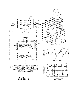
[0043] Fig. 1 is a schematic diagram of example modules, or
components, configured to non-
invasively compute cycle variability-related features or parameters to
generate, via a classifier
(e.g.. machine-learned classifier), one or more metrics associated with the
physiological state of a
patient in accordance with an illustrative embodiment. The modules or
components may be used
in a production application or the development of the cycle variability-
related features and other
classes of features.
[0044] The example analysis and classifiers described herein may be
used to assist a healthcare
provider in the diagnosis and/or treatment of cardiac- and cardiopulmonary-
related pathologies
and medical conditions, or an indicator of one. Examples include significant
coronary artery
disease (CAD), one or more forms of heart failure such as, e.g., heart failure
with preserved
ejection fraction (1-1FpEF), congestive heart failure, various forms of
arrhythmia, valve failure,
various forms of pulmonary hypertension, hypertrophic cardiomyopathy, among
various other
disease and conditions disclosed herein.
[0045] In addition, there exist possible indicators of a disease or
condition, such as an elevated
or abnormal left ventricular end-diastolic pressure (LVEDP) value as it
relates to some forms of
heart failure, abnormal left ventricular ejection fraction (LVEF) values as
they relate to some forms
of heart failure or an elevated mean pulmonary arterial pressure (mPAP) value
as it relates to
pulmonary hypertension and/or pulmonary arterial hypertension. Indicators of
the likelihood that
such indicators are abnormal/elevated or normal, such as those provided by the
example analysis
and classifiers described herein, can help a healthcare provider assess or
diagnose that the patient
has or does not have a given disease or condition. In addition to these
metrics associated with a
disease state of condition, other measurements and factors may be employed by
a healthcare
professional in making a diagnosis, such as the results of a physical
examination and/or other tests,
the patient's medical history, current medications, etc. The determination of
the presence or non-
presence of a disease state or medical condition can include the indication
(or a metric of measure
that is used in the diagnosis) for such disease.
[0046] In Fig. 1, the components include at least one non-invasive
biophysical signal recorder
or capture system 102 and an assessment system 103 that is located, for
example, in a cloud or
remote infrastructure or in a local system. Biophysical signal capture system
102 (also referred to
CA 03203044 2023- 6- 21
WO 2022/137167
PCT/1B2021/062193
as a biophysical signal recorder system), in this embodiment, is configured
to, e.g., acquire,
process, store and transmit synchronously acquired patient's electrical and
hemodynamic signals
as one or more types of biophysical signals 104. In the example of Fig. 1, the
biophysical signal
capture system 102 is configured to synchronously capture two types of
biophysical signals shown
5 as first biophysical signals 104a (e.g., synchronously acquired to other
first biophysical signals)
and second biophysical signals 104b (e.g., synchronously acquired to the other
biophysical signals)
acquired from measurement probes 106 (e.g., shown as probes 106a and 106b,
e.g., comprising
hemodynamic sensors for hemodynamic signals 104a, and probes 106c-106h
comprising leads for
electrical/cardiac signals 104b). In some embodiments, the non-invasive
biophysical signal
10 capture system 102 is configured to capture one type of biophysical
signals, e.g., first biophysical
signals 104a, second biophysical signals 104b, or any of the biophysical
signals described herein.
In the example shown in Fig. 1, the probes 106a-h are placed on, e.g., by
being adhered to or placed
next to, a surface tissue of a patient 108 (shown at patient locations 108a
and 108b). The patient
is preferably a human patient, but it can be any mammalian patient. The
acquired raw biophysical
signals (e.g., 106a and 106b) together form a biophysical-signal data set 110
(shown in Fig. 1 as a
first biophysical-signal data set 110a and a second biophysical-signal data
set 110b, respectively)
that may be stored, e.g., as a single file, preferably, that is identifiable
by a recording/signal
captured number and/or by a patient's name and medical record number.
[0047] In the Fig. 1 embodiment, the first biophysical-signal data
set 110a comprises a set of
raw photoplethysmographic, or hemodynamic, signal(s) associated with measured
changes in light
absorption of oxygenated and/or deoxygenated hemoglobin from the patient at
location 108a, and
the second biophysical-signal data set 110b comprises a set of raw cardiac or
biopotential signal(s)
associated with electrical signals of the heart. Though in Fig. 1, raw
photoplethysmographic or
hemodynamic signal(s) are shown being acquired at a patient's finger, the
signals may be
alternatively acquired at the patient's toe, wrist, forehead, earlobe, neck,
etc. Similarly, although
the cardiac or biopotential signal(s) are shown to be acquired via three sets
of orthogonal leads,
other lead configurations may be used (e.g., 11 lead configuration, 12 lead
configuration, etc.).
100481 Plots 110a' and 110b' show examples of the first biophysical-
signal data set 110a and
the second biophysical-signal data set II0a, respectively. Specifically, Plot
110a' shows an
example of an acquired photoplethysmographic or hemodynamic signal. In Plot
110a', the
photoplethysmographic signal is a time series signal having a signal voltage
potential as a function
CA 03203044 2023- 6- 21
WO 2022/137167
PCT/1B2021/062193
11
of time as acquired from two light sources (e.g., infrared and red-light
source). Plot 110b' shows
an example cardiac signal comprising a 3-channel potential time series plot.
In some
embodiments, the biophysical signal capture system 102 preferably acquires
biophysical signals
via non-invasive means or component(s). In alternative embodiments, invasive
or minimally-
invasively means or component(s) may be used to supplement or as substitutes
for the non-invasive
means (e.g., implanted pressure sensors, chemical sensors, accelerometers, and
the like). In still
further alternative embodiments, non-invasive and non-contact probes or
sensors capable of
collecting biophysical signals may be used to supplement or as substitutes for
the non-invasive
and/or invasive/minimally invasive means, in any combination (e.g., passive
thermometers,
scanners, cameras, x-ray, magnetic, or other means of non-contact or contact
energy data collection
system as discussed herein). Subsequent to signal acquisitions and recording,
the biophysical
signal capture system 102 then provides, e.g., sending over a wireless or
wired communication
system and/or a network, the acquired biophysical-signal data set 110 (or a
data set derived or
processed therefrom, e.g., filtered or pre-processed data) to a data
repository 112 (e.g., a cloud-
based storage area network) of the assessment system 103. In some embodiments,
the acquired
biophysical-signal data set 110 is sent directly to the assessment system 103
for analysis or is
uploaded to a data repository 112 through a secure clinician's portal.
[0049] Biophysical signal capture system 102 is configured with
circuitries and computing
hardware, software, firmware, middleware, etc., in some embodiments, to
acquire, store, transmit,
and optionally process both the captured biophysical signals to generate the
biophysical-signal
data set 110. An example biophysical signal capture system 102 and the
acquired biophysical-
signal set data 110 are described in U.S. Patent No. 10,542,898, entitled
"Method and Apparatus
for Wide-Band Phase Gradient Signal Acquisition," or U.S. Patent Publication
No. 2018/0249960,
entitled "Method and Apparatus for Wide-Band Phase Gradient Signal
Acquisition," each of which
is hereby incorporated by reference herein in its entirety.
[0050] In some embodiments, biophysical signal capture system 102
includes two or more
signal acquisition components, including a first signal acquisition component
(not shown) to
acquire the first biophysical signals (e.g., photoplethysmographic signals)
and includes a second
signal acquisition component (not shown) to acquire the second biophysical
signals (e.g., cardiac
signals). In some embodiments, the electrical signals are acquired at a multi-
kilohertz rate for a
few minutes, e.g., between 1 kHz and 10 kHz. In other embodiments, the
electrical signals are
CA 03203044 2023- 6- 21
WO 2022/137167
PCT/1B2021/062193
12
acquired between 10 kHz and 100 kHz. The hemodynamic signals may be acquired,
e.g., between
100 Hz and 1 kHz.
[0051] Biophysical signal capture system 102 may include one or
more other signal acquisition
components (e.g., sensors such as mechano-acoustic, ballistographic,
ballistocardiographic, etc.)
for acquiring signals. In other embodiments of the signal capture system 102,
a signal acquisition
component comprises conventional electrocardiogram (ECG/EKG) equipment (e.g.,
Holter
device, 12 lead ECG, etc.).
[0052] Assessment system 103 comprises, in some embodiments, the
data repository 112 and
an analytical engine or analyzer (not shown ¨ see Figs. 13A and 13B).
Assessment system 103
may include feature modules 114 and a classifier module 116 (e.g., an ML
classifier module). In
Fig. 1, Assessment system 103 is configured to retrieve the acquired
biophysical signal data set
110, e.g., from the data repository 112, and use it in the feature modules
114, which is shown in
Fig. 1 to include a cycle variability-associated feature module 120 and other
modules 122 (later
described herein). The features modules 114 compute values of features or
parameters, including
those of cycle variability-related features to provide to the classifier
module 116, which computes
an output 118, e.g., an output score, of the metrics associated with the
physiological state of a
patient (e.g., an indication of the presence or non-presence of a disease
state, medical condition,
or an indication of either). Output 118 is subsequently presented, in some
embodiments, at a
healthcare physician portal (not shown ¨ see Figs. 13A and 13B) to be used by
healthcare
professionals for the diagnosis and treatment of pathology or a medical
condition. In some
embodiments, a portal may be configured (e.g., tailored) for access by, e.g.,
patient, caregivers,
researchers, etc., with output 118 configured for the portal's intended
audience. Other data and
information may also be a part of output 118 (e.g., the acquired biophysical
signals or other
patient's information and medical history).
[0053] Classifier module 116 (e.g., ML classifier module) may include
transfer functions, loop
up tables, models, or operators developed based on algorithms such as but not
limited to decision
trees, random forests, neural networks, linear models, Gaussian processes,
nearest neighbor,
SVMs, Naïve Bayes, etc. In some embodiments, classifier module 116 may include
models that
are developed based on ML techniques described in concurrently filed U.S.
provisional patent
application entitled "Method and System to Non-Invasively Assess Elevated Left
Ventricular End-
Diastolic Pressure" having attorney docket no. 10321-048pv1; U.S. Patent
Publication No.
CA 03203044 2023- 6- 21
WO 2022/137167
PCT/1B2021/062193
13
20190026430, entitled "Discovering Novel Features to Use in Machine Learning
Techniques, such
as Machine Learning Techniques for Diagnosing Medical Conditions-; or U.S.
Patent Publication
No. 20190026431, entitled "Discovering Genomes to Use in Machine Learning
Techniques," each
of which is hereby incorporated by reference herein in its entirety.
[0054] Example Biophysical Signal Acquisition
[0055] Fig. 2 shows a biophysical signal capture system 102 (shown
as 102a) and its use in
non-invasively collecting biophysical signals of a patient in a clinical
setting in accordance with
an illustrative embodiment. In Fig. 2, the biophysical signal capture system
102a is configured to
capture two types of biophysical signals from the patient 108 while the
patient is at rest. The
biophysical signal capture system 102a synchronously acquires the patient's
(i) electrical signals
(e.g., cardiac signals corresponding to the second biophysical-signal data set
110b) from the torso
using orthogonally placed sensors (106c-106h; 106i is a 7th common-mode
reference lead) and (ii)
hemodynamic signals (e.g., PPG signals corresponding to the first biophysical-
signal data set
110a) from the finger using a photoplethysmographic sensor (e.g., collecting
signals 106a, 106b).
[0056] As shown in Fig. 2, the electrical and hemodynamic signals (e.g.,
104a, 104b) are
passively collected via commercially available sensors applied to the
patient's skin. The signals
may be acquired beneficially without patient exposure to ionizing radiation or
radiological contrast
agents and without patient exercise or the use of pharmacologic stressors. The
biophysical signal
capture system 102a can be used in any setting conducive for a healthcare
professional, such as a
technician or nurse, to acquire the requisite data and where a cellular signal
or Wi-Fi connection
can be established.
[0057] The electrical signals (e.g., corresponding to the second
biophysical signal data set
110b) are collected using three orthogonally paired surface electrodes
arranged across the patient's
chest and back along with a reference lead. The electrical signals are
acquired, in some
embodiments, using a low-pass anti-aliasing filter (e.g., ¨ 2kHz) at a multi-
kilohertz rate (e.g., 8
thousand samples per second for each of the six channels) for a few minutes
(e.g., 215 seconds).
In alternative embodiments, the biophysical signals may be
continuously/intermittently acquired
for monitoring, and portions of the acquired signals are used for analysis.
The hemodynamic
signals (e.g., corresponding to the first biophysical signal data set I I Oa)
are collected using a
photoplethysmographic sensor placed on a finger. The photo-absorption of red
light (e.g., any
wavelengths between 600-750 nm) and infrared light (e.g., any wavelengths
between 850-950nm)
CA 03203044 2023- 6- 21
WO 2022/137167
PCT/1B2021/062193
14
are recorded, in some embodiments, at a rate of 500 samples per second over
the same period. The
biophysical signal capture system 102a may include a common mode drive that
reduces common-
mode environmental noise in the signal. The photoplethysmographic and cardiac
signals were
simultaneously acquired for each patient. Jitter (inter-modality jitter) in
the data may be less than
about 10 microseconds ( s). Jitter among the cardiac signal channels may be
less than 10
microseconds, e.g., around ten femtoseconds (fs).
[0058] A signal data package containing the patient metadata and
signal data may be compiled
at the completion of the signal acquisition procedure. This data package may
be encrypted before
the biophysical signal capture system 102a transferring to the data repository
112. In some
embodiments, the data package is transferred to the assessment system (e.g.,
103). The transfer is
initiated, in some embodiments, following the completion of the signal
acquisition procedure
without any user intervention. The data repository 112 is hosted, in some
embodiments, on a cloud
storage service that can provide secure, redundant, cloud-based storage for
the patient's data
packages, e.g., Amazon Simple Storage Service (i.e., "Amazon S3"). The
biophysical signal
capture system 102a also provides an interface for the practitioner to receive
notification of an
improper signal acquisition to alert the practitioner to immediately acquire
additional data from
the patient.
[0059] Example Method of Operation
[0060] Figs. 3A-3B each shows an example method to use cycle
variability-related features or
their intermediate outputs in a practical application for diagnostics,
treatment, monitoring, or
tracking.
[0061] Estimation of Presence of Disease State or Indicating
Condition. Fig. 3A shows a
method 300a that employs cycle variability-related parameters or features to
determine estimators
of the presence of a disease state, medical condition, or indication of
either, e.g., to aid in the
diagnosis, tracking, or treatment. Method 300a includes the step of acquiring
(302) biophysical
signals from a patient (e.g., cardiac signals, photoplethysmographic signals,
ballistocardiographic
signals), e.g., as described in relation to Figs. 1 and 2 and other examples
as described herein. In
some embodiments, the acquired biophysical signals are transmitted for remote
storage and
analysis. In other embodiments, the acquired biophysical signals are stored
and analyzed locally.
[0062] As stated above, one example in the cardiac context is the
estimation of the presence
of abnormal left-ventricular end-diastolic pressure (LVEDP) or mean pulmonary
artery pressure
CA 03203044 2023- 6- 21
WO 2022/137167
PCT/1B2021/062193
(mPAP), significant coronary artery disease (CAD), abnormal left ventricular
ejection fraction
(LVEF), and one or more forms of pulmonary hypertension (PH), such as
pulmonary arterial
hypertension (PAH). Other pathologies or indicating conditions that may be
estimated include,
e.g., one or more forms of heart failure such as, e.g., heart failure with
preserved ejection fraction
5 (HFpF1), arrhythmia, congestive heart failure, valve failure,
hypertrophic cardiomyopathy, among
various other disease and medical conditions disclosed herein.
[0063] Method 300a further includes the step of retrieving (304)
the data set and determining
values of cycle variability-related features that describe the spectral or
information content that is
in-band to the frequency range of the signal and has a similar amplitude but
is not synchronized
10 with the cardiac cycle. Example operations to determine the values of
cycle variability-related
features are provided in relation to Figs. 4-12 later discussed herein. Method
300a further includes
the step of determining (306) an estimated value for a presence of a disease
state, medical
condition, or an indication of either based on an application of the
determined cycle variability-
related features to an estimation model (e.g., ML models). An example
implementation is
15 provided in relation to Figs. 13A and 13B.
[0064] Method 300a further includes the step of outputting (308)
estimated value(s) for the
presence of disease state or abnormal condition in a report (e.g., to be used
diagnosis or treatment
of the disease state, medical condition, or indication of either), e.g., as
described in relation to Figs.
1, 13A, and 13B and other examples described herein.
[0065] Diagnostics or Condition Monitoring or Tracking using Cycle
Variability Features.
Fig. 3B shows a method 300b that employs cycle-variability-related parameters
or features for the
monitoring health or controls of medical equipment or health monitoring
device. Method 300b
includes the step of obtaining (302) biophysical signals from a patient (e.g.,
cardiac signals,
photoplethysmographic signals, ballistocardiographic signals, etc.). The
operation may be
performed continuously or intermittently, e.g., to provide output for a report
or as controls for the
medical equipment or the health monitoring device.
[0066] Method 300b further includes determining (310) cycle-
variability-related value(s) from
the acquired biophysical data set, e.g., as described in relation to Figs. 4-
12.
[0067] Method 300b further includes outputting (312) the cycle-
variability-related value(s)
(e.g., in a report for use in diagnostics or as signals for controls). For
monitoring and tracking, the
output may be via a wearable device, a handheld device, or medical diagnostic
equipment (e.g.,
CA 03203044 2023- 6- 21
WO 2022/137167
PCT/1B2021/062193
16
pulse oximeter system, wearable health monitoring systems) to provide
augmented data associated
with health. In some embodiments, the outputs may be used in resuscitation
systems, cardiac or
pulmonary stress test equipment, pacemakers, etc.
[0068] Cycle Variability Features or Parameters
[0069] Figs. 4, 5, and 6 each shows an example cycle variability analysis
feature computation
module, for a total of three example modules configured to determine values of
cycle variability
features or parameters of biophysical signals in accordance with an
illustrative embodiment. The
cycle variability score analysis feature computation module 400 can calculate
a score-based metric
for cycle variability (shown in Fig 4, including CV score, CV relative score,
and CV ratio score).
The cycle variability distribution analysis feature computation module 500 can
calculate the
distribution of the values of cycle variability and provide a statistical
assessment as the feature
output of that distribution. The cycle-variability point-cloud analysis
feature computation module
600 can generate a point cloud from the cycle variability data to which an
encapsulated volumetric
object can be generated and various topological and morphological
characteristics of the
volumetric object can be determined, such as volume, porosity, surface,
perimeters. Figs. 7A, 7B,
and 7C, respectively, show example operations of the cycle variability
analysis feature
computation module of Figs. 4, 5, and 6.
[0070] Example #1 ¨ Cycle Variability Score Features
100711 Fig. 4 illustrates, as a first of three example feature or
parameter categories, an example
of the cycle variability score analysis feature computation module 400 (shown
as "CV Score
Feature(s)" 400) configured to determine values of the cycle-variability
associated score of one or
more acquired biophysical signals in accordance with an illustrative
embodiment. In the example
shown in Fig. 4, Module 400 is configured to output a cycle-variability score
402 a given signal.
For a cardiac cycle, e.g., with 3 channels, a cycle-variability score may be
generated for each of
the three channels. Module 400 may also generate a cycle-variability relative
score 404 that
normalizes (e.g., dynamic scaling, mean max, z-transform, etc.) each cycle-
variability score for a
given channel to all the calculated score (e.g., for all three channels). In
other embodiments non-
normalize values can be used for the score calculation. Module 400 may also
generate a cycle-
variability ratio score 406 that determines a ratio between the cycle-
variability score for a given
channel to the cycle-variability score for another channel.
CA 03203044 2023- 6- 21
WO 2022/137167
PCT/1B2021/062193
17
[0072] Table 2
shows an example set of 9 extractable cycle-variability features and their
corresponding description. In Table 2, features (shown with "*") have been
observed to have
significant utility in the assessment of the presence or non-presence of at
least one cardiac disease
or condition ¨ specifically, the determination of presence or non-presence of
elevated LVEDP.
The list of the specific features determined to have significant utility in
the assessment of the
presence or non-presence of abnormal or elevated LVEDP is provided in Table 7.
Table 2
Feature name Description
1 CV score X* Cycle variability score of channel X of cardiac signals
(CV)
2 CV score Y Cycle variability score of channel Y of cardiac
signals(CVy)
3 CV score Z Cycle variability score of channel Z of cardiac
signals(CVz)
CV,
4 CV relative X CV, + C Vy + C V,
CV
5 CV relative Y CV), + C Vy + C V,
CV,
6 CV relative Z CV, + CVy + CV,
C V,
7 CV ratio XY CVy
CV,
8 CV ratio XZ CV,
CV
9 CV ratio YZ CV,
[0073] Cycle
Variability Score Computation. Fig. 7A is a diagram of an exemplary method
700 to generate a cycle-variability score (e.g., "CV score X" CVx "CV score Y"
CV, "CV score
Z" CVz) of a biophysical signal in accordance with an illustrative embodiment.
Method 700
includes detecting peaks (702) in all, or a substantial portion, of the
biophysical signal for a quasi-
periodic cycle. For cardiac signals, the peaks may be points of ventricular
depolarization (also
commonly referred to as "R-peaks"), which are a point in the signal during
each cycle when the
electrical activation of the ventricles is maximal. In some embodiments,
Method 700 employs a
Pan-Tompkins algorithm for ventricular depolarization detection, for example,
as described in Pan
& Tompkins, A Real Time QRS Detection Algorithm, IEEE Transactions on
Biomedical
CA 03203044 2023- 6- 21
WO 2022/137167
PCT/1B2021/062193
18
Engineering, Volume 32-3, 230-236, 1985, the entirety of which is hereby
incorporated by
reference herein in its entirety. Other algorithms may be used to detect peaks
in the cardiac signal
data set ¨ examples include those described in Makwana et al. "Hilbert
transform based adaptive
ECG R-peak detection technique," International Journal of Electrical and
Computer Engineering,
2(5), 639 (2012); Lee et al., "Smart ECG Monitoring Patch with Built-in R-Peak
Detection for
Long-Term HRV Analysis," Annals of Biomedical Engineering. 44(7), 2292-3201
(2016); and
Kim et al., "Detection of R-Peaks in ECG Signal by Adaptive Linear Neuron
(ADALINE),"
Artificial Neural Network, presented at MATEC Web of Conferences, 54, 10001
(2016), each of
which is hereby incorporated by reference herein in its entirety. Various PPG
peak detectors may
be used for photoplethysmographic signals.
[0074] Method 700 then includes determining or creating (704) a
template-signal vector data
set (also referred to as a template cycle). The template-signal vector data
set represents a quasi-
periodic signal pattern of the subject (e.g., a heart-beat pattern for cardiac
signals). The term
"quasi-periodic" can also be referred to in more general terms as a
characteristic of a signal system
that cycles with, at a minimum, two frequency components, of which the ratio
is not a rational
number. In some embodiments, to determine the template-signal vector data set,
a median peak-
peak interval (e.g., R-R intervals for cardiac signals) is calculated using
the detected peak
locations. The cycle region is set around each peak and normalized for the
amplitude. The cycle
region includes regions of interest, for example, the P and completion of the
T wave for cardiac
signals.
[0075] In Fig. 8A, the detected peak locations of cardiac signal
104b (e.g., shown as 802a-
802g) are used to determine a median peak-to-peak interval (e.g., median R-R
peaks for portions
of the cardiac signal 104b as shown with 804a-804g) and to set a cycle region
(e.g., shown as 806a-
806f) around each peak (e.g., R-peaks for portions of cardiac signals 104b as
shown with 808a-
808g). Fig. SA further shows that the cycle region is set around the R-peak
and includes both the
P wave and the completion of the T wave for cardiac signal 104b. In Fig. 8A,
the ranges are from
about -20% to about +20% of the median interval (e.g., shown as 812a, 812b).
Each of the cycle
regions (e.g., 806a-806f) can be stored by a processor in a matrix (also
referred to as a -cycle
matrix"). The cycle matrix may have the size AlxN in which M is the number of
detected cycles,
and N is 40% (or other range) of the median peak-to-peak interval (e.g.,
median R-R intervals for
cardiac signals) in which the 40% of the peak-to-peak interval represents the
full temporal "width"
CA 03203044 2023- 6- 21
WO 2022/137167
PCT/1B2021/062193
19
of the cycle. Specifically, once the median peak-to-peak interval (e.g.,
median R-R interval for
cardiac signals) is known across the dataset, the signal can be divided in
half, e.g., to get the "20%"
that reaches both forward and backward in time from the peak (e.g., R-peak) to
capture the other
waves (e.g., T wave and P wave for a cardiac signal). Of course, other cycle
region lengths can be
used for cardiac signals and for the various distinct waves in brain signals,
etc. For example,
ranges that may be applied include, but are not limited to, from -10% to 10%, -
15% to 15%, -25%
to 25%. In addition, rather than a median of the peak-to-peak interval, the
mean of the peak-to-
peak interval may also be used.
[0076] Fig. 8B shows a plot of results of the normalization process
in accordance with an
illustrative embodiment. In Fig. 8B, each cycle region (e.g., shown as 806a-
806f in Fig. 8A) of the
biophysical-signal data set (e.g., cardiac signal data set 110b) is normalized
by a processor to
remove any offsets such that the average value of each cycle region is zero.
The normalized cardiac
signal data set, as shown, can have a range of "1" and "-1," though that range
can vary depending
on the distribution of the data. The normalization process can be employed in
embodiment when
calculating for the cycle variability feature. In other embodiments, non-
normalized signals may
be used. In addition to mean and scaling normalization, other normalization
methods may be
employed. Examples of other normalization methods include Z-transform and mean
Max, among
others.
[0077] Fig. 9A shows three plots of determined template-signal
vector data 900 (shown as
900a, 900b, and 900c) in reference to one of the cycles 902 (shown as 902a,
902b, and 902c). In
Fig. 9A, the template-signal vector data set is shown for each of the three
cardiac signals. In some
embodiments, the same template-signal vector data set calculated for one
representative cycle is
used to assess against all of the cycles of that given signal. Fig. 9B shows
in phase space the
determined template-signal vector data set 900 of Fig. 9A across the multiple
cycles. In the phase
space plot of Fig. 9B, each value of the template-signal vector data set
corresponding to the same
time instance is shown in the three-dimensional space. The values for all the
channels of the
acquired biophysical signals are also concurrently displayed in the X, Y, and
Z-axis.
100781 Referring back to Fig. 7A, Method 700 then includes
quantifying (706) the difference
between each detected biophysical cycle and the template-signal vector data
set. In some
embodiments, the template-signal vector data set is subtracted from each of
the detected
biophysical cycles to generate a residue data set. Fig. 9C shows a plot of the
residue 906 calculated
CA 03203044 2023- 6- 21
WO 2022/137167
PCT/1B2021/062193
between the detected biophysical cycle and the template-signal vector data set
in phase space.
Each axis in Fig. 9C represents a channel (channels X, Y, and Z) of the
acquired signals. As
discussed herein, the residue can be determined from normalized data or non-
normalized data
using mean and scaling normalization, z-transforms, mean Max, among others.
5 [0079] Referring to Fig_ 7A, Method 700 then includes combining the
resultant differences
across the detected phase cycles to create the final cycle variability score
(CVS) for the channel.
The cycle variability score, in some embodiments, is a median of the
calculated residue data set
for each given signal (e.g., 1 score per channel). In other embodiments, the
cycle variability score
is a median of the calculated residue data set for all of the signals (e.g., 1
score per set of channels).
10 In some embodiments, the cycle variability score is calculated from a
subset of the acquired
signals. In another embodiment, the cycle variability score is a mean of the
calculated residue data
set for a given signal. In yet another embodiment, the cycle variability score
is a mean of the
calculated residue data set for all of the signals, or a representative subset
of such signals.
[0080] In some embodiments, the cycle variability score is a Z-
score value for a given data
15 point in the template signal vector data set and is calculated as a
difference between the value of
the given data point and a mean of a set of cycles in which the difference is
then normalized by
the standard deviation of that given data point to the same indexed data value
of the set of cycles.
[0081] Cycle Variability Relative Score Computation. Fig. 7B shows
a diagram of a method
to generate cycle-variability relative scores 404 (shown as 404a, 404b, 404c)
that normalize each
20 cycle-variability score for a given channel to all the calculated score
(e.g., for all three channels).
In Fig. 7B, the cycle-variability relative scores 404a, 404b, 404c are shown
for three channels.
The cycle-variability relative score 404a, 404b, 404c for a channel n may be
calculated over the
sum of all the calculated scores, as shown in Equation 1:
cvn
(Equation 1)
zni cvr,
[0082] In Table 2, the cycle-variability relative scores 404 are shown as
"CV relative X,"
"CV relative Y," and "CV relative Z."
[0083] Cycle Variability Ratio Computation. Fig. 7C shows a diagram
to generate cycle-
variability ratio scores 406 (shown as 406a, 406b, and 406c) that normalize
each cycle-variability
score as a ratio between two channels. In Fig. 7C, the cycle-variability ratio
scores 406a, 406b,
406c are shown for three channels. In Table 2, the cycle-variability ratio
scores 406a, 406b, and
406c are shown as "CV ratio XY," "CV ratio XZ," and "CV ratio YZ,"
respectively.
CA 03203044 2023- 6- 21
WO 2022/137167 PCT/1B2021/062193
21
[0084] Example #2 ¨ Cycle Variability Statistical Distribution Feature
[0085] Fig. 5 illustrates, as a second of three example feature or
parameter categories, an
example cycle variability distribution analysis feature computation module 500
(shown as "CV
Distribution Feature(s)" 500) configured to determine an assessment comprising
statistical
parameters of the distribution of calculated cycle-variability values for the
acquired biophysical
signals in accordance with an illustrative embodiment. In the example shown in
Fig. 5, the
statistical assessment can include a mean (502), median (504), standard
deviation (506), skewness
(508), and kurtosis (510) of an assessed distribution.
[0086] Table 3 lists an example set of cycle-variability features and
their corresponding
description. In Table 3, features (shown with "*") have been observed to have
significant utility
in the assessment of the presence or non-presence of at least one cardiac
disease or condition ¨
specifically, the determination of presence or non-presence of elevated LVEDP.
The list of the
specific features determined to have significant utility in the assessment of
the presence or non-
presence of abnormal or elevated LVEDP is provided in Table 7.
Table 3
10 CV_X mean Mean of X CV
11 CV_X median Median of X CV
12 CV_X std* Std of X CV
13 CV_X Skew* Skewness of X CV
14 CV_X Kurt Kurtosis of X CV
15 CV_Y mean Mean of Y CV
16 CV_Y median Median of Y CV
17 CV_Y std Std of Y CV
18 CV_Y Skew Skewness of Y CV
19 CV_Y Kurt Kurtosis of Y CV
20 CV_Z mean Mean of Z CV
21 CV_Z median Median of Z CV
22 CV_Z std* Std of Z CV
23 CV_Z Skew Skewness of Z CV
24 CV Z Kurt Kurtosis of Z CV
CA 03203044 2023- 6- 21
WO 2022/137167
PCT/1B2021/062193
22
[0087] Fig. 10 is a diagram of an exemplary method 1000 to generate
a distribution (e.g.,
histogram) of cycle-variability residues of a biophysical signal in accordance
with an illustrative
embodiment. In Fig. 10, Method 1000 may include performing peak detection step
(702), a
template cycle creation step (704), and a residue distribution calculation
step (706) as described in
relation to Fig. 7A. Rather than combining all the calculated residue into a
single score for the
channel, or the set of channels, e.g., as described in relation to Fig. 7A,
Method 1000 performs a
statistical assessment (1002) of the calculated residue distribution. The
distribution can be, e.g., a
histogram that includes the calculated CV residue of each cycle of the
channel. The statistical
assessment may include a mean, median, standard deviation, skewness, and
kurtosis of the
determined distribution.
[0088] Cycle Variability Associated Features Example #3 - CV Model
Parameters
[0089] Fig. 6 illustrates, as a third of three example feature or
parameter categories, an
example cycle-variability point-cloud analysis feature computation module 600
(shown as "CV
Point Cloud Feature(s)" 600) configured to determine values of cycle-
variability point-cloud
features of one or more acquired biophysical signals in accordance with an
illustrative
embodiment. In Fig. 6, Module 600 is configured to generate a two- or three-
dimensional phase
space model (e.g., an alpha shape model) of the calculated CV residue; for
example, as described
in relation to Figs. 5, 6, and 7A, and determining geometric-based parameters
of that three-
dimensional phase space model (e.g., an alpha shape model).
[0090] For a cardiac cycle, e.g., with 3 channels, the three-dimensional
phase space model can
be generated with each channel serving as an axis of the model, or a two-
dimensional phase space
model can be generated from two channels. The geometric parameters that may be
assessed from
the three-dimensional phase space model include volume (602), porosity (604),
void volume (606),
surface area (608). The geometric parameter that may be assessed from the two-
dimensional phase
space model includes the perimeter (610).
[0091] Table 4 shows an example set of cycle-variability features
determined from a three-
dimensional phase space model and their corresponding description. In Table 4,
features (shown
designated with -*") have been observed to have significant utility in the
assessment of the
presence or non-presence of at least one cardiac disease or condition -
specifically, the
determination of presence or non-presence of elevated LVEDP. The list of the
specific features
CA 03203044 2023- 6- 21
WO 2022/137167 PCT/1B2021/062193
23
determined to have significant utility in the assessment of the presence or
non-presence of
abnormal or elevated LVEDP is provided in Table 7.
Table 4
25 C V_Volume* Alpha Shape volume
26 CV_VoidVolume* CV Convex Hull Volume
¨ CV Alpha Shape Volume
27 CV_Porosity CV Void Volume
CV Convex Volume
28 CV_SurfaceArea* Alpha Shape surface area
29 CV_PerimeterXY* Perimeter of the XY Alpha
Shape
30 CV_PerimeterXZ Perimeter of the XZ Alpha
Shape
31 CV_PerimeterYZ* Perimeter of the YZ Alpha
Shape
[0092] Fig. 11 is a diagram of an exemplary method 1100 to generate a two-
or three-
dimensional phase space model from the cycle-variability residue of a
biophysical signal in
accordance with an illustrative embodiment. In Fig. 11, Method 1100 may
include performing the
peak detection step (702), the template cycle creation step (704), and the
residue distribution
calculation step (706) as described in relation to Fig. 7A.
[0093] Method 1100 further includes generating (1102) a two- or three-
dimensional phase
space model from the calculated residues determined from the three channels of
the acquired data
set. The residues may be used generate a point-cloud map to which a
triangulation operation may
be applied. Examples of triangulation operation include alpha hull as well as
convex hull. Other
types of triangulation operations may be applied.
[0094] Fig. 9C shows an example point-cloud map 906 generated from the
residue data set of
a three-channel cardiac signal.
[0095] Fig. 12A shows an example three-dimensional phase space model 1
200 generated from
the point-cloud map 906 of Fig. 9C in accordance with an illustrative
embodiment. The two- or
three-dimensional phase space model 1200 may be colorized in some embodiments
to assess a
fourth-dimensional data. In Fig. 12A, there appears to be a structure in
viewing the CV residues
in phase space. Further, this structure is related to the temporal location
within the cardiac cycle,
CA 03203044 2023- 6- 21
WO 2022/137167
PCT/1B2021/062193
24
as can be seen in Fig. 12A in which color can be provided based on the radius
components of
signals in a spherical coordinate system. For example, as shown in Fig. 12A,
there appears to be
clustering of high radius amplitudes in certain regions of the image,
indicating that the residue in
those sections originates from the ventricular depolarization.
[0096] Fig. 12B is a plot illustrating a radius component of a given
signal, in spherical
coordinates, in the time domain that can be used to color Fig. 12A. In some
embodiments, color
information may be used as part of an extracted feature.
[0097] Experimental Results and Examples
[0098] Several development studies have been conducted to develop
feature sets, and in turn,
algorithms that can be used to estimate the presence or non-presence,
severity, or localization of
diseases, medical conditions, or an indication of either. In one study,
algorithms were developed
for the non-invasive assessment of abnormal or elevated LVEDP. As noted above,
abnormal or
elevated LVEDP is an indicator of heart failure in its various forms. In
another development study,
algorithms and features were developed for the non-invasive assessment of
coronary artery
disease.
[0099] As part of these two development studies, clinical data were
collected from adult human
patients using a biophysical signal capture system and according to protocols
described in relation
to Fig. 2. The subjects underwent cardiac catheterization (the current -gold
standard" tests for CAD
and abnormal LVEDP evaluation) following the signal acquisition, and the
catheterization results
were evaluated for CAD labels and elevated LVEDP values. The collected data
were stratified into
separate cohorts: one for feature/algorithm development and the other for
their validation.
[0100] Within the feature development phases, features were
developed, including the cycle-
variability-related features, to extract characteristics in an analytical
framework from biopotential
signals (as an example of the cardiac signals discussed herein) and photo-
absorption signals (as
examples of the hem odynamic or photoplethysmographic discussed herein) that
are intended to
represent properties of the cardiovascular system. Corresponding classifiers
were also developed
using classifier models, linear models (e.g., Elastic Net), decision tree
models (XGB Classifier,
random forest models, etc.), support vector machine models, and neural network
models to non-
invasively estimate the presence of an elevated or abnormal LVEDP. Univariate
feature selection
assessments and cross-validation operations were performed to identify
features for use in machine
learning models (e.g., classifiers) for the specific disease indication of
interest. Further description
CA 03203044 2023- 6- 21
WO 2022/137167
PCT/1B2021/062193
of the machine learning training and assessment are described in a U.S.
provisional patent
application concurrently filed herewith entitled "Method and System to Non-
Invasively Assess
Elevated Left Ventricular End-Diastolic Pressure" having attorney docket no.
10321-048py1,
which is hereby incorporated by reference herein in its entirety.
5 [0101] The univariate feature selection assessments evaluated many
scenarios, each defined
by a negative and a positive dataset pair using t-test, mutual information,
and AUC-ROC
evaluation. The t-test is a statistical test that can determine if there is a
difference between two
sample means from two populations with unknown variances. Here, the t-tests
were conducted
against a null hypothesis that there is no difference between the means of the
feature in these
10 groups, e.g., normal LVEDP vs. elevated (for LVEDP algorithm
development); CAD- vs. CAD+
(for CAD algorithm development). A small p-value (e.g., < 0.05) indicates
strong evidence against
the null hypothesis.
[0102] Mutual information (MI) operations were conducted to assess
the dependence of
elevated or abnormal LVEDP or significant coronary artery disease on certain
features. An MI
15 score greater than one indicates a higher dependency between the
variables being evaluated. MI
scores less than one indicates a lower dependency of such variables, and an MI
score of zero
indicates no such dependency.
[0103] A receiver operating characteristic curve, or ROC curve,
illustrates the diagnostic
ability of a binary classifier system as its discrimination threshold is
varied. The ROC curve may
20 be created by plotting the true positive rate (TPR) against the false
positive rate (FPR) at various
threshold settings. AUC-ROC quantifies the area under a receiver operating
characteristic (ROC)
curve ¨ the larger this area, the more diagnostically useful the model is. The
ROC, and AUC-
ROC, value is considered statistically significant when the bottom end of the
95% confidence
interval is greater than 0.50.
25 [0104] Table 6 shows an example list of the negative and a positive
dataset pair used in the
univariate feature selection assessments. Specifically, Table 6 shows positive
datasets being
defined as having an LVEDP measurement greater than 20 mmHg or 25 mmHg, and
negative
datasets were defined as having an LVEDP measurement less than 12 mmHg or
belonging to a
subject group determined to have normal LVEDP readings.
Table 6
Negative Dataset Positive Dataset
CA 03203044 2023- 6- 21
WO 2022/137167 PCT/1B2021/062193
26
12 (mmHg) 20 (mmHg)
12 (mmHg) 25 (mmHg)
Normal LVEDP 20 (mmHg)
Normal LVEDP 25 (mmHg)
[0105] Tables 7A, 7B, and 7C each shows a list of cycle-variability-
related features having
been determined to have utility in estimating the presence and non-presence of
elevated LVEDP
in an algorithm executing in a clinical evaluation system. The features of
Tables 7A, 7B, and 7C
and corresponding classifiers have been validated to have clinical performance
comparable to the
gold standard invasive method to measure elevated LVEDP.
Table 7
FA scenario LVEDP <= 12 (N=246) vs >=20 (N=209)
AUC (bottom of 95%
Feature name t-test p-value CI)
MI
CV Z std n/s 0.5184 1.0240
CV PerimeterYZ O. 0263 0.5140
n/s
CV Void Volume n/s 0.5236
n/s
CV SurfaceArea n/s 0.5352
n/s
CVscore X. n/s 0.5011
n/s
CV PerimeterXY n/s 0.5006
n/s
CV X Skew n/s 0.5091
n/s
CV Volume n/s 0.5285
n/s
CV X std n/s 0.5050 n/s
[0106] The determination that certain cycle-variability-related
features have clinical utility in
estimating the presence and non-presence of elevated LVEDP provides a basis
for the use of these
cycle-variability-related features or parameters, as well as other features
described herein, in
estimating for the presence or non-presence and/or severity and/or
localization of other disease,
medical condition, or an indication of either particularly, though not limited
to, heart disease or
conditions described herein.
[0107] The experimental results further indicate that intermediary data or
parameters of cycle-
variability-related features also have clinical utility in diagnostics as well
as treatment, controls,
monitoring, and tracking applications.
[0108] Example Clinical Evaluation System
CA 03203044 2023- 6- 21
WO 2022/137167
PCT/1B2021/062193
27
[0109] Fig. 13A shows an example clinical evaluation system 1300
(also referred to as a
clinical and diagnostic system) that implements the modules of Fig. 1 to non-
invasively compute
cycle-variability-related features or parameters, along with other features or
parameters, to
generate, via a classifier (e.g., machine-learned classifier), one or more
metrics associated with the
physiological state of a patient or subject according to an embodiment Indeed,
the feature modules
(e.g., of Figs. 1, 5-14) can be generally viewed as a part of a system (e.g.,
the clinical evaluation
system 1300) in which any number and/or types of features may be utilized for
a disease state,
medical condition, an indication of either, or combination thereof that is of
interest, e.g., with
different embodiments having different configurations of feature modules. This
is additionally
illustrated in Fig. 13A, where the clinical evaluation system 1300 is of a
modular design in which
disease-specific add-on modules 1302 (e.g., to assess for elevated LVEDP or
mPAP, CAD,
PH/PAH, abnormal LVEF, H.FpEF, hypertrophic cardiomyopathy, and others
described herein)
are capable of being integrated alone or in multiple instances with a singular
platform (i.e., a base
system 1304) to realize system 1300's full operation. The modularity allows
the clinical evaluation
system 1300 to be designed to leverage the same synchronously acquired
biophysical signals and
data set and base platform to assess for the presence of several different
diseases as such disease-
specific algorithms are developed, thereby reducing testing and certification
time and cost.
[0110] In various embodiments, different versions of the clinical
evaluation system 1300 may
implement the assessment system 103 (Fig. 1) by having included containing
different feature
computation modules that can be configured for a given disease state(s),
medical condition(s), or
indicating con diti on (s) of interest. In another embodiment, the clinical
evaluation system 1300
may include more than one assessment system 103 and maybe selectively utilized
to generate
different scores specific to a classifier 116 of that engine 103. In this way,
the modules of Figs. 1
and 13 in a more general sense may be viewed as one configuration of a modular
system in which
different and/or multiple engines 103, with different and/or multiple
corresponding classifiers 116,
may be used depending on the configuration of module desired. As such, any
number of
embodiments of the modules of Fig. 1, with or without the cycle-variability
specific feature(s),
may exist.
[0111] In Fig. 13A, System 1300 can analyze one or more biophysical-
signal data sets (e.g.,
110) using machine-learned disease-specific algorithms to assess for the
likelihood of elevated
LVEDP, as one example, of pathology or abnormal state. System 1300 includes
hardware and
CA 03203044 2023- 6- 21
WO 2022/137167
PCT/1B2021/062193
28
software components that are designed to work together in combination to
facilitate the analysis
and presentation of an estimation score using the algorithm to allow a
physician to use that score,
e.g., to assess for the presence or non-presence of a disease state, medical
condition, or an
indication of either.
[0112] The base system 1304 can provide a foundation of functions and
instructions upon
which each add-on module 1302 (which includes the disease-specific algorithm)
then interface to
assess for the pathology or indicating condition. The base system 1304, as
shown in the example
of Fig. 13A, includes a base analytical engine or analyzer 1306, a web-service
data transfer API
1308 (shown as "DTAPI" 1308), a report database 1310, a web portal service
module 1313, and
the data repository 111 (shown as 112a).
[0113] Data repository 112a, which can be cloud-based, stores data
from the signal capture
system 102 (shown as 102b). Biophysical signal capture system 102b, in some
embodiments, is a
reusable device designed as a single unit with a seven-channel lead set and
photoplethysmogram
(PPG) sensor securely attached (i.e., not removable). Signal capture system
102b, together with
its hardware, firmware, and software, provides a user interface to collect
patient-specific metadata
entered therein (e.g., name, gender, date of birth, medical record number,
height, and weight, etc.)
to synchronously acquire the patient's electrical and hemodynamic signals. The
signal capture
system 102b may securely transmit the metadata and signal data as a single
data package directly
to the cloud-based data repository. The data repository 112a, in some
embodiments, is a secure
cloud-based database configured to accept and store the patient-specific data
package and allow
for its retrieval by the analytical engines or analyzer 1306 or 1314.
[0114] Base analytical engine or analyzer 1306 is a secure cloud-
based processing tool that
may perform quality assessments of the acquired signals (performed via "SQA"
module 1316), the
results of which can be communicated to the user at the point of care. The
base analytical engine
or analyzer 1306 may also perform pre-processing (shown via pre-processing
module 1318) of the
acquired biophysical signals (e.g., 110¨ see Fig. 1). Web portal 1313 is a
secure web-based portal
designed to provide healthcare providers access to their patient's reports. An
example output of
the web portal 1313 is shown by visualization 1336. The report databases (RD)
1312 is a secure
database and may securely interface and communicate with other systems, such
as a hospital or
physician-hosted, remotely hosted, or remote electronic health records systems
(e.g., Epic, Cerner,
Allscrips, CureMD, Kareo, etc.) so that output score(s) (e.g., 118) and
related information may be
CA 03203044 2023- 6- 21
WO 2022/137167
PCT/1B2021/062193
29
integrated into and saved with the patient's general health record. In some
embodiments, web
portal 1313 is accessed by a call center to provide the output clinical
information over a telephone.
Database 1312 may be accessed by other systems that can generate a report to
be delivered via the
mail, courier service, personal delivery, etc.
[0115] Add-on module 1302 includes a second part 1314 (also referred to
herein as the
analytical engine (AE) or analyzer 1314 and shown as "AE add-on module" 1314)
that operates
with the base analytical engine (AE) or analyzer 1306. Analytical engine (AE)
or analyzer 1314
can include the main function loop of a given disease-specific algorithm,
e.g., the feature
computation module 1320, the classifier model 1324 (shown as "Ensemble" module
1324), and
the outlier assessment and rejection module 1324 (shown as "Outlier Detection"
module 1324).
In certain modular configurations, the analytical engines or analyzers (e.g.,
1306 and 1314) may
be implemented in a single analytical engine module.
[0116] The main function loop can include instructions to (i)
validate the executing
environment to ensure all required environment variables values are present
and (ii) execute an
analysis pipeline that analyzes a new signal capture data file comprising the
acquired biophysical
signals to calculate the patient's score using the disease-specific algorithm.
To execute the analysis
pipeline, AE add-on module 1314 can include and execute instructions for the
various feature
modules 114 and classifier module 116 as described in relation to Fig. 1 to
determine an output
score (e.g., 118) of the metrics associated with the physiological state of a
patient. The analysis
pipeline in the AE add-on module 1314 can compute the features or parameters
(shown as "Feature
Computation" 1320) and identify whether the computed features are outliers
(shown as "Outlier
Detection" 1322) by providing an outlier detection return for a signal-level
response of outlier
versus non-outlier based on the feature. The outliers may be assessed with
respect to the training
data set used to establish the classifier (of module 116). AE add-on module
1314 may generate
the patient's output score (e.g., 118) (e.g., via classifier module 1324)
using the computed values
of the features and classifier models. In the example of an evaluation
algorithm for the estimation
of elevated LVEDP, the output score (e.g., 118) is an LVEDP score.
101171 The clinical evaluation system 1300 can manage the data
within and across components
using the web-service DTAPIs 1308 (also may be referred to as HCPP web
services in some
embodiments). DTAPIs 1308 may be used to retrieve acquired biophysical data
sets from, and to
store signal quality analysis results to, the data repository 112a. DTAPIs
1308 may also be invoked
CA 03203044 2023- 6- 21
WO 2022/137167
PCT/1B2021/062193
to retrieve and provide the stored biophysical data files to the analytical
engines or analyzers (e.g.,
1306, 1314), and the results of the analytical engine's analysis of the
patient signals may be
transferred using DTAPI 1308 to the report database 1310. DTAPIs 1308 may also
be used, upon
a request by a healthcare professional, to retrieve a given patient data set
to the web portal module
5 1313, which may present a report to the healthcare practitioner for
review and interpretation in a
secure web-accessible interface.
[0118] Clinical evaluation system 1300 includes one or more feature
libraries 1326 that store
the cycle variability-related features 120 and various other features of the
feature modules 122.
The feature libraries 1326 may be a part of the add-on modules 1302 (as shown
in Fig. 13A) or the
10 base system 1304 (not shown) and are accessed, in some embodiments, by
the AE add-on module
1314.
[0119] Further details of the modularity of modules and various
configurations are provided
in a U.S. provisional patent application concurrently filed herewith entitled
"Modular Disease
Assessment System" having attorney docket no. 10321-060pv1, which is hereby
incorporated by
15 reference herein in its entirety.
[0120] Example Operation of the Modular Clinical Evaluation System
[0121] Fig. 13B shows a schematic diagram of the operation and
workflow of the analytical
engines or analyzers (e.g., 1306 and 1314) of the clinical evaluation system
1300 of Fig. 13A in
accordance with an illustrative embodiment.
20 [0122] Signal quality assessment /rejection (1330). Referring to Fig.
13B, the base analytical
engine or analyzer 1306 assesses (1330), via SQA module 1316, the quality of
the acquired
biophysical-signal data set while the analysis pipeline is executing. The
results of the assessment
(e.g., pass/fail) are immediately returned to the signal capture system's user
interface for reading
by the user. Acquired signal data that meet the signal quality requirements
are deemed acceptable
25 (i.e., "pass") and further processed and subjected to analysis for the
presence of metrics associated
with the pathology or indicating condition (e.g., elevated LVEDP or mPAP, CAD,
PH/PAR,
abnormal LVEF, HFpEF) by the AE add-on module 1314. Acquired signals deemed
unacceptable
are rejected (e.g., "fail"), and a notification is immediately sent to the
user to inform the user to
immediately obtain additional signals from the patient (see Fig. 2).
30 [0123] The base analytical engine or analyzer 1306 performs two sets
of assessments for signal
quality, one for the electrical signals and one for the hemodynamic signals.
The electrical signal
CA 03203044 2023- 6- 21
WO 2022/137167
PCT/1B2021/062193
31
assessment (1330) confirms that the electrical signals are of sufficient
length, that there is a lack
of high-frequency noise (e.g., above 170 Hz), and that there is no power line
noise from the
environment. The hemodynamic signal assessment (1330) confirms that the
percentage of outliers
in the hemodynamic data set is below a pre-defined threshold and that the
percentage and
maximum duration that the signals of the hemodynamic data set are railed or
saturated is below a
pre-defined threshold.
[0124] Feature Value Computation (1332). The AE add-on module 1314
performs feature
extraction and computation to calculate feature output values. In the example
of the LVEDP
algorithm, the AE add-on module 1314 determines, in some embodiments, a total
of 446 feature
outputs belonging to 18 different feature families (e.g., generated in modules
120 and 122),
including the cycle variability-related features (e.g., generated in module
120).
[0125] Additional descriptions of the various features, including
those used in the LVEDP
algorithm and other features and their feature families, are described in a US
provisional patent
application, concurrently filed herewith, entitled "Method and System to Non-
Invasively Assess
Elevated Left Ventricular End-Diastolic Pressure" having attorney docket no.
10321-048pv1; a
US provisional patent application, concurrently filed herewith, entitled
"Methods and Systems for
Engineering Visual Features From Biophysical Signals for Use in Characterizing
Physiological
Systems" having attorney docket no. 10321-049pv1; a US provisional patent
application,
concurrently filed herewith, entitled "Methods and Systems for Engineering
Power Spectral
Features From Biophysical Signals for Use in Characterizing Physiological
Systems" having
attorney docket no. 1 0321 -050pv1; a US provisional patent application,
concurrently filed
herewith, entitled "Method and System for Engineering Rate-Related Features
From Biophysical
Signals for Use in Characterizing Physiological Systems" having attorney
docket no. 10321-
051pvl ; a U.S. provisional patent application, concurrently filed herewith,
entitled "Methods and
Systems for Engineering Wavelet-Based Features From Biophysical Signals for
Use in
Characterizing Physiological Systems" having attorney docket no. 10321-052pv1
; a US
provisional patent application, concurrently filed herewith, entitled "Methods
and Systems for
Engineering photoplethysmographic Waveform Features for Use in Characterizing
Physiological
Systems" having attorney docket no. 10321-054pv1; a US provisional patent
application,
concurrently filed herewith, entitled "Methods and Systems for Engineering
Cardiac Waveform
Features From Biophysical Signals for Use in Characterizing Physiological
Systems" having
CA 03203044 2023- 6- 21
WO 2022/137167
PCT/1B2021/062193
32
attorney docket no. 10321-055pv1; a US provisional patent application,
concurrently filed
herewith, entitled "Methods and Systems for Engineering Conduction Deviation
Features From
Biophysical Signals for Use in Characterizing Physiological Systems" having
attorney docket no.
10321-056pv1 , each of which is hereby incorporated by reference herein in its
entirety.
[0126] Classifier Output Computation (1334). The AE add-on module 1314 then
uses the
calculated feature outputs in classifier models (e.g., machine-learned
classifier models) to generate
a set of model scores. The AE add-on module 1314 joins the set of model scores
in an ensemble
of the constituent models, which, in some embodiments, averages the output of
the classifier
models as shown in Equation 6 in the example of the LVEDP algorithm.
Modell + Model2 + ...+ Modeln
Ensemble estimation = _________________________
(Equation 6)
[0127] In some embodiments, classifier models may include models
that are developed based
on ML techniques described in U.S. Patent Publication No. 20190026430,
entitled "Discovering
Novel Features to Use in Machine Learning Techniques, such as Machine Learning
Techniques
for Diagnosing Medical Conditions"; or U.S. Patent Publication No.
20190026431, entitled
"Discovering Genomes to Use in Machine Learning Techniques," each of which is
hereby
incorporated by reference herein in its entirety.
[0128] In the example of the LVEDP algorithm, thirteen (13) machine-
learned classifier
models are each calculated using the calculated feature outputs. The 13
classifier models include
four ElasticNet machine-learned classifier models [9], four
RandomForestClassifier machine-
learned classifier models [10], and five extreme gradient boosting (XGB)
classifier models [11].
In some embodiments, the patient's metadata information, such as age, gender,
BMI value, may
be used. The output of the ensemble estimation may be a continuous score. The
score may be
shifted to a threshold value of zero by subtracting the threshold value for
presentation within the
web portal. The threshold value may be selected as a trade-off between
sensitivity and specificity.
The threshold may be defined within the algorithm and used as the
determination point for test
positive (e.g., "Likely Elevated LVEDP") and test negative (e.g., "Not Likely
Elevated LVEDP")
condition.
[0129] In some embodiments, the analytical engine or analyzer can
fuse the set of model scores
with a body mass index-based adjustment or an adjustment based on age or
gender. For example,
CA 03203044 2023- 6- 21
WO 2022/137167
PCT/1B2021/062193
33
the analytical engine or analyzer can average the model estimation with a
sigmoid function of the
patient BMI having the form sigmoid(x) = -1+1
[0130] Physician Portal Visualization (1336). The patient's report
may include a visualization
1336 of the acquired patient data and signals and the results of the disease
analyses. The analyses
are presented, in some embodiments, in multiple views in the report. In the
example shown in Fig.
13B, the visualization 1336 includes a score summary section 1340 (shown as
"Patient LVEDP
Score Summary" section 1340), a threshold section 1342 (shown as "LVEDP
Threshold Statistics"
section 1342), and a frequency distribution section 1344 (shown as "Frequency
Distribution"
section 1308). A healthcare provider, e.g., a physician, can review the report
and interpret it to
provide a diagnosis of the disease or to generate a treatment plan.
[0131] The healthcare portal may list a report for a patient if a
given patient's acquired signal
data set meets the signal quality standard. The report may indicate a disease-
specific result (e.g.,
elevated LVEDP) being available if the signal analysis could be performed. The
patient's
estimated score (shown via visual element 118a, 118b, 118c) for the disease-
specific analysis may
be interpreted relative to an established threshold.
[0132] In the score summary section 1340 shown in the example of
Fig. 13B, the patient's
score 118a and associated threshold are superimposed on a two-tone color bar
(e.g., shown in
section 1340) with the threshold located at the center of the bar with a
defined value of "0"
representing the delineation between test positive and test negative. The left
of the threshold may
be lightly shaded light and indicate a negative test result (e.g., "Not Likely
Elevated LVEDP")
while to the right of the threshold may be darkly shaded to indicate a
positive test result (e.g.,
"Likely Elevated LVEDP").
[0133] The threshold section 1342 shows reported statistics of the
threshold as provided to a
validation population that defines the sensitivity and specificity for the
estimation of the patient
score (e.g., 118). The threshold is the same for every test regardless of the
individual patient's
score (e.g., 118), meaning that every score, positive or negative, maybe
interpreted for accuracy
in view of the provided sensitivity and specificity information. The score may
change for a given
disease-specific analysis as well with the updating of the clinical
evaluation.
[0134] The frequency distribution section 1344 illustrates the
distribution of all patients in two
validation populations (e.g., (i) a non-elevated population to indicate the
likelihood of a false
positive estimation and (ii) an elevated population to indicate a likelihood
of a false negative
CA 03203044 2023- 6- 21
WO 2022/137167
PCT/1B2021/062193
34
estimation). The graphs (1346, 1348) are presented as smooth histograms to
provide context for
interpreting the patient's score 118 (e.g., 118b, 118c) relative to the test
performance validation
population patients.
[0135] The frequency distribution section 1340 includes a first
graph 1346 (shown as "Non-
Elevated LVEDP Population" 1346) that shows the score (118b), indicating the
likelihood of the
non-presence of the disease, condition, or indication, within a distribution
of a validation
population having non-presence of that disease, condition, or indication and a
second graph 1348
(shown as -Elevated LVEDP Population" 1348) that shows the store (118c),
indicates the
likelihood of the presence of the disease, condition, or indication, within a
distribution of validation
population having the presence of that disease, condition, or indication. In
the example of the
assessment of elevated LVDEP, the first graph 1346 shows a non-elevated LVEDP
distribution of
the validation population that identifies the true negative (TN) and false
positive (FP) areas. The
second graph 1348 shows an elevated LVEDP distribution of the validation
population that
identifies the false negative (TN) and true positive (FP) areas.
[0136] The frequency distribution section 1340 also includes interpretative
texts of the
patient's score relative to other patients in a validation population group
(as a percentage). In this
example, the patient has an LVEDP score of -0.08, which is located to the left
side of the LVEDP
threshold, indicating that the patient has -Not Likely Elevated LVEDP."
[0137] The report may be presented in the healthcare portal, e.g.,
to be used by a physician or
healthcare provider in their diagnosis for indications of left-heart failure.
The indications include,
in some embodiments, a probability or a severity score for the presence of a
disease, medical
condition, or an indication of either.
[0138] Outlier Assessment and Rejection Detection (1338). Following
the AE add-on module
1314 computing the feature value outputs (in process 1332) and prior to their
application to the
classifier models (in process 1334), the AE add-on module 1314 is configured
in some
embodiments to perform outlier analysis (shown in process 1338) of the feature
value outputs.
Outlier analysis evaluation process 1338 executes a machine-learned outlier
detection module
(ODM), in some embodiments, to identify and exclude anomalous acquired
biophysical signals by
identifying and excluding anomalous feature output values in reference to the
feature values
generated from the validation and training data. The outlier detection module
assesses for outliers
that present themselves within sparse clusters at isolated regions that are
out of distribution from
CA 03203044 2023- 6- 21
WO 2022/137167
PCT/1B2021/062193
the rest of the observations. Process 1338 can reduce the risk that outlier
signals are
inappropriately applied to the classifier models and produce inaccurate
evaluations to be viewed
by the patient or healthcare provider. The accuracy of the outlier module has
been verified using
hold-out validation sets in which the ODM is able to identify all the labeled
outliers in a test set
5 with the acceptable outlier detection rate (ODR) generalization.
[0139] While the methods and systems have been described in
connection with certain
embodiments and specific examples, it is not intended that the scope be
limited to the particular
embodiments set forth, as the embodiments herein are intended in all respects
to be illustrative
rather than restrictive. The cycle variability-related features discussed
herein may ultimately be
10 employed to make, or to assist a physician or other healthcare provider
in making, noninvasive
diagnoses or determinations of the presence or non-presence and/or severity of
other diseases,
medical conditions, or indication of either, such as, e.g., coronary artery
disease, pulmonary
hypertension and other pathologies as described herein using similar or other
development
approaches. In addition, the example analysis, including the cycle-variability-
related features, can
15 be used in the diagnosis and treatment of other cardiac-related
pathologies and indicating
conditions as well as neurological-related pathologies and indicating
conditions, such assessment
can be applied to the diagnosis and treatment (including, surgical, minimally
invasive, and/or
pharmacologic treatment) of any pathologies or indicating conditions in which
a biophysical signal
is involved in any relevant system of a living body. One example in the
cardiac context is the
20 diagnosis of CAD, and other diseases, medical condition, or indicating
conditions disclosed herein
and its treatment by any number of' therapies, alone or in combination, such
as the placement of a
stent in a coronary artery, the performance of an atherectomy, angioplasty,
prescription of drug
therapy, and/or the prescription of exercise, nutritional and other lifestyle
changes, etc. Other
cardiac-related pathologies or indicating conditions that may be diagnosed
include, e.g.,
25 arrhythmia, congestive heart failure, valve failure, pulmonary
hypertension (e.g., pulmonary
arterial hypertension, pulmonary hypertension due to left heart disease,
pulmonary hypertension
due to lung disease, pulmonary hypertension due to chronic blood clots, and
pulmonary
hypertension due to other diseases such as blood or other disorders), as well
as other cardiac-
related pathologies, indicating conditions and/or diseases. Non-limiting
examples of neurological-
30 related diseases, pathologies or indicating conditions that may be
diagnosed include, e.g., epilepsy,
schizophrenia, Parkinson's Disease, Alzheimer's Disease (and all other forms
of dementia), autism
CA 03203044 2023- 6- 21
WO 2022/137167
PCT/1B2021/062193
36
spectrum (including Asperger syndrome), attention deficit hyperactivity
disorder, Huntington's
Disease, muscular dystrophy, depression, bipolar disorder, brain/spinal cord
tumors (malignant
and benign), movement disorders, cognitive impairment, speech impairment,
various psychoses,
brain/spinal cord/nerve injury, chronic traumatic encephalopathy, cluster
headaches, migraine
headaches, neuropathy (in its various forms, including peripheral neuropathy),
phantom limb/pain,
chronic fatigue syndrome, acute and/or chronic pain (including back pain,
failed back surgery
syndrome, etc.), dyskinesia, anxiety disorders, indicating conditions caused
by infections or
foreign agents (e.g., Lyme disease, encephalitis, rabies), narcolepsy and
other sleep disorders, post-
traumatic stress disorder, neurological conditions/effects related to stroke,
aneurysms,
hemorrhagic injury, etc., tinnitus and other hearing-related
diseases/indicating conditions and
vision-related diseases/indicating conditions.
[0140] In addition, the clinical evaluation system described herein
may be configured to
analyze biophysical signals such as an electrocardiogram (ECG),
electroencephalogram (EEG),
gamma synchrony, respiratory function signals, pulse oximetry signals,
perfusion data signals;
quasi-periodic biological signals, fetal ECG signals, blood pressure signals;
cardiac magnetic field
signals, heart rate signals, among others.
[0141] Further examples of processing that may be used with the
exemplified method and
system disclosed herein are described in: U.S. Patent nos. 9,289,150;
9,655,536; 9,968,275;
8,923,958; 9,408,543; 9,955,883; 9,737,229; 10,039,468; 9,597,021; 9,968,265;
9,910,964;
10,672,518; 10,566,091; 10,566,092; 10,542,897; 10,362,950; 10,292,596;
10,806,349; U.S.
Patent Publication nos. 2020/0335217; 2020/0229724; 2019/0214137;
2018/0249960;
2019/0200893; 2019/0384757; 2020/0211713; 2019/0365265; 2020/0205739;
2020/0205745;
2019/0026430; 2019/0026431; PCT Publication nos. W02017/033164; W02017/221221;
W02019/130272; W02018/158749; W02019/077414; W02019/130273; W02019/244043;
W02020/136569; W02019/234587; W02020/136570; W02020/136571; U.S. Patent
Application nos. 16/831,264; 16/831,380; 17/132869; PCT Application nos.
PCT/IB2020/052889;
PCT/IB2020/052890, each of which is hereby incorporated by reference herein in
its entirety.
CA 03203044 2023- 6- 21