Note : Les descriptions sont présentées dans la langue officielle dans laquelle elles ont été soumises.
1
PREVENTING FOG ON A MEDICAL DEVICE VIEWPORT
This application is a division of Canadian Application No. 3,035,491 filed
September 7, 2016.
FIELD OF THE INVENTION
The invention, in some embodiments, relates to the field of medical devices
having a
viewport such as endoscopes, and more particularly, but not exclusively, to
methods and
devices for immunizing medical devices against accumulation of fog on the
viewport during a
medical procedure.
I0
BACKGROUND OF THE INVENTION
Endoscopes are widely used in medical procedures, particularly in minimally
invasive
surgical procedures. Here, an "endoscope" is intended to include any scope
that has a distal
end configured to be inserted into a patient's body, and a proximal end
configured to remain
outside the patient's body during the procedure. Typically, the distal end
comprises a viewport
such as a lens or a window or a bare end of an optical fiber or even a mirror
(such as a dentist
mirror for example). Through the viewport, the scope enables collecting an
image of the
surrounding of the viewport, e.g. using a light-sensitive device such as a
CCD. The viewport
may be aimed to collect light from in front of the device (namely from a
region coinciding with
the longitudinal axis of the device), or the viewport may be slanted in an
angle relative to the
longitudinal axis, or may be facing perpendicular to the longitudinal axis of
the device (as is
demonstrated for example in colonoscopies). The proximal end typically
includes or is
connected to a handle to be held by a medical practitioner, possibly including
user interface
components such as switches, navigating sticks, touch screens and touch pads.
Endoscopes include a vast range of scopes, for example bronchoscopes,
colonoscopes,
cystoscopes and laparoscopes. A laparoscope ¨ as a specific example ¨
comprises a rigid or
relatively rigid rod or shaft having a viewport, possibly including an
objective lens, at the distal
end, and an eyepiece and/or an integrated visual display at the proximal end.
The scope may
Date Recue/Date Received 2023-06-16
2
also be connected to a remote visual display device or a video camera to
record surgical
procedures.
In a laparoscopic procedure, the patient's abdominal or pelvic cavity is
accessed through
one or two or more relatively small incisions (typically between about 3mm and
about 15mm)
and a laparoscope may be inserted through one of the incisions to allow the
practitioner a view
of the internal organs to be operated on. The abdomen is typically inflated
with a gas through
the use of an insufflator ¨ carbon dioxide is usually used for insufflation ¨
to distend the
abdominal space by elevating the abdominal wall above the internal organs and
thereby create
a sufficient working and viewing space for the surgeon.
The local environment within a patient's abdominal space is generally humid
and warm
compared to the laparoscope which is being inserted. Consequently, the
viewport of the
laparoscope tends to blur, e.g. due to fog, that is to say due to condensation
of vapor on the
viewport, or, for example, due to accumulation of droplets, e.g. blood
droplets originating from
surgical activity during the procedure.
Some existing techniques used to clean the viewport of endoscopes require
retreating
the endoscope from the patient's body, rinsing the viewport or wiping it (e.g.
with a cloth) and
possibly drying the distal end and worming it, to reduce and slow down blur
formation after
introducing the endoscope back in the patient's body. Other existing
techniques include rinsing
the viewport inside the patient's body. US patent 8,047,215 discloses a
laparoscopic lens
cleaner which is suitable for maintaining the lens of a laparoscope in a
clean, dry condition
during a laparoscopic surgical procedure. An embodiment of the laparoscopic
lens cleaner
includes an elongated cleaner sheath having a sheath interior, a fluid conduit
provided in the
cleaner sheath, a fluid discharge nozzle provided in the sheath interior and
communicating with
the fluid conduit, a gas conduit provided in the cleaner sheath and a gas
discharge nozzle
provided in the sheath interior and communicating with the gas conduit. US
patent application
20150005582 discloses a method of defogging and cleaning a laparoscope. The
method
includes: inserting a laparoscope into a sheath; inserting the laparoscope and
sheath into a body
cavity; providing gas to a plurality of gas lumens within a wall of the sheath
such that the gas
flows through the gas lumens and over a lens of the laparoscope to defog the
lens while the
laparoscope is in the body cavity; and providing a fluid comprising a surface-
active agent to a
Date Recue/Date Received 2023-06-16
3
fluid lumen within the wall of the sheath such that the fluid flows through
the fluid lumen and
over the lens to clean the lens while the laparoscope is in the body cavity.
SUMMARY OF THE INVENTION
Aspects of the invention, in some embodiments thereof, relate to defogging ¨
namely
decreasing or preventing blur and fog ¨ on a viewport of a medical device.
More specifically,
aspects of the invention, in some embodiments thereof, relate to methods and
devices for
immunizing the medical device against accumulation of fog on the viewport
during a medical
procedure.
to As
discussed above there are existing techniques for maintaining a viewport of a
medical device clear during a medical procedure in which the medical device is
used inside a
patient's body. Such techniques involve active cleaning of the viewport,
either by removing
the medical device from the patient's body and cleaning the viewport with a
cloth or by rinsing,
or by rinsing the viewport (and possibly drying it using flow of gas) inside
the patient's body,
and are therefore less than optimal. Interruption of the medical procedure for
cleaning results
in lengthening the time of the procedure and may further cause various
complications resulting
from mind distraction of the medical practitioner or generally due to carrying
out activity steps
that are not medically required. Extracting the medical device from the
patient's body for
carrying out the cleaning is even worse, as such removal and then re-
introduction of the device
into the body may be a source of yet additional complications.
One reason that condensation of vapor on a viewport might cause blur, is that
the
condensed liquid ¨ e.g. water, possibly mixed with body fluids ¨ condenses
into droplets which
distort the light rays passing through the droplets, thereby ruining the
optical quality of the
viewport. In other words, each droplet might function as a lens, focusing or
diverging or
generally distorting the light rays passing therethrough in uncontrolled
directions. The total
effect of the multitude of droplets on the viewport is thus generating an
optically rough surface,
thereby preventing obtaining a sharp image from light passing the viewport (or
reflecting
therefrom).
There is thus provided, according to an aspect of some embodiments, a method
of
immunizing a viewport against fogging during use. According to some
embodiments the view
port may be a view port of a medical device such as an endoscope and the
process of
Date Recue/Date Received 2023-06-16
4
immunization may be provided prior to using the medical device in a medical
procedure. The
method comprises applying a plasma-generating electromagnetic field in a
closed chamber that
houses the viewport, in close vicinity to the viewport. The plasma treatment
of the viewport is
configured to increase hydrophilicity so as to achieve complete wetting of the
viewport by
water. Complete wetting is achieved by increasing the surface tension of the
treated surface of
the viewport to above the surface tension of water, namely above 0.072N/m.
Preferably, the
surface tension of the viewport surface is elevated to above 0.08N/m and even
above 0.1N/m
for a limited time period following the plasma treatment as explained above.
When the surface
tension of the treated surface of the viewport is greater than the surface
tension of water, water
to does not accumulate in droplets on the surface but rather wet the
surface, having a contact angle
of substantially 0 degrees. Thus, the method eliminates or at least
significantly reduces blur
due to fogging because condensation of moisture on the hydrophilic surface of
the viewport
results in a thin and even layer of fluid, thereby maintaining the optical
quality of the viewport
or at least limiting the degradation of the optical quality. Variations of
fluid thickness on the
viewport is reduced by the plasma treatment, and thereby variability in
optical lengths
associated with passing of light through the condensed fluid on the viewport
is reduced as well.
The effects of plasma treatment on hydrophilicity of a treated surface are
often
temporary, so that hydrophilicity of a treated surface tends to decrease over
time after the
exposure to plasma ends. The method thus further comprises using the viewport
(or the device
in which the viewport is installed) ¨ namely exposing the viewport to moisture
¨ soon after
applying the plasma. "Soon after" means within 24 hours, preferably within 6
hours and even
more preferably using the viewport within less than an hour after applying the
plasma thereto.
It is noted that according to the teachings herein, plasma is generated in a
Dielectric
Barrier Discharge (DBD) mode, to ensure uniformity of the plasma generating
electric field in
.. the vicinity of the view port, and hence to ensure the quality of the
plasma treatment. The
"quality" of the plasma treatment herein denotes the level of hydrophilicity
attained, and the
duration of time during which the electric field is activated to obtain that
hydrophilicity. In
other words, a high-quality plasma treatment achieves a relatively high level
of hydrophilicity
(e.g. obtaining a surface tension above that of water namely above 0.072N/M on
the treated
surface) within a relatively short duration (e.g. of 5 minutes, or 1 minute or
as short as 10
seconds or even as short as 5 seconds of activated electric field).
Date Recue/Date Received 2023-06-16
5
Plasma generation in a DBD mode may be effected, for example, by electrically
isolating one of the electrodes used for applying the field. Such isolation
may be realized by a
dielectric layer that isolates the electrode from the gas in the region where
plasma is generated;
or a DBD mode may be effected, for example, by a dielectric layer that
interrupts a line-of-
sight between two electrodes between which the plasma-generating field is
applied. For
example, according to some embodiments, a dentist's mirror may be treated
according to the
teachings herein by placing the distal end of the device including the mirror
with, e.g. a segment
of the metallic handle, in a close chamber, electrically connecting a cathode
to the metallic
handle and applying a RF high voltage to an anode which is electrically
isolated from the
to gaseous medium around the mirror. According to other exemplary
embodiments, a view port
made of a dielectric material such as glass or plastic and having no metallic
parts in a vicinity
thereof may be treated according to the teachings herein by being positioned
in between two
exposed electrodes used to apply the plasma-generating electric field, so that
the view port
itself is used as a dielectric barrier by interrupting the line of sight
between the electrodes.
Generating plasma in a DBD mode as described herein allows positioning the
electrodes at a relatively short distance from one another and at a short
distance from the treated
surface, and applying a relatively strong field while maintaining the field
relatively uniform in
close vicinity to the treated surface of the view port, thereby providing a
high-quality plasma
treatment to the treated surface ("relatively" here is used as compared to
generating plasma not
in a DBD mode).
According to an aspect of some embodiments there is further provided an
apparatus for
preparing an endoscope for an endoscopy procedure. The apparatus comprises a
protecting
shroud dimensioned to receive therein a distal end of the endoscope, the
distal end comprising
a viewport configured to enable collecting an image of the surrounding of the
viewport there
through. The apparatus further comprises a plasma generating field applicator,
electrically
associated with an electric power source and having a slot configured to
receive therein the
distal end of the endoscope shrouded within the protecting shroud. The plasma
generating field
applicator is configured to apply inside the slot an electric field suitable
for plasma generation
proximal the viewport. The protecting shroud is detachable from the distal end
of the endoscope
and from the plasma generating field applicator.
Date Recue/Date Received 2023-06-16
6
According to some embodiments the protecting shroud comprises at least one
electrode
and at least one shroud electric contact configured to electrically contact a
corresponding
applicator electric contact in the plasma generating field applicator when the
protecting shroud
is inserted into the slot. The at least one electrode is thereby configured to
apply a plasma
generating field within the protecting shroud upon receiving the electric
power from the plasma
generating field applicator.
According to an aspect of some embodiments there is provided an method of
preparing
an endoscope for an endoscopy procedure, comprising providing a protecting
shroud
dimensioned to receive therein a distal end of the endoscope, the distal end
comprising a
to viewport configured to allow collecting an image of the surrounding of
the viewport there
through. The method further comprises providing a plasma generating field
applicator, wherein
the protecting shroud is detachable from the distal end and from the plasma
generating field
applicator. The plasma generating field applicator is electrically associated
with an electric
power source and has a slot configured to receive therein the distal end of
the endoscope
shrouded within the protecting shroud. The plasma generating field applicator
is configured to
apply electric power suitable for plasma generation within the protecting
shroud. The method
further comprises positioning the distal end of the endoscope shrouded within
the protecting
shroud in the slot of the plasma generating field applicator, and activating
the power source to
generate plasma within the protecting shroud, thereby plasma-treating the
viewport of the distal
end. According to some embodiments the method further comprises preventing, by
the
protecting shroud, contamination of the plasma generating field applicator
with fluids dispersed
on the distal end.
According to an aspect of some embodiments there is further provided a method
of
preparing an endoscope for an endoscopy procedure, the endoscope comprising a
distal end
comprising a view port. The view port is made of a dielectric material and is
proximal to a
metallic segment at the distal end of the endoscope. The method comprises
placing the distal
end of the endoscope in a plasma chamber that has at least an anode and a
cathode wherein the
cathode electrically contacts the metallic segment. A line-of-sight between
the anode and the
cathode is interrupted by a dielectric barrier, and the method further
comprises applying a
plasma-generating electromagnetic field between the anode and the cathode,
thereby
generating plasma in a DBD mode in a vicinity of the view port. According to
some
Date Recue/Date Received 2023-06-16
7
embodiments, the electric barrier electrically isolates the anode from gas in
the vicinity of the
view port.
According to some embodiments the viewport is transparent such as a viewport
of a
laparoscope. According to some embodiments the viewport is a mirror such as in
a dentist's
mirror. According to some embodiments the viewport is made of glass or quartz
or plastic.
This invention separately provides an apparatus which can be used for plasma
treating
a view port of a medical instrument such as an endoscope, for activating an
external surface of
the view port so as to obtain a surface tension of the external surface which
is higher than the
surface tension of water.
to This invention separately provides a method of preparing a medical
instrument having
a view port, such as an endoscope, for a medical procedure, by plasma treating
the view port
for rendering the view port highly hydrophilic, thereby preventing blur due to
fogging on the
view port during use.
This invention separately provides a method of preparing a medical instrument
for a
medical procedure soon before the medical procedure or even during the medical
procedure.
The invention also provides an apparatus configured to provide plasma
treatment to a medical
instrument such as an endoscope soon before the medical procedure or even
during the medical
procedure in a clean and sterile environment.
According to one aspect of the invention, there is provided a protecting
shroud for
providing a sterility barrier during association of a plasma generator with a
medical device
having a viewport on a distal end thereof, comprising: a hollow cylinder
dimensioned to receive
the distal end of a medical device and engage a plasma generating field
applicator, said hollow
cylinder having an electrical feedthrough connecting an interior and an
exterior of the
protecting shroud; and a vacuum seal positioned within the hollow cylinder
between a proximal
opening and a distal end and adapted to circumscribe an external diameter of
the medical
device; wherein the protecting shroud is dispensable, disposable or
replaceable.
According to another aspect of the invention, there is provided a method of
preparing a
medical device having a viewport on a distal end comprising: providing a
protecting shroud
having a hollow cylinder dimensioned to receive the distal end of a medical
device and engage
a plasma generator, said hollow cylinder having an electrical feedthrough
connecting an
interior and an exterior of the protecting shroud, and a vacuum seal
positioned within the
Date Recue/Date Received 2023-06-16
8
hollow cylinder; providing a plasma generating field applicator having a slot
configured to
receive the protecting shroud; positioning a distal end of a medical device
within the protecting
shroud, and activating a power source to generate plasma within the protecting
shroud, thereby
plasma-treating the viewport on a distal end of the medical device.
Certain embodiments of the present invention may include some, all, or none of
the
above advantages. Further advantages may be readily apparent to those skilled
in the art from
the figures, descriptions, and claims included herein. Aspects and embodiments
of the
invention are further described in the specification hereinbelow and in the
appended claims.
Unless otherwise defined, all technical and scientific terms used herein have
the same
meaning as commonly understood by one of ordinary skill in the art to which
this invention
pertains. In case of conflict, the patent specification, including
definitions, governs. As used
herein, the indefinite articles "a" and "an" mean "at least one" or "one or
more" unless the
context clearly dictates otherwise.
BRIEF DESCRIPTION OF THE FIGURES
Some embodiments of the invention are described herein with reference to the
accompanying figures. The description, together with the figures, makes
apparent to a person
having ordinary skill in the art how some embodiments may be practiced. The
figures are for
the purpose of illustrative description and no attempt is made to show
structural details of an
embodiment in more detail than is necessary for a fundamental understanding of
the invention.
For the sake of clarity, some objects depicted in the figures are not to
scale.
In the Figures:
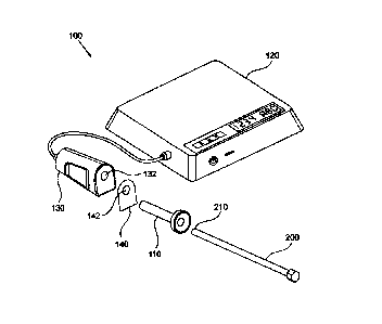
FIG. 1A schematically depicts an embodiment of an apparatus for preparing a
medical
device to a medical procedure, according to the teachings herein;
FIG. 1B schematically depicts a distal end of an endoscope, the distal end
comprising
a viewport suitable to be plasma-treated by the apparatus of FIG. 1A;
FIG. 1C schematically depicts a sterility screen of the apparatus of FIG. 1A,
comprising
a sterility sleeve for covering the plasma applicator of the apparatus of FIG.
1A, the sterility
sleeve being rolled prior to use;
Date Recue/Date Received 2023-06-16
9
FIG. 1D schematically depicts the sterility screen of FIG. 1C, wherein the
sterility
sleeve is partially unrolled to cover the plasma applicator;
FIG. 1E schematically depicts the sterility screen of FIG. 1C, wherein the
sterility
sleeve is unrolled thereby covering the plasma applicator;
FIG. 2 schematically depicts an embodiment of a protecting shroud of an
apparatus for
preparing a medical device to a medical procedure according to the teachings
herein, the
protecting shroud shrouding an endoscope to-be plasma-treated;
FIG. 3A schematically depicts a protecting shroud positioned inside a slot of
a plasma
applicator of the apparatus;
1 0 FIG. 3B schematically depicts a detail of the protecting shroud of FIG.
3A;
FIG. 3C schematically depicts another embodiments of a protecting shroud and a
generating field applicator for preparing a medical device to a medical
procedure according to
the teachings herein, and
FIG. 4 schematically depicts yet another embodiment of a protecting shroud of
an
apparatus for preparing a medical device to a medical procedure according to
the teachings
herein.
DETAILED DESCRIPTION OF SOME EMBODIMENTS
The principles, uses and implementations of the teachings herein may be better
understood with reference to the accompanying description and figures. Upon
perusal of the
description and figures present herein, one skilled in the art is able to
implement the teachings
herein without undue effort or experimentation. In the figures, like reference
numerals refer to
like parts throughout.
Figure 1A schematically depicts an apparatus 100, according to an aspect of
some
embodiments, for preparing a medical device 200 such as an endoscope, to a
medical
procedure. Medical device 200 comprises a distal end 210, schematically
depicted also in
Figure 1B. Distal end 210 comprises a viewport 220 configured to enable
collecting an image
of the surroundings of the viewport. Viewport 220 may be in some embodiments a
transparent
sheet such as a window or a lens, of material such as glass or quartz, or
plastic such as Perspex,
Date Recue/Date Received 2023-06-16
10
thereby allowing light from the outside of the medical device 200 to be
collected in the inside
of medical device 200, e.g. by a light sensitive device (not shown here) such
as a camera.
According to some embodiments viewport 220 may be a mirror, reflecting light
(rather than
transferring light there through) towards a light collecting apparatus (not
shown here) or a light
sensitive device. Viewport 220 comprises a surface 222 which during a medical
procedure may
be exposed to moisture. Consequently, if not treated, e.g. immunized against
fogging, surface
222 may thereby become covered with fog, such fog being the result of
accumulation of
droplets on the surface 222, e.g. (but not limited to) due to condensation of
vapor.
Apparatus 100 comprises a protecting shroud 110 dimensioned to receive therein
distal
end 210 of the medical device 200. Apparatus 100 further comprises an
operating unit 120 and
a plasma applicator 130 (also referred to herein as a plasma generating field
applicator)
connected to the operating unit 120. Plasma applicator 130 comprises a slot
132 configured to
receive therein distal end 210 of medical device 200, whereas distal end 210
is shrouded within
protecting shroud 110. In other words, for use, distal end 210 of medical
device 200 is inserted
into protecting shroud 110, and protecting shroud 110, with distal end 210
being shrouded
therein, is inserted into slot 132. According to some embodiments protecting
shroud 110 is
inserted into slot 132, and then distal end 210 is inserted and advanced into
protecting shroud
110.
According to some embodiments apparatus 100 further comprises a sterility
screen 140
having an opening 142. For use, protecting shroud 110 is inserted into slot
132 through opening
142 of sterility screen 140, as is further detailed and explained herein
below. According to
some embodiments protecting shroud 110 is a dispensable, disposable or
replaceable part,
being configured to be used during a single medical procedure carried out on a
single patient.
According to some embodiments, the protecting shroud functions as a sterility
barrier between
the endoscope which may be exposed to body fluids of the patient, and the
plasma applicator,
which may or may not be maintained sterile during use and after use. According
to some
embodiments sterility screen 140 facilitates maintaining plasma applicator 130
clear of body
fluids originating in the endoscope during use and after use. According to
some embodiments
sterility screen 140 facilitates maintaining the endoscope sterile against
contamination that may
originate in plasma applicator 130.
Date Recue/Date Received 2023-06-16
11
According to some embodiments sterility screen 140 is attached to a sterility
sleeve
144, as depicted schematically in Figures 1C, 1D and 1E, the sterility sleeve
extending between
the sterility screen and a sleeve distal end 146. According to some
embodiments sterility sleeve
144 may be soft like a sock. Prior to use, sterility sleeve 144 may be folded,
as schematically
depicted in Figure 1C. For use, sterility sleeve 144 may be unfolded to
encompass, envelop
and cover plasma applicator 130 or a portion thereof by inserting the plasma
applicator into the
sterility sleeve through the sleeve distal end 146. During use, sterility
sleeve 144 may be
disposed around plasma applicator 130 so as to envelop and cover plasma
applicator 130, so
that insertion of protecting shroud 110 through opening 142 and into slot 132,
and/or insertion
to of endoscope 200 into protecting shroud 110, may not contaminate plasma
applicator 130.
According to some embodiments the sterility sleeve may be substantially rigid,
having a shape
of e.g. a tube, being configured to house the protecting shroud therein.
According to some
embodiments sterility sleeve 144 comprises a double-sided sticky pad (not
shown here) in a
bottom portion thereof configured to stick on one side to plasma applicator
130 and to stick on
another side to a desk or a table or another working platform, thereby
attaching and stabilizing
plasma applicator to the working platform and facilitating inserting and
extracting protecting
shroud 110 (or endoscope 200) from plasma applicator 130. According to some
embodiments,
sterility screen 140 together with sterility sleeve 144, are attached to
protecting shroud 110, so
that insertion of protecting shroud 110 to the slot 132 and encapsulating
plasma applicator 130
with sterility sleeve 144 are performed substantially together.
Plasma applicator 130 is electrically associated with an electric power source
(not
shown here). The power source may be optionally situated in operating unit
120. Plasma
applicator 130 is further configured, when distal end 210, shrouded within
protecting shroud
110, is positioned inside slot 132, and upon activation of the power source,
to apply inside
protecting shroud 110 inside slot 132 an electric field suitable for plasma
generation proximal
viewport 222.
According to some embodiments plasma applicator 130 may be fluidly associated
with
a gas pump and additionally or alternatively with a gas reservoir (neither one
is shown here).
The gas pump and the gas reservoir may be used to controllably evacuate, or to
controllably
flush with a preferred gas, respectively, a vicinity of the distal end of the
endoscope, to facilitate
plasma ignition, as is further detailed and explained below. According to some
embodiments,
a preferred gas may be argon or nitrogen. According to some embodiments, a gas
pressure
Date Recue/Date Received 2023-06-16
12
suitable for plasma ignition after evacuation may be below 0.1Atm. According
to some
embodiments, the vicinity of the distal end of the endoscope may be pumped and
evacuated
and then flushed with a desired gas. According to some embodiments, the gas
pump and / or
the gas reservoir, as the case may be, may be optionally situated in the
operating unit 120.
Operating unit 120 is configured to enable a user of apparatus 100 to operate
and control
the apparatus. Operating unit 120 may thus comprise command switches and
controllers, such
as physical or virtual switches, buttons and controllers. The control unit may
further comprise
indicators for providing a user with required data and information for
operating the apparatus,
such as indication LEDs, displays and possibly an operating software for
providing a user with
operating and command screens to allow a user operate and command the
apparatus.
Figure 2 schematically depicts in a cross-sectional view, an embodiment of a
protecting
shroud 310 according to an aspect of some embodiments. Protecting shroud 310
is particularly
suitable for use with an endoscope 380, depicted schematically inside
protecting shroud 310 in
dashed lines. Endoscope 380 comprises a distal end 382 and an electrically
conducting surface
¨ e.g. a metallic surface 384 ¨ at distal end 382, proximal a viewport 390.
Viewport 390 further
comprises an external surface 392, which may be subject to plasma treatment as
described
herein.
Protecting shroud 310 comprises a hollow cylinder 312 extending between a
proximal
opening 314 and a cylinder distal end 316. Protecting shroud 310 further
comprises a vacuum
seal 320 comprising three 0-rings 320a, 320b and 320c, respectively. Vacuum
seal 320 is
adapted to fit an external dimension (e.g. an external diameter) of endoscope
380 so as to allow
insertion of endoscope 380 into protecting shroud 310 using a slight force,
e.g. by hand, as is
known in the art. Accordingly, vacuum seal 320 is configured to hold a
pressure difference (or
gas concentration difference) between an inside 322 of protecting shroud 310
and an outside
324 of protecting shroud 310 when endoscope 380 is positioned inside
protecting shroud 310.
Vacuum seal 320 may also assist in mechanically stabilizing endoscope 380
inside protecting
shroud 310, thereby assisting in preventing gas leakage between the inside 322
and the outside
324, and also assisting in plasma generation proximal viewport 390, as is
further explained
below.
Protecting shroud 310 further comprises a cathode 330 arranged on hollow
cylinder 312
and configured to establish an electrical feedthrough between the outside 324
of protecting
Date Recue/Date Received 2023-06-16
13
shroud 310 and the inside 322 thereof. Cathode 330 is flexible and
electrically exposed on the
inside 322 of protecting shroud 310 and on the outside thereof, thereby
allowing insertion of
endoscope 380 into protecting shroud 310 while forming an electric contact
between cathode
330 and metallic surface 384. Protecting shroud 310 further comprises an anode
340 arranged
proximal to cylinder distal end 316. Anode 340 may be shaped as a metallic
block having for
example a circular smooth surface 342 facing the inside 322. According to some
embodiments
the surface 342 may be curved. According to some embodiments (not shown here)
anode 340
may be shaped as a pointed tip pointing towards the inside 322. According to
some
embodiments anode 340 may be shaped as a ring. Anode 340 is mounted on a disk
344 made
of a dielectric material, so that disk 344 forms a dielectric barrier between
anode 340 and
cathode 330 and metallic surface 384 of the endoscope (which is on a same
potential as the
cathode). In other words, disk 344 is configured to ensure plasma generation
in a Dielectric
Barrier Discharge (DBD) mode of operation, by interrupting a line-of-sight
between the anode
340 and cathode 330 and metallic surface 384 of the endoscope, thereby forming
said dielectric
barrier. In a DBD mode, plasma may be generated more uniformly over the
available space in
the vicinity of the view port, whereas arcing or other types of specific and
narrow electric
transportation trajectories between the anode and the cathode are prevented.
It is noted that the thickness of the dielectric barrier has a strong effect
on the uniformity
of the plasma generating electric field in the vicinity of the view port, and
hence on the quality
of the plasma treatment. The "quality" of the plasma treatment herein denotes
the level of
hydrophilicity attained, and the duration of time during which the electric
field is activated to
obtain that hydrophylicity. In other words, a high-quality plasma treatment
achieves a relatively
high level of hydrophilicity (e.g. obtaining a surface tension above that of
water namely above
0.072N/M on the treated surface) within a relatively short duration (e.g. of 5
minutes, or 1
minute or as short as 10 second or even as short as 5 second of activated
electric field). The
thickness of the dielectric barrier should generally be as low as possible to
facilitate plasma
ignition, yet it should be large enough to prevent breakdown and arcing.
Exemplary thickness
of a dielectric material such as PET or polycarbonate in embodiments described
herein may be
in the range of about 0.3mm to about 3mm for RF electric field at frequencies
in the MHz range
(e.g. about 2MHz).
According to some embodiments anode 340 is configured to displace flexibly
relative
to hollow cylinder 312, to facilitate a reliable electrical contact between
anode 340 and a
Date Recue/Date Received 2023-06-16
14
feeding contactor as is explained further below. According to some embodiments
disc 344 may
be supported by springs 346 relative to the cylinder 312.
In operation a plasma generating electric power is supplied between anode 340
and
cathode 330 and consequently a plasma generating electric field in a DBD mode
is generated
between anode 340 and metallic surface 384 which is in contact with cathode
330. The plasma
generating electric field generates plasma in the space between anode 340 and
cathode 330 and
particularly in the vicinity of viewport 390 and adjacent external surface
392.
Figure 3A schematically depicts a portion of an embodiment of a plasma
applicator 348
suitable for use with protecting shroud 310a (protecting shroud 310a is
slightly different from
protecting shroud 310 of Figure 2, as is detailed below). Plasma applicator
348 comprises a
slot 350 configured for receiving therein protecting shroud 310a (wherein
endoscope 380 is
shrouded within protecting shroud 310a). Plasma applicator 348 further
comprises a cathode
contactor 352 configured to contact cathode 330 when protecting shroud 310a is
inside slot
350. An electric conductor 354 such as an electric wire, electrically
associated with cathode
contactor 352, may be used to supply electric power generated by a power
source (not shown
here) to cathode contactor 352 and to cathode 330. Plasma applicator 348
further comprises an
anode contactor 356 configured to contact anode 340 when protecting shroud
310a is inside
slot 350. An electric conductor 358 such as an electric wire, electrically
associated with anode
contactor 356 may be used to supply electric power generated by the power
source to anode
340. Anode contactor 356 may be supported flexibly, e.g. by a spring 360, to
facilitate a reliable
electric contact between anode contactor 356 and anode 340 when protecting
shroud 310a is
inserted to the slot.
It is noted that characteristics of the electric field that could generate
plasma in a gas
may depend strongly on characteristics of the gas itself, in addition to the
electrodes geometry
involved (such as shape and configuration of electrodes used for the
application of the electric
field, distance between the electrodes etc.). Generally, the higher the
pressure of the gas, the
higher the electric field should be to ignite plasma in the gas. Also, some
gases ignite at lower
fields than others. For example, plasma may be ignited in helium gas at
atmospheric pressure
and using an RF field (in a frequency between 1MHz and 15MHz) of about 7KV
over a distance
of lcm between electrodes, and at a voltage of about 200V if the gas is at a
pressure of 0.81(Pa.
With a similar configuration of electrodes and at similar field frequencies,
plasma may be
Date Recue/Date Received 2023-06-16
15
ignited in air at a voltage of about 20KV in atmospheric pressure and at a
voltage of about
800V in 0.81(Pa.
Thus, according to some embodiments, plasma applicator 348 is configured to
stream
gas from a gas reservoir (not shown here) to slot 350, or to pump air from
slot 350, to generate
a low-pressure atmosphere in the space between the electrodes 330 and 340, to
facilitate plasma
ignition. Thus, according to some embodiments, plasma applicator 348 is
connected to a hose
364 fluidly associating a gas reservoir (not shown here) containing a gas
suitable for plasma
generation therein such as helium or argon or nitrogen, with slot 350. A valve
366 controlled
by a control unit (not shown here) operable by a user, may be used to schedule
and regulate the
to flow of gas into slot 350. During operation, according to some
embodiments, after introducing
protecting shroud 310a with endoscope 380 therein into slot 350, valve 366 may
be opened to
allow gas flow into the slot. Protecting shroud 310a may be penetrable to gas
flow through
openings 368 between hollow cylinder 312 and disc 344, enabling the gas to
flow into
protecting shroud 310a and towards viewport 390. Excess of gas flowing into
slot 350 may
freely escape through the gap in slot 350 between protecting shroud 310a and
plasma applicator
348 (the gap being not sealed). Following a suitable time period of gas flow
(e.g. 5 seconds or
10 second or 30 second or even 1 minute) the electric power source may be
activated to supply
power to anode 340 and cathode 330 to generate a plasma generating electric
field near
viewport 390. According to some embodiments the gas reservoir may be portable
and suitable
for a single time use.
According to some embodiments, hose 364 may be used to pump gas (air) from
protecting shroud 310a and particularly from the space near viewport 390, to
facilitate plasma
ignition. Air may be sucked from the vicinity of viewport 390 through openings
368 towards
slot 350 and into hose 364. A vacuum seal 370 enables generating vacuum near
viewport 390
by withholding a pressure difference between a region near cylinder end 316
and a region near
opening 314 of protecting shroud 310a. According to some embodiments air may
be pumped
through hose 364 by a vacuum pump (not shown here), fluidly associated with
hose 364.
According to some embodiments hose 364 may be fluidly associated to a pumped
container
(not shown) which is continuously pumped, e.g by a small vacuum pump. Fluid
association is
provided through hose 364, the hose being in constant fluid communication with
the container
thereby being also continuously pumped. Opening valve 366 may result in
pumping slot 350
and particularly the space near viewport 390 by the vacuum pump or by the
pumped container,
Date Recue/Date Received 2023-06-16
16
depending on the particularities of the embodiment. The volume of the pumped
region in
fluidly connected parts of slot 350 and of protecting shroud 310a may be,
according to some
embodiments, smaller than lOcc, and a pumped container and hose of e.g. about
1000cc (1
liter) may suffice to establish a suitable vacuum level between e.g. about 0.1
atm and about
0.01atm within less than about 5 or less than about 10 seconds, which may be
sufficient for
plasma excitation for about 30 seconds or even about 1 minute to
satisfactorily plasma-treat
external surface 392.
According to some embodiments, depicted in detailed in Figure 3B, protecting
shroud
310a further comprises a sterility filter 372 positioned in openings 368 for
maintaining a
to sterility barrier between protecting shroud 310a and plasma applicator
348. By maintaining a
sterility barrier it is meant that microbial organisms may not penetrate
sterility filter 372,
wherein microbial organisms may include any form of prokaryotic cells or
eukaryotic cells,
including fungi and bacteria. The sterility filter is disposed according to
some embodiments
across cylinder end 316 in openings 368, so that gas flowing from plasma
applicator 348 into
protecting shroud 310a enters the protecting shroud sterile, and! or gas
flowing from the inside
322 of protecting shroud 310a into plasma applicator 348 enters the plasma
applicator sterile.
Thus, the sterility filter 372 prevents transfer of contamination from the
plasma applicator (e.g.
from surroundings of slot 350) onto endoscope 380, and! or prevents transfer
of contamination
from endoscope 380 onto the plasma applicator. Additionally or alternatively,
a sterility filter
may be positioned in the plasma applicator, or for example in hose 364.
Figure 3C schematically depicts a plasma applicator 448 and a corresponding
protecting
shroud 410 according to some exemplary embodiments. Plasma applicator 448 is
different from
plasma applicator 348 in comprising an applicator gas port 402 fluidly
associated with hose
364, and protecting shroud 410 comprises a shroud gas port 404 configured to
fluidly connect
to the applicator gas port 402. Fluid connectivity between the inside 322 of
the protecting
shroud and the outside 324 of protecting shroud 410 ¨ e.g. the space of slot
450 of the plasma
applicator ¨ is prevented by a vacuum seal 408, e.g. an 0-ring. Thus, when the
protecting
shroud 410 is inserted into the plasma applicator 448, the shroud gas port 404
fluidly connects
to the applicator gas port 402 thereby establishing fluid connectivity of hose
364 to the inside
322 of the protecting shroud. Consequently, a plasma-ignition facilitating gas
(such as helium
or argon) may be driven directly into the protecting shroud through hose 364,
and additionally
or alternatively, gas, and particularly air, may be pumped from the protecting
shroud through
Date Recue/Date Received 2023-06-16
17
hose 364. Fluid connectivity between the slot 450 and the inside 322 of the
protecting shroud
is thus prevented. A sterility filter 472 is positioned inside shroud gas port
404, for maintaining
a sterility barrier between the inside 322 of protecting shroud 410 and plasma
applicator 448.
As explained above regrading sterility filter 372 in Figure 3B, gas flowing
from plasma
applicator 448 into the inside 322 of protecting shroud 410 enters the
protecting shroud sterile,
and / or gas flowing from the inside 322 of protecting shroud 410 into plasma
applicator 448
enters the plasma applicator sterile. Thus, the sterility filter 472 prevents
transfer of
contamination from the plasma applicator (e.g. from surroundings of slot 450)
onto endoscope
380, and / or prevents transfer of contamination from endoscope 380 onto the
plasma
to applicator.
Protecting shroud 410 is further different form protecting shroud 310 in
having a ring
anode 440 shaped as a ring on an external circumference of hollow cylinder 312
near distal
cylinder end 316 (instead of anode 340 in protecting shroud 310). Hence hollow
cylinder 312,
being made of a dielectric material, functions as a dielectric barrier 444
between anode 440
and cathode 330 and metallic surface 384 of the endoscope, so that plasma is
generated in
protecting shroud 410 in a DBD mode of operation as described above regarding
protecting
shroud 310. According to some embodiments protecting shroud 410 comprises a
stopper 442
inside hollow cylinder 412. Stopper 442 is configured to limit advancement of
endoscope 380
into protecting shroud 410, so that a pre-determined, desired gap is
established between anode
440 and metallic surface 384 of the endoscope, thereby ensuring plasma
generation at a known
field (the field being determined by the voltage supplied between the cathode
and the anode
and the said gap). Stopper 442 may further be employed as a dielectric barrier
on the line of
sight between the anode and the cathode, thereby assisting in focusing plasma
towards the view
port 390.
When protecting shroud 410 is inserted into a slot 450 of plasma applicator
448, an
anode contactor 456 of plasma applicator 448 contacts ring anode 440. Anode
contactor 456 is
electrically associated with an electric conductor 458 which is configured to
connect to a power
supply (not shown here) to enable providing to ring anode 440 a plasma
generating electric
field as described above. It is noted that cathode 330 is of protecting shroud
410 is electrically
associated with cathode contactor 352 when protecting shroud 410 is inserted
into a slot 450
as described above. Thus, upon activation, a suitably connected power supply
may provide a
Date Recue/Date Received 2023-06-16
18
plasma generating electric field (in a DBD mode) between ring anode 440 and
the metallic
surface 384 of endoscope 380 to generated plasma in the vicinity of view port
390.
Figure 4 schematically depicts a protecting shroud 510 according to an aspect
of some
embodiments. Protecting shroud 510 is configured to enable facilitated plasma
ignition,
without pumping the space around the endoscope as described in the embodiments
above nor
without streaming gas into that space. In other words, protecting shroud
enables providing
plasma treatment to a view port of an endoscope according to the teachings
herein, using a
plasma applicator that is not connected neither to a gas reservoir nor to a
gas pump.
Accordingly, the protecting shroud does not have a gas port such as gas port
402, and is not
connected to a hose such as hose 364.
Protecting shroud 510 comprises hollow cylinder 312 extending between opening
314
and a cylinder end 316. Protecting shroud 510 is different from protecting
shroud 310 in that
hollow cylinder 312 is blind and sealed near cylinder end 316, thereby
substantially preventing
permeation or penetration of gas molecules through cylinder end 316.
Protecting shroud 510 is
further different from protecting shroud 310 in having a leakage seal 530
inside hollow cylinder
312, and a hermetic screen 518 in hollow cylinder 312 situated between leakage
seal 530 and
cylinder end 316. Hermetic screen 518 is configured to be impermeable to gas
molecules,
thereby defining a closed space 520, closed between hermetic screen 518 and
cylinder end 316.
Closed space 520 inside protecting shroud 510 is thus airtight, namely
maintained sealed from
the outside 324 of protecting shroud 510. Closed space 520 contains a gas
suitable for plasma
ignition, e.g. Argon, at a gas pressure of about 1 atmosphere, so that there
is, at most, only
minor pressure gradients over the hermetic screen.
Hermetic screen 518 is breakable, being thereby configured to break (tear
down) upon
insertion of an endoscope such as endoscope 380 into protecting shroud 510.
According to
some embodiments, protecting shroud 510 further comprises one or more tearing
needles 522
attached flexibly to hollow cylinder 312 near hermetic screen 518 outside of
closed space 520.
Tearing needles 522 are configured to lean flexibly towards hermetic screen
518 and to tear
the hermetic screen when pushed by an object inserted into the protecting
shroud. Thus, for
use, the endoscope may be inserted into protecting shroud 510 and affecting
tearing down of
hermetic screen 518 by pushing tearing needles 522 towards hermetic screen
518. The
endoscope may be further advanced until the viewport is between cathode 330
and anode 340.
Date Recue/Date Received 2023-06-16
19
It is noted that during insertion, the endoscope is first advanced through
leakage seal 530, then
hermetic screen 518 is broken and then the endoscope is further advanced to be
positioned in
place. Once hermetic screen 518 is broken, the gas inside space 520 is
prevented from freely
flowing towards opening 324 by a sealing formed between leakage seal 530 and
the endoscope.
During further advancement of the endoscope into the protecting shroud, the
free volume of
space 520 for the gas reduces, yet pressure build up in the region of closed
space 520 is
prevented, due to gas escape under a pressure difference across leakage seal
530. As a result,
when endoscope 380 is fully inserted into protecting shroud 510, closed space
520 and
particularly the space proximal the viewport, between anode 340 and cathode
330, comprises
substantially the gas that was contained in the space 520 before the tear-up
of hermetic screen
518, at approximately atmospheric pressure, thereby facilitating plasma
ignition therein.
According to some embodiments hermetic screen 518 may be made of Mylar or
metalized
Mylar or Kapton or metalized Kapton and the like.
There is thus provided according to an aspect of the invention an apparatus
(100 in
Figure 1A) for preparing an endoscope ((200 in Figure 1, 380 in Figures 2, 3A
and 3C) for an
endoscopy procedure. The apparatus comprises a protecting shroud (110 in
Figure 1A, 310,
310a in Figures 2 and 3A, 410 in Figure 3C, 510 in Figure 4) dimensioned to
receive therein a
distal end (210, 382) of the endoscope. The distal end comprises a view port
(220, 390)
configured to enable collecting an image of the surrounding of the view port
there through.
The apparatus further comprises a plasma generating field applicator (130,
348, 448),
electrically associated with an electric power source. The plasma generating
field applicator
has a slot (132, 350, 450) configured to receive therein the distal end of the
endoscope shrouded
within the protecting shroud. The plasma generating field applicator is
configured to apply
electric power suitable for plasma generation within the protecting shroud.
The protecting
shroud is detachable from the distal end of the endoscope and from the plasma
generating field
applicator.
According to some embodiments the view port of the endoscope may be
transparent or
may be a mirror.
According to some embodiments the apparatus further comprises a sterility
sleeve (144)
extending between a first end (146) and a second end (140), configured to
encapsulate the
plasma generating field applicator, having on the first end a first opening
configured to enable
Date Recue/Date Received 2023-06-16
20
inserting the plasma generating field applicator into the sterility sleeve,
and on second end a
second opening (142) configured to enable inserting the endoscope into the
plasma generating
field applicator. According to some embodiments the sterility sleeve is soft
and according to
some embodiments the sterility sleeve is rigid. The sterility sleeve is
detached from the plasma
generating field applicator. According to some embodiments the sterility
sleeve is attached to
the protecting shroud, and according to some embodiments the sterility sleeve
is detached from
the protecting shroud.
According to some embodiments the protecting shroud comprises at least one
electrode
(340, 440) and a first shroud electric contact (340, 440) electrically
connected to the electrode.
The first shroud electric contact is configured to electrically contact a
corresponding first
applicator electric contact (356, 456) in the plasma generating field
applicator when the
protecting shroud is inserted into the slot (350, 450). The at least one
electrode is thereby
configured to apply a plasma generating field inside (322) the protecting
shroud upon receiving
the electric power from the plasma generating field applicator.
According to some embodiments the protecting shroud further comprises a second
shroud electric contact (330), configured to contact the endoscope when the
distal end of the
endoscope is received within the protecting shroud. The second shroud electric
contact is
configured to electrically contact a second applicator electric contact (352)
when the protecting
shroud is inserted into the slot (350, 450).
According to some embodiments the protecting shroud comprises a hollow,
substantially rigid tube (312, 412) extending between an opening (314)
configured to receive
the distal end of the endoscope, and a distal end (316) of the protecting
shroud. According to
some embodiments the hollow tube is a hollow cylinder (312, 412).
According to some embodiments the protecting shroud further comprises a seal
(320,
530) positioned between the opening and the distal end along an inner
circumference of the
hollow tube, being dimensioned to encircle the endoscope (380), being thereby
configured to
sealingly contact the endoscope when the endoscope is received inside the
hollow tube.
According to some embodiments the seal comprises an 0-ring.
According to some embodiments the plasma generating field applicator (348,
448) is
connected to a hose (364). The hose is controllably fluidly connected to the
slot (350, 450).
According to some embodiments the plasma generating field applicator (348,
448) comprises
Date Recue/Date Received 2023-06-16
21
a controlled valve (366), controllably fluidly connecting the hose (364) with
the slot (350, 450).
According to some embodiments the plasma generating field applicator (348)
comprises an
applicator gas port (402) fluidly connected with the hose, and the protecting
shroud (410)
comprises a shroud gas port (404). The shroud gas port is configured to
sealingly connect with
the applicator gas port for fluidly connecting the hose with an inside (322)
of the protecting
shroud. The sealed connection between the shroud gas port and the applicator
gas port prevents,
e.g. by seal 408, flow communication between the inside (322) of the
protecting shroud (fluidly
associated with hose 364) and the slot (450), when the protecting shroud is
inserted into the
slot.
According to some embodiments the protecting shroud (510) comprises a seal
(530)
inside the hollow tube (312) configured to sealingly contact the endoscope
when the distal end
of the endoscope is inserted into the hollow tube. The protecting shroud (510)
further
comprises a hermetic screen (518) spanning across the hollow tube and
configured to thereby
define a closed and sealed space (520) between the hermetic screen and the
distal end (316) of
the hollow tube. According to some embodiments the protecting shroud further
comprises a
terrier (522) positioned inside the hollow tube between the seal (530) and the
hermetic screen
(518) being configured to tear down the hermetic sreen upon insertion of the
endoscope into
the hollow tube.
According to an aspect of some embodiments there is provided a method of
preparing
an endoscope for an endoscopy procedure. The method comprises providing a
protecting
shroud (110, 310, 310a, 410, 510) dimensioned to receive therein a distal end
(210, 382) of the
endoscope, the distal end comprising a view port (220, 390) configured to
allow collecting an
image of the surrounding of the view port there through. The method further
comprises
providing a plasma generating field applicator (130, 348, 448) electrically
associated with an
electric power source. The plasma generating field applicator has a slot (132,
350, 450)
configured to receive therein the distal end of the endoscope shrouded within
the protecting
shroud. The plasma generating field applicator is configured to apply electric
power suitable
for plasma generation within the protecting shroud (e.g. by the electrodes
330, 340 and 440).
The protecting shroud is detachable from the plasma generating field
applicator and from the
distal end of the endoscope. The method further comprises positioning the
distal end of the
endoscope shrouded within the protecting shroud in the slot of the plasma
generating field
Date Recue/Date Received 2023-06-16
22
applicator, and activating the power source to generate plasma within the
protecting shroud,
thereby plasma-treating the view port at the distal end of the endoscope.
According to some embodiments, the method further comprises preventing, by the
protecting shroud, contamination of the plasma generating field applicator
with fluids dispersed
on the distal end. According to some embodiments, the plasma generation field
applicator
comprises a hose (364) and the method further comprises controllably (by
closing and opening
valve 366) flowing a gas into an inside (322) of the protecting shroud, or
pumping the inside
of the protecting shroud via the hose.
According to an aspect of some embodiments there is further provided a method
of
preparing an endoscope (380) for an endoscopy procedure, the endoscope
comprising a distal
end (382) comprising a view port (390). The view port is made of a dielectric
material and is
proximal to a metallic segment (384) at the distal end of the endoscope. The
method comprises
placing the distal end of the endoscope in a closed plasma chamber (e.g.
protecting shrouds
310, 310a, 410 or 510, wherein the insertion of the endoscope seals the inside
322 of the
protecting shrouds, thereby defining a closed plasma chamber therein). The
closed plasma
chamber has at least an anode (340, 440) and a cathode (330) wherein the
cathode electrically
contacts the metallic segment. A line-of-sight between the anode and the
cathode is interrupted
by a dielectric barrier (344, 444), and the method further comprises applying
a plasma-
generating electromagnetic field between the anode and the cathode, thereby
generating plasma
in a DBD mode in a vicinity (322) of the view port. According to some
embodiments, the
electric barrier (444) electrically isolates the anode (440) from gas in the
vicinity (322) of the
view port. According to some embodiments of the method, the view port is
transparent or
alternatively is a mirror. According to some embodiments of the method the
view port is made
of glass or quartz or plastic.
It is appreciated that certain features of the invention, which are, for
clarity, described
in the context of separate embodiments, may also be provided in combination in
a single
embodiment. Conversely, various features of the invention, which are, for
brevity, described
in the context of a single embodiment, may also be provided separately or in
any suitable sub-
combination or as suitable in any other described embodiment of the invention.
No feature
described in the context of an embodiment is to be considered an essential
feature of that
embodiment, unless explicitly specified as such.
Date Recue/Date Received 2023-06-16
23
Although steps of methods according to some embodiments may be described in a
specific sequence, methods of the invention may comprise some or all of the
described steps
carried out in a different order. A method of the invention may comprise all
of the steps
described or only a few of the described steps. No particular step in a
disclosed method is to be
considered an essential step of that method, unless explicitly specified as
such.
Although the invention is described in conjunction with specific embodiments
thereof,
it is evident that numerous alternatives, modifications and variations that
are apparent to those
skilled in the art may exist. Accordingly, the invention embraces all such
alternatives,
modifications and variations that fall within the scope of the appended
claims. It is to be
understood that the invention is not necessarily limited in its application to
the details of
construction and the arrangement of the components and/or methods set forth
herein. Other
embodiments may be practiced, and an embodiment may be carried out in various
ways.
The phraseology and terminology employed herein are for descriptive purpose
and
should not be regarded as limiting. Citation or identification of any
reference in this application
shall not be construed as an admission that such reference is available as
prior art to the
invention. Section headings are used herein to ease understanding of the
specification and
should not be construed as necessarily limiting.
Date Recue/Date Received 2023-06-16