Note : Les descriptions sont présentées dans la langue officielle dans laquelle elles ont été soumises.
WO 2022/216374
PCT/US2022/017333
MULTI-COMPONENT DELIVERY SYSTEMS AND METHODS
CROSS-REFERENCE TO RELATED APPLICATION
[0001] This application claims the benefit of Provisional
Application No.
63/152,144, filed February 22, 2021, which is incorporated herein by reference
in its
entirety for all purposes.
FIELD
[0002] The present disclosure relates generally to systems and
methods for
delivering multi-component devices. More specifically, the disclosure relates
to systems
and methods for delivering endovascular devices that include individual
components to
a target site.
BACKGROUND
[0003] A variety of branched, anatomical passages may benefit
from treatment
in the form of an implanted, endoluminal device. One such passage is a
vascular
passage, such as an artery, with an aneurysm. Aortic disease and trauma such
as
aneurysms and dissections present a significant risk to a patient. That risk
is increased
based on the patient's condition. Such conditions or factors can include the
patient's
age and preexisting and/or related conditions such as cardiopulmonary bypass,
cardiac
arrest, circulatory arrest. These and other factors may limit the patient's
ability to
withstand and recover from surgery to repair the aortic disease. This same
issue exists
in other diseased and damaged tissues in the patients.
[0004] With respect to aneurysms, in order to prevent rupturing
of an aneurysm,
a stent graft may be introduced into a blood vessel percutaneously and
deployed to
span the aneurysmal sac. Stent grafts include a graft fabric secured to a
cylindrical
scaffolding or framework of one or more stents. The stent(s) provide rigidity
and
structure to hold the graft open in a tubular configuration as well as the
outward radial
force needed to create a seal between the graft and a healthy portion of the
vessel wall
and provide migration resistance. Blood flowing through the vessel can be
channeled
through the luminal surface of the stent graft to reduce, if not eliminate,
the stress on the
vessel wall at the location of the aneurysmal sac. Stent grafts may reduce the
risk of
rupture of the blood vessel wall at the aneurysmal site and allow blood to
flow through
the vessel without interruption.
CA 03206686 2023- 7- 27
WO 2022/216374
PCT/US2022/017333
[0005] Various endovascular repair procedures such as the
exclusion of an
aneurysm require a stent graft to be implanted adjacent to a vascular
bifurcation. Often
the aneurysm extends into the bifurcation requiring the stent graft to be
placed into the
bifurcation. A bifurcated stent graft is therefore required in these cases.
Modular stent
grafts, having a separate main body and branch component are often preferred
in these
procedures due to the ease and accuracy of deployment. See U.S. Patent
Application
No. 2008/0114446 to Hartley etal. for an example of a modular stent graft
having
separate main body and branch stent components. In the Hartley et al.
publication the
main body stent has a fenestration in the side wall that is tailored to engage
and secure
the side branch stent.
SUMMARY
[0006] An endoprosthesis including a main body is provided with
side branch
portals for providing fluidic access to side branches of a main lumen when the
main
body of the endoprosthesis is deployed in the main lumen. A method of
deployment of
the endoprosthesis is also provided
[0007] According to one example ("Example 1"), a method of
deploying includes
a multibranch stent graft at a target site having a main lumen and a first
branch lumen is
provided, the method including advancing a main guidewire to a target site;
advancing a
catheter including a main body of a multibranch stent graft along the main
guidewire
toward the main lumen of the target site, the main body having a first portion
and a
second portion, the main body defining a first portal operable to provide
fluidic access
from the main body to a first side branch extending from the target site when
the main
body is deployed at the target site, the first portal being pre-cannulated
with a first
secondary guidewire prior to advancing the main body along the main guidewire;
partially deploying the first portion of the main body in the main lumen of
the target site;
advancing a first sheath along the first guide member through the first
portal; advancing
a first articulatable wire or guide catheter through the first sheath;
positioning the first
articulatable wire or guide catheter into a first branch lumen of the target
site; partially
deploying the second portion of the main body in the main lumen of the target
site; fully
deploying the first portion and the second portion of the main body; advancing
a first
side branch body along the first articulatable wire or guide catheter into the
first branch
lumen of the target site; and deploying the first side branch body in the
first branch
lumen of the target site.
[0008] According to another example ("Example 2"), further to
Example 1, the
2
CA 03206686 2023- 7- 27
WO 2022/216374
PCT/US2022/017333
method includes deploying an embolic filter in the first branch lumen of the
target site.
[0009] According to another example ("Example 3"), further to
Example 2, the
method includes aspirating a filter sheath of the embolic filter.
[00010] According to another example ("Example 4"), further to Example 3, the
method includes removing the embolic filter after the first side branch body
has been
deployed.
[00011] According to another example ("Example 5"), further to any of the
preceding Examples, wherein the first guide member includes a first end that
is looped
around a cap of the catheter
[00012] According to another example ("Example 6"), further to any of the
preceding Examples, wherein the main body further defines a second portal and
a third
portal operable to provide fluidic access from the main body to a second side
branch
and a third side branch extending from the target site when the main body is
deployed
at the target site, the second portal being pre-cannulated with a second guide
member
and the third portal being pre-cannulated with a third guide member prior to
advancing
the main body along the main guidewire.
[00013] According to another example ("Example 7"), further to Example 6
further
includes advancing a second sheath along the second guide member through the
second portal; advancing a second articulatable wire or guide catheter through
the
second sheath; positioning the second articulatable wire or guide catheter
into a second
branch lumen of the target site; advancing a third sheath along the third
guide member
through the third portal; advancing a third articulatable wire or guide
catheter through
the third sheath; and positioning the third articulatable wire or guide
catheter into a third
branch lumen of the target site.
[00014] According to another example ("Example 8"), further to Example 7, the
method includes advancing a second side branch body along the second
articulatable
wire or guide catheter into the second branch lumen of the target site;
deploying the
second side branch body in the second branch lumen of the target site;
advancing a
third side branch body along the third articulatable wire or guide catheter
into the third
branch lumen of the target site; and deploying the third side branch body in
the third
branch lumen of the target site.
[00015] According to another example ("Example 9"), further to Example 8, the
method further includes removing the main guidewire, the first, second, and
third guide
members, and the first, second, and third sheaths.
[00016] According to another example ("Example 10"), further to Example 9,
3
CA 03206686 2023- 7- 27
WO 2022/216374
PCT/US2022/017333
wherein the catheter is removed prior to advancing the first, second, and
third sheaths.
[00017] According to another example ("Example 11"), an endoprosthesis
delivery system includes an elongate member having a first end and a second
end; an
end cap coupled to the first end of the elongate member; an endoprosthesis
including a
main body defining a main lumen and at least one side branch portal and at
least one
second body defining a secondary lumen; and at least one guide member
extending
through the at least one side branch portal and coupled to the end cap.
[00018] According to another example ("Example 12"), further to Example 11,
the
endoprosthesis delivery system further includes a constraining member
constraining the
main body of the endoprosthesis to the elongate member.
[00019] According to another example ("Example 13"), further to Example 12,
the
endoprosthesis delivery system, wherein the constraining member is operable to
constrain the main body at a constrained configuration and at a partially
deployed
configuration, the main body having a first diameter at the constrained
configuration, a
second diameter at the partially deployed configuration that is greater than
the first
diameter, and a third diameter at a deployed configuration that is greater
than the first
diameter and the second diameter.
[00020] According to another example ("Example 14"), further to Example 13,
the
endoprosthesis delivery system, wherein the constraining member includes a
first
portion and a second portion, wherein the first portion and the second portion
are
operable to independently constrain corresponding first and second portions of
the main
body at the constrained configuration and the partially deployed
configuration.
[00021] According to another example ("Example 15"), further to any one of
Examples 11-14, the endoprosthesis delivery system further includes a sheath
operable
to be advanced along the at least one guide member.
[00022] According to another example ("Example 16"), further to Example 15,
the
endoprosthesis delivery system further includes an articulatable wire or guide
catheter
operable to be advanced through the sheath.
[00023] According to another example ("Example 17"), further to Example 16,
the
endoprosthesis delivery system further includes at least one secondary branch
operable
to be advanced along the articulatable wire or guide catheter and to be
deployed at
least partially within the at least one side branch portal.
[00024] According to another example ("Example 18"), further to Example 17,
the
endoprosthesis delivery system further includes a removeable filter operable
to be
deployed downstream from a target site of the endoprosthesis.
4
CA 03206686 2023- 7- 27
WO 2022/216374
PCT/US2022/017333
[00025] According to another example ("Example 19"), further to Example 18,
the
endoprosthesis delivery system wherein the removeable filter includes a
central lumen
through which the articulatable or guide catheter wire is operable to extend.
[00026] According to another example ("Example 20"), further to any one of
Examples 11-19, the endoprosthesis delivery system, wherein the end cap is
curved.
[00027] According to another example ("Example 21"), further to the
endoprosthesis delivery system of any one of Examples 11-20, wherein the
endoprosthesis delivery system is curved from the end cap through the main
body.
[00028] According to another example ("Example 22"), an endoprosthesis
delivery system includes an elongate member having a first end and a second
end; an
endoprosthesis positioned longitudinally between the first end and second end
of the
elongate member, the endoprosthesis including a main body defining a main
lumen and
a side branch portal; a guide member extending through the side branch portal;
and a
guide member retainer removably coupled to the elongate member at a coupling
position, the guide member being coupled to the guide member retainer at a
position
between the side branch portal and the coupling position of the guide member
retainer.
[00029] According to another example ("Example 23"), further to the
endoprosthesis delivery system of Example 22, wherein the main body defines a
plurality of side branch portals.
[00030] According to another example ("Example 24"), further to the
endoprosthesis delivery system of either Example 22 or Example 23, further
includes a
plurality of guide members.
[00031] According to another example ("Example 25"), further to the
endoprosthesis delivery system of any one of Examples 22-24, wherein the guide
member retainer extends through loops formed at an end of each of the guide
members.
[00032] According to another example ("Example 26"), further to Example the
endoprosthesis delivery system of any one of Examples 22-25, wherein the guide
member retainer is operable to be selectively decoupled from the first
coupling position.
[00033] According to another example ("Example 27"), further to the
endoprosthesis delivery system of any one of Examples 22-26, wherein the
elongate
member includes a lock wire retainer positioned at the first end of the
elongate member.
[00034] According to another example ("Example 28"), further to the
endoprosthesis delivery system of Example 27, wherein the guide member
retainer is
releasably coupled to the lock wire retainer.
CA 03206686 2023- 7- 27
WO 2022/216374
PCT/US2022/017333
[00035] According to another example ("Example 29"), further to the
endoprosthesis delivery system of any one of Examples 22-28, further including
a side
branch body, wherein each guide member includes a first end, wherein each
first end of
the guide members is retained by the guide member retainer between the
coupling
position and the side branch portal when the side branch body is advanced
along the
guide member.
[00036] According to another example ("Example 30"), further to the
endoprosthesis delivery system of any one of Examples 22-29, wherein each
guide
member is operable to be removed from a corresponding side branch portal when
the
guide member retainer is released.
[00037] According to another example ("Example 31"), further to the
endoprosthesis delivery system of any one of Examples 22-30, further including
a
plurality of guide member retainers, wherein each guide member retainer is
coupled to a
corresponding guide member.
[00038] According to another example ("Example 32"), further to the
endoprosthesis delivery system of Example 29, wherein each guide member
retainer is
operable to be individually and selectively released from engagement at the
first
coupling position such that each guide member is operable to be individually
removed
from a corresponding side branch portal.
[00039] According to another example ("Example 33"), further to the
endoprosthesis delivery system of Example 22, wherein the elongate member
includes
a cap positioned at the first end of the elongate member, wherein the guide
member
retainer is coupled to the cap at the coupling position.
[00040] According to another example ("Example 34"), further to the
endoprosthesis delivery system of any one of Examples 22-33, the guide member
retainer is coupled to the elongate member at the first end of the elongate
member.
[00041] The foregoing Examples are just that, and should not be read to limit
or
otherwise narrow the scope of any of the inventive concepts otherwise provided
by the
instant disclosure. While multiple examples are disclosed, still other
embodiments will
become apparent to those skilled in the art from the following detailed
description, which
shows and describes illustrative examples. Accordingly, the drawings and
detailed
description are to be regarded as illustrative in nature rather than
restrictive in nature.
BRIEF DESCRIPTION OF THE DRAWINGS
[00042] The accompanying drawings are included to provide a further
6
CA 03206686 2023- 7- 27
WO 2022/216374
PCT/US2022/017333
understanding of the disclosure and are incorporated in and constitute a part
of this
specification, illustrate embodiments, and together with the description serve
to explain
the principles of the disclosure.
[00043] FIG. 1 is a view of a delivery system in accordance with an
embodiment;
[00044] FIG. 2 is a front view of an implantable device with a main body and
side
branches deployed in the aorta and adjacent side branches, in accordance with
an
embodiment;
[00045] FIG. 3 is a top view of a main body of an implantable device, the main
body including side branch portals through which side branch bodies can be
delivered
and deployed, in accordance with an embodiment;
[00046] FIG. 4 is a side view of a main body of an implantable device, the
main
body including a portal access feature for providing clearance for side branch
bodies
delivered and deployed through the side branch portals in accordance with an
embodiment;
[00047] FIG. 5 is an end view of a main body of an implantable device, the
interior opening of the side branch portal positioned in the lumen of the main
body, in
accordance with one embodiment;
[00048] FIG. 6 is an end view of a main body of an implantable device, a
portal
access feature projecting into the lumen of the main body in accordance with
one
embodiment;
[00049] FIG. 7 is a perspective view of a main body including side branch
portals
staggered along a longitudinal length of the main body in accordance with one
embodiment;
[00050] FIG. 8 is a perspective view of a main body including a side branch
portal
staggered relative to two side branch portals aligned along a longitudinal
length of the
main body in accordance with one embodiment;
[00051] FIG. 9 is a cut-away view of a patient's aorta in accordance with one
embodiment;
[00052] FIG. 10 is a view of a filtration system deployed in the vasculature
of a
patient in accordance with one embodiment;
[00053] FIG. 11A is a view of a main body of an implantable device being
delivered to a target site, the main body including side branch portals that
are pre-
cannulated in accordance with one embodiment;
[00054] FIG. 11 B is a view of a delivery system with a guide member retainer
retaining guide members via a lock wire retainer in accordance with one
embodiment;
7
CA 03206686 2023- 7- 27
WO 2022/216374
PCT/US2022/017333
[00055] FIG. 11C is a view of a delivery system with a lock wire, the delivery
system being steerable in accordance with one embodiment;
[00056] FIG. 12 is a view of a main body include a first and second region,
the
second region being partially deployed in accordance with one embodiment;
[00057] FIGS. 13a-13c are views of a sheath being advanced along guide
members that are cannulating side branch portals of a main body in accordance
with
one embodiment;
[00058] FIG. 14 is a view of an articulatable guide catheter
and/or wire that is
advanced through a sheath and positioned in a side branch of the target lumen
in
accordance with one embodiment;
[00059] FIG. 15 is a view of an articulatable wire advancing
through the filtration
system for a through-and-through configuration between access sites in
accordance
with one embodiment;
[00060] FIG. 16 is a view of articulatable wires positioned in
each of the side
branches of the target site in accordance with one embodiment;
[00061] FIG. 17 is a view of a main body partially deployed
along the full
longitudinal length of the main body for accurate placement of the main body
in the
target lumen in accordance with one embodiment;
[00062] FIG. 18 is a view of a main body fully deployed in the target lumen in
accordance with one embodiment;
[00063] FIG. 19 is view of side branch bodies being delivered to corresponding
branches of a target site in accordance with one embodiment;
[00064] FIG. 20 is a view of side branch bodies being deployed at
corresponding
branches of a target site in accordance with one embodiment;
[00065] FIG. 21 is a view of delivery systems for side branch bodies being
removed from the target site in accordance with one embodiment;
[00066] FIG. 22 is a view of a filtration system being aspirated prior to
removal of
delivery systems and the filtration system used for deploying an implantable
device to a
branched target site in accordance with one embodiment; and
[00067] FIG. 23 is a view of an implantable device implanted as a branched
target site prior to removal of a plurality of guidewires used to cannulate
each portion of
the branched target site in accordance with one embodiment.
DETAILED DESCRIPTION
Definitions and Terminology
8
CA 03206686 2023- 7- 27
WO 2022/216374
PCT/US2022/017333
[00068] This disclosure is not meant to be read in a restrictive manner. For
example, the terminology used in the application should be read broadly in the
context
of the meaning those in the field would attribute such terminology.
[00069] Persons skilled in the art will readily appreciate that
various aspects of
the present disclosure can be realized by any number of methods and apparatus
configured to perform the intended functions. Stated differently, other
methods and
apparatus can be incorporated herein to perform the intended functions. It
should also
be noted that the accompanying drawing figures referred to herein are not
necessarily
drawn to scale, but may be exaggerated to illustrate various aspects of the
present
disclosure, and in that regard, the drawing figures should not be construed as
limiting.
[00070] Certain relative terminology is used to indicate the
relative position of
components and features. For example, words such as "top", "bottom", "upper,"
"lower,"
"left," "right," "horizontal," "vertical," "upward," and "downward" are used
in a relational
sense (e.g., how components or features are positioned relative to one
another) and not
in an absolute sense unless context dictates otherwise. Similarly, throughout
this
disclosure, where a process or method is shown or described, the method may be
performed in any order or simultaneously, unless it is clear from the context
that the
method depends on certain actions being performed first.
[00071] With respect to terminology of inexactitude, the terms "about" and
"approximately" may be used, in certain instances, to refer to a measurement
that
includes the stated measurement and that also includes any measurements that
are
reasonably close to the stated measurement. Measurements that are reasonably
close
to the stated measurement deviate from the stated measurement by a reasonably
small
amount as understood and readily ascertained by individuals having ordinary
skill in the
relevant arts. Such deviations may be attributable to measurement error,
differences in
measurement and/or manufacturing equipment calibration, human error in reading
and/or setting measurements, minor adjustments made to optimize performance
and/or
structural parameters in view of differences in measurements associated with
other
components, particular implementation scenarios, imprecise adjustment and/or
manipulation of objects by a person or machine, and/or the like, for example.
[00072] As used herein, "couple" means join, connect, attach, adhere, affix,
or
bond, whether directly or indirectly, and whether permanently or temporarily.
[00073] As used herein, the term "elastomer" refers to a polymer or a mixture
of
polymers that has the ability to be stretched to at least 1.3 times its
original length and
to retract rapidly to approximately its original length when released. The
term
9
CA 03206686 2023- 7- 27
WO 2022/216374
PCT/US2022/017333
"elastomeric material" refers to a polymer or a mixture of polymers that
displays stretch
and recovery properties similar to an elastomer, although not necessarily to
the same
degree of stretch and/or recovery. The term "non-elastomeric material" refers
to a
polymer or a mixture of polymers that displays stretch and recovery properties
not
similar to either an elastomer or elastomeric material, that is, considered
not an
elastomer or elastomeric material as is generally known.
[00074] The term "film" as used herein generically refers to one or more of
the
membrane, composite material, or laminate.
[00075] The term "biocompatible material" as used herein generically refers to
any material with biocompatible characteristics including synthetic materials,
such as,
but not limited to, a biocompatible polymer, or a biological material, such
as, but not
limited to, bovine pericardium. Biocompatible material may comprise a first
film and a
second film as described herein for various embodiments.
[00076] For reference, the terms "circumference" and "diameter" are not meant
to
require a circular cross-section (although are inclusive of a circular cross-
section), and
are instead to be understood broadly to reference an outer surface or
dimension and
the dimension between opposing sides of the outer surface, respectively.
[00077] Although the embodiments herein may be described in connection with
various principles and beliefs, the described embodiments should not be bound
by
theory. For example, embodiments are described herein in connection with
vascular
stent grafts, and more specifically branched stent grafts. However,
embodiments within
the scope of this disclosure can be applied toward any endoprostheses of
similar
structure and/or function. Furthermore, embodiments within the scope of this
disclosure
can be applied in non-vascular applications.
Description of Various Embodiments
[00078]
Persons skilled in the art will readily appreciate that various
aspects of the
present disclosure can be realized by any number of methods and apparatuses
configured to perform the intended functions.
It should also be noted that the
accompanying drawing figures referred to herein are not necessarily drawn to
scale, but
may be exaggerated to illustrate various aspects of the present disclosure,
and in that
regard, the drawing figures should not be construed as limiting.
[00079] Devices, systems, and methods of endoluminally delivering a branchable
expandable implant in accordance with various embodiments are disclosed herein
for
treating disease of human vasculature. Although the description below and
figures are
CA 03206686 2023- 7- 27
WO 2022/216374
PCT/US2022/017333
illustrated in the context of treating the aorta 20, including the ascending
aorta 21, aortic
arch 22, and descending aorta 23, and branches therefrom, including the
brachiocephalic artery 24, the left common carotid artery 25, and the left
subclavian
artery 26, it should be appreciated that the present disclosure can be applied
to
treatment of other portions of the vasculature or, including, for example, any
disease
where a larger vessel and one or more branch vessels are to be treated.
Branchable, Expandable Implant
[00080] Referring to FIG. 2, an implantable device 10 can be delivered and
deployed in the aorta 20, the implantable device 10 including a main body 100
and
branch bodies 200. The main body 100 is deployed in the aortic arch 22 and the
branch
bodies 200 can be deployed in branching arteries (e.g., a first branch body
200a in the
brachiocephalic artery 24, a second branch body 200b in the left common
carotid artery
25, and a third branch body 200c in the left subclavian artery 26).
[00081] Although various configurations of the implantable device 10 are
contemplated with respect to the delivery systems and methods described
herein,
several discrete examples of an implantable device 10 are provided in detail
in order to
provide reference for the various components and steps of the delivery system
and
method of delivery and deployment. For example, FIG. 3 is an exemplary
embodiment
of an implantable device 10. The main body 100 includes a wall 104 forming the
main
lumen 102. The main body 100 has a first end 106 and a second end 108. At the
first
end 106, the main body 100 includes a first opening 107 and at the second end
108 the
main body 100 includes a second opening 109. Each of the openings 107, 109
provides
access to the main lumen 102 at the corresponding end 106, 108. Fluids are
operable
to flow through the main lumen 102 by passing through the first opening 107,
into the
main lumen 102, and out the second opening 109, defining a main body fluid
flow
direction. Or, the flow may be in the opposite direction, defining the main
body fluid flow
direction. The outer wall 104 substantially forms or defines the outer profile
of the main
body 100.
[00082] In some embodiments, the main body 100 is formed of a stent structure
120 and a graft member 130. The stent structure 120 is operable to maintain
patency of
the main body 100 and/or the main vessel (e.g., the aorta 20) when the main
body 100
is deployed. The stent structure 120 can be formed of various materials,
including, but
not limited to, metals, metal alloys, polymers, and any combination thereof to
provide
elastic or plastic properties (e.g., self-expanding or balloon-expandable
stents). The
11
CA 03206686 2023- 7- 27
WO 2022/216374
PCT/US2022/017333
graft member 130 is coupled to the stent structure 120 and forms the fluid
impermeable
or semi-permeable layer through which fluids may flow (e.g., blood).
[00083] The main body 100 further includes at least one side branch portal
110.
The side branch portal 110 is operable to provide fluidic access between the
main
lumen 102 and a branch vessel. The side branch portal 110 forms or is
positioned in an
opening 112 through the wall 104 along the outer profile of the main body 100.
In
certain instances, the side branch portal 110 extends through the wall 104 of
the main
body 100 longitudinally between the first end 106 and the second end 108 of
the main
body 100. Thus, fluid may flow through the first opening 107 and through the
side
branch portal 110. Some embodiments include a plurality of side branch portals
110.
For example, FIG. 3 illustrates a main body 100 included a first side branch
portal 110a,
a second side branch portal 110b, and a third side branch portal 110c. Any
number of
side branch portals 110 may be incorporated to accommodate the specific
anatomy into
which the device 10 is to be deployed.
[00084] Referring still to FIG. 3, in some embodiments, each of the side
branch
portals 110 includes a side branch stent structure 114 and a side branch graft
member
116. In various embodiments, the side branch stent structure 114 and side
branch graft
member 116 can be independent from, incorporated into, or integral with the
main body
stent structure 120 and the main body graft member 140. For example, as
illustrated in
FIGS. 3-5, the side branch stent structures 114 is separate or independent
from the
main body stent structure 120, whereas the side branch graft member 116 is
incorporated into the main body graft member 140 (e.g., sandwiched or
interposed
between layers of the main body graft member 140). In some embodiments, the
side
branch stent structure 114 extends from the main body stent structure 130 and
therefore represents a portion of the main body stent structure 130 rather
than an
independent stent structure. In still other embodiments, the side branch stent
structure
114 is coupled to the main body stent structure 130. Similarly, the side
branch graft
member 116 can be formed directly from the main body graft member 140 and
therefore
represent a portion of the main body graft member 140. In other embodiments,
the side
branch graft member 116 is coupled to the main body graft member 140 or, or in
still
other embodiment, is spaced from the main body graft member 140. It is
understood
that any combination of side branch stent structures 114 and side branch graft
member
116 embodiments is within the scope of this disclosure.
[00085] In some embodiments, the side branch portal 110 is positioned between
the first end 106 and the second end 108 of the main body 100 and does not
extend
12
CA 03206686 2023- 7- 27
WO 2022/216374
PCT/US2022/017333
beyond or increase the outer profile of the main body 100 (see FIGS. 4 and 5).
Stated
otherwise, the portion of an outer wall of the side branch portal 110 is
positioned along
the wall 104 of the main body 100 within the outer profile of the main body
100 (e.g.,
flush with the outer profile). Thus, the side branch portal 110 may extend
into the main
lumen 102 of the main body 100 without substantially increasing the outer
profile of the
main body 100 adjacent the exit location of the side branch portal 110 from
the main
body 100.
[00086] Each side branch portal 110 may be include a first end 118 and a
second
end 122 defining a first opening 119 and a second opening 121, respectively.
Fluids
travel through the side branch portal from the first end 118 to the second end
122 (or
vice versa) defining a side branch fluid flow direction. The side branch
portal 110 is
positioned such that the first opening 119 is positioned within or oriented
toward the
main lumen 102 of the main body 100 and the second opening 121 is positioned
exterior to or oriented away from the main body 100 (e.g., the first opening
119 is the
interior opening and the second opening 121 is the exterior opening of the
side branch
portal 110 relative to the wall 104 and main lumen 102 of the main body 100).
For
example, FIG. 5 illustrates those embodiments in which the first opening 119
of the side
branch portal 110 is positioned within the main lumen 102. The side branch
portal 110
may have various longitudinal lengths. Furthermore, when a plurality of side
branch
portals 110 are implemented, each side branch portal 110 may include various
lengths
or may be uniform in length. It is understood that in embodiments implementing
a
plurality of side branch portals 110, each side branch portal 110 may have an
independent diameter or geometric orifice area.
[00087] In some embodiments, the side branch portal 110 is
oriented such that
the side branch fluid flow direction is opposite to the main body fluid flow
direction (e.g.,
retrograde to the main body fluid flow direction). It is understood that
opposite or
retrograde in these embodiments is not limited to 180 degrees of difference,
but
generally encompasses a change in the direction of the fluid flowing that is
greater than
90 degrees. It is also understood that the direction of the fluid flow is with
respect to the
specific location along the longitudinal length of the main body 100 as the
main body
may conform to a curved anatomy. For example, in embodiments where the side
branch
fluid flow direction is opposite or retrograde to the main body fluid flow
includes those
embodiments in which the side branch portal 110 second opening 121 is
longitudinally
closer to the main body 100 first end 106 relative to the side branch portal
110 first
opening 119. By orienting the side branch portal 110 in the retrograde
orientation, a
13
CA 03206686 2023- 7- 27
WO 2022/216374
PCT/US2022/017333
surgeon may be able to perform the intervention and any subsequent
interventions from
a more advantageous access site (e.g., femoral access site to reduce trauma to
carotid
arteries, subclavian, or other arteries or decrease surgical presence in more
anatomically crowded portions of a patient such as around the neck or thorax
when
operating in the aortic arch). This orientation may be advantageous in some
presentations where access may difficult, obstructed, or dangerous from
certain access
sites.
[00088] In other embodiments, the side branch portal 110 is oriented such that
the side branch fluid flow direction is generally oriented with the main body
fluid flow
direction (e.g., antegrade to the main body fluid flow direction). In
embodiments where
the side branch fluid flow direction is antegrade to the main body fluid flow
includes
those embodiments in which the side branch portal 110 first opening 119 is
longitudinally closer to the main body 100 first end 106 relative to the side
branch portal
110 second opening 121. Antegrade orientations may be advantageous in some
embodiments to maintain more traditional fluid flow, especially in tissues or
anatomies
that may have unique geometries that would limit the use of a retrograde
orientation. In
embodiments implementing a plurality of side branch portals 110, the side
branch portal
may all have an antegrade orientation, may all have a retrograde orientation,
or may
include one or more branch portals with an antegrade orientation and one or
more
portals having a retrograde orientation.
[00089] The second opening 121 of the side branch portal 110 can be positioned
at various longitudinal positions between the first end 106 and the second end
108 of
the main body 100. For example, the second opening 121 of the side branch
portal 110
may be positioned generally at the midpoint between the first and second ends
106, 108
of the main body 100. In other embodiments, the second opening 121 of the side
branch portal 110 may be positioned closer to the first end 106 relative the
second end
108 or, alternatively, closer to the second end 108 relative to the first end
106 of the
main body 100. In those embodiments including a plurality of side branch
portals 110,
each second opening 121 may be aligned longitudinally along the length of the
main
body 100 (see FIG. 3), staggered along the length of the main body 100 (see
FIG. 7), or
a combination thereof (see FIG. 8).
[00090] The side branch portals 110 may be incorporated into the main body 100
in variety of ways. For example, the side branch portals 110 may be wrapped
between
layers of film in the graft member 130. It is noted that in those embodiments
in which a
plurality of side branch portals 110 are implements, a plug (not shown) may be
inserted
14
CA 03206686 2023- 7- 27
WO 2022/216374
PCT/US2022/017333
into any one or multiple side branch portals 110 if one or more of the side
branch portals
are not needed in a particular application. For example, a device 10 may
include three
side branch portals 110, but only two are needed for a patient (e.g., in the
aortic arch
with a bypass), one of the side branch portals 110 ay be closed (e.g., via a
plug).
[00091] In some embodiments, the stent structure 120 extends around an outer
periphery of the side branch portals 110. In embodiments implementing a side
branch
stent structure 114 that may implement materials that are more discreet or
provide less
holding or expansion force than the main body stent structure 120, the stent
structure
120 may extend around the side branch portals 110 to limit collapsing of the
side branch
portals 110 (and side branch stent structures 114 when included) during
delivery,
deployment, and used of the device 10. However, in some embodiments, the stent
structure 120 does not extend around the side branch portals 110.
[00092] Referring now to FIG. 4, the main body 100 includes a portal access
feature 150. The portal access feature 150 is operable to provide clearance
for branch
bodies 200 that are at least partially positioned and deployed within the side
branch
portal 110. For example, the portal access feature 150 may be a portion of the
wall 104
of the main body 100 that has a recessed outer profile. For example, in FIG.
4, the main
body 100 as illustrated includes a substantially circular cross-section along
the
longitudinal length of the main body 100 except at the longitudinal lengths of
the main
body 100 defining the portal access feature 150. FIG. 5 illustrates the main
body 100
from a side view looking through the main lumen 102. In this view, the
substantially
circular outer profile is illustrated. This view also depicts the profile of
the main body at
the portal access feature 150. The main body 100 at the portal access feature
150
includes a cross-section that is substantially circular with a truncated or
chord portion
152 of the wall 104 extending from a first position 154 of the wall 104 across
to a
second position 156 of the wall 104. As illustrated, the portal access feature
150
deviates from the typical outer profile of the remainder of the main body 100
such that
the portal access feature 150 appears to be radially inward from the remainder
of the
main body 100.
[00093] Referring again to FIG. 4, the portal access feature 150 is defined in
the
wall 104 of the main body 100 from at least the second opening 121 of the side
branch
portal 110 toward the first end 106 of the main body. The depth 158 of the
portal access
feature 150 is substantially equal to diameter of the side branch portal 110.
The portal
access feature 150 may extend from the second opening 121 of the side branch
portal
110 at the depth 158 for a predetermined length to define the entry portion
160. The
CA 03206686 2023- 7- 27
WO 2022/216374
PCT/US2022/017333
predetermined length of the entry portion 160 can provide sufficient space for
the
branch body 200 to exit the side branch portal 110 and turn or bend toward the
branch
vessel and defines an entry portion 160 of the portal access feature 150. The
entry
portion 160 in some embodiments is substantially flat, as is illustrated in
FIG. 4.
However, the entry portion 160, in some embodiments, can incorporate a
curvature. For
example, in some embodiments, the entry portion 160 includes an arcuate
profile. The
arcuate profile may allow a plurality of side branch portals 110 to be
implemented (e.g.,
each side branch portal 110 having the same diameter), where a bottom edge of
each
side branch portal 110 aligns with the entry portion 160 of the portal access
feature 150
and the top edge aligns with the outer profile of the main body 100 (not
shown). The
portal access feature 150 may also include a transition portion 162. The
transition
portion 162 includes the portion of the wall 104 that transitions into the
entry portion
160. The transition portion 162 may also be operable to accommodate the branch
body
200 as it exits the side branch portal 110. In some embodiments, the
transition portion
162 extends directly from the second opening 121 of the side branch portal 110
(not
shown). In still further embodiments, the portal access feature 150 is a
narrowing (not
shown) of the main body 100 proximate the second opening 121 of the side
branch
portal 110.
[00094] It is understood that the portal access feature 150 does not have to
begin
at the second opening 121 of the side branch portal 110. For example, in some
embodiments, the portal access feature 150 extends beneath the side branch
portals
110. The side branch portals may be positioned between the portal access
feature 150
and an outer layer of the graft member 130. In these embodiments, the portal
access
feature 150 extends from the side branch portal 110 toward the first end 106
of the main
body 100.
[00095] With further reference to FIG. 4, the portal access feature 150, in
some
embodiments, the portal access feature is free of any stent. In some
embodiments, the
stent structure 120 used to support the graft member 130 does not extend onto
the
portal access feature 150. For example, in those embodiments in which the
stent
structure 120 is helically wound, the stent structure 120 does not extend
across the
portal access feature 150, but instead has a longitudinal portion that extends
along the
length of the main body 100 proximate the portal access feature 150 and
extends away
from the portal access feature 150 at each end of the longitudinal portion. It
is
understood that the stent structure 120 can include various features such as
apices
170, sinusoidal shapes and so forth while generally still being helically
wound. In other
16
CA 03206686 2023- 7- 27
WO 2022/216374
PCT/US2022/017333
embodiments, the stent structure 120 may include a plurality of independent
rings that
are longitudinally spaced along the length of the main body 100. The rings of
the stent
structure 120 that are positioned at a shared longitudinal length of the main
body 100
with the portal access feature 150 may terminate proximate the portal access
feature
150 instead of extending fully around the main body 100, or may include a
longitudinal
portion that connects rings as discussed with respect to helical winding.
[00096] In other embodiments, the stent structure 120 can extend across the
portal access feature 150. For example, in an embodiment in which the stent
structure
120 extends across the portal access feature 150, the stent structure can be
formed
and/or shape set to accommodate and/or form the profile of the portal access
feature
150. The portion of the stent structure 120 defined over the portal access
feature 150
may be continuous with the remainder of the stent structure 120. For example,
in main
bodies 100 implementing a stent structure 120 that is helically disposed or
wrapped
about the main body 100, the stent structure 120 may substantially continue
the helical
path at the portal access feature 150. In some embodiments, the apices 170a of
the
stent structure 120 at the portal access feature 150 may be shorter than the
apices
170b around the remainder of the main body 100 (see FIG, 7). Furthermore, the
frequency may be decreased such that more apices are incorporated into a
circumferential length of the main body 100 at the portal access feature 150.
In other
embodiments, the stent structure 120 positioned at the portal access feature
150 is
shaped to outline or otherwise conform to the peripheral profile of the portal
access
feature 150. In these embodiments, the stent structure 120 of the portal
access feature
150 extends from or is coupled to the stent structure 120 of the remainder of
the main
body 100, but has a shape independent from or not conforming to the pattern of
the
stent structure 120 of the remainder of the main body 100.
[00097] In some embodiments, the portal access feature 150 may include a
portal access stent (not shown) that is independent from the stent structure
120 as
previously discussed. The independent stent member can is coupled to the graft
member 130 at the portal access feature 150. The independent stent member can
incorporate any number of configurations, including patterns operable to
conform to the
peripheral profile of the portal access feature 150.
[00098] The portal access feature 150 may further include a reinforcing
material.
The reinforcing material is operable to provide increased strength to the
portal access
feature 150. The reinforcing material can resist tear, puncture, and other
damage that
can be incurred by the portal access feature 150 as the device 10 is being
deployed.
17
CA 03206686 2023- 7- 27
WO 2022/216374
PCT/US2022/017333
For example, cannulation and/or delivery and deployment of the branch body 200
may
result in contacting the portal access feature, the reinforcing material being
sufficiently
sturdy to withstand tears or wear that could result in damage to the device
10. In some
embodiments, the reinforcing material is applied to the portal access feature,
is
incorporated into the graft member 130 at the portal access feature, or a
combination
thereof. Various materials may be implemented for the reinforcement material,
including
but not limited to dense ePTFE layers or multilayers.
Delivery System and Methods of Delivery and Deployment
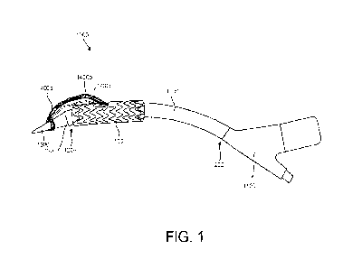
[00099] Referring to FIG. 1, a delivery system 1000 is
illustrated (not necessarily
to scale). The delivery system 1000 is operable to deliver a multi-component
implantable device (e.g., implantable device 10) to a target site. The
delivery system
includes a handle 1100, an elongate member 1200 having a first end 1202
coupled to
and/or extending from the handle 1100 and a second end 1204, a cap 1300
positioned
proximate the second end 1204 of the elongate member 1200, and at least one
guide
member 1400 extending at least partially along the elongate member 1200 toward
the
cap 1300. The elongate member 1200 and cap 1300 are operable to translate
along a
main guidewire 1500 (see FIG. 10). The delivery system 1000 can further
include at
least one sheath 1600 (see FIGS. 13a-13c), the sheath 1600 operable for use
with the
guide member 1400 (when there are a plurality of guide members 1400a, 1400b,
1400c,
each guide member 1400 has a corresponding sheath 1600). An articulatable
secondary guidewire 1700 (see FIG. 14) can be included for each sheath 1600.
The
delivery system 1000 may also include a constraining member 1800 (see FIG. 11)
that
is operable to constrain at least a portion of a multi-component implantable
device. It is
understood that an individual constraining member 1800 may be implemented for
each
discrete component of the multi-component implantable device. The delivery
system
1000 can be used in conjunction with filtration systems 2000 (e.g., to reduce
risk of
embolism, see FIG. 10). In some embodiments, the constraining member 1800 may
include a window 1802 for the side branch portals 110, the window 1802 of the
constraining member 1800 positioned overlaying the side branch portals such
that the
side branch portals 110 are accessible when the constraining member 1800 is
constraining the device 10 (see FIG. 24).
[000100] Referring to FIG. 9, an exemplary target site for delivery and
deployment
of a multi-component implantable device is illustrated. In this example, the
aorta 20 is
illustrated. However, it is understood that the delivery system 1000 may be
implemented
18
CA 03206686 2023- 7- 27
WO 2022/216374
PCT/US2022/017333
in any part of the vasculature that includes branched lumens as appropriate.
In this
example, the aorta 20 is illustrated, including the ascending aorta 21, aortic
arch 22,
and descending aorta 23, and branches therefrom, including the brachiocephalic
artery
24, the left common carotid artery 25, and the left subclavian artery 26.
[000101] FIG. 10 illustrates implementation of the filtration system 2000 in
connection with the delivery system 1000. The filtration system 2000 can
include a
plurality of deployable filters 2002 that can be deployed in discreet lumens,
including
side branch lumens, that are fluidically downstream from the target site at
which the
multi-component implantable device is to be implanted. The filtration system
2000 can
be intermittently flushed throughout the procedure. A main guidewire 1500 is
advanced
to the target site (e.g., the aortic arch). Although the main guidewire 1500
is illustrated
as coming from the descending aorta 23 (e.g., from a femoral access site), the
main
guidewire 1500 can be inserted from any appropriate access site.
[000102] Referring to FIG. 11, a multi-component implantable device is
advanced
to the target site via the guidewire 1500. For the purposes of the example
provided
herein, the multi-component implantable device will include the embodiment
disclosed
with respect to FIGS. 3-4. However, it is understood that the methods and the
delivery
system 1000 are not limited to delivering only the implantable device 10 as
described
with reference to FIG. 3 and 4. The implantable device (e.g., the main body
100) is
positioned on the elongate member 1200. For example, the implantable device 10
may
be constrained in a compressed configuration about the elongate member 1200.
For
example, the implantable device 10 can be constrained by a constraining member
1800.
The implantable device 10 may be positioned proximate the cap 1300 and the
second
end 1204 of the elongate member 1200.
[000103] As is illustrated in FIG. 11A, the delivery system 1000 may include a
plurality of guide members 1400a, 1400b, 1400c. The guide members 1400a,
1400b,
1400c are coupled (e.g., releasably coupled) to the delivery system 1000
proximate the
second end 1204 of the elongate member 1200. For example, in some embodiments,
the guide members 1400a, 1400b, 1400c are coupled to the cap 1300. The guide
members 1400a, 1400b, 1400c may each form a loop 1402 (one of which is
referenced
in FIG. 11A for ease of illustration) which can be fastened to or disposed
about at least
a portion of the cap 1300. In other embodiments, the guide members 1400a,
1400b,
1400c may implement a coupling system (not shown) for coupling the guide
members
1400 to the to the delivery system 1000 proximate the second end 1204 of the
elongate
member 1200, the coupling system including a feature, for example a ball tip,
that is
19
CA 03206686 2023- 7- 27
WO 2022/216374
PCT/US2022/017333
received by a corresponding member proximate the second end 1204 of the
elongate
member 1200 (e.g., the cap 1300 positioned proximate the second end 1204 of
the
elongate member 1200 may include a corresponding member). Other examples of
embodiments for matingly engaging or coupling the guide members 1400a, 1400b,
1400c at an end of the delivery system 1000 proximate the second end 1204 of
the
elongate member 1200 can be achieved by a variety of coupling arrangements,
including press fitting, threads, ball and detent, articulating clips or jaws,
hook and loop,
and magnetic. Any number of methods and structures may be implemented for
fastening the guide members 1400a, 1400b, 1400c proximate the second end 1204
of
the elongate member 1200, and the disclosed embodiments are not to be limiting
to the
scope of the disclosure. It is also understood that the guide members 1400 can
be
fastened at various other positions on the delivery system 1000. For example,
in some
embodiments, the guide member 1400 may be fixed to the elongate member 1200 or
other portions of the delivery system 1000. In some embodiments, the guide
members
1400 are fastened to an internal wall of the main body 100 (e.g., via a
releasable
suture). In some embodiments, the guide members 1400 can be retained at a
position
via a lock wire retainer 1902, which is described in further detail hereafter.
The lock wire
retainer 1902 may be implemented solely for capturing the guide members 1400
or may
be used in connection with other members for various other purposes, including
but not
limited to steering and positioning the main body 100 at the target site,
which will be
described hereafter. Further examples for coupling the guide members 1400 with
the
delivery system 1000 are provided hereafter and are discussed with regard to
FIGS.
27A-27C.
[000104] Referring to FIG. 11B, in some embodiments, a guide member retainer
1980 can be implemented with respect to the guide members 1400 in order to
retain the
guide members 1400 during delivery of the implantable device 10 and
advancement of
the sheaths 1600 along the guide members 1400. For example, as illustrated in
FIG.
11B, the delivery system 1000 includes an elongate member 1200 having a first
end
1202. At the first end 1202, the delivery system includes a lock wire retainer
1902 and
an end cap 1300 (e.g., the lock wire retainer 1902 is positioned between the
end cap
1300 and the first end 1202 of the elongate member 1200). The main body 100 is
positioned about the elongate member 1200 where the second region 3002 of the
main
body 100 is partially deployed and the first region 3000 is constrained. Guide
members
1400 are extending through the side branch portals 110 toward the first end
1202 of the
elongate member 1200. The guide members 1400 include a retaining member at the
CA 03206686 2023- 7- 27
WO 2022/216374
PCT/US2022/017333
end of each guide member 1400 (e.g., loops 1402). The guide member retainer
1980 is
releasably coupled to the lock wire retainer 1902 and extends along the
elongate
member 1200. The guide member retainer 1980 is operable to retain the guide
members 1400 at or proximate the first end 1202 of the elongate member 1200
(e.g., at
the lock wire retainer 1902, proximate the cap 1300, etc.). The guide member
retainer
1980 may be releasably coupled to the delivery system 1000 at a coupling
position, for
example, releasably and selectively coupled to the lock wire retainer 1902.
The guide
member retainer 1980 may capture, lasso, or otherwise retain the guide members
1400
at a longitudinal position relative to the elongate member 1200 such that the
guide
members 1400 are restricted from being retracted along the longitudinal length
of the
elongate member 1200. For example, the guide member retainer 1980 is fixedly
coupled to an end of each of the guide members 1400 (e.g., the loop 1402) such
that
the guide members 1400 are restricted from retracting when the guide member
retainer
1980 is engaged with the lock wire retainer 1902. The position where the guide
member
retainer 1980 is engaged with the guide members 1400 is generally at a
position
between the lock wire retainer 1902 and the side branch portals 110.
[000105] In some embodiments, the guide members 1400a, 1400b, 1400c may
implement a coupling system for coupling the guide members 1400 to the to the
guide
member retainer 1980 proximate the second end 1204 of the elongate member
1200,
the coupling system including a feature, for example a spherical tip, that is
received by a
corresponding member of the guide member retainer 1980. For example, the guide
member retainer 1980 may receive the spherical tip of the guide members 1400
through
an aperture or through a loop, where the diameter of the spherical tip of the
guide
members 1400 is greater than the diameter of the aperture or loop of the guide
member
retainer 1980. Other examples of embodiments for matingly engaging or coupling
the
guide members 1400a, 1400b, 1400c at an end of the delivery system 1000
proximate
the second end 1204 of the elongate member 1200 can be achieved by a variety
of
coupling arrangements, including press fitting, threads, ball and detent,
articulating clips
or jaws, hook and loop, and magnetic arrangements. Any number of methods and
structures may be implemented for fastening the guide members 1400a, 1400b,
1400c
proximate the second end 1204 of the elongate member 1200, and the disclosed
embodiments are not to be limiting to the scope of the disclosure. In some
embodiments, a plurality of guide member retainers 1980 may be implemented,
each
guide member retainer 1980 being operable to retain a corresponding guide
member
1400. Thus, each guide member 1400 may be independently retained and released
21
CA 03206686 2023- 7- 27
WO 2022/216374
PCT/US2022/017333
from engagement proximate the first end 1202 of the elongate member 1200. When
the
guide members 1400 are released, the guide members can be removed from the
corresponding side branch portal 110.
[000106] In some embodiments, the guide members 1400 may be coupled directly
to the lock wire retainer 1902. The guide member retainer 1980 and the guide
members
1400 (either directly or indirectly from the lock wire retainer 1902) may be
selectively
released from the lock wire retainer 1902. Each of the guide members 1400 may
be
selectively retained either collectively or individually.
[000107] Now referring to FIG. 11C, in various embodiments, the delivery
system
1000 may include a lock wire 1900. In such embodiments, the lock wire 1900 may
secure a steering line or lines 1850 to the catheter assembly. For example,
with
reference to FIG. 11C, delivery system 1000 comprises an elongate member 1200,
an
implantable device 10, at least one steering line 1850, and a lock wire 1900.
The lock
wire 1900 passes from outside of the body of the patient, through the elongate
member
1200, and exits at a point near a cap 1300. In some embodiments, at this point
the lock
wire 1900 interacts with the steering lines 1850, then reenters the elongate
member
1200 and continues to the cap 1300. In some embodiments, the lock wire 1900 is
coupled to a lock wire retainer 1902 (see also FIG. 1) that is positioned at
the second
end 1204 of the elongate members 1200, for example, between the cap 1300 and
the
implantable device 10. In such a configuration, the lock wire 1900 releasably
couples
the steering lines 1850 to delivery system 1000. Any manner in which the lock
wire
1900 may interact with the steering line or lines 1850 to maintain a
releasable coupling
between the steering line or lines 1850 and delivery system 1000 is within the
scope of
the present disclosure.
[000108] In various embodiments, each steering line may further include an end
loop. For example, each steering line 1850 comprises an end loop. The lock
wire 1900
may pass through each end loop, securing each steering line 1850 to delivery
system
1000. Any method of securing the steering line or lines 1850 to delivery
system 1000 is
within the scope of the invention.
[000109] In various embodiments, lock wires can be formed from metallic,
polymeric or materials and can include conventional medical grade materials
such as
nylon, polyacrylamide, polycarbonate, polyethylene, polyformaldehyde,
polymethylmethacrylate, polypropylene, polytetrafluoroethylene,
polytrifluorochlorethylene, polyvinylchloride, polyurethane, elastomeric
organosilicon
polymers; metals such as stainless steels, cobalt-chromium alloys and nitinol.
22
CA 03206686 2023- 7- 27
WO 2022/216374
PCT/US2022/017333
Elongated members or lock wires can also be formed from high strength polymer
fibers
such as ultra high molecular weight polyethylene fibers (e.g., Spectra ,
Dyneema
Purity , etc.) or aramid fibers (e.g., Technorae, etc.).
[000110] In various embodiments, a catheter assembly used to deliver an
expandable implant comprises a catheter shaft, an expandable implant, one or
more
sleeves, one or more steering lines, and a lock wire. In such configurations,
the
expandable implant is capable of bending, through tension applied to the one
or more
steering lines and corresponding displacement, to conform to curvature in the
vasculature of a patient. Tension can be applied to the steering lines 1850,
causing
expandable implant implantable device 10 to bend in a desired manner. For
example,
implantable device 10 can bend in a direction aligned with the location of the
steering
lines 1850. Once the implantable device 10 has been sufficiently bent,
consistent
tension is applied to steering lines 1850 to maintain the degree of bending.
In other
examples, the device 10 is configured to remain curved following tensioning of
the
steering lines 1850 absent a straightening force.
[000111] In various embodiments, tension can be applied to the steering lines
1850 by pulling the lines from the outside of the body of the patient. In
other
embodiments, the steering lines 1850 can be connected to one or more dials or
other
mechanisms for applying the tension at the trailing end of the elongate member
1200. In
this configuration, the dial can be used to apply a desired tension, as well
as maintain
the correct amount of tension once a desired angle of bending of implantable
device 10
has been achieved. Various embodiments may also comprise an indicator, scale,
gradient, or the like which demonstrates the amount of tension or displacement
of the
steering line, and/or the amount of bending in implantable device. In various
embodiments, the catheter assembly can comprise one more additional markings
(e.g.,
on a handle) that allow a user to determine the orientation of the steering
line with
respect to the vasculature.
[000112] After a sufficient degree of bending has been achieved in the
implantable
device 10, the implant can be rotated for final positioning in the treatment
area of the
vasculature. In various exemplary embodiments, the lock wire 1900 is engaged
with the
steering lines 1850 such that torsional rotation of the catheter shaft causes
the
implantable device 10 to rotate within the vasculature. However, any
configuration of the
delivery system 1000 which allows for rotation of implantable device 10 is
within the
scope of the present disclosure.
23
CA 03206686 2023- 7- 27
WO 2022/216374
PCT/US2022/017333
[000113] After the implantable device 10 is in position and expanded within
the
vasculature, the lock wire 1900 can be disengaged from delivery system 1000.
In
various embodiments, the lock wire 1900 is disengaged by applying sufficient
tension to
the lock wire 1900 from outside of the body of the patient. After the lock
wire 1900 is
disengaged, the steering lines 1850 can be released from coupling with the
elongate
member 1200 and can be removed from implantable device 10 and delivery system
1000.
[000114] With further reference to FIG. 11A, the guide members 1400a, 1400b,
1400c are each extending through a respective side branch portal 110 of the
main body
100 of the implantable device 10. Cannulation of the side branch portals 110
occurs
prior to insertion of the implantable device 10 into the patient via the
access site. Pre-
cannulation can shorten the procedure time and simplify the steps performed
during the
operation, which can reduce trauma to the patient's tissue and damage to the
implantable device 10. The guide members 1400a, 1400b, 1400c extend through
the
side branch portals 110 and through the second opening 109 of the main body
100 of
the implantable device 10. Thus, the guide members 1400a, 1400b, 1400c may be
positioned inside the main lumen 102 of the implantable device 10 from the
side branch
portals 110 to the second end 108 of the device. The guide members 1400a,
1400b,
1400c extend from the second opening 109 and toward the second end 1204 of the
elongate member 1200. In some embodiments, the guide members 1400a, 1400b,
1400c are routed through the handle 1100 (see FIG 1) and in other embodiments,
the
guide members 1400a, 1400b, 1400c are routed through other ports (not shown).
The
guide members 1400a, 1400b, 1400c may extend along the outside of the elongate
member 1200, or the guide members 1400a, 1400b, 1400c may extend through the
elongate member (not shown). In order to reduce tangling or crossing of the
guide
members 1400a, 1400b, 1400c, wire management devices (not shown) may be
implemented. For example, a wire management device may be provided to minimize
interaction of each of the plurality of guidewires and/or guide members with
each other
and other components of the delivery system 1000 in order to limit or prevent
tangling,
tying, or interference of the guidewires and/or guide members one with another
and
other components of the delivery system 1000, which obstructs advancement of
devices
along the guidewires and/or guide members. The wire management device
maintains
each of the guidewires and/or guide members in predetermined positions. The
wire
management device is operable to release portions of the guidewires and/or
guide
members when a device is advanced along the longitudinal length of the wire
24
CA 03206686 2023- 7- 27
WO 2022/216374
PCT/US2022/017333
management device, allowing the device and its branches to be advanced through
the
lumen of the patient. For example, the delivery system 1000 may include a wire
management device that releasably contains a plurality of guidewires and/or
guide
members. The wire management device may be configured to release a first
portion of
the at least one of the guidewires and/or guide members when a device is
advanced
along the main guidewire 1500 and configured to release a second portion one
of the
guidewires and/or guide members when the device is advanced along the main
guidewire 1500 to a second longitudinal position. Thus, the wire management
device
progressively (described also as step-wise, inch-by-inch, or sequentially)
releases the
guidewires as a device is advanced with respect to the delivery system 1000.
This
allows the guidewires to be appropriately positioned and to interact with the
device (e.g.,
pass into an internal lumen of the device) in accordance with delivery of the
device.
[000115] Referring now to FIG. 12, the implantable device 10 can be at least
partially deployed. For example, the main body 100 can be partially deployed
from a
first, constrained diameter to a second, partially constrained diameter that
is larger than
the first diameter. As is illustrated in FIG. 12, the main body 100 can also
include a first
region 3000 and a second region 3002. The first region 3000 extends from the
first end
106 to the second opening 121 of the side branch portal 110 and the second
region
3002 extends from the second opening 121 of the side branch portal 110 to the
second
end 108 of the main body 100. The dividing point between the first and second
regions
3000, 3002 may be defined at slightly different positions (e.g., generally
within about 3
cm of the side branch portals 110). In some embodiments, the first and second
regions
3000, 3002 can be independently constrained and/or deployed. For example, as
illustrated in FIG. 12, the second region 3002 is partially deployed to the
second,
partially constrained diameter whereas the first region 3000 is maintained at
the first,
constrained diameter. By partially deploying the second region 3002, the side
branch
portals 110 are operable to at least partially expand. Such constraining
members and
staged deployment may include, but are not necessarily limited to primary and
secondary sleeves of the constraining member 1800. The primary and secondary
sleeves may be used in series which allows for expansion or partial expansion
of a
portion of the main body 100 by releasing one of the primary or secondary
sleeves. This
permits access through the side branch portals, while still allowing the main
body 100 to
be manipulated relative to the target site. Furthermore, by maintaining the
first region
3000 in the first, constrained configuration, access through the second
opening 121 of
the side branch portal 110 is unrestructured or unblocked by the first region
3000 of the
CA 03206686 2023- 7- 27
WO 2022/216374
PCT/US2022/017333
main body 100. This also facilitates access to the branched lumens (e.g.,
brachiocephalic artery 24).
[000116] Referring now to FIGS. 13a-13c, a sheath 1600 is provided for each
guide member 1400. For example, when there is a first, second, and third guide
member 1400a, 1400b, 1400c for the first, second, and third side branch
portals 110a,
110b, 110c, a first, second, and third sheath 1600a, 1600b, 1600c (see FIG.
15) is
provided for each corresponding side branch body 200 and guide member 1400.
Each
sheath 1600 is operable to advance along each corresponding guide member 1400.
The sheath 1600 can be formed to move along the guide member 1400 by
surrounding
the guide member 1400, by using the guide member as a rail in a side-by-side
orientation, or as would otherwise permit the sheath 1600 the move
substantially along
the path of the guide member 1400. Because the side branch portals 110 are
already
pre-cannulated with the guide members 1400a, 1400b, 1400c, the sheath 1600 can
be
advanced through the second opening 109 of the elongate member and out the
second
opening 121 of the side branch portal 110. For example, a first end 1602 of
the sheath
1600 can be advanced through the vasculature of a patient and out the second
opening
121 of the side branch portal 110. The first end 1602 may be positioned
proximate the
corresponding branch of the vasculature (e.g., the brachiocephalic artery).
[000117] In some embodiments, the sheath 1600 includes a lumen through which
an articulatable secondary guide member or catheter 1700 can be inserted
(e.g., the
same lumen through which the guide members 1400 are passed). Various secondary
articulatable member or catheter 1700 may be implemented, including, but not
limited
to, steerable catheters and guidewires. For example, articulatable secondary
member or
catheter 1700 can be steered using at least one tether or tension member (not
shown)
coupled to a distal end of the articulatable guidewire 1700 (the articulatable
guide
member or catheter 1700 may be an integral unit, or may be a composite of
various
components for providing the articulating function, e.g., a guide catheter and
a
guidewire). The articulatable guide member or catheter 1700 may be steered by
applying tension to the tether or tension member. Various degrees of motion
can be
achieved using multiple tethers and/or tension members. Other embodiments may
include robotic or motor-driven guidewires. Various embodiments of an
articulatable
guidewire may be implemented in the delivery system 1000 and method. The
articulatable secondary guide member or catheter 1700 is advanced to the
treatment
site via the sheath 1600. For example, as illustrated in FIG. 14, a first
articulatable
secondary guide member or catheter 1700a is advanced to the target site via
the first
26
CA 03206686 2023- 7- 27
WO 2022/216374
PCT/US2022/017333
sheath 1600a through first side branch portal 110a. The articulatable
secondary
guidewire 1700 includes a leading end that can be articulated by the user at a
trailing
end (not shown). The leading end can bend or articulate to various
configurations and
positions. Once the leading end of the first articulatable secondary guide
member or
catheter 1700a is free from the first sheath 1600a, the user is able to
articulate the
leading end of the first articulatable secondary guide member or catheter
1700a into
position in the target branch corresponding to the first side branch portal
110a (e.g., the
brachiocephalic artery). Once the secondary guide member or catheter 1700a is
in
place, a guidewire 1702 may be advanced into position (e.g., through the
secondary
guide member or catheter 1700a). This step is repeated for each branch and
corresponding side branch portal 110 with a subsequent articulatable secondary
guide
member or catheter 1700. Referring to FIG. 15, the guidewire 1702 can be
advanced
through the filter 2002 that was deployed in the branch. Furthermore, the
guidewire
1702 can be advanced such that the guidewire 1702 extends out the filter's
access site
to create a through-and-through configuration of the guidewire 1702. FIG. 16
illustrated
each of the branches (e.g., brachiocephalic, left common carotid, and left
subclavian
arteries 24, 25, 26) being cannulated with corresponding articulatable
secondary
guidewires 1700, where the guidewires 1700 extend through a corresponding side
branch portal 110.
[000118] Referring now to FIG. 17, the first region 3000 is partially deployed
to the
second, partially constrained diameter. When the first region 3000 is
partially deployed,
the entire main body 100 is partially deployed to the second, partially
constrained
diameter. At this stage, the main body 100 can be adjusted to the appropriate
position
within the target site to facilitate optimal placement and performance of the
implantable
device 10. Once the desired positioning of the main body 100 is achieved, the
main
body 100 can be deployed to a third, deployed diameter (e.g., not constrained
by the
constraining member 1800). As illustrated in FIG. 18, once the main body 100
is fully
deployed, portions of the delivery system 1000 may be removed, including the
elongate
member 1200 and cap 1300, the at least one guide member 1400, the at least one
sheath 1600, and the constraining members 1800. As previously described, the
guide
members 1400 may be released, which may occur at this point in the procedure.
In
some embodiments, the main guidewire 1500 may also be removed. This results in
the
main body 100 remaining at the target site with secondary, articulatable
guidewires
1700 cannulating corresponding side branch portals 110 and branches (e.g.,
brachiocephalic, left common carotid, and left subclavian arteries 24, 25,
26).
27
CA 03206686 2023- 7- 27
WO 2022/216374
PCT/US2022/017333
[000119] Referring to FIG. 19, branch bodies 200 can be advanced along
corresponding secondary, articulatable guidewires 1700. The branch bodies 200
can be
advanced on independent components, for example, elongate members 4000 with
end
caps 4002, and so forth, similar to the components used to deliver the main
body 100.
The end caps 4002, or other independent components, can dilate the side branch
portals 110 in some embodiments as the branch bodies 200 are passing through
the
side branch portals 110. The branch bodies 200 are positioned such that a
first portion
202 is at least partially positioned in the branch of the target site and a
second portion is
positioned within the implantable device 10 (e.g., within the side branch
portal 110).
Once the branch bodies 200 are appropriately positioned, the branch bodies 200
are
deployed, as illustrated in FIG. 20. Referring to FIG. 21, the elongate
members 4000
and end caps 4002 used to deliver the branch bodies 200 are removed. FIG. 22
illustrates aspiration of the filtration system 2000. Once, the filtration
system 2000 is
aspirated, the filtration system 2000 can be removed, as illustrated in FIG.
23. The
remaining components can be removed from the patient (e.g., the main guidewire
1500
and the secondary, articulatable guidewires 1700) and the surgeon can commence
closure.
[000120] Referring to FIGS. 25-28, in some embodiments, another embodiment of
the device 10 is provided with a plurality of selectable side branch portals
510. FIG. 25
illustrates a side view of an example of an implantable device 10 having a
main body
500 and a plurality of selectable side branch portals 510 extending
therethrough. The
implantable device 10 also includes the side branches 502 extending from the
main
body 500 through the selectable side branch portal 510. The side branches 502
are
separate from the main body 500 (i.e., they are not integral with the main
body 500).
Because the side branches 502 are separate structures from the main body 500,
the
side branches 502 are coupled to the main body 500 to form the implantable
device. For
example, the main body 500 may be deployed in the abdominal aorta and the side
branches 502 may be deployed in the renal arteries and extend into the main
body 500
positioned in the abdominal aorta.
[000121] As shown in FIG. 26A, in some embodiments the main body 500 of the
implantable device 10 includes a tubular member 520 and a stent member 540. As
shown, the tubular member 520 has a first end 522 and a second end 524. The
tubular
member 520 forms a primary lumen 526 having a first opening 523 at the first
end 522
and a second opening 525 at the second end 524 of the tubular member 520. The
tubular member 520 includes a side branch portal 510 that includes a column
528
28
CA 03206686 2023- 7- 27
WO 2022/216374
PCT/US2022/017333
positioned within the primary lumen 526 and forms a secondary lumen 530 (see
FIG.
26B). The tubular member 520 defines an aperture 532 into the secondary lumen
530 at
a position longitudinally between the first end 522 and second end 524 of the
tubular
member 520. The column 528 defines a column opening 534 (see FIG. 26B)
proximal
the second end 524 of the tubular member 520. The stent member 540 supports
the
tubular member 520 in such a manner that the implantable device is operable to
be
configured in a delivery configuration and in a deployed configuration, or to
be
transitioned from a delivery configuration toward a deployed configuration.
[000122] In some embodiments, the tubular member 520 includes a first graft
member 541 defining the primary lumen 526 and a second graft member 542
coupled to
the first graft member 541 to form the column 528 defining the secondary lumen
530
between the first graft member 541 and the second graft member 542. For
example, the
first graft member 541 includes graft material formed in the shape of a tube
to define the
primary lumen 526. The second graft member 542 optionally includes graft
material that
is coupled to the first graft member 541 (e.g., via boding, adhesive, or by
otherwise
being coupled together) to form the secondary lumen 530. The graft materials
of the first
and second graft members 541, 542 may be the same material or different
materials as
desired. Though some materials may provide certain advantages over others, a
variety
of suitable graft materials may be implemented, and generally any suitable
graft
material may be implemented including those materials discussed herein.
[000123] In some embodiments the secondary lumen 530 extends at least
partially
along a longitudinal length of the main body 512. The secondary lumen 530 of
the
column 528 opens into the primary lumen 526 at the proximal opening of the
secondary
lumen 530. In some embodiments, the column 528 extends to the second end 524
of
the tubular member 520 such that the column opening 534 is positioned at or
coplanar
with the second opening 525 of the tubular member 520. In other embodiments,
the
column 528 extends toward the second end 524 of the tubular member 520 such
that
the column opening 534 is longitudinally spaced from the second opening 525 of
the
tubular member 520. In embodiments including a plurality of columns 528, the
column
openings 534 may be positioned at the same longitudinal length across, or in
different
terms, at the same longitudinal position along, the tubular member 520 or they
may be
staggered at two or more longitudinally-spaced positions along the length of
the tubular
member 520.
[000124] In some embodiments, the column 528, and consequently the secondary
lumen 530 are collapsible. For example, the column 528 may be unsupported by a
stent
29
CA 03206686 2023- 7- 27
WO 2022/216374
PCT/US2022/017333
member, although supported, collapsible embodiments are also contemplated.
Lack of
a support, or a suitably configured support, may allow the column 528 to be
collapsed
(radially collapsed) to seal the aperture 532 and limit the leaking or other
passing of
fluids (e.g., blood) through the aperture 532. In some embodiments, the
pressure (e.g.,
hydrostatic pressure, fluid pressure gradients, and/or pressure exerted by
fluids in
motion) that is exerted by the fluid collapses the column 528 such that the
column
coapts or seals against the tubular member 520 to limit flow through the
secondary
lumen 530 and consequently the aperture 532.
[000125] As illustrated in FIG. 26A, the column 528 may be sealed or closed
near
the first end 522 of the tubular member 520, or, in some embodiments not
shown, at the
first end 522. The secondary lumen 530 thus is operable to provide fluid
communication
between the exterior surface of the tubular member 520 between the first and
second
end 522, 524 and the primary lumen 526, for example, when the column 528 is
patent.
In some embodiments, the tubular member 520 may include a column 528 that is
unsealed (i.e., includes an opening) near the first end of the tubular member
520. In
such embodiments, an elongate member such as a delivery catheter may be
positioned
through the column 528. Referring to FIG. 26B, an end view of the main body
512 is
shown with the column opening 534 positioned proximate the second end 524 of
the
main body 512. In some embodiments, columns 528 extend to the second end 524
of
the main body 512. As illustrated, the secondary lumen 530 may be contained
within the
primary lumen 526.
[000126] Referring again to FIG. 26A, the main body 512 includes the stent
member 540. The stent member 540 may be formed of any suitable material as is
discussed hereafter. The stent member 540 is operable to support the tubular
member
520. The stent member 540 may be compressed into a delivery configuration and
may
be expanded into an expanded configuration, such as at deployment. The stent
member
540 may be a self-expanding stent or a balloon expandable stent. As
illustrated, the
stent member 540 includes a plurality of stent rings 544. Each stent ring 544
circumferentially supports the tubular member 520 at a longitudinal position
along the
length of the tubular member 520. For example, each stent ring 544 is
longitudinally
spaced from an adjacent stent ring 544. The stent rings 544 may each include
apices
546 with first apices 546a pointing toward the first end 522 and second apices
546h
pointing toward the second end 524. Various other configurations of stent
members 40
are contemplated herein including, but not limited to, helical stents
(including undulating
helical stents, diamond pattern stents, and others).
CA 03206686 2023- 7- 27
WO 2022/216374
PCT/US2022/017333
[000127] As illustrated in FIG. 26A, the tubular member 520 includes a
plurality of
apertures 532 spaced along the longitudinal length of the main body 512. The
apertures
532 may be positioned such that at least one stent ring is between the two
longitudinally
adjacent apertures 532. For example, a column 528 may include apertures 532
through
the tubular member 520 such that the apertures 532 are longitudinally spaced
along the
main body 512. The apertures are all in fluid communication with the secondary
lumen
530 of the column 528. The apertures 532 provide access points for the
secondary
branch at various longitudinal lengths along the main body 512.
[000128] Referring to FIG. 26C, the apertures 532 may be formed in a variety
of
shapes and size including circular profiles, a profile with a rounded edge and
a
substantially flat edge, ovular profiles, and so forth. The various shapes and
sizes may
be implemented to accommodate various side branches 502 and configurations
such as
angle of exit of the side branches 502 from the main body 512 at the apertures
532. In
some embodiments not shown, the apertures 532 may be irregularly spaced along
the
longitudinal length of the column 528. Furthermore, in some embodiments not
shown,
the apertures 532 may be circumferentially spaced within a column 528. For
example,
the apertures 532 may be staggered circumferentially and/or longitudinally.
[000129] In some embodiments, the main body 512 may include a plurality of
columns 528. For instance, the main body 512 may include two columns 528 that
are
circumferentially spaced from each other in order to deploy two side branches
514 into
the side branch lumens of the patient's anatomy. Furthermore, the main body
512 may
include a plurality of columns 528 that are associated with each side branch
lumen of
the patient's anatomy. For example, if the main body 512 is to be deployed in
the
abdominal aorta and the side branches 514 are to be deployed into the renal
arteries,
each patient may have a various positions circumferentially at which the renal
arteries
enter the aorta.
[000130] By having a plurality of columns 528 through which each side branch
514
may be deployed, the surgeon may select the appropriate columns 528 that best
conform to the patient's native anatomy without applying torsion to the
vessels when the
implantable device 10 is deployed. Thus, in one example, the main body 512 may
include three columns 528 on one circumferential side of the tubular member
520 and
three more columns 528 on an opposite circumferential side of the tubular
member 520.
Each column 528 is circumferentially spaced from the adjacent column 528 about
the
circumference of the tubular member 520. It is contemplated that any number of
columns 528 and the spacing of the columns 528 may be implemented, including
one,
31
CA 03206686 2023- 7- 27
WO 2022/216374
PCT/US2022/017333
two, three, four, five, six, seven, eight, or more columns 528 which may be
spaced
equally or variably about the circumference of the tubular member 520. It is
further
contemplated that the specific spacing may be determined by surveying the
average
circumferential spacing of side branches for a particular implementation in a
sample
population of patients to determine the spacing of the columns 528.
Circumferential
spacing of the column 528 allows for clocking of the main body 512 within the
patient's
anatomy with increased positions for appropriately positioning the side
branches 514
into the side branch vessels. As used herein, the term "clocking" refers to
the ability to
position features at a desired location about a circumference of an object.
This ability to
clock the one or more columns 528 can be further advantageous for use with
visualization, for example when the procedure is being performed via
fluoroscopy. This
simplifies placement by providing several entry points when dealing with the
two-
dimensional planes shown by visualization techniques and for parallax
associated with
such visualization. In some embodiments, the columns 528 may be irregularly
spaced
about the circumference of the main body 512 (e.g., non-uniform spacing
between the
columns 528). In some embodiments not shown, the column 528 extends
longitudinally
and at angle greater than zero relative to the main body 512 longitudinal
axis. For
example, the secondary lumen 530 extends along a secondary lumen axis that
extends
longitudinally at an angle greater than zero relative to an axis of the
primary lumen 526
(e.g., helically about the main body 512).
[000131] Referring again to FIG. 26A, the main body 512 may include a
constraining member receiver 50 positioned surrounding at least a portion of
the stent
member 40. For example, in those embodiments including a plurality of stent
rings 544,
a corresponding constraining member receiver 550 is positioned about each
stent ring
544. The constraining member receiver 550 may be formed from a variety of
materials
including graft materials, fibers, and so forth. The constraining member
receiver 550 is
operable to receive constraining members that can be retracted to partially
constrain or
collapse the stent rings 544 as is discussed hereafter.
[000132] In some embodiments, the tubular member 520 may include a scallop
552 at the first end 522. The scallop 552 is a facilitates placement of the
tubular
member 520 in a lumen including a side branch lumen that does not need a
prosthetic
side branch deployed. For example, when the implantable device 10 is
positioned in the
abdominal aorta and the superior mesenteric artery does not need a side branch
514
deployed therein, the scallop 552 may be positioned over the entrance into the
superior
mesenteric artery without blocking or restricting blood perfusion
therethrough. The
32
CA 03206686 2023- 7- 27
WO 2022/216374
PCT/US2022/017333
scallop 552 may include various shapes including straight edge profiles,
curved profiles,
and combinations thereof.
[000133] Referring now to FIGS. 27A-270, a catheter olive or cap 1300 is
positioned at the first end of the elongate member 1200 such that the main
body 512 of
the implantable device 10 is positioned longitudinally between the cap 1300
and the
second end of the elongate member 1200. Although an embodiment of the cap 1300
is
depicted in the drawings, it is in the scope of the disclosure that any
catheter olive or
cap may be implemented within the scope of this disclosure. The cap 1300 may
be
implement to atraumatically advance the delivery system 1000 through the
patient and
to dilate the surrounding anatomy where appropriate. For example, the cap 1300
may
include a leading tip which is advanced first through the patient's anatomy.
Referring to
FIGS. 27A-27C, the cap 1300 may include a guide member retainer 1302. However,
the
guide member retainer 1302 may comprise a passage through which the guide
members 1400 pass (see FIG. 27A). In this embodiment, the guide members 1400
may
pass through the cap 1300 and extend back through an aperture 532 of another,
oppositely positioned column 528. The guide member retainer 1302 may be
operable to
releasably retain a lock wire 1900 to which the guide members 1400 may be
coupled
(see FIG. 27B). The lock wire 1900 may be controlled via the lock wire lumen.
The
guide member retainer may be operable to received and releasably retain ends
of the
guide members 1400, for example via a friction fit or other coupling (see FIG.
27C).
Various embodiments of caps 1300 may be implemented specifically for coupling
the
guide members 1400 (e.g., the guide member retainers 1302). Such embodiments
include those discussed in U.S. Pat. Pub. No. 2020/0046534 by Chung et al.,
filed
August 13, 2019, the content of which is hereby expressly incorporated by
reference. In
some embodiments, the cap 1300 may be curved to facilitate clocking of the
device 10
as it is advanced to the target site from implantation.
[000134] Although the method was disclosed with reference to the aorta 20, the
systems and method described herein could be implemented on various lumens
where
branching occurs.
[000135] Catheters, introducer sheaths, hubs, handles and other components
usable in medical device delivery systems and methods disclosed herein can be
constructed using any suitable medical grade material or combination of
materials using
any suitable manufacturing process or tooling. Suitable medical grade
materials can
include, for example, nylon, polyacrylamide, polycarbonate, polyethylene,
polyformaldehyde, polymethylmethacrylate, polypropylene,
polytetrafluoroethylene,
33
CA 03206686 2023- 7- 27
WO 2022/216374
PCT/US2022/017333
expanded polytetrafluoroethylene, polytrifluorochlorethylene,
polyvinylchloride,
polyurethane, elastomeric organosilicon polymers, Pebaxe polyether block
amide, and
metals such as stainless steels and nitinol. Catheters can also include a
reinforcing
member, such as a layer of metal braid.
[000136] A biocompatible material for the graft components, discussed herein,
may be used. In certain instances, the graft may include a fluoropolymer, such
as a
polytetrafluoroethylene (PTFE) polymer or an expanded polytetrafluoroethylene
(ePTFE) polymer. In some instances, the graft may be formed of, such as, but
not
limited to, a polyester, a silicone, a urethane, a polyethylene terephthalate,
or another
biocompatible polymer, or combinations thereof. In some instances,
bioresorbable or
bioabsorbable materials may be used, for example a bioresorbable or
bioabsorbable
polymer. In some instances, the graft can include Dacron, polyolefins, carboxy
methylcellulose fabrics, polyurethanes, or other woven, non-woven, or film
elastomers.
[000137] It is understood that any of the components of the systems can also
include radiopaque markers to facilitate viewing on an x-ray fluoroscope
during an
implantation procedure. Any number, shape and location of radiopaque markers
can be
utilized as needed.
[000138] Delivery systems and methods disclosed herein are particularly suited
for
endoluminal delivery of branchable expandable implants for treating branched
vasculature. Expandable implants can include, for example, stents, grafts, and
stent
grafts. Further, expandable implants can include one or more stent components
with
one or more graft members disposed over and/or under the stent, which can
dilate from
a delivery configuration, through a range of larger intermediary
configurations, and
toward a deployed configuration engaged with vessel walls at a treatment site.
However, and as discussed below, any suitable combination and configuration of
stent
component(s) and graft member(s) is within the scope of the present
disclosure. For
example, stent components can have various configurations such as, for
example,
rings, cut tubes, wound wires (or ribbons) or flat patterned sheets rolled
into a tubular
form. Stent components can be formed from metallic, polymeric or natural
materials
and can comprise conventional medical grade materials such as nylon,
polyacrylamide,
polycarbonate, polyethylene, polyformaldehyde, polymethylmethacrylate,
polypropylene, polytetrafluoroethylene, polytrifluorochlorethylene,
polyvinylchloride,
polyurethane, elastomeric organosilicon polymers; metals such as stainless
steels,
cobalt-chromium alloys and nitinol and biologically derived materials such as
bovine
arteries/veins, pericardium and collagen. Stent components can also comprise
34
CA 03206686 2023- 7- 27
WO 2022/216374
PCT/US2022/017333
bioresorbable materials such as poly(amino acids), poly(anhydrides),
poly(caprolactones), poly(lactic/glycolic acid) polymers,
poly(hydroxybutyrates) and
poly(orthoesters).
[000139] Moreover, potential materials for graft members include, for example,
expanded polytetrafluoroethylene (ePTFE), polyester, polyurethane,
fluoropolymers,
such as perfluoroelastomers and the like, polytetrafluoroethylene, silicones,
urethanes,
ultra-high molecular weight polyethylene, aramid fibers, and combinations
thereof.
Other embodiments for a graft member material can include high strength
polymer
fibers such as ultra-high molecular weight polyethylene fibers (e.g., Spectra
, Dyneema
Purity , etc.) or aram id fibers (e.g., Technora , etc.). The graft member may
include a
bioactive agent. In one embodiment, an ePTFE graft includes a carbon component
along a blood contacting surface thereof. Any graft member which can be
delivered by
a catheter is in accordance with the present disclosure.
[000140] In addition, nitinol (NiTi) may be used as the material of the frame
or
stent (and any of the frames discussed herein), but other materials such as,
but not
limited to, stainless steel, L605 steel, polymers, MP35N steel, polymeric
materials,
Pyhnox, Elgiloy, or any other appropriate biocompatible material, and
combinations
thereof, can be used as the material of the frame. The super-elastic
properties and
softness of NiTi may enhance the conformability of the stent. In addition,
NiTi can be
shape-set into a desired shape. That is, NiTi can be shape-set so that the
frame tends
to self-expand into a desired shape when the frame is unconstrained, such as
when the
frame is deployed out from a delivery system. Other materials may also be used
as
appropriate, including but not limited to NiTiCo.
[000141] The invention of this application has been described above both
generically and with regard to specific embodiments. It will be apparent to
those skilled
in the art that various modifications and variations can be made in the
embodiments
without departing from the scope of the disclosure. Thus, it is intended that
the
embodiments cover the modifications and variations of this invention provided
they
come within the scope of the appended claims and their equivalents.
[000142] Any of a variety of bio-active agents may be implemented with any of
the
foregoing. For example, any one or more of (including portions thereof) the
implantable
device 10 and the delivery system 1000 may comprise a bio-active agent. Bio-
active
agents can be coated onto one or more of the foregoing features for controlled
release
of the agents. Such bio-active agents can include, but are not limited to,
thrombogenic
agents such as, but not limited to, heparin. Bio-active agents can also
include, but are
CA 03206686 2023- 7- 27
WO 2022/216374
PCT/US2022/017333
not limited to agents such as anti-proliferative/antimitotic agents including
natural
products such as vinca alkaloids (e.g., vinblastine, vincristine, and
vinorelbine),
paclitaxel, epidipodophyllotoxins (e.g., etoposide and teniposide),
antibiotics (e.g.,
dactinomycin (actinomycin D), daunorubicin, doxorubicin, and idarubicin),
anthracyclines, mitoxantrone, bleomycins, plicamycin (mithramycin) and
mitomycin,
enzymes (e.g., L-asparaginase which systemically metabolizes L-asparagine and
deprives cells which do not have the capacity to synthesize their own
asparagine);
antiplatelet agents such as G(GP) Ilb/Illa inhibitors and vitronectin receptor
antagonists;
anti-proliferative/antimitotic alkylating agents such as nitrogen mustards
(e.g.,
mechlorethamine, cyclophosphamide and analogs, melphalan, chlorambucil),
ethylenimines and methylmelamines (e.g., hexamethylmelamine and thiotepa),
alkyl
sulfonates-busulfan, nitrosoureas (e.g., carmustine (BCNU) and analogs,
streptozocin),
trazenes-dacarbazinine (DTIC); anti-proliferative/antimitotic antimetabolites
such as folic
acid analogs (e.g., methotrexate), pyrimidine analogs (e.g., fluorouracil,
floxuridine, and
cytarabine), purine analogs and related inhibitors (e.g., mercaptopurine,
thioguanine,
pentostatin and 2-chlorodeoxyadenosine {cladribine}); platinum coordination
complexes
(e.g., cisplatin and carboplatin), procarbazine, hydroxyurea, mitotane,
am inoglutethimide; hormones (e.g., estrogen); anti-coagulants (e.g., heparin,
synthetic
heparin salts and other inhibitors of thrombin); anti-platelet agents (e.g.,
aspirin,
clopidogrel, prasugrel, and ticagrelor); vasodilators (e.g., heparin,
aspirin); fibrinolytic
agents (e.g., plasminogen activator, streptokinase, and urokinase), aspirin,
dipyridamole, ticlopidine, clopidogrel, abciximab; antimigratory agents;
antisecretory
agents (e.g., breveldin); anti-inflammatory agents, such as adrenocortical
steroids (e.g.,
cortisol, cortisone, fludrocortisone, prednisone, prednisolone, 6a-
methylprednisolone,
triamcinolone, betamethasone, and dexamethasone), non-steroidal agents (e.g.,
salicylic acid derivatives, such as aspirin); para-aminophenol derivatives
(e.g.,
acetaminophen); indole and indene acetic acids (e.g., indomethacin, sulindac,
and
etodalac), heteroaryl acetic acids (e.g., tolmetin, diclofenac, and
ketorolac),
arylpropionic acids (e.g., ibuprofen and derivatives), anthranilic acids
(e.g., mefenamic
acid and meclofenamic acid), enolic acids (e.g., piroxicam, tenoxicam,
phenylbutazone,
and oxyphenthatrazone), nabumetone, gold compounds (e.g., auranofin,
aurothioglucose, and gold sodium thiomalate); immunosuppressives (e.g.,
cyclosporine,
tacrolimus (FK-506), sirolimus (rapamycin), azathioprine, and mycophenolate
mofetil);
angiogenic agents (e.g., vascular endothelial growth factor (VEGF)),
fibroblast growth
factor (FGF); angiotensin receptor blockers; nitric oxide donors; anti-sense
36
CA 03206686 2023- 7- 27
WO 2022/216374
PCT/US2022/017333
oligonucleotides and combinations thereof; cell cycle inhibitors, mTOR
inhibitors, growth
factor receptor signal transduction kinase inhibitors; retinoids; cyclin/CDK
inhibitors;
HMG co-enzyme reductase inhibitors (statins); and protease inhibitors.
Delivery systems and methods in accordance with various embodiments disclosed
herein can utilize removable guidewires to preserve branch portals for
guidewire
cannulation therethrough subsequent to compacting the expandable implant
toward a
delivery configuration for endoluminal delivery to the treatment site.
Removable
guidewire tube can comprise the same materials listed above for the catheter
materials.
[000143] Numerous characteristics and advantages of the present invention have
been set forth in the preceding description, including preferred and alternate
embodiments together with details of the structure and function of the
invention. The
disclosure is intended as illustrative only and as such is not intended to be
exhaustive. It
will be evident to those skilled in the art that various modifications may be
made,
especially in matters of structure, materials, elements, components, shape,
size and
arrangement of parts within the principals of the invention, to the full
extent indicated by
the broad, general meaning of the terms in which the appended claims are
expressed.
To the extent that these various modifications do not depart from the spirit
and scope of
the appended claims, they are intended to be encompassed therein. In addition
to being
directed to the embodiments described above and claimed below, the present
invention
is further directed to embodiments having different combinations of the
features
described above and claimed below.
[000144] The invention of this application has been described above both
generically and with regard to specific embodiments. It will be apparent to
those skilled
in the art that various modifications and variations can be made in the
embodiments
without departing from the scope of the disclosure. Thus, it is intended that
the
embodiments cover the modifications and variations of this invention provided
they
come within the scope of the appended claims and their equivalents.
37
CA 03206686 2023- 7- 27