Note : Les descriptions sont présentées dans la langue officielle dans laquelle elles ont été soumises.
P32W0 2022/200515
PCT/EP2022/057801
1
Assay
[0001] This invention relates to assays for allergy testing. The invention
further relates to cell
lines and kit for use in said assays, and methods of producing the cell lines.
BACKGROUND
[0002] Roughly one-third of the global population is suffering from allergic
hypersensitivity
disorders, according to recent estimations. For many patients allergies are
associated with a
marked reduction in physical and psychological well-being and lead to a
significant loss in
quality-of-life due to disease activity. To provide efficient and personalized
treatment options,
physicians are dependent on solid and reliable diagnostic tools.
[0003] Generally, allergy diagnosis is a complex and laborious multistep
procedure. It involves
the examination of the patient's medical history, serological determination of
total and allergen
specific immunoglobulin E (IgE) antibody levels and various in vivo allergen
skin prick tests
(SPT) or other in vivo allergen challenge protocols. Even though the
determined levels of total
and allergen specific IgE antibodies provide information about the atopic
status of an
individual, these values often poorly correlate with disease activity and
clinical symptoms.
Further, important parameters contributing to aggravation or suppression of
allergic responses
such as diversity and affinity of the allergen-specific IgE antibodies and the
presence of
allergen-specific IgG antibodies are frequently neglected in the
interpretation of diagnostic
laboratory results.
[0004] Occasionally, a functional basophil activation test (BAT) with whole
blood samples of
the patient is performed to determine reactivity against certain allergens.
While such assays
are useful as they provide important quantitative and functional information
about the allergic
status of an individual, they are hampered by the use of fresh whole blood
that has to be
processed within hours in specialized diagnostic laboratories. Immediate
analysis of whole
blood is associated with major logistical challenges as its storage is not
possible due to
instability of the biological material. More recently, diagnostic testing of
allergies based on
primary human blood-derived mast cells (i.e. MAT) has been suggested by
independent
groups. While the overall goal of this approach is appealing, the generation
of the cells is
laborious and requires extended culturing periods of more than two months.
Despite these
interesting recent developments, a convenient, safe, standardized and reliable
diagnostic
assay predicting functional reactivity against culprit allergens is still not
available.
[0005] Allergic patients are often advised to undergo allergen-specific
immunotherapy (AIT),
which has been reported to be one of the few disease modifying interventions
currently
available for allergy treatment. In AIT increasing doses of allergen (up-
dosing phase) are
applied to the patient until a certain maintenance dose (depends on allergen)
is reached. In
CA 03211629 2023- 9-8
P32W0 2022/200515
PCT/EP2022/057801
2
subcutaneous AIT (SCIT) protocols patients receive monthly allergen injections
over a
duration of three to five years after an initial up-dosing phase. Various
molecular and cellular
mechanisms such as the induction of allergen-specific protective IgG have been
described for
AIT, however, it remains still unclear why some patients respond better to the
treatment than
others. In fact, only 13% of patients show sustained unresponsiveness to an
allergen after one
year of AIT completion as assessed in a recent peanut desensitization study.
Currently, there
is no suitable read-out system available to assess whether and when a patient
is responding
to the treatment. Therefore, physicians usually perform an in vivo allergen
challenge test to
determine the degree of unresponsiveness after AIT, which is unpleasant for
the patient and
risks inducing an allergic reaction in case the patient has not responded to
the treatment.
BRIEF SUMMARY OF THE DISCLOSURE
[0006] According to a first aspect of the invention, there is provided a
method for producing
non-human conditionally immortalized mast cell progenitors, the method
comprising:
- introducing a nucleic acid molecule comprising an inducible homeobox gene
into myeloid
progenitor cells, wherein said myeloid progenitor cells are derived from a non-
human animal
and are engineered to express a heterologous high-affinity IgE receptor alpha
subunit
(FcERIa); and
- selecting for cells which contain the nucleic acid molecule.
The nucleic acid molecule may be recombinant.
[0007] According to a second aspect of the invention, there is provided a
method for
preparing mast cells comprising culturing non-human conditionally immortalized
mast cell
progenitors which are engineered to express a heterologous high-affinity IgE
receptor alpha
subunit (FcERIa), wherein the conditionally immortalized mast cell progenitors
further
comprise a homeobox gene, the expression of the homeobox gene being under the
control
of an inducer, and wherein the conditionally immortalized mast cell
progenitors are cultured
in the absence of the inducer.
[0008] The non-human conditionally immortalized mast cell progenitors used to
prepare the
mast cells may be those prepared using the method of the first aspect of the
invention.
[0009] In a third aspect, the present invention provides a non-human
conditionally
immortalized mast cell progenitor comprising a homeobox gene, the expression
of the
homeobox gene being under the control of an inducer, wherein said cell
expresses a
heterologous high-affinity IgE receptor alpha subunit (FcERIa). The homeobox
gene may be
comprised in a recombinant nucleic acid molecule.
CA 03211629 2023- 9-8
P32W0 2022/200515
PCT/EP2022/057801
3
[0010] The non-human conditionally immortalized mast cell progenitor may be
obtainable by
the method according to the first aspect of the invention.
[0011] In a fourth aspect of the invention, there is provided a composition
comprising a
population of non-human conditionally immortalized mast cell progenitors
according to the
third aspect of the invention. The composition may further comprise the
inducer which
controls the expression of the homeobox gene.
[0012] According to a fifth aspect of the invention, there is provided a non-
human mast cell,
wherein the mast cell comprises a homeobox gene, the expression of the
homeobox gene
being under the control of an inducer, and wherein the mast cell expresses a
heterologous
high-affinity IgE receptor alpha subunit (FccRla). The homeobox gene may be
comprised
within a recombinant nucleic acid molecule.
[0013] The mast cell may be obtainable by the method according to the second
aspect of
the invention.
[0014] According to a sixth aspect of the invention, there is provided a
composition
comprising a population of non-human mast cells according to the fifth aspect
of the
invention.
[0015] In a further aspect, the invention provides a method for determining
whether a patient
is allergic to an allergen and/or the severity of a patient's allergy to an
allergen, the method
comprising:
- incubating mast cells with a sample comprising patient antibodies;
- contacting the mast cells with the allergen; and
- detecting activation of the mast cells,
wherein the mast cells are non-human mast cells according to the fifth aspect
of the
invention.
[0016] In a further aspect, the invention provides a method for monitoring the
effectiveness
of a therapy that is being used, that may be used in the future, or that has
previously been
used to treat a patient allergic to an allergen, the method comprising:
- incubating mast cells with a first sample comprising patient antibodies;
- contacting the mast cells with the allergen;
- determining a first level of activation of the mast cells; and
- comparing the determined first level with a reference level,
CA 03211629 2023- 9-8
P32W0 2022/200515
PCT/EP2022/057801
4
wherein the mast cells are non-human mast cells according to the fifth aspect
of the
invention.
[0017] The therapy may be allergen-specific immunotherapy (AIT).
[0018] The method may be for monitoring the effectiveness of an anti-allergy
therapeutic
agent being administered to a patient in need thereof. The anti-allergy
therapeutic agent may
be an anti-IgE agent. In some embodiments, the anti-allergy therapeutic agent
is an agent
which induces protective IgG.
[0019] In another aspect of the invention, there is provided a method for
determining the
potency of an allergen preparation, the method comprising:
- incubating mast cells with IgE specific for the allergen;
- contacting the mast cells with a sample of the allergen preparation;
- determining a level of activation of the mast cells; and
- optionally, comparing the determined level of activation with a reference
level,
wherein the mast cells are non-human mast cells according to the fifth aspect
of the
invention.
[0020] In a further aspect of the invention, there is provided a method for
allergenicity
screening of a food additive or drug candidate, the method comprising:
- incubating mast cells with a sample comprising subject antibodies;
- contacting the mast cells with the food additive or drug candidate; and
- detecting activation of the mast cells,
wherein the mast cells are non-human mast cells according to the fifth aspect
of the
invention.
[0021] According to a further aspect of the invention, there is provided a
method for
determining a serum IgE concentration of a patient, the method comprising:
- incubating mast cells with a sample comprising IgE from the patient; and
- determining the amount of IgE bound to the surface of the mast cells;
wherein the mast cells are non-human mast cells according to the fifth aspect
of the
invention.
[0022] In some embodiments, the method is for determining the total IgE
concentration in
the sample. In other embodiments, the method is for determining the
concentration of an
allergen-specific IgE in the sample.
CA 03211629 2023- 9-8
P32W0 2022/200515
PCT/EP2022/057801
[0023] The invention further provides a kit for allergy testing, the kit
comprising:
- non-human mast cells according to the fifth aspect of the invention;
- a reagent for detecting activation of the mast cells.
DETAILED DESCRIPTION
5 [0024] The invention will now be described by way of example and with
reference to the
accompanying Figures, in which:
Figure 1 shows the generation and differentiation of conditional immortalized
Hoxb8 mast cell
progenitors: Fig.1A is a schematic overview of progenitor line generation and
differentiation;
Fig.1B shows the flow cytometric assessment of the selected Hoxb8 mast cell
progenitor line
upon removal of the inducer 4-0TH. Cells are first gated on side- and forward-
scatter (top row)
and subsequently analysed for c-kit/huFcERla expression (bottom row); Fig.1C
shows
morphological analysis of the selected Hoxb8 mast cell progenitor line at day
0, 2, 4 and 6 of
differentiation by toluidine staining;
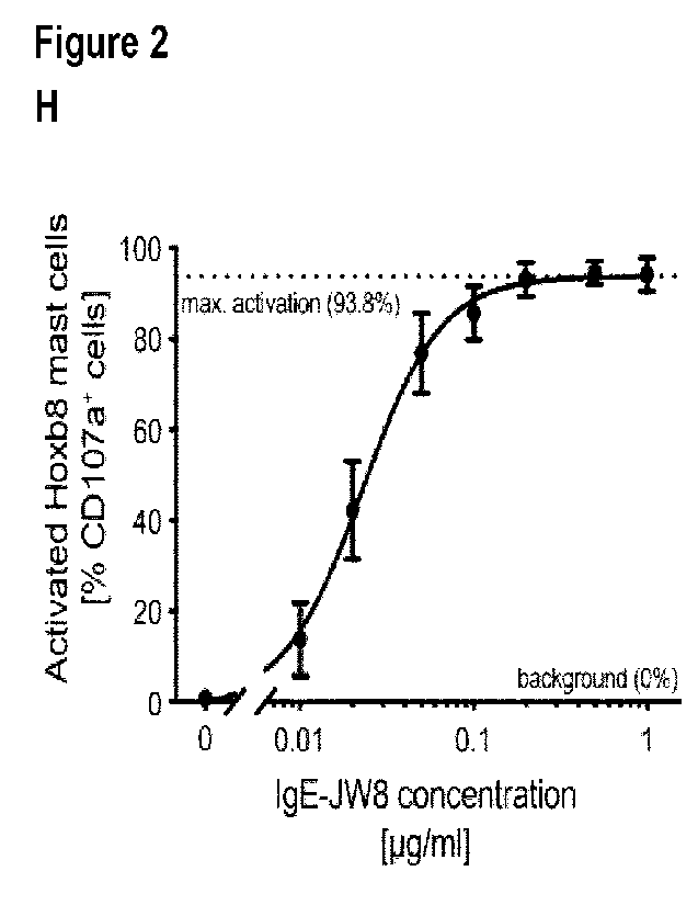
Figure 2 shows the functional characterization of Hoxb8 mast cells after 6
days of
differentiation: In Fig.2A, the absolute number of huFcERla receptors per cell
are shown in the
absence or presence (overnight) of human recombinant IgE; Fig.2B shows dose-
dependent
binding of human recombinant IgE to differentiated Hoxb8 mast cells as
assessed by flow
cytometry; In Fig.2C the correlation between total serum IgE as determined on
Hoxb8 mast
cells or by singleplex immunoassay is shown for 25 allergic patient sera; Fig.
20 shows
zo representative contour plots for antigen mediated activation of Hoxb8
mast cells in an IgE
dose-dependent manner, as measured by flow cytometry; Fig.2E shows
quantification of
antigen mediated activation of Hoxb8 mast cells in an IgE dose-dependent
manner; In Fig.2F
absolute cell counts of seeded progenitor cells after 5 days of
differentiation into harvested
Hoxb8 mast cells are shown; In Fig.2G a comparison of cell growth for
different allergic
effector cells is shown over time; In Fig.2H quantification of antigen
mediated activation of
Hoxb8 mast cells in an IgE dose-dependent manner after 5 weeks of progenitor
cell culture is
depicted. A non-linear regression curve was fitted to measured data points.
Data in Fig.2H are
shown as mean SEM; In Fig.2I the correlation between released 13-
hexosaminidase and the
cell surface activation marker CD107a is depicted for antigen activated Hoxb8
mast cells.
Statistical analysis in C and I was performed using a standard linear
regression model. Data
in A, B and E are shown as mean SEM;
Figure 3 Testing of allergic patient sera on Hoxb8 mast cells. Pre-defined
sera of allergic
patients were used to sensitize cells overnight. Dose-dependent activation as
measured by
flow cytometry is shown for different allergen sources: Fig.3A peanut extract;
Fig.3B
CA 03211629 2023- 9-8
P32W0 2022/200515
PCT/EP2022/057801
6
recombinant Fel dl; Fig.3C yellow jacket wasp venom; Fig.3D honey bee venom;
Fig.3E
house dust mite extract; Fig.3F common birch pollen extract; Fig.3G timothy
grass. Non-linear
regression curves were fitted to measured data points;
Figure 4 Allergen-specific immunotherapy monitoring with Hoxb8 mast cells:
Fig. 4A, cells
were sensitized overnight with an artificial serum containing either only
human recombinant
NIP-specific IgE (sIgE) or a combination of human recombinant NIP-specific IgE
(sIgE) and
IgG (sIgG). Dose-dependent activation of the cells as measured by flow
cytometry is shown;
Fig.4B, cells were sensitized overnight with sera from three timothy grass
allergic patients that
underwent AIT for at least 36 months and one serum of a patient undergoing
placebo
treatment. Dose-dependent activation of the cells as measured by flow
cytometry is shown;
Fig.4C, cells were sensitized overnight with either untreated 483 or IgG-
depleted sera from
two timothy grass allergic patients (solid and dashed lines) at 12 months post
AIT. Non-linear
regression curves were fitted to measured data points. Arrows indicate shift
of the curves; Fig.
4D, differentiated Hoxb8 mast cells were stained either with a control
antibody (isotype in light
grey) or with an anti-FcyRIlb antibody (anti-CD32b, dark grey). Flow
cytometric analysis is
shown in histogram representation with geometric mean fluorescence intensity
(geom. M FI);
Figure 5 High-throughput screening using cellular barcoding: Fig.5A, schematic
overview of
alternative embodiments of the basic workflow are illustrated; Fig.5B, the
deconvolution gating
strategy after acquisition of barcoded and pooled cells is depicted. Initially
cells are gated on
side- and forward-scatter. Then four different cell populations (1-4) are
identified based on
Pacific Blue labelling intensity. Each one of these four populations can be
further subdivided
into nine individual subpopulations based on the combination of Alexa Fluor
488 and Alexa
Fluor 647 labelling intensities; Fig.5C, individually barcoded cell
populations (A-F) have been
sensitized with a different concentration of human recombinant NIP-specific
IgE. Cells were
pooled and activated with NI P7BSA antigen. After acquisition the
deconvolution analysis was
performed to identify each individual cell subpopulation and to assess the
activation status by
quantifying CD107a as a cell surface activation marker; Fig.50, quantification
of deconvoluted
antigen mediated activation of Hoxb8 mast cells in an IgE dose-dependent
manner for this
high-throughput approach is shown. A non-linear regression curve was fitted to
measured data
points; Fig.5E, Individually barcoded cell population (1A-4H) have been
sensitized with sera
from eight different timothy grass allergic patients. Cells were pooled in
four separate tubes
and activated with four concentrations of allergen. After pooled acquisition
the deconvolution
analysis was performed to identify each individual cell subpopulation and to
assess the
activation status by quantifying CD107a as a cell surface activation marker;
Fig.5F,
quantification of deconvoluted dose-dependent allergen mediated activation of
Hoxb8 mast
cells for each individual patient sample is shown;
CA 03211629 2023- 9-8
P32W0 2022/200515
PCT/EP2022/057801
7
Figure 6 High-throughput screening of multiple allergens using cellular
barcoding. Individually
barcoded cell populations have been sensitized with two sera (Fig.6A and 6B)
from
polysensitized patients. The cells were stimulated with different recombinant
allergens or
allergen extracts. After pooled acquisition the deconvolution analysis was
performed to identify
each individual cell subpopulation and to assess the activation status by
quantifying CD107a
as a cell surface activation marker. Fig.6C and 60, Quantification of
deconvoluted dose-
dependent allergen mediated activation of Hoxb8 mast cells for individual
allergens with serum
from patient 1 (Fig.6C) and patient 2 (Fig.6D) is shown. Arrows indicate the
identified
allergens that led to activation of the cells; and
Figure 7 is a graph showing the optimization of Hoxb8 mast cell activation
sensitivity.
Differentiated Hoxb8 mast cells were either sensitized with JW8-IgE on day 5
of differentiation
and challenged with NI P7-BSA on day 6 (black circles) or sensitized with JW8-
IgE on day 6
of differentiation and challenged with NIP7-BSA on day 7 (black triangles).
The additional
resting phase of one day before sensitization increases the activation
sensitivity of the cells
by 2.8-fold (left shift of the activation curve).
Definitions
[0025] Unless otherwise stated, the following terms used in the specification
and claims have
the following meanings set out below.
[0026] "Myeloid progenitor cells" are a type of progenitor cells that
differentiate into only a few
cell types. Myeloid progenitor cells are precursors of red blood cells,
platelets, granulocytes,
monocyte-macrophages, dendritic cells, mast cells and osteoclasts.
[0027] "Mast cells" are cells which are present in virtually all vascularized
tissues of adult
mammals. Mast cells express on their surface the high-affinity receptor for
IgE (FcERI), which
can be activated by IgE and specific antigens to release mediators such as
histamine,
leukotrienes, prostaglandins, serine proteases, and various cytokines,
chemokines and
growth factors. As such, mast cells are critical effector cells of IgE-
associated allergic
disorders. Mature mast cells are c-kit and FccRI+.
[0028] It has been found that mast cells do not mature before leaving the bone
marrow, but
circulate through the vascular system as immature mast cell progenitors. Thus,
mast cells
progenitors are precursors of mature mast cells which, in vivo, differentiate
into mature mast
cells under the influence of growth factors. In the context of the present
invention, a "mast cell
progenitor" is a cell which is capable of differentiating into a mature mast
cell under certain
conditions.
CA 03211629 2023- 9-8
P32W0 2022/200515
PCT/EP2022/057801
8
[0029] The phrase "high-affinity IgE receptor (FcERI)" refers to the receptor
for the Fc region
of innnnunoglobulin E (IgE), an antibody isotype involved in the allergic
response. FcERI is a
tetrameric receptor complex that binds Fc portion of the E heavy chain of IgE.
It consists of
four polypeptide chains: an extracellular alpha chain (FcERIa), a beta chain
(FcERI8), and two
gamma chains (FcERly). The extracellular binding domain of the a-chain binds
with high
affinity to the Fc region of IgE, whereas the other chains are responsible for
the transduction
of initial cross-linking signals into the cell.
[0030] As used herein, the term "antibody" will be understood to include all
antibodies and
antigen binding fragments thereof, including whole antibodies, dimeric,
trimeric and
multimeric antibodies; bispecific antibodies; chimeric antibodies; recombinant
and
engineered antibodies, and fragments thereof. The term "antibody" is thus used
to refer to
any antibody-like molecule that has an antigen binding region, and this term
includes
antibody fragments that comprise an antigen binding domain such as Fab', Fab,
F(ab')2,
single domain antibodies (DABs), TandAbs dimer, Fv, scFv (single chain Fv),
dsFv, ds-scFv,
Fd, linear antibodies, minibodies, diabodies, bispecific antibody fragments,
bibody, tribody
(scFv-Fab fusions, bispecific or trispecific, respectively); sc-diabody;
kappa(lambda) bodies
(scFv-CL fusions); Bispecific T-cell Engager (BITE) (scFv-scFv tandems to
attract T cells);
dual variable domain (DVD)-Ig (bispecific format); small immunoprotein (SIP)
(kind of
minibody); SMIP ("small modular immunopharmaceutical" scFv-Fc dimer; DART (ds-
stabilized diabody "Dual Affinity ReTargeting"); small antibody mimetics
comprising one or
more CDRs and the like.
Production of mast cell progenitors
[0031] According to a first aspect of the invention, there is provided a
method for producing
non-human conditionally immortalized mast cell progenitors, the method
comprising:
- introducing a nucleic acid molecule comprising an inducible homeobox gene
into myeloid
progenitor cells, wherein said myeloid progenitor cells are derived from a non-
human animal
and are engineered to express a heterologous high-affinity IgE receptor alpha
subunit
(FcERIa); and
- selecting for cells which contain the nucleic acid molecule.
It may be that the nucleic acid molecule is a recombinant nucleic acid
molecule.
[0032] By "heterologous high-affinity IgE receptor alpha subunit (FcERIa)" it
will be
understood that the gene encoding FcERla gene is derived from a different
species to the
non-human animal from which the myeloid progenitor cells are derived. In other
words, the
non-human animal is transgenic for the FcERla gene. In some embodiments, the
FceRla
CA 03211629 2023- 9-8
P32W0 2022/200515
PCT/EP2022/057801
9
gene is derived from the species for which the mast cells are to be used in
the diagnostic
and monitoring assays described herein. In some embodiments, the FcERla gene
is human
FcERla (accession no. NM_002001.4; Gene ID: 2005). For example, the myeloid
progenitor
cells may be mouse cells, and the FcERla may be human FcERla. Mice which are
transgenic
for human FcERla and which have the murine FcERla gene knocked out are
described by
Dombromovicz etal., J. Immunol. 1996, 15; 157(4): 1645-51, and can be obtained
from the
Jackson Laboratory (Strain B6.Cg-Fcer/atm/Knt Tg(FCER1A)1Bhk/J; Stock no.
010506).
[0033] The homeobox gene may be selected from HoxB8, HoxA9, Lhx2 (LH2) and
TLX1
(Hox11). Lhx2 (LH2) and TLX1 (Hox11) have been shown to have the potential to
immortalize multipotent haematopoietic progenitors (Pinto etal., EMBO J. 1998
Oct
1;17(19):5744-56; Zhang etal., Oncogene 1999 Apr 1;18(13):2273-9). In some
embodiments, the homeobox gene is HoxB8. Hox genes of mammalian origin are
well-
known in the art. In some embodiments, the HoxB8 gene is mouse HoxB8. The
mouse
HoxB8 gene is available under GenBank accession no. NM_010461 (Gene ID: 15416)
(encoding mouse HoxB8 protein accession No. NP_034591).
[0034] The homeobox gene is comprised within the nucleic acid molecule. The
nucleic acid
molecule may be a recombinant nucleic acid molecule. The recombinant nucleic
acid
molecule may comprise the homeobox gene operably linked to an inducible
promoter. For
example, the nucleic acid molecule may comprise an exogenous inducible
expression
cassette which comprises the homeobox gene. Introduction of the homeobox gene
into the
myeloid progenitor cells can be achieved using any conventional recombinant
technology for
introducing nucleic acids into host cells including, but not limited to,
transduction,
transfection or electroporation.
[0035] In some embodiments, the nucleic acid molecule may be any type of
molecule which
is suitable for transfection, such as a vector (e.g. a cosmid, plasmid or
viral vector).
[0036] It will thus be appreciated that the non-human conditionally
immortalized mast cell
progenitors and the non-human mast cells of the invention contain an exogenous
homeobox
gene, i.e. a homeobox gene which has been introduced into the cell in addition
to, or instead
of, the chromosomal homeobox gene(s) already present. In some embodiments, the
homeobox gene comprised within the nucleic acid molecule is heterologous, i.e.
belonging to
a different species to the species from which the mast cells or mast cell
progenitors were
derived. References herein to a "homeobox gene" thus refer to a homeobox gene
which is
introduced into the myeloid progenitor cells via a nucleic acid molecule (e.g.
a recombinant
nucleic acid molecule) and not to any native homeobox genes which may be
present in the
cell chromosome, unless otherwise stated.
CA 03211629 2023- 9-8
P32W0 2022/200515
PCT/EP2022/057801
[0037] In some embodiments the nucleic acid molecule is a viral vector.
Suitable viral
vectors for the introduction of heterologous genes into cells (e.g. mammalian
cells) will be
known to the skilled person and include a herpes simplex viral vector, an
adenoviral vector,
an adeno-associated viral vector (AAV), or a retroviral vector, for example
but not limited to,
5 an HIV retroviral vector, a lentivirus, a VL 30 vector, a MSCV retroviral
vector, or a Harvey
Murine Sarcoma Vector. In some embodiments, the viral vector is a lentiviral
particle.
[0038] Methods for subcloning the mouse Hoxb8 gene into a lentiviral system
and
generation of viral particles has been described by Salmanidis et al., Cell
Death Differ 2013;
20:1370-80, and Gurzeler etal., Allergy 2013;68:604-13.
10 [0039] Methods for the introduction of a viral vector into host cells
(i.e. transduction) will be
known to those skilled in the art. In some embodiments, cells may be
transfected by spin
infection.
[0040] Cells into which the nucleic acid molecule has been successfully
introduced (e.g.
transduced cells) can be selected for by applying a selective pressure to the
myeloid
progenitor cells such that only those which contain the nucleic acid molecule
survive. For
example, the nucleic acid molecule may additionally comprise a gene conferring
resistance
to an antibiotic, in which case cells containing the nucleic acid molecule can
be selected for
by culturing the cells in the presence of the antibiotic (e.g. by culturing
the cells in a culture
medium comprising the antibiotic). Performing antibiotic selection enables
elimination of
cells which do not contain the nucleic acid molecule, resulting in a more
homogenous (but
still polyclonal) cell population.
[0041] In some embodiments, the cells are cultured in a culture medium
comprising the
antibiotic. Suitable antibiotics include, but are not limited to, puromycin
and blasticidin. In
some embodiments, the antibiotic is added to the culture medium 1, 2, 3, 4 or
5 days,
preferably 2-4 days, after the introduction of the nucleic acid molecule (e.g.
after
transduction). The presence of the antibiotic in the culture medium may be
maintained until
outgrowth of the surviving cells (i.e. the transduced cells).
[0042] The mast cell progenitors produced by the method are conditionally
immortalized. By
"immortalized" it will be understood that the cells are capable of
proliferating indefinitely. As
such, the cells can be maintained in culture for long periods of time.
[0043] By "conditionally immortalized", it will be understood that the
homeobox gene (e.g.
the homeobox gene present in the nucleic acid molecule) is not constitutively
expressed but
instead expression of the gene is controlled by an exogenous agent. As will be
known by the
skilled person, conditional expression of a gene can be achieved, for example,
by inserting
the gene downstream of an inducible promoter such that the gene is expressed
the
CA 03211629 2023- 9-8
P32W0 2022/200515
PCT/EP2022/057801
11
presence of the inducer. Any suitable inducible expression system may be used
to control
expression of the homeobox gene, such as the "Tet-on" or "Tet off" system. It
has been
previously demonstrated that, while a native hoxb gene system is present in
mouse mast
cells, these hoxb genes are not expressed in either mast cell progenitors or
terminally
differentiated mature mast cells. It is known that hoxb gene expression is
restricted to
embryonal development and haematopoietic stem cells, and is silenced in more
lineage
committed progenitors. It will therefore be appreciated by those skilled in
the art that any
native chromosomal homeobox genes present in the mast cells or mast cell
progenitors of
the invention, or the myeloid progenitor cells from which they derive, are not
expressed,
either constitutively or by internal inducing agents. As such, only the
homeobox gene which
is introduced into the cells via the nucleic acid molecule can be expressed in
the cells of the
invention.
[0044] Thus, in some embodiments, expression of the homeobox gene is
controlled by an
inducer. Myeloid progenitor cells which contain the nucleic acid molecule may
be cultured in
the presence of the inducer. For example, the culture medium may comprise the
inducer.
This causes expression of the homeobox gene, thereby immortalizing the cells.
In some
embodiments, the inducer is 4-hydroxytamoxifen (4-0HT). This inducer may be
used with
the expression system pF-5xUAS-gene_of interest-GEV16.
[0045] In some embodiments, the culture medium used to culture the cells
containing the
nucleic acid molecule comprises interleukin-3 (IL-3).
[0046] Thus, following the introduction of the nucleic acid molecule into the
myeloid
progenitor cells, the cells may be cultured in culture medium comprising IL-3
and the inducer
(e.g. 4-0HT). An antibiotic may be added to the culture medium (e.g. after 1-5
days,
preferably after 2-4 days) to select for transduced cells, as described above.
[0047] Non-human conditionally immortalized mast cell progenitors obtained by
the method
described above may then be tested for their growth, viability and/or
functional performance
properties, in order to select the best performing cell lines.
[0048] In some embodiments, the method further comprises carrying out a single
cell
dilution of the conditionally immortalized mast cell progenitors, followed by
clonal expansion
so as to obtain a monoclonal conditionally immortalized mast cell progenitor
cell line.
[0049] The myeloid progenitor cells which are used to prepare the
conditionally immortalized
mast cell progenitors may be derived from a non-human animal by:
- providing whole bone marrow previously obtained from the animal;
- enriching the bone marrow for hematopoietic progenitor cells; and
CA 03211629 2023- 9-8
P32W0 2022/200515
PCT/EP2022/057801
12
- culturing the hematopoietic progenitor cells in the presence of IL-3.
[0050] In some embodiments, the method may additionally comprise the step of
obtaining
the whole bone marrow from the animal.
[0051] The bone marrow may be enriched for hematopoietic progenitor cells by
magnetic
cell separation, using a lineage depletion cocktail. As will be known by the
skilled person,
this process is commonly used in the art for the depletion of committed
leukocyte
populations (i.e. mature haematopoietic cells such as T cells, B cells, NK
cells, platelets
etc.), thereby enabling rare cell populations such as progenitor cells to be
enriched. Lineage
cell depletion kits are commercially available, such as the BD IMag set from
BD Biosciences,
Europe.
[0052] Following the enrichment step, the hematopoietic progenitor cells
remaining are
incubated in the presence of IL-3. Incubation may be carried out for at least
24 hours, at
least 36 hours, at least 48 hours, or at least 72 hours. In some embodiments,
incubation is
carried out for up to 7 days. Preferably, incubation is carried out for about
48 hours.
Preferably, the IL-3 is derived from the same species from which the myeloid
progenitor cells
were derived. For example, in embodiments in which the myeloid progenitor
cells are
derived from a mouse, the IL-3 is preferably murine IL-3. It may be that the
hematopoietic
progenitor cells are cultured in WEHI-3B cell-conditioned medium, which
provides a source
of murine IL-3.
[0053] The non-human animal may be any suitable animal, such as a sheep, pig,
cow,
horse, goat, dog, primate, rabbit or rodent. In some embodiments, the non-
human animal is
a rodent (e.g. a mouse, rat, hamster, guinea pig or gerbil). The non-human
animal may be a
mouse.
[0054] The present invention thus provides a non-human conditionally
immortalized mast
cell progenitor. The progenitor cell comprises a nucleic acid molecule (e.g. a
recombinant
nucleic acid molecule) comprising a homeobox gene, the expression of the
homeobox gene
being under the control of an inducer, wherein said cell expresses a
heterologous high-
affinity IgE receptor alpha subunit (FcERIa). The non-human conditionally
immortalized mast
cell progenitor may be obtainable by the methods described herein.
[0055] Also provided is a composition comprising a population of non-human
conditionally
immortalized mast cell progenitors according to the invention. The composition
may further
comprise the inducer which controls the expression of the homeobox gene. In
some
embodiments the composition comprises IL-3. The composition may further
comprise a
suitable medium, buffer and/or salts for maintaining the cells. In some
embodiments, the
composition may comprise one or more cytokines (e.g. in addition to IL-3),
proteins, and/or
CA 03211629 2023- 9-8
P32W0 2022/200515
PCT/EP2022/057801
13
growth factors. For example, the composition may comprise one or more (or all)
of: IP-10,
MIP-1 a, MIP-2, VEGF, IFNB-1, IL-16, IL-20, and MCP-5.
[0056] By virtue of the inducible homeobox gene, the conditionally
immortalized mast cell
progenitors of the invention have a high replicative rate and near-unlimited
renewal potential.
Advantageously, this enables on-demand differentiation to produce large
numbers of mast
cells within a few days. The invention thus provides a conditionally
immortalized mast cell
progenitor line that features remarkable self-renewal potential. The
progenitor cells of the
invention can be kept in culture for months without losing self-renewal
potential. The
progenitor cells may also be frozen (e.g. using liquid nitrogen) for storage,
ensuring a stock
of progenitors from which a virtually unlimited number of mast cells can be
generated.
Production of mast cells
[0057] The non-human conditionally immortalized mast cell progenitors of the
invention may
be cultured in the absence of the inducer and in the presence of IL-3 so as to
produce
differentiated mast cells. In the absence of the inducer, the progenitor cells
no longer
express the homeobox gene and differentiate along the myeloid lineage.
[0058] Thus, a method for preparing mast cells comprises culturing non-human
conditionally
immortalized mast cell progenitors which are engineered to express a
heterologous high-
affinity IgE receptor alpha subunit (FcERIa), wherein the conditionally
immortalized mast cell
progenitors further comprise a nucleic acid molecule (e.g. a recombinant
nucleic acid
molecule) comprising a homeobox gene, the expression of the homeobox gene
being under
the control of an inducer, and wherein the conditionally immortalized mast
cell progenitors
are cultured in the absence of the inducer and in the presence of IL-3. In
some
embodiments, the non-human conditionally immortalized mast cell progenitors
are cultured
in WEH3b cell-conditioned medium.
[0059] Guerzeler etal. (Allergy 68 (2013) 604-613) describes a method
comprising
conditionally immortalizing myeloid progenitors using Hoxb8 in the presence of
IL-3.
However, Guerzeler et a/. only reports the generation of c-kit negative
basophils using this
method.
[0060] In some embodiments, the non-human conditionally immortalized mast cell
progenitors are cultured in the absence of the inducer and in the presence of
IL-3 for at least
5 days, or at least 6 days.
[0061] In some embodiments, the method for preparing mast cells comprises:
- on day 0, adding the non-human conditionally immortalized mast cell
progenitors to a
culture medium comprising IL-3, wherein the culture medium lacks the inducer;
and
CA 03211629 2023- 9-8
P32W0 2022/200515
PCT/EP2022/057801
14
- on day 5 or day 6, harvesting the (mature) mast cells.
[0062] In some embodiments, the mast cells are harvested on day 6.
Surprisingly, the
present inventors have found that waiting until day 6 to harvest the
differentiated mast cells
can increase their sensitivity to activation upon antigen challenge.
[0063] In some embodiments, the method comprises replacing the culture medium
before
the mast cells are harvested. For example, the culture medium may be replaced
on the day
before the day on which the mast cells are harvested.
[0064] In some embodiments, the method comprises replacing the culture medium
on day 5,
and continuing to culture the cells (in the absence of the inducer and in the
presence of IL-3)
for at least 2, 4, 6, 8, 10, 12, 14, 16, 18, 20, 22, 24, 30 or 36 hours.
[0065] In some embodiments, the method comprises replacing the culture medium
on day 5,
and harvesting the cells on day 6.
[0066] In some embodiments, the method comprises:
- on day 0, adding the non-human conditionally immortalized mast cell
progenitors to a first
culture medium comprising IL-3, wherein the first culture medium lacks the
inducer;
- on day 5, removing the cells from the first medium and adding the cells
to a second
medium comprising IL-3, wherein the second culture medium lacks the inducer;
and
- on day 6, harvesting the (mature) mast cells.
[0067] The first medium may be different to the second medium, or the first
medium and the
second medium may be the same. In some embodiments, the concentration of IL-3
in the
second medium is higher than the concentration of IL-3 in the first medium.
The
concentration of IL-3 in the second medium may be 2-, 5-, 10-, 20-, 50-or 100-
times higher
than the concentration of IL-3 in the first medium.
[0068] In some embodiments, the cells are washed after being removed from the
first
medium and before being added to the second medium.
[0069] It will be appreciated that, in any of the embodiments described
herein, cells are
cultured under appropriate conditions which will be known to those skilled in
the art, such as
those described herein. For example, the mast cell progenitors may be cultured
at 37 C,
optionally under 5% CO2.
[0070] As described above, clonal expansion of a single non-human
conditionally
immortalized mast cell progenitor may be carried out to generate a monoclonal
cell line from
which the mast cells are generated. Thus, in any of the methods described
herein which
CA 03211629 2023- 9-8
P32W0 2022/200515
PCT/EP2022/057801
utilise mast cells, the mast cells may be monoclonal. Alternatively, the mast
cells may be
polyclonal.
Cells & characterization
[0071] The present invention provides a non-human conditionally immortalized
mast cell
5 progenitor (also referred to hereinbelow as "progenitor cells"). The
progenitor cell comprises
a homeobox gene, the expression of the homeobox gene being under the control
of an
inducer, wherein said cell expresses a heterologous high-affinity IgE receptor
alpha subunit
(FcERIa). The non-human conditionally immortalized mast cell progenitor may be
obtainable
by the methods described herein. The homeobox gene may be comprised in
recombinant
10 nucleic acid molecule.
[0072] The invention further provides a non-human mast cell, wherein the mast
cell
comprises a homeobox gene, the expression of the homeobox gene being under the
control
of an inducer, and wherein the mast cell expresses a heterologous high-
affinity IgE receptor
alpha subunit (FcERIa). The mast cell may be obtainable by the methods
described herein.
15 The homeobox gene may be comprised in recombinant nucleic acid molecule.
[0073] Also provided is a composition comprising a population of non-human
mast cells, as
described herein. In some embodiments the composition comprises IL-3. The
composition
may further comprise a suitable medium, buffer and/or salts for maintaining
the cells.
[0074] Thus, both the non-human conditionally immortalized mast cell
progenitors, and the
non-human mast cells derived therefrom, express FcERla on the cell surface.
Expression of
FcERla may be confirmed by staining the cells with an anti-FcERla antibody,
for example
using the methods described herein.
[0075] The mast cells can be distinguished from the progenitor cells from
which they are
derived by a number of methods. For example, the mast cells may be identified
by their
expression of c-kit (CD117). It has been observed that during differentiation
of the progenitor
cells into mast cells, by culturing the progenitor cells in the absence of the
inducer, c-kit
expression gradually increases over time. Thus, in a population of non-human
mast cells
according to the invention, at least 80%, at least 85%, at least 90%, at least
93%, at least
95%, at least 96%, at least 97%, at least 98% or at least 99% of the mast
cells may be c-kit
positive. In some embodiments, 100% of the mast cells in a population are c-
kit positive (for
example, in a monoclonal population). Mast cells which result from the
differentiation of
conditionally-immortalized mast cell progenitors, as described herein, may
thus be identified
as being c-kit and FcERla double positive. The mast cells may display an even
distribution
of both FcERla (e.g. human FcERIa) and c-kit on the cell surface. c-kit
expression can be
CA 03211629 2023- 9-8
P32W0 2022/200515
PCT/EP2022/057801
16
detected by staining cells with anti-c-kit (e.g. anti-mouse c-kit) antibodies
using well-known
techniques, such as the methods described herein.
[0076] Upon differentiation of the conditionally-immortalized mast cell
progenitors, the cells
gradually lose Hoxb8 protein expression. This clearly distinguishes the
progenitors (having
high Hoxb8 protein expression) from the differentiated mast cells (low or no
Hoxb8
detectable). Expression of Hoxb8 (e.g. as detected using Western Blot) may no
longer be
detectable by around day 2 of differentiation.
[0077] The differentiated mast cells may be further characterised by the
number of FcERla
(e.g. human FccRla) receptors per cell. It may be that, in the absence of IgE
sensitization,
the mast cells of the invention display from 5000 to 24000 receptors per cell
(rpc), from
10000 to 22000 rpc, from 15000 to 20000 rpc or from 17000 to 19000 rpc, for
example
approximately 18000 to 18500 rpc. Incubation of the mast cells with IgE (e.g.
recombinant
human IgE) may increase the amount of receptors on the cells by from 4- to 6-
fold, e.g.
approximately 5-fold. It may be that in the presence of IgE sensitization, the
mast cells of the
invention display from 70000 to 120000 rpc, from 80000 to 110000 rpc, or from
85000 to
110000 rpc, e.g. about 90000 rpc.
[0078] It has been found by the present inventors that the number of receptors
on the
conditionally-immortalized mast cell progenitors is higher than the number of
receptors on
the mast cells. In the absence of IgE sensitization, the mast cell progenitors
of the invention
display more than 24000, more than 28000, more than 30000, or more than 32000
receptors
per cell (rpc), for example from about 30000 to about 38000 or from about
32000 to about
36000 rpc (e.g. about 35000 rpc). In the presence of IgE sensitization, the
mast cell
progenitors of the invention display more than 90000, more than 95000 or more
than 100000
rpc, for example from about 98000 to about 110000 rpc or from about 10000 to
about
108000 rpc, e.g. about 106000 rpc.
[0079] Differentiated mast cells may also be identified by their morphology.
Morphological
analysis of the cells may be carried out by staining the cells (e.g. using
toluidine blue) and/or
by imaging the stained cells, for example using microscopy. Such methods will
be known to
the skilled person, and are described in more detail below. The mast cell
phenotype is
characterised by increased cellular granularity and rnetachrornatic elements,
relative to the
progenitor cells from which they are derived. For example, the differentiated
mast cells may
be identified by the accumulation of granules, which may be observed as pink
or purple dots
when stained. The parent progenitor cells have no such granules.
[0080] Mast cells may further be identified by detecting the expression and/or
secretion of
beta-hexosaminidase, histamine, and/or mast cell proteases (e.g. mouse mast
cell
CA 03211629 2023- 9-8
P32W0 2022/200515
PCT/EP2022/057801
17
proteases -1, -4 and -5). These molecules are increasingly expressed (and
stored in
granules) upon maturation of the mast cells, and are released upon activation.
The
expression of mast cell proteases may be detected by detecting the
corresponding mRNA
(e.g. using qt-PCR, RNA sequencing or microarray), or by detecting the
proteins themselves
(e.g. using a western blot of cell lysates, ELISA, immunofluorescence or
microscopy).
[0081] Advantageously, the non-human mast cells of the present invention may
display a
maximal activation of at least 30%, at least 40%, at least 50%, at least 60%,
at least 70%, at
least 80%, at least 85%, at least 90%, at least 92%, at least 94%, at least
95%, at least 96%
or at least 97% (e.g. about 95%, or about 97%). The maximal activation may be
determined
by the methods described herein. For example, the maximal activation may be
determined
by the amount of p-hexosaminidase (or another suitable mediator, such as
histamine, a mast
cell protease, or leukotriene) released by cells upon challenge as a
percentage of the total
amount of p-hexosaminidase (or other mediator) (released plus leftover in the
cells after
challenge). Alternatively, the maximal activation may be determined by
quantifying a cell-
surface marker (such as CD107a) that is exposed upon degranulation, e.g. by
flow
cytometry.
[0082] It has been observed that the conditionally-immortalized mast cell
progenitors of the
invention grow faster than previously characterised progenitor cell lines. It
may be that the
conditionally-immortalized mast cell progenitors of the invention have a
doubling time of less
than 35 hours, less than 32 hours or less than 30 hours (e.g. about 29 hours),
when cultured
at 37 C. Cells may be cultured with 5% CO2 in a suitable medium, such as RPMI-
1640
medium AQmedia (Sigma) supplemented with 10% FCS Sera Pro (Pan Biotech), 10%
WEHI-3b supernatant, Penicillin 100U/ml, 100pg/m1Streptomycin (100x
Penicillin/Streptomycin, Gibco) and 100nM 4-Hydroxytamoxifen (Sigma).
Furthermore,
differentiation of the mast cell progenitors of the invention into the mature
mast cells of the
invention is much faster than previously described bone marrow-derived mast
cells.
Differentiation to mature mast cells may take less than 14 days, less than 10
days, or less
than 8 days. For example, differentiation to mature mast cells from the mast
cell progenitors
of the invention may take approximately 5 or 6 days. In contrast, prior art
methods which
involve differentiating bone marrow-derived cells take a number of weeks to
produce mast
cells.
[0083] The conditionally-immortalized mast cell progenitors of the invention
may be stored in
culture for at least 4, at least 5, at least 6, at least 8, at least 10 or at
least 12 weeks, or at
least 4, 6, 8, 10 or 12 months. Thus, in some embodiments fully functional
mast cells can be
CA 03211629 2023- 9-8
P32W0 2022/200515
PCT/EP2022/057801
18
differentiated from progenitors that have been cultured for at least 4, 5, 6,
8, 10 or 12 weeks
or at least 4, 6, 8, 10 or 12 months.
[0084] The differentiated mast cells may be stored in culture (while retaining
viability and
functionality) for at least 5, at least 7 or at least 10 days, at 37 C. These
characteristics
make the mast cells of the invention particularly useful for functional assays
and diagnosis.
[0085] The fact that the non-human mast cells of the invention can be derived
from the
same progenitor cells in a standard operating procedure makes the cells
remarkably
homogenous, stable and highly reproducible. Additionally, the mast cells of
the invention
feature an unprecedented signal-to-noise ratio upon allergen-mediated
activation. While
maximal activation of most previously described allergic effector cell lines
lies between 40-
60%, the non-human mast cells of the invention can be activated to almost
100%, indicating
the dynamic range and exceptional sensitivity of the system. Importantly,
these activation
parameters remain constant after multiple months of progenitor cell culture,
and prolonged
use of the cells does not affect their viability.
Assays
Functional allergy assay
[0086] The mast cells of the invention find particular utility in allergy
testing. In a further
aspect of the invention, there is provided a method for determining whether a
patient is
allergic to an allergen and/or the severity of a patient's allergy to an
allergen, the method
comprising:
- incubating mast cells with a sample comprising patient antibodies;
- contacting the mast cells with the allergen; and
- detecting activation of the mast cells,
wherein the mast cells are non-human mast cells according to the fifth aspect
of the
invention. The mast cells may be obtained by the methods described herein.
[0087] Antigen or allergen mediated aggregation of FccRla-bound IgE on mast
cells leads to
their activation and immediate degranulation. Mast cells store a number of
different chemical
mediators, such as histamine, p-hexosaminidase, interleukins, leukotriene 04
(LTC4),
proteoglycans and various enzymes, in granules. "Degranulation" is a cellular
process by
which, upon activation, mast cells release the contents of their granules into
the surrounding
environment, i.e. the surrounding tissue in vivo, or the cell culture
supernatant in the case of
an in vitro assay. The activation of mast cells can therefore be detected by
detecting and/or
quantifying chemical mediators (e.g. P-hexosaminidase) in the cell culture
supernatant.
CA 03211629 2023- 9-8
P32W0 2022/200515
PCT/EP2022/057801
19
Alternatively, cell surface markers that are exposed upon degranulation may be
detected
and/or quantified, e.g. by flow cytonnetry.
[0088] The mast cells and methods described herein can be used to detect
allergies against
any potential allergen, including air-borne allergens, food allergens (e.g.
lactose, egg protein,
fish, nuts, wheat and soy), drug allergens (e.g. penicillin, tetracycline, non-
steroidal anti-
inflammatory drugs, anaesthetics), environmental allergens (e.g. pollen,
birch, timothy grass,
animal hair, saliva or dander, mould, latex, dust mites) and venoms (e.g. wasp
and bee
stings, mosquito bites).
[0089] The sample comprising patient antibodies may comprise neat or diluted
patient
serum. In some embodiments, the sample comprises antibodies which have been
isolated
from patient serum, in a suitable medium or buffer. For example, the
antibodies may have
been extracted from the patient serum by purification or fluid exchange.
[0090] Therefore, in any of the methods described herein, antibodies may be
isolated from
patient serum prior to incubating the mast cells with the patient antibodies.
The antibodies
may be isolated using well-known methods of antibody purification which
include, but are not
limited to, using protein A or protein G columns, ion exchange or metal
chelate
chromatography, ammonium sulfate precipitation, and Melon Gel chromatography.
[0091] In other embodiments, the patient serum is subjected to a fluid
exchange process in
which the fluid phase of the serum is replaced by a suitable medium, thereby
obtaining a
sample comprising patient antibodies. The sample containing the antibodies is
then
incubated with the mast cells. The fluid exchange process may be carried by
spinning the
serum sample through a size exclusion (e.g.100-kDa cut-off) column, into the
selected
medium. Preferably, the volume of serum is equal to the volume of medium so
that the
antibody concentration remains unchanged. This processing step ensures that
antibodies
remain in the medium, but that low molecular weight compounds (e.g. smaller
than 100 kDa)
are removed.
[0092] In any of the methods described herein, the method may further
comprise:
- optionally, obtaining serum from the patient;
- isolating antibodies from the patient serum to obtain a sample comprising
patient
antibodies.
[0093] In any of the methods described herein, the mast cells may be incubated
with the
sample comprising patient antibodies for at least 8 hours, at least 10 hours,
preferably at
least 12 hours (e.g. overnight).
CA 03211629 2023- 9-8
P32W0 2022/200515
PCT/EP2022/057801
[0094] The step of contacting the mast cells with the allergen may be carried
out by directly
adding the allergen to the mast cell culture.
[0095] In some embodiments, the method is carried out in the absence of a wash
step
between the steps of incubating the mast cells with the antibodies and
contacting the mast
5 cells with the allergen. Surprisingly, a protocol in which the cells are
not washed prior to
allergen challenge has been found to have several advantages. Firstly, the
omission of a
wash step more closely mimics in vivo conditions in which mast cells are
constantly exposed
to serum. It also allows the role of allergen-specific IgG or other unknown
modulatory factors
in serum samples to be assessed upon allergen challenge. Furthermore, it has
been
10 unexpectedly observed that the maximal activation of the mast cells is
significantly higher
when cells were not washed after sensitization and before allergen challenge
(Table 1).
Without being bound by theory, it is thought that the IgE-allergen complex
formation may
occur more efficiently in the absence of a wash step, leading to enhanced
cross-linking of
FccRI on the cell surface.
15 [0096] Detecting activation of the mast cells may comprise detecting the
release of a
mediator, or the expression of a surface marker, that is indicative of the
presence of IgE
specific for the allergen in the patient serum sample.
[0097] In some embodiments, detecting activation of the mast cells comprises
detecting the
expression of a surface marker. The surface marker may be a lysozyme
associated
20 membrane glycoprotein (LAMP-1, LAMP-2 or LAMP-3), CD203c, 0D63 or
CD107a. In some
embodiments the surface marker is CD107a.
[0098] In some embodiments, detecting the expression of the surface marker
comprises
contacting the mast cells with an antibody specific for the surface marker,
and detecting
antibodies bound to the cells. For example, in embodiments therein the surface
marker is
CD107a, detecting the expression of the surface marker may comprising
contacting the mast
cells with an anti-CD107a antibody. The antibody may be added to the mast cell
culture at
the same time as the allergen, or after the allergen. The method may further
comprise
quantifying the antibodies bound to the cells, for example using flow
cytometry.
[0099] In some embodiments, detecting activation of the mast cells comprises
detecting the
release of a mediator. The mediator may be 6-hexosaminidase, a protease,
histamine or a
leukotriene (e.g. LTC4). In some embodiments the mediator is p-hexosaminidase.
The
release of p-hexosaminidase may be detected by adding a substrate of p-
hexosaminidase to
the culture supernatant and/or cell pellet lysates, and detecting the product
of the enzyme-
substrate reaction. For example, the substrate may be 4-nitrophenyl N-acetyl-p-
D-
glucosaminidase, which is a chromogenic substrate of 6-hexosaminidase. The
release of a
CA 03211629 2023- 9-8
P32W0 2022/200515
PCT/EP2022/057801
21
protease may be detected using a substrate of the protease, e.g. a tryptase
substrate such
as acetyl-Orn-Phe-Arg-AMC, or by ELISA or innnnunoblot of cell supernatant.
[00100] Alternatively, mast cell activation may be detecting by detecting a pH
change
resulting from degranulation. Changes in pH may be detected using reagents
such as pH-
sensitive fluorophores or pH indicator solutions.
[00101] Detecting activation of the mast cells may comprise determining a
level of
activation. The level of activation may correspond to the severity of the
allergy of the patient
to the allergen. Thus, the level of mast cell activation can be used for
clinical grading of the
allergy. This, in turn, may be used to inform the type of treatment or
management of the
patient's allergy. The less allergen that is required to reach maximal
activation of the mast
cells, and/or the higher the level of maximal activation, the more allergic
the patient is to the
allergen. The level of activation may be determined, for example, by
quantifying the amount
of surface marker expressed by the mast cells, the amount of mediator released
by the cells,
or the extent of a pH change.
[00102] In any of the assay methods described herein, the method may further
comprise
culturing non-human conditionally immortalized mast cell progenitors so as to
produce the
mast cells. The mast cell progenitors may be cultured as described
hereinabove. Thus,
mast cells may be generated as required for use in an assay.
[00103] Accordingly, in a further aspect the invention provides a method for
determining
whether a patient is allergic to an allergen and/or the severity of a
patient's allergy to an
allergen, the method comprising:
- providing a population of non-human conditionally immortalized mast cell
progenitors, each
mast cell progenitor comprising a homeobox gene, the expression of the
homeobox gene
being under the control of an inducer, wherein said cell expresses a
heterologous high-
affinity IgE receptor alpha subunit (FcERIa);
- culturing the mast cell progenitors in the absence of the inducer and in
the presence of IL-3
for at least 5 days so as to produce non-human conditionally immortalized mast
cells;
- incubating the mast cells with a sample comprising patient antibodies;
- contacting the mast cells with the allergen; and
- detecting activation of the mast cells.
[00104] The homeobox gene may be comprised in recombinant nucleic acid
molecule.
CA 03211629 2023- 9-8
P32W0 2022/200515
PCT/EP2022/057801
22
[00105] The step of culturing the mast cell progenitors so as to produce non-
human
conditionally immortalized mast cells may be carried out using any of the
methods described
herein.
[00106] In some embodiments the method comprises culturing the mast cell
progenitors for
at least 6 days, prior to incubating the resulting mast cells with the sample.
[00107] In some embodiments, culturing the mast cell progenitors comprises:
- on day 0, adding the non-human conditionally immortalized mast cell
progenitors to a
culture medium comprising IL-3, wherein the culture medium lacks the inducer;
and
- on day 5 or day 6, harvesting the (mature) mast cells.
[00108] In some embodiments, the mast cells are harvested on day 6.
[00109] In some embodiments, the method comprises:
- on day 0, adding the non-human conditionally immortalized mast cell
progenitors to a first
culture medium comprising IL-3, wherein the first culture medium lacks the
inducer;
- on day 5, removing the cells from the first medium and adding the cells
to a second
medium comprising IL-3, wherein the second culture medium lacks the inducer;
and
- on day 6, harvesting the (mature) mast cells.
[00110] The first medium may be different to the second medium, or the first
medium and
the second medium may be the same. In some embodiments, the concentration of
IL-3 in
the second medium is higher than the concentration of IL-3 in the first
medium. The
concentration of IL-3 in the second medium may be 2-, 5-, 10-, 20-, 50- or 100-
times higher
than the concentration of IL-3 in the first medium.
[00111] In some embodiments, the cells are washed after being removed from the
first
medium and before being added to the second medium.
[00112] The invention thus provides a functional allergy screening assay with
remarkable
diagnostic potential. The methods of the invention, which are based on the
passive
sensitization of mast cells that are transgenic for a high-affinity IgE
receptor (e.g. human
FcERIa) with IgE from patient serum, provides comprehensive information on the
allergic
status of the patient and overcomes many of the challenges and limitations
associated with
current diagnostic tools.
[00113] The functional assay of the invention, which is based on antibodies
derived from
serum, rather than whole blood, has the advantage that patient samples can be
frozen and
stored for later analysis without losing biological activity. This facilitates
sample handling and
CA 03211629 2023- 9-8
P32W0 2022/200515
PCT/EP2022/057801
23
allows for pro- as well as retrospective analysis of individual patient
samples or entire
sample cohorts.
High-throughput multiplex assays
[00114] Conveniently, the method may be multiplexed. Thus, in some embodiments
the
method is for determining whether the patient is allergic to multiple
allergens. In such
embodiments, the method may comprise:
- separately incubating each of a plurality of mast cell populations with a
sample comprising
patient antibodies;
- labelling each of the plurality of mast cell populations with a different
detectable label;
- after incubating the mast cell populations with the samples comprising
patient antibodies,
separately contacting each of the plurality of mast cell populations with a
different allergen;
- pooling the plurality of mast cell populations; and
- detecting activation of the mast cells in each population,
wherein each mast cell population comprises non-human mast cells as described
herein.
[00115] The step of labelling each of the plurality of mast cell populations
with a different
detectable label may be carried out before or after the step of incubating the
plurality of mast
cell populations with the samples comprising patient antibodies.
[00116] In a further embodiment, the method is for determining whether
multiple patients
are allergic to an allergen, the method comprising:
- separately incubating each of a plurality of samples, each sample comprising
antibodies
from a different patient, with one of a plurality of mast cell populations,
- labelling each of the plurality of mast cell populations with a different
detectable label;
- pooling the plurality of mast cell populations;
- contacting each of the plurality of mast cell populations with the
allergen; and
- simultaneously detecting activation of the mast cells in each population,
wherein each mast cell population comprises non-human mast cells as described
herein.
[00117] By each sample comprising antibodies from a different patient", it
will be
understood that a sample comprises multiple antibodies derived from a single
patient, and
that each sample is patient-specific. In this way, each patient sample is
paired with a
detectable label which is different to the detectable label used for each
other patient. For
example, the method may comprise testing whether a first patient and a second
patient are
CA 03211629 2023- 9-8
P32W0 2022/200515
PCT/EP2022/057801
24
allergic to an allergen. A sample comprising antibodies from the first patient
may be
incubated with a first population of mast cells, which are labelled with a
first detectable label.
A sample comprising antibodies from the second patient may be incubated with a
second
population of mast cells, which are labelled with a second detectable label
that is different to
the first detectable label. After the first and second mast cell populations
are pooled and
contacted with the allergen, activation of the first mast cell population can
be distinguished
from activation of the second mast cell population by virtue of the different
detectable labels.
[00118] The step of labelling each of the plurality of mast cell population
with a different
detectable label may be carried out before or after the step of incubating the
mast cell
populations with the samples.
[00119] In some embodiments, the plurality of mast cell populations are pooled
prior to
contacting the mast cells with the allergen. Alternatively, the plurality of
mast cell populations
may be pooled after contacting the mast cells with the allergen, and before
detecting
activation.
[00120] In the methods described about, the mast cell populations may be
labelled with any
suitable detectable label, such as fluorescent labels (including fluorescently-
labelled
antibodies), radioactive labels, or nucleic acid (e.g. oligonucleotide)
labelling.
[00121] The use of a cellular labelling or "barcoding" strategy allows
simultaneous testing of
multiple allergens or multiple patient sera in a high-throughput manner,
resulting in a rapid
and standardized diagnostic procedure. A high-throughput approach may be
useful in
clinical trials, for example for assessing the efficacy of drug candidates
which modify serum
IgE levels.
Monitoring anti-allergy therapy
[00122] In a further aspect, the invention provides a method for monitoring
the effectiveness
of a therapy that is being used, that may be used in the future, or that has
previously been
used to treat a patient who is allergic to an allergen, the method comprising:
- incubating mast cells with a first sample comprising patient antibodies;
- contacting the mast cells with the allergen;
- determining a first level of activation of the mast cells; and
- comparing the determined first level with a reference level,
wherein the mast cells are non-human mast cells according to the fifth aspect
of the
invention.
CA 03211629 2023- 9-8
P32W0 2022/200515
PCT/EP2022/057801
[00123] In embodiments wherein the effectiveness of a current or past therapy
is being
monitored, the reference level may be a baseline level of mast cell activation
which was
determined using a sample comprising antibodies which were obtained from the
patient prior
(e.g. obtained from patient serum) prior to the initiation of therapy. The
method may further
5 comprise determining a reference level of mast cell activation.
[00124] In some embodiments, the method comprises providing a first sample
comprising
patient antibodies. The first sample may be provided by, optionally, obtaining
a serum
sample from the patient, and diluting the serum sample or isolating the
antibodies from the
serum sample (e.g. as described above).
10 [00125] In some embodiments, the therapy is allergen-specific
immunotherapy (AIT). AIT is
an important disease modifying approach for the treatment of allergic
patients. The cells and
methods of the invention make it possible to assess whether and when a patient
is
responsive to AIT, through longitudinal tracking of serum antibody reactivity.
This helps to
reduce or eliminate the need for in vivo allergen challenges to determine
whether or not AIT
15 has been successful.
[00126] The patient may undergo AIT for at least 6 months, at least 12 months,
or at least 2,
3, 4 or 5 years. The patient may be currently undergoing AIT. The
effectiveness of the AIT
may be determined at regular intervals during the treatment period. For
example, the
method for monitoring the effectiveness of AIT may be carried out
approximately every 3, 4,
20 6 or 12 months for a part or the whole of the duration of treatment,
starting from initiation of
the AIT. Thus, a serum sample may be obtained from the patient at 3, 4, 6 or
12 month
intervals, following initiation of AIT, and antibodies present in the serum
contacted with the
mast cells. It may be that the effectiveness of the treatment is monitored
more frequently for
an initial period of time, and less frequently for a subsequent period of
time. For example,
25 monitoring may be carried out every 3 or 4 months during the first 12,
18 or 24 months of
treatment, and every 6 or 12 months during the subsequent year(s) of
treatment.
[00127] The method for monitoring the effectiveness of a therapy as described
herein may
be used to identify patients who respond to the therapy (e.g. AIT), and/or
patients who do
not respond to the therapy (e.g. AIT). For example, it may be that the patient
has been
undergoing therapy (e.g. AIT) for a period of time (e.g. 6 or 12 months, 18
months or 2
years) and no significant change in the first level of activation of the mast
cells is observed
relative to the reference level is observed. In such cases, it may be
determined that the
patient is non-responsive to the therapy (e.g. AIT). It may be that a
reduction in the first level
of activation of the mast cells relative to the reference level indicates that
a patient is
responsive to AIT. The reduction may be at least 5%, at least 7%, at least
10%, at least
CA 03211629 2023- 9-8
P32W0 2022/200515
PCT/EP2022/057801
26
15% or at least 20%. The classification of patients as responders or non-
responders to
therapy (e.g. AIT) can help clinicians to decide whether to proceed with
therapy, or whether
to use alternative treatments.
[00128] Thus, in some embodiments wherein the first sample comprises
antibodies
obtained from the patient at least 6 months, at least 12 months, at least 18
months, at least
20 months, or at least 24 months following initiation of AIT, and the first
level of activation of
the mast cells is not significantly reduced relative to the reference level,
the patient is
determined to be non-responsive to AIT. In such embodiments, it may be that
the AIT is
discontinued.
[00129] In some embodiments, the method is for monitoring the effectiveness of
AIT in a
patient who has completed the therapy. In other words, the method may be used
to
determine whether tolerance to an allergen persists after the completion of
AIT. For
example, the method may be used to determine whether the patient remains
tolerant to the
allergen at least 3 months, at least 6 months, at least 12 months, at least 1
year, at least 2
years, at least 3 years or at least 5 years after the completion of AIT. In
such embodiments,
a serum sample is taken from the patient at the appropriate time point, and
activation of
mast cells using antibodies present in the serum is tested using the method
described
above.
[00130] In some embodiments, the method comprises determining whether the
patient has
become tolerant to the allergen, i.e. as a result of the therapy. A patient
may be determined
as being tolerant to the allergen if the method results in substantially no
activation of the
mast cells.
[00131] In some embodiments, the method is for monitoring the effectiveness of
a therapy
which comprises treatment of the patient with an anti-allergy therapeutic
agent. This may be
useful for assessing the effectiveness of, for example, anti-IgE biologicals
which are used to
treat IgE-dependent conditions such as atopic asthma and certain food
allergies. The anti-
allergy therapeutic agent may be an antibody, a DARPin, an affimer, a
monobody, an
anticalin or an affi body.
[00132] In some embodiments the anti-allergy therapeutic agent modulates serum
IgE
levels and/or specificities. The anti-allergy therapeutic agent may be an anti-
IgE agent. An
"anti-IgE agent" is an agent which specifically binds to IgE, thereby reducing
or inhibiting its
function. In some embodiments, the anti-allergy therapeutic agent is an anti-
IgE antibody.
The anti-IgE agent may be a suppressing agent (e.g. an IgE suppressing
antibody, such as
Quilizumab) or a neutralizing agent (e.g. an IgE neutralizing antibody, such
as Omalizumab
CA 03211629 2023- 9-8
P32W0 2022/200515
PCT/EP2022/057801
27
or ligelizumab). In some embodiments, the anti-allergy therapeutic agent is an
IgE
modulating agent, such as Rituxinnab.
[00133] In some embodiments, the anti-allergy therapeutic agent is an agent
which induces
protective IgG.
[00134] In some embodiments, the method for monitoring the effectiveness of
the therapy
may further comprise detecting the induction of protective (i.e. antigen-
specific) IgG.
Detecting whether protective IgG has been induced in the patient may help to
determine
whether or not the therapy is working, and whether it should be continued.
Detecting the
induction of protective IgG may be carried out by:
- incubating mast cells with a second sample comprising patient antibodies,
wherein the
second sample is IgG-depleted;
- contacting the mast cells with the allergen;
- determining a second level of activation of the mast cells; and
- comparing the determined second level with the reference level and/or
with the determined
first level.
[00135] The second sample may be derived from the same serum sample as the
first
sample, or it may be derived from a different serum sample. IgG depletion may
be carried
out using Protein G- or Protein A- coupled beads (except for IgG3). For
example, IgG
depletion may be carried out Protein G Spin Columns (e.g. supplied by
ThermoFisher).
[00136] In some embodiments the method is for monitoring the effectiveness of
a potential
therapy, i.e. one that may be used in the future. For example, the method may
be used to
assess the efficacy of a drug candidate for modifying serum IgE levels, for
example as part
of clinical trials.
[00137] The mast cell assay thus provides a suitable tool to functionally
follow, and
optionally quantify, the treatment response of allergic patients undergoing a
therapy such as
AIT. Not only does the assay support clinicians in discriminating responders
from non-
responders early on during treatment, it also aids in determining the
timepoint of tolerance
induction. Furthermore, the assay enables the induction of allergen-specific
IgG (e.g. during
AIT) to be examined, and the protective function of IgG to be assessed. The
functional assay
of the invention may reduce or eliminate the need for risky in vivo allergen
challenges to be
carried out.
Further functional assays
CA 03211629 2023- 9-8
P32W0 2022/200515
PCT/EP2022/057801
28
[00138] In another aspect of the invention, there is provided a method for
determining the
potency of an allergen preparation, the method comprising:
- incubating mast cells with IgE specific for the allergen;
- contacting the mast cells with a sample of the allergen preparation;
- determining a level of activation of the mast cells; and
- optionally, comparing the determined level of activation with a reference
level,
wherein the mast cells are non-human mast cells as described herein.
[00139] Such a method may be used for detecting variations in potency between
different
batches of the same allergen preparation. For example, the method may be used
for the
standardization of allergen preparations to be used in AIT. In some
embodiments, the
method is for preparing a standardised allergen preparation. The method may
further
comprise adjusting the potency of the allergen preparation, for example to
bring the potency
to within a target range.
[00140] In a further aspect of the invention, there is provided a method for
allergenicity
screening of a food additive or drug candidate, the method comprising:
- incubating mast cells with a sample comprising subject antibodies;
- contacting the mast cells with the food additive or drug candidate; and
- detecting activation of the mast cells,
wherein the mast cells are non-human mast cells according to the fifth aspect
of the
invention.
[00141] In some embodiments, the sample comprises a pool of antibodies
obtained from the
sera of a plurality of subjects, e.g. at least 10, 50, 100, 200, 500, 700 or
1000 subjects. In
this way, the likelihood of a food additive or a drug candidate producing an
allergic response
in a population can be determined.
[00142] According to a further aspect of the invention, there is provided a
method for
determining the serum IgE concentration of a patient, the method comprising:
- incubating mast cells with a sample comprising IgE from the patient; and
- determining the amount of IgE bound to the surface of the mast cells;
wherein the mast cells are non-human mast cells according to the fifth aspect
of the
invention.
CA 03211629 2023- 9-8
P32W0 2022/200515
PCT/EP2022/057801
29
[00143] In some embodiments, the method is for determining the total IgE
concentration in
the sample. In some embodiments, the method is for determining the
concentration of an
allergen-specific IgE present in the sample.
[00144] Determining the amount of IgE bound to the surface of the mast cells
may
comprise:
- contacting the mast cells with an agent that specifically binds to the
IgE;
- determining the amount of the agent bound to the cells.
The amount of agent bound to the cells may then be compared with a reference.
This
enables the concentration of the agent bound to the IgE, and thus the IgE
concentration, to
be determined.
[00145] In some embodiments, for example when the method is for determining
total IgE
concentration, the agent is an antibody, i.e an anti-IgE antibody. The
antibody may be
labelled with a detectable label, e.g. a fluorescent dye. The concentration of
the anti-IgE
antibody, and thus the IgE concentration, may then be determined by detecting
the
detectable label on the cells, e.g. by determining the level of fluorescence
using standard
techniques in the art, such as FAGS.
[00146] In some embodiments, for example when the method is for determining
the
concentration of an allergen-specific IgE, the agent is a labelled allergen
(i.e. the cognate
allergen for the IgE being detected). The allergen may be labelled with a
detectable label,
such as a fluorescent dye. The concentration of the labelled allergen, and
thus the allergen-
specific IgE concentration, may then be determined by detecting the detectable
label on the
cells, e.g. by determining the level of fluorescence. In such embodiments, the
method may
further comprise labelling the antigen (e.g. with a tag or fluorochrome).
[00147] The IgE concentration (total and/or allergen-specific) may be
determined during or
after the patient has been treated with an IgE-modulating agent, such as an
anti-IgE
antibody. In such embodiments, the determined IgE concentration may be
compared with an
IgE concentration in a sample taken from the patient prior to the initiation
of treatment. This
can be used to determine the effectiveness of the treatment.
[00148] The sample may comprise neat or diluted serum obtained from the
patient.
Alternatively the sample may comprise IgE which has been isolated from serum
obtained
from the patient, for example by purification or fluid exchange, in a suitable
medium or
buffer, as described above.
[00149] The reference may be a standard curve. A standard curve may be
generated by
incubating the mast cells with a series of known concentrations of IgE,
contacting the mast
CA 03211629 2023- 9-8
P32W0 2022/200515
PCT/EP2022/057801
cells with the agent (e.g. a fluorescently-labelled anti-IgE antibody or
allergen), and
determining the amount of agent bound to the mast cells (e.g. by determining
the level of
fluorescence). The amount of agent (or, for example, the level of
fluorescence) can then be
plotted against each IgE concentration in order to generate the standard
curve.
5 [00150] The invention further provides a kit for allergy testing, the kit
comprising:
- non-human mast cells according to the fifth aspect of the invention;
- a reagent for detecting activation of the mast cells.
[00151] In some embodiments, the kit further comprises one or more allergens,
and/or a
positive control. The positive control may be an anti-IgE antibody (e.g. a
monoclonal
10 antibody) which is specific for the or each allergen.
[00152] The reagent for detecting activation of the mast cells may be an
antibody (e.g. an
antibody specific for a lysozyme associated membrane glycoprotein (LAMP), such
as
CD107a or CD203c), a protease substrate (e.g a tryptase substrate), a
substrate of beta-
hexosaminidase or a reagent that is responsive to changes in pH (e.g. pH-
sensitive
15 fluorophores or pH indicator solutions).
[00153] In any of the aspects or embodiments described herein, the patient may
be a
mammal, preferably a human. The FccRla may be derived from the same species as
the
patient. Thus, it is preferred that the FcERla is human FcERla (huFcERIa).
EXAMPLES
20 The invention is further illustrated by the following examples.
Example 1
Materials and methods
Reagents
[00154] Human chimeric NIP-specific JW8-IgE was purchased from NBS-C
BioScience
25 (Vienna, AUT). Human chimeric NIP-specific JW183-IgG was purchased from
BioRad
Laboratories (Cressier, CH). Isolated human Immunoglobulins IVIG Hizentra has
been
received from CSL Behring (King of Prussia, Pennsylvania, USA). Recombinant
nnurine IL-3
was purchased from Peprotec (London, UK). Allergens NIP7-BSA and NIP24-BSA
were
purchased from LGC Biosearch Technologies. Various CAST Allergens in the
following
30 were received from Buhlmann Laboratories AG (Schonenbuch, CH): peanut
extract (BAG-
F13), cat recombinant Fel d 1 (BAG2-FELD1), wasp yellow jacket venom (BAG2-
I3), honey
bee venom (BAG2-I1), house dust mite extract (BAG-D1), common birch extract
(BAG-T3)
and timothy grass extract (BAG-G6). VivaSpin 2m1ultrafiltration spin columns,
100kDa
CA 03211629 2023- 9-8
P32W0 2022/200515
PCT/EP2022/057801
31
MWCO PES membrane were purchased from Sartorius (Gottingen, GER). IgG
depletion
columns NAb Protein G Spin Columns, 0.2nnL were purchased from Thermo Fisher
Scientific. Pacific Blue¨ Succinimidyl ester (NHS), Alexa Fluor 488
Succinimidyl ester (NHS)
and Alexa Fluor 647 Succinimidyl ester (NHS) were all purchased from Thermo
Fisher
Scientific (Waltham, MA, USA). Hoxb8 progenitor cells were cultured in RPM1-
1640 medium
AQmedia (Sigma) complemented with 10% FCS Sera Pro (Pan Biotech), 10% WEHI-3b
supernatant (self-made), Penicillin 100U/ml, 100pg/mIStreptomycin (100x
Penicillin/Streptomycin, Gibco) and 100nM 4-Hydroxctamoxifen (Sigma).
Sensitization and
activation of the cells was performed in activation medium (BMMC medium)
composed of
RPMI-1640 w/ stable Glutamine, 2.0g/I NaHCO3 (Seraglob) complemented with 10%
Hyclone FCS (Fisher Scientific, NH, USA), penicillin 100U/ml, 100pg/m1
streptomycin (100x
penicillin/streptomycin, Merck, Darmstadt, Germany), 10mM HEPES buffer
solution (stock-
solution 1M, Life Technologies, CA, USA), 1mM Sodium Pyruvate (stock-solution
100mM,
100x, Gibco), 4mM L-Glutamine (stock-solution 200mM, 100x, Gibco), lx Non
Essential
Amino Acids (stock-solution 100x, Gibco), 30ng/m1 mouse recombinant IL-3
(Peprotec,
London, UK), 50 M 2-Mercaptoethanol (stock-solution 14.3M, Merk). Tyrode's
Buffer used
for p-Hexosaminidase release assay was composed of 10mM Hepes, 130mM NaCI, 5mM
KCI, 1.9mM CaCl2, 2.1mM MgCl2, 5.6mM L-Glucose, 0.1% BSA endotoxin-free
dissolved in
distilled H20. For flow cytometry, we used the following antibodies: anti-
human IgE FITC
(clone Ige21, Thermo Fisher Scientific, MA, USA), monoclonal mouse anti-human
FccRla
APC (clone AER-37, Thermo Fisher Scientific, MA, USA), monoclonal rat anti-
mouse
CD200R FITC (clone OX-110, AbD Serotec), monoclonal rat anti-mouse CD117 cKit
PE
(clone 2B8, Thermo Fisher Scientific, MA, USA), monoclonal rat anti-mouse
CD107a
(LAM P1) APC and PE (clone 1D4B), IgG2aK, BioLegend, San Diego, California,
USA,
monoclonal mouse anti-human CD117 cKit PE (clone A3C6E2), IgG1K BioLegend, San
Diego, California, USA, monoclonal rat anti-mouse CD63 (LAMP-3) APC (clone NVG-
2),
IgG2ak, BioLegend, San Diego, California, USA, And the appropriate isotype
controls:
mouse IgG1K Isotype control FITC (clone P3.6.2.8.1, Thermo Fisher Scientific,
MA, USA),
mouse IgG2b Isotype control APC (clone eBMG2b, Thermo Fisher Scientific, MA,
USA), rat
IgG2bk Isotype control PE (clone eB149/10H5), eBioscience, San Diego,
California, USA,
mouse IgG1K Isotype control PE (MOPC-21), BioLegend, San Diego, California,
USA. Flow
cytometry was performed using a BD FACSCanto device (BD Bioscience, Franklin
Lakes,
NJ, USA) and results were evaluated with FlowJo Version 10.1 (Ashland, OR,
USA) unless
stated otherwise.
Cell culturing of different allergic effector cells.
CA 03211629 2023- 9-8
P32W0 2022/200515
PCT/EP2022/057801
32
[00155] Bone marrow derived mast cells (BMMCs): Mice lacking the murine FcgRla
but
transgenic for the human FcERla (sTG) and mice double transgenic for human IgE
and
human FccRla (dTG) were euthanized by CO2 asphyxiation. Femur, tibia and
humerus were
removed and the isolated bone marrow cells were grown in BMMC medium made up
of
RPMI-1640 w/ stable Glutamine, 2.0g/I NaHCO3 (Seraglob) complemented with 10%
Hyclone FCS (Fisher Scientific, NH, USA), penicillin 100U/ml, 100pg/m1
streptomycin
(Merck, Darmstadt, Germany), 10mM HEPES buffer solution (stock-solution 1M,
Life
Technologies, CA, USA), 1mM Sodium Pyruvate (Gibco), 4mM L-Glutamine (Gibco),
lx Non
Essential Amino Acids (Gibco), 30ng/m1 mouse recombinant IL-3 (Peprotec,
London, UK),
50 M 2-Mercaptoethanol (Merk). For the first two weeks, the medium was changed
every
second day and cells were cultured at a concentration of 2x10⁶ cells/mL in a
T75 cell culture
flask (Greiner Bio One, KremsmOnster, AUT) and kept in a humidified 37 C
incubator with
5% CO2. Afterwards, the medium was changed twice a week and the cells diluted
to lx 106
cells/mi.
[00156] HMC-1: HMC-1 cells were cultured at 37 C and 5% CO2 in filter
sterilized (0.22pm,
Sartorius) Iscove's modified Dulbecco's medium (IMDM lx) + GlutaMAX-I + 25mM
HEPES
(Gibco) supplemented with 10% Hyclone FCS (Fisher Scientific, NH, USA) and
1.2mM 1-
Thioglycerol (Sigma). The cells were passaged every 3 days to be diluted to
3.5x10⁵ cells/ml
and kept in a T25 cell culture flask (Greiner Bio One, KremsmOnster, AUT).
[00157] RBL-2H3a: RBL-2H3a cells were cultured at 37 C and 5% CO2 in filter
sterilized
(0.22pm, Sartorius) RPM1-1640 medium w/ stable Glutamine, 2.0g/I NaHCO3
(Seraglob)
complemented with 10% Hyclone FCS (Fisher Scientific, NH, USA) and 500 M
Geneticin G-
418 Sulphate (Gibco). Cells were passaged every 3 days by removing the culture
medium,
two rinsing steps with lx PBS, pH 7.4 (Insel Group) and detaching the cells
for 5min at 37 C
and 5% CO2 with Trypsin-EDTA Solution 0.25% (Sigma). Trypsin-EDTA was quenched
by
addition of culture medium and the cells were diluted to 2x10⁵ cells/ml and
kept in a T75 cell
culture flask (Greiner Bio One, KremsmOnster, AUT).
[00158] LUVA: LUVA cells were cultured at 37 C and 5% CO2 in filter sterilized
(0.22pm,
Sartorius) StemPro-34 SMF medium (Gibco) comlemented with StemPro-34 nutrient
supplement (Gibco), penicillin 100U/ml, 100pg/mIstreptomycin (Merck,
Darmstadt,
Germany), 2mM L-Glutamine (Gibco) and 100pg/m1Primocin (Invivo Gen, San Diego,
California, USA). The cells were passaged every 2-3 days to be diluted to
5x10⁵ cells/ml and
kept in a T25 cell culture flask (Greiner Bio One, KremsmOnster, AUT).
CA 03211629 2023- 9-8
P32W0 2022/200515
PCT/EP2022/057801
33
[00159] To assess the expression of hFcERla receptor on the above-mentioned
cells, 5x10⁴
cells were washed twice with 200p,I of PBS pH 7.4 at 600xg for 5min at 4 C.
Subsequently,
the cells were stained with the anti-human FcERla antibody or its according
Isotype control
antibody for 15min, light protected at RT. Flow cytometry was performed using
a BD
FACSCanto device (BD Bioscience, Franklin Lakes, NJ, USA) and results were
evaluated
with FlowJo Version 10.1 (Ashland, OR, USA).
[00160] To assess IgE-mediated activation activation of the above-mentioned
cells, 5x10⁴
cells per well were seeded in a 96-well round-bottom plate. Washing was
performed once
with 200111 of PBS pH 7.4 at 600xg for 5min at 4 C and the cells were
resuspended in 25 .1
corresponding medium containing increasing concentrations (0.01-5014/ml) of
JVV8-IgE and
incubated overnight at 37 C, 5% CO2. Subsequently, 25111 of 2X antigen NIP24-
BSA diluted in
medium and containing either the staining antibody anti-CD107a or anti-CD63
was added to
the cells, yielding a total activation volume of 50 I and an antigen
concentration for
challenge of 10Ong/ml. The cells were incubated for 25min at 37 C, 5% CO2. The
cells were
washed twice with PBS pH 7.4 at 600xg for 5nnin at 4 C and measured by flow
cytometry.
Receptor quantification and IgE binding capacity of differentiated mast cells.
[00161] To quantitatively determine the surface density of the human FcERla on
Hoxb8
mast cells the QIFIKITe(BIOCYTEX) (Code K0078, Dako, Denmark / Aligent, Santa
Clara,
California) was used according to the manufacturer's instructions. Therefore,
1x10⁵
differentiated Hoxb8 mast cells were incubated overnight with 5 g/mIJW8-1gE or
no IgE
before they were incubated with 114 of the unconjugated monoclonal mouse anti-
human
FcERla antibody CRA-1 (Abnova, Taipeh, China) or an irrelevant monocolonal
mouse anti-
human CD32 (IV.3) antibody (Stem Cell Technologies, Vancouver, Canada) of the
same
Isotype respectively. Subsequently, the cells were stained with F(a1:02
fragment of FITC-
conjugated goat anti-mouse immunoglobulins provided with the Kit. Set-Up Beads
and
Calibration Beads from the Kit were prepared as recommended by the
manufacturer. The
amount of receptor per cell (rpc) was deduced from the calibration curve
established with the
MFI values of calibration bead sample.
[00162] To assess the IgE binding to the human FcERI receptor by flow
cytometry,
differentiated Hoxb8 mast cells were incubated overnight with increasing
concentrations
(0.1-500 g/m1) of JW8-IgE. Subsequently, the cells were stained with the ahu-
IgE.
[00163] To deduce IgE concentrations from human serum samples by flow
cytometry,
differentiated Hoxb8 mast cells were incubated overnight with 25 different
human serum
samples used undiluted and in dilutions of 1:2, 1:5, 1:10. Afterwards, cells
were proceeded
CA 03211629 2023- 9-8
P32W0 2022/200515
PCT/EP2022/057801
34
as described above. IgE concentrations in the serum samples were calculated
from data
interpolation into the standard curve established with IgE-JW8 using GraphPad,
Prism 8
(GraphPad Holdings LLC, San Diego, California, USA).
Activation of differentiated mast cells with recombinant proteins and patient
sera.
[00164] For the determination of the Hoxb8 mast cell activation by flow
cytometry,
differentiated Hoxb8 mast cells were incubated overnight with increasing
concentrations
(0.01-5 g/m1) of JW8-IgE for cells sensitization. Subsequently, the cells were
stimulated by
direct addition of 10Ong/m1 of the antigen NIP24.-BSA and the staining
antibody anti-CD107a.
Only now, the cells were washed to prepare for acquisition.
[00165] Differentiated Hoxb8 mast cells were treated as described above for
overnight IgE-
JW8 sensitization and then stimulated with 10Ong/m1 of the antigen NIP7-BSA
diluted in
Tyrode's buffer (10mM Hepes, 130mM NaCI, 5mM KCI, 1.9mM CaCl2, 2.1mM MgCl2,
5.6mM
L-Glucose, 0.1% BSA endotoxin-free dissolved in distilled H20) was added. The
cells were
incubated 60min at 37 C, 5% 002. Subsequently, the cells were centrifuged at
600xg for
5min at 4 C. 50 I of the supernatants were transferred to a 96-well flat-
bottom plate, while
the remaining supernatants were removed from the cells. Then the cells were
lysed by
addition of 100[11 0.5% Triton-X made up in Tyrode's buffer and resuspended
thoroughly.
Again, 50 1 of the pellet lysate was transferred to the 96-well flat-bottom
plate. 50 I of the
substrate pNAG solution (4mM 4-nitrophenyl N-acetyl-B-D-glucosaminidase in
substrate
buffer containing 201mM Na2HPO4. and 438mM citric acid in ddH20, pH 4.5,
Sigma) were
added to the supernatants and pellet lysates and incubated for 60min at 37 C
(no CO2). The
enzyme-substrate reaction was stopped by addition of 100 I 0.2M glycine pH
10.7 to each
well and the absorbance at 405nm wavelength was measured on a standard ELISA
plate
reader SpectraMax M5 (Molecular Devices LCC, San Jose, California, USA). The
percentage of the net release of p-hexosaminidase from the RBL-2H3a cells upon
challenge
was calculated by dividing the amount of released by the total amount of p-
hexosaminidase
(released plus leftover in the cells after challenge). Methods for the
determination of
percentage of net release of p-hexosaminidase, or another mediator, from cells
will be
known to those skilled in the art. Such methods are described by Kuehn etal.,
Measuring
Mast Cell Mediator Release, Curr. Protoc. Immunol. 2010 Nov; Chapter 7, Unit
7.38.
[00166] Serum samples from patients with defined allergies against peanut,
cat, wasp,
honey bee, house dust mite, birch and timothy grass were prepared for Hoxb8
mast cell
sensitization employing VivaSpin 2mlultrafiltration spin columns (MWCO
100kDa). The
columns were used according to the manufacturer's instructions to perform a
buffer
exchange of the human serum samples with BMMC medium. To determine the
activation of
CA 03211629 2023- 9-8
P32W0 2022/200515
PCT/EP2022/057801
Hoxb8 mast cell passively sensitized with human patient serum by flow
cytometry, 5x10⁴
differentiated Hoxb8 mast cells per condition were resuspended in processed
serum
samples and incubated overnight for IgE-sensitization of the cells.
Subsequently, the cells
were either washed prior to the addition of the cognate antigen and the
staining antibody
5 anti-CD107a, or the wash step was omitted (as indicated). The allergens
were titrated and
the cells challenged with 1-50ng/m1 allergen for peanut, 0.1-10Ong/mlfor cat,
1.1-10Ong/m1
for wasp, 1-10Ong/mlfor honey bee, common birch and timothy grass, and 3.7-
1000ng/m1
for house dust mite. The cells were incubated for activation, then washed and
acquired.
[00167] To assess the effect of allergen-specific IgG possibly present in
allergic patients
10 after AIT, an artificial serum has been assembled by mixing 240ng/mIJW8-
1gE with 10mg/m1
IVIG human immunoglobulins, either in presence or absence of 5O4/mlJW183-IgG.
These
artificially assembled serum samples were added to differentiated Hoxb8 mast
cells and
incubated overnight. Subsequently, the cognate antigen N1P7-BSA and the
staining antibody
anti-CD107a were added to the cells for stimulation. The antigen was added in
increasing
15 concentrations of 0.07ng/m1-333.33 g/mIto assess dose-dependent
activation. Following
the stimulation, the cells were washed and cell activation was assessed by
flow cytometry
using a CytoFLEX S 4L 13C (B2-R3-V4-Y4) plus 96 DW plate loader, Beckman
Coulter Life
Sciences (Brea, California, USA). Results were evaluated with FlowJo Version
10.1
(Ashland, OR, USA).
20 [00168] Serum or plasma samples from SCIT patients at time points 0 (pre-
treatment) as
well as 3, 6, 9, 12, 24 and 36 months of therapy (post-treatment) were
prepared for Hoxb8
mast cell sensitization with VivaSpin 2m1ultrafiltration spin columns (MWCO
100kDa) as
described above. To determine the effect of SCIT on the activation of Hoxb8
mast cell
passively sensitized with human patient serum, differentiated Hoxb8 mast cells
were
25 incubated overnight with the according processed serum samples.
Subsequently, the
cognate antigen timothy grass was added to the cells for stimulation in
increasing
concentrations in the range of 5-10Ong/mItogether with the staining antibody
anti-CD107a.
Only after stimulation, the cells were washed and acquired.
[00169] To investigate the effect of IgG in the serum of SCIT patients, IgG
was removed
30 from serum samples of patients post 12 months of therapy using NAb
Protein G Spin
Columns (Thermo Fisher Scientific) according to the manufacturer's
instructions. Two runs
of IgG-depletion were performed on 400 I of serum before proceeding with the
buffer
exchange step using the VivaSpin 2m1ultrafiltration spin columns as described
above.
Sensitization and activation of differentiated Hoxb8 mast cells was performed
as before.
35 Fluorescent cell barcoding of differentiated mast cells.
CA 03211629 2023- 9-8
P32W0 2022/200515
PCT/EP2022/057801
36
[00170] In order to design a setup to allow a high-throughput format for the
Hoxb8 mast cell
activation, differentiated Hoxb8 mast cells labeled with 36 (4x3x3) unique
fluorescent
barcodes. Therefore, all possible concentration combinations of the
fluorescent dyes Pacific
Blue succinimidyl ester (40, 6, 0.5, 0 g/m1), Alexa Fluor 488 succinimidyl
ester (40, 2,
0p.g/m1) and Alexa Fluor 647 succinimidyl ester (8, 0.5, 0p.g/m1) were
prepared in PBS pH
7.4 and added to the cells. For covalent amine-coupling, the cells were
incubated for 25min
at RT according to the manufacturer's instructions. Subsequently, the cells
were washed
before being pooled in a 5m1 FACS tube for flow cytometry measurements were
performed
using a BD FACS LSR 11 SORP device (BD Bioscience, Franklin Lakes, NJ, USA).
Single
cell populations were deconvolved using FlowJo Version 10.1 (Ashland, OR,
USA).
[00171] To assess activation of Hoxb8 mast cells in the high-throughput
format,
differentiated Hoxb8 mast cells per well were incubated overnight with
increasing
concentrations (0.01-2 g/m1) of JW8-IgE for sensitization. Then, the cells
were washed
before labeling with 9 (3x3) unique fluorescent barcodes was performed as
described above
using the fluorescent dyes Alexa Fluor 488 succinimidyl ester (40, 2, Op.g/m1)
and Alexa
Fluor 647 succinimidyl ester (0.5, 0 g/m1). Following the covalent amine-
coupling the cells
were washed prior to the addition of the cognate antigen NIP7-BSA at 10Ong/m1
and the
staining antibody anti-CD107a. After activation of the single cell conditions,
the cells were
washed, pooled a 5m1 FACS tube and flow cytometry was performed using a BD
FACS LSR
11 SORP device (BD Bioscience, Franklin Lakes, NJ, USA). The barcoded cell
populations
were deconvolved and activation of each population was determined with FlowJo
Version
10.1 (Ashland, OR, USA).
[00172] To demonstrate the high-throughput format for the activation of Hoxb8
mast cells
after passive sensitization with serum samples from human allergic patients,
differentiated
Hoxb8 mast cells were incubated overnight with VivaSpin 2m1ultrafiltration
spin columns
(MWCO 100kDa) processed serum samples from 8 defined timothy grass allergic
patients.
Then, washing was performed prior to the labeling the Hoxb8 mast cells with 36
(4x3x3)
unique fluorescent barcodes with the fluorescent dyes Pacific Blue
succinimidyl ester (40, 6,
0.5, 0 ,g/m1), Alexa Fluor 488 succinimidyl ester (40, 2, 0 ,g/m1) and Alexa
Fluor 647
succinimidyl ester (8, 0.5, 0n/m1). Subsequently, the cells were washed and
the 9
conditions with the same Pacific Blue barcode were pooled into one 5m1 FACS
tube. For
activation, one concentration of the cognate antigen timothy grass (0, 10, 50,
10Ong/m1) and
the staining antibody anti-CD107a were separately added to one of the 4 FACS
tubes. After
activation, washing was performed for the 4 FAGS tubes and once more after
pooling the
cells of all 4 tubes into a single one. Flow cytometry was performed using a
BD FACS LSR 11
CA 03211629 2023- 9-8
P32W0 2022/200515
PCT/EP2022/057801
37
SORP (upgrade) device (BD Bioscience, Franklin Lakes, NJ, USA) and results
were
evaluated with FlowJo Version 10.1 (Ashland, OR, USA). The barcoded cell
populations
were deconvolved and activation of each population was determined with FlowJo
Version
10.1 (Ashland, OR, USA).
Generation and differentiation of conditional Hoxb8-immortalized progenitors
[00173] Subcloning of the mouse Hoxb8 coding sequence into the pF-5xUAS-SV40-
puro-
Gev16 lentiviral vector system and generation of viral particles has been
described
elsewhere (Tsai et al., J. Allergy Clin. lmmunol. 2020; 145(885-6), Agache et
al., Allergy
2015; 70:335-65). In short, lentiviral particles carrying a 4-hydroxytamoxifen
(4-0HT)
inducible Hoxb8 expression system were produced in HEK 293T cells by transient
transfection using X-tremeGENE HP transfection reagent (Roche Diagnostics,
Rotkreuz,
CH). A total of 15 pg DNA was transfected per 10-cm tissue culture dish, using
a ratio of
pMD2.VSV-G (envelope proteins):pCMVOR8.2 (packaging elements):pF-5xUAS-
Hoxb8(mm)-Sv40puroGev16 of 2:5:3 with 30 pl of transfection reagent. Medium
was
replaced the next day and virus-containing supernatant collected 24 h and 48 h
later.
Supernatants were pooled and passed through a 0.2 pm filter and used fresh for
infection.
Hematopoietic progenitor cells were enriched from bone marrow of B6.Cg-
Fcer1a"1" Tg(FCER1A)1Bhk/J mice by magnetic cell separation using a lineage
depletion
cocktail (BD IMagTm, BD Biosciences Europe) following the manufacturer's
instructions.
5x10⁵ lineage-marker depleted cells were incubated for 48 h in complete RPM!
medium
(RPMIc= RPMI-1640 AQmedia TM, 10% FCS, 1% Penicillin/Streptomycin, 50 IAM 2-
mercaptoethanol) in the presence of 300-400 pg/ml IL-3 (added as WEHI-3B cell
conditioned
medium as a source for murine IL-3). Cells were spin infected with"ndHoxb8
lentiviral
particles (1 mL virus containing supernatant, supplemented with 8
pg/mIpolybrene) at 30 C
for 90 min. Cells were transferred to RPM l /IL-3 medium and Hoxb8 expression
induced by
addition of 0.1 pM 4-0HT. Puromycin selection (1 g/ml) was started 4 days
after infection
and maintained until outgrowth of surviving cells. Obtained cell lines were
cultured in
RPMIG/IL-3/4-OHT and selected based on combination of best growth, viability
and functional
performance in the functional assay. The best performing line was furthermore
used for
testing of subclones by limited single cell dilution and clonal expansion in
96-well plates
using 50% pre-conditioned growth medium. To differentiate progenitors into
mature allergic
effector cells, cells were washed twice in PBS and reseeded at 7.5x10⁴
cells/ml in RPMIG/IL-
3 medium for 5-6 days.
Flow cytometric and morphological characterization of cells.
CA 03211629 2023- 9-8
P32W0 2022/200515
PCT/EP2022/057801
38
[00174] To characterize the Hoxb8 mast cell progenitor line at various stages
of their
differentiation, the cells were stained at day 0, 2, 4 or 6 after
differentiation start with anti-
mouse cKit CD117 and anti-human FcERla antibodies. To visualize the expression
of the
surface markers and IgE binding to huFcERla in a spatial context by
multispectral imaging
flow cytometry, 1x10⁵ differentiated Hoxb8 mast cells were incubated with anti-
mouse cKit
CD117 and anti-human FcERla antibodies, as well as the according isotypes.
Hoxb8 mast
cells after overnight incubation with 0, 0.5, 5 g/mIJW8-1gE were stained with
anti-human
IgE and anti-human FcERla antibodies. Expression and distribution of the
surface marker
anti-mouse cKit and anti-human FcERla and the binding of IgE-JW8 to the
receptor was
assessed using an Amnis lmageStream X MKII and the corresponding IDEAS
software
(Luminex corporation, Austin, TX, USA).
[00175] To visualize the progressive increase in cellular granularity of the
Hoxb8 mast cell
progenitor line trough differentiation, 5x10⁴ cells were immobilized by use of
a Cellspin l
centrifuge (Tharmac, Wiesbaden, Germany) on microscopy slides (Thermo Fisher
Scientific,
Waltham, Massachusetts, USA) at day 0, 2, 4 or 6 after differentiation start.
Cells were
stained with 1% Toluidine Blue in methanol solution (Insel Apotheke),
dehydrated, cleared
and mounted with Cyto Seal XYLT" (Thermo Fisher Scientific, Waltham,
Massachusetts,
USA). Images were acquired in brightfield mode using the HCX PL APO objective
for 63x
magnification at the LEICA DMI4000 microscope and the corresponding LAS V4.2
software.
Human samples and animals
[00176] Human serum samples of allergic donors were received from the Center
of
Laboratory Medicine at the University Hospital Bern with approval from the
local ethics
committee (KEK 2017-01590). Human serum samples of allergic donors that
underwent
allergen-specific subcutaneous immunotherapy were received from an study
approved from
the Regional Committee on Biomedical Research Ethics (M2009-0121) at Aarhus
University
Hospital. Informed consent was obtained in accordance with the Helsinki
Declaration. Mice
transgenic for human FcERla and with the murine FcERla knocked out were
obtained from
Prof. Jean-Pierre Kinet. Double transgenic mice expressing human IgE and human
FcERla
were licensed from GenOway S.A. All animal experimentation was approved from
the local
animal committee (BE66/18).
Statistics
[00177] Statistical analysis and calculation of linear as well as non-linear
fitting models as
indicated in the figure legends were carried out in Prism 8.0 software
(GraphPad Software,
La Jolla, Calif). VVherever suitable individual datapoints are shown. For all
other graphs, data
are displayed as mean SEM.
CA 03211629 2023- 9-8
P32W0 2022/200515
PCT/EP2022/057801
39
Results
Generation and differentiation of Hoxb8 immortalized progenitor cells
[00178] To generate a virtually unlimited source of allergic effector cells to
be used in a
functional diagnostic test for human IgE-dependent type-1 hypersensitivity
reaction, we
sought to conditionally immortalize mast cell progenitors from mice that are
transgenic for
the human high-affinity IgE receptor (huFccRlatg). For this purpose, we
isolated whole bone
marrow from the femurs of huFccRlatg mice, performed a linage depletion (i.e.
removal of
committed leukocyte populations) and cultured the remaining myeloid progenitor
cells for two
days in differentiation medium containing murine IL-3 (Fig 1, A) . In a next
step, we spin-
infected these cells with a previously described 4-hydroxytamoxifen (4-0HT)
inducible
homeobox B8 (Hoxb8) expression system including a puromycin resistance gene.
Antibiotic
pressure in the presence of 4-0HT and murine IL-3 containing differentiation
medium
resulted in the selection of immortalized progenitor cells with extensive
growth and self-
renewal potential.
[00179] It has previously been demonstrated that shut-down of exogenous Hoxb8
expression upon withdrawal of 4-0HT in such stably transduced progenitor
cultures readily
induces cell differentiation along the myeloid lineage. Depending on the
cytokines present,
generation of neutrophils, macrophages and basophils has been described. Thus,
we first
assessed whether our progenitor cells might differentiate into mature allergic
effector cells
(i.e. basophils or mast cells) in the presence of IL-3. Importantly, the
generation of mast cells
using conditional Hoxb8 has not yet been reported. Indeed, we observed
differentiation into
allergic effector cells upon 4-0HT removal from the culture medium (Fig. 1,
B). We decided
to further proceed with the cells showing the most uniform expression of c-kit
With the goal
to ultimately obtain a robust mast cell progenitor line, we characterized
these cells at various
stages of differentiation (i.e. day 0-6). Flow cytometric analysis
demonstrates that c-kit
expression gradually increased over time and that by day 6 more than 90% of
the cells were
c-kit and huFccRla double-positive (Fig 1, B) . To further visualize these
cell surface markers
in a spatial context, we additionally performed image stream flow cytometry.
These
measurements revealed an even distribution of both huFccRla and c-kit on the
cell surface
and confirmed up-regulation of c-kit upon differentiation. Toluidine blue
staining of the cells
at various differentiation stages additionally indicates a progressive
increase of cellular
granularity and a clear mast cell phenotype with metachromatic elements (Fig
1, C). In
addition, we performed limited dilution of the selected polyclonal progenitor
cells and
subsequent clonal expansion that resulted in the generation of a monoclonal
Hoxb8 mast
cell line (i.e. NT-1). NT-1 showed remarkably similar characteristics as the
parental
polyclonal line in terms of c-kit/huFccRla expression and IgE-binding. We
ultimately decided
CA 03211629 2023- 9-8
P32W0 2022/200515
PCT/EP2022/057801
to continue using the polyclonal line for subsequent experiments. Together,
this data
provides strong evidence that our selection strategy resulted in the
identification of an
immortalized huFGERla transgenic mast cell progenitor line, from which
differentiated Hoxb8
mast cells can be derived in as little as 5 days upon removal of 4-0HT from
the cell culture
5 in the presence of murine IL-3 containing differentiation medium.
Cellular and functional characterization of differentiated Hoxb8 mast cells
[00180] To further characterize differentiated Hoxb8 mast cells, we quantified
the absolute
amount of huFcERla receptors per cell (rpc) and compared it to levels measured
on either
bone marrow derived mast cells from single transgenic huFcERla mice (i.e.
BMMCa-sTG),
10 double transgenic hulgE/huFcERla mice (i.e. BMMCa-dTG), rat basophilic
leukemia cells
that were stably transfected with huFGERla (i.e. RBL-2H3a) and the human mast
cell lines
HMC-1 and LUVA (Fig 2, A and see Table 1). In the absence of IgE
sensitization, Hoxb8
mast cells display an average of 18,415 rpc, while BMMCa-sTG express almost
twice as
many receptors with 35,160 rpc. BMMCa-dTG showed an intermediate level with
25,007 rpc
15 and RBL-2H3a a low amount with 1,084 rpc. Surface expression of huFcERla
was
undetectable on both HMC-1 and LUVA. Overnight incubation in the presence of 5
pg/ml
recombinant human IgE (hulgE) increases the amount of huFcERla about 5-fold on
every
cell line that has been analyzed (Fig 2, A and see Table 1). This finding is
in line with
previous reports demonstrating that IgE stabilizes its receptor on the mast
cell surface.
20 Image stream flow cytometry analysis further reveals a homogenous
distribution and co-
localization of huFcERla with hulgE. No clustering or aggregation on the Hoxb8
mast cell
surface has been observed. Titration experiments with recombinant human IgE-
JW8 on
Hoxb8 mast cells demonstrates that IgE can be detected at concentrations in a
linear range
from >1 pg/ml to <100 pg/ml by flow cytometry. Thus, this assay is able to
detect IgE binding
25 with a dynamic range of more than 2 logs (Fig 2, B). Next, we assessed
whether it is
possible to use the Hoxb8 mast cell system to determine total IgE
concentrations in human
serum samples. Using standard curves, we quantified IgE levels in sera from 25
individuals
on Hoxb8 mast cells and compared those results to total serum IgE values
measured by the
current gold-standard singleplex immunoassay (i.e. lmmunoCAPTM, Phadia).
Remarkably,
30 the results demonstrate that the Hoxb8 mast cell system is accurate and
shows a high
degree of correlation (r = 0.972, p<0.0001) with the measurements by
singleplex
immunoassay (Fig 2, C).
Table 1: Characterisation of different allergic effector cells
Hoxb8 mast BMMCa- BMMCa-
Characteristics HMC-1 RBL-2H3a
LUVA
cells sTG dTG
CA 03211629 2023- 9-8
P32W0 2022/200515
PCT/EP2022/057801
41
FcERla expression Yes Yes Yes No Yes
No
Number of FcERla
per cell 18415 35160 25007 1084
(absence IgE)
Number of FcERla
per cell 90831 200374 188475 5519
(presence IgE)
Maximal Activation
-95% -60.8% -67.5%
no wash
(CD107a) (CD107a) (CD107a)
(CD63/CD107a)
(Activation Marker)
Maximal Activation
-60% -41.5% -41.5%
with wash
(CD107a) (CD107a) (CD107a)
(CD63/CD107a)
(Activation Marker)
[00181] It is well established that antigen or allergen mediated aggregation
of FccRla-bound
IgE on mast cells leads to their activation and immediate degranulation.
However, to be
functionally active huFccRla has to pair with murine y-chains containing an
intracellular
signaling domain (i.e. formation of the trimeric ay2 receptor) and ideally
associate with the
murine 13-chain also containing an intracellular signaling domain and serving
as a signal
amplifier (i.e. formation of the tetrameric 013y2 receptor). To assess the
expression of a-, 3-
and y-chains in Hoxb8 mast cells we performed western blot analysis. All three
chains are
detectable, strongly suggesting that the heterotetrameric apy2 receptor form
is expressed on
the surface of Hoxb8 mast cells. Activation and degranulation of allergic
effector cells can be
assessed by various means. On the one hand, researchers quantify specific
enzymes or
mediators (e.g. p-hexosaminidase) that get released upon degranulation in the
cell culture
supernatants. Alternatively, cell surface markers that get exposed upon
degranulation, such
as the lysosome-associated membrane protein-1 (LAMP-1 or CD107a), may be
quantified
by flow cytometry. To assess whether huFceRla in differentiated Hoxb8 mast
cells is
functionally active, we sensitized cells with increasing concentrations of
antigen-specific IgE-
JW8 and challenged them with a fixed concentration of the cognate antigen
(NIP24-BSA).
Using cell surface CD107a positivity as a read-out (Fig 2, D) our results
indicate dose-
dependent activation of Hoxb8 mast cells, which is highly reproducible in its
maximal
activation and which overall shows a remarkable signal-to-noise ratio (Fig 2,
E). While
maximal activation is reached at -95% - a value that has previously neither
been observed
with mast cell lines nor with bone marrow derived mast cells (Table 1) -
background
activation remains low at <1%. We also tested Hoxb8 mast cells derived from a
monoclonal
progenitor line (i.e. NT-1), which behaves almost identically to cells derived
from its parental
polyclonal line. Besides the exceptionally high maximal activation values,
Hoxb8 mast cells
CA 03211629 2023- 9-8
P32W0 2022/200515
PCT/EP2022/057801
42
show other favorable characteristics. The total yield of differentiated cells
after 5 days is 5.8-
times the number of seeded progenitor cells (Fig. 2, F), which is in a similar
range as
previously described for human blood-derived mast cell cultures. However, with
a calculated
doubling time of 28.8 hours, the HoxB8 progenitor cultures grow clearly faster
than other
previously described mast cell or basophil lines including BMMCa-sTG, BMMCa-
dTG, RBL-
2H3a, HMC-1 or LUVA (Fig 2, G). Importantly, HoxB8 mast cells are still
functional after 5
weeks of progenitor cell culturing without losing activity (Fig 2, H). Since
previous studies
involving in-vitro models of mast cell activation preferentially used
quantification of p-
hexosaminidase in the supernatants rather than flow cytometric analysis of
activation
markers, we also assessed how these two parameters relate to each other for
Hoxb8 mast
cells. By performing both measurements we find that there is a close
correlation of released
p-hexosaminidase and exposed CD107a surface marker (Fig 2, I).
[00182] The differentiated Hoxb8 mast cells were found to have exceptional
survival and
maintenance of reactivity in the assay of at least 7 days in differentiation
medium at room
temperature without 02 or CO2 supplementation in closed tubes. This finding
underscores
the robustness of the cells.
Probing for IgE-mediated reactions using Hoxb8 mast cells
[00183] We have shown that IgE from human serum readily binds to huFceRla on
differentiated Hoxb8 mast cells and demonstrated that these cells immediately
degranulate
upon IgE cross-linking by antigenic stimuli. Next, we wanted to assess whether
passive
sensitization of Hoxb8 mast cells with pre-defined sera from allergic patients
could be used
to test allergen-specific activation. To get a representative picture for
different allergen
sources, we incubated Hoxb8 mast cells with sera from either peanut, cat,
wasp, honey bee,
house dust mite, birch or timothy grass allergic individuals and additionally
determined total
and allergen-specific IgE in these samples by singleplex immunoassay (Table
2). We
additionally made sure that sera from different RAST classes are represented
in the tested
samples. For all allergens used, we observed dose-dependent activation of
Hoxb8 mast
cells (Fig 3, A-G), suggesting that this experimental setup is suitable to
screen patients for
unknown allergies. Interestingly, activation did not correlate with the amount
of either
allergen-specific or total IgE in the serum, nor with the ratio thereof,
indicating that additional
parameters such as the presence of protective allergen-specific IgG influence
the outcome
in this functional assay. These data strongly suggest that the Hoxb8 mast cell
assay can be
used to identify IgE-mediated allergies to virtually any allergen as well as
to determine the
severity of the allergic response based on maximal activation at a given
allergen
concentration.
CA 03211629 2023- 9-8
P32W0 2022/200515
PCT/EP2022/057801
43
[00184] Table 2: Samples for allergy screening
Specific
Sample Total IgE Ratio % RAST class
Allergen
IgE
rAra h2
1 91 11.5 12.637 3
(Peanut)
rAra h2
2 259 37.8 14.595 4
(Peanut)
rAra h2
3 565 61.7 10.920 5
(Peanut)
rFel di
4 851 100 11.751 6
(Cat)
rFel di
409 33.6 8.215 4
(Cat)
rFel dl
6 385 58.7 15.247 5
(Cat)
rVes vi and rVes v5
7 200 34.34 17.170 4
(Wasp)
rVes vi and rVes v5
8 151 51.92 34.384 4
(Wasp)
rVes vi and rVes v5
9 62 16.21 26.145 3
(Wasp)
rApi m1
83 11.3 13.614 3
(Honey Bee)
rApi m1
11 258 48.4 18.760 4
(Honey Bee)
rApi m1
12 434 59.9 13.802 5
(Honey Bee)
13 132 11.5 8.712 3
nDer p1 & nDer p2
(house dust mite)
14 708 99.1 13.997 5
nDer pi & nDer p2
(house dust mite)
1339 155.3 11.598 6 nDer p1 & nDer p2
(house dust mite)
rBet vi
16 405 100 24.691 6
(Birch)
rBet vi
17 118 26.3 22.288 4
(Birch)
rBet vi
18 145 6.34 4.372 3
(Birch)
19 146 16.8 11.507 3
rPhl pi & rPhl p5b
(Timothy Grass)
764 100 13.089 6 rPhl p1 & rPhl p5b
(Timothy Grass)
21 494 100 20.243 6
rPhl p1 & rPhl p5b
(Timothy Grass)
22 198 34.2 17.273 4
rPhl p1 & rPhl p5b
(Timothy Grass)
23 883 100 11.325 6
rPhl pi & rPhl p5b
(Timothy Grass)
24 845 100 11.834 6
rPhl pi & rPhl p5b
(Timothy Grass)
Monitoring AIT with differentiated Hoxb8 mast cells
[00185] It is well understood that the induction of protective IgG represents
one of the
5 mechanisms underlying the establishment of tolerance upon allergen-
specific
CA 03211629 2023- 9-8
P32W0 2022/200515
PCT/EP2022/057801
44
immunotherapy (AIT). Nevertheless, there is no standardized and validated
functional assay
available to test the implications of allergen-specific IgG induction and to
monitor treatment
success of AIT. Here, we addressed the question of whether passive
sensitization of Hoxb8
mast cells with patient sera could be useful for the monitoring of allergic
patients undergoing
AIT and whether this approach might help to predict treatment outcome. As a
proof-of-
concept, we sensitized Hoxb8 mast cells with a fixed concentration of
recombinant NIP-
specific human IgE-JW8 in the presence or absence of a 200-fold excess of NIP-
specific
IgG. Upon activation of the cells with different concentrations of NIP7-BSA
antigen, we
observed a shift of the activation curve towards higher antigen concentrations
for the NIP-
specific IgG containing sample. Additionally, a decrease in maximal activation
became
apparent if NIP-specific IgG was present (Fig 4, A) . Next, we tested serum
samples of
timothy grass allergic patients that had undergone AIT over the course of at
least 36 months
(Table 3). This longitudinal observation revealed that patient sera showing
reactivity on
Hoxb8 mast cells to timothy grass at baseline (i.e. before treatment
initiation) became
unresponsive after 12 months of treatment or later timepoints (Fig 4, B) . On
the other hand,
serum from a placebo treated control patient showed no obvious change in
reactivity over
the timeframe of 36 months. To assess whether unresponsiveness of patient sera
post-SCIT
were due to the presence of protective allergen-specific IgG, we depleted IgG
from 12 month
post-SCIT sera and compared them to untreated 12 month post-SCIT sera. While
the
untreated SCIT sera induced as expected no activation, the IgG depleted sera
became
highly reactive and dose-dependently activated the Hoxb8 mast cells upon
timothy grass
allergen challenge (Fig 4, C). Further, our findings re-emphasize the
importance of allergen-
specific IgG induction during SCIT that has previously been reported. We
further checked
whether Hoxb8 mast cells express the inhibitory receptor FcyRIIB (i.e. CD32b),
which could
potentially be engaged by human IgG:allergen immune complexed with similar
affinity than
by mouse IgG and might thereby inhibit Hoxb8 mast cell activation. Flow
cytometric analysis
showed a clear shift for the entire cell population when stained with an anti-
CD32b antibody
as compared with its isotype control, confirming the presence of FcyRIIB on
the surface of
Hoxb8 mast cells (Fig. 4D). Together, these data strongly indicate that the
functional Hoxb8
mast cell assay is helpful in longitudinal monitoring of patients undergoing
AIT treatment and
for the clinical interpretation of therapy outcome.
Table 3: Samples for SCIT screening.
Specific Specific Specific Specific
IgE IgE RAST IgG4 IgG4
Sample
Allergen
(pre- (post class (pre- (post-
treatment) treatment) treatment) treatment)
CA 03211629 2023- 9-8
P32W0 2022/200515
PCT/EP2022/057801
rPhl; all major and
1 32.8 48.5 4 0.01 0.7
minor
(Timothy Grass)
rPhl; all major and
2 42.2 35.5 4 0.1 6.8
minor
(Timothy Grass)
rPhl; all major and
3 25.3 201.2 4 0.1 8.1
minor
(Timothy Grass)
rPhl; all major and
Ctrl 34.6 41.2 4 0.01 0.01
minor
(Timothy Grass)
High-throughput allergy screening with differentiated Hoxb8 mast cells
[00186] In a next step we investigated whether an allergy screening assay
based on Hoxb8
5 mast cells activation could be adapted onto a high-throughput format. For
this purpose, we
labeled sensitized cells with varying concentrations of fluorescent dyes (i.e.
Pacific Blue,
Alexa Fluor 488, Alexa Fluor 647) using covalent amine-coupling protocols (Fig
5, A(i).
However, it will be appreciated that in alternative embodiments the mast cells
could be
labelled prior to sensitization (Fig. 5, AO). Each batch of cells receives a
unique cellular
10 barcode based on a particular fluorescent label. Flow cytometric
analysis of the Hoxb8 mast
cells reveals a nice separation of the differentially labeled cell populations
(Fig 5, B). As a
gating strategy, we chose to first separate SSCh'gh/FSChigh viable cells
according to their
pacific blue fluorescence intensity and subsequently to plot Alexa Fluor 488
against Alexa
Fluor 647 signals. In a proof-of-concept experiment, we used different
concentrations of
15 recombinant human NIP-specific human IgE-JW8 to sensitize individually
labeled cell
populations before pooling all of them in a single tube to which we
subsequently added a
fixed concentration of NIP7-BSA antigen for activation. Flow cytometric
acquisition and
analysis allowed us to retrieve the individual cell populations and monitor
their activation
status (Fig 5, C). The percentage of activation as determined by CD107a cell
surface
20 positivity of each identified cell population nicely correlated with the
amount of sensitization
(Fig 5, C-D). In a next step, we used eight pre-defined sera (Table 4) from
timothy grass
allergic patients to sensitize individually labeled cell populations before
pooling those in four
tubes. To each of the four tubes a certain concentration of timothy grass
extract (i.e. 0, 10,
50, 10Ong/m1) was added for activation. All cells were pooled for acquisition
and
25 subsequently deconvoluted to identify individual cell populations based
on their fluorescence
CA 03211629 2023- 9-8
P32W0 2022/200515
PCT/EP2022/057801
46
barcode (Fig 5, E) . Each serum sample except for the no serum control shows
allergen
dose-dependent cell activation (Fig 5, F), strongly supporting that the
fluorescent cell
barcoding based high-throughput approach is suitable to screen multiple sera
in one
experimental run.
Table 4: High-throughput screening samples
RAST
Sample Total IgE Specific IgE Ratio %
Allergen
class
rPhl p1 & rPhl p5b
A 146 16.8 11.507 3
(Timothy Grass)
rPhl p1 & rPhl p5b
198 34.2 17.273 4
(Timothy Grass)
rPhl p1 & rPhl p5b
385 72.1 18.727 5
(Timothy Grass)
rPhl p1 & rPhl p5b
477 66.6 13.962 5
(Timothy Grass)
rPhl p1 & rPhl p5b
764 100 13.089 6
(Timothy Grass)
rPhl p1 & rPhl p5b
883 100 11.325 6
(Timothy Grass)
rPhl p1 & rPhl p5b
494 100 20.243 6
(Timothy Grass)
rPhl p1 & rPhl p5b
845 100 11.834 6
(Timothy Grass)
[00187] Next, we investigated whether an individual patient serum could be
screened
against multiple allergens in such a high-throughput approach. To do so, we
incubated
barcoded cells with processed sera from two polysensitized patients (Table 5)
and
separately stimulated them with various allergens. After activation, cells
from an individual
patient serum were pooled for acquisition and subsequently deconvoluted to
identify
individual cell populations based on their fluorescence barcode. While we
detect reactivity
against grass-mix as well as cat and birch allergens in serum of patient 1
(Fig. 6, A and C),
the serum of patient 2 reacts against house dust mite and cat allergens (Fig.
6, B and D).
These data clearly indicate that the established high-throughput format is a
suitable and
rapid set-up to functionally screen and identify polysensitized patients.
Table 5: High-throughput poly-sensitization samples
CA 03211629 2023- 9-8
P32W0 2022/200515
PCT/EP2022/057801
47
Specific
Sample Total IgE Allergen Specific IgE
Allergen
IgE
rBet V1
rAra h2
Sample 1 1044 >100 24.6
(birch)
(peanut)
rFel d1
rDer p2
Sample 2 626 >100 28.9
(cat)
(house dust mite)
Example 2: Optimization of Hoxb8 mast cell activation sensitivity
Method
Hoxb8 progenitor mast cells were differentiated at 37 C and 5% CO2 for 5 days
in StableCell
RPMI-1640 medium, complemented with 10% FCS Sera Pro, 10% WEHI-3b supernatant,
100
U/mL penicillin, 100 mg/mL streptomycin, and 50 mM 2-mercaptoethanol in the
absence of
Tamoxifen (4-0HT). Subsequently cells were washed with PBS and 5 x 10⁴
differentiated
Hoxb8 mast cells seeded in 25 pl BMMC medium (i.e. RPMI-1640 w/ stable
glutamine, 2.0 g/L
NaHCO complemented with 10% Hyclone FCS, 100 U/mL penicillin, 100 mg/mL
streptomycin,
10 mM HEPES buffer solution, 1 mM sodium pyruvate, 4 mM L-glutamine, 13
nonessential
amino acids, 30 ng/ mL mouse recombinant IL-3, and 50 mM 2-mercaptoethanol)
per well in
a 96-well plate. Cells were either directly sensitized overnight with
different concentrations of
JW8-IgE (day 5-6 setting; circles) or left one more day in BMMC medium and
then sensitized
overnight with different concentrations of JW8-IgE (day 6-7 setting;
triangles) at 37 C and 5%
CO2. The following morning sensitized cells were challenged for 25 minutes
with 100 ng/ml
NIP-BSA antigen in the presence of an anti-CD107a antibody and activation was
measured
by flow cytonnetry (i.e. quantification of CD107a+ cells).
Results
As compared to the day 5-6 setting, the day 6-7 setting increases the
sensitivity of the
sensitized Hoxb8 mast cells to be activated upon NIP7-BSA antigen challenge by
2.8-fold as
the JW8-IgE concentration for half-maximal activation decreases from 0.075
pg/ml to 0.026
pg/m I.
CA 03211629 2023- 9-8
P32W0 2022/200515
PCT/EP2022/057801
48
The invention may be defined by any of the following clauses:
Clauses
1. A method for producing non-human conditionally immortalized mast cell
progenitors, the
method comprising:
- introducing a nucleic acid molecule comprising an inducible homeobox gene
into myeloid
progenitor cells, wherein said myeloid progenitor cells are derived from a non-
human animal
and are engineered to express a heterologous high-affinity IgE receptor alpha
subunit
(FccRla);
- selecting for cells which contain the nucleic acid molecule. The nucleic
acid molecule may
be recombinant.
2. The method of clause 1, wherein the homeobox gene is selected from HoxB8,
HoxA9,
Lhx2 (LH2) and TLX1 (Hox11).
3. The method of clause 2, wherein the homeobox gene is HoxB8.
4. The method of any one of clauses 1 to 3, wherein the nucleic acid molecule
further
comprises a gene conferring resistance to an antibiotic, and wherein selecting
for cells which
contain the nucleic acid molecule comprises culturing the cells in a culture
medium
comprising the antibiotic.
5. The method of clause 4, wherein expression of the homeobox gene is
controlled by an
inducer, and wherein the culture medium further comprises the inducer.
6. The method of clause 5, wherein the inducer is 4-hydroxytamoxifen (4-0HT).
7. The method of any one of clauses 4 to 7, wherein the culture medium further
comprises
interleukin-3 (IL-3).
8. The method of any preceding clause, wherein the nucleic acid molecule is a
vector,
optionally a viral vector.
9. The method of clause 8, wherein the viral vector is a lentiviral particle.
CA 03211629 2023- 9-8
P32W0 2022/200515
PCT/EP2022/057801
49
10. The method of any preceding clause, further comprising carrying out a
single cell dilution
of the conditionally immortalized mast cell progenitors, followed by clonal
expansion so as to
obtain a monoclonal conditionally immortalized mast cell progenitor cell line.
11. The method of any preceding clause, further comprising deriving the
myeloid progenitor
cells from a non-human animal by:
- providing whole bone marrow previously obtained from the animal;
- enriching the bone marrow for hematopoietic progenitor cells, optionally
using magnetic cell
separation; and
- culturing the hematopoietic progenitor cells in the presence of IL-3,
optionally wherein the
cells are cultured in WEHI-3B cell-conditioned medium.
12. The method of any preceding clause, wherein the non-human animal is a
rodent,
optionally a mouse.
13. The method of any preceding clause, wherein the heterologous FcERla is
human
FcERIa.
14. The method of clause 5, or any one of clauses 6 to 13 when dependent on
clause 5,
wherein the method further comprises culturing the non-human conditionally
immortalized
mast cell progenitors in the absence of the inducer and in the presence of IL-
3 so as to
obtain differentiated mast cells.
15. The method of clause 14, wherein the non-human conditionally immortalized
mast cell
progenitors are cultured for at least 5 days, optionally wherein the non-human
conditionally
immortalized mast cell progenitors are cultured for at least 6 days.
16. The method of clause 14 or clause 15, wherein the non-human conditionally
immortalized mast cell progenitors are cultured in WEH3b cell-conditioned
medium.
17. A method for preparing mast cells comprising culturing non-human
conditionally
immortalized mast cell progenitors which are engineered to express a
heterologous high-
affinity IgE receptor alpha subunit (FcERIa), wherein the conditionally
immortalized mast cell
progenitors further comprise a homeobox gene, the expression of the homeobox
gene being
under the control of an inducer, and wherein the conditionally immortalized
mast cell
progenitors are cultured in the absence of the inducer and in the presence of
IL-3, optionally
wherein the homeobox gene is comprised within a recombinant nucleic acid
molecule.
CA 03211629 2023- 9-8
P32W0 2022/200515
PCT/EP2022/057801
18. The method of any one of clauses 14 to 17, wherein the mast cells are
double positive
for c-kit and heterologous FcERIa.
19. A non-human conditionally immortalized mast cell progenitor comprising a
homeobox
gene, the expression of the homeobox gene being under the control of an
inducer, wherein
5 said cell expresses a heterologous high-affinity IgE receptor alpha
subunit (FcERIa)
optionally wherein the homeobox gene is comprised within a recombinant nucleic
acid
molecule..
20. The cell of clause 19, wherein the cell is obtainable by the method of any
one of clauses
1 to 14.
lo 21. A composition comprising:
- a population of non-human conditionally immortalized mast cell
progenitors according to
clause 19 or clause 20; and
- the inducer.
22. A non-human mast cell, wherein the mast cell comprises a homeobox gene,
the
15 expression of the homeobox gene being under the control of an inducer,
and wherein the
mast cell expresses a heterologous high-affinity IgE receptor alpha subunit
(FcERIa).Optionally the homeobox gene is comprised within a recombinant
nucleic acid
molecule.
23. The non-human mast cell of clause 22, wherein the mast cell is obtainable
by the
20 method of any one of clauses 14 to 18.
24. The non-human mast cell of clause 22 or clause 23, wherein the mast cell
is c-kit
positive.
25. A composition comprising a population of non-human mast cells according to
any of
clauses 22 to 24.
25 26. The composition of clause 25, wherein at least 95% of the mast cell
population is c-kit
positive.
27. The composition of clause 25 or clause 26, wherein the mast cell
population displays
from 15000-20000 FcERla receptors per cell, in the absence of IgE
sensitization.
CA 03211629 2023- 9-8
P32W0 2022/200515
PCT/EP2022/057801
51
28. The composition of any of clauses 25 to 27, wherein the mast cell
population displays
from 80000 to 110000 FccRla receptors per cell, in the presence of IgE
sensitization.
29. The composition of any of clauses 25 to 28, wherein the mast cells display
a maximal
activation of at least 90%, optionally at least 95%.
30. The composition of any of clauses 25 to 29, wherein the mast cells have a
doubling time
of less than 35 hours, optionally less than 30 hours.
31. A method for determining whether a patient is allergic to an allergen
and/or the severity
of a patient's allergy to an allergen, the method comprising:
- incubating mast cells with a sample comprising patient antibodies;
- contacting the mast cells with the allergen; and
- detecting activation of the mast cells,
wherein the mast cells are non-human mast cells according to clauses 22-24, or
a
population of non-human mast cells comprised within a composition according to
clauses
25-30.
32. The method of clause 31, wherein the sample is neat or diluted patient
serum.
33. The method of clause 31, wherein the sample comprises antibodies which
have been
isolated from patient serum, e.g. in a suitable medium or buffer.
34. The method of any one of clauses 31 to 33, wherein the mast cells are
incubated with
the sample comprising patient antibodies for at least 10 hours, preferably at
least 12 hours
(e.g. overnight).
35. The method of any one of clauses 31-34, wherein the method is carried out
in the
absence of a wash step between the steps of incubating the mast cells with the
sample
comprising patient antibodies and contacting the mast cells with the allergen.
36. The method of any one of clauses 31-35, wherein detecting activation of
the mast cells
comprises detecting the release of a mediator, detecting the expression of a
surface marker,
or detecting a pH change that is indicative of the presence of IgE specific
for the allergen in
the patient serum sample.
CA 03211629 2023- 9-8
P32W0 2022/200515
PCT/EP2022/057801
52
37. The method of clause 36, wherein the surface marker is a lysozyme
associated
membrane glycoprotein (such as LAMP-1, LAMP-2 or LAMP-3), CD203c, 0063 or
CD107a,
optionally wherein the surface marker is CD107a.
38. The method of clause 37, wherein detecting the expression of the surface
marker
comprises contacting the mast cells with an antibody specific for the surface
marker, and
detecting antibodies bound to the cells.
39. The method of clause 38, wherein the method comprises quantifying the
antibodies
bound to the cells, optionally using flow cytometry.
40. The method of clause 36, wherein the mediator is p-hexosaminidase, a
protease,
histamine or a leukotriene (e.g. LTC4).
41. The method of any one of clauses 31 to 40, wherein detecting activation of
the mast cells
further comprises determining a level of activation, the level of activation
corresponding to
the severity of the allergy of the patient to the allergen.
42. The method of any one of clauses 31 to 41, wherein the method is for
determining
whether the patient is allergic to multiple allergens, the method comprising:
- separately incubating each of a plurality of mast cell populations with a
sample comprising
patient antibodies;
- labelling each of the plurality of mast cell populations with a different
detectable label;
- after incubating the mast cell populations with the samples comprising
patient antibodies,
separately contacting each of the plurality of mast cell populations with a
different allergen;
- pooling the plurality of mast cell populations; and
- detecting activation of the mast cells in each population,
wherein each mast cell population comprises non-human mast cells according to
any of
clauses 22 to 24.
43. The method of clause 42, wherein the step of labelling each of the
plurality of mast cell
populations with a different detectable label is carried out before or after
the step of
incubating each of the plurality of mast cell populations with the samples
comprising patient
antibodies.
CA 03211629 2023- 9-8
P32W0 2022/200515
PCT/EP2022/057801
53
44. The method of any one of clauses 31 to 41, wherein the method is for
determining
whether multiple patients are allergic to an allergen, the method comprising:
- separately incubating each of a plurality of samples, each sample
comprising antibodies
from a different patient, with one of a plurality of mast cell populations;
- labelling each of the plurality of mast cell populations with a different
detectable label;
- pooling the plurality of mast cell populations;
- contacting each of the plurality of mast cell populations with the
allergen; and
- simultaneously detecting activation of the mast cells in each population,
wherein each mast cell population comprises non-human mast cells according to
any of
clauses 22 to 24.
45. The method of clause 44, wherein the step of labelling each of the
plurality of mast cell
population with a different detectable label is carried out before or after
the step of incubating
the mast cell populations with the samples comprising patient antibodies.
46. The method of clause 44 or clause 45, wherein the plurality of mast cell
populations are
pooled prior to contacting the mast cells with the allergen.
47. The method of clause 44 or clause 45, wherein the plurality of mast cell
populations are
pooled after contacting the mast cells with the allergen, and before detecting
activation.
48. The method of any one of clauses 44 to 47, wherein the detectable label is
a fluorescent
dye.
49. A method for monitoring the effectiveness of a therapy that is being used,
that may be
used in the future, or that has previously been used to treat a patient
allergic to an allergen,
the method comprising:
- incubating mast cells with a first sample comprising patient antibodies;
- contacting the mast cells with the allergen;
- determining a first level of activation of the mast cells; and
- comparing the determined first level with a reference level,
CA 03211629 2023- 9-8
P32W0 2022/200515
PCT/EP2022/057801
54
wherein the mast cells are non-human mast cells according to any of clauses 22
to 24.
50. The method of clause 49, wherein the reference level is a baseline level
of mast cell
activation determined using a sample comprising antibodies which were obtained
from the
patient prior to initiation of therapy.
51. The method of clause 49 or clause 50, wherein the therapy is allergen-
specific
immunotherapy (AIT).
52. The method of clause 51, wherein the patient is treated with AIT for at
least 6 months, or
at least 1, 2, 3, 4 or 5 years.
53. The method of clause 51 or clause 52, wherein the method is carried out at
regular time
intervals following initiation of AIT, optionally when the method is carried
out approximately
every 3, 4, 6 or 12 months.
54. The method of any one of clauses 49 to 53, wherein the method is for
identifying patients
who are non-responsive to the therapy.
55. The method of any one of clauses 49 to 53, wherein the method is for
identifying patients
who become tolerant to the allergen.
56. The method of clause 49 or clause 50, wherein the method is for monitoring
the
effectiveness of a therapy which comprises treatment of the patient with an
anti-allergy
therapeutic agent.
57. The method of clause 56, wherein the anti-allergy therapeutic agent is an
anti-IgE agent,
optionally wherein the anti-allergy therapeutic agent is an antibody, a
DARPin, an affimer, a
monobody, an anticalin or an affi body.
58. The method of clause 56, wherein the anti-allergy therapeutic agent is an
agent which
induces protective IgG.
59. The method of any one of clauses 49 to 58, the method further comprising
detecting the
induction of protective (antigen-specific) IgG by:
- incubating mast cells with a second sample comprising patient antibodies,
wherein the
second sample is IgG-depleted;
- contacting the mast cells with the allergen;
- determining a second level of activation of the mast cells; and
CA 03211629 2023- 9-8
P32W0 2022/200515
PCT/EP2022/057801
- comparing the determined second level with the reference level and/or
with the determined
first level.
60. A method for determining the potency of an allergen preparation, the
method comprising:
- incubating mast cells with IgE specific for the allergen;
5 - contacting the mast cells with a sample of the allergen preparation;
- determining a level of activation of the mast cells; and
- optionally, comparing the determined level of activation with a reference
level,
wherein the mast cells are non-human mast cells according to any of clauses 22
to 24.
61. The method of clause 60, wherein the method is for detecting variations in
potency
10 between different batches of the same allergen preparation.
62. The method of clause 60 or clause 61, wherein the method is for preparing
a
standardised allergen preparation, and the method further comprises adjusting
the potency
of the allergen preparation.
63. A method for allergenicity screening of a food additive or drug candidate,
the method
15 comprising:
- incubating mast cells with a sample comprising subject antibodies;
- contacting the mast cells with the food additive or drug candidate; and
- detecting activation of the mast cells,
wherein the mast cells are non-human mast cells according to any of clauses 22
to 24.
20 64. The method of clause 63, wherein the sample comprises antibodies
obtained from a
plurality of individuals, e.g. at least 10, 50, 100, 200, 500, 700 or 1000
individuals.
65. A method for determining the serum IgE concentration of a patient, the
method
comprising:
- incubating mast cells with a sample comprising IgE from the patient; and
25 - determining the amount of IgE bound to the surface of the mast cells,
CA 03211629 2023- 9-8
P32W0 2022/200515
PCT/EP2022/057801
56
wherein the mast cells are non-human mast cells according to any of clauses 22
to 24.
66. The method of clause 65, wherein the method is for determining the total
IgE
concentration in the sample.
67. The method of clause 65, wherein the method is for determining the
concentration of an
allergen-specific IgE in the sample.
68. The method of any one of clauses 65 to 67, wherein determining the amount
of IgE
bound to the surface of the mast cells comprises:
- contacting the mast cells with an agent that specifically binds to the
IgE; and
- determining the amount of the agent bound to the cells.
69. The method of clause 68, wherein the method further comprises comparing
the amount
of agent bound to the cells with a reference.
70. The method of clause 69, wherein the reference is a standard curve.
71. The method of any one of clauses 68 to 70, wherein the agent is an anti-
IgE antibody or
the cognate allergen of the allergen-specific IgE.
72. The method of any one of clauses 68 to 71, wherein the agent is labelled
with a
detectable label, e.g. a fluorescent dye.
73. The method of any one of clauses 31 to 72, wherein the method further
comprises
culturing non-human conditionally immortalized mast cell progenitors so as to
produce the
mast cells.
74. the method of clause 73, wherein culturing the non-human conditionally
immortalized
mast cell progenitors is carried out in accordance with clause 17.
75. The method of clause 73 or clause 74, wherein culturing non-human
conditionally
immortalized mast cell progenitors so as to produce the mast cells comprises:
- providing a population of non-human conditionally immortalized mast cell
progenitors, each
mast cell progenitor comprising a homeobox gene, the expression of the
homeobox gene
being under the control of an inducer, wherein each cell expresses a
heterologous high-
affinity IgE receptor alpha subunit (FcsRla);
- culturing the mast cell progenitors in the absence of the inducer and in
the presence of IL-3
for at least 5 days so as to produce the non-human conditionally immortalized
mast cells.
CA 03211629 2023- 9-8
P32W0 2022/200515
PCT/EP2022/057801
57
76. A kit for allergy testing, the kit comprising:
- non-human mast cells according to any of clauses 22 to 24;
- a reagent for detecting activation of the mast cells;
- optionally, one or more allergens; and
- optionally, a positive control (e.g. monoclonal IgE which is specific for
the or each allergen).
77. The kit of clause 76, wherein the reagent is an antibody (e.g. an antibody
specific for a
lysozyme associated membrane glycoprotein (LAMP), such as CD107a or CD203c), a
protease substrate (e.g a tryptase substrate), a substrate of beta-
hexosaminidase or a
lo reagent that is responsive to changes in pH (e.g. pH-sensitive
fluorophores or pH indicator
solutions).
78. The method of any one of clauses 1 to 17 or 31 to 75, the cells of any one
of clauses 19,
20, or 22 to 24, the composition of any one of clauses 21 or 25 to 30, or the
kit of any one of
clauses 76 to 77, wherein the patient is human and the FccRla is human FccRla
(huFccRla).
CA 03211629 2023- 9-8