Note : Les descriptions sont présentées dans la langue officielle dans laquelle elles ont été soumises.
WO 2023/278385
PCT/US2022/035232
PATIENT-SPECIFIC ADJUSTMENT OF SPINAL IMPLANTS, AND
ASSOCIATED SYSTEMS AND METHODS
CROSS-REFERENCE TO RELATED APPLICATION
[0001] The present application claims priority to U.S.
Provisional Patent
Application No. 63/215,784, filed June 28, 2021, the disclosure of which is
incorporated
by reference herein in its entirety.
TECHNICAL FIELD
[0002] The present disclosure is generally related to designing
and implementing
medical care, and more particularly to systems and methods for designing and
implementing patient-specific adjustment of spinal implants.
BACKGROUND
[0003] Orthopedic implants are used to correct numerous
different maladies in a
variety of contexts, including spine surgery, hand surgery, shoulder and elbow
surgery,
total joint reconstruction (arthroplasty), skull reconstruction, pediatric
orthopedics, foot
and ankle surgery, musculoskeletal oncology, surgical sports medicine, and
orthopedic
trauma. Spine surgery itself may encompass a variety of procedures and
targets, such
as one or more of the cervical spine, thoracic spine, lumbar spine, or sacrum,
and may
be performed to treat a deformity or degeneration of the spine and/or related
back pain,
leg pain, or other body pain. Common spinal deformities that may be treated
using an
orthopedic implant include irregular spinal curvature such as scoliosis,
lordosis, or
kyphosis (hyper- or hypo-), and irregular spinal displacement (e.g.,
spondylolisthesis).
Other spinal disorders that can be treated using an orthopedic implant include
osteoarthritis, lumbar degenerative disc disease or cervical degenerative disc
disease,
lumbar spinal stenosis, and cervical spinal stenosis.
BRIEF DESCRIPTION OF THE DRAWINGS
[0004] The accompanying drawings illustrate various embodiments
of systems,
methods, and embodiments of various other aspects of the disclosure. Any
person with
ordinary skill in the art will appreciate that the illustrated element
boundaries (e.g.,
boxes, groups of boxes, or other shapes) in the figures represent one example
of the
CA 03224316 2023- 12- 27
WO 2023/278385
PCT/US2022/035232
boundaries. It may be that in some examples one element may be designed as
multiple
elements or that multiple elements may be designed as one element. In some
examples, an element shown as an internal component of one element may be
implemented as an external component in another, and vice versa. Furthermore,
elements may not be drawn to scale. Non-limiting and non-exhaustive
descriptions are
described with reference to the following drawings. The components in the
figures are
not necessarily to scale, with emphasis instead being placed upon illustrating
principles.
[0005] Figure 1 is a network connection diagram illustrating a
system for providing
patient-specific medical care, according to an embodiment.
[0006] Figure 2 illustrates a computing device suitable for use
in connection with
the system of Figure, according to an embodiment.
[0007] Figure 3 is a flow diagram illustrating a method for
providing patient-specific
medical care, according to an embodiment.
[0008] Figures 4A-4C illustrate exemplary data sets that may be
used and/or
generated in connection with the methods described herein, according to an
embodiment. Figure 4A illustrates a patient data set. Figure 4B illustrates
reference
patient data sets. Figure 4C illustrates similarity scores and outcome scores
for the
reference patient data sets of Figure 4B.
[0009] Figure 5 is a flow diagram illustrating another method
for providing patient-
specific medical care, according to an embodiment.
[0010] Figure 6 is a partially schematic illustration of an
operative setup and
associated computing systems for providing patient-specific medical care,
according to
an embodiment.
[0011] Figures 7A-7D illustrate an exemplary patient data set
that may be used
and/or generated in connection with the methods described herein, according to
an
embodiment.
[0012] Figures 8A and 8B illustrate an exemplary virtual model
of a patient's spine
that may be used and/or generated in connection with the methods described
herein,
according to an embodiment.
[0013] Figures 9A-1-9B-2 illustrate an exemplary virtual model
of a patient's spine
in a pre-operative anatomical configuration and a corrected anatomical
configuration.
-2-
CA 03224316 2023- 12- 27
WO 2023/278385
PCT/US2022/035232
More specifically, Figures 9A-1 and 9A-2 illustrate the pre-operative
anatomical
configuration of the patient; and Figures 9B-1 and 9B-2 illustrate the
corrected
anatomical configuration.
[0014] Figure 10 illustrates an exemplary surgical plan for a
patient-specific
surgical procedure that may be used and/or generated in connection with the
methods
described herein, according to an embodiment.
[0015] Figure 11 illustrates an exemplary surgical plan report
detailing the surgical
plan shown in Figure 10 for surgeon review and that may be used and/or
generated in
connection with the methods described herein, according to an embodiment.
[0016] Figures 12A and 12B illustrate an exemplary patient-
specific implant that
can be used and/or generated in connection with the methods described herein,
according to an embodiment.
[0017] Figure 13 illustrates a segment of a patient's spine
after several patient-
specific implants have been implanted therein, according to an embodiment.
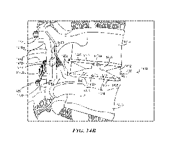
[0018] Figures 14A and 14B are schematic anterior and side
illustrations,
respectively, of a deployed patient-specific intervertebral body fusion device
deployed
between a first vertebra (e.g., a relatively superior vertebra) and a second
vertebra (e.g.,
a relatively inferior vertebra), according to an embodiment.
[0019] Figure 15 is a flow diagram illustrating a process for
patient-specific
adjustment of spinal implants, according to an embodiment.
[0020] Figure 16A shows a patients spine and a remote device
for controlling
actuation of intervertebral implants, according to an embodiment.
[0021] Figure 16B illustrates an exemplary corrective plan that
may be used and/or
generated in connection with the systems and methods described herein,
according to
an embodiment.
[0022] Figures 17A-17D show a patients spine in different
configurations,
according to an embodiment.
[0023] Figure 18 is a side view of an intervertebral body
fusion device and a
fixation for the spine, according to an embodiment.
-3-
CA 03224316 2023- 12- 27
WO 2023/278385
PCT/US2022/035232
DETAILED DESCRIPTION
[0024] The present technology is directed to systems and
methods for designing
and implementing patient-specific adjustment of spinal implants. Spinal
fusion, also
called spondylodesis or spondylosyndesis, is a neurosurgical or orthopedic
surgical
technique that joins two or more vertebrae. Spinal fusion can be used to treat
a variety
of conditions affecting any level of the spine¨lumbar, cervical, and thoracic.
In general,
spinal fusion is performed to decompress and stabilize the spine, and the
result can
prevent any movement between the fused vertebrae. Spinal fusion is most
commonly
performed to relieve the pain and pressure from mechanical pain of the
vertebrae or on
the spinal cord that results when a disc wears out (e.g., resulting from
degenerative disc
disease). Other common pathological conditions that are treated by spinal
fusion
include spinal stenosis, spondylolisthesis, spondylosis, spinal fractures,
scoliosis, and
kyphosis.
[0025] A spine fusion surgery can utilize intervertebral body
fusion (IBF) devices.
The IBF device can help to restore a height between vertebral bodies, restore
lordotic
and coronal misalignment, and/or stabilize the spine until bony fusion occurs
between
vertebral bodies. Example IBF devices can be configured for anterior lumbar
interbody
fusion (ALIF), lateral lumbar interbody fusion (LLIF), oblique lateral
interbody fusion,
posterior lumbar interbody fusion (PLIF), or transforaminal lumbar interbody
fusion
(TLIF). In some embodiments, the IBF device can be a cervical cage. IBF
devices can
also have multiple expandable mechanisms that provide intraoperative
adjustability. In
some embodiments, expandable IBF devices also provide adjustability (e.g., pre-
, intra-,
and/or postoperative adjustability) of, for example, spinal curvature,
vertebral heights,
lordotic restoration, and/or corona! restoration.
[0026] Although the disclosure herein primarily describes
systems and methods
for treatment planning in the context of orthopedic surgery, the technology
may be
applied equally to medical treatment and devices in other fields (e.g., other
types of
surgical practice). Additionally, although many embodiments herein describe
systems
and methods with respect to implanted devices, the technology may be applied
equally
to other types of medical devices (e.g., non-implanted devices).
[0027] For example, in many of the embodiments disclosed
herein, a computer
system receives implant sensor readings from one or more implant sensors of a
spinal
-4-
CA 03224316 2023- 12- 27
WO 2023/278385
PCT/US2022/035232
implant configured in a first physical configuration according to a corrective
plan for the
patient. The implant sensor readings are received after the surgery is
performed and
are indicative of a load applied by a spine of the patient on the spinal
implant. The
computer system extracts a feature vector from the implant sensor readings
using a
machine learning module of the computer system. The feature vector is
indicative of a
target correction. The computer system generates implant electrical signals
using the
machine learning module based on the feature vector. The machine learning
module
is trained based on patient data sets to generate the implant electrical
signals to adjust
the load, e.g., to achieve the target correction. The computer system
transmits the
implant electrical signals to the spinal implant to cause the spinal implant
to move the
spinal implant to a second physical configuration for the target correction.
In some
embodiments, the spinal implant is adjusted post-operatively in accordance
with a
predetermined adjustable-implant corrective plan that accounts for one or more
of
disease progression, additional surgical interventions, aging, sensed metrics,
or the
like.
[0028] In some embodiments, the computer system receives
patient data. An
anatomical configuration of the patients spine is determined based on the
received
patient data. The computer system identifies the target correction based on
the
anatomical configuration and available adjustability of the spinal implant,
wherein the
identified target correction is used to extract the feature vector.
[0029] In some embodiments, the corrective plan comprises
criteria for actuating
the spinal implant.
[0030] In some embodiments, the computer system receives device
sensor
readings from one or more device sensors embedded in an intervertebral fusion
device
implant implanted in the patient during the surgery. The device sensor
readings are
received after the surgery is performed and before the implant sensor readings
are
received. The computer system generates device electrical signals using the
machine
learning module based on the device sensor readings. The machine learning
module
is trained based on the patient data sets to generate the device sensor
readings to
reduce physical discomfort caused by the intervertebral fusion device.
[0031] In some embodiments, the feature vector is further
indicative of at least one
of lumbar lordosis (LL), Cobb angles, coronal parameters, sagittal parameters,
pelvic
-5-
CA 03224316 2023- 12- 27
WO 2023/278385
PCT/US2022/035232
parameters, disc height, segment flexibility, bone quality, or rotational
displacement of
the spine of the patient.
[0032] In some embodiments, configuring the spinal implant in
the second physical
configuration includes adjusting at least one of a screw, a cage, a plate, a
rod, a disk, a
spacer, an expandable device, a stent, a bracket, a tie, a scaffold, a
fixation device, an
anchor, a nut, a bolt, a rivet, a connector, a tether, a fastener, or a joint
replacement of
the spinal implant using the one or more implant actuators.
[0033] In some embodiments, configuring the spinal implant in
the second physical
configuration includes adjusting a reservoir coupled to the spinal implant to
modify an
amount of at least one of a pharmaceutical, biologic, biochemical, narcotic,
or a steroid
delivered to the patient.
[0034] In some embodiments, a method of providing medical care
includes
comparing a patient data set of a patient to be treated with multiple
reference patient
data sets (e.g., data from previously treated patients). The method can
include
selecting a subset of the reference patient data sets, e.g., based on
similarity of the
reference patient data set to the patient data set and/or whether the
reference patient
had a favorable treatment outcome. The selected subset can be used to generate
a
surgical procedure and/or medical device design that is likely to produce a
favorable
treatment outcome for the particular patient. In some embodiments, the
selected subset
is analyzed to identify correlations between patient pathology, surgical
procedures,
device designs, and/or treatment outcomes, and these correlations are used to
determine a personalized treatment protocol with a higher likelihood of
success.
[0035] In the context of orthopedic surgery, systems with
improved computing
capabilities (e.g., predictive analytics, machine learning, neural networks,
artificial
intelligence (Al)) can use large data sets to define improved or optimal
surgical
interventions and/or implant designs for a specific patient. The patient's
entire data can
be characterized and compared to aggregated data from groups of prior patients
(e.g.,
parameters, metrics, pathologies, treatments, outcomes). In some embodiments,
the
systems described herein use this aggregated data to formulate potential
treatment
solutions (e.g., surgical plans and/or implant designs for spine and
orthopedic
procedures) and analyze the associated likelihood of success. These systems
can
-6-
CA 03224316 2023- 12- 27
WO 2023/278385
PCT/US2022/035232
further compare potential treatment solutions to determine an optimal patient-
specific
solution that is expected to maximize the likelihood for a successful outcome.
[0036] For example, if a patient presents with a spinal
deformity pathology that can
be described with data including lumbar lordosis, Cobb angles, corona!
parameters
(e.g., coronal balance, global coronal balance, coronal pelvic tilt, etc.),
sagittal
parameters (e.g., pelvic incidence (PI), sacral slope, thoracic kyphosis,
etc.) and/or
pelvic parameters, an algorithm using these data points as inputs can be used
to
describe an optimal surgical plan and/or implant design to correct the subject
pathology
and improve the patient's outcome. As additional data inputs are used to
describe the
pathology (e.g., disc height, segment flexibility, bone quality, rotational
displacement),
the algorithm can use these additional inputs to further define an optimal
surgical plan
and/or implant design for that particular patient and their pathology.
[0037] In some embodiments, the present technology can
automatically or at least
semi-automatically determine a corrected anatomical configuration for a
subject patient
suffering from one or more deformities. For example, the computing systems
described
herein can apply mathematical rules for select parameters (e.g., LL, Cobb
angles, etc.)
and/or identify similar patients by analyzing reference patient data sets,
and, based on
the rules and/or comparison to other patients, can provide a recommended
anatomical
configuration that represents the optimal outcome if the subject patient were
to undergo
surgery. In some embodiments, the systems and methods described herein
generate
a virtual model of the corrected/recommended anatomical configuration (e.g.,
for
surgeon review).
[0038] In some embodiments, the present technology can also
automatically or at
least semi-automatically generate a surgical plan for achieving a previously
identified
corrected anatomical configuration for a subject patient. For example, based
off the
virtual model of the corrected anatomical configuration, the systems and
methods
herein can determine a type of surgery (e.g., spinal fusion surgery, non-
fusion surgery,
etc.), a surgical approach (e.g., anterior, posterior, etc.), and/or spinal
parameters for
the corrected anatomical configuration (e.g., LL, Cobb angles, etc.). The
surgical plan
can be transmitted to a surgeon for review and approval. In some embodiments,
the
present technology can also design one or more patient-specific implants for
achieving
the corrected anatomical configuration via the surgical plan.
-7-
CA 03224316 2023- 12- 27
WO 2023/278385
PCT/US2022/035232
[0039]
In some embodiments, the present technology provides systems and
methods that generate multiple anatomical models of the patient. For example,
a first
model may show the patient's native (e.g., pre-operative) anatomical
configuration, and
a second model may provide a simulation of the patients corrected (e.g., post-
operative) anatomical configuration. The second virtual model may optionally
include
one or more virtual implants shown as implanted at one or more target regions
of the
patient. Spine metrics (e.g., LL, Cobb angles, coronal parameters, sagittal
parameters,
pelvic parameters, etc.) can also be provided for both the pre-operative
anatomical
configuration and expected post-operative anatomical configuration.
[0040]
In some embodiments, the present technology includes generating,
designing, and/or providing patient-specific medical procedures for multiple
locations
within a patient. For example, the present technology can include identifying
at least
two target regions or sites within a patient (e.g., a first vertebral level
and a second
vertebral level) for surgical intervention. The present technology can then
design at
least two patient-specific implants for implantation at the at least two
target regions.
The at least two patient-specific implants can each be specifically designed
for their
respective target region, and thus can have different geometries.
In some
embodiments, the corrected anatomical configuration of the patient is only
achieved by
implanting each of the at least two patient-specific implants. In the context
of spinal
surgery, for example, the present technology may provide a first patient-
specific
interbody device to be implanted between the L2 and L3 vertebrae, a second
patient-
specific interbody device to be implanted between the L3 and L4 vertebrae, and
a third
patient-specific interbody device to be implanted between the L4 and L5
vertebrae.
[0041]
In some embodiments, the present technology can predict, model, or
simulate disease progression within a particular patient to aid in diagnosis
and/or
treatment planning. The simulation can be done to model and/or estimate future
anatomical configurations and/or spine metrics of the patient (a) if no
surgical
intervention occurs, or (b) for a variety of different surgical intervention
options. The
progression modeling can thus be used to determine the optimal time for
surgical
intervention and/or to select which surgical intervention provides the best
long-term
outcomes. In some embodiments, the disease progression modelling is performed
using one or more machine learning models trained based on multiple reference
patients.
-8-
CA 03224316 2023- 12- 27
WO 2023/278385
PCT/US2022/035232
[0042]
In a particular non-limiting example, the present technology includes a
method for providing patient-specific medical care for a subject patient. The
method
can include receiving a patient-data set for the subject patient that includes
one or more
images of the patient's spinal region showing the patient's native anatomical
configuration. The method can further include determining a corrected
anatomical
configuration for the subject patient that is different than the native
anatomical
configuration and creating a virtual model of the corrected anatomical
configuration.
The method can further include generating a surgical plan and designing one or
more
patient-specific implants for achieving the corrected anatomical configuration
in the
subject patient.
In representative embodiments, the foregoing method can be
performed by a system storing computer-executable instructions that, when
executed,
cause the system to perform the steps of method.
[0043]
In a particular non-limiting example, the present technology includes a
method for designing a patient-specific orthopedic implant for a subject
patient. The
method can include receiving a patient data set of the subject patient, the
patient data
set including spinal pathology data for the subject patient. The patient data
set can be
compared to multiple reference patient data sets to identify one or more
similar patient
data sets in the reference patient data sets, with each identified similar
patient data set
corresponding to a reference patient having similar spinal pathology to the
subject
patient and who received treatment with an orthopedic implant. The method can
further
include selecting a subset of the one or more similar patient data sets based
on whether
the similar patient data sets indicated the reference patient had a favorable
outcome
following implantation of their orthopedic implant. The method can further
include
identifying, for at least one similar reference patient of the selected
subset, surgical
procedure data and design data for the respective orthopedic implant that
produced the
favorable outcome in the similar reference patient. Based on the design data
and the
surgical data that produced the favorable outcome in the similar reference
patient, the
patient-specific orthopedic implant for the subject patient and a surgical
procedure for
implanting the patient-specific orthopedic implant into the subject patient
can be
designed. In some embodiments, the method can further include outputting
fabrication
instructions for causing a manufacturing system to manufacture the patient-
specific
orthopedic implant according to the generated design. In representative
embodiments,
-9-
CA 03224316 2023- 12- 27
WO 2023/278385
PCT/US2022/035232
the foregoing method can be performed by a system storing computer-executable
instructions that, when executed, cause the system to perform the steps of
method.
[0044] In some embodiments, IBF devices are personalized to
patient-specific
features and/or concerns in accordance with a pre-operative plan for height
restoration,
lordotic and coronal correction, and/or optimal endplate coverage. For
example, an
expandable IBF device in accordance with the present technology can include
patient-
specific endplates that can achieve optimal surface area contact and/or
provide a
mechanism to tailor the medical intervention from the IBF device (e.g., tailor
the
segmental height restoration, the lordotic correction, and/or the corona!
correction). In
some embodiments, the patient-specific endplates are the result of additive
and/or
subtractive manufacturing. The patient-specific endplates can then be
connected to an
expandable mechanism that can also provide a predetermined height restoration,
lordotic correction, and/or coronal correction via one or more expansion
mechanisms
(e.g., an expandable jack, scissor jack mechanism, screw drive mechanism,
etc.). In
some such embodiments, the expansion mechanism includes one or more joints
(e.g.,
ball joints), hinges, or other connections that can be precisely adjusted to a
predetermined angle and then temporarily or permanently locked.
[0045] In an example embodiment, the IBF device includes an
expansion
mechanism configured to be locked at a desired expansion configuration to
facilitate
the fusion. The expansion mechanism can include a first lockable ball joint on
an upper
surface of the mechanism and a second lockable ball joint on a lower surface
of the
mechanism. The IBF device also includes a first endplate connected to the
mechanism
at the first lockable ball joint. In some embodiments, the first endplate
includes a
superior surface having one or more patient-specific features configured to
engage and
mate with the topology of an inferior surface of the superior vertebra. The
IBF device
also includes a second endplate connected to the mechanism at the second
lockable
ball joint. In some embodiments, the second endplate includes an inferior
surface
having one or more patient-specific features configured to engage and mate
with the
topology of a superior surface of the inferior vertebra.
[0046] In some embodiments, the patient-specific features of
the first and/or
second endplates can improve the match between the IBF device and the
vertebrae
being fused, thereby increasing the traction of the I BF device at the joint.
For example,
-10-
CA 03224316 2023- 12- 27
WO 2023/278385
PCT/US2022/035232
the one or more patient-specific features can correspond to topographical
features on
the surfaces of the vertebrae at the vertebral joint to customize the fit of
the first and
second endplates. In some embodiments, the patient-specific features of the
first
and/or second endplates include one or more features that help facilitate a
prescribed
medical treatment. For example, the first and/or second endplates can include
a slope
that helps provide a lordotic and/or coronal correction to the patient's
spine. In some
embodiments, the expandable main body includes a screw jack mechanical
expansion
mechanism. In some embodiments the expandable main body includes a scissor
jack
mechanical expansion mechanism.
[0047] For ease of reference, patient-specific implants are
sometimes described
herein with reference to top and bottom, upper and lower, upwards and
downwards,
and/or horizontal plane, x-y plane, vertical, or z-direction relative to the
spatial
orientation of the embodiments shown in the figures. It is to be understood,
however,
that the patient-specific implants can be moved to, and used in, different
spatial
orientations without changing the structure and/or function of the disclosed
embodiments of the present technology.
[0048] Embodiments of the present disclosure will be described
more fully
hereinafter with reference to the accompanying drawings in which like numerals
represent like elements throughout the several figures, and in which example
embodiments are shown. Embodiments of the claims may, however, be embodied in
many different forms and should not be construed as limited to the embodiments
set
forth herein. The examples set forth herein are non-limiting examples and are
merely
examples among other possible examples.
[0049] The words "comprising," "having," "containing," and
"including," and other
forms thereof, are intended to be equivalent in meaning and be open ended in
that an
item or items following any one of these words is not meant to be an
exhaustive listing
of such item or items, or meant to be limited to only the listed item or
items.
[0050] As used herein and in the appended claims, the singular
forms "a," "an,"
and "the" include plural references unless the context clearly dictates
otherwise.
[0051] Further, although primarily discussed herein as a method
for customizing
intervertebral body fusion devices and the resulting IBF devices, one of skill
in the art
will understand that the scope of the invention is not so limited. For
example, the
-11-
CA 03224316 2023- 12- 27
WO 2023/278385
PCT/US2022/035232
patient-specific customization methods disclosed herein can also be used to
customize
implants for various other medical procedures, such as for insertion at
another joint in
a patient's body. Accordingly, the scope of the invention is not confined to
any subset
of embodiments and is confined only by the limitations set out in the appended
claims.
[0052] Figure 1 is a network connection diagram illustrating a
computing system
100 for providing patient-specific medical care, according to an embodiment.
As
described in further detail herein, the system 100 is configured to generate a
medical
treatment plan for a patient. In some embodiments, the system 100 is
configured to
generate a medical treatment plan for a patient suffering from an orthopedic
or spinal
disease or disorder, such as trauma (e.g., fractures), cancer, deformity,
degeneration,
pain (e.g., back pain, leg pain), irregular spinal curvature (e.g., scoliosis,
lordosis,
kyphosis), irregular spinal displacement (e.g., spondylolisthesis, lateral
displacement
axial displacement), osteoarthritis, lumbar degenerative disc disease,
cervical
degenerative disc disease, lumbar spinal stenosis, or cervical spinal
stenosis, or a
combination thereof. The medical treatment plan can include surgical
information,
surgical plans, technology recommendations (e.g., device and/or instrument
recommendations), and/or medical device designs. For example, the medical
treatment
plan can include at least one treatment procedure (e.g., a surgical procedure
or
intervention) and/or at least one medical device (e.g., an implanted medical
device (also
referred to herein as an "implant" or "implanted device") or implant delivery
instrument).
[0053] In some embodiments, the system 100 generates a medical
treatment plan
that is customized for a particular patient or group of patients, also
referred to herein as
a "patient-specific" or "personalized" treatment plan. The patient-specific
treatment plan
can include at least one patient-specific surgical procedure and/or at least
one patient-
specific medical device that are designed and/or optimized for the patient's
particular
characteristics (e.g., condition, anatomy, pathology, condition, medical
history). For
example, the patient-specific medical device can be designed and manufactured
specifically for the particular patient, rather than being an off-the-shelf
device. However,
it shall be appreciated that a patient-specific treatment plan can also
include aspects
that are not customized for the particular patient. For example, a patient-
specific or
personalized surgical procedure can include one or more instructions,
portions, steps,
etc. that are non-patient-specific. Likewise, a patient-specific or
personalized medical
device can include one or more components that are non-patient-specific,
and/or can
-12-
CA 03224316 2023- 12- 27
WO 2023/278385
PCT/US2022/035232
be used with an instrument or tool that is non-patient-specific. Personalized
implant
designs can be used to manufacture or select patient-specific technologies,
including
medical devices, instruments, and/or surgical kits. For example, a
personalized surgical
kit can include one or more patient-specific devices, patient-specific
instruments, non-
patient-specific technology (e.g., standard instruments, devices, etc.),
instructions for
use, patient-specific treatment plan information, or a combination thereof.
[0054] The system 100 includes a client computing device 102,
which can be a
user device, such as a smart phone, mobile device, laptop, desktop, personal
computer,
tablet, phablet, or other such devices known in the art. As discussed further
herein, the
client computing device 102 can include one or more processors and memory
storing
instructions executable by the one or more processors to perform the methods
described herein. The client computing device 102 can be associated with a
healthcare
provider that is treating the patient. Although Figure 1 illustrates a single
client
computing device 102, in alternative embodiments, the client computing device
102 can
instead be implemented as a client computing system encompassing multiple
computing devices, such that the operations described herein with respect to
the client
computing device 102 can instead be performed by the computing system and/or
the
computing devices.
[0055] The client computing device 102 is configured to receive
a patient data set
108 associated with a patient to be treated. The patient data set 108 can
include data
representative of the patients condition, anatomy, pathology, medical history,
preferences, and/or any other information or parameters relevant to the
patient. For
example, the patient data set 108 can include medical history, surgical
intervention data,
treatment outcome data, progress data (e.g., physician notes), patient
feedback (e.g.,
feedback acquired using quality of life questionnaires, surveys), clinical
data, provider
information (e.g., physician, hospital, surgical team), patient information
(e.g.,
demographics, sex, age, height, weight, type of pathology, occupation,
activity level,
tissue information, health rating, comorbidities, health related quality of
life (HRQL)),
vital signs, diagnostic results, medication information, allergies, image data
(e.g.,
camera images, magnetic resonance imaging (MRI) images, ultrasound images,
computerized aided tomography (CAT) scan images, positron emission tomography
(PET) images, X-Ray images), diagnostic equipment information (e.g.,
manufacturer,
model number, specifications, user-selected settings/configurations, etc.), or
the like.
-13-
CA 03224316 2023- 12- 27
WO 2023/278385
PCT/US2022/035232
In some embodiments, the patient data set 108 includes data representing one
or more
of patient identification number (ID), age, gender, body mass index (BM!), LL,
Cobb
angle(s), PI, disc height, segment flexibility, bone quality, rotational
displacement,
and/or treatment level of the spine.
[0056] The client computing device 102 is operably connected
via a
communication network 104 to a server 106, thus allowing for data transfer
between
the client computing device 102 and the server 106. The communication network
104
may be a wired and/or a wireless network. The communication network 104, if
wireless,
may be implemented using communication techniques such as Visible Light
Communication (VLC), Worldwide Interoperability for Microwave Access (WiMAX),
Long term evolution (LTE), Wireless local area network (WLAN), Infrared (IR)
communication, Public Switched Telephone Network (PSTN), Radio waves, and/or
other communication techniques known in the art.
[0057] The server 106, which may also be referred to as a
"treatment assistance
network" or "prescriptive analytics network," can include one or more
computing devices
and/or systems. As discussed further herein, the server 106 can include one or
more
processors, and memory storing instructions executable by the one or more
processors
to perform the methods described herein. In some embodiments, the server 106
is
implemented as a distributed "cloud" computing system or facility across any
suitable
combination of hardware and/or virtual computing resources.
[0058] The client computing device 102 and server 106 can
individually or
collectively perform the various methods described herein for providing
patient-specific
medical care. For example, some or all of the steps of the methods described
herein
can be performed by the client computing device 102 alone, the server 106
alone, or a
combination of the client computing device 102 and the server 106. Thus,
although
certain operations are described herein with respect to the server 106, it
shall be
appreciated that these operations can also be performed by the client
computing device
102, and vice-versa.
[0069] The server 106 includes at least one database 110
configured to store
reference data useful for the treatment planning methods described herein. The
reference data can include historical and/or clinical data from the same or
other patients,
data collected from prior surgeries and/or other treatments of patients by the
same or
-14-
CA 03224316 2023- 12- 27
WO 2023/278385
PCT/US2022/035232
other healthcare providers, data relating to medical device designs, data
collected from
study groups or research groups, data from practice databases, data from
academic
institutions, data from implant manufacturers or other medical device
manufacturers,
data from imaging studies, data from simulations, clinical trials, demographic
data,
treatment data, outcome data, mortality rates, or the like.
[0060] In some embodiments, the database 110 includes multiple
reference
patient data sets, each patient reference data set associated with a
corresponding
reference patient. For example, the reference patient can be a patient that
previously
received treatment or is currently receiving treatment. Each reference patient
data set
can include data representative of the corresponding reference patients
condition,
anatomy, pathology, medical history, disease progression, preferences, and/or
any
other information or parameters relevant to the reference patient, such as any
of the
data described herein with respect to the patient data set 108. In some
embodiments,
the reference patient data set includes pre-operative data, intra-operative
data, and/or
post-operative data. For example, a reference patient data set can include
data
representing one or more of patient ID, age, gender, BMI, LL, Cobb angle(s),
PI, disc
height, segment flexibility, bone quality, rotational displacement, and/or
treatment level
of the spine. As another example, a reference patient data set can include
treatment
data regarding at least one treatment procedure performed on the reference
patient,
such as descriptions of surgical procedures or interventions (e.g., surgical
approaches,
bony resections, surgical maneuvers, corrective maneuvers, placement of
implants or
other devices). In some embodiments, the treatment data includes medical
device
design data for at least one medical device used to treat the reference
patient, such as
physical properties (e.g., size, shape, volume, material, mass, weight),
mechanical
properties (e.g., stiffness, strength, modulus, hardness), and/or biological
properties
(e.g., osteo-integration, cellular adhesion, anti-bacterial properties, anti-
viral
properties). In yet another example, a reference patient data set can include
outcome
data representing an outcome of the treatment of the reference patient, such
as
corrected anatomical metrics, presence of fusion, HRQL, activity level, return
to work,
complications, recovery times, efficacy, mortality, and/or follow-up
surgeries.
[0061] In some embodiments, the server 106 receives at least
some of the
reference patient data sets from multiple healthcare provider computing
systems (e.g.,
systems 112a-112c, collectively 112). The server 106 can be connected to the
-15-
CA 03224316 2023- 12- 27
WO 2023/278385
PCT/US2022/035232
healthcare provider computing systems 112 via one or more communication
networks
(not shown). Each healthcare provider computing system 112 can be associated
with
a corresponding healthcare provider (e.g., physician, surgeon, medical clinic,
hospital,
healthcare network, etc.). Each healthcare provider computing system 112 can
include
at least one reference patient data set (e.g., reference patient data sets
114a-114c,
collectively 114) associated with reference patients treated by the
corresponding
healthcare provider. The reference patient data sets 114 can include, for
example,
electronic medical records, electronic health records, biomedical data sets,
etc. The
reference patient data sets 114 can be received by the server 106 from the
healthcare
provider computing systems 112 and can be reformatted into different formats
for
storage in the database 110. Optionally, the reference patient data sets 114
can be
processed (e.g., cleaned) to ensure that the represented patient parameters
are likely
to be useful in the treatment planning methods described herein.
[0062] As described in further detail herein, the server 106
can be configured with
one or more algorithms that generate patient-specific treatment plan data
(e.g.,
treatment procedures, medical devices) based on the reference data. In some
embodiments, the patient-specific data is generated based on correlations
between the
patient data set 108 and the reference data. Optionally, the server 106 can
predict
outcomes, including recovery times, efficacy based on clinical end points,
likelihood of
success, predicted mortality, predicted related follow-up surgeries, or the
like. In some
embodiments, the server 106 can continuously or periodically analyze patient
data
(including patient data obtained during the patient stay) to determine near
real-time or
real-time risk scores, mortality prediction, etc.
[0063] In some embodiments, the server 106 includes one or more
modules for
performing one or more steps of the patient-specific treatment planning
methods
described herein. For example, in the depicted embodiment, the server 106
includes a
data analysis module 116 and a treatment planning module 118. In alternative
embodiments, one or more of these modules may be combined with each other, or
may
be omitted. Thus, although certain operations are described herein with
respect to a
particular module or modules, this is not intended to be limiting, and such
operations
can be performed by a different module or modules in alternative embodiments.
-16-
CA 03224316 2023- 12- 27
WO 2023/278385
PCT/US2022/035232
[0064] The data analysis module 116 is configured with one or
more algorithms
for identifying a subset of reference data from the database 110 that is
likely to be useful
in developing a patient-specific treatment plan. For example, the data
analysis module
116 can compare patient-specific data (e.g., the patient data set 108 received
from the
client computing device 102) to the reference data from the database 110
(e.g., the
reference patient data sets) to identify similar data (e.g., one or more
similar patient
data sets in the reference patient data sets). The comparison can be based on
one or
more parameters, such as age, gender, BMI, LL, PI, and/or treatment levels.
The
parameter(s) can be used to calculate a similarity score for each reference
patient. The
similarity score can represent a statistical correlation between the patient
data set 108
and the reference patient data set. Accordingly, similar patients can be
identified based
on whether the similarity score is above, below, or at a specified threshold
value. For
example, as described in greater detail below, the comparison can be performed
by
assigning values to each parameter and determining the aggregate difference
between
the subject patient and each reference patient. Reference patients whose
aggregate
difference is below a threshold can be considered to be similar patients.
[0065] The data analysis module 116 can further be configured
with one or more
algorithms to select a subset of the reference patient data sets, e.g., based
on similarity
to the patient data set 108 and/or treatment outcome of the corresponding
reference
patient. For example, the data analysis module 116 can identify one or more
similar
patient data sets in the reference patient data sets, and then select a subset
of the
similar patient data sets based on whether the similar patient data set
includes data
indicative of a favorable or desired treatment outcome. The outcome data can
include
data representing one or more outcome parameters, such as corrected anatomical
metrics, presence of fusion, HRQL, activity level, complications, recovery
times,
efficacy, mortality, or follow-up surgeries. As described in further detail
below, in some
embodiments, the data analysis module 116 calculates an outcome score by
assigning
values to each outcome parameter. A patient can be considered to have a
favorable
outcome if the outcome score is above, below, or at a specified threshold
value.
[0066] In some embodiments, the data analysis module 116
selects a subset of
the reference patient data sets based at least in part on user input (e.g,
from a clinician,
surgeon, physician, healthcare provider). For example, the user input can be
used in
identifying similar patient data sets. In some embodiments, weighting of
similarity
-17-
CA 03224316 2023- 12- 27
WO 2023/278385
PCT/US2022/035232
and/or outcome parameters can be selected by a healthcare provider or
physician to
adjust the similarity and/or outcome score based on clinician input.
In further
embodiments, the healthcare provider or physician can select the set of
similarity and/or
outcome parameters (or define new similarity and/or outcome parameters) used
to
generate the similarity and/or outcome score, respectively.
[0067]
In some embodiments, the data analysis module 116 includes one or more
algorithms used to select a set or subset of the reference patient data sets
based on
criteria other than patient parameters. For example, the one or more
algorithms can be
used to select the subset based on healthcare provider parameters (e.g., based
on
healthcare provider ranking/scores such as hospital/physician expertise,
number of
procedures performed, hospital ranking, etc.) and/or healthcare resource
parameters
(e.g., diagnostic equipment, facilities, surgical equipment such as surgical
robots), or
other non-patient related information that can be used to predict outcomes and
risk
profiles for procedures for the present healthcare provider. For example,
reference
patient data sets with images captured from similar diagnostic equipment can
be
aggregated to reduce or limit irregularities due to variation between
diagnostic
equipment. Additionally, patient-specific treatment plans can be developed for
a
particular health-care provider using data from similar healthcare providers
(e.g.,
healthcare providers with traditionally similar outcomes, physician expertise,
surgical
teams, etc.). In some embodiments, reference healthcare provider data sets,
hospital
data sets, physician data sets, surgical team data sets, post-treatment data
set, and
other data sets can be utilized. By way of example, a patient-specific
treatment plan to
perform a battlefield surgery can be based on reference patient data from
similar
battlefield surgeries and/or datasets associated with battlefield surgeries.
In another
example, the patient-specific treatment plan can be generated based on
available
robotic surgical systems. The reference patient data sets can be selected
based on
patients that have been operated on using comparable robotic surgical systems
under
similar conditions (e.g., size and capabilities of surgical teams, hospital
resources, etc.).
[0068]
The treatment planning module 118 is configured with one or more
algorithms to generate at least one treatment plan (e.g., pre-operative plans,
surgical
plans, post-operative plans, etc.) based on the output from the data analysis
module
116. In some embodiments, the treatment planning module 118 is configured to
develop and/or implement at least one predictive model for generating the
patient-
-18-
CA 03224316 2023- 12- 27
WO 2023/278385
PCT/US2022/035232
specific treatment plan, also known as a "prescriptive model." The predictive
model(s)
can be developed using clinical knowledge, statistics, machine learning, Al,
neural
networks, or the like. In some embodiments, the output from the data analysis
module
116 is analyzed (e.g., using statistics, machine learning, neural networks,
Al) to identify
correlations between data sets, patient parameters, healthcare provider
parameters,
healthcare resource parameters, treatment procedures, medical device designs,
and/or
treatment outcomes. These correlations can be used to develop at least one
predictive
model that predicts the likelihood that a treatment plan will produce a
favorable outcome
for the particular patient. The predictive model(s) can be validated, e.g., by
inputting
data into the model(s) and comparing the output of the model to the expected
output.
[0069]
In some embodiments, the treatment planning module 118 is configured to
generate the treatment plan based on previous treatment data from reference
patients.
For example, the treatment planning module 118 can receive a selected subset
of
reference patient data sets and/or similar patient data sets from the data
analysis
module 116, and determine or identify treatment data from the selected subset.
The
treatment data can include, for example, treatment procedure data (e.g.,
surgical
procedure or intervention data) and/or medical device design data (e.g.,
implant design
data) that are associated with favorable or desired treatment outcomes for the
corresponding patient. The treatment planning module 118 can analyze the
treatment
procedure data and/or medical device design data to determine an optimal
treatment
protocol for the patient to be treated. For example, the treatment procedures
and/or
medical device designs can be assigned values and aggregated to produce a
treatment
score. The patient-specific treatment plan can be determined by selecting
treatment
plan(s) based on the score (e.g., higher or highest score; lower or lowest
score; score
that is above, below, or at a specified threshold value). The personalized
patient-
specific treatment plan can be based on, at least in part, the patient-
specific
technologies or patient-specific selected technology.
[0070]
Alternatively or in combination, the treatment planning module 118 can
generate the treatment plan based on correlations between data sets. For
example,
the treatment planning module 118 can correlate treatment procedure data
and/or
medical device design data from similar patients with favorable outcomes
(e.g., as
identified by the data analysis module 116).
Correlation analysis can include
transforming correlation coefficient values to values or scores. The
values/scores can
-19-
CA 03224316 2023- 12- 27
WO 2023/278385
PCT/US2022/035232
be aggregated, filtered, or otherwise analyzed to determine one or more
statistical
significances. These correlations can be used to determine treatment
procedure(s)
and/or medical device design(s) that are optimal or likely to produce a
favorable
outcome for the patient to be treated.
[0071] Alternatively or in combination, the treatment planning
module 118 can
generate the treatment plan using one or more Al techniques. Al techniques can
be
used to develop computing systems capable of simulating aspects of human
intelligence, e.g., learning, reasoning, planning, problem solving, decision
making, etc.
Al techniques can include, but are not limited to, case-based reasoning, rule-
based
systems, artificial neural networks, decision trees, support vector machines,
regression
analysis, Bayesian networks (e.g., naïve Bayes classifiers), genetic
algorithms, cellular
automata, fuzzy logic systems, multi-agent systems, swarm intelligence, data
mining,
machine learning (e.g., supervised learning, unsupervised learning,
reinforcement
learning), and hybrid systems.
[0072] In some embodiments, the treatment planning module 118
generates the
treatment plan using one or more trained machine learning models. Various
types of
machine learning models, algorithms, and techniques are suitable for use with
the
present technology. In some embodiments, the machine learning model is
initially
trained on a training data set, which is a set of examples used to fit the
parameters (e.g.,
weights of connections between "neurons" in artificial neural networks) of the
model.
For example, the training data set can include any of the reference data
stored in
database 110, such as multiple reference patient data sets or a selected
subset thereof
(e.g., multiple similar patient data sets).
[0073] In some embodiments, the machine learning model (e.g., a
neural network
or a naïve Bayes classifier) may be trained on the training data set using a
supervised
learning method (e.g., gradient descent or stochastic gradient descent). The
training
dataset can include pairs of generated "input vectors" with the associated
corresponding "answer vector" (commonly denoted as the target). The current
model
is run with the training data set and produces a result, which is then
compared with the
target, for each input vector in the training data set. Based on the result of
the
comparison and the specific learning algorithm being used, the parameters of
the model
are adjusted. The model fitting can include both variable selection and
parameter
-20-
CA 03224316 2023- 12- 27
WO 2023/278385
PCT/US2022/035232
estimation. The fitted model can be used to predict the responses for the
observations
in a second data set called the validation data set. The validation data set
can provide
an unbiased evaluation of a model fit on the training data set while tuning
the model
parameters. Validation data sets can be used for regularization by early
stopping, e.g.,
by stopping training when the error on the validation data set increases, as
this may be
a sign of overfitting to the training data set. In some embodiments, the
validation data
set error can fluctuate during training, such that ad-hoc rules may be used to
decide
when overfitting has truly begun. Finally, a test data set can be used to
provide an
unbiased evaluation of a final model fit on the training data set.
[0074] To generate a treatment plan, the patient data set 108
can be input into the
trained machine learning model(s). Additional data, such as the selected
subset of
reference patient data sets and/or similar patient data sets, and/or treatment
data from
the selected subset, can also be input into the trained machine learning
model(s). The
trained machine learning model(s) can then calculate whether various candidate
treatment procedures and/or medical device designs are likely to produce a
favorable
outcome for the patient. Based on these calculations, the trained machine
learning
model(s) can select at least one treatment plan for the patient. In
embodiments where
multiple trained machine learning models are used, the models can be run
sequentially
or concurrently to compare outcomes and can be periodically updated using
training
data sets. The treatment planning module 118 can use one or more of the
machine
learning models based the model's predicted accuracy score.
[0075] The patient-specific treatment plan generated by the
treatment planning
module 118 can include at least one patient-specific treatment procedure
(e.g., a
surgical procedure or intervention) and/or at least one patient-specific
medical device
(e.g., an implant or implant delivery instrument). A patient-specific
treatment plan can
include an entire surgical procedure or portions thereof. Additionally, one or
more
patient-specific medical devices can be specifically selected or designed for
the
corresponding surgical procedure, thus allowing for the various components of
the
patient-specific technology to be used in combination to treat the patient.
[0076] In some embodiments, the patient-specific treatment
procedure includes
an orthopedic surgery procedure, such as spinal surgery, hip surgery, knee
surgery,
jaw surgery, hand surgery, shoulder surgery, elbow surgery, total joint
reconstruction
-21 -
CA 03224316 2023- 12- 27
WO 2023/278385
PCT/US2022/035232
(arthroplasty), skull reconstruction, foot surgery, or ankle surgery. Spinal
surgery can
include spinal fusion surgery, such as PLIF, ALIF, transverse or TLIF, LLIF,
direct lateral
lumbar interbody fusion (DLIF), or extreme lateral lumbar interbody fusion
(XLIF). In
some embodiments, the patient-specific treatment procedure includes
descriptions of
and/or instructions for performing one or more aspects of a patient-specific
surgical
procedure. For example, the patient-specific surgical procedure can include
one or
more of a surgical approach, a corrective maneuver, a bony resection, or
implant
placement.
[0077] In some embodiments, the patient-specific medical device
design includes
a design for an orthopedic implant and/or a design for an instrument for
delivering an
orthopedic implant. Examples of such implants include, but are not limited to,
screws
(e.g., bone screws, spinal screws, pedicle screws, facet screws), interbody
implant
devices (e.g., intervertebral implants, expandable intervertebral implants,
etc.), cages,
plates, rods, discs, fusion devices, spacers, rods, expandable devices,
stents, brackets,
ties, scaffolds, fixation devices, anchors, nuts, bolts, rivets, connectors,
tethers,
fasteners, joint replacements, hip implants, or the like. Examples of
instruments
include, but are not limited to, screw guides, cannulas, ports, catheters,
insertion tools,
or the like.
[0078] A patient-specific medical device design can include
data representing one
or more of physical properties (e.g., size, shape, volume, material, mass,
weight),
mechanical properties (e.g., stiffness, strength, modulus, hardness), and/or
biological
properties (e.g., osteo-integration, cellular adhesion, anti-bacterial
properties, anti-viral
properties) of a corresponding medical device. For example, a design for an
orthopedic
implant can include implant shape, size, material, and/or effective stiffness
(e.g., lattice
density, number of struts, location of struts, etc.). In some embodiments, the
generated
patient-specific medical device design is a design for an entire device.
Alternatively,
the generated design can be for one or more components of a device, rather
than the
entire device.
[0079] In some embodiments, the design is for one or more
patient-specific device
components that can be used with standard, off-the-shelf components. For
example,
in a spinal surgery, a pedicle screw kit can include both standard components
and
patient-specific customized components. In some embodiments, the generated
design
-22-
CA 03224316 2023- 12- 27
WO 2023/278385
PCT/US2022/035232
is for a patient-specific medical device that can be used with a standard, off-
the-shelf
delivery instrument. For example, the implants (e.g., screws, screw holders,
rods) can
be designed and manufactured for the patient, while the instruments for
delivering the
implants can be standard instruments. This approach allows the components that
are
implanted to be designed and manufactured based on the patient's anatomy
and/or
surgeon's preferences to enhance treatment. The patient-specific devices
described
herein are expected to improve delivery into the patient's body, placement at
the
treatment site, and/or interaction with the patient's anatomy.
[0080] In embodiments where the patient-specific treatment plan
includes a
surgical procedure to implant a medical device, the treatment planning module
118 can
also store various types of implant surgery information, such as implant
parameters
(e.g., types, dimensions), availability of implants, aspects of a pre-
operative plan (e.g.,
initial implant configuration, detection and measurement of the patient's
anatomy, etc.),
FDA requirements for implants (e.g., specific implant parameters and/or
characteristics
for compliance with FDA regulations), or the like. In some embodiments, the
treatment
planning module 118 can convert the implant surgery information into formats
useable
for machine-learning based models and algorithms. For example, the implant
surgery
information can be tagged with particular identifiers for formulas or can be
converted
into numerical representations suitable for supplying to the trained machine
learning
model(s). The treatment planning module 118 can also store information
regarding the
patient's anatomy, such as two- or three-dimensional images or models of the
anatomy,
and/or information regarding the biology, geometry, and/or mechanical
properties of the
anatomy. The anatomy information can be used to inform implant design and/or
placement.
[0081] The treatment plan(s) generated by the treatment
planning module 118 can
be transmitted via the communication network 104 to the client computing
device 102
for output to a user (e.g., clinician, surgeon, healthcare provider, patient).
In some
embodiments, the client computing device 102 includes or is operably coupled
to a
display 122 for outputting the treatment plan(s), surgical plan(s), corrective
plan(s), etc.
For example, the surgical plan can be displayed to illustrate a predicted post-
operative
outcome. The corrective plan can be an adjustable-implant corrective plan
displayed
to illustrate post-operative implant adjustments to compensate for one or more
of
disease progression, predicted new disease(s), anatomical changes, future
-23-
CA 03224316 2023- 12- 27
WO 2023/278385
PCT/US2022/035232
interventions, etc. The display 122 can display adjustability of one or more
implants
123 according to the corrective plan.
[0082]
The display 122 can include a graphical user interface (GUI) for visually
depicting various aspects of the treatment plan(s). For example, the display
122 can
show various aspects of a surgical procedure to be performed on the patient,
such as
the surgical approach, treatment levels, corrective maneuvers, tissue
resection, and/or
implant placement. To facilitate visualization, a virtual model of the
surgical procedure
can be displayed. As another example, the display 122 can show a design for a
medical
device to be implanted in the patient, such as a two- or three-dimensional
model of the
device design. The display 122 can also show patient information, such as two-
or
three-dimensional images or models of the patient's anatomy where the surgical
procedure is to be performed and/or where the device is to be implanted. The
client
computing device 102 can further include one or more user input devices (not
shown)
allowing the user to modify, select, approve, and/or reject the displayed
treatment
plan(s).
[0083]
In some embodiments, the medical device design(s) generated by the
treatment planning module 118 can be transmitted from the client computing
device 102
and/or server 106 to a manufacturing system 124 for manufacturing a
corresponding
medical device. The manufacturing system 124 can be located on site or off
site. On-
site manufacturing can reduce the number of sessions with a patient and/or the
time to
be able to perform the surgery whereas off-site manufacturing can be useful to
make
the complex devices.
Off-site manufacturing facilities can have specialized
manufacturing equipment.
In some embodiments, more complicated device
components can be manufactured off site, while simpler device components can
be
manufactured on site.
[0084]
Various types of manufacturing systems are suitable for use in accordance
with the embodiments herein. For example, the manufacturing system 124 can be
configured for additive manufacturing, such as three-dimensional (3D)
printing,
stereolithography (SLA), digital light processing (DLP), fused deposition
modeling
(FDM), selective laser sintering (SLS), selective laser melting (SLM),
selective heat
sintering (SHM), electronic beam melting (EBM), laminated object manufacturing
(LOM), powder bed printing (PP), thermoplastic printing, direct material
deposition
-24-
CA 03224316 2023- 12- 27
WO 2023/278385
PCT/US2022/035232
(DMD), inkjet photo resin printing, or like technologies, or combination
thereof.
Alternatively or in combination, the manufacturing system 124 can be
configured for
subtractive (traditional) manufacturing, such as CNC machining, electrical
discharge
machining (EDM), grinding, laser cutting, water jet machining, manual
machining (e.g.,
milling, lathe/turning), or like technologies, or combinations thereof. The
manufacturing
system 124 can manufacture one or more patient-specific medical devices based
on
fabrication instructions or data (e.g., CAD data, 3D data, digital blueprints,
stereolithography data, or other data suitable for the various manufacturing
technologies described herein). Different components of the system 100 can
generate
at least a portion of the manufacturing data used by the manufacturing system
124. The
manufacturing data can include, without limitation, fabrication instructions
(e.g.,
programs executable by additive manufacturing equipment, subtractive
manufacturing
equipment, etc.), 3D data, CAD data (e.g., CAD files), CAM data (e.g., CAM
files), path
data (e.g., print head paths, tool paths, etc.), material data, tolerance
data, surface finish
data (e.g., surface roughness data), regulatory data (e.g., FDA requirements,
reimbursement data, etc.), or the like. The manufacturing system 124 can
analyze the
manufacturability of the implant design based on the received manufacturing
data. The
implant design can be finalized by altering geometries, surfaces, etc. and
then
generating manufacturing instructions.
In some embodiments, the server 106
generates at least a portion of the manufacturing data, which is transmitted
to the
manufacturing system 124.
[0085]
The manufacturing system 124 can generate CAM data, print data (e.g.,
powder bed print data, thermoplastic print data, photo resin data, etc.), or
the like and
can include additive manufacturing equipment, subtractive manufacturing
equipment,
thermal processing equipment, or the like. The additive manufacturing
equipment can
be 3D printers, stereolithography devices, DLP devices, FDM devices, SLS
devices,
SLM devices, EBM devices, LOM devices, powder bed printers, thermoplastic
printers,
DMD devices, or inkjet photo resin printers, or like technologies. The
subtractive
manufacturing equipment can be CNC machines, electrical discharge machines,
grinders, laser cutters, water jet machines, manual machines (e.g., milling
machines,
lathes, etc.), or like technologies. Both additive and subtractive techniques
can be used
to produce implants with complex geometries, surface finishes, material
properties, etc.
The generated fabrication instructions can be configured to cause the
manufacturing
-25-
CA 03224316 2023- 12- 27
WO 2023/278385
PCT/US2022/035232
system 124 to manufacture the patient-specific orthopedic implant that matches
or is
therapeutically the same as the patient-specific design. In some embodiments,
the
patient-specific medical device can include features, materials, and designs
shared
across designs to simplify manufacturing. For example, deployable patient-
specific
medical devices for different patients can have similar internal deployment
mechanisms
but have different deployed configurations. In some embodiments, the
components of
the patient-specific medical devices are selected from a set of available pre-
fabricated
components and the selected pre-fabricated components can be modified based on
the
fabrication instructions or data.
[0086] The treatment plans described herein can be performed by
a surgeon, a
surgical robot, or a combination thereof, thus allowing for treatment
flexibility. In some
embodiments, the surgical procedure can be performed entirely by a surgeon,
entirely
by a surgical robot, or a combination thereof. For example, one step of a
surgical
procedure can be manually performed by a surgeon and another step of the
procedure
can be performed by a surgical robot. In some embodiments the treatment
planning
module 118 generates control instructions configured to cause a surgical robot
(e.g.,
robotic surgery systems, navigation systems, etc.) to partially or fully
perform a surgical
procedure. The control instructions can be transmitted to the robotic
apparatus by the
client computing device 102 and/or the server 106.
[0087] Following the treatment of the patient in accordance
with the treatment
plan, treatment progress can be monitored over one or more time periods to
update the
data analysis module 116 and/or treatment planning module 118. Post-treatment
data
can be added to the reference data stored in the database 110. The post-
treatment
data can be used to train machine learning models for developing patient-
specific
treatment plans, patient-specific medical devices, or combinations thereof.
[0088] It shall be appreciated that the components of the
system 100 can be
configured in many different ways. For example, in alternative embodiments,
the
database 110, the data analysis module 116 and/or the treatment planning
module 118
can be components of the client computing device 102, rather than the server
106. As
another example, the database 110 the data analysis module 116, and/or the
treatment
planning module 118 can be located across multiple different servers,
computing
-26-
CA 03224316 2023- 12- 27
WO 2023/278385
PCT/US2022/035232
systems, or other types of cloud-computing resources, rather than at a single
server
106 or client computing device 102.
[0089] Additionally, in some embodiments, the system 100 can be
operational with
numerous other computing system environments or configurations. Examples of
computing systems, environments, and/or configurations that may be suitable
for use
with the technology include, but are not limited to, personal computers,
server
computers, handheld or laptop devices, cellular telephones, wearable
electronics, tablet
devices, multiprocessor systems, microprocessor-based systems, programmable
consumer electronics, network PCs, minicomputers, mainframe computers,
distributed
computing environments that include any of the above systems or devices, or
the like.
[0090] Figure 2 illustrates a computing device 200 suitable for
use in connection
with the system 100 of Figure 1, according to an embodiment. The computing
device
200 can be incorporated in various components of the system 100 of Figure 1,
such as
the client computing device 102 or the server 106. The computing device 200
includes
one or more processors 210 (e.g., CPU(s), GPU(s), HPU(s), etc.). The
processor(s)
210 can be a single processing unit or multiple processing units in a device
or distributed
across multiple devices. The processor(s) 210 can be coupled to other hardware
devices, for example, with the use of a bus, such as a PCI bus or SCSI bus.
The
processor(s) 210 can be configured to execute one more computer-readable
program
instructions, such as program instructions to carry out of any of the methods
described
herein.
[0091] The computing device 200 can include one or more input
devices 220 that
provide input to the processor(s) 210, e.g., to notify it of actions from a
user of the device
200. The actions can be mediated by a hardware controller that interprets the
signals
received from the input device and communicates the information to the
processor(s)
210 using a communication protocol. Input device(s) 220 can include, for
example, a
mouse, a keyboard, a touchscreen, an infrared sensor, a touchpad, a wearable
input
device, a camera- or image-based input device, a microphone, or other user
input
devices.
[0092] The computing device 200 can include a display 230 used
to display
various types of output, such as text, models, virtual procedures, surgical
plans,
implants, graphics, and/or images (e.g., images with voxels indicating
radiodensity units
-27-
CA 03224316 2023- 12- 27
WO 2023/278385
PCT/US2022/035232
or Hounsfield units representing the density of the tissue at a location). In
some
embodiments, the display 230 provides graphical and textual visual feedback to
a user.
The processor(s) 210 can communicate with the display 230 via a hardware
controller
for devices. In some embodiments, the display 230 includes the input device(s)
220 as
part of the display 230, such as when the input device(s) 220 include a
touchscreen or
is equipped with an eye direction monitoring system. In alternative
embodiments, the
display 230 is separate from the input device(s) 220. Examples of display
devices
include an LCD display screen, an LED display screen, a projected,
holographic, or
augmented reality display (e.g., a heads-up display device or a head-mounted
device),
and so on.
[0093] Optionally, other I/O devices 240 can also be coupled to
the processor(s)
210, such as a network card, video card, audio card, USB, firewire or other
external
device, camera, printer, speakers, CD-ROM drive, DVD drive, disk drive, or Blu-
Ray
device. Other I/O devices 240 can also include input ports for information
from directly
connected medical equipment such as imaging apparatuses, including MRI
machines,
X-Ray machines, CT machines, etc. Other I/O devices 240 can further include
input
ports for receiving data from these types of machine from other sources, such
as across
a network or from previously captured data, for example, stored in a database.
[0094] In some embodiments, the computing device 200 also
includes a
communication device (not shown) capable of communicating wirelessly or wire-
based
with a network node. The communication device can communicate with another
device
or a server through a network using, for example, TCP/IP protocols. The
computing
device 200 can utilize the communication device to distribute operations
across multiple
network devices, including imaging equipment, manufacturing equipment, etc.
[0095] The computing device 200 can include memory 250, which
can be in a
single device or distributed across multiple devices. Memory 250 includes one
or more
of various hardware devices for volatile and non-volatile storage, and can
include both
read-only and writable memory. For example, a memory can comprise random
access
memory (RAM), various caches, CPU registers, read-only memory (ROM), and
writable
non-volatile memory, such as flash memory, hard drives, floppy disks, CDs,
DVDs,
magnetic storage devices, tape drives, device buffers, and so forth. A memory
is not a
propagating signal divorced from underlying hardware; a memory is thus non-
transitory.
-28-
CA 03224316 2023- 12- 27
WO 2023/278385
PCT/US2022/035232
In some embodiments, the memory 250 is a non-transitory computer-readable
storage
medium that stores, for example, programs, software, data, or the like. In
some
embodiments, memory 250 can include program memory 260 that stores programs
and
software, such as an operating system 262, one or more treatment assistance
modules
264, and other application programs 266. The treatment assistance module(s)
264 can
include one or more modules configured to perform the various methods
described
herein (e.g., the data analysis module 116 and/or treatment planning module
118
described with respect to Figure 1). Memory 250 can also include data memory
270
that can include, e.g., reference data, configuration data, settings, user
options or
preferences, etc., which can be provided to the program memory 260 or any
other
element of the computing device 200.
[0096] Figure 3 is a flow diagram illustrating a method 300 for
providing patient-
specific medical care, according to an embodiment. The method 300 can include
a data
phase 310, a modeling phase 320, and an execution phase 330. The data phase
310
can include collecting data of a patient to be treated (e.g., pathology data),
and
comparing the patient data to reference data (e.g., prior patient data such as
pathology,
surgical, and/or outcome data). For example, a patient data set can be
received (block
312). The patient data set can be compared to multiple reference patient data
sets
(block 314), e.g., in order to identify one or more similar patient data sets
in the
reference patient data sets. Each of the reference patient data sets can
include data
representing one or more of age, gender, BMI, LL, Cobb angle(s), PI, disc
height,
segment flexibility, bone quality, rotational displacement, or treatment level
of the spine.
[0097] A subset of the reference patient data sets can be
selected (block 316),
e.g., based on similarity to the patient data set and/or treatment outcomes of
the
corresponding reference patients. For example, a similarity score can be
generated for
each reference patient data set, based on the comparison of the patient data
set and
the reference patient data set. The similarity score can represent a
statistical correlation
between the patient data and the reference patient data set. One or more
similar patient
data sets can be identified based, at least partly, on the similarity score.
[0098] In some embodiments, each patient data set of the
selected subset includes
and/or is associated with data indicative of a favorable treatment outcome
(e.g., a
favorable treatment outcome based on a single target outcome, aggregate
outcome
-29-
CA 03224316 2023- 12- 27
WO 2023/278385
PCT/US2022/035232
score, outcome thresholding). The data can include, for example, data
representing
one or more of corrected anatomical metrics, presence of fusion, health
related quality
of life, activity level, or complications. In some embodiments, the data is or
includes an
outcome score, which can be calculated based on a single target outcome, an
aggregate outcome, and/or an outcome threshold.
[0099] Optionally, the data analysis phase 310 can include
identifying or
determining, for at least one patient data set of the selected subset (e.g.,
for at least
one similar patient data set), surgical procedure data and/or medical device
design data
associated with the favorable treatment outcome. The surgical procedure data
can
include data representing one or more of a surgical approach, a corrective
maneuver,
a bony resection, or implant placement. The at least one medical device design
can
include data representing one or more of physical properties, mechanical
properties, or
biological properties of a corresponding medical device. In some embodiments,
the at
least one patient-specific medical device design includes a design for an
implant or an
implant delivery instrument.
[0100] In the modeling phase 320, a surgical procedure and/or
medical device
design is generated (block 322). The generating step can include developing at
least
one predictive model based on the patient data set and/or selected subset of
reference
patient data sets (e.g., using statistics, machine learning, neural networks,
Al, or the
like). The predictive model can be configured to generate the surgical
procedure and/or
medical device design.
[0101] In some embodiments, the predictive model includes one or
more trained
machine learning models that generate, at least partly, the surgical procedure
and/or
medical device design. For example, the trained machine learning model(s) can
determine multiple candidate surgical procedures and/or medical device designs
for
treating the patient. Each surgical procedure can be associated with a
corresponding
medical device design. In some embodiments, the surgical procedures and/or
medical
device designs are determined based on surgical procedure data and/or medical
device
design data associated with favorable outcomes, as previously described with
respect
to the data analysis phase 310. For each surgical procedure and/or
corresponding
medical device design, the trained machine learning model(s) can calculate a
probability
of achieving a target outcome (e.g., favorable or desired outcome) for the
patient. The
-30-
CA 03224316 2023- 12- 27
WO 2023/278385
PCT/US2022/035232
trained machine learning model(s) can then select at least one surgical
procedure
and/or corresponding medical device design based, at least partly, on the
calculated
probabilities.
[0102] The execution phase 330 can include manufacturing the
medical device
design (block 332). In some embodiments, the medical device design is
manufactured
by a manufacturing system configured to perform one or more of additive
manufacturing, 3D printing, stereolithography, DLP, FDM, SLS, SLM, EBM, LOM,
PP,
thermoplastic printing, DMD, or inkjet photo resin printing. The execution
phase 330
can optionally include generating fabrication instructions configured to cause
the
manufacturing system to manufacture a medical device having the medical device
design.
[0103] The execution phase 330 can include performing the
surgical procedure
(block 334). The surgical procedure can involve implanting a medical device
having the
medical device design into the patient. The surgical procedure can be
performed
manually, by a surgical robot, or a combination thereof. In embodiments where
the
surgical procedure is performed by a surgical robot, the execution phase 330
can
include generating control instructions configured to cause the surgical robot
to perform,
at least partly, the patient-specific surgical procedure.
[0104] The method 300 can be implemented and performed in
various ways. In
some embodiments, one or more steps of the method 300 (e.g., the data phase
310
and/or the modeling phase 320) can be implemented as computer-readable
instructions
stored in memory and executable by one or more processors of any of the
computing
devices and systems described herein (e.g., the system 100), or a component
thereof
(e.g., the client computing device 102 and/or the server 106). Alternatively,
one or more
steps of the method 300 (e.g., the execution phase 330) can be performed by a
healthcare provider (e.g., physician, surgeon), a robotic apparatus (e.g., a
surgical
robot), a manufacturing system (e.g., manufacturing system 124), or a
combination
thereof. In some embodiments, one or more steps of the method 300 are omitted
(e.g.,
the execution phase 330).
[0105] Figures 4A-4C illustrate exemplary data sets that may be
used and/or
generated in connection with the methods described herein (e.g., the data
analysis
phase 310 described with respect to Figure 3), according to an embodiment.
Figure 4A
-31 -
CA 03224316 2023- 12- 27
WO 2023/278385
PCT/US2022/035232
illustrates a patient data set 400 of a patient to be treated. The patient
data set 400 can
include a patient ID and multiple pre-operative patient metrics (e.g., age,
gender, BMI,
LL, PI, and treatment levels of the spine (levels)). Figure 4B illustrates
multiple
reference patient data sets 410. In the depicted embodiment, the reference
patient data
sets 410 include a first subset 412 from a study group (Study Group X), a
second subset
414 from a practice database (Practice Y), and a third subset 416 from an
academic
group (University Z). In alternative embodiments, the reference patient data
sets 410
can include data from other sources, as previously described herein. Each
reference
patient data set can include a patient ID, multiple pre-operative patient
metrics (e.g.,
age, gender, BMI, LL, PI, and treatment levels of the spine (levels)),
treatment outcome
data (Outcome) (e.g., presence of fusion (fused), HRQL, complications), and
treatment
procedure data (Surg. Intervention) (e.g., implant design, implant placement,
surgical
approach).
[0106] Figure 40 illustrates comparison of the patient data set
400 to the reference
patient data sets 410. As previously described, the patient data set 400 can
be
compared to the reference patient data sets 410 to identify one or more
similar patient
data sets from the reference patient data sets. In some embodiments, the
patient
metrics from the reference patient data sets 410 are converted to numeric
values and
compared the patient metrics from the patient data set 400 to calculate a
similarity score
420 ("Pre-op Similarity") for each reference patient data set. Reference
patient data
sets having a similarity score below a threshold value can be considered to be
similar
to the patient data set 400. For example, in the depicted embodiment,
reference patient
data set 410a has a similarity score of 9, reference patient data set 410b has
a similarity
score of 2, reference patient data set 410c has a similarity score of 5, and
reference
patient data set 410d has a similarity score of 8. Because each of these
scores are
below the threshold value of 10, reference patient data sets 410a-d are
identified as
being similar patient data sets.
[0107] The treatment outcome data of the similar patient data
sets 410a-d can be
analyzed to determine surgical procedures and/or implant designs with the
highest
probabilities of success. For example, the treatment outcome data for each
reference
patient data set can be converted to a numerical outcome score 430 ("Outcome
Quotient") representing the likelihood of a favorable outcome. In the depicted
embodiment, reference patient data set 410a has an outcome score of 1,
reference
-32-
CA 03224316 2023- 12- 27
WO 2023/278385
PCT/US2022/035232
patient data set 410b has an outcome score of 1, reference patient data set
410c has
an outcome score of 9, and reference patient data set 410d has an outcome
score of 2.
In embodiments where a lower outcome score correlates to a higher likelihood
of a
favorable outcome, reference patient data sets 410a, 410b, and 410d can be
selected.
The treatment procedure data from the selected reference patient data sets
410a, 410b,
and 410d can then be used to determine at least one surgical procedure (e.g.,
implant
placement, surgical approach) and/or implant design that is likely to produce
a favorable
outcome for the patient to be treated.
[0108] In some embodiments, a method for providing medical care
to a patient is
provided. The method can include comparing a patient data set to reference
data. The
patient data set and reference data can include any of the data types
described herein.
The method can include identifying and/or selecting relevant reference data
(e.g., data
relevant to treatment of the patient, such as data of similar patients and/or
data of similar
treatment procedures), using any of the techniques described herein. A
treatment plan
can be generated based on the selected data, using any of the techniques
described
herein. The treatment plan can include one or more treatment procedures (e.g.,
surgical
procedures, instructions for procedures, models or other virtual
representations of
procedures), one or more medical devices (e.g., implanted devices, instruments
for
delivering devices, surgical kits), or a combination thereof.
[0109] In some embodiments, a system for generating a medical
treatment plan is
provided. The system can compare a patient data set to multiple reference
patient data
sets, using any of the techniques described herein. A subset of the reference
patient
data sets can be selected, e.g., based on similarity and/or treatment outcome,
or any
other technique as described herein. A medical treatment plan can be generated
based
at least in part on the selected subset, using any of the techniques described
herein.
The medical treatment plan can include one or more treatment procedures, one
or more
medical devices, or any of the other aspects of a treatment plan described
herein, or
combinations thereof.
[0110] In further embodiments, a system is configured to use
historical patient
data. The system can select historical patient data to develop or select a
treatment
plan, design medical devices, or the like. Historical data can be selected
based on one
or more similarities between the present patient and prior patients to develop
a
-33-
CA 03224316 2023- 12- 27
WO 2023/278385
PCT/US2022/035232
prescriptive treatment plan designed for desired outcomes. The prescriptive
treatment
plan can be tailored for the present patient to increase the likelihood of the
desired
outcome. In some embodiments, the system can analyze and/or select a subset of
historical data to generate one or more treatment procedures, one or more
medical
devices, or a combination thereof. In some embodiments, the system can use
subsets
of data from one or more groups of prior patients, with favorable outcomes, to
produce
a reference historical data set used to, for example, design, develop or
select the
treatment plan, medical devices, or combinations thereof.
[0111] Figure 5 is a flow diagram illustrating a method 500 for
providing patient-
specific medical care, according to another embodiment of the present
technology. The
method 500 can begin in step 502 by receiving a patient data set for a
particular patient
in need of medical treatment. The patient data set can include data
representative of
the patients condition, anatomy, pathology, symptoms, medical history,
preferences,
and/or any other information or parameters relevant to the patient. For
example, the
patient data set 808 can include surgical intervention data, treatment outcome
data,
progress data (e.g., surgeon notes), patient feedback (e.g., feedback acquired
using
quality of life questionnaires, surveys), clinical data, patient information
(e.g.,
demographics, sex, age, height, weight, type of pathology, occupation,
activity level,
tissue information, health rating, comorbidities, health related quality of
life (HRQL)),
vital signs, diagnostic results, medication information, allergies, diagnostic
equipment
information (e.g., manufacturer, model number, specifications, user-selected
settings/configurations, etc.) or the like. The patient data set can also
include image
data, such as camera images, MRI images, ultrasound images, CAT scan images,
PET
images, X-Ray images, and the like. In some embodiments, the patient data set
includes data representing one or more of patient identification number (ID),
age,
gender, BMI, LL, Cobb angle(s), PI, disc height, segment flexibility, bone
quality,
rotational displacement, and/or treatment level of the spine. The patient data
set can
be received at a server, computing device, or other computing system. For
example, in
some embodiments the patient data set can be received by the server 106 shown
in
Figure 1 or the computing system 606 described below with respect to Figure 6.
In
some embodiments, the computing system that receives the patient data set in
step
502 also stores one or more software modules (e.g., the data analysis module
116
and/or the treatment planning module 118, shown in Figure 1, or additional
software
-34-
CA 03224316 2023- 12- 27
WO 2023/278385
PCT/US2022/035232
modules for performing various operations of the method 500). Additional
details for
collecting and receiving the patient data set are described below with respect
to Figures
6-7D.
[0112]
In some embodiments, the received patient data set can include disease
metrics such as LL, Cobb angles, corona! parameters (e.g., coronal balance,
global
coronal balance, coronal pelvic tilt, etc.), sagittal parameters (e.g., PI,
sacral slope,
thoracic kyphosis, etc.) and/or pelvic parameters. The disease metrics can
include
micro-measurements (e.g., metrics associated with specific or individual
segments of
the patients spine) and/or macro-measurements (e.g., metrics associated with
multiple
segments of the patient's spine). In some embodiments, the disease metrics are
not
included in the patient data set, and the method 500 includes determining
(e.g.,
automatically determining) one or more of the disease metrics based on the
patient
image data, as described below.
[0113]
Once the patient data set is received in step 502, the method 500 can
continue in step 503 by creating a virtual model of the patients native
anatomical
configuration (also referred to as "pre-operative anatomical configuration").
The virtual
model can be based on the image data included in the patient data set received
in step
502. For example, the same computing system that received the patient data set
in
step 502 can analyze the image data in the patient data set to generate a
virtual model
of the patient's native anatomical configuration. The virtual model can be a
two- or
three-dimensional visual representation of the patient's native anatomy. The
virtual
model can include one or more regions of interest, and may include some or all
of the
patients anatomy within the regions of interest (e.g., any combination of
tissue types
including, but not limited to, bony structures, cartilage, soft tissue,
vascular tissue,
nervous tissue, etc.). As a non-limiting example, the virtual model can
include a visual
representation of the patient's spinal cord region, including some or all of
the sacrum,
lumbar region, thoracic region, and/or cervical region. In some embodiments,
the virtual
model includes soft tissue, cartilage, and other non-bony structures.
In other
embodiments, the virtual model only includes the patient's bony structures. An
example
of a virtual model of the native anatomical configuration is described below
with respect
to Figures 8A and 8B. In some embodiments, the method 500 can optionally omit
creating a virtual model of the patients native anatomy in step 503, and
proceed directly
from step 502 to step 504.
-35-
CA 03224316 2023- 12- 27
WO 2023/278385
PCT/US2022/035232
[0114] In some embodiments, the computing system that generated
the virtual
model in step 502 can also determine (e.g., automatically determine or
measure) one
or more disease metrics of the patient based on the virtual model. For
example, the
computing system may analyze the virtual model to determine the patients pre-
operative LL, Cobb angles, corona! parameters (e.g., coronal balance, global
coronal
balance, coronal pelvic tilt, etc.), sagittal parameters (e.g., PI, sacral
slope, thoracic
kyphosis, etc.) and/or pelvic parameters. The disease metrics can include
micro-
measurements (e.g., metrics associated with specific or individual segments of
the
patients spine) and/or macro-measurements (e.g., metrics associated with
multiple
segments of the patient's spine).
[0115] The method 500 can continue in step 504 by creating a
virtual model of a
corrected anatomical configuration (which can also be referred to herein as
the "planned
configuration," "optimized geometry," "post-operative anatomical
configuration," or
"target outcome") for the patient. For example, the computing system can,
using the
analysis procedures described previously, determine a "corrected" or
"optimized"
anatomical configuration for the particular patient that represents an ideal
surgical
outcome for the particular patient. This can be done, for example, by
analyzing multiple
reference patient data sets to identify post-operative anatomical
configurations for
similar patients who had a favorable post-operative outcome, as previously
described
in detail with respect to Figures 1-40 (e.g., based on similarity of the
reference patient
data set to the patient data set and/or whether the reference patient had a
favorable
treatment outcome). This may also include applying one or more mathematical
rules
defining optimal anatomical outcomes (e.g., positional relationships between
anatomic
elements) and/or target (e.g., acceptable) post-operative metrics/design
criteria (e.g.,
adjust anatomy so that the post-operative sagittal vertical axis is less than
7mm, the
post-operative Cobb angle less than 10 degrees, etc.). Target post-operative
metrics
can include, but are not limited to, target coronal parameters, target
sagittal parameters,
target PI angle, target Cobb angle, target shoulder tilt, target iliolumbar
angle, target
coronal balance, target Cobb angle, target lordosis angle, and/or a target
intervertebral
space height. The different between the native anatomical configuration and
the
corrected anatomical configuration may be referred to as a "patient-specific
correction"
or "target correction."
-36-
CA 03224316 2023- 12- 27
WO 2023/278385
PCT/US2022/035232
[0116] Once the corrected anatomical configuration is
determined, the computing
system can generate a two- or three-dimensional visual representation of the
patient's
anatomy with the corrected anatomical configuration. As with the virtual model
created
in step 503, the virtual model of the patients corrected anatomical
configuration can
include one or more regions of interest, and may include some or all of the
patient's
anatomy within the regions of interest (e.g., any combination of tissue types
including,
but not limited to, bony structures, cartilage, soft tissue, vascular tissue,
nervous tissue,
etc.). As a non-limiting example, the virtual model can include a visual
representation
of the patient's spinal cord region in a corrected anatomical configuration,
including
some or all of the sacrum, lumbar region, thoracic region, and/or cervical
region. In
some embodiments, the virtual model includes soft tissue, cartilage, and other
non-bony
structures. In other embodiments, the virtual model only includes the patients
bony
structures. An example of a virtual model of the native anatomical
configuration is
described below with respect to Figures 9A-1-9B-2.
[0117] The method 500 can continue in step 506 by generating
(e.g., automatically
generating) a surgical plan for achieving the corrected anatomical
configuration shown
by the virtual model. The surgical plan can include pre-operative plans,
operative plans,
post-operative plans, and/or specific spine metrics associated with the
optimal surgical
outcome. For example, the surgical plans can include a specific surgical
procedure for
achieving the corrected anatomical configuration. In the context of spinal
surgery, the
surgical plan may include a specific fusion surgery (e.g., PLIF, ALIF, TLIF,
LLIF, DLIF,
XLIF, etc.) across a specific range of vertebral levels (e.g., L1-L4, L1-L5,
L3-112, etc.).
Of course, other surgical procedures may be identified for achieving the
corrected
anatomical configuration, such as non-fusion surgical approaches and
orthopedic
procedures for other areas of the patient. The surgical plan may also include
one or
more expected spine metrics (e.g., LL, Cobb angles, coronal parameters,
sagittal
parameters, and/or pelvic parameters) corresponding to the expected post-
operative
patient anatomy. The surgical plan can be generated by the same or different
computing system that created the virtual model of the corrected anatomical
configuration. In some embodiments, the surgical plan can also be based on one
or
more reference patient data sets as previously described with respect to
Figures 1-40.
In some embodiments, the surgical plan can also be based at least in part on
surgeon-
specific preferences and/or outcomes associated with a specific surgeon
performing the
-37-
CA 03224316 2023- 12- 27
WO 2023/278385
PCT/US2022/035232
surgery. In some embodiments, more than one surgical plan is generated in step
506
to provide a surgeon with multiple options. An example of a surgical plan is
described
below with respect to Figure 10.
[0118] After the virtual model of the corrected anatomical
configuration is created
in step 504 and the surgical plan is generated in step 506, the method 500 can
continue
in step 508 by transmitting the virtual model of the corrected anatomical
configuration
and the surgical plan for surgeon review. In some embodiments, the virtual
model and
the surgical plan are transmitted as a surgical plan report, an example of
which is
described with respect to Figure 11. In some embodiments, the same computing
system used in steps 502-506 can transmit the virtual model and surgical plan
to a
computing device for surgeon review (e.g., the client computing device 102
described
in Figure 1 or the computing device 602 described below with respect to Figure
6). This
can include directly transmitting the virtual model and the surgical plan to
the computing
device or uploading the virtual model and the surgical plan to a cloud or
other storage
system for subsequent downloading. Although step 508 describes transmitting
the
surgical plan and the virtual model to the surgeon, one skilled in the art
will appreciate
from the disclosure herein that images of the virtual model may be included in
the
surgical plan transmitted to the surgeon, and that the actual model need not
be included
(e.g., to decrease the file size being transmitted). Additionally, the
information
transmitted to the surgeon in step 508 may include the virtual model of the
patient's
native anatomical configuration (or images thereof) in addition to the virtual
model of
the corrected anatomical configuration. In embodiments in which more than one
surgical plan is generated in step 506, the method 500 can include
transmitting more
than one surgical plan to the surgeon for review and selection.
[0119] The surgeon can review the virtual model and surgical
plan and, in step
510, either approve or reject the surgical plan (or, if more than one surgical
plan is
provided in step 508, select one of the provided surgical plans). If the
surgeon does
not approve the surgical plan in step 510, the surgeon can optionally provide
feedback
and/or suggested modifications to the surgical plan (e.g., by adjusting the
virtual model
or changing one or more aspects about the plan). Accordingly, the method 500
can
include receiving (e.g., via the computing system) the surgeon feedback and/or
suggested modifications. If surgeon feedback and/or suggested modifications
are
received in step 512, the method 500 can continue in step 514 by revising
(e.g.,
-38-
CA 03224316 2023- 12- 27
WO 2023/278385
PCT/US2022/035232
automatically revising via the computing system) the virtual model and/or
surgical plan
based at least in part on the surgeon feedback and/or suggested modifications
received
in step 512. In some embodiments, the surgeon does not provide feedback and/or
suggested modifications if they reject the surgical plan. In such embodiments,
step 512
can be omitted, and the method 500 can continue in step 514 by revising (e.g.,
automatically revising via the computing system) the virtual model and/or the
surgical
plan by selecting new and/or additional reference patient data sets. The
revised virtual
model and/or surgical plan can then be transmitted to the surgeon for review.
Steps
508, 510, 512, and 514 can be repeated as many times as necessary until the
surgeon
approves the surgical plan. Although described as the surgeon reviewing,
modifying,
approving, and/or rejecting the surgical plan, in some embodiments the surgeon
can
also review, modify, approve, and/or reject the corrected anatomical
configuration
shown via the virtual model.
[0120] Once surgeon approval of the surgical plan is received
in step 510, the
method 500 can continue to step 513 by determining whether a suitable
adjustable-
implant corrective plan ("corrective plan") for providing one or more post-
operative
implant adjustments can be generated based on the approved surgical plan. For
example, the computing system can perform one or more simulations in which one
or
more implants perform post-operative adjustments designed to achieve simulated
outcomes (e.g., outcomes of the surgical plan, corrective plan, etc.). If the
simulated
outcomes meet suitability criteria (e.g., user inputted criteria, threshold
metrics from the
surgical plan, etc.), the computing system can determine that the corrective
plan is
suitable for the surgical plan. In some embodiments, the computing system can
request
modification to the surgical plan in order to generate corrective plans for
the modified
surgical plan. This provides flexibility to coordinate surgical plans and
adjustable
implant corrective plans.
[0121] If the computing system determines that a suitable
corrective plan can be
generated, the method 500 can continue to step 515. The computing system can
analyze, for example, one or more surgical plans, designs of adjustable
implants,
selectable sensor readings, and/or design parameters to generate a corrective
plan that
meets one or more outcome criteria. In some embodiments, reference data sets
can
be used to determine a patient-specific post-operative corrective plan. For
example,
step 513 can include one or more acts of step 316 of Figure 3 and select a
subset of
-39-
CA 03224316 2023- 12- 27
WO 2023/278385
PCT/US2022/035232
reference data sets with adjustable-implant data based on similarity criteria.
This allows
the computing system to use reference data that meets criteria for designing
implants
(e.g., non-adjustable implants, non-adjustable implants, etc.). In some
embodiments,
the physician can input design parameters, including, without limitation,
ranges or
values for disk heights, anterior disk heights, posterior disk heights,
lordosis angles, and
other parameters disclosed herein. Example design parameters for adjustable
implants
and physician approval are discussed in connection with Figure 16B and can be
used
to generate patient-specific post-operative corrective plans in the form of
corrective
plans (e.g., implant specific corrective plans, multi-implant corrective
plans, etc.) used
to perform single level or multi-level post-operative spinal adjustments.
[0122] In some embodiments, the computing system can generate a
corrective
plan based on physician inputted design parameters by, for example, selecting
levels
for treatment, number of implants (e.g., per level, per spinal segment, etc.),
and designs
for the respective implants. The corrective plan can include post-operative
adjustments
that can be, for example, incorporated into post-operative therapy or other
plans
disclosed herein. In some embodiments, the computing system can determine, for
example, types of adjustable implants (e.g., expandable disks, rods,
expandable cages,
etc.), range of motion for individual implants, sensing capabilities of
implants, or the like
based on computer generated simulations using, for example, three-dimensional
models of the patient's anatomy. The physician can approve or revise the
corrective
plan.
[0123] The method 500 can proceed to step 517 to design (e.g.,
via the same
computing system that performed one or more of steps 502-514) patient-specific
implant(s) based on the surgical plan and the corrective plan. The patient-
specific
implant(s) can have post-implantation configurations for achieving targeted
corrections,
such as corrections in the surgical plan. The adjustability of the patient-
specific implants
can then be designed based on the corrective plan, which can include, without
limitation,
one or more implant configurations (e.g., undeployed or collapsed
configurations,
partially deployed or expanded configurations, fully deployed or fully
expanded
configurations), ranges of motion, relationships between implants (e.g.,
configuration
relationships, loading relationships, etc.), sensing capabilities, or the
like. In some
embodiments, the computer system can, for example, simulate disease
progressions,
natural age-related anatomical changes, target spinal configurations, or other
-40-
CA 03224316 2023- 12- 27
WO 2023/278385
PCT/US2022/035232
anatomical changes to design implants capable of achieving outcomes via post-
operative in-vivo adjustments. In some embodiments, the computer system can
simulate multiple scenarios of anatomical changes. The scenarios can be ranked
to
prioritize and/or select design parameters. The system can request physician
input to,
for example, select the rankings. In some embodiments, the computer system can
score and rank the scenarios. The highest-ranking scenario can be selected to
design
implants that meet a confidence score threshold.
[0124] The method 500 can continue in step 518 by manufacturing
the patient-
specific implant. The implant can be manufactured using additive manufacturing
techniques, such as 3D printing, stereolithography, DLP, FDM, SLS, SLM, EBM,
LOM,
PP, thermoplastic printing, DMD, or inkjet photo resin printing, or like
technologies, or
combination thereof. Alternatively or additionally, the implant can be
manufactured
using subtractive manufacturing techniques, such as CNC machining, EDM,
grinding,
laser cutting, water jet machining, manual machining (e.g., milling,
lathe/turning), or like
technologies, or combinations thereof. The implant may be manufactured by any
suitable manufacturing system (e.g., the manufacturing system 124 shown in
Figure 1
or the manufacturing system 630 described below with respect to Figure 6). In
some
embodiments, the implant is manufactured by the manufacturing system executing
the
computer-readable fabrication instructions generated by the computing system
in step
516.
[0125] Once the implant is manufactured in step 518, the method
500 can continue
in step 520 by implanting the patient-specific implant into the patient. The
surgical
procedure can be performed manually, by a robotic surgical platform (e.g., a
surgical
robot), or a combination thereof. In embodiments in which the surgical
procedure is
performed at least in part by a robotic surgical platform, the surgical plan
can include
computer-readable control instructions configured to cause the surgical robot
to
perform, at least partly, the patient-specific surgical procedure. Additional
details
regarding a robotic surgical platform are described below with respect to
Figure 6.
[0126] At step 512, if the suitable corrective plan is not
identified for the approved
surgical plan, the method 500 can proceed to step 516 to design (e.g., via the
same
computing system that performed steps 502-514) patient-specific implant(s)
based on
the corrected anatomical configuration and the surgical plan. For example, the
patient-
-41 -
CA 03224316 2023- 12- 27
WO 2023/278385
PCT/US2022/035232
specific implant can be specifically designed such that, when it is implanted
in the
particular patient, it directs the patients anatomy to occupy the corrected
anatomical
configuration (e.g., transforming the patients anatomy from the native
anatomical
configuration to the corrected anatomical configuration). The patient-specific
implant
can be designed such that, when implanted, it causes the patients anatomy to
occupy
the corrected anatomical configuration for the expected service life of the
implant (e.g.,
years or more, 10 years or more, 20 years or more, 50 years or more, etc.). In
some
embodiments, the patient-specific implant is designed solely based on the
virtual model
of the corrected anatomical configuration and/or without reference to pre-
operative
patient images.
[0127] The patient-specific implant can be any of the implants
described herein or
in any patent references incorporated by reference herein. For example, the
patient-
specific implant can include one or more of screws (e.g., bone screws, spinal
screws,
pedicle screws, facet screws), interbody implant devices (e.g., intervertebral
implants),
cages, plates, rods, discs, fusion devices, spacers, rods, expandable devices,
stents,
brackets, ties, scaffolds, fixation device, anchors, nuts, bolts, rivets,
connectors, tethers,
fasteners, joint replacements (e.g., artificial discs), hip implants, or the
like. A patient-
specific implant design can include data representing one or more of physical
properties
(e.g., size, shape, volume, material, mass, weight), mechanical properties
(e.g.,
stiffness, strength, modulus, hardness), and/or biological properties (e.g.,
osteo-
integration, cellular adhesion, anti-bacterial properties, anti-viral
properties) of the
implant. For example, a design for an orthopedic implant can include implant
shape,
size, material, and/or effective stiffness (e.g., lattice density, number of
struts, location
of struts, etc.). An example of a patient-specific implant designed via the
method 500
is described below with respect to Figures 12A and 12B.
[0128] In some embodiments, designing the implant in step 516
can optionally
include generating fabrication instructions for manufacturing the implant. For
example,
the computing system may generate computer-executable fabrication instructions
that
that, when executed by a manufacturing system, cause the manufacturing system
to
manufacture the implant.
[0129] In some embodiments, the patient-specific implant is
designed at step 516
only after the surgeon has reviewed and approved the virtual model with the
corrected
-42-
CA 03224316 2023- 12- 27
WO 2023/278385
PCT/US2022/035232
anatomical configuration and the surgical plan. Accordingly, in some
embodiments, the
implant design is neither transmitted to the surgeon with the surgical plan in
step 508,
nor manufactured before receiving surgeon approval of the surgical plan.
Without being
bound by theory, waiting to design the patient-specific implant until after
the surgeon
approves the surgical plan may increase the efficiency of the method 500
and/or reduce
the resources necessary to perform the method 500.
[0130] The method 500 can then proceed to steps 518 and 520 as
discussed
above. The method 500 can be implemented and performed in various ways. In
some
embodiments, steps 502-516 can be performed by a computing system (e.g., the
computing system 606 described below with respect to Figure 6) associated with
a first
entity, step 518 can be performed by a manufacturing system associated with a
second
entity, and step 520 can be performed by a surgical provider, surgeon, and/or
robotic
surgical platform associated with a third entity. Any of the foregoing steps
may also be
implemented as computer-readable instructions stored in memory and executable
by
one or more processors of the associated computing system(s).
[0131] Figure 6 is a schematic illustration of an operative
setup including select
systems and devices that can be used to provide patient-specific medical care,
such as
for performing the method 500 described with respect to Figure 5. As shown,
the
operative setup includes a computing device 602, a computing system 606, a
cloud
608, a manufacturing system 630, and a robotic surgical platform 650. The
computing
device 602 can be a user device, such as a smart phone, mobile device, laptop,
desktop, personal computer, tablet, phablet, or other such devices known in
the art. In
operation, a user (e.g., a surgeon) can collect, retrieve, review, modify, or
otherwise
interact with a patient data set using the computing device 602. The computing
system
606 can include any suitable computing system configured to store one or more
software modules for identifying reference patient data sets, determining
patient-
specific surgical plans, generating virtual models of patient anatomy,
designing patient-
specific implants, or the like. The one or more software modules can include
algorithms,
machine-learning models, Al architectures, or the like for performing select
operations.
The cloud 608 can be any suitable network and/or storage system, and may
include
any combination of hardware and/or virtual computing resources. The
manufacturing
system 630 can be any suitable manufacturing system for producing patient-
specific
implants, including any of those previously described herein. The robotic
surgical
-43-
CA 03224316 2023- 12- 27
WO 2023/278385
PCT/US2022/035232
platform 650 (referred to herein as the platform 650) can be configured to
perform or
otherwise assist with one or more aspects of a surgical procedure.
[0132]
In a representative operation, the computing device 602, the computing
system 606, the cloud 608, the manufacturing system 630, and the platform 650
can be
used to provide patient-specific medical care, such as to perform the method
500
described with respect to Figure 5. For example, the computing system 606 can
receive
a patient data set from the computing device 602 (e.g., step 502 of the method
500). In
some embodiments, the computing device 602 can directly transmit the patient
data set
to the computing system 606. In other embodiments, the computing device 602
can
upload the patient data set into the cloud 608, and the computing system 606
can
download or otherwise access the patient data set from the cloud. Once the
computing
system 606 receives the patient data set, the computing system 606 can create
a virtual
model of the patients native anatomical configuration (e.g., step 503 of the
method
500), create a virtual model of the corrected anatomical configuration (e.g.,
step 504 of
the method 500), and/or generate a surgical plan for achieving the corrected
anatomical
configuration (e.g., step 506 of the method 500). The computing system can
perform
the foregoing operations via the one or more software modules, which in some
embodiments include machine learning models or other Al architectures. Once
the
virtual models and the surgical plan are created, the computing system 606 can
transmit
the virtual models and the surgical plan to the surgeon for review (e.g., step
508 of the
method 500). This can include, for example, directly transmitting the virtual
models and
the surgical plan to the computing device 602 for surgeon review.
In other
embodiments, this can include uploading the virtual models and the surgical
plan to the
cloud 608. A surgeon can then download or otherwise access the virtual models
and
the surgical plan from the cloud 608 using the computing device 602.
[0133]
The surgeon can use the computing device 602 to review the virtual models
and the surgical plan. The surgeon can also approve or reject the surgical
plan and
provide any feedback regarding the surgical plan using the computing device
602. The
surgeon's approval, rejection, and/or feedback regarding the surgical plan can
be
transmitted to, and received by, the computing system 606 (e.g., steps 510 and
512 of
the method 500). The computing system 606 can than revise the virtual model
and/or
the surgical plan (e.g., step 514 of the method 500). The computing system 606
can
-44-
CA 03224316 2023- 12- 27
WO 2023/278385
PCT/US2022/035232
transmit the revised virtual model and surgical plan to the surgeon for review
(e.g., by
uploading it to the cloud 608 or directly transmitting it to the computing
device 602).
[0134] The computing system 606 can also design the patient-
specific implant
based on the corrected anatomical configuration and the surgical plan (e.g.,
step 516
of the method 500) using, the one or more software modules. In some
embodiments,
software modules rely on one or more algorithms, machine learning models, or
other Al
architectures to design the implant. Once the computing system 606 designs the
patient-specific implant, the computing system 606 can upload the design
and/or
manufacturing instructions to the cloud 608. The computing system 606 may also
create fabrication instructions (e.g., computer-readable fabrication
instructions) for
manufacturing the patient-specific implant. In such embodiments, the computing
system 606 can upload the fabrication instructions to the cloud 608.
[0135] The manufacturing system 630 can download or otherwise
access the
design and/or fabrication instructions for the patient-specific implant from
the cloud 608.
The manufacturing system can then manufacture the patient-specific implant
(e.g., step
518 in the method 500) using additive manufacturing techniques, subtractive
manufacturing techniques, or other suitable manufacturing techniques.
[0136] The robotic surgical platform 650 can perform or
otherwise assist with one
or more aspects of the surgical procedure (e.g., step 520 of the method 500).
For
example, the platform 650 can prepare tissue for an incision, make an
incision, make a
resection, remove tissue, manipulate tissue, perform a corrective maneuver,
deliver the
implant to a target site, deploy the implant at the target site, adjust the
implant at the
target site, manipulate the implant once it is implanted, secure the implant
at the target
site, explant the implant, suture tissue, etc. The platform 650 can therefore
include one
or more arms 655 and end effectors for holding various surgical tools (e.g.,
graspers,
clips, needles, needle drivers, irrigation tools, suction tools, staplers,
screw driver
assemblies, etc.), imaging instruments (e.g., cameras, sensors, etc.), and/or
medical
devices (e.g., the implant 600) and that enable the platform 650 to perform
the one or
more aspects of the surgical plan. Although shown as having one arm 655, one
skilled
in the art will appreciate that the platform 650 can have multiple arms (e.g.,
two, three,
four, or more) and any number of joints, linkages, motors, and degrees of
freedom. In
some embodiments, the platform 650 may have a first arm dedicated to holding
one or
-45-
CA 03224316 2023- 12- 27
WO 2023/278385
PCT/US2022/035232
more imaging instruments, while the remainder of the arms hold various
surgical tools.
In some embodiments, the tools can be releasably secured to the arms such that
they
can be selectively interchanged before, during, or after an operative
procedure. The
arms can be moveable through a variety of ranges of motion (e.g., degrees of
freedom)
to provide adequate dexterity for performing various aspects of the operative
procedure.
[0137] The platform 650 can include a control module 660 for
controlling operation
of the arm(s) 655. In some embodiments, the control module 660 includes a user
input
device (not shown) for controlling operation of the arm(s) 655. The user input
device
can be a joystick, a mouse, a keyboard, a touchscreen, an infrared sensor, a
touchpad,
a wearable input device, a camera- or image-based input device, a microphone,
or other
user input devices. A user (e.g., a surgeon) can interact with the user input
device to
control movement of the arm(s) 655.
[0138] In some embodiments, the control module 660 includes one
or more
processors for executing machine-readable operative instructions that, when
executed,
automatically control operation of the arm 655 to perform one or more aspects
of the
surgical procedure. In some embodiments, the control module 660 may receive
the
machine-readable operative instructions (e.g., from the cloud 608) specifying
one or
more steps of the surgical procedure that, when executed by the control module
660,
cause the platform 650 to perform the one or more steps of the surgical
procedure. For
example, the machine-readable operative instructions may direct the platform
650 to
prepare tissue for an incision, make an incision, make a resection, remove
tissue,
manipulate tissue, perform a corrective maneuver, deliver the implant 600 to a
target
site, deploy the implant 600 at the target site, adjust a configuration of the
implant 600
at the target site, manipulate the implant 600 once it is implanted, secure
the implant
600 at the target site, explant the implant 600, suture tissue, and the like.
The operative
instructions may therefore include particular instructions for articulating
the arm 655 to
perform or otherwise aid in the delivery of the patient-specific implant.
[0139] In some embodiments, the platform 650 can generate
(e.g., as opposed to
simply receiving) the machine-readable operative instructions based on the
surgical
plan. For example, the surgical plan can include information about the
delivery path,
tools, and implantation site. The platform 650 can analyze the surgical plan
and develop
executable operative instructions for performing the patient-specific
procedure based
-46-
CA 03224316 2023- 12- 27
WO 2023/278385
PCT/US2022/035232
on the capabilities (e.g., configuration and number of robotic arms,
functionality of end
effectors, guidance systems, visualization systems, etc.) of the robotic
system. This
enables the operative setup shown in Figure 6 to be compatible with a wide
range of
different types of robotic surgery systems.
[0140] The platform 650 can include one or more communication
devices (e.g.,
components having VLC, WiMAX, LTE, WLAN, IR communication, PSTN, Radio
waves, Bluetooth, and/or Wi-Fi operability) for establishing a connection with
the cloud
608 and/or the computing device 602 for accessing and/or downloading the
surgical
plan and/or the machine-readable operative instructions. For example, the
cloud 608
can receive a request for a particular surgical plan from the platform 650 and
send the
plan to the platform 650. Once identified, the cloud 608 can transmit the
surgical plan
directly to the platform 650 for execution. In some embodiments, the cloud 608
can
transmit the surgical plan to one or more intermediate networked devices
(e.g., the
computing device 602) rather than transmitting the surgical plan directly to
the platform
650. A user can review the surgical plan using the computing device 602 before
transmitting the surgical plan to the platform 650 for execution. Additional
details for
identifying, storing, downloading, and accessing patient-specific surgical
plans are
described in U.S. Application No. 16/990,810, filed August 11,2020, the
disclosure of
which is incorporated by reference herein in its entirety.
[0141] The platform 650 can include additional components not
expressly shown
in Figure 6. For example, in various embodiments the platform 650 may include
one or
more displays (e.g., LCD display screen, an LED display screen, a projected,
holographic, or augmented reality display (e.g., a heads-up display device or
a head-
mounted device)), one or more I/O devices (e.g., a network card, video card,
audio card,
USB, firewire or other external device, camera, printer, speakers, CD-ROM
drive, DVD
drive, disk drive, or Blu-Ray device), and/or a memory (e.g., RAM, various
caches, CPU
registers, ROM, and writable non-volatile memory, such as flash memory, hard
drives,
floppy disks, CDs, DVDs, magnetic storage devices, tape drives, device
buffers, and so
forth). In some embodiments, the foregoing components can be generally similar
to the
like components described in detail with respect to computing device 200 in
Figure 2.
[0142] Without being bound by theory, using a robotic surgical
platform to perform
various aspects of the surgical plans described herein is expected to provide
several
-47-
CA 03224316 2023- 12- 27
WO 2023/278385
PCT/US2022/035232
advantages over conventional operative techniques. For example, use of robotic
surgical platforms may improve surgical outcomes and/or shorten recovery times
by,
for example, decreasing incision size, decreasing blood loss, decreasing a
length of
time of the operative procedure, increasing the accuracy and precision of the
surgery
(e.g., the placement of the implant at the target location), and the like. The
platform 650
can also avoid or reduce user input errors, e.g., by including one or more
scanners for
obtaining information from instruments (e.g., instruments with retrieval
features), tools,
the patient specific implant 600 (e.g., after the implant 600 has been gripped
by the arm
655), etc. The platform 650 can confirm use of proper instruments prior to and
during
the surgical procedure. If the platform 650 identifies an incorrect instrument
or tool, an
alert can be sent to a user that another instrument or tool should be
installed. The user
can scan the new instrument to confirm that the instrument is appropriate for
the surgical
plan. In some embodiments, the surgical plan includes instructions for use, a
list of
instruments, instrument specifications, replacement instruments, and the like.
The
platform 650 can perform pre- and post-surgical checking routines based on
information
from the scanners.
[0143] Figures 7A-13 further illustrate select aspects of
providing patient-specific
medical care, e.g., in accordance with the method 500. For example, Figures 7A-
7D
illustrate an example of a patient data set 700 (e.g., as received in step 502
of the
method 500). The patient data set 700 can include any of the information
previously
described with respect to the patient data set. For example, the patient data
set 700
includes patient information 701 (e.g., patient identification no., patient
MRN, patient
name, sex, age, BMI, surgery date, surgeon, etc., shown in Figures 7A and 7B),
diagnostic information 702 (e.g., Oswestry Disability Index (0D1), VAS-back
score,
VAS-leg score, Pre-operative pelvic incidence, pre-operative lumbar lordosis,
pre-
operative PI-LL angle, pre-operative lumbar coronal cobb, etc., shown in
Figures 7B
and 7C), and image data 703 (x-ray, CT, MRI, etc., shown in Figure 7D). In the
illustrated embodiment, the patient data set 700 is collected by a healthcare
provider
(e.g., a surgeon, a nurse, etc.) using a digital and/or fillable report that
can be accessed
using a computing device (e.g., the computing device 602 shown in Figure 6).
In some
embodiments, the patient data set 700 can be automatically or at least
partially
automatically generated based on digital medical records of the patient.
Regardless,
once collected, the patient data set 700 can be transmitted to the computing
system
-48-
CA 03224316 2023- 12- 27
WO 2023/278385
PCT/US2022/035232
configured to generate the surgical plan for the patient (e.g., the computing
system 606
shown in Figure 6).
[0144] Figures 8A and 8B illustrate an example of a virtual
model 800 of a patient's
native anatomical configuration (e.g., as created in step 503 of the method
500). In
particular, Figure 8A is an enlarged view of the virtual model 800 of the
patient's native
anatomy and shows the patients native anatomy of their lower spinal cord
region. The
virtual model 800 is a three-dimensional visual representation of the
patient's native
anatomy. In the illustrated embodiment, the virtual model includes a portion
of the
spinal column extending from the sacrum to the L4 vertebral level. Of course,
the virtual
model can include other regions of the patient's spinal column, including
cervical
vertebrae, thoracic vertebrae, lumbar vertebrae, and the sacrum. The
illustrated virtual
model 800 only includes bony structures of the patient's anatomy, but in other
embodiments may include additional structures, such as cartilage, soft tissue,
vascular
tissue, nervous tissue, etc.
[0145] Figure 8B illustrates a virtual model display 850
(sometimes referred to
herein as the "display 850) showing different views of the virtual model 800.
The virtual
model display 850 includes a three-dimensional view of the virtual model 800,
one or
more corona! cross-section(s) 802 of the virtual model 800, one or more axial
cross
section(s) 804 of the virtual model 800, and/or one or more sagittal cross-
section(s) 806
of the virtual model 800. Of course, other views are possible and can be
included on
the virtual model display 850. In some embodiments, the virtual model 800 may
be
interactive such that a user can manipulate the orientation or view of the
virtual model
800 (e.g., rotate), change the depth of the displayed cross-sections, select
and isolate
specific bony structures, or the like.
[0146] Figures 9A-1-9B-2 demonstrate an example of a virtual
model of a patients
native anatomical configuration (e.g., as created in step 503 of the method
500) and a
virtual model of the patients corrected anatomical configuration (e.g., as
created in step
504 of the method 500). In particular, Figures 9A-1 and 9A-2 are anterior and
lateral
views, respectively, of a virtual model 910 showing a native anatomical
configuration of
a patient, and Figures 9B-1 and 9B-2 are anterior and lateral views,
respectively, of a
virtual model 920 showing the corrected anatomical configuration for the same
patient.
Referring first to Figure 9A-1, the anterior view of the virtual model 910
illustrates the
-49-
CA 03224316 2023- 12- 27
WO 2023/278385
PCT/US2022/035232
patient has abnormal curvature (e.g., scoliosis) of their spinal column. This
is marked
by line X, which follows a rostral-caudal axis of the spinal column. Referring
next to
Figure 9A-1, the lateral view of the virtual model 910 illustrates the patient
has collapsed
discs or decreased spacing between adjacent vertebral endplates, marked by
ovals Y.
Figures 9B-1 and 9B-2 illustrate the corrected virtual model 920 accounting
for the
abnormal anatomical configurations shown in Figures 9A-1 and 9A-2. For
example,
Figure 9B-1, which is an anterior view of the virtual model 920, illustrates
the patients
spinal column having corrected alignment (e.g., the abnormal curvature has
been
reduced). This correction is shown by line X, which also follows a rostral-
caudal axis of
the spinal column. Figure 9B-2, which is a lateral view of the virtual model
920,
illustrates the patient's spinal column having restored disc height (e.g.,
increased
spacing between adjacent vertebral endplates), also marked by ovals Y. The
lines X
and the ovals Y are provided in Figures 9A-1-9B-2 to more clearly demonstrate
the
correction between the virtual models 910 and 920, and are not necessarily
included on
the virtual models generated in accordance with the present technology.
[0147]
Figure 10 illustrates an example of a surgical plan 1000 (e.g., as
generated
in step 506 of the method 500). The surgical plan 1000 can include pre-
operative
patient metrics 1002, predicted post-operative patient metrics 1004, one or
more patient
images (e.g., the patient images 703 received as part of the patient data
set), the virtual
model 910 (which can be the model itself or one or more images derived from
the model)
of the patient's native anatomical configuration (e.g., pre-operative patient
anatomy),
and/or the virtual model 920 (which can be the model itself or one or more
images
derived from the model) of the patient's corrected anatomical configuration
(e.g.,
predicted post-operative patient anatomy). The virtual model 920 of the
predicted post-
operative patient anatomy can optionally include one or more implants 1012
shown as
implanted in the patients spinal cord region to demonstrate how patient
anatomy will
look following the surgery. Although four implants 1012 are shown in the
virtual model
920, the surgical plan 1000 may include more or fewer implants 1012, including
one,
two, three, five, six, seven, eight, or more implants 1012.
[0148]
The surgical plan 1000 can include additional information beyond what is
illustrated in Figure 10. For example, the surgical plan 1000 may include pre-
operative
instructions, operative instructions, and/or post-operative instructions.
Operative
instructions can include one or more specific procedures to be performed
(e.g., PLIF,
-50-
CA 03224316 2023- 12- 27
WO 2023/278385
PCT/US2022/035232
ALIF, TLIF, LLIF, DLIF, XLIF, etc.) and/or one or more specific targets of the
operation
(e.g., fusion of vertebral levels L1-L4, anchoring screw to be inserted in
lateral surface
of L4, etc.). Although the surgical plan 1000 is demonstrated in Figure 10 as
a visual
report, the surgical plan 1000 can also be encoded in computer-executable
instructions
that, when executed by a processor connected to a computing device, cause the
surgical plan 1000 to be displayed by the computing device. In some
embodiments,
the surgical plan 1000 may also include machine-readable operative
instructions for
carrying out the surgical plan. For example, the surgical plan can include
operative
instructions for a robotic surgical platform to carry out one or more steps of
the surgical
plan 1000.
[0149] Figure 11 provides a series of images illustrating an
example of a patient
surgical plan report 1100 that includes the surgical plan 1000 and that may be
transmitted to a surgeon for review and approval (e.g., as transmitted in step
508 of the
method 500). The surgical plan report 1100 can include a multi-page report
detailing
aspects of the surgical plan 1000. For example, the multi-page report may
include a
first page 1101 demonstrating an overview of the surgical plan 1000 (e.g., as
shown in
Figure 10), a second page 1102 illustrating patient images (e.g., such as the
patient
images 703 received in step 502 and shown in Figure 7D), a third page 1103
illustrating
an enlarged view of the virtual model of the corrected anatomical
configuration (e.g.,
the virtual model 920 shown in Figure 9), and a fourth page 1104 prompting the
surgeon
to either approve or reject the surgical plan 900. Of course, additional
information about
the surgical plan can be presented with the report 1100 in the same or
different formats.
In some embodiments, if the surgeon rejects the surgical plan 1000, the
surgeon can
be prompted to provide feedback regarding the aspects of the surgical plan
1000 the
surgeon would like adjusted.
[0150] The patient surgical plan report 1100 can be presented
to the surgeon on
a digital display of a computing device (e.g., the client computing device 102
shown in
Figure 1 or the computing device 602 shown in Figure 6). In some embodiments,
the
report 1100 is interactive and the surgeon can manipulate various aspects of
the report
1100 (e.g., adjust views of the virtual model, zoom-in, zoom-out, annotate,
etc.).
However, even if the report 1100 is interactive, the surgeon generally cannot
directly
change the surgical plan 1000. Rather, the surgeon may provide feedback and
-51 -
CA 03224316 2023- 12- 27
WO 2023/278385
PCT/US2022/035232
suggested changes to the surgical plan 1000, which can be sent back to the
computing
system that generated the surgical plan 1000 for analysis and refinement.
[0151]
Figure 12A illustrates an example of a patient-specific implant 1200
(e.g.,
as designed in step 516 and manufactured in step 518 of the method 500), and
Figure
12B illustrates the implant 1200 implanted in the patient. The implant 1200
can be any
orthopedic or other implant specifically designed to induce the patient's body
to conform
to the previously identified corrected anatomical configuration.
In the illustrated
embodiment, the implant 1200 is an vertebral interbody device having a first
(e.g.,
upper) surface 1202 configured to engage an inferior endplate surface of a
superior
vertebral body and a second (e.g., lower) surface 1204 configured to engage a
superior
endplate surface of an inferior vertebral body. The first surface 1202 can
have a patient-
specific topography designed to match (e.g., mate with) the topography of the
inferior
endplate surface of the superior vertebral body to form a generally gapless
interface
therebetween. Likewise, the second surface 1204 can have a patient-specific
topography designed to match or mate with the topography of the superior
endplate
surface of the inferior vertebral body to form a generally gapless interface
therebetween.
The implant 1200 may also include a recess 1206 or other feature configured to
promote
bony ingrowth. Because the implant 1200 is patient-specific and designed to
induce a
geometric change in the patient, the implant 1200 is not necessarily
symmetric, and is
often asymmetric. For example, in the illustrated embodiment, the implant 1200
has a
non-uniform thickness such that a plane defined by the first surface 1202 is
not parallel
to a central longitudinal axis A of the implant 1200. Of course, because the
implants
described herein, including the implant 1200, are patient-specific, the
present
technology is not limited to any particular implant design or characteristic.
Additional
features of patient-specific implants that can be designed and manufactured in
accordance with the present technology are described in U.S. Patent
Application Nos.
16/987,113 and 17/100,396, the disclosures of which are incorporated by
reference
herein in their entireties.
[0152]
The patient-specific medical procedures described herein can involve
implanting more than one patient-specific implant into the patient to achieve
the
corrected anatomical configuration (e.g., a multi-site procedure).
Figure 13, for
example, illustrates a lower spinal cord region having three patient specific
implants
1300a-1300c implanted at different vertebral levels. More specifically, a
first implant
-52-
CA 03224316 2023- 12- 27
WO 2023/278385
PCT/US2022/035232
1300a is implanted between the L3 and L4 vertebral bodies, a second implant
1300b is
implanted between the L4 and L5 vertebral bodies, and a third implant 1300c is
implanted between the L5 vertebral body and the sacrum. Together, the implants
1300a-c can cause the patient's spinal cord region to assume the previously
identified
corrected anatomical configuration (e.g., transforming the patient's anatomy
from its
pre-operative diseased configuration to the post-operative optimized
configuration). In
some embodiments, more or fewer implants are used to achieve the corrected
anatomical configuration. For example, in some embodiments one, two, four,
five, six,
seven, eight, or more implants are used to achieve the corrected anatomical
configuration. In embodiments involving more than one implant, the implants do
not
necessarily have the same shape, size, or function. In fact, the multiple
implants will
often have different geometries and topographies to correspond to the target
vertebral
level at which they will be implanted. As also shown in Figure 13, the patient-
specific
medical procedures described herein can involve treating the patient at
multiple target
regions (e.g., multiple vertebral levels).
[0153] In addition to designing patient-specific medical care
based off reference
patient data sets, the systems and methods of the present technology may also
design
patient-specific medical care based off disease progression for a particular
patient. In
some embodiments, the present technology therefore includes software modules
(e.g.,
machine learning models or other algorithms) that can be used to analyze,
predict,
and/or model disease progression for a particular patient. The machine
learning models
can be trained based off multiple reference patient data sets that includes,
in addition
to the patient data described with respect to Figure 1, disease progression
metrics for
each of the reference patients. The progression metrics can include
measurements for
disease metrics over a period of time. Suitable metrics may include
spinopelvic
parameters (e.g., LL, pelvic tilt, sagittal vertical axis (SVA), cobb angle,
coronal offset,
etc.), disability scores, functional ability scores, flexibility scores, VAS
pain scores, or
the like. The progression of the metrics for each reference patient can be
correlated to
other patient information for the specific reference patient (e.g., age, sex,
height, weight,
activity level, diet, etc.).
[0154] In some embodiments, the present technology includes a
disease
progression module that includes an algorithm, machine learning model, or
other
software analytical tool for predicting disease progression in a particular
patient. The
-53-
CA 03224316 2023- 12- 27
WO 2023/278385
PCT/US2022/035232
disease progression module can be trained based on reference patient data sets
that
includes patient information (e.g., age, sex, height, weight, activity level,
diet) and
disease metrics (e.g., diagnosis, spinopelvic parameters such as LL, pelvic
tilt, SVA,
cobb angle, coronal offset, etc., disability scores, functional ability
scores, flexibility
scores, VAS pain scores, etc.). The disease metrics can include values over a
period
of time. For example, the reference patient data may include values of disease
metrics
on a daily, weekly, monthly, bi-monthly, yearly, or other basis. By measuring
the metrics
over a period of time, changes in the values of the metrics can be tracked as
an estimate
of disease progression and correlated to other patient data.
[0155]
In some embodiments, the disease progression module can therefore
estimate the rate of disease progression for a particular patient. The
progression may
be estimated by providing estimated changes in one or more disease metrics
over a
period of time (e.g., X% increase in a disease metric per year). The rate can
be constant
(e.g., 5% increase in pelvic tilt per year) or variable (e.g., 5% increase in
pelvic tilt for a
first year, 10% increase in pelvic tilt for a second year, etc.). In some
embodiments, the
estimated rate of progression can be transmitted to a surgeon or other
healthcare
provider, who can review and update the estimate, if necessary.
[0156]
As a non-limiting example, a particular patient who is a fifty-five-year-
old
male may have an SVA value of 6mm. The disease progression module can analyze
patient reference data sets to identify disease progression for individual
reference
patients having one or more similarities with the particular patient (e.g.,
individual
patients of the reference patients who have an SVA value of about 6 mm and are
approximately the same age, weight, height, and/or sex of the patient). Based
on this
analysis, the disease progression module can predict the rate of disease
progression if
no surgical intervention occurs (e.g., the patient's VAS pain scores may
increase 5%,
10%, or 15% annually if no surgical intervention occurs, the SVA value may
continue to
increase by 5% annually if no surgical intervention occurs, etc.).
[0157]
The systems and methods described herein can also generate
models/simulations based on the estimated rates of disease progression,
thereby
modeling different outcomes over a desired period of time.
Additionally, the
models/simulations can account for any number of additional diseases or
conditions to
predict the patients overall health, mobility, or the like. These additional
diseases or
-54-
CA 03224316 2023- 12- 27
WO 2023/278385
PCT/US2022/035232
conditions can, in combination with other patient health factors (e.g.,
height, weight,
age, activity level, etc.) be used to generate a patient health score
reflecting the overall
health of the patient. The patient health score can be displayed for surgeon
review
and/or incorporated into the estimation of disease progression. Accordingly,
the present
technology can generate one or more virtual simulations of the predicted
disease
progression to demonstrate how the patient's anatomy is predicted to change
over time.
Physician input can be used to generate or modify the virtual simulation(s).
The present
technology can generate one or more post-treatment virtual simulations based
on the
received physician input for review by the healthcare provider, patient, etc.
[0158] In some embodiments, the present technology can also
predict, model,
and/or simulate disease progression based on one or more potential surgical
interventions. For example, the disease progression module may simulate what a
patient's anatomy may look like 1, 2, 5, or 10 years post-surgery for several
surgical
intervention options. The simulations may also incorporate non-surgical
factors, such
as patient age, height, weight, sex, activity level, other health conditions,
or the like, as
previously described. Based on these simulations, the system and/or a surgeon
can
select which surgical intervention is best suited for long-term efficacy.
These
simulations can also be used to determine patient-specific corrections that
compensate
for the projected diseases progression.
[0159] Accordingly, in some embodiments, multiple disease
progression models
(e.g., two, three, four, five, six, or more) are simulated to provide disease
progression
data for several different surgical intervention options or other scenarios.
For example,
the disease progression module can generate models that predict post-surgical
disease
progression for each of three different surgical interventions. A surgeon or
other
healthcare provider can review the disease progression models and, based on
the
review, select which of the three surgical intervention options is likely to
provide the
patient with the best long-term outcome. Of course, selecting the optimal
intervention
can also be fully or semi-automated, as described herein.
[0160] Based off of the modeled disease progression, the
systems and methods
described herein can also (i) identify the optimal time for surgical
intervention, and/or
(ii) identify the optimal type of surgical procedure for the patient. In some
embodiments,
the present technology therefore includes an intervention timing module that
includes
-55-
CA 03224316 2023- 12- 27
WO 2023/278385
PCT/US2022/035232
an algorithm, machine learning model, or other software analytical tool for
determining
the optimal time for surgical intervention in a particular patient. This can
be done, for
example, by analyzing patient reference data that includes (i) pre-operative
disease
progression metrics for individual reference patients, (ii) disease metrics at
the time of
surgical intervention for individual reference patients, (iii) post-operative
disease
progression metrics for individual reference patients, and/or (iv) scored
surgical
outcomes for individual reference patients. The intervention timing module can
compare the disease metrics for a particular patient to the reference patient
data sets
to determine, for similar patients, the point of disease progression at which
surgical
intervention produced the most favorable outcomes.
[0161] As a non-limiting example, the reference patient data
sets may include data
associated with reference patients' SVA. The data can include (i) SVA values
for
individual patients over a period of time before surgical intervention (e.g.,
how fast and
to what degree the SVA value changed), (ii) SVA of the individual patients at
the time
of surgical intervention, (iii) the change in SVA after surgical intervention,
and (iv) the
degree to which the surgical intervention was successful (e.g., based on pain,
quality of
life, or other factors). Based on the foregoing data, the intervention timing
module can,
based on a particular patient's SVA value, identify at which point surgical
intervention
will have the highest likelihood of producing the most favorable outcome. Of
course,
the foregoing metric is provided by way of example only, and the intervention
timing
module can incorporate other metrics (e.g., LL, pelvic tilt, SVA, cobb angle,
coronal
offset, disability scores, functional ability scores, flexibility scores, VAS
pain scores)
instead of or in combination with SVA to predict the time at which surgical
intervention
has the highest probability of providing a favorable outcome for the
particular patient.
[0162] The intervention timing module may also incorporate one
or more
mathematical rules based on value thresholds for various disease metrics. For
example, the intervention timing module may indicate surgical intervention is
necessary
if one or more disease metrics exceed a predetermined threshold or meet some
other
criteria. Representative thresholds that indicate surgical intervention may be
necessary
include SVA values greater than 7mm, a mismatch between lumbar lordosis and
pelvic
incidence greater than 10 degrees, a cobb angle of greater than 10 degrees,
and/or a
combination of cobb angle and LL/PI mismatch greater than 20 degrees. Of
course,
other threshold values and metrics can be used; the foregoing are provided as
-56-
CA 03224316 2023- 12- 27
WO 2023/278385
PCT/US2022/035232
examples only and in no way limit the present disclosure. In some embodiments,
the
foregoing rules can be tailored to specific patient populations (e.g., for
males over 50
years of age, an SVA value greater than 7 mm indicates the need for surgical
intervention). If a particular patient does not exceed the thresholds
indicating surgical
intervention is recommended, the intervention timing module may provide an
estimate
for when the patient's metrics will exceed one or more thresholds, thereby
providing the
patient with an estimate of when surgical intervention may become recommended.
[0163] The present technology may also include a treatment
planning module that
can identify the optimal type of surgical procedure for the patient based on
the disease
progression of the patient. The treatment planning module can be an algorithm,
machine learning model, or other software analytical tool trained or otherwise
based on
multiple reference patient data sets, as previously described. The treatment
planning
module may also incorporate one or more mathematical rules for identifying
surgical
procedures. As a non-limiting example, if a LL/PI mismatch is between 10 and
20
degrees, the treatment planning module may recommend an anterior fusion
surgery,
but if the LL/PI mismatch is greater than 20 degrees, the treatment planning
module
may recommend both anterior and posterior fusion surgery. As another non-
limiting
example, if a SVA value is between 7 mm and 15mm, the treatment planning
module
may recommend posterior fusion surgery, but if the SVA is above 15 mm, the
treatment
planning module may recommend both posterior fusion surgery and anterior
fusion
surgery. Of course, other rules can be used; the foregoing are provided as
examples
only and in no way limit the present disclosure.
[0164] Without being bound by theory, incorporating disease
progression
modeling into the patient-specific medical procedures described herein may
even
further increase the effectiveness of the procedures. For example, in many
cases it
may be disadvantageous operate after a patient's disease progresses to an
irreversible
or unstable state. However, it may also be disadvantageous to operate too
early, before
the patient's disease is causing symptoms and/or if the patient's disease may
not
progress further. The disease progression module and/or the intervention
timing
module can therefore help identify the window of time during which surgical
intervention
in a particular patient has the highest probability of providing a favorable
outcome for
the patient.
-57-
CA 03224316 2023- 12- 27
WO 2023/278385
PCT/US2022/035232
[0165]
Figures 14A and 14B are schematic anterior and side illustrations,
respectively, of a deployed patient-specific IBF device 1430 ("device 1430")
deployed
between a first vertebra 1410 (e.g., a relatively superior vertebra) and a
second vertebra
1420 (e.g., a relatively inferior vertebra) in accordance with some
embodiments of the
present technology. The device 1430 can be in a collapsed or low-profile
configuration
for delivery to the disc space between the first and second vertebrae 1410,
1420. For
example, the collapsed device 1430 can be inserted manually with surgical
navigation
or via a surgical robot and then expanded at the implantation site. Once
deployed, as
described in more detail below, the device 1430 can provide one or more
adjustments,
corrections (e.g., corrections to the alignment of the first and second
vertebrae 1410,
1420, spinal segments, etc.), or the like. Figure 14B shows the device 1430
fully
expanded along a vertical axis, indicated by arrows 1437, to space apart the
first and
second vertebrae 1410, 1420. As discussed in more detail below, the device
1430 can
be contoured and/or otherwise customized to match the contours of the first
and second
vertebrae 1410, 1420. An auxiliary implant 1453 is also illustrated.
In some
embodiments, the auxiliary implant 1453 is patient-specific and includes a rod
1457
curved to provide spacing between vertebrae. Fasteners 1459 can couple the rod
1457
to the vertebrae. Auxiliary patient-specific implants can include, without
limitation, rod
and screw systems, interspinous spacers, or other orthopedic implants. In some
embodiments, the device 1430 can also be used with non-patient-specific
devices and
implants. The systems disclosed herein can design implants for implantation at
different
locations to provide a specific treatment.
[0166]
In the illustrated embodiment of Figure 14A, the device 1430 includes an
expansion body or main body 1431 operable to controllably expand or deploy the
device
1430. The main body 1431 has a deployed or expanded configuration selected
based
on the treatment to be performed. The internal components of the main body
1431 can
be designed and manufactured to achieve a desired treatment plan. Central
reservoir
1432 can be configured to expand under hydraulic or pneumatic pressure. The
patient-
specific expansion can be selected based, at least in part, on the design of
other
components of the device 1430. The device 1430 can have a patient-specific
design
selected to, for example, enhance fusion (e.g., bone growth to the vertebral
bodies),
enhance fixation between the vertebral bodies, limit stresses in the vertebral
bodies, or
-58-
CA 03224316 2023- 12- 27
WO 2023/278385
PCT/US2022/035232
the like. For example, the device 1430 can have a volume or receiving window
1445
for receiving material, such as material that promotes bone ingrowth.
[0167] The main body 1431 can be configured to expand from a
collapsed
configuration to an expanded configuration (illustrated in Figures 14A and
14B) and can
include one or more expansion mechanisms (e.g., screw jack mechanisms, wedges,
scissor mechanisms, etc.), angled or sloped surfaces, inflatable members, or
other
components for causing deployment. Additionally, the main body 1431 can
include
linkages, pin connections, linkage assemblies, or other components for
connecting
various other components. In some embodiments, the device 1430 includes a
drive
feature 1436 (e.g., a drive head, a screw head, a bolt head, etc.) coupleable
to a drive
instrument. The drive feature 1436 can be connected to one or more drive
elements
(e.g., threaded bodies, wedge members, drive shafts, etc.) of the device 1430.
In some
embodiments, the drive feature 1436 can be rotated in opposite directions to
controllably expand or collapse the device 1430. The main body 1431 can
include an
outer covering or bellows 1439 that surrounds internal movable components. In
other
embodiments, the internal moving components can be exposed to the surrounding
environment and upper and lower components 1434, 1435 can inhibit or limit
movement
of tissue between components of the main body 1431.
[0168] As further illustrated in Figures 14A and 14B, the
device 1430 can further
include a first endplate 1440 (e.g., a superior endplate) and a lockable joint
1438a
connecting the first endplate 1440 to the main body 1431. The device 1430 can
further
include a second endplate 1450 (e.g., an inferior endplate) connected to the
main body
1431 via a lockable joint 1438b. The lockable joints 1438a, 1438b can be
selectively
transitioned between an unlocked configuration and a locked configuration. In
the
unlocked configuration, the lockable joints 1438a, 1438b enable the first
endplate 1440
and the second endplate 1450 to move relative to the main body 1431. In the
locked
configuration, the lockable joints 1438a, 1438b prevent or at least reduce the
first
endplate 1440 and the second endplate 1450 from moving relative to the main
body.
Accordingly, in the unlocked configuration, the lockable joints 1438a, 1438b
can be
configured to provide a desired range of motion that permits the endplates
1440, 1450
to conform to patient anatomy. In some embodiments, the lockable joints 1438a,
1438b
are ball joints, hinges, tethers, or other connections that allow relative
movement
between the endplates 1440, 1450. The lockable joints 1438a, 1438b can be
connected
-59-
CA 03224316 2023- 12- 27
WO 2023/278385
PCT/US2022/035232
to opposite ends of the main body 1431 such that the lockable joints 1438a,
1438b are
moved along with the respective endplates 1440, 1450, respectively, during
expansion
of the device 1430. In some embodiments, the maximum range of motion of the
lockable joints 1438a, 1438b is selected based on a desired range of motion
for the
spinal segment. In some embodiments, the position, configuration, and/or
motion
provided by the lockable joints 1438a, 1438b after locking can be selected
based on
the desired range of motion.
[0169] The first endplate 1440 includes a first surface 1442
(e.g., a superior
surface) that mates with an inferior surface 1412 of the first vertebra, and a
second
surface 1444 (e.g., an inferior surface) that mates with the lockable joint
1438a and/or
the upper component 1434. Further, as illustrated in Figures 14A and 14B, the
first
surface 1442 is customized to the patient-specific topology of the inferior
vertebral
surface 1412 of the first vertebra 1410. For example, as illustrated with
respect to
Figure 14A, the inferior vertebral surface 1412 can include patient-specific
feature 1414,
such as the illustrated recessed region, valley, or divot. A flat endplate
that was not
customized to the patient-specific topology of the inferior vertebral surface
1412 would
result in a gap 1462 at the patient-specific feature 1414 (i.e., the gap 1462
where the
first endplate 1440 does not contact the first vertebra 1410). However, in the
illustrated
embodiment, the contoured first surface 1442 matches the inferior vertebral
surface
1412 to increase the area of contact, thereby limiting or reducing stresses,
such as
stresses in the first vertebra 1410 and/or the device 1430. Further, as a
result of the
more complete contact made by the first endplate 1440, the device 1430 is
expected to
have more optimal surface area contact with the first vertebra 1410 to improve
the
traction of the device 1430 and/or improve the expected outcome for a medical
treatment using the device 1430.
[0170] The contoured first surface 1442 of the device 1430 can
also reduce or limit
motion between the first vertebra 1410 and the device 1430. The reduced motion
can
help reduce spinal fusion time. In some embodiments, the contoured first
surface 1442
can have a thickened or protruding region that is substantially geometrically
concurrent
to the patient-specific feature 1414 along the inferior vertebral surface
1412. This
further helps the endplate 1440 to seat against the first vertebra 1410. When
an axial
load is applied to device 1430, the customized mating at the interface can
limit, reduce,
or substantially prevent relative movement between device 1430 and the first
vertebra
-60-
CA 03224316 2023- 12- 27
WO 2023/278385
PCT/US2022/035232
1410. In some procedures, the device 1430 can be configured to provide a
generally
gapless interface when the device 1430 is in a fully expanded, implanted
configuration.
[0171] Similarly, the second endplate 1450 includes a first
surface 1452 (e.g., an
inferior surface) that mates with a superior surface 1422 of the second
vertebra 1420
and a second surface 1454 (e.g., a superior surface) that mates with the
lockable joint
1438b and/or the lower component 1435. Further, as illustrated in Figures 14A
and
14B, the first surface 1452 is customized to the patient-specific topology of
the superior
surface 1422 of the second vertebra 1420. For example, as illustrated with
respect to
Figure 14A, the superior surface 1422 can include multiple patient-specific
features
1424, such as the illustrated valleys or divots. An endplate that was not
customized to
the patient-specific topology of the superior surface 1422 would accordingly
include one
or more gaps 1464 corresponding to the patient-specific features 1424 where
the
second endplate 1450 does not contact the second vertebra 1420. In contrast,
the
patient-specific topology of the second endplate 1452 can be contoured to
occupy the
gap 1464 and therefore contact the superior surface 1422 of the second
vertebra 1420
at the patient-specific features 1424. As a result of the more complete
contact made
by the second endplate 1450, the device 1430 is expected to have more optimal
surface
area contact with the second vertebra 1420 to improve the traction of the
device 1430
and/or improve the expected outcome for a medical treatment using the device
1430.
[0172] In some embodiments, the first and second endplates
1440, 1450 can
additionally, or alternatively, be customized to a medical treatment
prescribed for the
patient. In some embodiments, the first and second endplates 1440, 1450 are
configured to help provide a height restoration, lordotic correction, and/or
corona!
correction. For example, the first and second endplates 1440, 1450 can vary in
thickness in an x-y plane (e.g., thereby containing a slope) to help provide
the lordotic
and/or corona! corrections. In some embodiments, the height restoration,
lordotic
correction, and/or coronal correction provided by the first and second
endplates 1440,
1450 can be patient-specific (e.g., based on the amount of prescribed
corrections and/or
factors specific to the patient, described in more detail below).
[0173] In some embodiments, the first and second endplates
1440, 1450 can
additionally, or alternatively, be customized to account for load bearing
strengths,
toughness, fatigue characteristics, or properties that may vary along the
endplates
-61 -
CA 03224316 2023- 12- 27
WO 2023/278385
PCT/US2022/035232
1440, 1450. For example, the first and second endplates 1440, 1450 can
compensate
for strong and/or weak zones identified in the first and second vertebrae
1410, 1420
that are specific to the patient. In some embodiments, the first and second
endplates
1440, 1450 can be configured to apply more force to identified strong or high
load
bearing zones (e.g., zones comprising bone or tissue with a relative high
yield strength,
fracture toughness, etc.) in the first and second vertebrae 1410, 1420 and/or
apply less
force (or no force) to identified weak zones (e.g., zones comprising bone or
tissue with
a relative low yield strength, fracture toughness, etc.).
[0174]
In some embodiments, the first and second endplates 1440, 1450 can
additionally, or alternatively, be customized to achieve a desired fit. The
desired fit can
be designed to, for example, reduce motion between the device 1430 and the
vertebral
bodies, facilitate seating during the implantation procedure, increase
friction, or the like.
The first and second endplates 1440, 1450 can include anchors, texturing,
protrusions,
or other suitable elements selected to provide the desired fit.
[0175]
In addition to (or in alternative to) the patient-specific features of
the first
and second endplates 1440, 1450, the device 1430 can be configured to provide
a
precise, predetermined height restoration, lordosis angle correction, and/or
coronal
angle correction. In some embodiments, the device 1430 can be intraoperatively
adjusted. For example, a surgical instrument can be connected to the first and
second
endplates 1440, 1450 to intraoperatively adjust the lockable joints 1438 until
a
predetermined lordotic and/or coronal segmental correction is provided by the
slope of
the first and second endplates 1440, 1450, then the lockable joints 1438 can
be locked.
In another example, a surgical instrument can be intraoperatively connected to
the drive
feature 1436 of the device 1430 to expand the device 1430 until a
predetermined height
restoration is achieved. Once the predetermined height restoration is
achieved, the
device 1430 can be locked at the configuration that provides the predetermined
height
restoration. In some embodiments, the device 1430 is pre-operatively adjusted
and
locked, then inserted to achieve the predetermined correction. In some
embodiments,
one or more components of the device 1430 are pre-operatively adjusted and
locked
while other components are intraoperatively adjusted.
For example, in some
embodiments, the lockable joints 1438a, 1438b can be pre-operatively adjusted
and
locked to achieve the predetermined angulation of the first and second
endplates 1440,
-62-
CA 03224316 2023- 12- 27
WO 2023/278385
PCT/US2022/035232
1450, while the device 1430 is expanded in-situ to provide the predetermined
height
restoration.
[0176] In some intraoperative embodiments, the device 1430 is
inserted and
adjusted to achieve the optimal height and angular correction under surgical
navigation
guidance. For example, a surgical instrument can be used to monitor the
expansion
and/or angular correction of the device 1430. In some embodiments, the device
1430
includes lockable mechanical and/or electrical stoppers (not shown) that can
be pre-
operatively set to stop the expansion and/or angle correction at predetermined
points.
For example, the device 1430 can include a lockable mechanical mechanism that
prevents the device 1430 from expanding past the predetermined height
restoration. In
these embodiments, the intraoperative adjustments can be accurately adjusted
to the
predetermined configuration without additional surgical instrumentation by
adjusting
until the lockable stoppers prevent further adjustment.
[0177] Without being bound by theory, the device 1430 is
expected to provide
several advantages over conventional IBF devices. First, the device 1430 can
be
configured for two types of adjustments: (1) increasing the space/distance
between the
first endplate 1440 and the second endplate 1450 (e.g., expansion), such as to
restore
appropriate intervertebral spacing, and (2) selectively and independently
changing the
angle of the first endplate 1440 and the second endplate 1450 relative to the
main body
1431 via manipulation of the lockable joints 1438a, 1438b, such as to restore
appropriate intervertebral alignment. Second, as set forth in detail above,
the device
1430 is designed with patient-specific features that are expected to improve
performance of the device by improving fit, optimizing load-bearing regions,
reducing
the likelihood of an overcorrection, or the like. Of course, other advantages
of the device
1430 and the present technology will be apparent to those skilled in the art
based on
this Detailed Description and the Figures. The present technology is therefore
not
limited by the foregoing advantages. Indeed, additional examples and features
of
expandable IBF devices are described in U.S. Patent Application No.
17/835,777, the
disclosure of which is incorporated by reference herein in its entirety.
[0178] In additional to the foregoing, in some embodiments, as
shown in Figure
14B, the device 1430 includes one or more sensors 1476 (sometimes referred to
as
device sensors), e.g., embedded in, or otherwise coupled to, the device 1430.
The
-63-
CA 03224316 2023- 12- 27
WO 2023/278385
PCT/US2022/035232
sensors 1476 can be pressure sensors, temperature sensors, transducers,
accelerometers, fluid sensors, etc. The sensors 1476 measure, when the device
1430
is implanted in the patient, at least one of a pressure exerted by the body of
the patient
on the device 1430, a temperature of the body of the patient, movement of the
body of
the patient, etc. In some embodiments, the device 1430 includes one or more
actuators
1474 embedded in the device 1430. The actuators 1474 receive electrical
signals
generated off-site (e.g., from a controller (not shown) positioned external to
the patient)
and move parts of the device 1430 to place the device 1430 in different
physical
configurations as described above. For example, the actuators 1474 can provide
one
or more adjustments or corrections to the physical configuration of the device
1430,
expand the device 1430 along the vertical axis, (indicated by arrows 1437 to
space
apart the first and second vertebrae 1410, 1420), rotate the spatial
orientation of the
first endplate 1440 and/or the second endplate 1450 relative to the main body
1431 (via
the first and second lockable joints 1438a, 1438b), contour and/or otherwise
position
the device 1430 to match the contours of the first and second vertebrae 1410,
1420,
etc.
[0179]
In some embodiments, as shown in Figure 14B, the auxiliary implant 1453
includes one or more sensors 1480 (sometimes referred to as implant sensors)
embedded in the auxiliary implant 1453. The auxiliary implant 1453 is
sometimes
referred to as a "spinal implant." The sensors 1480 can be pressure sensors,
temperature sensors, transducers, accelerometers, fluid sensors, etc. Similar
to the
sensors 1480 on the device 1430, the sensors 1480 measure, when the spinal
implant
1453 is implanted in the patient, at least one of a pressure exerted by the
body of the
patient on the spinal implant 1453, a temperature of the body of the patient,
movement
of the body of the patient, etc. However, unlike the device 1430, the spinal
implant 1453
is not positioned in a disc space between the first vertebra 1410 and the
second vertebra
1420, and therefore the sensors 1480 are positioned to measure one or more
physiologic parameters at a position spaced apart from the device 1430. In
some
embodiments, the parameter(s) measured by the sensors 1476 on the device 1430
and
the sensors 1480 on the spinal implant 1453 are compared to aid in determining
an
appropriate adjustment to the device 1430 and/or the spinal implant 1453.
For
example, the sensors 1476 may detect a first pressure value indicative of a
first load on
the device 1430, and the sensors 1480 may detect a second pressure value
indicative
-64-
CA 03224316 2023- 12- 27
WO 2023/278385
PCT/US2022/035232
of a second load on the spinal implant 1453. If the relationship between the
first load
and the second load is outside a predetermined range (e.g., within 50% of,
with 75% of,
within 80% of a threshold or range) or does not meet another predetermined
metric
(e.g., the first load being greater than the second load by particular value,
such as at
least 10% greater than, at least 20% greater than, at least 50% greater than,
etc.), then
one or both of the device 1430 and the spinal implant 1453 can be adjusted
until the
sensed parameters satisfy the predetermined range or other metric.
[0180] Accordingly, in some embodiments, the spinal implant
1453 includes one
or more actuators 1478 (sometimes referred to as implant actuators) embedded
in the
spinal implant 1453. The actuators 1478 receive electrical signals generated
off-site
(e.g., from a controller (not shown) positioned external to the patient) and
move parts
of the spinal implant 1453 to place the spinal implant 1453 in different
physical
configurations, as described below.
[0181] For example, in some embodiments, the rod 1457 includes
a first moveable
portion 1470a that can be moved relative to a second moveable portion 1470b of
the
rod 1457 using the one or more actuators 1478 embedded in the spinal implant
1453.
In some embodiments, the rod 1457 includes an inner member 1472 that can be
moved
relative to the fasteners 1459 that couple the rod 1457 to the vertebrae, such
that the
rod 1457 telescopes. For example, the one or more actuators 1478 are operable
to
controllably expand or deploy the spinal implant 1453. The spinal implant 1453
has a
deployed or expanded configuration selected based on the treatment to be
performed.
The patient-specific expansion can be selected based, at least in part, on the
design of
other components of the spinal implant 1453. The rod 1457 and the device 1430
can
be configured to provide a desired range of motion. In some embodiments, the
maximum range of motion of the rod 1457 is selected based on a desired range
of
motion for the device 1430.
[0182] The spinal implant 1453 can be configured to expand from
a collapsed
configuration to an expanded configuration (illustrated in Figure 14B) and can
include
one or more expansion mechanisms (e.g., screw jack mechanisms, wedges, scissor
mechanisms, etc.), angled or sloped surfaces, inflatable members, or other
components
for causing deployment. Additionally, the spinal implant 1453 can include
linkages, pin
connections, linkage assemblies, or other components for connecting various
other
-65-
CA 03224316 2023- 12- 27
WO 2023/278385
PCT/US2022/035232
components. In some embodiments, the spinal implant 1453 includes a drive
feature
(e.g., a drive head, a screw head, a bolt head, etc.) coupleable to a drive
instrument.
[0183] In some embodiments, a computer system can be coupled to
the spinal
implant 1453 and/or the device 1430 in either a wired or wireless
configuration. The
computer system can be the same as or similar to the computing system 100
illustrated
and described in more detail with reference to Figure 1. The computer system
can be
implemented using any of the components illustrated and described in more
detail with
reference to Figures 1 and 2. The computer system is used to program and
adjust the
physical configuration of the spinal implant 1453 and/or the device 1430
during and/or
after surgery to balance the load exerted by the body of the patient on the
spinal implant
1453 and/or the device 1430. The computer system thus adjusts mechanical
properties
of the spinal implant 1453 and/or the device 1430. During surgery, the spinal
implant
1453 and/or the device 1430 is positioned within the patient without loads
being applied
(i.e., not simulating actual loading when the patient is standing). The
initial position of
the spinal implant 1453 and/or the device 1430 is sometimes referred to as a
"small
delivery configuration."
[0184] After surgery, the spinal implant 1453 and/or the device
1430 is in a load
bearing configuration. The computer system adjusts the configuration of the
spinal
implant 1453 and/or the device 1430 while the patient is awake after surgery
to reduce
pain and/or physical discomfort experienced by the patient in real-time;
additionally, the
configuration can be adjusted to provide an improved biomechanical system
including
alignment, balance, or spacing. In some embodiments, for example, the computer
system receives implant sensor readings from the one or more implant sensors
1480
embedded in the spinal implant 1453. The spinal implant 1453 is configured in
a first
physical configuration. The implant sensor readings are received after the
surgery is
performed and the patient is awake. For example, the implant sensor readings
are
digital and/or analog signals implemented using the methods illustrated and
described
in more detail with reference to Figure 2. The implant sensor readings are
indicative of
a load applied by a spine of the patient on the spinal implant 1453. The load
can cause
physical discomfort to the patient when the spinal implant 1453 is configured
in the first
physical configuration.
-66-
CA 03224316 2023- 12- 27
WO 2023/278385
PCT/US2022/035232
[0185] In some embodiments, the computer system receives device
sensor
readings from the one or more device sensors 1476 embedded in the
intervertebral
fusion device 1430 implanted in the patient during the surgery. The device
sensor
readings are received after the surgery is performed and before the implant
sensor
readings are received. This is because the fusion device 1430 can be adjusted
shortly
after surgery (before vertebrae have fused) and the rod 1457 can be adjusted
months
later. Accordingly, in some embodiments whether the device 1430 or the rod
1457 is
adjusted depends at last in part on an amount of time elapsed following the
surgery.
For example, if the computer system determines an adjustment is necessary to
reduce
patient discomfort, improve load bearing, or the like, the computer system can
also
determine whether to adjust the device 1430 or the rod 1457. In some
embodiments, if
the adjustment is to be performed intraoperatively and/or before a certain
time has
elapsed post-surgery, such as within 3 days, within 1 week, within 2 weeks,
etc., the
computer system can adjust the device 1430. If the adjustment is to be
performed
postoperatively and/or after a certain amount of time has elapsed post-
surgery, such as
more than 3 days post-surgery, more than 1 week post-surgery, more than 2
weeks
post-surgery, etc., the computer system can adjust the rod 1457. In this way,
the
computer system can utilize time elapsed since surgery to help determine
appropriate
adjustments to reduce patient discomfort, improve load bearing, and otherwise
improve
long-term surgical outcomes. In some embodiments, the device 1430 and/or the
rod
1457 can be adjusted based upon, for example, a surgical plan, a corrective
plan, a
post-operative therapy plan, or the like. For example, the computer system can
generate a corrective plan with a post-operative adjustment protocol, as
described with
reference to Figure 5. The device 1430 and/or the rod 14572 can be programmed
to
perform one or more post operative adjustment(s) according to a predetermined
schedule. The schedule can include adjustments at specific time(s),
adjustments based
on sensor reading(s), physician input, etc. In some embodiments, the device
1430
and/or the rod 157 can perform intra- or post-operative adjustments based on
real-time
collected data to achieve one or more targeted outcomes, such as range of
motion,
pelvic parameters within targeted ranges, or the like. The device 1430 and/or
the rod
157 can confirm adjustments achieve based on their sensor readings and can
communicate with each other to monitor and/or confirm spinal corrections.
-67-
CA 03224316 2023- 12- 27
WO 2023/278385
PCT/US2022/035232
[0186] The computer system extracts a feature vector from the
implant sensor
readings using a machine learning module of the computer system. The feature
vector
is indicative of the physical discomfort caused by the load. In some
embodiments, the
feature vector is further indicative of at least one of lumbar lordosis, Cobb
angles,
coronal parameters, sagittal parameters, pelvic parameters, disc height,
segment
flexibility, bone quality, or rotational displacement of the spine of the
patient. The use
of machine learning to implement the embodiments disclosed herein is
illustrated and
described in more detail with reference to Figure 1. The feature vector
includes features
that are individual measurable properties or characteristics of the raw
implant sensor
readings. For example, the features can be numeric or structural. In one
embodiment,
the machine learning module applies dimensionality reduction (e.g., via linear
discriminant analysis (LDA), principle component analysis (PCA), or the like)
to reduce
the amount of data in the features for content items to a smaller, more
representative
set of data.
[0187] The computer system generates implant electrical signals
using the
machine learning module based on the feature vector. The machine learning
module
is trained based on patient data sets to generate the implant electrical
signals to balance
the load, such that the physical discomfort caused by the load is reduced. The
use of
patient data sets is illustrated and described in more detail with reference
to Figures
4A-40 and 7A-7D. In some embodiments, the computer system generates device
electrical signals using the machine learning module based on the device
sensor
readings. The machine learning module is trained based on the patient data
sets to
generate the device sensor readings to reduce physical discomfort caused by
the
intervertebral fusion device 1430. The computer system transmits the implant
electrical
signals to the one or more implant actuators 1478 embedded in the spinal
implant 1453
to cause the one or more implant actuators 1478 to configure the spinal
implant 1453
in a second physical configuration, such that the load is balanced. The
machine
learning or artificial intelligence module can be run in a feedback loop, such
that a first
patient is treated and the machine learning module learns to treat the first
patient and
other patients.
[0188] In some embodiments, configuring the spinal implant 1453
in the second
physical configuration includes adjusting at least one of a screw, a cage, a
plate, the
rod 1457, a disk, a spacer, an expandable device, a stent, a bracket, a tie, a
scaffold, a
-68-
CA 03224316 2023- 12- 27
WO 2023/278385
PCT/US2022/035232
fixation device, an anchor, a nut, a bolt, a rivet, a connector, a tether, a
fastener, or a
joint replacement of the spinal implant using the one or more implant
actuators. In other
embodiments, the spinal implant 1453 includes a reservoir containing at least
one of a
narcotic or a steroid. Alternatively, the reservoir can be coupled to the
spinal implant
1453. Configuring the spinal implant 1453 in the second physical configuration
can
include adjusting the reservoir to modify an amount of the narcotic or a
steroid delivered
to the patient. The adjustment of biochemical properties provides pain
management,
infection reduction, and a favorable biological response.
[0189]
Figure 15 is a flow diagram illustrating a process 1500 for patient-
specific
adjustment of spinal implants. In some embodiments, the process 1500 of Figure
15 is
performed by a computer system, e.g., the example computing system 100
illustrated
and described in more detail with reference to Figure 1 Particular entities,
for example,
the data analysis module 116, illustrated and described in more detail with
reference to
Figure 1, perform some or all of the steps of the process in other
embodiments.
Likewise, embodiments may include different and/or additional steps, or
perform the
steps in different orders.
[0190]
The computer system receives (1504) implant sensor readings from one or
more implant sensors embedded in a spinal implant configured in a first
physical
configuration. The implant sensors are the same as or similar to the sensors
1476
and/or 1480 illustrated and described in more detail with reference to Figures
14A and
14B. The spinal implant can be the same as or similar to the spinal implant
1453 and/or
the device 1430 illustrated and described in more detail with reference to
Figures 14A
and 14B. The spinal implant is implanted in a patient during a surgery. The
implant
sensor readings are received after the surgery is performed and are indicative
of a load
applied by a spine of the patient on the spinal implant. The load causes
physical
discomfort to the patient when the spinal implant is configured in the first
physical
configuration.
[0191]
The computer system extracts (1508) a feature vector from the implant
sensor readings using a machine learning module of the computer system. The
feature
vector is indicative of the physical discomfort caused by the load.
In some
embodiments, the feature vector is further indicative of at least one of
lumbar lordosis,
Cobb angles, coronal parameters, sagittal parameters, pelvic parameters, disc
height,
-69-
CA 03224316 2023- 12- 27
WO 2023/278385
PCT/US2022/035232
segment flexibility, bone quality, or rotational displacement of the spine of
the patient.
The use of machine learning to implement the embodiments disclosed herein is
illustrated and described in more detail with reference to Figure 1.
[0192] The computer system generates (1512) implant electrical
signals using the
machine learning module based on the feature vector. The machine learning
module
is trained based on patient data sets to generate the implant electrical
signals to balance
the load, such that the physical discomfort caused by the load is reduced. The
use of
patient data sets is illustrated and described in more detail with reference
to Figures
4A-40 and 7A-7D. In some embodiments, the computer system generates device
electrical signals using the machine learning module based on the device
sensor
readings. The machine learning module is trained based on the patient data
sets to
generate the device sensor readings to reduce physical discomfort caused by
the
intervertebral fusion device 1430.
[0193] The computer system transmits (1516) the implant
electrical signals to one
or more implant actuators embedded in the spinal implant to cause the one or
more
implant actuators to configure the spinal implant in a second physical
configuration,
such that the load is balanced. The implant actuators can be the same as or
similar to
the actuators 1474 and/or 1478 illustrated and described in more detail with
reference
to Figures 14A and 14B. The machine learning or artificial intelligence module
can be
run in a feedback loop, such that a first patient is treated and the machine
learning
module learns to treat other patients.
[0194] As one skilled in the art will appreciate from the
disclosure herein, the
device 1430 is provided as a simple schematic example of a patient-specific
IBF device.
Because the patient-specific implants described herein are designed to match
individual
patient anatomy, the size, shape, and geometry of the patient-specific implant
will vary
according to individual patient anatomy. The present technology is thus not
limited to
any particular IBF device or implant design and can therefore include other
implants
beyond those illustrated or described herein, including replacements for other
discs or
joints not expressly described herein. In various embodiments, a spinal
procedure may
include implantation of implants at one or multiple levels.
[0195] Figure 16A shows a patients spine 2030 with
intervertebral implants
located at each individual level. The implants 2000a-g (collectively,
"implants 2000")
-70-
CA 03224316 2023- 12- 27
WO 2023/278385
PCT/US2022/035232
can be tailored to fit with anatomical features at the individual levels. Non-
invasive post-
operative spine adjustments can be performed using a remote device or
controller 2049
that controls actuation of the implants 2000. The remote device 2049 can
communicate
wirelessly with selected implants or all of the implants.
[0196] An implant 2000a is implanted at a level 2031 with
normal endplates free
from any defect in the surface topology. The endplates of the implant 2000a
can have
convex shapes that match the illustrated concave endplates of the adjacent
vertebrae
at the level 2031. The implant 2000a can have an actuation mechanism 2001 that
can
be powered by an externally applied field (e.g., magnetic field or another
field) provided
by the remote device 2049. The actuation mechanism 2001 can include
inductively
rechargeable power sources, actuation elements, processors,
transmitters/receivers,
etc. The position, number, and capabilities of the actuation mechanisms can be
selected based on the available adjustability (e.g., range
expansion/contraction, drive
force, etc.).
[0197] The implants 2000 can have patient specific features. An
implant 2000b is
implanted at a level 2032 with a severe concave shape in the superior and
inferior
vertebra. The implant 2000b has large convex contours that match the
corresponding
concave shape of the superior vertebra. An implant 2000c is implanted at a
level 2033
with a superior endplate having a focal defect adjacent, but not on, a
longitudinal side
of the superior vertebra. The implant 2000d has an upper endplate 2052 with a
contouring feature 2056 generally corresponding to the focal defect to better
fit the
superior endplate. Focal defects in a patient's spine can range from
relatively small
cavities (e.g., as shown at the level 2033) to relatively large valleys (e.g.,
as shown at
the level 2034). Further, focal defects can include protrusions (not shown)
where
excess bone and/or cartilage is collected, requiring concave contouring
features in the
endplates of the implants to match them. An implant 2000e is implanted a level
2035
with corner defects in the superior and inferior vertebrae. Corner defects are
located at
least partially on longitudinal sides of the vertebrae. Corner defects can
include missing
corners that are cut off at varying angles, protrusions (not shown) at the
corners, and/or
rough topology at the corners (e.g., on the missing corner, on the protrusion,
and/or on
the otherwise normal surface of the corner). The implant 2000e has an upper
endplate
2052 with a periphery contour 2058 configured to fit the corner defect in the
superior
endplate and a lower endplate 2053 with a periphery contour 2060 configured to
fit the
-71 -
CA 03224316 2023- 12- 27
WO 2023/278385
PCT/US2022/035232
corner defect in the inferior endplate. Other adjacent levels, such as level
2036, can be
formed by endplates with relatively smooth and planar or straight topologies.
In such
embodiments, an implant 2000f with relatively smooth contouring can be
implanted at
level 2036.
[0198]
An implant 2000g is implanted at level 2037 with a superior vertebra
having
erosive defects on the inferior surface of the superior vertebra. The external
device
2049 can command the implant 2000g to move to a target position. As
illustrated,
erosive defects can span the entire surface of a vertebra and include multiple
valleys
and peaks therein. In some patients, erosive defects can be contained to a
focal region
and/or a corner region of a surface. In some patients, erosive defects can
include one
or more deep valleys and/or one or more tall peaks. As illustrated, the
implant 2000g
can have an upper endplate 2052 configured to mate with the erosive defects in
the
superior vertebra.
[0199]
Figure 16B illustrates an exemplary corrective plan 2100 (e.g., as
generated in step 515 of the method 500) for a patient-specific surgical
procedure that
may be used and/or generated in connection with the methods described herein,
according to an embodiment. The corrective plan 2100 can be an adjustable-
implant
corrective plan that incorporates all or some of surgical plans or other plans
disclosed
herein.
The corrective plan 2100 can include, without limitation, intra- and/or
pre-
operative patient metrics (e.g., pre-operative patient metrics 1002 discussed
in
connection with Figures 10-13), predicted post-operative patient metrics
(e.g., predicted
post-operative patient metrics 1004 discussed in connection with Figures 10-
13), and
adjustment metrics 2110.
[0200]
The adjustment metrics 2110 can include any number of planned
adjustments to an adjustable spinal implant. The illustrated corrective plan
2100
includes planned adjustments 2120a, 2120b, 2120c (collectively "adjustments
2120").
Each adjustment 2120 can include associated post-adjustment metrics reviewable
by
the physician. For example, the physician can review and approve these metrics
by
selecting an approve button. The computing system can then design the
adjustable
implants based on the approved adjustment (e.g., design the adjustable implant
to have
an adjustable range of motion capable of accommodating the approved
adjustment(s)).
If the physician wants to modify adjustments, the physician can select the
modify button.
-72-
CA 03224316 2023- 12- 27
WO 2023/278385
PCT/US2022/035232
The physician can then input one or more parameters or metrics for adjustment.
The
computing system can update the spinal model accordingly to the inputted
parameters
or metrics. Arrows can (e.g., arrows 2130a, 2130b, 2130c) indicate
adjustments, such
as range of motion, adjustment values, etc. Adjustments 2 and 3 include
adjustment
indicators (illustrated as arrows) showing planned adjustments, such as the
adjustments discussed in connection with Figures 17A-17D. The physician can
approve/select individual target intra-operative configurations and/or post-
operative
configurations for different loading conditions.
[0201]
Figures 17A-17D show a patients spine in different orientations resulting
in
different loading of implants. Figure 17A shows the patients spine in a
generally
horizontally orientation. For example, the implants can be implanted when the
patient's
body is generally horizontal so that the spine is generally unloaded. During
surgery, it
may be difficult to determine how loading of spine will compare to the
predicted loading.
Accordingly, the implants can be reconfigured post-operatively to move the
spine to a
target post-operative configuration. Figures 17B-17D show adjustment for the
post-
operative patients spine in a vertical orientation (e.g., sitting or
standing), although post-
operative adjustments can also be performed with the patient in other
orientations. This
allows for post-operative adjustments based on post-operative loading, dynamic
visualization, etc.
[0202]
In spinal fusion procedures, implants can be adjusted shortly after
surgery
(e.g., hours, days, etc.) to position at vertebrae for fusion.
In spinal alignment
procedures, the implants can be vertebral discs adjusted periodically to
compensate for
patient improvement, disease progression, etc. For example, Adjustments 1-3 of
Figures 17B-17D can be performed monthly, yearly, or at physician determined
intervals. The number of adjustment sessions, period between adjustment
sessions,
and alterations to the spine can be selected based on a treatment plan,
patient recovery,
or the like. For example, the system of Figure 1 and computer device of Figure
2 can
be used to generate correction and adjustments plans. For example, a computer
system can be used to determine a corrected anatomical configuration of a
patient for
achieving a target treatment outcome. The computer system can predict disease
progression for a disease affecting the patient's spine based on a patient
data set of a
patient using at least one machine learning model. The computer system can
identify
the actuatable implant configured to be implanted in the patient to achieve
the corrected
-73-
CA 03224316 2023- 12- 27
WO 2023/278385
PCT/US2022/035232
anatomical configuration. The actuatable implant is movable between multiple
configurations to compensate for the predicted disease progression based on
the target
treatment outcome. The at least one machine learning model can determine
whether
to reconfigure the at least one device based on post-adjustment images. The
post-
adjustment images can include dynamic sit/stand x-ray images, and in some
adjustment
procedures, the spine can be visualized (e.g., using fluoroscopy) while
invasively or
non-invasively actuating the actuatable implant.
[0203] In some embodiments, one or more anatomical corrections
for the patient
are generated based on pre-adjustment images and a patient-specific pre-
surgical
correction plan. A computer system can generate a series of corrected
anatomical
models representing anatomical changes over a period of time based on a
patient-
specific correction to the native anatomy and a predicted disease progression.
The
corrected anatomical models can be viewed and modified by a user as part of
the pre-
surgical correction plan. The pre-surgical correction plan can be generated by
comparing a patient data set to a plurality of reference patient data sets to
identify one
or more similar patient data sets in the plurality of reference patient data
sets, and each
similar patient data set corresponds to a reference patient that (a) has
similar spinal
pathology data as the patient and/or (b) received treatment with an post-
operative
adjustable orthopedic implant. In some embodiments, a virtual model of the
spine is
generated. The predicted disease progression using the virtual model. An
actuatable
implant can be designed to fit the virtual model throughout the predicted
disease
progression. Simulations can be modified and rerun based on the post-operative
adjustments (see Figures 17A-17D). Additional implants configured to cooperate
with
the actuatable implant can be designed to achieve the target treatment outcome
and be
configured for multi-level adjustments. The plans disclosed herein an provide
results
(e.g., analytics for each level, overall spine correction score, etc.) from
simulations of
the multi-level adjustments.
[0204] Figure 18 shows an implant system 2400 including
implants 2200a, 2200b
(also referred to herein as "patient devices" 2200a, 2200b) that can
selectively and
independently be non-invasively reconfigured. A computer system can receive a
patient
data set of a patient and can compare the patient data set to a plurality of
reference
patient data sets to identify one or more similar patient data sets in the
plurality of
reference patient data sets. A subset of the one or more similar patient data
sets can
-74-
CA 03224316 2023- 12- 27
WO 2023/278385
PCT/US2022/035232
be selected such that each similar patient data set of the selected subset
includes data
indicative of a favorable treatment outcome. The system can identify for at
least one
similar patient data set of the selected subset, surgical procedure data and
medical
device design data and implant adjustment data associated with the favorable
treatment
outcome. Based on the medical device design data and implant adjustment data
surgical procedure data and the medical device design data, the computer
system
generates at least one patient-specific surgical procedure and at least one
patient-
specific medical device design for the patient. At least one patient-specific
medical
device design is configured to be actuated non-invasively using an internal
actuator
2450 (shown in phantom line).
[0205] In some embodiments, a series of corrected anatomical
models represent
predicted anatomical changes over a period of time based on a patient-specific
correction to the patient's native anatomy and a predicted disease
progression. A
plurality of treatment locations can be identified along the patient's spine.
The implants
2200a, 2200b can be designed for each treatment location based on the patient-
specific
correction and to compensate for the anatomical changes by post-operative
adjustments to the implants. The physician can review the corrected anatomical
models
and can modify/approve the corrected anatomical models, as discussed above.
[0206] The implants 2200a, 2200b can be configured to implement
a corrective
treatment plan(s), correction, and/or to correct for deformities of the spine
described by,
without limitation, lumbar lordosis, Cobb angles, corona! parameters (e.g.,
coronal
balance, global coronal balance, coronal pelvic tilt, etc.), sagittal
parameters (e.g., pelvic
incidence, sacral slope, thoracic kyphosis, etc.), pelvic parameters,
combinations
thereof, or the like. The implant system 2400 can store, for example,
algorithms that
use collected data (e.g., sensor readings, device setting/configuration data,
etc.) as
inputs used to determine the pathology (e.g., discs height, segment
flexibility, bone
quality, rotational displacement), and the algorithm can use these additional
inputs to
further define optimal implant configurations for the current pathology. To
treat multiple
disease/conditions, the implant system 2400 can predict outcomes for one or
more
conditions/diseases based on adjustments. The predicted outcomes can then be
ranked, categorized, weighted, aggregated, etc. to evaluate the patient's
condition/state
and determine whether to adjust implant configurations, adjustment protocols,
or patient
monitoring plans, etc. This allows conditions/diseases to be ranked and
prioritized. In
-75-
CA 03224316 2023- 12- 27
WO 2023/278385
PCT/US2022/035232
some embodiments, the implants can collect data to track fatigue or service
life. In
some embodiments, the implant system 2400 uses sensor readings for
diagnostics.
[0207]
The patient devices 2200a, 2200b can be moved concurrently or
sequentially to provide targeted corrections. Additional sensor data can be
collected
during this process to monitor adjustments. If applied loads reach maximum
allowable
values, the patient devices 2200a, 2200b can reduce actuation speeds or
determine an
alternative target configuration.
The implant system 2400 then determines
configurations for one or more of the patient devices 2200a, 2200b to provide
the target
anatomical configuration or correction. In some implementations, the implant
system
2400 can generate the target anatomical configuration or correction based on
sensor
readings. If the patient devices 2200a, 2200b detect values (e.g., strains,
loads, forces,
pressures, etc.) that are outside of an acceptable range, the patient devices
2200a,
2200b move to another configuration to keep the detected values within the
range,
thereby limiting or avoiding unwanted states or conditions.
[0208]
If a threshold correction cannot be achieved, a notification can be
transmitted to a remote device to alert the patient and/or healthcare
provider. Additional
treatments or surgical procedures can then be performed. In some embodiments,
the
patient devices 2200a, 2200b are programmed with one or more target anatomical
configuration for the patient, such a spine curvature, vertebral spacing, etc.
The patient
devices 2200a, 2200b can communicate with each other to monitor the current
anatomical configuration of the patient. If the anatomical configuration is
outside an
acceptable range or above/below a predetermined value, the patient devices
2200,
2200b can automatically move anatomical features (e.g., vertebral bodies) to
an
acceptable configuration for height restoration, angle correction (e.g.,
lordosis angle
correction, coronal angle correction, etc.), or the like.
[0209]
The difference between the native anatomical configuration and the
corrected anatomical configuration may be referred to as a "patient-specific
correction"
or "target correction." The embodiments, features, systems, devices,
materials,
methods, and techniques described herein may, in certain embodiments, be
applied to
or used in connection with any one or more of the embodiments, features,
systems, or
devices.
In some embodiments, mathematical rules defining patient-specific
corrections, optimal anatomical outcomes (e.g., positional relationships
between
-76-
CA 03224316 2023- 12- 27
WO 2023/278385
PCT/US2022/035232
anatomic elements) and/or post-operative metrics/design criteria (e.g.,
adjusted
anatomies) are used so that post-operative metrics are within an acceptable
range for
a patient state (e.g., laying down, standing vertical, etc.). Target post-
operative metrics
can include, but are not limited to, target coronal parameters, target
sagittal parameters,
target pelvic incidence angle, target Cobb angle, target shoulder tilt, target
iliolumbar
angle, target coronal balance, target lordosis angle, and/or a target
intervertebral space
height. For example, the sagittal vertical axis can be less than a set value
(e.g., 6 mm,
7 mm, 9 mm, etc.) and the post-operative Cobb angle less than a set value
(e.g., 8
degrees, 9 degrees, 10 degrees, etc.), etc., when the patient stands
vertically.
[0210]
The implanted patient device 2200a can be an artificial disk with sensors
2438a, 2438b (collectively "sensors 438) configured to measure pressures,
loads, or
forces applied by the first vertebra 410 (e.g., a relatively superior
vertebra) and a second
vertebra 2420 (e.g., a relatively inferior vertebra). The patient device 2200a
can
transmit sensor readings from the sensors 2438a, 2438b to the patient device
2200b,
illustrated as an extendable rod assembly with sensors (e.g., force sensors,
pressure
sensors, etc.) and a controller 2469. The patient devices 2200a, 2200b can
receive
signals transmitted from a controller positioned external to the patient, and
in response
move to another configuration based on the received signals. In other
embodiments,
the patient device 2200a generates and transmits commands to the patient
device
2200b, or vice versa. Both patient devices 2200a and 2200b can expand to
increase
the height of an intervertebral space 2410 to, for example, reduce or
eliminate nerve
compress while maintaining target characteristics (e.g., curvature, alignment,
etc.) of
the spine. The amount of expansion provided by each device can be
substantially the
same (e.g., to maintain current spine curvature along that section of the
spine) or
substantially different (e.g., to alter the spine curvature along that section
of the spine),
etc. In some embodiments, the characteristics of the patient device 2200a are
controlled based on the sensor readings. Such characteristics include
compressibility,
range of articulation, etc.
[0211]
The implanted patient devices 2200 may have modules or programs that
incorporate one or more mathematical rules based on ranges or thresholds for
various
disease metrics. For example, an intervention timing module may indicate
surgical
intervention is necessary if one or more disease metrics exceed a
predetermined
threshold or meet some other criteria.
Representative thresholds that indicate
-77-
CA 03224316 2023- 12- 27
WO 2023/278385
PCT/US2022/035232
adjustment may be necessary include SVA values greater than 7 mm, a mismatch
between lumbar lordosis and pelvic incidence greater than 10 degrees, a Cobb
angle
of greater than 10 degrees, and/or a combination of Cobb angle and LL/PI
mismatch
greater than 20 degrees. In some embodiments, indications of adjustment can be
based on a percentage change over a period of time. For example, if the SVA
value
increases by 20% over a period of time, the patient devices 200 can
reconfigure
themselves to lower the SVA value within 10% of a target SVA value. Other
threshold
values and metrics can be used; the foregoing are provided as examples only
and in no
way limit the present disclosure. In some embodiments, the foregoing rules can
be
tailored to specific patient populations (e.g., for males over 50 years of age
or females
over 40 years of age, etc.). If a particular patient does not exceed the
thresholds
indicating adjustment is recommended, the implanted patient devices 2200a,
2200b
may provide an estimate for when the patient's metrics will exceed one or more
thresholds, thereby providing the patient with an estimate of when adjustment
may
become recommended. The estimations can be transmitted to an external device
for
viewing.
[0212] The present technology may also include a treatment
planning module that
can identify the optimal type of correction based on the disease progression
of the
patient. The treatment planning module can be an algorithm, machine learning
model,
or other software analytical tool trained or otherwise based on a plurality of
reference
patient data sets, as previously described. The treatment planning module may
also
incorporate one or more mathematical rules for identifying adjustments to
counter or
slow disease progression. As a non-limiting example, if a LL/PI mismatch is
between 5
and 10 degrees, the treatment planning module may recommend adjustment to
lower
LL/PI mismatch below 5 degrees.
[0213] The patient device 2200 can include patient-specific
endplates 2415 and a
main body 2417. The endplates can be configured to engage the vertebral bodies
2410
and 2420, respectively. The main body 2417 can include electronics (e.g.,
processors,
storage devices, communication devices, etc.), sensors 2438a, 2438b, and
actuation
devices 2450. In some embodiments, an actuation device is fluidically driven
and the
sensors 2438a, 2438b are pressure sensors.
-78-
CA 03224316 2023- 12- 27
WO 2023/278385
PCT/US2022/035232
[0214] In some embodiments, the implant 2000a and/or the
implant 2000b can
also be used with non-patient-specific devices and implants. The systems
disclosed
herein can include any number of implants designed for implantation at
different
locations to provide a specific treatment.
[0215] In some procedures, the patient device 2200a can be in a
collapsed or low-
profile configuration between the first and second vertebrae 2410, 2420. For
example,
the device 2200a can be inserted manually in the collapsed configuration with
surgical
navigation or via a surgical robot and then expanded at the implantation site.
Once
deployed/expanded, as described in more detail below, the device 2200a can
provide
one or more adjustments, corrections (e.g., corrections to the alignment of
the first and
second vertebrae 2410, 2420, spinal segments, etc.), or the like. Figure 18
shows the
implant 2200a fully expanded along a vertical axis, indicated by arrows 2437,
2439, to
hold apart the first and second vertebrae 2410, 2420. When the implant 2200a
is
implanted, the patient can be laying generally horizontal so that the spine is
generally
unloaded. After surgery, the patient can stand vertically or perform one or
more tasks
while the sensors 2438a, 2438b continuously or periodically collect data for
adjustment
of the patient device 2200a. An external device such as a controller or other
computing
system can control data collection, including sampling rates, detection
schedules, etc.
The system 2400 can transmit the data to the remote device or network. The
remote
device or network can determine one or more settings (e.g., height, stiffness,
position
of endplates, etc.) based on the received data. The settings are sent to the
implants
2200a, 2200b so that the implants move to respective new target
configurations.
Accordingly, the implants 2200a, 2200b can also be referred to as "networked"
implants.
The process can be performed any number of times (e.g., monthly, yearly, etc.)
for
treatment adjustability.
[0216] The implants 2200a, 2200b can be programmed to detect
adverse events,
such as excessive loading, displacement (e.g., lateral movement), or the like.
The
implants 2200a, 2200b can automatically control adjustment to compensate for
the
adverse event. For example, if posterior loading exceeds a threshold value,
the implant
2200a can command the fixation rod implant 2200b to lengthen to reduce the
posterior
loading on the implant 2200a. The number, configurations, and capabilities of
the
patient devices 2200a, 2200b can be selected based on the treatment. For
example,
-79-
CA 03224316 2023- 12- 27
WO 2023/278385
PCT/US2022/035232
an intervertebral device (e.g., cage, artificial disc, etc.) can be at each
level for
treatment.
[0217] The networked systems and devices disclosed herein can
include a data
storage element storing patient-specific data, a retrieval feature for
accessing patient-
specific data, or the like. A data storage module having a memory storing data
and a
retrieval module configured to transmit the patient-specific surgical plan
from the data
storage module to a surgical platform can be configured to execute one or more
aspects
of the patient-specific surgical plan. Patient-specific data is therefore
linked to the
patient-specific implant. Data can be accessed after the implant is implanted.
Data can
be used to confirm aspects of the implant/surgery (e.g., is the implant
correctly
positioned) and be combined, aggregated, and analyzed with post-implantation
data
(e.g., state of implant data, configuration data, sensor data, eta). U.S. App.
No.
16/990,810 discloses features, systems, devices, materials, and methods that
can be
incorporated into or used with the networked systems and devices disclosed
herein.
U.S. App. No. 16/990,810 is incorporated herein by reference in its entirety.
[0218] In some embodiments, the present technology can also
predict, model,
and/or simulate disease progression. For example, a disease progression module
may
simulate what a patients anatomy may look like one, two, five, or 10 years
post-surgery
for several surgical intervention options. The simulations may also
incorporate non-
surgical factors, such as patient age, height, weight, sex, activity level,
other health
conditions, or the like, as previously described. Based on these simulations,
the system
and/or a surgeon can select which surgical intervention is best suited for
long-term
efficacy. These simulations can also be used to determine patient-specific
corrections
that compensate for the projected diseases progression. The networked systems
and
devices can generate data to monitor and predict disease progression. In some
embodiments, one or more of the implantable devices includes a disease
progression
module for local analysis of data. In other embodiments, a remote computing
device
can include the disease progression module. As the implanted networked systems
adjust corrections, the disease progression module can continuously or
periodically
predict disease progression.
[0219] The systems disclosed herein can also include multiple
disease
progression models (e.g., two, three, four, five, six, or more) that are
simulated to
-80-
CA 03224316 2023- 12- 27
WO 2023/278385
PCT/US2022/035232
provide disease progression data for several different surgical intervention
options or
other scenarios. For example, the disease progression module can generate
models
that predict post-surgical disease progression for each of three different
surgical
interventions. A surgeon or other healthcare provider can review the disease
progression models and, based on the review, select which of the three
surgical
intervention options is likely to provide the patient with the best long-term
outcome. Of
course, selecting the optimal adjustments can also be fully or semi-automated,
as
described herein. The implanted networked system can be programmed with the
multiple disease progression models. The disease progression models can be
modified
based on the collected data and healthcare provider, etc.
[0220] In some embodiments, networked implants can be used to
correct
numerous different maladies in a variety of contexts, including spine surgery,
hand
surgery, shoulder and elbow surgery, total joint reconstruction
(arthroplasty), skull
reconstruction, pediatric orthopedics, foot and ankle surgery, musculoskeletal
oncology,
surgical sports medicine, or orthopedic trauma. The implants can dynamically
correct
for irregular spinal curvature, such as scoliosis, lordosis, or kyphosis
(hyper- or hypo-),
and irregular spinal displacement (e.g., spondylolisthesis). As such,
corrections can be
varied over time to compensate for disease progress and growth of the patient
(e.g.,
devices implanted when patient is not fully grown, etc.). The networked
devices can be
designed to treat osteoarthritis, lumbar degenerative disk disease or cervical
degenerative disk disease, lumbar spinal stenosis, or cervical spinal
stenosis.
[0221] As one skilled in the art will appreciate, any of the
software functions
described previously may be combined or distributed into one or more software
functions or devices for performing the operations described herein.
Accordingly, any
of the operations described herein can be performed by any of the computing
devices
or systems described herein, unless expressly noted otherwise.
[0222] As one skilled in the art will appreciate, any of the
software modules
described previously may be combined into a single software module for
performing the
operations described herein. Likewise, the software modules can be distributed
across
any combination of the computing systems and devices described herein, and are
not
limited to the express arrangements described herein. Accordingly, any of the
-81 -
CA 03224316 2023- 12- 27
WO 2023/278385
PCT/US2022/035232
operations described herein can be performed by any of the computing devices
or
systems described herein, unless expressly noted otherwise.
Examples
[0223] The present technology is illustrated, for example,
according to various
aspects described below. Various examples of aspects of the present technology
are
described as numbered examples (1, 2, 3, etc.) for convenience. These are
provided
as examples and do not limit the present technology. It is noted that any of
the
dependent examples can be combined in any suitable manner, and placed into a
respective independent example. The other examples can be presented in a
similar
manner.
1. A method for treating a patient, comprising:
receiving, by a computer system, implant sensor readings from one or more
implant sensors of a spinal implant implanted in a patient and configured
in a first physical configuration according to an adjustable-implant
corrective plan for the patient, the implant sensor readings indicative of a
load applied by a spine of the patient on the spinal implant;
extracting, by the computer system, a feature vector from the implant sensor
readings using a machine learning module of the computer system, the
feature vector indicative of a target correction according to the adjustable-
implant corrective plan;
generating, by the computer system, implant electrical signals using the
machine
learning module and based on the feature vector, the machine learning
module trained based on patient data sets to generate the implant
electrical signals to adjust the load to achieve the target correction; and
transmitting, by the computer system, the implant electrical signals to the
spinal
implant to cause the spinal implant to move the spinal implant to a second
physical configuration for the target correction.
2. The method of example 1, further comprising:
receiving, by the computer system, patient data;
-82-
CA 03224316 2023- 12- 27
WO 2023/278385
PCT/US2022/035232
determining, by the computer system, an anatomical configuration of the
patient's spine based on the received patient data, and
identifying, by the computer system, the target correction based on the
anatomical configuration and available adjustability of the spinal implant,
wherein the identified target correction is used to extract the feature
vector.
3. The method of example 1 or 2, wherein the corrective plan comprises
criteria for actuating the spinal implant.
4. The method of any of examples 1-3, further comprising:
receiving, by a computer system, device sensor readings from one or more
device sensors embedded in an intervertebral fusion device implant
implanted in the patient, the device sensor readings received before the
implant sensor readings are received from the spinal implant; and
generating, by the computer system, device electrical signals using the
machine
learning module and based on the device sensor readings, wherein the
device electrical signals include instructions for adjusting a configuration
of the device.
5. The method of any of examples 1-4, wherein the feature vector is further
indicative of at least one of lumbar lordosis, Cobb angles, coronal
parameters, sagittal
parameters, pelvic parameters, disc height, segment flexibility, bone quality,
or
rotational displacement of the spine of the patient.
6. The method of any of examples 1-5, wherein configuring the spinal
implant in the second physical configuration comprises:
adjusting at least one of a screw, a cage, a plate, a rod, a disk, a spacer,
an
expandable device, a stent, a bracket, a tie, a scaffold, a fixation device,
an anchor, a nut, a bolt, a rivet, a connector, a tether, a fastener, or a
joint
replacement of the spinal implant using the one or more implant actuators.
-83-
CA 03224316 2023- 12- 27
WO 2023/278385
PCT/US2022/035232
7. The method of any of examples 1-5, wherein configuring the spinal
implant in the second physical configuration comprises:
adjusting a reservoir coupled to the spinal implant to modify an amount of at
least
one of a pharmacological, a biological, a biochemical, a narcotic, or a
steroid delivered to the patient.
8. A non-transitory, computer-readable storage medium storing computer
instructions, which when executed by one or more computer processors, cause
the one
or more computer processors to:
receive implant sensor readings from one or more implant sensors of a spinal
implant implanted in a patient and configured in a first physical
configuration according to a corrective plan for the patient, the implant
sensor readings indicative of a load applied by a spine of the patient on
the spinal implant;
extract a feature vector from the implant sensor readings using a machine
learning module, the feature vector indicative of a target correction
according to a corrective plan;
generate implant electrical signals using the machine learning module and
based
on the feature vector, the machine learning module trained based on
patient data sets to generate the implant electrical signals to adjust the
load to achieve the target correction; and
transmit the implant electrical signals to the spinal implant to cause the
spinal
implant to move the spinal implant to a second physical configuration for
the target correction.
9. The non-transitory, computer-readable storage medium of example 8,
wherein the computer instructions further cause the one or more computer
processors
to:
receive device sensor readings from one or more device sensors of an
intervertebral fusion device implant implanted in the patient, the device
sensor readings received before the implant sensor readings are received
from the spinal implant; and
-84-
CA 03224316 2023- 12- 27
WO 2023/278385
PCT/US2022/035232
generate device electrical signals using the machine learning module and based
on the device sensor readings, the device electrical signals including
instructions for adjusting a configuration of the device.
10. The non-transitory, computer-readable storage medium of example 8 or
9, wherein the feature vector is further indicative of at least one of lumbar
lordosis, Cobb
angles, coronal parameters, sagittal parameters, pelvic parameters, disc
height,
segment flexibility, bone quality, or rotational displacement of the spine of
the patient.
11. The non-transitory, computer-readable storage medium of any of
examples 8-10, wherein configuring the spinal implant in the second physical
configuration comprises:
adjusting at least one of a screw, a cage, a plate, a rod, a disk, a spacer,
an
expandable device, a stent, a bracket, a tie, a scaffold, a fixation device,
an anchor, a nut, a bolt, a rivet, a connector, a tether, a fastener, or a
joint
replacement of the spinal implant using one or more implant actuators.
12. The non-transitory, computer-readable storage medium of any of
examples 8-10, wherein configuring the spinal implant in the second physical
configuration comprises:
adjusting a reservoir coupled to the spinal implant to modify an amount of at
least
one of a pharmacological, a biological, a biochemical, a narcotic, or a
steroid delivered to the patient.
13. A system, comprising:
one or more computer processors; and
a non-transitory, computer-readable storage medium storing computer
instructions, which when executed by the one or more computer
processors, cause the one or more computer processors to:
receive implant sensor readings from one or more implant sensors of a
spinal implant implanted in a patient and configured in a first
physical configuration, the implant sensor readings indicative of a
load applied by a spine of the patient on the spinal implant;
-85-
CA 03224316 2023- 12- 27
WO 2023/278385
PCT/US2022/035232
extract a feature vector from the implant sensor readings using a machine
learning module of the system, the feature vector indicative of a
target correction according to a corrective plan;
generate implant electrical signals using the machine learning module and
based on the feature vector, the machine learning module trained
based on patient data sets to generate the implant electrical signals
to adjust the load to achieve the target correction; and
transmit the implant electrical signals to the spinal implant to cause the
spinal implant to move the spinal implant to a second physical
configuration for the target correction.
14. The system of example 13, wherein the computer instructions further
cause the one or more computer processors to:
receive device sensor readings from one or more device sensors embedded in
an intervertebral fusion device implant implanted in the patient, the device
sensor readings received before the implant sensor readings are received
from the spinal implant; and
generate device electrical signals using the machine learning module and based
on the device sensor readings, the device electrical signals including
instructions for adjusting a configuration of the device.
15. The system of example 13 or 14, wherein the feature vector is further
indicative of at least one of lumbar lordosis, Cobb angles, coronal
parameters, sagittal
parameters, pelvic parameters, disc height, segment flexibility, bone quality,
or
rotational displacement of the spine of the patient.
16. The system of any of examples 13-15, wherein configuring the spinal
implant in the second physical configuration comprises:
adjusting at least one of a screw, a cage, a plate, a rod, a disk, a spacer,
an
expandable device, a stent, a bracket, a tie, a scaffold, a fixation device,
an anchor, a nut, a bolt, a rivet, a connector, a tether, a fastener, or a
joint
replacement of the spinal implant using the one or more implant actuators.
-86-
CA 03224316 2023- 12- 27
WO 2023/278385
PCT/US2022/035232
17. The system of any of examples 13-15 wherein configuring the spinal
implant in the second physical configuration comprises:
adjusting a reservoir coupled to the spinal implant to modify an amount of at
least
one of a pharmacological, a biological, a biochemical, a narcotic, or a
steroid delivered to the patient.
18. A computer-implemented method for treating a spine, the method
comprising:
determining, by a computer system, a corrected anatomical configuration of a
patient for achieving a target treatment outcome;
predicting, by the computer system, disease progression for a disease
affecting
a spine of the patient based on a patient data set of the patient using at
least one machine learning model; and
identifying, by the computer system, an actuatable implant configured to be
implanted in the patient to achieve the corrected anatomical configuration,
wherein the actuatable implant is movable between a plurality of
configurations following implantation to compensate for the predicted
disease progression based on the target treatment outcome.
19. The computer-implemented method of example 18, further comprising
designing, by the computer system, one or more additional implants configured
to
cooperate with the actuatable implant to achieve the target treatment outcome.
20. The computer-implemented method of example 18 or 19, further
comprising:
generating, by the computer system, a virtual model of the spine;
simulating, by the computer system, the predicted disease progression using
the
virtual model; and
designing, by the computer system, the actuatable implant to fit the virtual
model
throughout the predicted disease progression.
-87-
CA 03224316 2023- 12- 27
WO 2023/278385
PCT/US2022/035232
21. The computer-implemented method of any of examples 18-20, further
cornprising:
simulating, by the computer system, the predicted disease progression and
adjustment of the actuatable implant for viewing by a physician;
receiving, by the computer system, physician input for the simulation; and
simulating, by the computer system, at least one treatment outcome for the
patient based on the received physician input, the predicted disease
progression, and one or more adjustments of the actuatable implant.
22. The computer-implemented method of any of examples 18-21, wherein
the target treatment outcome comprises a range of an acceptable spinal
parameter,
wherein the adjustability of the actuatable implant is selected to achieve the
target
treatment outcome for a planned service life.
23. The computer-implemented method of any of examples 18-22, further
cornprising:
selecting, by the computer system, at least one matching prior patient from
one
or more similar prior patients;
acquiring, by the computer system, disease progression data of the at least
one
matching prior patient; and
determining, by the computer system, a patient-specific implant adjustment
plan
to compensate for the disease progression based on the acquired disease
progression data.
24. The computer-implemented method of any of examples 18-23, further
cornprising:
generating, by the computer system, a plurality of disease progression and
implant scenarios;
displaying, by the computer system, the disease progression and implant
scenarios; and
receiving, by the computer system, a selection of one or more of the disease
progression scenarios for determining a minimum adjustability of the
actuatable implant.
-88-
CA 03224316 2023- 12- 27
WO 2023/278385
PCT/US2022/035232
25. The computer-implemented method of example 24, wherein at least one
of the disease progression and implant scenarios is generated based on at
least one
of:
a predicted rate of progression for the disease;
a patient health score; or
a planned treatment period.
26. The computer-implemented method of any of examples 18-25, wherein
the predicted rate of progression is determined based on one or more reference
patient
data sets.
27. A computer-implemented method for providing patient-specific medical
care, the method comprising:
receiving, by a computer system, a patient data set of a patient;
comparing, by the computer system, the patient data set to a plurality of
reference patient data sets to identify one or more similar patient data sets
in the plurality of reference patient data sets;
selecting, by the computer system, a subset of the one or more similar patient
data sets, wherein each similar patient data set of the selected subset
includes data indicative of a favorable treatment outcome;
identifying, by the computer system, for at least one similar patient data set
of
the selected subset, medical device design data and implant adjustment
data associated with the favorable treatment outcome; and
generating, by the computer system, at least one patient-specific medical
device
design for the patient based on the medical device design data and
implant adjustment data, wherein at least one patient-specific medical
device design is configured to be actuated non-invasively to adjust a
configuration of the medical device post-operatively.
28. The computer-implemented method of example 27, wherein the
comparing comprises:
generating, by the computer system, for each reference patient data set, a
similarity score based on a comparison of spinal pathology data of the
-89-
CA 03224316 2023- 12- 27
WO 2023/278385
PCT/US2022/035232
patient data set and spinal pathology data of the reference patient data
set, wherein the similarity score is based, at least partly, on whether an
adjustable implant was used; and
identifying, by the computer system, the one or more similar patient data sets
based, at least partly, on the similarity score.
29. The computer-implemented method of example 27 or 28, wherein:
at least one of the similar patient data sets corresponds to a reference
patient
that (a) has similar spinal pathology data as the patient and/or (b) received
treatment with a respective orthopedic implant with at least one actuator,
and
at least one of the similar patient data sets of the selected subset includes
data
indicating that the treatment with the respective orthopedic implant
received by the reference patient produced the favorable treatment
outcome;
the computer-implemented method further comprising determining, by the
computer system, parameters for expansion or contraction of the at least
one patient-specific medical device design based on the selected subset.
30. The computer-implemented method of example 27, wherein the
comparing comprises:
comparing, by the computer system, the patient data set and the reference
patient data sets;
generating, by the computer system, for each reference patient data set, a
similarity score based on a comparison of the patient data set and the
respective reference patient data set, and
identifying, by the computer system, the one or more similar patient data sets
based, at least partly, on the similarity score and whether the patient
received a post-operative actuatable implant.
31. The computer-implemented method of example 30, wherein the similarity
score represents a statistical correlation between the patient data set and
the respective
reference patient data set.
-90-
CA 03224316 2023- 12- 27
WO 2023/278385
PCT/US2022/035232
32. A computer-implemented method for designing a patient-specific
orthopedic implant, the method comprising:
comparing, by the computer system, a patient data set to a plurality of
reference
patient data sets to identify one or more similar patient data sets in the
plurality of reference patient data sets, wherein each similar patient data
set corresponds to a reference patient that (a) has similar spinal pathology
data as the patient and (b) received treatment with a post-operative
actuatable orthopedic implant;
identifying, by the computer system, for at least one similar patient data,
design
data for a respective implant and actuation data for a surgical procedure
for implanting the respective implant in the corresponding reference
patient; and
generating, by the computer system, based on the design data and the
adjustment data, a design for the actuatable orthopedic implant for an
anatomy of the patient such that actuation of the actuatable orthopedic
implant is remotely controlled by an external controller.
33. The computer-implemented method of example 32, further comprising
selecting, by the computer system, a subset of the one or more similar patient
data sets
used to identify the design data, wherein each similar patient data set of the
selected
subset includes data indicating one or more adjustments to the implant
received by the
reference patient that produced a favorable treatment outcome.
34. The computer-implemented method of example 32 or 33, further
comprising using, by the computer system, a trained machine learning model to:
determine a plurality of implant adjustment plans for a period of time and a
corresponding plurality of orthopedic implant designs for treating the
patient,
determine, for each of the plurality of implant adjustment plans and each of
the
corresponding plurality of orthopedic implant designs, a probability of
achieving a target treatment outcome for the patient for the period of time,
and
-91 -
CA 03224316 2023- 12- 27
WO 2023/278385
PCT/US2022/035232
select at least one of the plurality of implant adjustment plans and at least
one of
the corresponding plurality of orthopedic implant designs, based, at least
partly, on the determined probability of achieving the target treatment
outcome for the period of time.
35. A computer-implemented method comprising:
generating, by a computer system, an anatomical model of at least a portion of
a patient, wherein the anatomical model describes a native anatomy of
the patient;
generating, by the computer system, a series of corrected anatomical models
representing anatomical changes over a period of time based on a patient-
specific correction to the native anatomy and a predicted disease
progression;
determining, by the computer system, a plurality of treatment locations along
a
spine of the patient; and
designing, by the computer system, implants for respective treatment locations
based on the patient-specific correction to compensate for the anatomical
changes by post-operative actuation of the implants.
36. The computer-implemented method of example 35, wherein the implants
are configured to cause the portion of the patient to substantially match the
corrected
anatomical model when the implants are implanted at the plurality of treatment
locations.
37. The computer-implemented method of example 35 or 36, wherein the
anatomical model is a virtual model of at least a portion of the spine.
38. The computer-implemented method of any of examples 35-37, further
comprising comparing, by the computer system, the anatomical model and the
corrected anatomical model to determine the plurality of treatment locations.
-92-
CA 03224316 2023- 12- 27
WO 2023/278385
PCT/US2022/035232
39. A computer-implemented method for non-invasive anatomical
adjustments, the method comprising:
obtaining, by the computer system, pre-adjustment images of a spine of a
patient
in a vertical position to apply loads to at least one device implanted along
the spine;
determining, by the computer system, one or more anatomical corrections for
the
patient based on the pre-adjustment images and a patient-specific pre-
surgical correction plan;
non-invasively causing, by the computer system, actuation of the at least one
device to be actuated from a first configuration to a second configuration
to provide the one or more anatomical corrections by moving the spine
toward a target anatomical configuration of the patient-specific pre-
surgical correction plan;
obtaining post-adjustment images of the patient with the at least one device
in
the second configuration; and
determining whether to reconfigure the at least one device based on the post-
adjustment images.
40. The computer-implemented method of example 39, wherein
the pre-adjustment images include at least one of standing X-ray images or
sitting X-ray images, and
the post-adjustment images include at least one of standing X-ray images or
sitting X-ray images.
41. The computer-implemented method of example 39 or 40, wherein
obtaining the pre-adjustment images comprises imaging the spine to generate
dynamic
sit/stand images while actuating the at least one device.
42. The computer-implemented method of any of examples 39-41, wherein:
the at least one device includes a plurality of interbody fusion devices, each
implanted at a different level on the spine, and
-93-
CA 03224316 2023- 12- 27
WO 2023/278385
PCT/US2022/035232
the non-invasively actuation of the at least one device includes reconfiguring
the
interbody fusion devices to move a post-operative spine of the patient to
the target anatomical configuration for spinal fusion to occur.
43.
The computer-implemented method of any of examples 39-42, further
including:
obtaining pre-operative images of the patient;
determining post-operative adjustability for the at least one device based on
the
patient-specific correction plan; and
designing the at least one device with the post-operative adjustability.
Conclusion
[0224]
The foregoing detailed description has set forth various embodiments of
the devices and/or processes via the use of block diagrams, flowcharts, and/or
examples. Insofar as such block diagrams, flowcharts, and/or examples contain
one or
more functions and/or operations, it will be understood by those within the
art that each
function and/or operation within such block diagrams, flowcharts, or examples
can be
implemented, individually and/or collectively, by a wide range of hardware,
software,
firmware, or virtually any combination thereof. In some embodiments, several
portions
of the subject matter described herein may be implemented via Application
Specific
Integrated Circuits (ASICs), Field Programmable Gate Arrays (FPGAs), digital
signal
processors (DSPs), or other integrated formats. However, those skilled in the
art will
recognize that some aspects of the embodiments disclosed herein, in whole or
in part,
can be equivalently implemented in integrated circuits, as one or more
computer
programs running on one or more computers (e.g., as one or more programs
running
on one or more computer systems), as one or more programs running on one or
more
processors (e.g., as one or more programs running on one or more
microprocessors),
as firmware, or as virtually any combination thereof, and that designing the
circuitry
and/or writing the code for the software and or firmware would be well within
the skill of
one of skill in the art in light of this disclosure. In addition, those
skilled in the art will
appreciate that the mechanisms of the subject matter described herein are
capable of
being distributed as a program product in a variety of forms, and that an
illustrative
embodiment of the subject matter described herein applies regardless of the
particular
-94-
CA 03224316 2023- 12- 27
WO 2023/278385
PCT/US2022/035232
type of signal bearing medium used to actually carry out the distribution.
Examples of
a signal bearing medium include, but are not limited to, the following: a
recordable type
medium such as a floppy disk, a hard disk drive, a CD, a DVD, a digital tape,
a computer
memory, etc.; and a transmission type medium such as a digital and/or an
analog
communication medium (e.g., a fiber optic cable, a waveguide, a wired
communications
link, a wireless communication link, etc.).
[0225] Those skilled in the art will recognize that it is
common within the art to
describe devices and/or processes in the fashion set forth herein, and
thereafter use
engineering practices to integrate such described devices and/or processes
into data
processing systems. That is, at least a portion of the devices and/or
processes
described herein can be integrated into a data processing system via a
reasonable
amount of experimentation. Those having skill in the art will recognize that a
typical
data processing system generally includes one or more of a system unit
housing, a
video display device, a memory such as volatile and non-volatile memory,
processors
such as microprocessors and DSPs, computational entities such as operating
systems,
drivers, graphical user interfaces, and applications programs, one or more
interaction
devices, such as a touch pad or screen, and/or control systems including
feedback
loops and control motors (e.g., feedback for sensing position and/or velocity,
control
motors for moving and/or adjusting components and/or quantities). A typical
data
processing system may be implemented utilizing any suitable commercially
available
components, such as those typically found in data computing/communication
and/or
network computing/communication systems.
[0226] The herein described subject matter sometimes
illustrates different
components contained within, or connected with, different other components. It
is to be
understood that such depicted architectures are merely examples, and that in
fact many
other architectures can be implemented which achieve the same functionality.
In a
conceptual sense, any arrangement of components to achieve the same
functionality is
effectively "associated" such that the desired functionality is achieved.
Hence, any two
components herein combined to achieve a particular functionality can be seen
as
"associated with" each other such that the desired functionality is achieved,
irrespective
of architectures or intermediate components. Likewise, any two components so
associated can also be viewed as being "operably connected," or "operably
coupled,"
to each other to achieve the desired functionality, and any two components
capable of
-95-
CA 03224316 2023- 12- 27
WO 2023/278385
PCT/US2022/035232
being so associated can also be viewed as being "operably couplable" to each
other to
achieve the desired functionality. Specific examples of operably couplable
include but
are not limited to physically mateable and/or physically interacting
components and/or
wirelessly interactable and/or wirelessly interacting components and/or
logically
interacting and/or logically interactable components.
[0227] The embodiments, features, systems, devices, materials,
methods and
techniques described herein may, in some embodiments, be similar to any one or
more
of the embodiments, features, systems, devices, materials, methods and
techniques
described in the following:
U.S. Application No. 16/048,167, filed on July 27, 2017, titled "SYSTEMS AND
METHODS FOR ASSISTING AND AUGMENTING SURGICAL
PROCEDURES";
U.S. Application No. 16/242,877, filed on January 8, 2019, titled "SYSTEMS
AND METHODS OF ASSISTING A SURGEON WITH SCREW PLACEMENT
DURING SPINAL SURGERY";
U.S. Application No. 16/207,116, filed on December 1, 2018, titled "SYSTEMS
AND METHODS FOR MULTI-PLANAR ORTHOPEDIC ALIGNMENT";
U.S. Application No. 16/352,699, filed on March 13, 2019, titled "SYSTEMS AND
METHODS FOR ORTHOPEDIC IMPLANT FIXATION";
U.S. Application No. 16/383,215, filed on April 12, 2019, titled "SYSTEMS AND
METHODS FOR ORTHOPEDIC IMPLANT FIXATION";
U.S. Application No. 16/569,494, filed on September 12, 2019, titled "SYSTEMS
AND METHODS FOR ORTHOPEDIC IMPLANTS";
U.S. Application No. 62/773,127, filed on November 29, 2018, titled "SYSTEMS
AND METHODS FOR ORTHOPEDIC IMPLANTS";
U.S. Application No. 62/928,909, filed on October 31, 2019, titled "SYSTEMS
AND METHODS FOR DESIGNING ORTHOPEDIC IMPLANTS BASED ON
TISSUE CHARACTERISTICS";
-96-
CA 03224316 2023- 12- 27
WO 2023/278385
PCT/US2022/035232
U.S. Application No. 16/735,222, filed January 6, 2020, titled "PATIENT-
SPECIFIC MEDICAL PROCEDURES AND DEVICES, AND ASSOCIATED
SYSTEMS AND METHODS";
U.S. Application No. 16/987,113, filed August 6, 2020, titled "PATIENT-
SPECIFIC ARTIFICIAL DISCS, IMPLANTS AND ASSOCIATED SYSTEMS
AND METHODS";
U.S. Application No. 16/990,810, filed August 11, 2020, titled "LINKING
PATIENT-SPECIFIC MEDICAL DEVICES WITH PATIENT-SPECIFIC DATA,
AND ASSOCIATED SYSTEMS, DEVICES, AND METHODS";
U.S. Application No. 17/463,054, filed August 31, 2021, titled "BLOCKCHAIN
MANAGED MEDICAL IMPLANTS;"
U.S. Application No. 17/085,564, filed October 30, 2020, titled "SYSTEMS AND
METHODS FOR DESIGNING ORTHOPEDIC IMPLANTS BASED ON TISSUE
CHARACTERISTICS"; and
U.S. Application No. 17/100,396, filed November 20, 2020, titled "PATIENT-
SPECIFIC VERTEBRAL IMPLANTS WITH POSITIONING FEATURES."
[0228] All of the above-identified patents and applications are
incorporated by
reference in their entireties. In addition, the embodiments, features,
systems, devices,
materials, methods and techniques described herein may, in certain
embodiments, be
applied to or used in connection with any one or more of the embodiments,
features,
systems, devices, or other matter.
[0229] The ranges disclosed herein also encompass any and all
overlap, sub-
ranges, and combinations thereof. Language such as "up to," "at least,"
"greater than,"
"less than," "between," or the like includes the number recited. Numbers
preceded by
a term such as "approximately," "about," and "substantially" as used herein
include the
recited numbers (e.g., about 10%=10%), and also represent an amount close to
the
stated amount that still performs a desired function or achieves a desired
result. For
example, the terms "approximately," "about," and "substantially" may refer to
an amount
that is within less than 10% of, within less than 5% of, within less than 1%
of, within less
than 0.1% of, and within less than 0.01% of the stated amount.
-97-
CA 03224316 2023- 12- 27
WO 2023/278385
PCT/US2022/035232
[0230] From the foregoing, it will be appreciated that various
embodiments of the
present disclosure have been described herein for purposes of illustration,
and that
various modifications may be made without departing from the scope and spirit
of the
present disclosure. Accordingly, the various embodiments disclosed herein are
not
intended to be limiting.
-98-
CA 03224316 2023- 12- 27