Note : Les descriptions sont présentées dans la langue officielle dans laquelle elles ont été soumises.
CA 03224921 2023-12-20
WO 2022/269351
PCT/IB2022/000349
MICROPARTICLE TISSUE SCAFFOLD COMPOSITIONS, APPARATUSES,
METHODS OF PREPARATION, AND USES THEREOF
CROSS-REFERENCE TO RELATED APPLICATIONS.
[0001] This
application claims the benefit of priority under 35 U.S.C. 119(e) to U.S.
Provisional
Application No. 63/212,993, filed June 21, 2021, the contents of which are
herein incorporated by
reference in its entirety.
FIELD OF INVENTION
[0002]
Embodiments of the present disclosure relate to injectable microparticle
scaffold
compositions for reconstructive use.
BACKGROUND OF INVENTION
[0003] A high incidence of soft tissue damage and loss occurs among patients
caused by acute tissue
injuries, disease, or elective procedures, such as breast lumpectomy or tumor
removal. The results of
these kinds of injuries and treatment procedures and surgeries are in most
cases, unpleasing
aesthetically, leading to scars and deformed tissue, which often require
sequential subsequent surgeries
for reconstruction. Additional defects can be attributed to loss of skin
proteins, flexibility, and
smoothness as a result of aging processes.
[0004] Current
treatment options available rely on degradable fillers or fat grafting, which
is a
surgical procedure with a low success rate and is associated with patient
morbidity. Most biodegradable
dermal fillers suffer either from a temporary effect or from unnatural
outcomes. Permanent fillers such
as silicon or polymethyl-methacrylate microspheres (PMMA) are now rarely used
& considered unsafe
as they are known to lead to serious adverse clinical outcomes, including
recurrent hematomas, edema,
hypertrophic scarring, nodule formation, and cancer in some instances.
[0005]
Accordingly, there is a need for an appropriate agent that can assist in
reconstruction of lost
or decreased soft tissue volume, rejuvenation, or substitution of defective or
absent tissue.
[0006] Gelatin
provides for an attractive implantable biomaterial for tissue engineering and
regenerative medicine. In order to obtain a cross-linked structure, UV light
applied to commercially
available gelatin modified with pendant methacrylate groups, e.g., gelatin
methacrylate (GelMA)
fabricates crosslinked hydrogels using radical polymerization. In this
example, crosslinking leaves
behind toxic free radicals and has sub-optimal biocompatibility.
[0007]
Therefore, there is a need for a safe, inexpensive, cost-efficient implantable
tissue support for
reconstructive uses, for example, body contouring and bio-stimulation.
Moreover, the tissue support
1
CA 03224921 2023-12-20
WO 2022/269351
PCT/IB2022/000349
should not induce a harmful immune system response (i.e., lack of
immunogenicity). It should also be
degradable within a short timeframe not to risk granulomas.
[0008] Yet
another need is to enhance the seeding and survival of cell therapies injected
into tissues.
Those cells usually do not retain very well and there is a need to assist them
by providing a supportive
biocompatible scaffold, to serve as an initial and intermediate bed for their
attachment in the treated
tissue.
SUMMARY
[0009] One
object of the present disclosure is to provide an improved microparticle
porous scaffold
composition, optionally crosslinked enzymatically, and optionally injectable
into the body or through
injectors with needle.
[0010] Another
object can be directed to a plurality of microparticles, comprising: a cross-
linked
protein, where the cross-linked protein comprises at least one RGD (Arg-Gly-
Asp) motif; where the
plurality of microparticles is essentially or substantially cross-linker¨free;
and where the plurality of
microparticles is water insoluble. One aspect of the plurality of
microparticles is that the cross-linked
protein can be selected from a group consisting of: gelatin, collagen, casein,
elastin, tropoelastin,
albumin, engineered protein thereof, and the like, or any combinations
thereof; or in other aspects, the
cross-linked protein is selected from: non-recombinant gelatin, recombinant
gelatin, non-recombinant
collagen, recombinant collagen, engineered protein or synthetic protein
thereof, any engineered
polymer with a RGD motif linked thereto, and the like, or combinations
thereof. Some aspects provide
such plurality of particles of the disclosure composed of: lyophilized foam
particles; particle sizes (e.g.,
dry or wet particles) selected from: 0.1 gm ¨ 2000 gm (e.g., 40 gm ¨100 gm; 60
gm ¨90 gm); at least
two different particle sizes selected from: 0.1 gm ¨2000 gm (e.g., 40 gm ¨ 100
gm; 60 gm ¨90 gm);
a mean particle size selected from 0.1 gm ¨2000 gm (e.g., 30 gm ¨500 gm; 40 gm
¨ 100 gm; 60 gm
¨90 gm), or combinations thereof
[0011] In other
objects of the disclosure, a method of preparing the plurality of
microparticles
described here, comprise: (a) mixing a cross-linkable protein solution and a
cross-linker solution, where
the cross-linkable protein solution comprises dissolving a cross-linkable
protein or engineered polymer
comprising at least one RGD (Arginine-Glycine-Aspartate (Arg-Gly-Asp)) motif
or linked thereto in a
liquid; and where the cross-linker solution comprises dissolving a cross-
linker in a liquid; (b) forming
a cross-linked foam comprising the mixed cross-linkable protein solution and
cross-linker solution of
(a); (c) removing the cross-linker from the cross-linked foam of (b) to form a
cross-linker¨free foam;
and (d) reducing in size: the formed cross-linked foam of (b), the cross-
linker¨free foam of (c), or
combinations of the formed cross-linked foam of (b) and the cross-linker¨free
foam of (c), to form a
plurality of microparticles comprising size-reduced cross-linked foam of (b)
and/or size-reduced cross-
linker¨free foam of (c). Another aspect of the method provides the mixing of
(a) having steps of: (al)
2
CA 03224921 2023-12-20
WO 2022/269351
PCT/IB2022/000349
preparing the cross-linkable protein solution, by: adding a cross-linkable
protein to a liquid (e.g., water,
saline, PBS) at 50 C while stirring or continuously stirring; and dissolving
the cross-linkable protein to
form the cross-linkable protein solution; (a2) preparing the cross-linker
solution, by: adding a cross-
linker to a liquid (e.g., water, saline, PBS) at 25 C while stirring or
continuously stirring; and dissolving
the cross-linker to form the cross-linker solution. In some aspects of the
method disclosed here, the
cross-linked foam of (b) is enzymatically cross-linked, where the cross-linker
is transglutaminase (e.g.,
microbial transglutaminase). Further aspects of the method of the disclosure
of preparing the plurality
of microparticles, where the formation of the cross-linked foam of (b)
comprises: whipping or agitating
to aerate or stirring with gas or air (e.g., argon, carbon dioxide, helium,
hydrogen, krypton, methane,
neon, nitrogen, oxygen, ozone, water vapor, xenon) or without. In some
aspects, the method comprises
stirring the cross-linker solution of (a) without gas or air to form a
confluent crosslinked protein (b) that
is not a foam, which can be further treated and reduced in size in the same
manner as that performed
for the foam of (c) ¨ (g). The cross-linkable protein solution of (a) while
adding the cross-linker solution
of (a) at 37 C, to form the cross-linked foam of (b). Whipping allows for
aeration in the cross-linked
foam formation. In some embodiments, formation of the cross-linked foam of (b)
can occur by stirring
or mixing without gas or air. In yet another aspect of the method, the
reducing of (d), comprises cutting
(e.g., dicing, chopping, meshing) the formed cross-linked foam of (b) into
large pieces of foam having
a size of 0.2 mm - 20 mm (e.g., 1 mm ¨ 19 mm; 2 mm ¨ 18 mm; 3 mm ¨ 17 mm; 4 mm
¨ 16 mm; 5
mm ¨ 15 mm; 6 mm ¨ 14 mm; 7 mm ¨ 13 mm; 8 mm ¨ 12 mm; 9 mm ¨ 11 mm); 0.5 mm or
greater
(e.g., 0.6 mm; 0.7 mm; 0.8 mm; 0.9 mm; 1 mm; 2 mm; 3 mm; 4 mm; 5 mm; 6 mm; 7
mm; 8 mm; 9
mm; 10 mm; 11 mm; 12 mm; 13 mm; 14 mm; 15 mm; 16 mm; 17 mm; 18 mm; 19 mm; 20
mm); 20
mm or less (e.g., 19.5 mm; 18.5 mm; 17.5 mm; 16.5 mm; 15.5 mm; 14.5 mm; 13.5
mm; 12.5 mm; 11.5
mm; 10.5 mm; 9.5 mm; 8.5 mm; 7.5 mm; 6.5 mm; 5.5 mm; 4.5 mm; 3.5 mm; 2.5 mm;
1.5 mm). Some
aspects of the method further directed to removing of (c) can comprise:
removing the cross-linker or
crosslinking enzyme by, for example, washing the cross-linked foam of (b),
where the cross-linked
foam of (b) is reduced in size by cutting (e.g., diced, chopped, meshed) into
pieces, where washing
occurs by agitating the pieces of cross-linked foam in a liquid at 45 C ¨ 55 C
(e.g., 50 C) to form
washed foam pieces; and sieving the washed foam pieces of (c 1) on a mesh
sieve (e.g., one or more
mesh sieves; 35 US Mesh # ¨ 5000 US Mesh #; 2.5 mm ¨ 500 mm; 0.5 mm), thereby
forming cross-
linker¨free foam pieces (e.g., 0.2 mm ¨ 20mm).
[0012] Another
object of the method can be directed to further comprising: (e) freezing the
cross-
linker¨free foam of (c) or plurality of particles of (d); (f) drying (e.g.,
lyophilizing, freeze-drying, oven
drying, room temperature drying, ambient drying) the frozen cross-linker¨free
foam of (e); and (g)
reducing in size the lyophilized cross-linker¨free foam of (f) to form a
plurality of cross-linked foam
particles. The plurality of cross-linked foam particles of the method
comprises a particle size (e.g., dry
or wet particles) of 0.1 gm ¨ 2000 gm (e.g., 5 gm ¨ 150 gm; 40 gm ¨ 100 gm; 60
gm ¨ 90 gm). In one
3
CA 03224921 2023-12-20
WO 2022/269351
PCT/IB2022/000349
aspect of the method, the cross-linkable protein can be selected from:
gelatin, collagen, casein, elastin,
tropoelastin, albumin, engineered protein thereof, and the like, or any
combinations thereof, where the
cross-linkable protein can further be selected from the group consisting of:
non-recombinant gelatin,
recombinant gelatin, non-recombinant collagen, recombinant collagen, and the
like, or engineered
polymer comprising at least one RGD motif or linked thereto, and the like, or
any combinations thereof.
Some aspects can be directed to the cross-linker, where the cross-linker is an
enzyme, such as but not
limited to, transglutaminase or oxidative enzyme. Non-limiting examples of
such cross-linkers: natural
transglutaminase, modified transglutaminase, recombinant transglutaminase,
microbial
transglutaminase (mTG), tissue transglutaminase (tTG), keratinocyte
transglutaminase, epidermal
transglutaminase, prostate transglutaminase, neuronal transglutaminase, human
transglutaminase,
Factor XIII, natural oxidative enzyme, modified oxidative enzyme, lysyl
oxidase, tyrosinase, laccase,
peroxidase, and the like, or any combinations thereof. Moreover, in another
aspect of the methods of
the disclosure, the freezing of (e) can occur at -18 C ¨ 25 C for a minimum of
2 hours (e.g., 3 hours, 4
hours, 5-25 hours); the lyophilizing of (f) can occur at -50 C 10 C, 0.01
mbar ¨ 0.1 mbar (e.g., 0.04
mbar ¨ 0.05 mbar), and 24 hours ¨96 hours (e.g., 48 hours).
[0013] Yet
another embodiment is to stir the protein solutions of (a) without gas or air
to form a
confluent crosslinked protein (b) that is not a foam, which can be further
treated and reduced in size in
the same manner as that performed for the foam of: (c) removing the cross-
linker from the cross-linked
foam or block of (b) to form a cross-linker¨free foam or block; and reducing
in size, the formed cross-
linked foam or block of (b); (e) freezing the cross-linker¨free foam or block
of (c) or plurality of
particles of (d); (f) lyophilizing the frozen cross-linker¨free foam or block
of (e); (g) reducing in size
the lyophilized cross-linker¨free foam of (f) to form a plurality of cross-
linked foam particles.
[0014] A
further aspect of the methods of the disclosure are directed to the reducing
in size of (g)
that comprises: pulverizing the lyophilized cross-linker¨free foam of (e) to
form the plurality of cross-
linker¨free foam particles; separating by size, the plurality of cross-
linker¨free foam particles. The
reducing in size can result in a plurality of cross-linker¨free foam particles
having a particle size of 0.1
jim ¨ 2000 gm (dry or wet particle size). Another aspect of the methods of the
disclosure provides for
the separating by size of the pulverized lyophilized cross-linker¨free foam of
(e), by sieving the plurality
of cross-linker¨free foam particles to generate the plurality of cross-linked
foam particles having one
or more, or at least two different particle size ranges.
[0015] A
further object of the disclosure can be directed to a composition, comprising:
(a) the
plurality of microparticles of the disclosure; and (b) a carrier, where the
cross-linked protein is selected
from: gelatin, collagen, casein, elastin, tropoelastin, albumin, engineered
protein thereof, and the like,
or any combinations thereof; or the cross-linked protein is selected from: non-
recombinant gelatin,
recombinant gelatin, non-recombinant collagen, recombinant collagen, and the
like, or engineered
polymer comprising at least one RGD motifs or any combinations thereof Further
aspects can provide
4
CA 03224921 2023-12-20
WO 2022/269351
PCT/IB2022/000349
for a carrier of the composition that is a hydrogel, where the carrier can be
selected from: gelatin;
collagen; alginate; hyaluronic acid; carboxymethyl cellulose; poly(ethylene
oxide) (PEO); poly(vinyl
alcohol) (PVA); poly(propylene fumarate) (PPF); polyethylene glycol (PEG); and
the like, or any
combinations thereof; or the carrier is selected from: gelatin (e.g., non-
crosslinked, crosslinked, in situ
crosslinking); collagen (e.g., non-crosslinked, crosslinked); alginate (e.g.,
non-crosslinked,
crosslinked); hyaluronic acid (e.g., non-crosslinked, crosslinked); PEG;
carboxymethyl cellulose; and
the like, or combinations thereof. Aspects of these compositions also provide
a concentration of the
plurality of microparticles in the carrier of: 1 mg/ml or greater (e.g., 10
mg/ml; 20 mg/ml; 30 mg/ml;
40 mg/ml; 50 mg/ml; 60 mg/ml; 70 mg/ml; 80 mg/ml; 90 mg/ml; 100 mg/ml; 110
mg/ml; 120 mg/ml;
130 mg/ml; 140 mg/ml; 150 mg/ml; 200 mg/ml; 300 mg/ml); 300 mg/ml or less
(e.g., 290 mg/ml; 280
mg/ml; 270 mg/ml; 260 mg/ml; 250 mg/ml; 240 mg/ml; 230 mg/ml; 220 mg/ml; 210
mg/ml; 200 mg/ml;
190 mg/ml; 180 mg/ml; 170 mg/ml; 160 mg/ml; 155 mg/ml; 145 mg/ml; 135 mg/ml;
125 mg/ml; 115
mg/ml; 105 mg/ml; 95 mg/ml; 85 mg/ml; 75 mg/ml; 65 mg/ml; 55 mg/ml; 45 mg/ml;
35 mg/ml; 25
mg/ml; 15 mg/ml; 5 mg/ml); or 1 mg/ml ¨300 mg/ml (e.g., 2 mg/ml -295 mg/ml; 4
mg/ml - 285 mg/ml;
6 mg/ml - 275 mg/ml; 8 mg/ml - 265 mg/ml; 12 mg/ml - 255 mg/ml; 14 mg/ml - 245
mg/ml; 16 mg/ml
- 235 mg/ml; 18 mg/ml - 225 mg/ml; 22 mg/ml - 215 mg/ml; 24 mg/ml - 205 mg/ml;
26 mg/ml - 195
mg/ml; 28 mg/ml - 185 mg/ml; 32 mg/ml - 175 mg/ml; 34 mg/ml - 165 mg/ml; 36
mg/ml - 153 mg/ml;
38 mg/ml - 143 mg/ml; 42 mg/ml - 133 mg/ml; 52 mg/ml - 123 mg/ml; 62 mg/ml -
113 mg/ml; 72
mg/ml - 103 mg/ml; 82 mg/ml - 93 mg/ml).
[0016] Another
object of the disclosure provide a tissue scaffold, comprising: the plurality
of
microparticles of the disclosure, and in some aspects, further comprises a
hydrogel carrier, where the
hydrogel carrier is selected from: gelatin, collagen, alginate, hyaluronic
acid, carboxymethyl cellulose,
and the like, or any combinations thereof In some other aspects, the tissue
scaffold is configured as a
foam, and the cross-linked protein microparticles comprise at least one
different particle sizes, where
the particle size can be 0.1 gm ¨ 2000 gm.
[0017] Yet a
further object of the disclosure can be directed to an apparatus, comprising
the
composition of the disclosure comprising a plurality of microparticles and a
carrier, where the apparatus
is, in some aspects, a syringe, a cartridge, or a vial. Another aspect
provides a syringe comprising a
needle or a cannula selected from 14 gauge to 39 gauge (e.g., 25 gauge ¨ 30
gauge, 27 gauge - 30
gauge). In one aspect of the apparatus, the apparatus is sterilizable or
configured for sterilization.
[0018] In
another object of the disclosure, use of the composition comprising a
plurality of
microparticles and a carrier, and/or the plurality of microparticles of the
disclosure, can be for body
contouring in a subject (either human or animal). An aspect of the use
provides for body contouring
selected from: soft tissue reconstruction, volume restoration, breast
augmentation, biostimulation (of
cells, e.g., of skin), and the like, or combinations thereof. In some aspects,
biostimulation can be
selected from: fibroblast stimulation, collagen production stimulation, neo-
collagenesis, tissue
CA 03224921 2023-12-20
WO 2022/269351
PCT/IB2022/000349
regrowth, wound closure and the like, or combinations thereof Another aspect
of the use is directed to
the composition of the disclosure and/or the plurality of the microparticles
of the disclosure, where the
composition and/or the plurality of microparticles are configured in the
apparatus described here, which
can be, for example, a syringe, a cartridge, or a vial.
[0019] One
object of the disclosure also provides for a method of treating a subject in
need of body
contouring, comprising administering the composition of the disclosure at a
site of the subject in need
of body contouring, where administration in one aspect is by injection.
Another aspect provides for
administration of the composition of the disclosure which results in: (a)
stimulating fibroblasts; (b)
stimulating collagen production; (c) inducing neo-collagenesis; (d) inducing
tissue regrowth; (e)
providing a tissue scaffold; or the like, or (f) any combinations thereof
BRIEF DESCRIPTION OF FIGURES
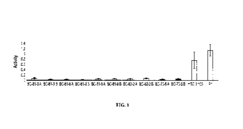
[0020] FIG. 1
shows a graphical representation of the average activity of crosslinker, mTG,
in
various microparticle batches, where the amount of mTG used for gelatin
crosslinking was 3 g ¨ 10 g.
P1: assay positive control; mTG (1:100): positive control; Y-axis: mTG average
activity; X-axis:
microparticle batches and positive controls.
[0021] FIGs. 2A-2D show a representative histopathological evaluation of a
subcutaneous area
implanted with the composition of the disclosure, 30 days following injection,
using Masson's
Trichrome (MT) staining. The scales for each are as follows: 1000gm (FIG. 2A);
200 g (FIG. 2B);
and 50 g (FIGs. 2C - 2D). Black arrows indicate the implanted composition of
the disclosure. Gray
arrows indicate neo-collagenesis. The white arrow shows the interaction
between infiltrating fibroblasts
and the scaffold.
[0022] FIG. 3
demonstrates a linear correlation between particles size to injection force,
using a 1
ml syringe with 27 gauge (G) needle.
[0023] FIG. 4 shows an exemplary Scanning Electron Microscopy (SEM) image of
particles greater
than 0.1 gm (i.e., 100 nm) (e.g., 104 nm; 105 nm; 112 nm; 145 nm; 150 nm; 275
nm).
[0024] FIG. 5 shows an exemplary SEM image of microparticles having a particle
size range of 60
gm ¨100 gm (e.g., 75.69 gm; 88.38 gm; 91.56 gm; 99.68 gm).
[0025] FIG. 6A shows an exemplary light microscopy image of particles up to
2000 gm. The
particles were hydrated before imaging (scale 500 gm). FIG. 6B shows dry
particle sizes of: 558 gm,
862 gm, and 986 gm (scale 200 gm). FIG. 6C shows a wet particle having a
particle size of 1808 gm
(scale 500 gm).
[0026] FIG. 7A shows dry gelatin microparticles (scale 100 gm). FIG. 7B shows
hydrated or wet
gelatin microparticles (scale 100 gm).
6
CA 03224921 2023-12-20
WO 2022/269351
PCT/IB2022/000349
[0027] FIG. 8
shows a graphical representation of size distribution of dry and hydrated
microparticles. Dry microparticles (Left Column): Particle sizes: 70 gm- 170
gm. Hydrated
microparticles (Right Column): 90 gm - 310 gm.
[0028] FIG. 9 shows a representative frequency sweep graph, displaying on the
Y-axis the storage
modulus G' (Pa)A, Loss modulus G" (Pa)o, and the complex viscosity ij* (Pa.$)
0, of 0.75% gelatin
carrier at 6 C to the frequency f (Hz) on the X-axis.
[0029] FIG. 10
shows a representative size distribution of exemplary foam microparticles of
the
sample (8 gr mTG), where the 96% ethanol sample (circle) peaked at 14 volume %
at a size of 80 gm,
the DDW instant sample (diamond) peaked at 10 volume % at a size of 120 gm;
and the DDW at 24
hours sample (square) peaked at 11 volume % at a size of 140 gm. See, TABLE 6.
[0030] FIG. 11 shows the injectability (N) of an exemplary formulation of
crosslinked gelatin foam
microparticles with different saline volumes (1.5 ml; 2 ml; 3 ml; 4 m1).
[0031] FIG. 12 shows representative histologic photographs of implants stained
with H&E
(Hematoxylin and Eosin which stains cell nuclei a purplish blue, and the
extracellular matrix and
cytoplasm pink) and MT, Mason Trichrome (produce red keratin, muscle fibers
and implant, blue
collagen and bone, light red or pink cytoplasm, and dark brown to black cell
nuclei) in pig and rat skin
at 7-, 30-, and 180-days post-implantation (H&E- pig Day 7 and Day 30, Rat Day
7 and Day 30, and
MT-pig Day 180), the arrows showing sites of the implanted composition of the
disclosure.
[0032] FIG. 13 shows representative histologic photographs of implants stained
with H&E
(Hematoxylin and Eosin which stains cell nuclei a purplish blue, and the
extracellular matrix and
cytoplasm pink) and Mason Trichrome (produce red keratin, muscle fibers and
implant, blue collagen
and bone, light red or pink cytoplasm, and dark brown to black cell nuclei) at
7-, 30-, and 180-days
post-implantation (H&E- pig Day7 and Mason Trichrome pig Day 30 and Day 180,
Rat Day 7 and Day
30) showing the implanted formulation (black arrows) of new collagen fibers
stained in blue (white
arrows).
[0033] FIG. 14 shows an SDS-PAGE of 1 mg/ml FPs prepared in water. Collagenase
was added to
the suspension to degrade the FPs. Molecular weight marker (M); microbial
transglutaminase (7 jig
protein in 20 id) (1); gelatin (10 jig protein in 20 id) (2); collagenase (1.7
U in 20 al) (3); foam particles
(FPs) (degraded with collagenase, 10 jig protein in 20 id) (4).
[0034] FIG. 15
shows a calibration curve of arginine. R2 value of 0.999 indicates high
linearity.
Arginine concentration (gg/m1) (X-axis) to emission intensity (Y-axis).
[0035] FIG. 16
shows fluorescence emissions spectra of free arginine, raw materials, and
crosslinked
gelatin particles.
7
CA 03224921 2023-12-20
WO 2022/269351
PCT/IB2022/000349
[0036] FIG. 17 shows RGD quantification in raw materials non-crosslinked
gelatin and crosslinked
gelatin particles (i.e., FPs and confluent particles).
[0037] FIG. 18 shows the amount of RGD sequences or motifs (gg/mg) (Y-axis) on
FPs with
different crosslinked gelatin particle size ranges (X-axis) of less than 63
gm; 63 gm ¨99 gm; and more
than 99 gm.
[0038] FIG. 19 shows the amount of RGD (gg/mg) (Y-axis) in relation to various
weight ratios of
gelatin:mTG, gelatin, and microbial transglutaminase (mTG).
[0039] FIG. 20A and FIG. 20B show light microscopy images of human induced
pluripotent stem
(iPS) cells grown on foam particle microcarriers of the disclosure
differentiated into cardiomyocytes.
FIG. 20A has a scale of 50 gm and FIG. 20B has a scale 200 gm.
[0040] FIG. 20A and FIG. 20B show light microscopy images of foam particles
(FPs) produced
from foam crosslinked gelatin fibers. Scale 100 gm.
DETAILED DESCRIPTION
[0041] Detailed embodiments of the present disclosure are described here;
however, it is to be
understood that the disclosed embodiments are merely illustrative of the
invention that can be embodied
in various forms. In addition, each of the examples given in connection with
the various embodiments
of the invention is intended to be illustrative, and not restrictive.
[0042] Porous and biodegradable polymer scaffolds can be utilized as a
structural supporting matrix
or as cell adhesive substrates. It is an object of the disclosure to provide a
safe, non-toxic, inexpensive
or low cost, implantable tissue support that does not induce an immune
response or lacks
immunogenicity. In one instance, the implantable tissue support of the
disclosure is synthetic and/or
lacks or is essentially free of non-human components. Using a material with
inherent cell-binding
elements can improve the performance of implants by allowing direct cell
attachment and local
remodeling. A tripeptide motif (e.g., RGD (Arginine (Arg) -Glycine (Gly) ¨
Aspartate (Asp))) is found
within extracellular matrix proteins, such as but not limited to, bone
sialoprotein, collagen, fibrinogen,
fibronectin, gelatin, laminin, osteopontin, and vitronectin, facilitates cell
adhesion, cell membrane
binding, and cell attachment. The RGD motif is an inte grin-binding domain
within ECM proteins. For
example, gelatin, derived from collagen, contains the RGD motif that is useful
for cell adhesion.
[0043] Particles
[0044] In various embodiments of the disclosure, microparticles or a
plurality of microparticles; their
methods of preparation; compositions comprising the plurality of
microparticles; apparatuses, such as
syringes or vials, comprising the compositions of the disclosure; scaffolds or
tissue scaffolds comprising
the plurality of microparticles or compositions of the disclosure; uses of the
disclosed plurality of
8
CA 03224921 2023-12-20
WO 2022/269351
PCT/IB2022/000349
microparticles or compositions of the disclosure; and methods of treating a
subject by administering the
plurality of microparticles or compositions of the disclosure are provided
here.
[0045] As used herein, the term "subject" refers to any organism to which a
composition in
accordance with the disclosure can be administered, e.g., for experimental,
diagnostic, prophylactic,
and/or therapeutic purposes. Typical subjects include any animal (e.g.,
mammals such as mice, rats,
rabbits, dogs, cats, non-human primates, and humans, etc.). A subject in need
thereof is typically a
subject for whom it is desirable to treat a disease, disorder, or condition as
described herein. For
example, a subject in need thereof can seek or be in need of treatment,
require treatment, be receiving
treatment, can be receiving treatment in the future, or a human or animal that
is under care by a trained
professional for a particular disease, disorder, or condition. In some
embodiments, the subject is in
need of body contouring, including but not limited to: soft tissue
reconstruction, volume restoration,
breast augmentation, biostimulation (of cells, e.g., of skin), and the like,
or combinations thereof In
some embodiments, biostimulation can be selected from: fibroblast stimulation,
collagen production
stimulation, neo-collagenesis, tissue regrowth, wound closure and the like, or
combinations thereof
[0046] One
embodiment of the disclosure is directed to a microparticle or a plurality of
microparticles having: a cross-linked protein, where the protein of the cross-
linked protein comprises
at least one RGD (Arg-Gly-Asp) motif, which conveys, inter al/a, cell adhesion
properties. In some
embodiments, the cross-linked protein having several RGD motifs, but at least
one RGD motif, can be
sufficiently and/or more advantageously exposed by reducing in particle size
and increasing the surface
area of the microparticles. The microparticle or the plurality of
microparticles described here comprises
a cross-linked protein that has an RGD motif in an amount of 0.1 g/mg ¨ 50
g/mg (e.g., 0.2 g/mg ¨
45 g/mg; 0.3 g/mg ¨ 40 g/mg; 0.4 g/mg ¨ 35 g/mg; 0.5 g/mg ¨ 30 g/mg;
0.6 g/mg ¨ 25 g/mg;
0.7 g/mg ¨ 20 g/mg; 0.8 g/mg ¨ 15 g/mg; 0.9 g/mg ¨ 10 g/mg; 1 g/mg ¨ 5
g/mg); of 0.1
g/mg or greater (e.g., 2 g; 4 g; 6 g; 8 g; 10 g; 12 g; 14 g; 16 g; 18
g; 20 g; 22 g; 24 g;
26 g; 28 g; 30 g; 32 g; 34 g; 36 g; 38 g; 40 g; 42 g; 44 g; 46 g;
48 g; 50 g); or of 50
g/mg or less (e.g., 49 g; 47 g; 45 g; 43 g; 41 g; 39 g; 37 g; 35 g; 33
g; 31 g; 29 g; 27 g;
25 g; 23 g; 21 g; 19 g; 17 g; 15 g; 13 g; 11 g; 9 g; 7 g; 5 g; 3
g; 1 g; 0.9 g; 0.7 g; 0.5
g; 0.3 g; 0.1 g).
[0047] The
microparticle or plurality of microparticles, including the cross-linked
protein, is cross-
linker¨free or essentially cross-linker¨free, where "cross-linker¨free" as
used here means absent cross-
linkers or containing nominal amounts of cross-linkers, which can be present,
but have no effect on the
function or use of the plurality of microparticles or cross-linked protein.
The cross-linked protein can
be stabilized into, for example, a foam, into a confluent hydrogel, or into
fibers, where the cross-linking
occurred by enzymatic crosslinking. In some embodiments, the enzymatic
crosslinking was performed
using an enzyme, but subsequently removed by, for example, washing the enzyme
out of the particles
or inactivating the cross-linker or crosslinking enzyme. One embodiment
comprises the use of a
9
CA 03224921 2023-12-20
WO 2022/269351
PCT/IB2022/000349
transglutaminase enzyme for cross-linking the protein of the cross-linked
protein, one upon completion
of cross-linking, the enzyme is washed out of the cross-linked protein(s). In
a further embodiment, the
transglutaminase enzyme is or comprises a microbial transglutaminase enzyme.
[0048]
Accordingly, the final microparticle or plurality of microparticles comprises
cross-linked
proteins that are cross-linker¨free. In some aspects, the protein of the cross-
linked protein of the final
microparticle or plurality of microparticles comprises proteins previously
cross-linked or pre-cross-
linked proteins, where the cross-linked proteins have been washed to remove
any cross-linkers, such
that the final microparticle or plurality of microparticles comprising cross-
linked proteins is cross-
linker¨free or essentially or substantially cross-linker¨free. The protein of
the cross-linked protein can
be selected from, but not limited to, gelatin, collagen, casein, elastin,
tropoelastin, albumin, engineered
protein thereof, and the like, or any combinations thereof Other aspects of
the embodiment can be
directed to proteins of the cross-linked proteins comprising: non-recombinant
gelatin, recombinant
gelatin, non-recombinant collagen, recombinant collagen, engineered protein
thereof, engineered
polymer comprising at least one RGD motif or linked thereto, and the like, or
any combinations thereof.
Furthermore, the microparticles or plurality of microparticles of the
disclosure comprise at least one or
more cross-linked proteins, where the at least one or more cross-linked
proteins comprise at least one
RGD (Arg-Gly-Asp) motif; where the microparticle or plurality of
microparticles is cross-linker¨free
(i.e., absent or essentially absent of cross-linker(s)); and the microparticle
or plurality of microparticles
is water-insoluble or essentially water-insoluble. Some embodiments of the
disclosure are directed to
a plurality of microparticles that are pre-crosslinked, water insoluble, and
cross-linker¨free, and are not
water soluble.
[0049] Another
embodiment is directed to a plurality of microparticles of the disclosure,
where the
microparticles comprise particles of foam or particles having a foam-like
property, where the plurality
of microparticles or foam particles comprise cross-linked proteins that are
cross-linker¨free. In some
aspects of the embodiments of the disclosure, the cross-linked proteins are
stabilized into a foam, into
a confluent hydrogel, or into fibers (such as in electro-spinning), where the
cross-linking occurred by
enzymatic crosslinking. As used here, "foam" means a dispersion of gas bubbles
in a liquid, solid, or
semi-solid (e.g., gel). In other instances, disclosed here, a foam can
comprise or be configured as
particles. These foam particles either retain properties of a foam or are
derived from foam thereby
having "foam-like" properties. Moreover, the plurality of microparticles or
foam particles can be
composed of lyophilized particles, including lyophilized foam particles
comprising cross-linked
proteins that are cross-linker free. "Foam particles" (FPs) as used herein
means that they originate from
a stable protein foam, and are not necessarily foam in their own structure,
after the pulverization. This
can depend on the size of the gas bubbles in the initial cross-linker¨free
foam of (c) and the size of the
resulting lyophilized and size-reduced particles of (g). If the gas bubbles
are smaller than the particle
size, they can contain closed cells of the foam; however, if the particles are
smaller than the gas bubbles,
CA 03224921 2023-12-20
WO 2022/269351
PCT/IB2022/000349
then bubbles or full bubbles cannot remain enclosed in the particles. In
either event, the performance
and the intention of the embodiments described here is not impeded and is not
to be limited to a foam
structure.
[0050] One embodiment is directed to foam or foam particles that is reduced in
size and comprises
or is configured as particles, including microparticles, by cutting (e.g.,
chopping, dicing); using
compression, lump breakers, pulverizers, mills (e.g., impact mills, flour
mills, full-screen hammer mills,
mega hammer mills, air classifying mills, jet mills, ball mills, pebble mills,
rod mills); grinders (fine
grinders, blade grinders), and the like, or combinations thereof. By reducing
in size, the cross-linked
foam to form particles, such as microparticles, allows for exposure of several
RGD motifs to a large
surface area, which facilitates cell adhesion and bio-stimulation. Those
embodiments using at least two
different particle sizes also benefit from enhanced surface area. Particle
sizes can be analyzed or
measured by any technique commonly known and/or used by persons of ordinary
skill in the art. Non-
limiting examples of such methods, techniques, or tools for measuring particle
size include: Particle
Size Analyzers (PSA); high definition image processing; image particle
analysis (IPA) (e.g., optical
microscopes, scanning electron microscopes (SEMs), transmission electron
microscopes (TEMs));
dynamic image analysis (DIA); static laser light scattering (SLS, also known
as laser diffraction);
dynamic light scattering (DLS); acoustic spectroscopy; sieve analysis (e.g.,
dry sieving, wet sieving);
and the like, or any combinations thereof
[0051] In some
embodiments, the plurality of microparticles, including but not limited to
those
derived from foam cross-linked proteins, comprises a particle size of 0.1 gm ¨
2000 gm (e.g., 0.2 gm ¨
1499 gm; 0.4 gm ¨ 1450 gm; 0.5 gm ¨ 1425 gm; 0.6 gm ¨ 1400 gm; 0.7 gm ¨ 1350
gm; 0.8 gm ¨ 1300
gm; 0.9 gm ¨1250 gm; 1 jim ¨1200 gm; 2 gm ¨1150 gm; 3 gm ¨1100 gm; 4 gm ¨1050
gm; 5 jim ¨
1000 gm; 6 gm ¨ 950 gm; 7 gm ¨ 900 gm; 8 gm ¨ 850 gm; 9 gm ¨ 800 gm; 10 gm ¨
750 gm; 11 ¨700 gm; 12 gm ¨650 gm; 13 gm ¨600 gm; 14 gm ¨ 550 gm; 15 jim
¨500 gm; 16 gm ¨450 gm; 17
jim ¨400 gm; 18 gm ¨350 gm; 19 gm ¨300 gm; 20 gm ¨250 gm; 25 jim ¨200 gm; 30
gm ¨ 150 gm;
40 gm ¨ 100 gm; 60 gm ¨ 90 gm); 0.1 gm or greater (e.g., 0.5 gm; 1 gm; 5 gm;
15 gm; 25 gm; 35 gm;
45 gm; 55 gm; 65 gm; 75 gm; 85 gm; 95 gm; 100 gm; 105 gm; 115 gm; 125 gm; 135
gm; 145 gm; 150
gm; 200 gm; 250 gm; 500 gm; 1000 gm; 2000 gm); or 2000 gm or less (e.g., 1250
gm; 1000 gm; 750
gm; 500 gm; 250 gm; 200 gm; 150 gm; 140 gm; 130 gm; 120 gm; 110 gm; 90 gm; 80
gm; 70 gm; 60
gm; 50 gm; 40 gm; 30 gm; 20 gm; 10 gm; 5 gm; 4 gm; 3 gm; 2 gm). Other
embodiments directed to
such plurality of microparticles comprises a mean particle size of: 0.1 gm ¨
2000 gm (e.g., 0.2 gm ¨
1499 gm; 0.4 gm ¨ 1450 gm; 0.5 gm ¨ 1425 gm; 0.6 gm ¨ 1400 gm; 0.7 gm ¨ 1350
gm; 0.8 gm ¨ 1300
gm; 0.9 gm ¨1250 gm; 1 jim ¨1200 gm; 2 gm ¨1150 gm; 3 gm ¨1100 gm; 4 gm ¨1050
gm; 5 jim ¨
1000 gm; 6 gm ¨ 950 gm; 7 gm ¨ 900 gm; 8 gm ¨ 850 gm; 9 gm ¨ 800 gm; 10 gm ¨
750 gm; 11 ¨700 gm; 12 gm ¨650 gm; 13 gm ¨600 gm; 14 gm ¨ 550 gm; 15 jim
¨500 gm; 16 gm ¨450 gm; 17
jim ¨400 gm; 18 gm ¨350 gm; 19 gm ¨300 gm; 20 gm ¨250 gm; 25 jim ¨200 gm; 30
gm ¨ 150 gm;
11
CA 03224921 2023-12-20
WO 2022/269351
PCT/IB2022/000349
40 gm ¨100 gm; 60 gm ¨ 90 gm); 0.1 gm or greater (e.g., 5 gm; 15 gm; 25 gm; 35
gm; 45 gm; 55 gm;
65 gm; 75 gm; 85 gm; 95 gm; 100 gm; 105 gm; 115 gm; 125 gm; 135 gm; 145 gm;
150 gm; 200 gm;
250 gm; 500 gm; 1000 gm; 2000 gm); or 2000 gm or less (e.g., 1250 gm; 1000 gm;
750 gm; 500 gm;
250 gm; 200 gm; 150 gm; 140 gm; 130 gm; 120 gm; 110 gm; 90 gm; 80 gm; 70 gm;
60 gm; 50 gm;
40 gm; 30 gm; 20 gm; 10 gm; 5 gm; 4 gm; 3 gm; 2 gm). In embodiments of the
disclosure, a "mean
particle size" as used here means the average particle size of the plurality
of microparticles. In some
embodiments, a "particle size" refers to a dry particle size. Some
embodiments, a "particle size" refers
to a wet particle size. In some embodiments, a wet or hydrated particle has a
greater particle size than
a dry particle of the same dry size, by a factor of, for example, 1.4 to 2.8
with an average factor of 1.67
(1.65 ¨ 1.67). See, e.g., TABLE 4.
[0052] Further
embodiments of the disclosure are directed to the plurality of microparticles
described
here where the plurality of microparticles can comprise at least two different
particle sizes. The particle
sizes can be selected from any of the particle sizes disclosed here, including
but not limited to: 0.1 gm
¨2000 gm (e.g., 0.2 gm ¨ 1900 gm; 0.3 gm ¨ 1800 gm; 0.4 gm ¨ 1700 gm; 0.5 jim
¨ 1600 gm; 0.6 gm
¨ 1500 gm; 0.7 gm ¨ 1400 gm; 0.8 gm ¨ 1300 gm; 0.9 gm ¨ 1250 gm; 1 jim ¨
1200 gm; 2 gm ¨ 1150
gm; 3 gm ¨ 1100 gm; 4 gm ¨ 1050 gm; 5 gm ¨ 1000 gm; 6 gm ¨ 950 gm; 7 gm ¨ 900
gm; 8 gm ¨ 850
gm; 9 gm ¨ 800 gm; 10 gm ¨ 750 gm; 11 gm ¨ 700 gm; 12 gm ¨ 650 gm; 13 gm ¨ 600
gm; 14 gm ¨
550 gm; 15 jim ¨ 500 gm; 16 gm ¨450 gm; 17 gm ¨ 400 gm; 18 gm ¨350 gm; 19 gm
¨300 gm; 20
gm ¨ 250 gm; 25 gm ¨ 200 gm; 30 gm ¨ 150 gm; 40 gm ¨ 100 gm; 60 gm ¨ 90 gm);
0.1 gm or greater
(e.g., 0.5 gm; 1 gm; 5 gm; 15 gm; 25 gm; 35 gm; 45 gm; 55 gm; 65 gm; 75 gm; 85
gm; 95 gm; 100
gm; 105 gm; 115 gm; 125 gm; 135 gm; 145 gm; 150 gm; 200 gm; 250 gm; 500 gm;
1000 gm; 2000
gm); or 2000 gm or less (e.g., 1250 gm; 1000 gm; 750 gm; 500 gm; 250 gm; 200
gm; 150 gm; 140
gm; 130 gm; 120 gm; 110 gm; 90 gm; 80 gm; 70 gm; 60 gm; 50 gm; 40 gm; 30 gm;
20 gm; 10 gm; 5
gm; 4 gm; 3 gm; 2 gm). Other embodiments directed to the plurality of
microparticles comprising at
least two different particle sizes comprise a mean particle size selected
from, but not limited to: 0.1 gm
¨2000 gm (e.g., 0.2 gm ¨ 1499 gm; 0.4 gm ¨ 1450 gm; 0.5 jim ¨ 1425 gm; 0.6 gm
¨ 1400 gm; 0.7 gm
¨1350 gm; 0.8 gm ¨1300 gm; 0.9 gm ¨1250 gm; 1 jim ¨1200 gm; 2 gm ¨1150 gm; 3
gm ¨1100
gm; 4 gm ¨ 1050 gm; 5 gm ¨ 1000 gm; 6 gm ¨ 950 gm; 7 gm ¨ 900 gm; 8 gm ¨ 850
gm; 9 gm ¨ 800
gm; 10 gm ¨ 750 gm; 11 gm ¨ 700 gm; 12 gm ¨ 650 gm; 13 gm ¨ 600 gm; 14 gm ¨550
gm; 15 ¨500 gm; 16 gm ¨450 gm; 17 gm ¨400 gm; 18 gm ¨ 350 gm; 19 gm ¨300
gm; 20 gm ¨250 gm; 25
jim ¨200 gm; 30 gm ¨ 150 gm; 40 gm ¨ 100 gm; 60 gm ¨90 gm); 0.1 gm or greater
(e.g., 0.5 gm; 1
gm; 5 gm; 15 gm; 25 gm; 35 gm; 45 gm; 55 gm; 65 gm; 75 gm; 85 gm; 95 gm; 100
gm; 105 gm; 115
gm; 125 gm; 135 gm; 145 gm; 150 gm; 200 gm; 250 gm; 500 gm; 1000 gm; 2000 gm);
or 2000 gm or
less (e.g., 1250 gm; 1000 gm; 750 gm; 500 gm; 250 gm; 200 gm; 150 gm; 140 gm;
130 gm; 120 gm;
110 gm; 90 gm; 80 gm; 70 gm; 60 gm; 50 gm; 40 gm; 30 gm; 20 gm; 10 gm; 5 gm; 4
gm; 3 gm; 2
inn).
12
CA 03224921 2023-12-20
WO 2022/269351
PCT/IB2022/000349
[0053] Methods of Preparing Particles of the Disclosure
[0054] Although gelatin microspheres have been previously fabricated using
various methods and
techniques, including water-in-oil emulsion, electrospray, spray-drying, and
microfluidic
emulsification to name a few. Using these methods, the gelatin is cross-linked
by several types of
chemical crosslinking agents such as, 1-ethyl-3-(3-dimethyl aminopropy1)-
carbodiimide (EDC) and N-
hydroxysuccinimide (NHS), glycidoxyproyltrimethoxysilane (GPTMS),
glutaraldehyde, and genipin.
The water-in-oil method is a commonly used laboratory technique but possesses
many drawbacks
including difficulties in scaling up to industrial scale and the use of oil
and chemical crosslinkers results
in toxicity issues requiring extensive removal of the residual oils and
chemical crosslinkers.
[0055] One embodiment of the disclosure is directed to a method of preparing
the plurality of
microparticles of the disclosure, comprising: (a) mixing a cross-linkable
protein solution and a cross-
linker solution, where the cross-linkable protein solution comprises
dissolving a cross-linkable protein
comprising at least one RGD (Arg-Gly-Asp) motif or linked thereto (e.g.,
gelatin (e.g., recombinant
gelatin, non-recombinant gelatin, in situ crosslinking), collagen (e.g.,
recombinant collagen, non-
recombinant collagen), casein, tropoelastin, elastin, albumin, engineered
protein thereof, and the like,
or any combinations thereof; or non-recombinant gelatin, recombinant gelatin,
non-recombinant
collagen, recombinant collagen, any engineered protein thereto, engineered
polymer comprising RGDs
or linked thereto, and the like, or any combinations thereof) in a liquid
(e.g., water, saline, PBS); and
where the cross-linker solution comprises dissolving a cross-linker or an
enzyme cross-linker (e.g.,
transglutaminase (e.g., natural transglutaminase, modified transglutaminase,
recombinant
transglutaminase, microbial transglutaminase (mTG), tissue transglutaminase
(tTG), keratinocyte
transglutaminase, epidermal transglutaminase, prostate transglutaminase,
neuronal transglutaminase,
human transglutaminase, Factor XIII, and the like, or any combinations
thereof), oxidative enzyme
(e.g., natural oxidative enzyme, modified oxidative enzyme, lysyl oxidase,
tyrosinase, laccase,
peroxidase, and the like, or any combinations thereof) in a liquid (e.g.,
water, saline, PBS), where the
cross-linker is in an amount sufficient to crosslink the cross-linkable
protein to form a cross-linked
foam/block, non-foam cross-linked hydrogel, or fibers (such as in
electrospinning). Another
embodiment is directed to the cross-linker in an amount sufficient to convert
the cross-linkable protein
from soluble to insoluble at a temperature ranging from 10 C ¨ 40 C. The
method of preparing the
plurality of microparticles of the disclosure further comprises: (b) forming a
cross-linked foam/block,
a non-foam cross-linked hydrogel, or fibers (such as in electro-spinning)
comprising the mixed cross-
linkable protein and the cross-linker of (a); (c) pulverizing the non-foam
cross-linked hydrogel, fibers,
or cross-linked foam of (b); (d) removing the cross-linker from the cross-
linked formulation or product
of (c) to form a cross-linker¨free foam or hydrogel or fibers (e.g.,
essentially or substantially cross-
linker¨free); and (e) reducing in size: the formed cross-linked product of
(d), the cross-linker¨free
product of (d), or combinations of the formed cross-linked foam of (d) and the
cross-linker¨free foam
13
CA 03224921 2023-12-20
WO 2022/269351
PCT/IB2022/000349
or hydrogel of (d), to form a plurality of particles and/or microparticles
comprising size-reduced cross-
linked foam or hydrogel of (b) and/or size-reduced cross-linker¨free foam or
hydrogel of (d). In some
embodiments, the plurality of particles and/or microparticles comprising size-
reduced cross-linked
foam or hydrogel of (b) and/or size-reduced cross-linker¨free foam or hydrogel
of (d) can be sterilized
by any appropriate method that does not substantially alter functionality,
physico-chemical properties,
stability, toxicity, or biological effects, including but not limited to:
filtration, autoclaving (e.g., 110 C
¨ 134 C; 15 mins ¨40 mins; 5 psi - 20 psi), irradiation (e.g., Ultraviolet
(UV); gamma; electron beam
(e-beam); X-rays). Some embodiments of sterilization include UV treatment
under an exposure of 5
mins ¨ 720 mins (e.g., 100 mins, 150 mins, 200 mins, 250 mins) and UV
wavelength of 10 nm - 400
nm (e.g., 200 nm ¨ 270 nm). Additional embodiments include gamma irradiation
of 10 kGy ¨ 50 kGy
(e.g., 15 kGy, 20 kGy, 25 kGy, 30 kGy, 35 kGy, 40 kGy, 45 kGy). Vetten et al.
disclose various
sterilization techniques and parameters useful that can be applied herein and
is incorporated by
reference in its entirety (see, Nanomedicine. 10(7):1391-1399, 2014).
[0056] Some
embodiments are directed to methods of preparing a plurality of microparticles
as
described here where the crosslinking occurs in vitro as a production-
controlled step, in contrast to other
formulations that are mixed at a point of care and injected, thereby allowing
for crosslinking to occur
in situ. In order to remove the transglutaminase crosslinker enzyme as
described in the disclosed
method, after the crosslinking reaction occurs, multiple repeated and extended
washings are performed.
[0057] In another embodiment, the disclosed method of preparing a plurality of
microparticles is
directed to the cross-linkable protein solution, comprising: (i) adding a
cross-linkable protein to a liquid
(e.g., water, saline, PBS) at a temperature sufficient to dissolve the cross-
linkable protein, such as a
temperature greater than or equal 25 C (e.g., 30 C, 37 C, 40 C, 45 C, 50 C),
where the cross-linkable
protein is selected from, but not limited to, a protein comprising at least
one RGD (Arg-Gly-Asp) motif
(e.g., gelatin (e.g., non-recombinant gelatin, recombinant gelatin), collagen
(e.g., non-recombinant
collagen, recombinant collagen), casein, albumin, and any combinations
thereof), at a temperature
sufficient to dissolve the cross-linkable protein, essentially dissolve, or
completely dissolve, such as but
not limited to 40 C ¨ 60 C, e.g., 50 C, while continuously stirring; and (ii)
dissolving, essentially
dissolving, or completely dissolving the cross-linkable protein in the liquid
to form the cross-linkable
protein solution.
[0058] Some
embodiments are directed to fabricating foamed cross-linked gelatin
microparticles
(MPs) by a crosslinking reaction with a transglutaminase enzyme (e.g.,
microbial transglutaminase
(mTG); recombinant transglutaminase; bacterial transglutaminase). Briefly, a
transglutaminase (e.g.,
mTG) solution can be added to liquid state gelatin in a whipping machine or
for mixing or stirring by
any other means with or without gas or air (e.g., argon, carbon dioxide,
helium, hydrogen, krypton,
methane, neon, nitrogen, oxygen, ozone, water vapor, xenon, or any
combinations thereof). In some
embodiments, the method comprises forming a cross-linked foam by whipping the
cross-linkable
14
CA 03224921 2023-12-20
WO 2022/269351
PCT/IB2022/000349
protein solution of (a) while adding the cross-linker solution of (a) at 37 C
to form the cross-linked
foam of (b). Other embodiments are directed to the method of the disclosure
comprising stirring or
mixing the cross-linkable protein solution of (a) while adding the cross-
linker solution of (a) at 37 C
without gas or air to form the non-foam cross-linked block of (b).
[0059] While mixing and foaming, the gelatin is crosslinked until a three-
dimensional (3D) foam
structure stabilizes. Afterward, the formulated foam is incubated at 45 C and
then chopped into large
or gross slices or pieces (e.g., 0.05 cm ¨2 cm; 0.5 mm- 20 mm). The chopped
slices are washed several
times at 50 C for the removal of excess crosslinker or transglutaminase (e.g.,
mTG). After washing,
the foamed gelatin is freeze-dried using, for example, a lyophilizer. For the
creation of MPs, the dry
crosslinked foamed gelatin is milled and sieved into microparticles in several
size ranges (e.g., 0.1 gm
¨ 10 mm). The MPs can be sterilized by any means, including those described
here, that does not
negatively impact the structure, function, or performance of the
microparticles, including, but not
limited to, radiation.
[0060] In
another embodiment of the disclosure, cross-linked gelatin microparticles
(MPs) can be
fabricated by a crosslinking reaction with a transglutaminase enzyme (e.g.,
microbial transglutaminase
(mTG); recombinant transglutaminase; bacterial transglutaminase). Briefly,
transglutaminase (e.g.,
mTG) solution is added to liquid state gelatin. The gelatin is stirred (with
no foaming) until crosslinking
into a stable three-dimensional (3D) structure, forming a crosslinked gelatin
structure. Afterward, the
formulated structure is incubated at 45 C and then chopped into large or gross
slices or pieces (e.g.,
0.05 cm ¨2 cm; 0.5 mm- 20 mm). The chopped slices are washed several times at
50 C for the removal
of excess crosslinker or transglutaminase (e.g., mTG). After washing, the
crosslinked gelatin is then
freeze-dried using, for example, a lyophilizer. For the creation of MPs, the
dry crosslinked gelatin is
milled and sieved into microparticles in several size ranges (e.g., 0.1 gm ¨
10 mm). The MPs can be
sterilized by any means, including those described here, that does not
negatively impact the structure,
function, or performance of the microparticles, including, but not limited to,
radiation.
[0061] A further embodiment provides such method of preparing a plurality of
microparticles
directed to the cross-linker solution, comprising: (i) adding a cross-linker
to a liquid (e.g., water, saline,
PBS) at a temperature sufficient to dissolve the cross-linker, essentially
dissolve, or completely
dissolve, such as but not limited to room temperature, 15 C ¨ 27 C, e.g., 25
C, while continuously
stirring; and (ii) dissolving, essentially dissolving, or completely
dissolving the cross-linker in the liquid
to form the cross-linker solution. Other embodiments can be directed to such
methods of preparing a
plurality of microparticles, where the cross-linkable protein is cross-linked
in the presence of, or when
mixed with, the cross-linker of the disclosure. In a further embodiment, the
cross-linker is an enzyme
(e.g., transglutaminase, such as microbial transglutaminase) that when mixed
with the cross-linkable
protein, forms enzymatically cross-linked proteins, enzymatically cross-linked
foams, or enzymatically
cross-linked particles, or enzymatically cross-linked fibers. The cross-linked
foam of (b) in methods of
CA 03224921 2023-12-20
WO 2022/269351
PCT/IB2022/000349
preparing a plurality of microparticles can be formed by: (b 1) whipping the
cross-linkable protein
solution of (a) or (b2) mixing or stirring with or without gas or air (e.g.,
argon, carbon dioxide, helium,
hydrogen, krypton, methane, neon, nitrogen, oxygen, ozone, water vapor, xenon,
or combinations
thereof), while adding the cross-linker solution of (a) at a temperature
sufficient for the whipping,
stirring to form the cross-linked foam of (b) or cross-linked block of (b),
respectively, where, for
example, the whipping or mixing or stirring occurs at a temperature of 30 C ¨
40 C (e.g., 37 C).
[0062] The
removal of the cross-linker from the cross-linked protein, cross-linked foam,
and/or
microparticles or compositions comprising the same, in one embodiment, is
beneficial from a safety
and regulatory position, as well as cost. Accordingly, some embodiments can be
directed to such
methods of the disclosure where removing of (c), comprises: washing the cross-
linked foam or block
of (b), where the cross-linked foam or block of (b) is reduced in size as
described here, where the
washing occurs by agitating the pieces of cross-linked foam in a liquid (e.g.,
water, saline, PBS) at a
temperature (e.g., 40 C ¨ 60 C; 45 C ¨ 55 C, such as 50 C) and time (e.g., 5
mins ¨ 1 hour; 10 mins ¨
45 mins; 15 mins ¨30 mins) sufficient to remove or essentially remove the
cross-linker from the cross-
linked foam; and reducing in size by, for example, sieving the washed foam
pieces to a desired size
using an appropriate mesh sieve, such as but not limited to, a sieve with 35
mesh # - 5000 mesh # (500
gm to 2.5 gm), for example, a 0.5 mm mesh or 35 mesh # equivalent, sieve,
thereby forming cross-
linker¨free foam pieces of the description comprising pieces of desirable
sizes.
[0063] In one embodiment, such methods can provide a reducing in size of (d),
comprising: cutting
(e.g., dicing, chopping, meshing) the formed cross-linked foam or block of
(b), the cross-linker¨free
foam or block of (c), or combinations of cross-linked foam or block of (b) and
the cross-linker¨free
foam or block of (c). Other non-limiting examples of techniques, methods, or
tools for reducing in size
the cross-linked foam or block of (b) and/or the cross-linker¨free foam or
block of (c) include: cutting
(e.g., dicing, chopping, meshing, sieving), using compression, lump breakers,
pulverizers, mills (e.g.,
impact mills, flour mills, full-screen hammer mills, mega hammer mills, air
classifying mills, jet mills,
ball mills, pebble mills, rod mills); grinders (fine grinders, blade
grinders), and the like, or combinations
thereof. The reduction in size of such methods can occur to form a plurality
of particles of 0.1 gm ¨ 10
mm (e.g., 0.2 gm ¨9 mm; 0.3 gm ¨8 mm; 0.4 gm ¨7 mm; 0.5 jim ¨7 mm; 1 jim ¨6
mm; 5 jim ¨5
mm; 10 gm ¨ 4 mm; 20 gm ¨ 1 mm; 40 gm ¨ 500 gm; 60 gm ¨ 200 gm; 90 gm ¨ 150
gm; 95 gm ¨ 100
gm); greater than 1 gm (e.g., 2 gm, 4 gm, 6 gm, 8gm, 12 gm, 15 gm, 25 gm, 35
gm, 45 gm, 55 gm, 65
gm, 75 gm, 85 gm, 95 gm, 105 gm, 115 gm, 125 gm, 135 gm, 145 gm, 150 gm, 200
gm, 300 gm, 400
gm, 500 gm, 1 mm, 3 mm, 5 mm, 7 mm, 9 mm); 10 mm or less (e.g., 8 mm, 6 mm, 4
mm, 2 mm, 900
gm, 800 gm, 700 gm, 600 gm, 550 gm, 450 gm, 350 gm, 250 gm, 175 gm, 165 gm,
155 gm, 140 gm,
130 gm, 120 gm, 100 gm, 90 gm, 80 gm, 70 gm, 60 gm, 50 gm, 40 gm, 30 gm, 20
gm, 10 gm, 5 gm,
3 gm, 1 gm). Additional embodiments provide such methods where the reducing of
(d) results in the
formed cross-linked foam of (b) or cross-linked foam pieces with a size of 0.5
mm ¨ 10 mm (e.g., 1
16
CA 03224921 2023-12-20
WO 2022/269351
PCT/IB2022/000349
mm ¨ 8 mm; 2 mm ¨ 7 mm; 3 mm ¨ 6 mm; 4 mm ¨ 5 mm); 0.5 mm or greater (e.g.,
1.5 mm, 2.5 mm,
3.5 mm, 4.5 mm, 5.5 mm, 6.5 mm, 7.5 mm, 8.5 mm, 9.5 mm); 10 mm or less (e.g.,
9 mm, 8 mm, 7 mm,
6 mm, 5 mm, 4 mm, 3 mm, 2 mm, 1 mm).
[0064] In yet
another embodiment, such methods of preparing a plurality of microparticles,
comprises: (e) freezing the cross-linker¨free foam or block of (c) or
plurality of particles of (d);
lyophilizing the frozen cross-linker¨free foam or block of (e); and reducing
in size the lyophilized cross-
linker¨free foam or block of (f) to form a plurality of cross-linked foam or
block particles. Other
embodiments of the methods of preparing a plurality of microparticles
comprises: drying the cross-
linker¨free foam or block of (c) or plurality of particles of (d); and
reducing in size the dried cross-
linker¨free foam or block of (c) to form a plurality of dried cross-linked
foam or block particles. The
plurality of cross-linked foam particles comprises a particle size of, for
example, 0.1 gm ¨ 2000 gm
(e.g., 0.2 gm ¨ 1499 gm; 0.4 gm ¨ 1450 gm; 0.5 jim ¨ 1425 gm; 0.6 gm ¨ 1400
gm; 0.7 gm ¨ 1350 gm;
0.8 gm ¨1300 gm; 0.9 gm ¨1250 gm; 1 jim ¨1200 gm; 2 gm ¨1150 gm; 3 gm ¨1100
gm; 4 gm ¨
1050 gm; 5 gm ¨ 1000 gm; 6 gm ¨ 950 gm; 7 gm ¨ 900 gm; 8 gm ¨ 850 gm; 9 gm ¨
800 gm; 10 gm ¨
750 gm; 11 jtm ¨700 gm; 12 gm ¨650 gm; 13 gm ¨ 600 gm; 14 gm ¨550 gm; 15 jim
¨500 gm; 16
jim ¨450 gm; 17 gm ¨400 gm; 18 gm ¨350 gm; 19 gm ¨300 gm; 20 gm ¨250 gm; 25
jim ¨200 gm;
30 gm ¨ 150 gm; 40 gm ¨ 100 gm; 60 gm ¨ 90 gm). Such methods comprise cross-
linkable proteins
selected from the group consisting of: gelatin, collagen, casein, albumin,
tropoelastin, elastin and any
combinations thereof; or non-recombinant gelatin, recombinant gelatin, non-
recombinant collagen,
recombinant collagen, any engineered protein thereof, engineered polymer
comprising at least one RGD
motif or linked thereto, and the like, or any combinations thereof A further
embodiment of such
methods also comprises an enzyme cross-linker, where the enzyme cross-linker
can be selected from
transglutaminase or oxidative enzyme. Other embodiments of the disclosure
provide for an enzyme
cross-linker selected from the group consisting of: natural transglutaminase,
modified transglutaminase,
recombinant transglutaminase, microbial transglutaminase (mTG), tissue
transglutaminase (tTG),
keratinocyte transglutaminase, epidermal transglutaminase, prostate
transglutaminase, neuronal
transglutaminase, human transglutaminase, Factor XIII, and the like, or any
combinations thereof.
Some other embodiments can provide for an enzyme cross-linker selected from
the group consisting of:
natural oxidative enzyme, modified oxidative enzyme, lysyl oxidase,
tyrosinase, laccase, peroxidase,
and the like, or any combinations thereof
[0065] Further embodiments provide for such methods, where the freezing of (e)
occurs at a
temperature sufficient for preparation of lyophilization, where the
temperature comprises -18 C ¨25 C
(e.g., -15 C ¨ 23 C; -10 C ¨ 20 C; -5 C ¨ 15 C; 0 C ¨ 10 C; -18 C or greater
(e.g., -16 C; -14 C; -
12 C; -8 C; -6 C; -4 C; -2 C; 2 C; 4 C; 6 C; 8 C; 10 C; 12 C; 14 C; 16'; 18';
20'; 22 C; 24 C) or
25 C or less (e.g., 23'; 21 C; 19'; 17'; 15 C; 13 C; 11 C; 9 C; 7 C; 5 C; 3 C;
1 C; -1 C; -3 C; -5 C;
-7 C; -9 C; -11 C; -13 C; -15 C; -17 C) for a time sufficient for preparation
of lyophilization, where
17
CA 03224921 2023-12-20
WO 2022/269351
PCT/IB2022/000349
the time comprises: 2 hours - 25 hours (e.g., 7 hours -24 hours; 9 hours - 22
hours; 11 hours -20
hours; 13 hours - 18 hours; 15 hours - 16 hours); 5 hours or greater (e.g., 6
hours; 8 hours; 10 hours;
12 hours; 14 hours; 16 hours; 18 hours; 20 hours; 22 hours; 24 hours); or 25
hours or less (e.g., 23
hours; 21 hours; 19 hours; 17 hours; 15 hours; 13 hours; 11 hours; 9 hours; 7
hours; 5 hours).
[0066] Yet other embodiments can be directed to such methods, where the
lyophilizing of (f) occurs
at a temperature selected from: -50 C 10 C (e.g., -60 C - -40 C; -55 C - -35
C; -50 - -30 C; -45
- -25 ; -40 C - -20 C; -35 - -30 C; -60 C or greater (e.g., -58 C; -56 C; -54
C; -52 C; -50 C; -48 C;
-46 C; -44 C; -42 C; -40 C); -40 C or less (e.g., -41 C; -43 C; -45 C; -47 C; -
49 C; -51 C; -53 C; -
55 C; -57 C; -59 C), at an atmosphere of: 0.01 mbar - 0.1 mbar (e.g., 0.02
mbar - 0.08 mbar; 0.04
mbar - 0.06 mbar); 0.01 mbar or greater (e.g., 0.03 mbar; 0.05 mbar; 0.07
mbar; 0.09 mbar); or 0.1
mbar or less; 0.08 mbar; 0.06 mbar; 0.04 mbar; 0.02 mbar; 0.01 mbar); for a
time of 24 hours -96 hours
(e.g., 48 hours - 95 hours; 50 hours - 94 hours; 52 hours - 92 hours; 54 hours
- 90 hours; 56 hours -
88 hours; 58 hours -86 hours; 60 hours - 84 hours; 62 hours -82 hours; 64
hours - 80 hours; 66 hours
-78 hours; 68 hours -76 hours; 70 hours -74 hours); 48 hours or greater (e.g.,
49 hours; 51 hours; 53
hours; 55 hours; 57 hours; 59 hours; 61 hours; 63 hours; 65 hours; 67 hours;
69 hours; 71 hours; 73
hours; 75 hours; 77 hours; 79 hours; 81 hours; 83 hours; 85 hours; 87 hours;
89 hours); or 96 hours or
less (e.g., 94 hours; 92 hours; 90 hours; 88 hours; 86 hours; 84 hours; 82
hours; 80 hours; 78 hours; 76
hours; 74 hours; 72 hours; 70 hours; 68 hours; 66 hours; 64 hours; 62 hours;
60 hours; 58 hours; 56
hours; 54 hours; 52 hours; 50 hours; 48 hours; 36 hours); where the
temperature, the pressure, and the
time are sufficient to result in a lyophilized frozen cross-linker-free foam
of (c) or a lyophilized plurality
of particles of (d), where a "lyophilized" product, such as but not limited
to, a lyophilized cross-linker-
free foam of (c) or a lyophilized plurality of particles of (d), as used here,
is meant a product with a
moisture content of 4% or less (e.g., 3.8%; 3.6%; 3.4%; 3.2%; 3%; 2.8%; 2.6%;
2.4%; 2.2%; 2%; 1.8%;
1.6%; 1.4%;1.2%; 1%; 0.8%; 0.6%; 0.4%; 0.2%; 0.08%; 0.06%; 0.04%; 0.02%; 0%);
0% or greater
(e.g., 0.01%; 0.03%; 0.05%; 0.07%; 0.09%; 1.1%; 1.3%; 1.5%; 1.7%; 1.9%; 2.1%;
2.3%; 2.5%; 2.7%;
2.9%; 3.1%; 3.3%; 3.5%; 3.7%; 3.9%); or 0% - 4% (0.5% - 3.5%; 0.7% - 3.3%;
0.9% - 3.1%; 1.1% -
2.9%; 1.3% - 2.7%; 1.5% - 2.5%). The temperature, pressure, and time necessary
to sufficiently
lyophilize the cross-linker-free foam or plurality of particles are understood
by those of ordinary skill
in the art, and would not take undue experimentation to optimize these
parameters.
[0067] In yet another embodiment, such methods of preparing a plurality of
microparticles,
comprises: (e) freezing the cross-linker-free foam or block of (c) or the
plurality of microparticles of
(d); and/or (f) drying the cross-linker-free foam or hydrogel block of (c) or
(e) or the plurality of
microparticles of (d). Some embodiments are directed to drying, including but
not limited to,
lyophilizing or freeze-drying, oven drying, and room temperature or ambient
temperature drying. The
method of preparing a plurality of microparticles, further comprising: (g)
reducing in size the dried
cross-linker-free foam or hydrogel block of (e) and/or (f) to form a plurality
of cross-linked foam
18
CA 03224921 2023-12-20
WO 2022/269351
PCT/IB2022/000349
particles or non-foam cross-linked hydrogel particles. The plurality of cross-
linked microparticles
comprises a particle size of, for example, 0.1 gm ¨2000 gm (e.g., 0.2 gm ¨
1499 gm; 0.4 gm ¨ 1450
gm; 0.5 jim ¨ 1425 gm; 0.6 gm ¨ 1400 gm; 0.7 gm ¨ 1350 gm; 0.8 gm ¨ 1300 gm;
0.9 gm ¨ 1250 gm;
1 jim ¨1200 gm; 2 gm ¨1150 gm; 3 gm ¨1100 gm; 4 gm ¨1050 gm; 5 jim ¨1000 gm; 6
gm ¨950
gm; 7 gm ¨ 900 gm; 8 gm ¨ 850 gm; 9 gm ¨ 800 gm; 10 gm ¨ 750 gm; 11 gm ¨ 700
gm; 12 gm ¨ 650
gm; 13 gm ¨ 600 gm; 14 gm ¨ 550 gm; 15 gm ¨ 500 gm; 16 gm ¨ 450 gm; 17 gm ¨
400 gm; 18 gm ¨
350 gm; 19 gm ¨300 gm; 20 gm ¨250 gm; 25 jim ¨200 gm; 30 gm ¨ 150 gm; 40 gm ¨
100 gm; 60
gm ¨ 90 gm).
[0068] Another embodiment can be directed to such methods, where the reducing
in size of (g),
comprises: pulverizing the dried (e.g., lyophilized) cross-linker¨free foam or
hydrogel block of (e) to
form the plurality of cross-linker¨free foam particles; and separating by size
the plurality of cross-
linker¨free foam or hydrogel particles of the disclosure. Such methods, where
the plurality of cross-
linker¨free foam particles or hydrogel particles comprises a particle size of,
for example, 0.1 gm ¨2000
gm, can comprise the reducing in size or separating by size of the plurality
of cross-linker¨free foam
particles, which occurs by sieving the plurality of cross-linker¨free foam
particles sufficient to generate
the plurality of cross-linked foam particles having different particle size
ranges selected from a particle
size or mean particle size of 0.1 gm ¨ 2000 gm, where the different particle
size ranges comprise at
least two different particle size ranges.
[0069] Compositions
[0070] In some embodiments, the disclosure can be directed to a composition
comprising (a) the
plurality of microparticles as described here; and with or without (b) a
carrier. The compositions of the
disclosure comprises (a) a plurality of microparticles, where the plurality of
microparticles comprises a
cross-linked protein, where the cross-linked protein comprises at least one
RGD (Arg-Gly-Asp) motif;
where the plurality of microparticles is cross-linker¨free; where the
plurality of microparticles is all or
independently water insoluble; and with or without (b) a carrier. Moreover,
the compositions of the
disclosure are injectable. Some embodiments are directed to compositions
comprising the plurality of
microparticles comprising at least two different particle sizes in a range of
0.1 gm ¨ 2000 gm (e.g., 5
jim ¨ 150 gm); or combinations thereof
[0071] In
another embodiment, compositions of the disclosure comprise: (a) a plurality
of
microparticles as described here, where the microparticles or plurality of
microparticles comprise a
cross-linked protein, where the protein of the cross-linked protein comprises
at least one RGD (Arg-
Gly-Asp) motif, where the plurality of microparticles is essentially or
substantially cross-linker-free,
where the plurality of microparticles is water insoluble; and optionally (b) a
carrier. Such compositions
comprising the plurality of microparticles of the disclosure comprises at
least two different particle sizes
in a range of 0.1 gm ¨ 2000 gm (e.g., 0.2 gm ¨ 1499 gm; 0.4 gm ¨ 1450 gm; 0.5
jim ¨ 1425 gm; 0.6
19
CA 03224921 2023-12-20
WO 2022/269351
PCT/IB2022/000349
jim ¨ 1400 gm; 0.7 gm ¨ 1350 gm; 0.8 gm ¨ 1300 gm; 0.9 gm ¨ 1250 gm; 1 jim ¨
1200 gm; 2 gm ¨
1150 gm; 3 gm ¨ 1100 gm; 4 gm ¨ 1050 gm; 5 gm ¨ 1000 gm; 6 gm ¨ 950 gm; 7 gm ¨
900 gm; 8 gm
¨850 gm; 9 gm ¨ 800 gm; 10 gm ¨ 750 gm; 11 gm ¨ 700 gm; 12 gm ¨ 650 gm; 13 gm
¨ 600 gm; 14
jim ¨550 gm; 15 jim ¨500 gm; 16 gm ¨450 gm; 17 gm ¨400 gm; 18 gm ¨350 gm; 19
gm ¨300 gm;
20 gm ¨ 250 gm; 25 gm ¨ 200 gm; 30 gm ¨ 150 gm; 40 gm ¨ 100 gm; 60 gm ¨ 90 gm)
or the at least
two different particle sizes comprise a mean particle size in a range of 0.1
gm ¨ 2000 gm (e.g., 0.2 gm
¨ 1499 gm; 0.4 gm ¨ 1450 gm; 0.5 jim ¨ 1425 gm; 0.6 gm ¨ 1400 gm; 0.7 gm ¨
1350 gm; 0.8 gm ¨
1300 gm; 0.9 gm ¨ 1250 gm; 1 jim ¨ 1200 gm; 2 gm ¨ 1150 gm; 3 gm ¨ 1100 gm; 4
gm ¨ 1050 gm; 5
gm ¨ 1000 gm; 6 gm ¨ 950 gm; 7 gm ¨ 900 gm; 8 gm ¨ 850 gm; 9 gm ¨ 800 gm; 10
gm ¨ 750 gm; 11
gm ¨700 gm; 12 gm ¨650 gm; 13 gm ¨600 gm; 14 gm ¨550 gm; 15 jim ¨500 gm; 16 gm
¨450 gm;
17 gm ¨ 400 gm; 18 gm ¨ 350 gm; 19 gm ¨ 300 gm; 20 gm ¨ 250 gm; 25 gm ¨ 200
gm; 30 gm ¨ 150
gm; 40 gm ¨ 100 gm; 60 gm ¨90 gm); and (b) a carrier.
[0072] Yet another embodiment of the disclosure provides such compositions as
disclosed here,
where the cross-linked protein is selected from the group consisting of:
gelatin, collagen, elastin,
tropoelastin, casein, albumin, any engineered proteins thereof, similar
proteins thereof, and the like, or
combinations thereof In a further embodiment, the cross-linked protein can be
selected from the group
consisting of: non-recombinant gelatin, recombinant gelatin, non-recombinant
collagen, recombinant
collagen, engineered protein thereof, any engineered polymer comprising a RGD
motif or linked
thereto, and the like, or any combinations thereof. Other embodiments can be
directed to a plurality of
microparticles and such compositions described here comprising such plurality
of microparticles, where
the protein of the cross-linked protein comprises gelatin or collagen. In a
further embodiment, the
plurality of microparticles and described compositions comprising such
plurality of microparticles are
directed to proteins of the cross-linked protein that are comprised of
gelatin.
[0073] The carrier, in some composition embodiments, can comprise a hydrogel.
Some aspects of
the embodiment provide a hydrogel carrier, where a "hydrogel" as used here in
one embodiment means
a gel or semi-solid hydrophilic polymer of at least 10% H20. The carrier
and/or lubricant can also be
selected from the group consisting of, but not limited to: gelatin (e.g.,
crosslinked (2% w/v); non-
crosslinked gelatin (0.25%-2% w/v)); or in situ crosslinked gelatin (0.1% w/v -
10% w/v); collagen
(e.g., crosslinked; non-crosslinked); alginate; carboxymethyl cellulose (CMC)
(1%-3.5% w/v);
poly(ethylene oxide) (PEO); poly(vinyl alcohol) (PVA); poly(propylene
fumarate) (PPF); polyethylene
glycol (PEG); glycosaminoglycan polymers such as hyaluronic acid (HA) (e.g.,
crosslinked and non-
crosslinked HA (0.01%-10% w/v)); and the like, or any combinations thereof The
carrier can comprise
a single carrier or a mixture of two or more carriers (e.g., a first carrier
and a second carrier of the same
different weight average molecular weights). Non-limiting examples of the
carrier include
glycosaminoglycan polymers (e.g., hyaluronic acid, crosslinked hyaluronic
acid, keratan sulfate,
chondroitin sulfate, and/or heparin), extracellular matrix protein polymers
(e.g., gelatin, collagen,
CA 03224921 2023-12-20
WO 2022/269351
PCT/IB2022/000349
elastin, and/or fibronectin). Other embodiments are directed to compositions
of the disclosure
comprising a plurality of microparticles and a carrier, where the carrier is
selected from the group
consisting of: gelatin; collagen; alginate; glycosaminoglycan (GAG);
polyethylene glycol (PEG);
carboxymethyl cellulose; and combinations thereof. Some embodiments provide
for compositions of
the disclosure comprising a carrier selected from the group consisting of:
uncrosslinked chondroitin
sulfate polymers, uncrosslinked dermatan sulfate polymers, uncrosslinked
keratan sulfate polymers,
uncrosslinked heparan polymers, uncrosslinked heparan sulfate polymers,
uncrosslinked hyaluronan
polymers, uncrosslinked glycosaminoglycan polymers, uncrosslinked elastin
and/or fibronectin, and
any combinations thereof
[0074] Further
embodiments provided herein is an injectable composition comprising
crosslinked
hyaluronic acid carrier and plurality of microparticles, wherein the
crosslinked hyaluronic acid has a
crosslink density of about 3 mol% to about 40 mol%.
[0075] In some
embodiments where there are at least two carriers, the first carrier may
comprise
hyaluronic acid with a weight average molecular weight of about 200 kDa to
about 1 MDa, and
optionally wherein the second carrier comprises hyaluronic acid with a weight
average molecular
weight of about 200 kDa to about 5 MDa. In some embodiments, the hyaluronic
acid polymer may have
a concentration of about 0.1% w/v to 10% w/v.
[0076] The
average particle size of the protein microparticles in some embodiments
involving the
compositions described herein may be selected to suit the need of each
application. For example, smaller
average particle size may be desirable for treatment of fine lines and
wrinkles, while larger average
particle size may be more suitable for vocal fold augmentation or even large
volume reconstruction
(e.g., breast reconstruction).
[0077] Other
embodiments are directed to compositions of the disclosure comprising: a
plurality of
microparticles described here and a carrier. Non-limiting examples of a
carrier useful in embodiments
of the disclosure is selected from the group consisting of: non-crosslinked
gelatin; non-crosslinked
collagen; non-crosslinked alginate; non-crosslinked hyaluronic acid; and
combinations thereof
Whereas a non-active crosslinker as stored, can be added and reacted with a
non-crosslinked carrier in
situ, thereby maintaining the particles in place for injection. Another
embodiment provides, for
example, a non-crosslinked gelatin cross-linkable protein and active cross-
linker enzyme that can
crosslink in situ, thereby maintaining the particles in a hydrogel for a
longer time in situ as compared
to with the non-active cross-linker.
[0078] In one
embodiment, the compositions of the disclosure comprising a plurality of
microparticles that are cross-linker free, yet comprise cross-linked proteins,
and a carrier, where the
compositions have a concentration of the plurality of microparticles in the
carrier of: 1 mg/ml or greater
(e.g., 10 mg/ml; 20 mg/ml; 30 mg/ml; 40 mg/ml; 50 mg/ml; 60 mg/ml; 70 mg/ml;
80 mg/ml; 90 mg/ml;
21
CA 03224921 2023-12-20
WO 2022/269351
PCT/IB2022/000349
100 mg/ml; 110 mg/ml; 120 mg/ml; 130 mg/ml; 140 mg/ml; 150 mg/ml; 200 mg/ml;
300 mg/ml); 300
mg/ml or less (e.g., 290 mg/ml; 280 mg/ml; 270 mg/ml; 260 mg/ml; 250 mg/ml;
240 mg/ml; 230 mg/ml;
220 mg/ml; 210 mg/ml; 200 mg/ml; 190 mg/ml; 180 mg/ml; 170 mg/ml; 160 mg/ml;
155 mg/ml; 145
mg/ml; 135 mg/ml; 125 mg/ml; 115 mg/ml; 105 mg/ml; 95 mg/ml; 85 mg/ml; 75
mg/ml; 65 mg/ml; 55
mg/ml; 45 mg/ml; 35 mg/ml; 25 mg/ml; 15 mg/ml; 5 mg/ml); or 1 mg/ml ¨300 mg/ml
(e.g., 2 mg/ml -
295 mg/ml; 4 mg/ml - 285 mg/ml; 6 mg/ml - 275 mg/ml; 8 mg/ml - 265 mg/ml; 12
mg/ml - 255 mg/ml;
14 mg/ml - 245 mg/ml; 16 mg/ml - 235 mg/ml; 18 mg/ml - 225 mg/ml; 22 mg/ml -
215 mg/ml; 24
mg/ml - 205 mg/ml; 26 mg/ml - 195 mg/ml; 28 mg/ml - 185 mg/ml; 32 mg/ml - 175
mg/ml; 34 mg/ml
- 165 mg/ml; 36 mg/ml - 153 mg/ml; 38 mg/ml - 143 mg/ml; 42 mg/ml - 133 mg/ml;
52 mg/ml - 123
mg/ml; 62 mg/ml - 113 mg/ml; 72 mg/ml - 103 mg/ml; 82 mg/ml - 93 mg/ml).
[0079] In some embodiments involving the foam particles described here, the
population of the foam
particles can have an elastic modulus of at least about 0.5 kPa or greater (as
measured at a 0.1 Hz -10
Hz frequency sweep).
[0080] Some embodiments provide the microparticle or plurality of
microparticles described here,
where at least about 40% (e.g., at least about 50%, at least about 60%, at
least about 70%, or more) of
the microparticle pores have an aspect ratio of about 1.0 to about 2Ø
[0081] In additional embodiments involving the particle described here, the
pores of the particle have
an average aspect ratio of about 1 to about 2.5.
[0082] Further embodiments provide microparticles or a plurality of
microparticles described here,
where the microparticles can be hydrated, for example, in an aqueous solution,
including, but not limited
to water, saline, a buffered solution, such as a phosphate buffered solution,
or combinations thereof
[0083] Tissue Scaffolds
[0084] Another embodiment provides a tissue scaffold, comprising: a
plurality of microparticles as
described here, where the plurality of microparticles comprises cross-linked
protein microparticles,
where the plurality of microparticles comprises a protein of the cross-linked
protein selected from, for
example, gelatin; collagen; and combinations thereof, where the plurality of
microparticles is water
insoluble; and where the plurality of microparticles comprise a particle size
of 1 jim ¨2000 jim (e.g., 5
jim ¨ 150 iim) or a mean particle size of 1 jim ¨ 1500 jim (e.g., 5 jim ¨ 150
iim). In some embodiments,
the tissue scaffold further comprises a hydrogel carrier, where the hydrogel
carrier is selected from, but
not limited to, gelatin; collagen; alginate; hyaluronic acid; carboxymethyl
cellulose; poly(ethylene
oxide) (PEO); poly(vinyl alcohol) (PVA); poly(propylene fumarate) (PPF);
polyethylene glycol (PEG),
and the like, or any combinations thereof. Other embodiments are directed to
such tissue scaffolds,
comprising a dispersion of cross-linked protein microparticles, or a
dispersion of the plurality of
microparticles as described here, in a hydrogel carrier. Further embodiments
provide for such a tissue
scaffold, where the tissue scaffold is configured as a foam. In yet another
embodiment, the tissue
22
CA 03224921 2023-12-20
WO 2022/269351
PCT/IB2022/000349
scaffold of the disclosure comprising the plurality of microparticles of cross-
linked protein
microparticles, where the plurality of microparticles is cross-linker¨free and
water insoluble, and the
cross-linked protein microparticles or plurality of microparticles comprise at
least two different or
independent particle sizes. Yet other embodiments provide for such tissue
scaffolds of the disclosure,
where the tissue scaffold comprises or is configured in a three-dimensional
shape. One embodiment is
directed to the tissue scaffold having at least two different or independent
particle sizes that comprise a
particle size selected from: 1 gm ¨2000 gm (e.g., 5 gm ¨ 120 gm; 40 gm ¨ 100
gm; 60 gm ¨90 gm).
[0085] Apparatus
[0086] Further embodiments of the disclosure provide an apparatus comprising
the composition
described here. In one embodiment, the apparatus of the disclosure comprises
such compositions
comprising: a plurality of microparticles, where the plurality of
microparticles comprises a cross-linked
protein, where the protein of the cross-linked protein comprises at least one
RGD (Arg-Gly-Asp) motif,
where the plurality of microparticles comprising cross-linked proteins or
composition comprising the
plurality of microparticles is cross-linker¨free and water insoluble; and a
carrier, such as a hydrogel,
where the apparatus is a syringe, cartridge, or a vial. Other embodiments
provide a syringe comprising:
(a) a plurality of microparticles comprising cross-linked gelatin, where the
plurality of microparticles
is essentially or substantially cross-linker free and water insoluble; and (b)
a hydrogel carrier, or
compositions comprising the same, where the syringe and/or its contents
therein, are sterilized,
sterilizable, or configured for sterilization. Non-limiting examples of
sterilization methods, techniques,
or tools thereof include: steam sterilization (e.g., autoclave); flaming; heat
sterilization (e.g., hot air
ovens for dry heat sterilization; glass bead sterilizers); chemical
sterilization (e.g., ethylene oxide gas
sterilization, nitrogen dioxide sterilization, sterilization using
glutaraldehyde and formaldehyde
solution, hydrogen peroxide sterilization (e.g., liquid and vaporized),
peracetic acid sterilization);
radiation sterilization (e.g., electromagnetic radiation using ultraviolet
(UV) light sterilization (e.g.,
UV-C or germicidal UV sterilization (e.g., far-UVC sterilization); gamma rays,
X-ray; or irradiation by
electron beams); broad-spectrum UV (including but not limited to, UV-A, UV-B,
and UV-C
wavelengths, or any combinations thereof); low-temperature sterilization
(e.g., vaporized hydrogen
peroxide, peracetic acid immersion, ozone)); and the like, or any combinations
thereof. Further
embodiments of the disclosure provide an apparatus, such as a syringe, where
the syringe comprises a
needle selected from 14 gauge (G) to 39G (e.g., 18 gauge ¨ 30 gauge; 20 gauge
¨ 29 gauge; 22 gauge
¨ 27 gauge; 25 gauge ¨ 26 gauge; 27 gauge ¨ 30 gauge; 17G; 18G; 19G; 20G; 21G;
22G; 23G; 24G;
25G; 26G; 27G; 28G; 29G; 30G), where the lower the gauge (i.e., thicker
needle), the easier it is to
inject the plurality of microparticles or compositions of the disclosure;
whereas, the higher the gauge
(i.e., thinner needle), the less damaging to the dermis of a subject in need
of the tissue scaffold, plurality
of microparticles; or compositions comprising the plurality of microparticles.
Some embodiments for
dermatological applications can include, for example, a syringe apparatus that
can be attached to or
23
CA 03224921 2023-12-20
WO 2022/269351
PCT/IB2022/000349
configured to attach to several different needles, such as 27 gauge ¨ 39 gauge
needles. For instance,
syringes with a 27 gauge needle with the said biomaterial that allow for
injection of the particles and/or
compositions of the disclosure while maintaining an injection force or load of
2 N ¨ 70 N (e.g., 3 N ¨
60 N; 4N ¨ 50 N; 5 N ¨ 40 N; 6 N ¨ 30 N; 7 N ¨ 20 N); 70 N or less than 70 N
(e.g., 65 N; 55 N; 45 N;
35 N; 25 N; 15 N; 5 N); 2 N or greater than 2 N (e.g., 3 N; 4 N; 5 N; 6 N; 7
N; 8 N; 9 N; 10 N; 11 N;
12N; 13N; 14N; 15N; 16N; 17N; 18N; 19 N; 20 N; 21 N; 22 N; 23 N; 24 N; 25 N;
26 N; 27 N; 28
N; 29 N; 30 N; 40 N; 50 N; 60 N; 70 N) is encompassed by some methods of the
disclosure. The tissue
scaffold can be, in some embodiments, a porous, gelatin tissue scaffold. Other
such embodiments
provide a three-dimensional tissue scaffold. Further embodiments can be
directed to an apparatus, such
as a syringe, comprising a plurality of microparticles, a composition
comprising the plurality of
microparticles of the disclosure, or a tissue scaffold described here.
[0087] Further
embodiments of the disclosure provide an apparatus, such as a syringe, a
cartridge, a
vial, or an additive manufacturing device (such as for biofabrication)
comprising the microparticles of
the disclosure, where the microparticles are placed into a plate or supportive
hydrogel, tissue, or onto
or into a subject's body.
[0088] In some embodiments of the disclosure, the compositions of any of the
embodiments
described here, provide an injectable composition that can be pre-loaded in an
apparatus or delivery
apparatus, such as a syringe. In some embodiments, the syringe is coupled to a
tube via a handle so that
the composition can be injected through the tube. This tube can further be
coupled to an endoscope or
cystoscope during a procedure. The needle can be a hollow needle that is
attached to the tube. The
tube can be positioned within and moveable within an outer sheath tube. The
needle can be moveable
between a retracted position within the outer sheath tube and an extended
position in which the needle
tip is outside the outer sheath tube to control injection of the compositions.
In some embodiments, the
outer sheath tube, with the needle and inner tube inside the outer sheath
tube, is inserted into the channel
of an endoscope. The delivery apparatus can include a handle that can be
actuated by a user to move
the inner tube distally relative to the outer tube sheath, thereby advancing
the needle distally through
the outer sheath tube toward an extended position in which the needle tip is
exposed for injection of the
compositions or plurality of microparticles as described here into a tissue or
region of interest.
[0089] In other embodiments for small volume bulking applications, the
composition or plurality of
microparticles can be injected with a 14 gauge ¨ 39 gauge needle using an
average extrusion force of
no more than about 30 N. Examples of small volume bulking applications
include, but are not limited
to a dermal filler for skin tissue (e.g., treatment of facial skin tissue
having a facial line, wrinkle, or a
scar to be filled), bulking of urethra (e.g., treatment for stress-urinary
incontinence), bulking of cervical
tissue (e.g., treatment for cervical insufficiency), and bulking of vocal fold
(e.g., correction of vocal
fold paralysis or other causes of vocal fold insufficiency).
24
CA 03224921 2023-12-20
WO 2022/269351
PCT/IB2022/000349
[0090] Uses and Methods of Treatment
[0091] Yet a further embodiment provides a use of the plurality of
microparticles, the composition
comprising the plurality of microparticles, the tissue scaffold, the apparatus
comprising the plurality of
microparticles and/or the composition comprising the plurality of
microparticles, for any one or more
of: body contouring, tissue engineering, regenerative medicine, and aesthetic
dermatology, where some
embodiments further provide for body contouring selected from the group
consisting of: soft tissue
reconstruction, volume restoration, breast augmentation, biostimulation, and
the like, or any
combinations thereof Another use embodiment of the disclosure comprises
biostimulation, as used
here, that is selected from the group consisting of: fibroblast stimulation,
collagen production
stimulation, neo-collagenesis (i.e., process of making new collagen), tissue
regrowth, inducing
angiogenesis, providing a tissue scaffold, and the like, or any combinations
thereof Yet a further use
embodiment of the disclosure relates to the composition and/or the plurality
of microparticles
configured in the apparatus described here, where the apparatus is, for
example, a syringe, a cartridge,
or a vial.
[0092] A method of the disclosure provides for a method of treating a subject
(animal, including
human) in need of body contouring as described here, comprising administering
the composition of a
plurality of microparticles and a carrier, at a site of the subject in need of
body contouring. Such a
method comprises administering by, for example, injecting the composition of a
plurality of
microparticles and a carrier, at a site of the subject in need of body
contouring. Another embodiment
of the disclosure provides for such method of treating a subject in need of
body contouring, where
administering comprises: stimulating fibroblasts; stimulating collagen
production; inducing neo-
collagenesis; inducing tissue regrowth; inducing angiogenesis; providing a
tissue scaffold; and the like,
or any combinations thereof.
[0093] In some
embodiments of the disclosure, the plurality of microparticles suspended in a
hydrogel carrier to form a composition, can be injected at a site of a subject
via a sterile syringe
containing the composition, where the subject is in need of therapeutic and/or
aesthetic applications.
The compositions or formulations described here can be injected into the
subcutaneous layer (aka
subcutis, hypodermis), soft tissue, and mammalian glands as needed. This
technique can be utilized in
conjunction with others in order to visualize the injection placement, for
example, ultrasounds and X-
rays. Furthermore, injecting a tissue scaffold into a subject, using minimally
invasive procedures,
minimizes: the risk of infection from performing open surgery, costs
associated with surgery, and/or
potential for medical malpractice since exposure of the body cavity is
minimized. Also, the methods
of treating a subject described here by injecting the plurality of
microparticles suspended in a hydrogel
carrier as a composition also reduces recovery time and pain as compared to
typical surgery that requires
a large excision or opening greater than the size of a syringe and/or needle
used here.
CA 03224921 2023-12-20
WO 2022/269351
PCT/IB2022/000349
[0094] Some
embodiments are directed to administration types of the composition or
formulation
described her, where the compositions or formulations are administered into
the subcutaneous layer
(also known as subcutis or hypodermis) of the skin. Skin can include facial
skin, buttocks skin, or any
soft tissue. Compositions and formulations described here also include
administration to the mammary
gland or into fat tissues for breast reconstruction procedures in a subject.
Pre-clinical data demonstrated
gelatin microparticles of the disclosure with non-crosslinked gelatin carrier
that were injected into the
subcutaneous (SC) layer of the skin in a rat model and a pig model, as well as
into the mammary gland
in the pig model. See, e.g., EXAMPLE 2.
[0095] In some
embodiments, a plurality of microparticles of the disclosure or a composition
comprising a plurality of microparticles as a scaffold can be used to provide
immediate physical and
mechanical stabilization of a tissue defect or provide skin lifting/expansion
through the biomechanical
strength of the scaffold, which can be an implant. The implant of the
disclosure can be used as a
transitory scaffold for soft tissue support and repair to reinforce
deficiencies where weakness or voids
exist that require the addition of material to obtain the desired surgical
outcome. After implantation,
the implant and/or ingrown native tissue resulting from the implant, can
maintain at least 10% volume
of the time zero implant volume (i.e., 100% time 0 volume) after 1 month, 3
months, or 6 months' time.
The implant can act as a filler for, e.g., body contouring, reconstruction,
breast augmentation, that does
not immediately degrade, and is replaced with tissue stimulated by the implant
(e.g., stimulating
fibroblasts and/or collagen production; inducing neo-collagenesis; inducing
tissue regrowth; providing
a tissue scaffold; or the like, or any combinations thereof). Since the
implant can act as a biostimulator,
stimulated cells or tissue remain for 3 months ¨ 6 months in a subject, in a
volume of, for example, 10%
¨ 100% (e.g., 20% ¨ 50%) volume of the initial implant. New cells or tissue
can be induced by the
implant and replaces the microparticle implant.
[0096] Another embodiment provides for the plurality of microparticles and/or
composition of the
disclosure that act as a biostimulant and tissue scaffold. For example, when
the plurality of
microparticles and/or composition is administered, fibroblasts and collagen
production can be
stimulated, neo-collagenesis and/or tissue regrowth can be induced, and/or a
tissue scaffold utilized, all
within the boundaries of a safe, effective, and inexpensive tissue scaffold
and/or biostimulant for use in
therapeutic, aesthetic dermatology, and reconstructive procedures or surgery.
[0097] As illustrated in EXAMPLE 16, some embodiments of the disclosure
provide a use of the
composition comprising a plurality of microparticles or the plurality of
microparticles, where the
composition or the plurality of microparticles functions as a microcarrier for
living cells, for either in
vitro or in vivo applications. In vitro cultures of cells comprising, for
example, foam particles (FPs)
described here can be utilized for the production of proteins, biomaterials
for research, medical purposes
such as micro-organs for drug development, microstructures for tissue
engineering, or as agents for
enhancement of cell-based therapies. In some embodiments, these cells can
proliferate and be
26
CA 03224921 2023-12-20
WO 2022/269351
PCT/IB2022/000349
maintained in vast numbers, for example, in a continuous manner. However, this
can be difficult to
achieve in standard two-dimensional cell culture methods (i.e., superficial
culture in plastic plates,
flasks, etc.) and the microparticles (e.g., FPs) of the disclosure, for
example, can act as microcarriers
allowing for three-dimensional, suspended culture, optimally utilizing culture
volume and media, and
enabling single batch as well as continuous culture processes. Furthermore,
such microcarriers can
provide support for cells used in vivo in cell-based therapies, e.g., cells
injected into tissues, enhancing
cell survival in vivo. The microparticles of the disclosure (e.g., FPs) can be
also used for cells
differentiation using stem cells or satellite cells with optimal conditions
for the desired cells line
differentiation, such as environmental conditions, seeding concentration and
choice of medium.
[0098] Another embodiment provides for ex vivo tissue engineering, for
example, 3-dimensional
(3D) scaffolding to support cell growth and promote formation of tissue-like
micro-organs for
implantation or for use in drug screening or for protein manufacturing.
[0099] In some
embodiments, the compositions or plurality of microparticles of the disclosure
can
be used for in vitro tissue or cell culturing, where upon contacting cells
(e.g., mammalian cells) with
the compositions or plurality of microparticles, the cells proliferate and as
such express specific
proteins, and the cells can be expanded to express more proteins. Some
embodiments are directed to
mammalian cells, which require an RGD rich scaffold to grow or expand. Non-
limiting examples of
cells include: fibroblasts, epithelial, Chinese hamster ovary (CHO), NSO and
5p2/0 murine myeloma
cell, HEK293 cells, Human diploid (HeLa) cells, baby hamster kidney (BHK21)
cells, and the like,
which express proteins selected from the group consisting of: structural
extracellular matrix (ECM)
components such as collagen, elastin, gelatin, hormones, monoclonal
antibodies, enzymes, FC-fusion
protein, cytokines and growth factors, clotting factors, respectively. For
example, the compositions or
plurality of microparticles of the disclosure can be used as a microcarrier or
scaffold for cell attachment,
growth, expansion, or combinations thereof, where the cells can be any
commonly known and used
mammalian, adherent cells, such as but not limited to: fibroblasts,
epithelial, Chinese hamster ovary
(CHO), NSO and 5p2/0 murine myeloma cell, HEK293 cells, Human diploid (HeLa)
cells, baby hamster
kidney (BHK21) cells, cardiomyocytes, induced pluripotent stem cells, and the
like. In some aspects,
fibroblasts, cardiomyocytes, and induced pluripotent stem cells are commonly
used cells that are
representative of other cell types used for expression and research on small
organs.
[0100]
Additional embodiments are directed to uses of the compositions or plurality
of
microparticles described here, for protein purification by in vitro tissue or
cell culturing. In some
embodiments, protein purification can be accomplished by collecting the
expressed proteins, filtering
the proteins in the culture medium, where filtering or separating the proteins
that are water soluble from
the microparticles that are water insoluble, occurs by, for example,
filtration or centrifugation, for
collection.
27
CA 03224921 2023-12-20
WO 2022/269351
PCT/IB2022/000349
[0101] Some
embodiments are directed to a method of producing a protein (e.g., cell-free),
comprising: growing or culturing a plurality of protein-producing or -
expressing cells in a cell culture
comprising a plurality of microparticles or a composition comprising a
plurality of microparticles
described here and culture medium, under conditions sufficient to culture the
cells and inducing protein
expression or synthesis. In some embodiments, the cells are mammalian cells
(e.g., fibroblasts,
epithelial cells, Chinese hamster ovary (CHO), NSO and 5p2/0 murine myeloma
cell, HEK293 cells,
Human diploid (HeLa) cells, baby hamster kidney (BHK21) cells which can be
used to produce a
protein selected from the group of: structural ECM components such as
collagen, elastin, gelatin,
hormones, monoclonal antibodies, enzymes, FC-fusion protein, cytokines and
growth factors consisting
of: Hormones: Choriogonadotropin alfa, Follitropin alfa, Follitropin beta,
Luteinizing hormone,
Osteogenic protein-1, Thyrotropin alfa, Clotting factors, Factor VIII, Factor
IX, Insulin, Somatropin,
collagen, _antibodies: Adalimumab, Alemtuzumab, Bevacizumab, Brentuximab,
Denosumab,
Golimumab, Ibritumomab tiuxetan, Ipilimumab, Obinutuzumab, Omalizumab,
Pertuzumab,
Rituximab, Siltuximab, Tocilizumab, Trastuzumab, Vedolizumab, Ado-
trastuzumabemtansine,
Ustekinumab. Enzymes: Agalsidase beta, Alglucosidase alfa, Alteplase,
Elosulfase, GalNAc 4-
sulfatase, Human DNase, Hyaluronidase, Imiglucerase, Laronidase, Tenecteplase,
growth factors: and
cytokines: Darbepoetin alfa, Interferon beta-la, Epoetin alfa, Epoetin beta,
Epoetin theta, and the like.
[0102] Embodiments of the disclosure are also directed to a method of
culturing any of the
aforementioned cells (e.g., mammalian, adherent) on a microcarrier, where a
microcarrier is a plurality
of microparticles described here, having a dry particle size of 5 gm ¨2000 gm
(e.g., 99 gm ¨700 gm).
In some embodiments, the cells are adherent, mammalian cells, such as human
fibroblasts, epithelial
cells, Chinese hamster ovary (CHO), NSO and 5p2/0 murine myeloma cell, HEK293
cells, Human
diploid (HeLa) cells, baby hamster kidney (BHK21) cells, and any of the
aforementioned cells which
are common and representative of the type of cells useful for in vitro cell
culturing for protein expression
or purification.
[0103] Some
embodiments of the disclosure provide for a method of producing a protein,
e.g., a cell-
free protein, comprising: growing a plurality of protein-producing cells in a
cell culture comprising the
plurality of microparticles of the disclosure and culture medium, where the
growing occurs under
conditions that induce protein synthesis, thereby producing a cell-free
protein. Non-limiting examples
of protein-producing cells include: fibroblasts for collagen production,
epithelial cells, Chinese hamster
ovary (CHO) for production of monoclonal antibodies such as: Adalimumab,
Alemtuzumab,
Bevacizumab, Brentuximab, Denosumab, Golimumab, Ibritumomab tiuxetan,
Ipilimumab,
Obinutuzumab, Omalizumab, Pertuzumab, Rituximab, Siltuximab, Tocilizumab,
Trastuzumab,
Vedolizumab, Ado-trastuzumabemtansine, Ustekinumab or enzymes production such
as: Agalsidase
beta, Alglucosidase alfa, Alteplase, Elosulfase, GalNAc 4-sulfatase, Human
DNase, Hyaluronidase,
Imiglucerase, Laronidase, Tenecteplase, or hormone production such as:
Choriogonadotropin alfa,
28
CA 03224921 2023-12-20
WO 2022/269351
PCT/IB2022/000349
Follitropin alfa, Follitropin beta, Luteinizing hormone, Osteogenic protein-1,
Thyrotropin alfa, Clotting
factors, Factor VIII, Factor IX, Insulin, Somatropin; NSO and Sp2/0 murine
myeloma cells for
producing monoclonal antibodies such as: Belimumab, Natalizumab, Ofatumumab,
Palivizumab,
Ramucirumab, Abciximab, Basiliximab, Canakinumab, Cetuximab, Infliximab; and
HEK293 cells,
Human diploid (HeLa) cells, baby hamster kidney (BHK21) cells for the
production of Clotting factors
such as Factor VIIa or Factor VIII. In some embodiments, the methods of
producing a cell-free, or
essentially cell-free, protein described here, produces a protein or cell-free
protein selected from the
group consisting of: collagen; a hormone; a monoclonal antibody; an enzyme; a
growth factor; a
cytokine; and combinations thereof
[0104] In some
embodiments, a method of producing a differentiated cell or differentiated
cells,
comprises: growing a plurality of cells, including but not limited to induced
pluripotent stem cells,
dermal stem cells, epidermal stem cells, and the like. The plurality of cells
are grown in a cell culture
or cell culture medium comprising a plurality of microparticles or composition
comprising the plurality
of microparticles of the disclosure (i.e., a cross-linked protein comprising
at least one RGD motif, where
the plurality of microparticles does not comprise of, does not substantially
comprise of cross-linker, or
is cross-linker¨free, or essentially cross-linker¨free), wherein the cells are
grown under conditions
sufficient to induce cell differentiation, thereby producing differentiated
cells. For example, the
aforementioned plurality of cells can be differentiated into functional cells,
such as functional
cardiomyocyte s.
[0105]
Additional embodiments of the disclosure are directed to a method of culturing
cells (e.g.,
mammalian, adherent) on a microcarrier, where a microcarrier comprises a
plurality of microparticles
described here, having a dry particle size of 5 jim ¨ 2000 gm. In some
embodiments, the cells are
adherent, mammalian cells suitable for differentiation, such as induced
pluripotent stem cells (iPS),
embryonic stem cells, hematopoietic stem cell, mesenchymal stem cell,
satellite cells, and any of the
aforementioned cells.
[0106] All
terms used herein are intended to have their ordinary meaning in the art
unless otherwise
provided. All concentrations are in terms of percentage by weight of the
specified component relative
to the entire weight of the topical composition, unless otherwise defined.
[0107] As used herein, "a" or "an" shall mean one or more. As used herein when
used in conjunction
with the word "comprising," the words "a" or "an" mean one or more than one.
As used herein
"another" means at least a second or more.
[0108] As used
herein, all ranges of numeric values include the endpoints and all possible
values
disclosed between the disclosed values. The exact values of all half-integral
numeric values are also
contemplated as specifically disclosed and as limits for all subsets of the
disclosed range. For example,
a range of from 0.1% to 3% specifically discloses a percentage of 0.1%, 1%,
1.5%, 2.0%, 2.5%, and
29
CA 03224921 2023-12-20
WO 2022/269351
PCT/IB2022/000349
3%. Additionally, a range of 0.1 to 3% includes subsets of the original range
including from 0.5% to
2.5%, from 1% to 3%, from 0.1% to 2.5%, etc. It will be understood that the
sum of all weight % of
individual components will not exceed 100%.
[0109] By
"consist essentially" it is meant that the ingredients include only the listed
components
along with the normal impurities present in commercial materials and with any
other additives present
at levels which do not affect the operation of the invention as described in
the disclosed embodiments,
for instance at levels less than 5% by weight or less than 1% or even 0.5% by
weight.
EXAMPLES
[0110] The
following examples illustrate specific aspects of the instant description. The
examples
should not be construed as limiting, as the example merely provides specific
understanding and practice
of the embodiments and its various aspects.
[0111] For example, the examples here describe the preparation of the
enzymatically (mTG)
crosslinked gelatin foam microparticles which form a tissue scaffold in
essence, describe rheological
properties of the composition of the disclosure in the context of
injectability, and demonstrate the safety
and effectiveness of the microparticles and compositions of the disclosure as
an injectable dermal filler,
showing low inflammation and significant neo-collagenesis in animal model
experiments.
[0112] EXAMPLE 1: Preparation of crosslinked gelatin foam microparticles.
[0113] Microparticles of crosslinked gelatin foam were prepared as follows:
[0114] 1) Gelatin
powder was gradually added to water at 50 C under continuous stirring, until
completely dissolved.
[0115] 2)
Separately, microbial transglutaminase (mTG) was gradually added to water at
25 C
under continuous stirring, until completely dissolved.
[0116] 3)
Dissolved gelatin solution was whipped into a foam at ¨37 C, using a whipping
machine to aerate, or agitated or stirred, for example, with gas or air (e.g.,
argon, carbon dioxide, helium,
hydrogen, krypton, methane, neon, nitrogen, oxygen, ozone, water vapor,
xenon).
[0117] 4)
Dissolved mTG solution was gradually added into the gelatin solution, during
stirring,
for example, without gas or air, which continued until a crosslinked gelatin
confluent hydrogel block
was formed.
[0118] 5) Foam or
hydrogel block was diced or cut into pieces (e.g., 5 mm ¨ 20 mm (i.e., 2 cm)).
[0119] 6) Diced
hydrogel block or foam was washed twice, by agitating in water at 50 C and
filtering until mTG enzyme was removed or washed away, or the majority of
enzyme was removed to
form a crosslinker-free diced hydrogel or foam.
CA 03224921 2023-12-20
WO 2022/269351
PCT/IB2022/000349
[0120] 7) The
washed, diced crosslinked gelatin foam or hydrogel block was frozen overnight
(e.g. 2 hours ¨25 hours) on a tray at -18 C, and then lyophilized for 48
hours, 0.04 mbar ¨ 0.05 mbar.
[0121] 8) The
lyophilized foam or hydrogel block was pulverized either using a jet mill or
blade
grinder, and separated into particle size groups (e.g., 0.1 gm ¨ 2000 gm) by
passing the powder through
sieves (e.g., 35 US Mesh # ¨ 5000 US Mesh #; 2.5 gm - 500 gm).
[0122] Gelatin
was crosslinked with different microbial transglutaminase concentrations to
generate
a stable structure. The stable structure was chopped, milled, and sieved to
several size ranges to form
the microparticles, and washed to remove the transglutaminase as mentioned in
the production process.
The microparticles were diluted to different concentrations in a carrier or
lubricant before injection.
The microparticles and carrier were injected as a confluent homogenous gel-
like fluid, with no presence
of air (which is composed primarily of nitrogen and oxygen, and can also
include small amounts of, for
example, carbon dioxide, hydrogen, helium, argon, neon, etc.). Crosslinked
gelatin foam microparticles
of desired size range (e.g., 0.1 gm ¨2000 gm) were dispersed in a liquid
carrier of choice (e.g., gelatin;
hyaluronic acid; carboxymethyl cellulose; water) (see, e.g., TABLES 1-3), or
mixed with dry powder
of the carrier/ lubricant component, filled into syringes, and sterilized by
autoclave or radiation.
[0123] The amount of mTG crosslinker in the microparticles was measured using
a mTG activity
assay and SDS PAGE. The concentrations tested showed values of mTG activity
and mTG protein
lower than that of the positive control, demonstrating that the microparticles
were essentially or
substantially cross-linker free as the term is used herein. See, FIG. 1.
[0124] EXAMPLE 2: Histopathological Evaluation of Crosslinked Gelatin Foam
Microparticle
Formulation.
101251 The
acute and sub-chronic reaction to a subcutaneously injected formulation of
crosslinked
gelatin foam microparticles in a rat skin model was performed to evaluate
safety, tolerability, and
performance for tissue augmentation and skin remodeling. Parameters evaluated
were external skin
reactions as well as cell and tissue responses to the implanted crosslinked
gelatin foam microparticle
formulation.
[0126] For this experiment, a formulation of the disclosure was tested, which
was composed of 125
mg of lyophilized gelatin foam microparticles, sterilized by radiation (10
Kilo Gray), and suspended in
1.2 ml sterile saline. The dry gelatin foam microparticles were mixed with
saline 3 hours prior to
injection. Preparation of the microparticles is described in more detail in
EXAMPLE 1.
[0127] The gelatin foam microparticle formulation was implanted by injection
into the subcutaneous
tissue of three rats. Each rat was implanted at one to two sites with 0.3 ml
of the formulation at each
site. One site was injected with 0.3 ml of a competitive product, RadiesseTM
(Merz Aesthetics; a
collagen stimulator composed of calcium hydroxyapatite microspheres in aqueous
gel carrier), which
31
CA 03224921 2023-12-20
WO 2022/269351 PCT/IB2022/000349
was used a positive control. Implantation sites were collected for
histopathological evaluation by
Hematoxylin & Eosin (HE) and Masson's trichrome (MT) staining at day 7 and day
30.
[0128] Histopathological evaluation was based on a semi-quantitative
scoring method, and evaluated
by an independent pathologist, in a "blinded" fashion. The evaluation of
implant tolerability and
performance consisted of parameters of local tissue response at the site of
implantation, presence of
necrosis, cavity formation, type of cell infiltration, presence of foreign
body response, amount of new
collagen fibers (neo-collagenesis), and material absorption. Each parameter
was scored on a scale of
0-4 (where each number of the scale represented: 0-no change, 1-minimal, 2-
mild, 3-moderate, 4-
severe). See, FIGs. 12-13.
[0129] A histopathological evaluation of the injectability, safety,
tolerability and performance of
gelatin foam microparticle formulations of the disclosure for tissue
augmentation and skin remodeling
was performed.
[0130] The following formulations were prepared with various amounts of
crosslinked gelatin foam
microparticles per milliliter of product and various carrier hydrogels.
[0131] TABLE 1: In vivo Tested Formulations.
Carrier Particles Sites (syringe
# of
Type Concen- Free/ Dry/ Size Weight quantity)
Syringes
tration % Xlink Suspension ( m) (mg)/ 1
mL
1 CMC 2.5% Free Dry 60-99 120 1 mL/site
x 3 8
sites
2 HA 1% Free Suspension 60-99 60 1
mL/site x 3 8
(3Molar) sites
3 Gelatin 0.5% Free Dry 60-99 120 1 mL/site
x 3 8
sites
4 Gelatin 2% Xlinked Dry 60-99 60 1 mL/site
x 3 12
sites
Gelatin 0.5% Free Dry 60-99 60 1 mL/site x 3
8
sites
6 Gelatin 0.5% Free Dry 60-99 30 1 mL/site
x 3 8
sites
7 HA 1% Free Suspension 60-99 30 1
mL/site x 3 8
(3Molar) sites
8 Neg. 1 mL/site x 2
Control sites
(saline)
[0132] All of the formulations (# 1-8) of TABLE 1 were injected
successfully to each injection site:
1 ml per 3 cm x 3 cm square in a pig belly. Carboxymethyl cellulose (CMC);
hyaluronic acid (HA);
32
CA 03224921 2023-12-20
WO 2022/269351
PCT/IB2022/000349
[0133] The pig skin was examined for macroscopic adverse events for one week
and the injection
sites were observed to lack any adverse reactions.
[0134] Results:
[0135] The disclosed gelatin foam microparticle formulation implants
collected from two sites on
day 7, showed a foreign body response graded 1 and neo-collagenesis graded 1-
2. Implants from three
sites were collected on day 30, which showed neo-collagenesis grade 2 with
foreign body response
graded 1-2, and some absorption of the implant was noticeable. Also observed,
was an abundance of
fibroblasts connecting with the implant or composition comprising a plurality
of microparticles
described here and penetrating the implant or composition comprising a
plurality of microparticles. No
necrosis, cavity formation, nor edema was present, at any site and time point,
proving good tissue
tolerability (FIG. 2A-FIG. 2D).
[0136] In comparison, a similar amount of the positive control, RadiesseTM
(Merz Aesthetics), was
implanted subcutaneously and collected on day 30 for histopathological
evaluation. In the implanted
site, neo-collagenesis was graded 0-1, and predominant foreign body response
was graded 3. Whereas
the gelatin foam microparticle formulation of the disclosure was graded 2 for
neo-collagenesis and for
predominant foreign body response, graded 1-2. The RadiesseTM implants
permitted cell migration
around the particles but did not allow infiltration into the particle bulk as
seen with gelatin foam
microparticles
[0137] In summary, the gelatin foam microparticle formulation of the
disclosure was found to be
safe. The implant was observed to be highly tolerable with no negative effect
on tissues such as muscles,
blood vessels, nerves and epidermis. The implant promotes skin regeneration by
stimulating neo-
collagenesis, which is superior to the positive control competitive product.
101381 EXAMPLE 3: Injectability characterization of gelatin foam microparticle
formulation.
[0139] The gelatin foam or hydrogel microparticle formulation of the
disclosure was developed, in
one embodiment, for aesthetic dermatology, and reconstructive surgery, to
provide an optimal scaffold
support for fibroblast stimulation and tissue regrowth. A product that solves
the tremendous need for a
safe and injectable bio-stimulant, with an immediate clinical outcome, while
lifting the skin as similarly
produced using dermal fillers and with a long-lasting result, is desired.
[0140] Injectability is considered to be the ability of a product that is
successfully administered by a
syringe and appropriate needle. Injectability of the gelatin foam
microparticle formulation of the
disclosure was assessed using the Lloyd compression system (LLOYD
Instruments). This method was
developed for the characterization of an adhesive 3D foam structure according
to ASTM F2900-11
Standard guide and characterization of hydrogels used in regenerative
medicine. This analytical method
33
CA 03224921 2023-12-20
WO 2022/269351
PCT/IB2022/000349
provided mechanical data of the force needed to inject the material through a
syringe. Needle size and
syringe brand and size affect the force.
[0141] The purpose of this study was to assess different, and optimal
formulations for administration
of the gelatin foam microparticle formulation of the disclosure. The
challenges in developing such a
product included generating a uniform cohesive paste that could support the
gelatin foam particles of
the disclosure to maintain their 3D structure in the target tissue and to
prevent early clearance, such as
3 months to 2 years. An ideal formulation would be stable during a reasonable
period of storage time
in refrigeration (e.g., 4 C) or room temperature (e.g., 20 C ¨ 25 C).
[0142] Formulation is referred to as the specific combination of gelatin
foam microparticle size,
particle to carrier ratio, carrier type, and carrier concentration.
[0143] Results
[0144] The effect of particle size on injection force was tested. Different
size particles were
suspended in carboxymethyl cellulose (CMC) carrier. Specifically, 120 mg of
each particle size (e.g.,
30 gm ¨ 100 gm) was suspended in 1 ml of 1% CMC. Injection force was measured
using 1 ml syringes
and 27 gauge needles. A linear correlation (dotted line) was observed between
particle size and the
force measured (solid line) (FIG. 3).
[0145] Formulations
[0146] Dozens of different formulations were initially tested. Once a
prepared formulation texture
was identified as visually smooth and coherent, a syringe was prepared and its
injectability tested.
Accordingly, optimization of each of the formulations was performed in a step-
by-step manner. The
study results presented here used particles sizes 50 gm-100 gm (e.g., 60 gm ¨
90 gm), yet additional
formulations containing smaller particle sizes less than 60 gm (e.g., 30 gm,
40gm) were also prepared.
[0147] TABLE 2: Formulations as an Injected Medical Product or Implant.
Carrier Particles Sterilization
Type Concentration Free/xlink Dry/ Size Weight (mg)
suspension (gm)
1 CMC 2.5% Free Dry 60-90 120 mg/ 1 ml Autoclave
2 HA 1% Free Suspension 60-90 70
mg/ 1 ml Autoclave
(3M)
3 Gelatin 2% Free Dry 60-90 110 mg/ 1 ml Autoclave
[0148] Stability after storage at 2 C-8 C was tested. Three time points
were tested for formulation
#1 in TABLE 2, namely time points of 1 day, 3-4 days, and 7 days. Prior to
force tests, syringes were
equilibrated to room temperature. As can be seen in TABLE 3, there was no
significant change in
injection force, after the tested time points of 1 day, 3-4 days, and 7 days
of refrigerated storage.
34
CA 03224921 2023-12-20
WO 2022/269351 PCT/IB2022/000349
[0149] TABLE 3: Injection Force After Refrigerated Storage of Formulation #1.
Sample Replicate # Aging/ Particle Syringe
Needle Load
Stability Size Type Gauge (N)
CMC 2.5% + 60 gm 1 1 day 39
2 1 day 37
CMC 2.5% + 60 gm 1 4 days 42
60 gm 1 mL 30
2 3 days 42
CMC 2.5% + 60 gm 1 1 week 36
2 1 week 40
[0150] Gelatin foam microparticle formulations with various particle sizes
and carrier hydrogels
were prepared. The particles were milled to particle sizes of 30 gm, 40 gm, 60
gm, and 90 gm.
Injectability was found to be affected by particle size in a linear
correlation of particle size to injection
force needed. Data were obtained for two different needle sizes: 30 G and 27
G, which are known to
be suitable for minimally invasive dermatology application (FIG. 3).
101511 EXAMPLE 4: Morphological Shape of Gelatin Foam Microparticle
Formulations in
Different Size Ranges.
[0152] The morphological structure of a gelatin foam microparticle (MP) was
evaluated using High
Resolution Scanning Electron Microscopy (HR-SEM) and bright field microscopy.
Morphological
parameters such as the shape, size and size distribution of MPs, and porosity
and surface texture of the
MPs were investigated.
[0153] A small quantity of MPs sample was placed in a 1.5 ml microcentrifuge
flip-cap tube, for
transportation. Samples were prepared for high resolution scanning electron
microscopy (HR-SEM)
(Technion; "Soft Material Electron Microscopy" unit). Specifically, double-
sided adhesive carbo-tape
pieces were adhered to designated metallic molds, onto which the sample MPs
were spread and adhered
evenly.
[0154] Analysis: several measurements were done on images, using the HR-SEM
software. All other
analyses are principally qualitative, as visual assessment of the images and
graphical representation.
See, FIGs. 4-6, FIG. 7A-7B, FIG. 8.
[0155] As seen in the HR-SEM images, particles in several size ranges were
prepared. Particles
having a particle size of greater than (or down to) 0.1 gm were imaged. See,
FIG. 4; 104 nm, 105 nm,
112 nm, 145 nm, 150 nm, 275 nm. MPs having a particle size of 60 gm ¨99 gm
were observed. See,
FIG. 5; 75.69 gm; 88.38 gm; 91.56 gm; 99.68 gm. The size range was controlled
and adjusted during
the milling and sieving production steps of the particles.
[0156] Large particles up to 2000 gm in size were observed using bright
field microscopy. The
particles were hydrated before imaging. Crosslinked gelatin was milled and
sieved to a size range of
CA 03224921 2023-12-20
WO 2022/269351
PCT/IB2022/000349
99 gm ¨710 gm. The resultant particles' swelling factor (wet/dry) was 1.65.
Once wet, the particles'
swell and size increased by a factor of 1.65 from their initial size, up to
about 2000 gm. See, FIG. 6
which shows a hydrated gelatin particle of 1172 gm.
[0157] EXAMPLE 5: Particle Size Determination.
[0158]
Lyophilized microparticles of crosslinked gelatin were dispersed in phosphate
buffered saline
(PBS) for 24 h at room temperature (RT). Hydrated microparticles were
visualized under the light
microscope and compared to the dry particles. The size distribution was
manually evaluated by ImageJ
software. See, FIG. 7A, FIG. 7B, FIG. 8.
[0159] TABLE 4: Summary of Exemplary Microparticle Size Ranges and Average.
Size (jam) Dry particles (n=55) Wet particles (n=97)
Minimum 60 85
Maximum 155 303
Average (std) 101 20 167 42
101601 EXAMPLE 6: Mechanical Properties of the Gelatin Foam Microparticle
Formulations
[0161] The mechanical properties of the gelatin foam microparticle formulation
were prepared and
tested after mixing and storage for 1 hour at room temperature (RT) and at 6
C.
[0162] Wet and dry formulations of gelatin foam microparticles (MPs) (i.e., 60
gm ¨ 99 gm, 120
mg/m1) in 0.5%, 0.75% or 1% of non-crosslinked gelatin carrier were prepared
and introduced in a 1
ml syringe. The formulation's mechanical properties were measured using an AG-
R2 Rheometer with
a frequency sweep test in a range of 0.1 Hz ¨ 10 Hz. See, FIG. 9. These
different oscillation frequencies
correspond to different levels of shear force that are being applied on the
samples. Measurement of gel
stiffness and thus its ability to resist deformation under applied pressure
can give an indication of how
the formulation is extruded through an injection needle or cannula, or how the
formulation is subjected
to movements of the facial musculature and overlying skin after.
[0163] The dry
particle formulation resulted in a lower modulus than that of the wet particle
formulation. This can be due to the difference between the formulations: the
dry particle formulation
was sterilized before mixing with liquid while the wet particle formulations
were sterilized after mixing
with a liquid. Moreover, the percentage of the non-crosslinked gelatin carrier
was lower in the dry
particle formulation.
36
CA 03224921 2023-12-20
WO 2022/269351 PCT/IB2022/000349
[0164] TABLE 5: Mechanical Properties of Gelatin Microparticles in Non-
Crosslinked Gelatin
Carrier at Room Temperature (RT) and 6 C.
Gelatin % Storage Storage Loss tan 6 Complex
(w/v) Temperature modulus modulus viscosity
(G'), [Pa] (G"), [Pa] (1*), [Pas]
Wet Formulation
0.75 RT 3300 300 200 10 0.059
0.01 530 40
1 RT 3400 200 200 20
0.058 0.02 550 40
0.75 6 C 3300 100 189 4 0.056 0.01 530 20
1 6 C 3800 200 219 2 0. 058 0. 03 600 30
Dry Formulation
0.5 RT 1458 103 0.07 232
[0165] EXAMPLE 7: Size Characterization of Foam Gelatin Microparticles.
[0166] Samples from different batches of foam particles made with different
amounts of microbial
transglutaminase (mTG) per 20-gram gelatin were taken for measurements in the
Mastersizer 3000, (at
the ITT Faculty of Biotechnology and Food Engineering, Technion, Haifa, IL).
Samples were dispersed
in deuterium-depleted water (DDW) or in 96% ethanol immediately before
measurement or dispersed
in water for 24 hours before measurement.
[0167] TABLE 6: Average Diameter Values of Foam Particles Measured Using a
Mastersizer 3000.
Sample
Treatment Dx (10) Dx (50) Dx (90) Dx (99)
Name
DDW, instant 65 1 137 3 230 5 305 6
BC-81-9 DDW-24h 38 1 133 1 241 2 337 2
96% Ethanol, instant 50 5 79 1 117 2 142 2
DDW, instant 53 14 129 2 225 3 304 6
BC-82-0 DDW-24h 61 3 137 0.5 235 3 320 10
96% Ethanol, instant 51.9 0.4 82 1 123 2 154 5
Measurements were performed in 5 replicates (n=5).
[0168] Results:
[0169] FIG. 10 displays the size distribution of non-hydrated foam MPs (BC 81-
9) and hydrated
foam MP. Foam MPs were dispersed in deuterium-depleted water (DDW) (either
instantly (DDW) or
for 24 hours (DDW, 24 hours)), and in 96% ethanol. The dispersion of the
particles in 96% ethanol
was the non-hydrated state of the particles, which indicated the dry particle
size after sieving.
[0170] When the particles were dispersed in 96% ethanol, a narrower size
distribution was observed,
with a size range of 50 gm ¨ 150 gm, and Dx (50) of approximately 80 gm. Foam
MPs were sieved to
37
CA 03224921 2023-12-20
WO 2022/269351
PCT/IB2022/000349
a size range of 60 gm ¨ 99 gm, resulting in dry particles having a size Dx
(50) of 80 gm was
approximately in the middle the expected sieving range.
101711 When the particles were dispersed in water (DDW), a wider size
distribution was observed.
This was due to water uptake of the gelatin foam MPs, which caused swelling of
the particles, and
resulted in a shift in the size distribution plot. No significant difference
was found between Dx (50) of
particles hydrated in water instantly or after 24 hours of hydration, which
indicated that foam MPs were
fully hydrated soon after the dispersion in water.
101721 The resulting ratio between the Dx (50) of the hydrated particles
(DDW-24hr5) to the non-
hydrated particles (96% ethanol) was 1.67.
101731 EXAMPLE 8: Injectability of foam gelatin microparticles in different
saline dilutions.
101741 220 mg foam microparticles with 10 mg of non-crosslinked gelatin used
as a carrier were
mixed with 1.5 ml, 2 ml, and 3 ml of saline in a 2.5 ml syringe in a syringe
to syringe (STS) manner for
30 seconds.
101751 Injectability was measured 10 min after mixing using a Lloyd's
mechanical testing instrument
with a 27-gauge (27G) needle.
101761 TABLE 7: Gelatin foam MPs in different saline mixing volumes.
Saline Mixing Injectability (N) Injectability Plot
Volume
1.5 41 1 (n=3)
:z
:z
:z
:z
:z
TWO*
2 24 1 (n=3)
:37 I. 17. :17. 7 7. 7 7 7 7
"
Nicsoftio
38
CA 03224921 2023-12-20
WO 2022/269351
PCT/IB2022/000349
3 12 5 (n=2)
:::::=
Pg0M5K
4 3 1 (n=3)
[0177] As displayed in TABLE 7 and in FIG. 11 the saline mixing volume affects
the injectability
values of the crosslinked gelatin foam MPs formulation. When the mixing volume
was increased from
1.5 ml to 4 ml, the injectability decreased from 41 N to 3 N, respectively.
This ability to adjust the
injectability of the formulation could be an advantage when injecting the
formulation in different
locations and volumes depend on the tissue resistance.
101781 EXAMPLE 9: Sterility of foam gelatin microparticles formulations
101791 The sterility of the gelatin foam MPs was evaluated using endotoxin and
bioburden tests after
sterilization using E-beam radiation of 12 kGy (Sor-Van, IL).
[0180] For evaluating endotoxin levels, 20 mg of foam gelatin MPs were
dispersed in 5 ml
endotoxin-free water with 4U or 8U collagenase and incubated at 37 C under 150
rpm shaking
overnight until full degradation of the foam MPs. The endotoxin values were
quantified using
EndoZymeTM II assay.
[0181] For
evaluation of and quantification of bacterial levels or microbial
contamination in water,
raw materials, or finished products for safety purposes of a manufactured
product, a bioburden test was
performed. In compliance with ISO 11737-1, a bioburden test was performed
externally at Miloda
laboratories (SOP 200.04.01). A sample (0.1 g) was placed in 1 ml Buffered
Sodium Chloride-Peptone
(BSCP) + 0.1% Tween. Extraction was performed by hand mixing for 60 sec, then
1 ml of the extraction
was plated on Tryptic Soy Agar (TSA) plate and incubated at 30 C ¨ 35 C. An
amount of
microorganisms that grew on the plate was counted after 72 hours. Afterward,
the petri dishes were
transferred to 25 C for another 72 hours and then the number of yeast and
molds were counted as
colony forming units (CFU).
39
CA 03224921 2023-12-20
WO 2022/269351
PCT/IB2022/000349
[0182] TABLE 8: Endotoxins and Bioburden of Foam Gelation MPs After
Sterilization.
Sample (batch number) Endotoxin [EU/mg] EU/ Device Bioburden
BC-83-1 & BC-83-2 0.0399 8.782 <1 CFU/ gram
BC-83-3 & BC-83-4 0.0466 10.255 <1 CFU/ gram
BC-83-5 & BC-83-6 0.0126 2.761 <1 CFU/ gram
[0183] As seen in TABLE 8, the endotoxin level (endotoxin unit (EU)) of the
foam particle samples
was between 0.0126 EU/mg to 0.0466 EU/mg. When calculating the EU per device
(of 220 mg of foam
particles (FPs)), the EU value was up to 10.2, which is under the acceptable
EU value of 20 EU per
device demonstrating the sterility of the formulation and validation of the
sterility method using E-beam
radiation. This was also demonstrated by a bioburden test with colony forming
unit (CFU)/gram (g)
smaller than or less than 1.
[0184] EXAMPLE 10: Water Insolubility Testing of Microparticles (MPs)
101851
Microparticles of the disclosure were placed in a well (6 plate well) with 5
ml saline and
incubated at 55 C, while on a shaker at 100 rpm. Water insolubility of the MPs
was assessed at time
zero (before incubation), 1 hour, and 4 days for visual assessment. No
distinction was visually observed
over the time course.
[0186] In
another study, microparticles of the disclosure were placed in a filter that
was pre-dried at
60 C overnight. The filter was weighted with 50 mg ¨ 55 mg particles and
placed in 2 ml Eppendorf
tube. Water was added inside the filter to make sure that the particles were
covered and the filter mesh
was in contact with the water (-2.5 m1). The filter and Eppendorf tube were
covered with aluminum
foil, sealed with tape and placed for incubation at 60 C. After 1 h or 48 h,
the samples were washed
and dried to measure weight loss. The samples were washed with 3 ml ¨ 4 ml
water each, (300 jtl ¨
400 jul in 10 rounds) and placed at 60 C overnight to dry. The filter with the
FPs sample was weighted
after the dryness process. The study was performed in triplicate. The weight
ratios of dry FPs before
incubation or soaking in water and after incubation in water at 60 C for 1 h
and 48 h were 1.006 and
1.005, respectively. Thus, the material dry mass remained unchanged when
incubated in water for 1
hour or 48 hours, even when the water was warm (60 C). Hence, FPs are
crosslinked and not water
soluble.
CA 03224921 2023-12-20
WO 2022/269351
PCT/IB2022/000349
[0187] EXAMPLE 11: Animal Implantation Safety Study
[0188] In a pig
implantation safety study, different types of the formulation were injected
into the
subcutaneous tissue (also known as, subcutis, hypodermis) of the pig. The
injection sites were analyzed
up to 180 days post-injection (BC010 study in swine model). Microparticles
(MPs) + carrier or
lubricant formulations tested showed good acute and sub-chronic tolerability
and was determined as
safe. Fibroblast recruitment was shown even in the early time point of day 7,
continued by a later
process of neo-collagen production seen at day 30. Collagen stimulation was
demonstrated, and with
the support of local angiogenesis (new capillaries formation), lead to the
generation of new, vivid
collagenous tissue.
[0189] In a rat
implantation study, MPs and carrier or lubricant formulations were injected
into the
subcutaneous tissue (also known as, subcutis, hypodermis) of the rat. The
study showed high safety
and tolerability of the injected formulation in different dosages up to 2 ml
per injection site (in a rat
model that is an extreme 100-fold overdosing) with no adverse events, edema,
or necrosis up to 30 days
post-injection. Collagen stimulation was demonstrated, and with the support of
local angiogenesis
(formation of new capillaries), lead to the generation of new collagenous
tissue.
[0190] In a rat
implantation study, FPs with carrier dry particles were mixed with a different
hydrating liquid: saline, phosphate buffered saline (PBS), or water for
injection (WFI). For example,
110 mg FPs and 5 mg non-crosslinked gelatin were mixed with 1 ml saline or
WFI. The study showed
high safety and tolerability of the injected formulation in different liquids,
with no adverse events,
edema, or necrosis up to 30 days post-injection. Collagen stimulation was
demonstrated, and with the
support of local angiogenesis (formation of new capillaries), lead to the
generation of new collagenous
tissue.
[0191] In a rat
implantation study, FPs dry particles were mixed with different carriers: (a)
120 mg
FPs were mixed with 0.5 ml saline and with 0.5 ml crosslinked Hyaluronic Acid
(HA; is 3000 KD;
10mg/m1; 0.05 BDDE/lmg HA); (b) 120 mg FPs were mixed with a hygroscopic dry
powder of 5 mg
non-crosslinked gelatin (see, e.g., U.S. Patent Nos. 10,596,194 and 11,331,412
regarding the particles)
and dry powder of 12.5 enzyme units of microbial transglutaminase (mTG) mixed
with 1 ml saline.
Both formulations (a) and (b) showed high safety and tolerability with no
adverse events, edema, or
necrosis up to 30 days post-injection. Collagen stimulation was observed (only
around the FPs and not
around the hyaluronic acid), and lead to the generation of new collagenous
tissue indicating that
crosslinked gelatin microparticles FPs were critical to cell ingrowth and
remodeling.
[0192] In a rat
implantation study, dry FPs were fabricated by crosslinking the gelatin with
different
concentrations of mTG. 120 mg FPs of various crosslinking mTG formulations
were mixed with 1 ml
saline. All formulations showed high safety and tolerability with no adverse
events, edema, or necrosis
41
CA 03224921 2023-12-20
WO 2022/269351
PCT/IB2022/000349
up to 30 days post-injection. Collagen stimulation was demonstrated, and with
the support of local
angiogenesis (formation of new capillaries), lead to the generation of new
collagenous tissue.
[0193] EXAMPLE 12: Evaluation of in vivo Implanted Formulations
[0194] In a pig implantation study (BC010), the injected MPs and carrier
formulations were observed
in the implantation sites at day 7 with remnants seen at a 1-month time point.
At 180 days, the MPs
and carrier formulations were fully degraded with no remnants seen. In a rat
study (PCR007), the
injected MPs and carrier formulations were present in the implantation sites 1-
month post-injection.
[0195] For the pig experiment, a formulation of the disclosure was tested,
which was composed of
30 mg - 120 mg of lyophilized gelatin foam microparticles in different
carriers to a final volume of 1
ml, sterilized by autoclave. Preparation of the microparticles was described
in more detail in
EXAMPLE 1.
[0196] For the rat experiment, a formulation of the disclosure was tested,
which was composed of
220 mg of lyophilized gelatin foam microparticles mixed with 10 mg of carrier
powder, sterilized by
radiation (10 Kilo Gray), and suspended in 2 ml sterile saline. The dry
gelatin foam microparticles
were mixed with 2 ml saline immediately prior to injection. Preparation of the
microparticles is
described in more detail in EXAMPLE 1.
[0197] The gelatin foam microparticle formulation was implanted by injection
into the subcutaneous
tissue of 2 pigs and 18 rats. Each rat was implanted at one to four sites with
0.3 ml to 2 ml of the
formulation at each site. The arrows show the implanted composition of the
disclosure. Implantation
sites were collected for histopathological evaluation by Hematoxylin & Eosin
(HE) and Masson's
trichrome (MT) staining at day 7 and day 30 in the rat model and at day 7, day
30, and day 180 in the
pig model. Implants were stained with H&E (Hematoxylin and Eosin which stains
cell nuclei a purplish
blue, and the extracellular matrix and cytoplasm pink) and MT, Mason Trichrome
(produce red keratin,
muscle fibers and implant, blue collagen and bone, light red or pink
cytoplasm, and dark brown to black
cell nuclei) in pig and rat skin at 7-, 30-, and 180-days post-implantation
(H&E- pig Day 7 and Day 30,
Rat Day 7 and Day 30, and MT-pig Day 180). See, FIG. 12.
[0198] The
microparticle composition or formulation described here can be classified as
biodegradable, which possesses an advantage in risk mitigation. Other
commercially available
products, such as calcium hydroxyapatite (CaHA) or poly-L-lactic acid (PLA),
showed that degradation
rates can cause numerous adverse events and complications. Treatment with CaHA
had the highest
complication rate with the most common adverse events of nodule and granuloma
formation in the
injected tissue. The CaHA-CMC (calcium hydroxyapatite-carboxymethylcellulose)
implants permitted
cell migration around the particles but did not allow infiltration into the
particle bulk as seen with the
gelatin microparticles.
42
CA 03224921 2023-12-20
WO 2022/269351
PCT/IB2022/000349
[0199] In a rat
study, the injected microparticle formulations were present in the
implantation sites
1-month post-injection.
[0200] EXAMPLE 13: Evaluation of Biostimulation Process
[0201] Based on
pig and rat implantation studies as described here, the biostimulation process
of the
material was observed as early as 7 days post-injection. This was shown by an
ongoing collagenesis
process (grade 2) and the formation of new collagen fibers in the injected
area. Representative
histologic photographs of implants stained with H&E (Hematoxylin and Eosin
which stains cell nuclei
a purplish blue, and the extracellular matrix and cytoplasm pink) and Mason
Trichrome (produce red
keratin, muscle fibers and implant, blue collagen and bone, light red or pink
cytoplasm, and dark brown
to black cell nuclei) at 7-, 30-, and 180-days post-implantation (H&E- pig
Day7 and Mason Trichrome
pig Day 30 and Day 180, Rat Day 7 and Day 30). The implanted formulation
(black arrows) of new
collagen fibers stained in blue (white arrows). See, FIG. 13.
[0202] EXAMPLE 14: mTG Residues in Foam Particles (FPs) measured by SDS-PAGE:
[0203] This study was to analyze by SDS-PAGE a qualitative measurement of
microbial
transglutaminase (mTG) enzyme residues in FPs product. The appearance of a mTG
band in FPs
suspension indicated the presence of the enzyme. The study was performed in
duplicate. See, FIG. 14.
[0204] Test
controls of mTG (1), gelatin (2), and collagenase (3) showed results in the
expected
protein pattern and size. See, FIG. 14.
[0205] The FPs result (4) showed no evidence or trace of mTG enzyme in the
suspension. This
suggested that the FPs contained none or a very minute amount of mTG if at all
and mTG enzyme was
undetectable as measured by this method.
[0206] EXAMPLE 15: RGD Quantification
[0207] A fluorometric assay was used to quantify the amount of RGD (Arginine-
Glycine-Aspartate)
motifs on the surface of crosslinked gelatin microparticles and raw materials,
through the amino groups
of arginine. The reaction between amino groups of arginine with 9,10-
phenanthrenequinone produced
a fluorescent compound. The reaction typically occurs at a high pH followed by
acidification that
produces a fluorescent compound or molecule.
[0208] Samples
and standards were separately mixed with a 9,10-phenanthrenequinone reagent in
a
high pH environment and incubated at 60 C, 100 rpm for 3 hours. Then, the
mixture was mixed with
HC1 and incubated at room temperature (RT) for 1 hour to obtain the
fluorescent molecule. The
fluorescence intensity was measured with an excitation wavelength of 312 nm
and emission of 395 nm.
Blank control without RGD was prepared with deionized water. Samples were
tested in triplicates.
43
CA 03224921 2023-12-20
WO 2022/269351
PCT/IB2022/000349
[0209] Results:
[0210] The fluorescence emission of Arginine was measured in different
concentrations and shown
in the calibration curve of arginine of FIG. 15.
[0211] The fluorescence emissions spectra of different materials were measured
and shown in FIG.
16. At a wavelength of 395 nm, the curves from top to bottom are: Arg 80
g/ml; Non-crosslinked
gelatin; BC-82-8; BC-81-8; BC-82-7; BC-82-5; BC-82-4; BC-82-6; mTG; Blank,
respectively.
[0212] RGD motifs in the materials tested were quantification and
calculated according to the
Arginine calibration curve (FIG. 15). The free arginine used in the
calibration curve had two primary
amino acid groups and the RGD motif sequence had one amino groups.
Calculations included the
fluorescence emissions at a wavelength of 395 nm.
[0213] FIG. 17 shows the amount of RGD ( g/mg) in non-crosslinked gelatin,
microbial
transglutaminase, foam particles (FPs), and confluent particles. Non-
crosslinked gelatin had over 30
g/mg of the RGD motif, and the (FPs) and confluent particles had about 12
g/mg and 14 g/mg,
respectively. Whereas microbial transglutaminase (mTG) essentially had zero or
a barely detectable
amount of RGD.
[0214] FIG. 18 shows the measured amount of RGD on cross-linked foam particles
(FPs) in different
size ranges.
[0215] The RGD amount (Y-axis) on FPs crosslinked with different amounts of
mTG (X-axis) was
also measured and shown in FIG. 19. Various batches of different gelatin to
enzyme (mTG) weight
ratios are illustrated on the X-axis (BC-82-7; BC-81-8; BC-82-5; BC-82-4; BC-
82-6; and BC-82-8) as
compared to gelatin or mTG alone.
[0216] Conclusion:
[0217] Increasing amounts of arginine showed a characteristic increase in
emission at a wavelength
of 395 nm, seen also in the gelatin raw material. mTG showed a minimal
fluorescence emission
spectrum at a wavelength of 395 nm. The low signal in the mTG can be due to a
neglible amount of
arginine by weight with respect to the total enzyme weight, indicating that
quantification of RGD in the
FPs was referred to as the crosslinked gelatin. This demonstrated that the RGD
sequence could be
quantified by using the fluorometric method.
[0218] As expected, the amount of RGD in the non-crosslinked gelatin was
higher than the
crosslinked gelatin microparticles, which served as a positive control in this
study. The gelatin was
soluble, which allowed for a large or high amount of exposed RGD sites in
comparison to the insoluble
crosslinked particles, in which some of the RGD sites were trapped or
unexposed. Although non-
crosslinked gelatin demonstrated more RGDs, use of non-crosslinked gelatin was
not practical for the
44
CA 03224921 2023-12-20
WO 2022/269351
PCT/IB2022/000349
suggested microparticle since at 37 C, it dissolved or disintegrated very
quickly with no biological
effect.
[0219] A
positive characteristic emission was also seen in the crosslinked gelatin
microparticles,
both in the confluent and in the foam particles. Though the confluent
particles and the foam particles
were different in their manufacturing method and in size, the RGD amount on
the particle surface was
similar.
[0220] FPs in different size ranges showed similar RGD amount. There was no
correlation found
between the amount of RGD and the amount of mTG used to crosslink the FPs.
Integration of the
results showed that the amount of RGD on the crosslinked gelatin particles
range between 11 gg/mg ¨
36 Jig/mg.
[0221] EXAMPLE 16: In vitro culture of primary fibroblasts on microparticles
in suspension
[0222] Primary
bovine dermal fibroblasts were incubated with microparticles of the disclosure
and
placed in non-tissue culture dishes. For comparison, cells were seeded in non-
tissue culture dishes
without microparticles, and in regular tissue culture dish. Viability was
measured.
[0223] Cells: Primary bovine dermal fibroblasts (BDFs) were isolated from a 14-
months old male
calf, using the explant method. In the explant method, a small piece of skin,
for example from the calf,
was placed on a tissue culture dish until a sizable outgrowth of cells was
produced. This technique was
historically employed as a model of wound healing. Cells were dispersed from
adherence to culture
plates by washing the cell monolayer of ¨80% confluence with PBS for 5
minutes, followed by
enzymatic dispersion with 0.25% trypsin for 4 minutes. For this experiment,
cells at passage 5 were
used.
[0224]
Microparticles: Microparticles described in this disclosure, in the dry
particle size range of
100 gm ¨ 700 gm were used. Microparticles were suspended for hydration in
complete culture medium
for 48 hours prior to incubation with cells.
[0225]
Adherence: Suspended cells, ¨1x10⁵, were added to 120 mg of microparticle
suspension, to
a final volume of 4 ml in a 15 ml cap tube in complete culture medium. The
cells and microparticles
were pipetted for mixing and placed in a cell culture incubator (37 C, 5% CO2)
for two hours to allow
for sufficient cell-microparticle adherence.
[0226] Seeding:
The cell-microparticle suspension was gently suspended and seeded in a non-
tissue
culture 96-well U-shape bottom plate. BDFs in the same ratio of cells/medium
volume were seeded in
wells of the same plate as control. As another control, BDFs in the same ratio
of cells/medium volume
were seeded in wells of regular 96-well tissue culture plate.
[0227]
Viability: Cell viability was measured 7 days after seeding using the Alamar
Blue viability /
proliferation / cytotoxicity assay (Bio-Rad). Half the medium volume in each
tested well was removed
CA 03224921 2023-12-20
WO 2022/269351
PCT/IB2022/000349
and replaced with fresh medium, supplemented with 20% (v/v) of Alamar Blue
reagent to a final 10%
concentration. Reagent was also added to a well with medium but without cells,
to serve as negative
control. After 4 hours of incubation with the reagent, 60 1.11_, medium was
extracted from each test or
control well and diluted 1:10 in PBS. Absorbance was measured at 570 nm and
600 nm (Shimadzu
UV-1280 spectrophotometer). Viability was calculated according to the
equation: ((02 x Al) - (01 x
A2)) / ((R1 x N2) ¨ (R2 x NO) *100, and expressed as percent reduction of
Alamar Blue, whereas: 01
= molar extinction coefficient (E) of oxidized Alamar Blue (blue) at 570 nm,
02 = E of oxidized Alamar
Blue at 600 nm, R1 = E of reduced Alamar Blue (red) at 570 nm, R2 = E of
reduced Alamar Blue at
600 nm, Al = absorbance of test wells at 570 nm, A2 = absorbance of test wells
at 600 nm, N1 =
absorbance of negative control well (media plus Alamar Blue without cells) at
570 nm, N2 = absorbance
of negative control well (media plus Alamar Blue without cells) at 600 nm. The
culture was eventually
dried fixated and analyzed by SEM for surface morphology.
102281 Results:
The viability of the cells that were adhered to the microparticles of the
disclosure
was comparable to that of the same number of seeded cells on a flat bottom 96-
well tissue culture plate.
The cells seeded in a non-tissue culture 96-well U-shape bottom plate without
microparticles exhibited
limited viability if any at all. SEM analysis demonstrated stretched
fibroblasts with collagen fibers
deposition around the cells.
[0229]
Conclusion: The microparticles of the disclosure provided a surface for cell
adherence and
support of primary bovine dermal fibroblast cells. The studies described here
also demonstrated the
viability of the fibroblast cells in the presence of the microparticles of the
disclosure, which functioned
as microcarriers in a suspension. Whereas cells incubated without the
microparticles of the disclosure
did not survive. See, TABLE 9.
[0230] TABLE 9: Viability of Bovine Dermal Fibroblasts (BDFs)
Assay Viability (% reduction of alamarBlue SD)
BDFs with microparticles, seeded on non-tissue
68.93 12.29
culture 96-well dish
BDFs without microparticles, seeded on non-tissue
22.24 0.85
culture 96-well dish
BDFs without microparticles, seeded on tissue
70.27 2.86
culture 96-well dish
[0231] Primary bovine dermal fibroblast cells were isolated from a 14-months
old male calf The
cell culturing occured under standard conditions (e.g., 100% relative humidity
(RH), 37 C, 5% CO2)
46
CA 03224921 2023-12-20
WO 2022/269351
PCT/IB2022/000349
with growth medium comprising high glucose DMEM supplemented with 10% fetal
calf serum (FCS),
L-glutamine, Na-pyruvate, and antibiotics and/or antimycotics. About 100,000
cells were cultured by
incubating the cells with the microcarrier or plurality of microparticles of
the disclosure (120 mg) in a
final volume of growth medium (4 ml) for a time sufficient to allow adherence
of the cells to the
microcarrier, which was about two hours, resuspending the cells and
microcarrier of the disclosure, and
seeding the suspension in a non-tissue culture plate (e.g., 96-well U-shape
bottom plate). Cells in the
same ratio of cells to medium volume were seeded without microcarrier in wells
of the same plate as
control, and cells in the same ratio of cells to medium volume were seeded
without microcarrier in wells
of regular 96-well tissue culture plate wells as another control.
[0232] Viability was measured, for example, three days post-seeding, using
the alamarBlue
viability/proliferation/cytotoxicity assay (Bio-Rad), in accordance with the
recommended manufacturer
instructions. Viability (at day 3) of cells seeded on the microcarrier was
similar to that of cells seeded
on a tissue culture plate, while cells seeded directly on non-tissue culture
plate were not viable. See,
TABLE 9.
[0233] For example, 6x10⁶ of induced pluripotent stem (iPS) cells were
incubated with the FPs of
the disclosure (particle size range 500 gm ¨ 2000 gm) in an incubator for an
overnight on a shaker (80
RPM) in an uncoated 55 mm petri dish for adhesion to the FPs. Afterward, the
cell aggregates were
incubated 37 C for proliferation and differentiation for 8 days. The cell
aggregate started to beat after
7 days of culture indicating that the iPS cells were differentiated
successfully into cardiomyocytes and
were functional.
[0234] Example 17: In vitro culturing of induced pluripotent stem cells for
cell differentiation
[0235] iPS cells (6 million) were incubated to adhere with 100 mg of FPs
(particle size range 25 gm-
2000 gm) in an incubator for overnight incubation on a shaker (80 RPM) in an
uncoated 55 mm petri
dish. Afterward, the resulting cell aggregates were incubated at 37 C for
proliferation and
differentiation for 8 days.
[0236] Results: The cells loaded with FPs were observed under light microscope
(See, FIG. 20A;
FIG. 20B). Beating of the cells loaded FPs was observed after 8 days,
indicating that the iPS cells were
differentiated successfully into cardiomyocytes.
[0237] Example 18: Foam gelatin particles produced from crosslinked gelatin
fibers:
[0238] Crosslinked gelatin was prepared from 1 g milled gelatin, with lg mTG.
The powders were
mixed and placed in a 10 ml syringe (Syringe 1). An additional syringe
(Syringe 2) was filled with 8
ml saline and connected to Syringe 1. The saline of Syringe 2 was mixed with
the powder of Syringe
1 through a syringe-to-syringe mixing method for 60 seconds. The produced foam
was injected through
various sized needles: 27G, 25G, and 21G needles into a cold (4 C) microbial
transglutaminase (mTG)
47
CA 03224921 2023-12-20
WO 2022/269351
PCT/IB2022/000349
solution with a concentration of 0.2% w/v placed in a petri dish and
maintained at room temperature
(RT) for 2 hours. Half of the petri dishes was stored for an additional 1 hour
at 37 C. Finally, generated
fibers were filtered from the mTG solution, dried at RT overnight, milled by
mortar and pestle, and
their morphology was characterized by light microscopy.
[0239] Results:
light microscopy images of the particles showed that foam particles (FPs)
having a
particle size range of 22 gm ¨ 752 gm were successfully prepared from foam
crosslinked gelatin fibers.
See, FIG. 21A; FIG. 21B.
[0240] As various changes can be made in the above-described subject matter
without departing
from the scope and spirit of the present disclosure, it is intended that all
subject matter contained in the
above description, or defined in the appended claims, be interpreted as
descriptive and illustrative of
the present disclosure. Many modifications and variations of the present
disclosure are possible in light
of the above teachings. Accordingly, the present description is intended to
embrace all such
alternatives, modifications and variances which fall within the scope of the
appended claims.
[0241] All
documents cited or referenced herein and all documents cited or referenced in
the herein
cited documents, together with any manufacturer's instructions, descriptions,
product specifications,
and product sheets for any products mentioned herein or in any document
incorporated by reference
herein, are hereby incorporated by reference, and can be employed in the
practice of the disclosure.
48