Note : Les descriptions sont présentées dans la langue officielle dans laquelle elles ont été soumises.
WO 2023/017319
PCT/IB2022/000468
- 1 ¨
HIGH SENSITIVITY ANALYTE NETWORK DETECTION FLOW ASSAYS
AND RELATED METHODS
TECHNICAL FIELD
Articles (e.g., lateral flow assays) and methods for the detection of an
analyte on
a lateral flow assay are generally described.
BACKGROUND
The rapid analysis of biological and chemical targets can be important for
preliminary or emergency medical screening and this explains the wide use of
certain
rapid tests for performing the analysis. Many such rapid tests are based on
chromatographic techniques using lateral flow assays on paper. Some of these
lateral
flow devices are generally based on nanoparticles and offer only limited
sensitivity.
Despite their limitation in sensitivity, these nanoparticle-based tests are
preferred
because of their simplicity and low production cost. However, improved later
flow
assays with increased sensitivity are desired.
SUMMARY
Articles (e.g., flow assays, lateral flow assays) and methods for the
detection of
an analyte within an interconnected network (e.g., a lattice) are generally
described. The
subject matter of the present disclosure involves, in some cases, interrelated
products,
alternative solutions to a particular problem, and/or a plurality of different
uses of one or
more systems and/or articles.
In one aspect, a flow assay is described, the flow assay comprising a
substrate
having an upstream position and a downstream position and a binding region
comprising
a plurality of capture reagents positioned at the downstream position, wherein
the flow of
at least a portion of the plurality of capture reagents is restricted when an
analyte binds to
at least a portion of the capture reagents.
In another aspect, a method for detecting a plurality of analytes in a sample
using
a flow assay, the method comprising, introducing the sample to an upstream
position of
substrate, wherein the substrate comprises a plurality of capture reagents
positioned at a
binding region at a downstream position, flowing the sample from the upstream
position
CA 03228664 2024- 2-9
WO 2023/017319
PCT/IB2022/000468
¨ 2 ¨
towards the downstream position, allowing at least a portion of the plurality
of analytes
of the sample to bind with at least a portion of the plurality of capture
reagents,
restricting a flow along the flow assay after at least a portion of the
plurality of analytes
of the sample bind with at least a portion of the plurality of capture
reagents, detecting at
least one of the analytes.
In another aspect, a flow assay is described comprising a substrate having an
upstream position and a downstream position and a binding region comprising a
plurality
of capture reagents positioned at the downstream position, wherein the
plurality of
capture reagents is configured to form an interconnected network with a
plurality of
analytes, the interconnected network comprising a mixture of the plurality of
capture
reagents and the plurality of analytes interconnected with one another.
In another aspect, a flow assay is described comprising a substrate having an
upstream position and a downstream position; a binding region comprising a
plurality of
capture reagents positioned at the downstream position; and an interconnected
network at
the downstream position or between the upstream position and the downstream
position,
wherein the interconnected network comprises a mixture of the plurality of
capture
reagents and the plurality of analytes interconnected with one another.
In another aspect, a method for detecting a plurality of analytes in a sample
using
a flow assay is described, the method comprising introducing the sample at an
upstream
position of a substrate, wherein the substrate comprises a plurality of
capture reagents
positioned at a binding region at a downstream position; flowing the sample
from the
upstream position towards the downstream position; flowing a plurality of
detection
reagents from the upstream position towards the downstream position; allowing
the
plurality of analytes of the sample to bind with at least some of the
plurality of capture
reagents; forming an interconnected network comprising a mixture of the
plurality of
capture reagents and the plurality of analytes; and detecting at least one the
analytes.
In another aspect, a flow assay is described comprising a substrate having an
upstream position and a downstream position; a binding region comprising a
plurality of
capture reagents positioned at the downstream position; a plurality of
detection reagents
positioned upstream of the plurality of capture reagents; and a cassette
enclosing at least
a portion of the substrate, wherein the cassette comprises a first portion and
a second
portion opposing the first portion, wherein the substrate is positioned
between the first
and second portions, wherein the second portion comprises a protrusion
extending
CA 03228664 2024- 2-9
WO 2023/017319
PCT/IB2022/000468
¨ 3 ¨
towards the first portion, and wherein the protrusion presses the substrate
against the first
portion.
Other advantages and novel features of the present disclosure will become
apparent from the following detailed description of various non-limiting
embodiments of
the invention when considered in conjunction with the accompanying figures. In
cases
where the present specification and a document incorporated by reference
include
conflicting and/or inconsistent disclosure, the present specification shall
control.
BRIEF DESCRIPTION OF THE DRAWINGS
Non-limiting embodiments of the present invention will be described by way of
example with reference to the accompanying figures, which are schematic and
are not
intended to be drawn to scale. In the figures, each identical or nearly
identical
component illustrated is typically represented by a single numeral. For
purposes of
clarity, not every component is labeled in every figure, nor is every
component of each
embodiment of the invention shown where illustration is not necessary to allow
those of
ordinary skill in the art to understand the invention. In the figures:
FIGS. 1A-1C schematically illustrate the detection of an analytc using a flow
assay, according to some embodiments;
FIGS. 1D-1F schematically illustrate the detection of a plurality of analytes
within an interconnected network using a flow assay, according to some
embodiments;
FIGS. 1G-1L schematically illustrate interconnected networks comprising at
least
one analyte and at least one capture reagent, according to some embodiments;
FIGS. 2A-2B are schematic depictions of a cassette that includes a protrusion
that
can press a substrate against a first portion of the cassette, according to
some
embodiments;
FIGS. 2C-2F are schematic views of a cassette and its included protrusion,
according to some embodiments;
FIG. 3 is photographic image of a substrate, according to one set of
embodiments;
FIG. 4 shows photographic images of a substrate cut into small piece,
according
to some embodiments;
CA 03228664 2024- 2-9
WO 2023/017319
PCT/IB2022/000468
¨ 4 ¨
FIG. 5 is a photographic image of small pieces of cut substrate within a 96-
well
plate, according to some embodiments;
FIG. 6 is a bar chart indicating the absorbance of various piece of the cut
substrate, according to some embodiments;
FIG. 7 is a photographic image of two lateral flow assays after use, comparing
the flow profile of the two assays, according to some embodiments;
FIG. 8 shows photographic images of lateral flow assays comparing the use of
different types and sizes of antibodies as capture reagents, according to some
embodiments;
FIGS. 9-10 arc images using an antigen from respiratory syncytial virus (RSV)
as
the analyte, according to some embodiments;
FIG. 11 shows time lapse microscope images of a strip with fluorescently
labeled
antibodies during a lateral flow assay run, comparing a positive and negative
sample to
determine the flow properties of the antibody, according to some embodiments;
FIG. 12 shows time lapse microscope images of the labeled enzyme detection
reagent during flow through the membrane for positive and negative samples,
according
to some embodiments;
FIG. 13 shows fluorescent imaging of the strip for a positive and negative
sample
before and after TMB addition, according to some embodiments; and
FIG. 14 is an image of a strip following TMB addition and development for a
positive and negative sample, according to some embodiments.
DETAILED DESCRIPTION
The present disclosure describes flow assays and methods for the detection of
an
analyte (e.g., within an interconnected network, a precipitate, and/or a
lattice). In some
embodiments, flow on the lateral flow assay is reduced (e.g., restricted), for
example,
when an analyte binds to a reagent (e.g., a capture reagent, a capture
antibody) within a
binding region. Advantageously, reducing of the flow rate (e.g., when the
analyte binds
to a capture reagent) may improve in detection of the analyte relative to
conventional
lateral flow assays where flow of the analyte is not reduced upon binding of
the analyte
to a capture reagent. In some instances, the flow assay is a lateral flow
assay that flows
the analyte on a surface of a substrate, such a paper substrate. Many existing
lateral flow
CA 03228664 2024- 2-9
WO 2023/017319
PCT/IB2022/000468
¨ 5 ¨
assays are based on chromatographic techniques on paper substrates and may
use, for
example, nanoparticles to visually detect an analyte, if present within a
sample to be
analyzed. However, these colorimetric techniques provide only limited
sensitivity. By
contrast, enzymatic techniques, such as ELISA (enzyme-linked immunosorbent
assay),
may provide a higher sensitivity relative to most existing lateral flow assays
because
ELISA uses an enzymatic reaction and the addition of an enzyme substrate,
which can
improve the sensitivity of the assay. However, the ELISA process may be
prohibitively
time consuming and may require more complex processing relative to lateral
flow
assays.
It has been discovered, within the context of this disclosure, that the
sensitivity
and the performance of lateral flow assays can be improved by replacing
nanoparticles
used in traditional lateral flow assays with an enzyme (i.e., a capture
reagent) and a
chromogenic substrate (i.e., a detection reagent) for the enzyme. By using a
chromogenic
enzyme substrate in combination with features described herein, it was
unexpectedly
discovered that the sensitivity of the lateral flow assay could be
dramatically improved
relative to many existing lateral flow assays. Without wishing to be bound by
any
particular theory, it is believed the improved sensitivity is achieved at
least in part by the
formation of an interconnected network (e.g., a lattice, a precipitate)
including a capture
reagent, and, optionally a detection reagent, and it is believed that the
formation of this
interconnected network may increase the limit of detection of the assay,
resulting in
improved sensitivity. It should be understood that the articles and methods
described
herein may be applied to any suitable flow assays, including microfluidic
channel
systems, and the aspects described herein are not limited to lateral flow
assays.
In an exemplary embodiment, a sample suspected of containing an analyte (or
some other species of interest) is deposited onto an upstream position of a
substrate, such
as a strip of nitrocellulose paper, wherein the strip includes a detection
reagent, such as
an enzyme, at an upstream position and a capture reagent, such as a capture
antibody, at
downstream position. The strip may include a wicking material that flows the
sample
towards the downstream capture reagent. In some cases, as the sample flows
downstream, an analyte (if present) within the sample can bind to the
detection reagent
(e.g., detection enzyme), where the analyte and the detection reagent may flow
towards
the downstream capture antibody. In some cases, the analyte and capture
antibody may
bind and form a complex which may further form an interconnected network
(e.g., a
CA 03228664 2024- 2-9
WO 2023/017319
PCT/IB2022/000468
¨ 6 ¨
precipitate, a lattice). This interconnected network may stop the flow, or
significantly
reduce the flow rate, of at least some components of the assay, such as the
detection
reagent(s). Details about this interconnected network are described further
below, but
briefly, and without wishing to be bound by any particular theory, it is
believed that the
formation of an interconnected network may result in the formation of a
precipitate
and/or lattice, in which additional analytc, capture antibody, and optionally
the detection
reagent, may bind or be trapped (e.g., trapped within pores of the
interconnected
network). The presence of this interconnected network within the binding
region may
result in a signal (e.g., a visual signal). In some cases, the signal is
visually observable.
In some cases, a stain or developing reagent, such as TMB
(3,3'.5,5'-tetramethylbenzicline) to be applied to the binding region to
generate a signal
(e.g., a colorimetric signal). In some instances, during use of such
development reagents
on the strip may result in backflow of the development reagent and/or sample
or other
components of the assay (i.e., flow from a downstream to upstream). However,
it has
also been discovered, as described by this disclosure, that the strip may (at
least partially)
be enclosed in a cassette that includes a protrusion (e.g., a bridge)
extending from a first
portion of the cassette to a second, opposing portion of the cassette that may
press the
strip against the first portion of the cassette, which can reduce or prevent
backflow of the
sample (or a component within the sample, such as a detection reagent).
Formation of an interconnected network of a plurality of capture reagents
(e.g.,
capture antibodies) and a plurality of analytes is described in more detail
below, but
briefly, it is believed, without wishing to be bound by theory, that this
interconnected
network improves the sensitivity (e.g., lowers the limit of detection) of the
flow assay
compared to certain existing assays in which an analyte binds to a capture
reagent but
without forming an interconnected network. As described in more detail below,
the
interconnected network may comprise a precipitate and/or a lattice including a
plurality
of analytes and/or capture reagents (e.g., a mixture of analytes and capture
reagents)
wherein at least some of the analytes and/or capture reagents are connected
with one
another. The connections between the plurality of analytes and/or capture
reagents may
be a covalent interaction (e.g., a bond) and/or a non-covalent interaction
(e.g., ionic
interactions, hydrogen bonding, hydrophobic interactions, van der Waals
interactions). In
some embodiments, the connections between the plurality of analytes and/or
capture
reagents can be a complementary interaction, such as an antigen-antibody
pairing (e.g.,
CA 03228664 2024- 2-9
WO 2023/017319
PCT/IB2022/000468
¨ 7 ¨
one or more antigen-antibody pairings). Other connections are possible.
Details
regarding the interconnected network within the flow assay are described
further below.
Turning to the figures, specific, non-limiting embodiments are described in
further detail. It should be understood that the various components, features,
systems,
assays, and methods described relative to these embodiments may be used either
individually and/or in any desired combination as this disclosure is not
limited to only
the specific embodiments described herein.
FIG. lA depicts a schematic diagram of a flow assay 100 used for the detection
of an analyte. In the figure, the flow assay 100 includes a substrate 110
comprising a
surface with an upstream position 112 and a downstream position 114. Located
near the
upstream position 112 on the substrate 110 is a sample region 116 and a
detection
reagent region 118, where the sample and detection reagent(s), respectively,
can be
deposited. When introducing the sample onto the substrate (e.g., in the sample
region),
flow of the sample (or an analyte within the sample) flows from an upstream
position to
a downstream position (e.g., via capillary action, wicking).
The flow assay 100 may also include a detection reagent 130, deposited in the
detection reagent region 118. A capture reagent 120, which may be configured
to bind to
the detection reagent 130 and/or an analyte within the sample, may be
positioned at a
binding region 122. Accordingly, in some cases, the capture reagent 120 is
configured to
bind to a particular analyte of interest and may have a size, shape, and/or
composition
complementary to the detection reagent in order to facilitate binding with
both the
detection reagent and the analyte of interest.
In FIG. 1B, a sample comprising an analyte 140 has been deposited at the
sample
region 116, i.e., at a position upstream from the binding region 122. The
detection
reagent 130 and the analyte 140 may flow (e.g., via wicking) towards the
downstream
position 114 towards binding region 122, shown as a flow 142 in the figure.
First, the
detection reagent 130 may bind to the capture reagent 120. Next, after the
analyte 140
flows towards the binding region 122, the analyte 140 may bind to capture
reagent 120,
as schematically shown in FIG. 1C. Upon binding, the analyte-capture reagent-
detection
reagent complex may be detected (e.g., visually by an external user). Once
capture
reagent 120 binds the analyte 140, the capture reagent 120 may be configured
to remain
(e.g., not move) from the binding region 122 (e.g., even as flow of continues
from the
upstream to the downstream position), as illustrated schematically in the
figure.
CA 03228664 2024- 2-9
WO 2023/017319
PCT/IB2022/000468
¨ 8 ¨
In some embodiments, a plurality of analytes is present within a sample, and
the
flow assay may detect one or more of the plurality of analytes. For example,
as
illustrated schematically in FIG. ID, a plurality of analytes 140 is present
within the
sample region 116 and a plurality of capture reagents 120 is present within
the binding
122. Flow may be initiated (e.g., flow 142) and the plurality of analytes 140
and the
detection reagent 130 may flow towards the plurality of capture reagents 120
(e.g., from
an upstream position to a downstream position), as shown in the figure.
The plurality of analytes and/or capture reagent may form an interconnected
network (or be configured to form an interconnected network) comprising at
least some
of the plurality of analytes, capture reagents, and/or detection reagents. For
example, as
shown in FIG. 1E, an interconnected network 150 including a plurality of
analytes and a
plurality of capture reagents is shown. Advantageously, formation of the
interconnected
network may improve the sensitivity of the flow assay compared to certain
existing flow
assays. Details regarding the interconnected network are described further
below.
After formation of the interconnected network, the analyte may be detected.
For
example, as shown in FIG. 1F, a staining reagent 160 is dispensed from a
pipette 162 on
to the binding region 122, which includes the interconnected network 150. The
staining
reagent may change the detection reagent and/or analyte so that the reagent
generates a
signal if the analyte is present. For example, in the figure, the detection
reagent 130B,
which may bind to or simply be blocked or trapped by the interconnected
network, may
generate a signal indicating the presence of at least one analyte 140 and
exposure to the
staining reagent 160.
The interconnected network (e.g., precipitate, lattice) may have a variety of
configurations. FIGS. 1G-1K schematically depict some exemplary
configurations. For
example, in FIG. 1G, the interconnected network 150 comprises a plurality of
capture
reagents 120 and a plurality of analytes 140 and are connected by non-covalent
interactions between the plurality of analytes 140. In contrast, FIG. 1H
depicts the
interconnected network joined by at least one bond 152 between two analytes
140. FIG.
11 schematically depicts a configuration for the interconnected network 150
where the
bond 152 is between two capture reagents 120. In FIG. 1J, the capture reagent
120
bridges two analytes forming an agglutinate for the interconnected network
150. And
FIG. 1K schematically depicts a configuration of the interconnect network 150
in which
the analyte 140 bridges two capture reagents 120 forming an agglutinate for
the
CA 03228664 2024- 2-9
WO 2023/017319
PCT/IB2022/000468
¨ 9 ¨
interconnected network 150. In some embodiments, the capture reagent is
configured to
bind two or more analytes. For example, FIG. 1L schematically shows capture
reagent
120 configured to bind two analytes 140, such that the interconnected network
150
comprises the capture reagent 120 binding two analytes. In some embodiments,
at least
some of the analytes may bind more than one capture reagent and the at least
some
capture reagents may also bind more than one analyte and form a chain or
agglutination
of capture reagents and analytes. That is so say, in some embodiments, the
interconnected network comprises a chain or agglutination comprising a
plurality of
capture reagents and/or analytes. Of course, other configurations of the
interconnected
network are possible, and the configurations arc not limited to those
schematically
depicted in the figures. More details regarding the interconnected network are
described
below and elsewhere herein.
The formation of an interconnected network (e.g., a lattice) within the flow
assay
may affect the flow properties of the assay. For example, the interconnected
network
may block pores of the substrate, so that flow is reduced, restricted, and/or
stopped on at
least some portions of the substrate. In some embodiments, backflow (i.e.,
flow from a
downstream position to an upstream position) is possible. In order to mitigate
or
eliminate backflow, it was discovered that the substrate can be enclosed
(e.g., at least
partially enclosed) in a cassette comprising a protrusion that presses the
substrate against
the cassette. By way of illustration (and not limitation), FIG. 2A and FIG. 2B
depict
schematic diagrams of such a cassette. FIG. 2A shows a top view of a cassette
200 with a
sample window 210 for depositing a sample and a viewing window 220 for viewing
the
binding region of a substrate. FIG. 2B depicts a cross-sectional view of the
cassette 200,
showing a first portion 230 and a second portion 240 of the cassette 200,
which has a
protrusion 250 extending towards the opposing first portion 230 of the
cassette. The
protrusion may press the substrate against the first portion of the cassette,
which may
advantageously reduce or eliminate backflow of one or more components of a
flow assay
(e.g., of a detection reagent, of a sample solvent). And while FIGS. 2A-2B
depict one
protrusion of the cassette, it should be understood that one or more
protrusions may be
present. Additional details regarding the cassette and its protrusion(s) are
described
further below and elsewhere herein.
As mentioned above, the articles and methods described herein may be used for
flow assays. For example, in some embodiments, the flow assay is a lateral
flow assay.
CA 03228664 2024- 2-9
WO 2023/017319
PCT/IB2022/000468
- 10 ¨
As understood by those skilled in the art, a lateral flow assay is a
diagnostic technique to
confirm the presence (or absence) of an analyte. Lateral flow assays typically
include a
planar substrate, such as a sheet or a strip of a material (e.g.. paper) that
can absorb a
liquid sample, and may include a test line where a user can view the results
of the assay
and a control line so the user can ensure the assay is functioning properly. A
sample may
be deposited at a sample depositing region at an upstream position of the
lateral flow
assay and the sample can then flow from an upstream position of the substrate
towards a
downstream position of the substrate (e.g., towards the test line and/or
control line of the
substrate). However, it should be noted that while the articles and methods
described
herein may be suitable for lateral flow assays, other types of flow assays arc
possible.
For example, flow assays may also include chromatographic techniques (e.g.,
thin layer
chromatography, liquid chromatography, reverse phase chromatography), and the
sample
(or an analyte within the sample) may flow through one or more stationary
phases of a
chromatography-based assay with the assistance of one or more mobile phases
(e.g.,
solvents) as this disclosure is not so limited. Gel assays or microfluidic
channel assays
are also possible.
For some embodiments, the substrate of the flow assay has an upstream position
and a downstream position. In some embodiments, the substrate comprises a
detection
reagent (or a plurality of detection reagents) positioned at an upstream
position of the
substrate. In some embodiments, a sample is introduced to an upstream position
of the
substrate (e.g., within a sample loading region or within a sample deposition
region).
The substrate may comprise any suitable material. In an exemplary embodiment,
the substrate is or comprises cellulose (e.g., nitrocellulose). However, other
materials are
possible. For example, in some embodiments, the substrate comprises or is
formed of a
polymer or a polymeric material (e.g., cellulose fibers, nylon, PVDF, a
polymer gel),
without limitation. Other non-limiting examples of substrate materials include
paper,
glass, quartz, capillary tubes, gels, packed beads (e.g., silica beads), and
woven meshes.
In some embodiments, the substrate comprises an absorptive material (e.g.,
cotton,
cellulose fiber, absorption pad). In some embodiments, the substrate is or
comprises one
or more membranes or membrane materials that may selectively pass certain
species
while rejecting other species (e.g., not permitting an interconnected network
comprising
one or more analytes to pass while allowing other components to pass). In some
embodiments, the substrate includes a flexible or hard material to facilitate
easy handling
CA 03228664 2024- 2-9
WO 2023/017319
PCT/1B2022/000468
- 11 ¨
of the substrate. In some embodiments, the substrate comprises one or more
portions of
layers. In some such embodiments, each portion and/or layer may independently
include
or be formed of a suitable material.
In some embodiments, the substrate is a porous substrate. For example, in some
embodiments, the substrate has a porosity greater than or equal to 20%,
greater than or
equal to 25%, greater than or equal to 30%, greater than or equal to 40%, or
greater than
or equal to 50%. In some embodiments, the porosity of the substrate is less
than or equal
to 70%, less than or equal to 60%, less than or equal to 50%, less than or
equal to 40%,
less than or equal to 30%, less than or equal 25%, or less than or equal to
20%.
Combinations of the above-referenced ranges are also possible (e.g., greater
than or
equal to 20% and less than or equal to 40%). Other ranges are possible.
In some embodiments, a porous substrate may have pores of a particular pore
size. For example, in some embodiments, an average pore size of the pores of a
substrate
is greater than or equal to 1 pm, greater than or equal to 2 p.m, greater than
or equal to 5
pm, greater than or equal to 10 pm, greater than or equal to 15 pm, greater
than or equal
to 20 pm, greater than or equal to 25 pm, greater than or equal to 30 pm,
greater than or
equal to 40 lam, or greater than or equal to 50 um. In some embodiments, an
average
pore size of the pores of the substrate is less than or equal to 50 pm, less
than or equal to
40 Lam, less than or equal to 30 p.m, less than or equal to 25 Lam, less than
or equal to 20
Lam, less than or equal to 15 pm, less than or equal to 10 Lam, less than or
equal to 5 pm,
less than or equal to 2 pm, or less than or equal to 1 pm. Combinations of the
above-
referenced ranges are also possible (e.g., greater than or equal to 1 Lam or
less than or
equal to 50 pm). Other ranges are possible.
In some embodiments, the substrate comprises one or more layers of materials
(e.g., pads of material) that can be stacked on top of one another and/or one
or more
layers placed adjacent to one another to form the substrate. Retention and/or
rejection
can be selected based on a variety of factors, including but not limited to,
size, charge,
and/or mass of a species (e.g., of an analyte, of an analyte-capture antibody
complex, of
an interconnected network comprising one or more analytes). For example, in
some
embodiments, the substrate comprises a membrane that may allow some species to
pass
through while retaining other species based on size exclusion. In some
embodiments, the
particular pore size, charge, or other properties of the substrate may be
important in
CA 03228664 2024- 2-9
WO 2023/017319
PCT/IB2022/000468
¨ 12 ¨
determining the difference of flow between the free analyte and the analyte
within an
interconnected network.
It is also noted that, as used herein, when a portion or a layer of a
substrate is
referred to as being "adjacent" to another portion or layer, it can be
directly adjacent to
the portion or layer, or one or more intervening components (e.g., portions,
layers
including, but not limited to, a membrane, a pad, a polymer layer, a glass
layer, a
coating, and/or a fluid) also may be present. A portion or layer that is
"directly adjacent"
to another portion of layer means that no intervening component is present.
The substrate or layers of the substrate may have any suitable thickness. In
some
embodiments, the substrate (or a layer of the substrate) has a thickness of
greater than or
equal to 100 pm, greater than or equal to 250 lam, greater than or equal to
500 lam,
greater than or equal to 750 pm, greater than or equal to 1 min, greater than
or equal to 2
mm, greater than or equal to 3 mm, greater than or equal to 4 mm, greater than
or equal
to 5 mm, or greater than or equal to 10 mm. In some embodiments, the substrate
(or a
layer of the substrate) has a thickness of less than or equal to 10 mm, less
than or equal
to 5 mm, less than or equal to 4 mm, less than or equal to 3 mm, less than or
equal to 2
mm, less than or equal to 1 nun, less than or equal to 750 jam, less than or
equal to 500
fam, less than or equal to 250 pm, or less than or equal to 100 pm.
Combinations of the
above-referenced ranges are also possible (e.g., greater than or equal to 100
pm and less
than or equal to 5 mm). Other ranges are possible.
In some embodiments, a binding region is positioned on the substrate. The
binding region may comprise a test line where capture reagents (e.g., capture
antibodies)
may be deposited. In some embodiments, the binding region also comprises a
control
line, in which a control reagent (e.g., another capture antibody) may be used
in order to
ensure the flow assay adequately gives a signal to a user. In some
embodiments, the
binding region comprising a plurality of capture reagents positioned at the
downstream
position. In some embodiments, an interconnected networking comprising one or
more
analytes is deposited before or within the binding region.
In some embodiments, the binding region comprises one or more capture
reagents wherein the capture reagent is a single type of capture antibody
positioned at or
on a surface of the substrate at the binding region. That is, the capture
antibody of the
single type may bind a single, specific antigen and may be the only type of
antibody
present within the lateral flow assay (i.e., within the binding region of the
flow assay, or
CA 03228664 2024- 2-9
WO 2023/017319
PCT/IB2022/000468
¨ 13 ¨
within all of the reagents used for the assay for a particular analyte of
interest). In some
embodiments there may be more than one capture reagent, but these may be of
the same
type of the single type of capture reagent where each target the same
analyte). In some
such embodiments, the binding region includes capture antibodies only of the
single type
such that all the capture antibodies each can bind an analyte, each at the
same position of
the antibody. However, other embodiments, there may be more than one capture
reagent,
but each may target a different analyte, or a different portion of the same
analyte (e.g.,
wherein the capture reagent(s) comprise polyclonal antibodies, which each may
target a
different epitope of the analyte or antigen).
The articles and methods described herein may be used to detect an analyte
within a sample. The sample may be any suitable sample containing an analyte
of
interest or suspected of containing the analyte of interest. As such, in some
cases, the
sample (or at least a portion of the sample) does not contain the analyte but
may be
suspected of containing an analyte of interest.
The sample may be obtained from any suitable subject, such as a human or an
animal, or may obtained from the environment for environmental testing of
analyte (e.g.,
detection of an analyte in sewage or river water). In some embodiments, the
sample is
obtained from a cell (e.g., a human cell). In some embodiments, the sample can
be
obtained from a nasal swab of a patient in order to determine if the patient
is afflicted
with a disease. In some embodiments, the sample is (or is obtained from) a
blood sample,
a saliva sample, or a urine sample. In some embodiments, the sample may be
processed
(e.g., physically processed, chemically processed) prior to its introduction
to a sample
region in order to (at least partially) purify the sample and/or analyte. For
example, the
analyte may be contained in a solid sample (e.g., a stool sample) and the
solid sample
may be further processed (e.g., suspended in a solvent, filtered) to produce a
liquid
sample.
In some embodiments, the sample may include a solvent for dissolving and/or
suspending sample components (e.g., an analyte contained within the sample).
In some
embodiments, the sample solvent may also facilitate flow of the sample along
the flow
assay (e.g., via capillary action, via wicking). In some embodiments, the
solvent may
also be used to wash the flow assay, for example, to wash unbound detection
reagents
and/or capture reagents from the assay. In some embodiments, the solvent is an
aqueous
solvent (e.g., water). However, other solvents are possible. The solvent can
be selected
CA 03228664 2024- 2-9
WO 2023/017319
PCT/IB2022/000468
¨ 14 ¨
based on a variety of factors including, but not limited, the ability to
dissolve sample
components and compatibility with substrate materials of the substrate. Non-
limiting
examples of other solvents include acetone, acetonitrile (MeCN), benzene,
butanol,
carbon tetrachloride, chloroform, dichloromethane (DCM), dimethyl formarnide
(DMF),
dimethylsulfoxidc (DMSO), dioxanc, ethyl acetate, diethyl ether, isopropyl
alcohol, ethyl
alcohol, methanol, tetrahydrofuran (THF), toluene, or water. Other solvents
arc possible.
In some embodiments, the sample (or the solvent of a sample) flows via
wicking,
such as in a lateral flow assay. However, the sample may also flow via other
mechanisms, such as via capillary forces or via an applied negative or
positive pressure
(e.g., via vacuum, via pump, using a compressed gas). In some embodiments, the
flow
assay is operatively associated with an external pump to provide flow.
The articles and methods described herein may be suitable for detecting a
variety
of analytes. In an exemplary embodiment, the sample contains an analyte that
is or is
derived from a pathogen, such as an antigen (e.g., a viral protein or nucleic
acid). In
some such embodiments, the analyte comprises nucleic acid from or related to
SARS-
CoV-2. However, other analytes or antigens are possible. In some embodiments,
the
analyte comprises proteins, peptides, hoiutones, toxins, nucleic acids (e.g.,
nucleic acid
fragments), and/or gene fragments, or some other appropriate biomarker. In
some
embodiments, the analyte may be derived from a virus, a bacterium, a fungus, a
plant
cell, or an animal cell (e.g., a human cell). In some embodiments, the sample
is obtained
from a fluid sample of a user or a patient (e.g., blood, urine, or saliva).
In some embodiments, the analyte may be detected at a relatively low limit of
detection (LOD) compared to certain existing lateral flow assays. In cases
where the
analyte is derived from a pathogen, the LOD can be determined by measuring the
Median Tissue Culture Infectious Dose (TCID50) assay. As understood by those
skilled
in the art, this assay is performed by adding a serial dilution of a sample
containing the
pathogen of interest (e.g., a virus) to a plurality of cells in a 96 well
plate format. The
type of cell is specifically selected to show a cytopathic effect, i.e., a
morphological
change upon infection of the cells by the pathogen of interest or,
alternatively, cell death.
After an incubation period, the cells are inspected for CPE or cell death and
each well is
classified as infected or not infected. In some cases, a colorimetric or
fluorometric
readout can assist with this classification, which may increase assay
sensitivity. The
dilution at which 50% of the wells show a CPE or cell death is used to
calculate the
CA 03228664 2024- 2-9
WO 2023/017319
PCT/IB2022/000468
¨ 15 ¨
TCID50. In some embodiments, the concentration of pathogen is less than or
equal to
1,000 TCID5o/mL, less than or equal to 200 TCID50/mL less than or equal to 32
TCID50/mL, less than or equal to 24 TCID50/naL, less than or equal to 20
TCID50/mL,
less than or equal to 15 TCID50/mL, less than or equal to 10 TCID50/mL, less
than or
equal to 5 TCID5o/mL, less than or equal to 3 TCID5o/mL, less than or equal to
1
TC1D50/mL, less than or equal to 0.5 TCID5o/mL, less than or equal to 0.4
TC1D50/mL,
less than or equal to 0.3 TCID50/mL, 0.2 TCID5o/mL or less than or equal to
0.1
TCID50/mL. In some embodiments, the TC50 is greater than or equal to 0.1
TCID50/mL,
greater than or equal to 0.5 TCID50/mL, greater than or equal to 1 TCID50/mL,
greater
than or equal to 3 TCID50/mL, greater than or equal to 5 TCID50/mL, greater
than or
equal to 10 TCID5o/mL, greater than or equal to 15 TCID5o/mL, greater than or
equal to
TCID50/mL, greater than or equal to 24 TCID5o/mL, greater than or equal to 32
TCID50/mL, great than or equal to 200 TCID5o/mL, or greater than or equal to
1,000
TCID50/mL. Combinations of the above-referenced ranges are also possible
(e.g., greater
15 than or equal to 0.1 TCID5o/mL and less than or equal to 32 TCID5o/mL.).
Other ranges
are possible.
As mentioned above, the flow assays described herein may include one or more
capture reagents (e.g., a plurality of capture reagents). The capture reagents
may be
configured to bind to an analyte and may also be configured to bind to one or
more
20 detection reagents. In some embodiments, each of the plurality of
capture reagents may,
independently, bind to one or more analytes (e.g., two analytes, three
analytes). In some
embodiments, a plurality of capture reagents is positioned at a downstream
position on a
surface of the substrate. In some embodiments, a method comprises allowing a
plurality
of analytes (or at least a portion of the plurality of analytes) of the sample
to bind with at
least some of the plurality of capture reagents. In some embodiments, the
plurality of
capture reagents are configured to form an interconnected network with a
plurality of
analytes and/or a plurality of detection, or some other capture reagents.
In some embodiments, the capture reagent (e.g., a plurality of capture
reagents) is
positioned (e.g., immobilized, bound, bonded) on the substrate, for example,
within a
binding region of the substrate and/or positioned on a test line (e.g., within
the binding
region) of the substrate. The capture reagent may be positioned on the
substrate in a
variety of ways, such as via a covalent interaction or via a non-covalent
interaction (e.g.,
absorbed, adsorbed, electrostatic interactions, dispersion forces, or
combinations
CA 03228664 2024- 2-9
WO 2023/017319
PCT/IB2022/000468
¨ 16 ¨
thereof). In an exemplary embodiment, the capture reagent is a capture
antibody (for
example IgG, IgM, IgA, IgD or IgE antibodies) configured to complementarily
bind to
one or more analytes (e.g., an antigen or a nucleic acid derived from a
virus). In an
exemplary embodiment, the capture reagent is also configured to form an
interconnected
network with other capture reagents and/or with analytes and/or detection
reagents.
However, other capture reagents are possible, as this disclosure is not so
limited. In some
embodiments, the capture reagent comprises an aptamer (e.g., a protein, a
oligonucleotide) configured bind to a specific target molecule.
In some embodiments, the capture reagent may be located, deposited,
immobilized, or positioned (e.g., prior to first use) at a downstream position
relative to
the detection reagent and/or the sample introduction region (e.g., in a
binding region
positioned downstream the sample introduction region). However, in other
embodiments,
the capture reagent may be, alternatively or additionally, located together
with the
detection reagent (e.g., prior to first use).
In some embodiments, the capture reagent may be configured to complementarily
bind to both the detection reagent and also the analyte. In some such
embodiments,
binding to both the detection reagent and the analyte may allow the capture
reagent to
remain attached to the binding region of the substrate. However, in some
embodiments,
when the capture reagent binds the detection reagent but not the analyte, the
capture
reagent may be configured to be removed (e.g., detached, released) from the
binding
region of the substrate during use when a liquid flows across the binding
region. In some
embodiments, when the capture reagent binds the detection reagent but not the
analyte,
the capture reagent does not form an interconnected network, and can be washed
or
flowed to a different position on the substrate (e.g., passed the binding
region, to a waste
region).
In some embodiments, the capture reagent may not be bound to the substrate
(e.g., prior to first use) but may subsequently bind to or be positioned on
the substrate
(e.g., at the binding region) when it binds the detection reagent and/or the
analyte. In
some such embodiments, the capture reagent may form an interconnected network
with
detection reagents and/or analytes. In some such embodiments, the capture
reagent is
configured not to flow (or flow at a slower rate) on or in the substrate
(e.g., away from
the binding region of the substrate, during first use of the flow assay) if
the capture
reagent is bound to an analyte. That is to say, in some such embodiments, the
analyte
CA 03228664 2024- 2-9
WO 2023/017319
PCT/IB2022/000468
¨ 17 ¨
(and/or a liquid carrying the analyte), when it binds to the capture reagent,
may stop
flowing or have a reduced flow rate and/or may cause another component of the
assay to
stop flowing or have a reduced flow rate (e.g., flow of upstream capture
and/or detection
reagents may be reduced or stopped when the analyte binds to the capture
reagent and/or
forms a network comprising the analyte along with capture and/or detection
reagents). In
some embodiments, the flow of capture and/or detection reagents is restricted
(e.g., as a
result of the capture reagent-analyte interactions) such that more of the
capture reagent
remains on the substrate. However, for embodiments in which the sample does
not
comprise the analyte (e.g., the target analyte), or comprises relatively less
analyte, less or
none of the capture and/or detection reagents have their flow restricted.
Thus, when a
developing reagent (e.g., TMB) is applied to substrate (e.g., a binding
reagent of the
substrate), more development of the capture and/or detection reagents occurs
relative to
when less (or no analyte) is present within the sample. Without wishing to be
bound by
any particular theory, it is believed that restriction of flow due to the
analyte-capture
reagent interactions may be stronger when there is more analyte present (and
thus more
restriction of the flow rate, relative to a sample containing no or less
analyte) and this
difference may enable quantitative or semi-quantitative measurement of the
amount (e.g.,
concentration) of analyte within the sample. It is believed this is achieved
when more
analyte is present within the sample, since more analyte leads to more
restriction in the
flow and hence it becomes easier to detect the analyte (e.g., visually detect
the analyte),
as it can bind to more capture reagent, and can also bind to more detection
reagent, on
the substrate than when there is less analyte (or no analyte) in the sample.
In some
embodiments, this difference in the restriction of flow may be due to
formation of an
analyte-capture reagent network (e.g., a lattice) when there is more analyte
in the sample
relative to a sample with less analyte (or free of the analyte). However, it
should be
understood that the difference in restriction may occur even in the absence of
the
formation analyte-capture reagent network.
In some embodiments, flow is restricted when a sample comprising the analyte
(e.g., at least one analyte, a plurality of analytes) flows on the lateral
flow assay (e.g.,
flows passed and/or binds one or more capture reagents), wherein flow is not
restricted
when the sample is free of analyte. These events may occur before the sample
and/or
liquid has reached the end of the substrate (e.g., the furthermost downstream
end of the
substrate).
CA 03228664 2024- 2-9
WO 2023/017319
PCT/IB2022/000468
¨ 18 ¨
In some embodiments, the flow of a sample and/or the flow of a liquid (e.g., a
solvent) on the assay may be restricted, reduced, or otherwise slowed,
relative to an
initial flow rate. By way of illustration, and not limitation, a sample may
flow on the
assay at a flowrate of 2 mailsec, and, for example, after at least one analyte
of the
plurality of analytes binds to a portion of the plurality of capture reagents,
the flowrate of
the sample may be restricted 1 mm/sec, or slower. Without wishing to be bound
by any
particular theory, it is believed that the formation of a mesh or network
comprising
analyte(s) and/or capture reagent may reduce the flow rate of the sample, for
example,
due to filing of pores on the substrate (i.e., size exclusion). Alternatively,
without
wishing to be bound by any theory the binding of analyte to capture reagent
may block
the pores in the nitrocellulose membrane (or other substrate) either due to
physical
hindrance, charge based hindrance or some other effect. In some embodiments
the flow
restriction is due to formation of an interconnected network between the
analytes,
capture reagents (and in some embodiments other constituents of the sample
liquid such
as salts and surfactants). In other embodiments simply the individual analyte-
capture
reagents may simply block the flow thus forming a blocking network which is
not
interconnected. Without wishing to be bound by any particular theory this may
be due to
blocking of the pores of a nitrocellulose membrane or other substrate due to
size. charge
or other restrictions. In some embodiments, the flow rate of the sample and/or
the flow
of a liquid on/in the assay is reduced by at least 0.9, 0.8, 0.6, 0.5, 0.4,
0.2, or 0.1 times
the flow rate of the sample or liquid prior to the flow being restricted
(e.g., compared to
the flow rate of the sample/liquid at an upstream portion of the substrate,
such as at any
one of regions 112, 116, 118, and/or 110 shown in FIG. 1A). In some
embodiments, the
flow rate of the sample and/or the flow of a liquid on/in the assay is reduced
by less than
or equal to 0.1, 0.2, 0.4, 0.5, 0.6,0.8, or 0.9, times the flow rate of the
sample or liquid
prior to the flow being restricted (e.g., compared to the flow rate of the
sample/liquid at
an upstream portion of the substrate, such as at any one of regions 112, 116,
118, and/or
110 shown in FIG. 1A). Combinations of the above-referenced ranges are also
possible.
These events may occur before the sample and/or liquid has reached the end of
the
substrate (e.g., the furthermost downstream end of the substrate).
In some embodiments, the flow rate of the sample and/or the flow of a liquid
(e.g., a solvent) on/in the assay is reduced by greater than or equal to 0.1
mm/sec, greater
than or equal to 0.2 mm/sec, greater than or equal to 0.3 mm/sec, greater than
or equal to
CA 03228664 2024- 2-9
WO 2023/017319
PCT/IB2022/000468
¨ 19 ¨
0.4 mm/sec, greater than or equal to 0.5 mm/sec, greater than or equal to 1
mm/sec,
greater than or equal to 2 mm/sec, greater than or equal to 3 mm/sec or
greater than or
equal to 5 mm/see, relative to an initial flow rate of the sample (or liquid)
on the lateral
flow (e.g., relative to before the flowrate of the sample is restricted). In
some
embodiments, the flow rate of the sample and/or the flow of a liquid (e.g., a
solvent)
on/in the assay is reduced by less than or equal to 5 mm/scc, less than or
equal to 3
mm/sec, less than or equal to 2 mm/sec, less than or equal to 1 mm/sec, less
than or equal
to 0.5 mm/sec, less than or equal to 0.4 mm/sec, less than or equal to 0.3
mm/sec, less
than or equal to 0.2 mm/sec, or less than or equal to 0.1 mm/sec, relative to
an initial
flow rate of the sample (or liquid) on the later flow assay (e.g., relative to
before the flow
rate of the sample is restricted, such as compared to the flow rate of the
sample/liquid at
an upstream portion of the substrate, such as at any one of regions 112, 116,
118, and/or
110 shown in FIG. 1A). Combinations of the above-referenced ranges are also
possible
(e.g., greater than or equal to 0.1 mm/sec and less than or equal to 5
mm/sec). Other
ranges are possible. For some embodiments, the flow rate of the sample/liquid
is slowed,
but not stopped (for example, the flowrate is not 0 mm/sec). For some other
embodiments, the flow rate of the sample is stopped. These events may occur
before the
sample and/or liquid has reached the end of the substrate (e.g., the
furthermost
downstream end of the substrate).
In some embodiments, restriction and/or reduction of a flow rate of a
sample/liquid may occur in any one of the flow assays described herein. In
some
embodiments, the flow assay comprises a substrate having an upstream position
and a
downstream position, and a binding region comprising a plurality of capture
reagents
positioned at the downstream position, wherein the plurality of capture
reagents is
configured to form an interconnected network with a plurality of analytes, the
interconnected network comprising a mixture of the plurality of capture
reagents and the
plurality of analytes interconnected with one another. Other configurations
are also
possible. The method may involve, for example, introducing the sample to an
upstream
position of substrate, wherein the substrate comprises a plurality of capture
reagents
positioned at a binding region at a downstream position, flowing the sample
from the
upstream position towards the downstream position, allowing at least a portion
of the
plurality of analytes of the sample to bind with at least a portion of the
plurality of
capture reagents, restricting a flow along the flow assay after at least a
portion of the
CA 03228664 2024- 2-9
WO 2023/017319
PCT/IB2022/000468
¨ 20 ¨
plurality of analytes of the sample bind with at least a portion of the
plurality of capture
reagents, and detecting at least one of the analytes. Other steps are also
possible.
In some embodiments the capture reagent is positioned on the substrate but is
not
bound (or bound lightly) to the substrate so that it can flow along the
substrate when a
liquid (e.g., a solvent) is applied. However, when the capture reagent binds
an analyte
and/or a detection reagent, the resulting complex may not move (or moves more
slowly)
along the substrate. In some embodiments, the complex forms or is within an
interconnected network, and the interconnected network may not move or may
stop
within the binding region of the substrate. More details regarding the
interconnected
network are described below.
In some embodiments, a chemical species, such as a binding entity, may be
attached (e.g., bonded) to the capture reagent, for example, to facilitate its
attachment to
the substrate. In some embodiments, the chemical species is or comprises
biotin, a
biotinylated derivative, or other suitable binding entity. That is, the
capture reagent may
be biotinylated, which may facilitate binding of the capture reagent to the
substrate (e.g.,
a binding region on the substrate). In some embodiments, the capture reagent
includes, is
attached to, or is bonded to a detection reagent, e.g., via attachment to
biotin on the
capture reagent. However, other binding entities other than biotin are
possible, as this
disclosure is not so limited.
As mentioned above, various embodiments may also include one or more
detection reagents (e.g., a plurality of detection reagents). For example, in
some
embodiments, a plurality of detection reagents is positioned at an upstream
position on a
surface of a substrate. The detection reagent, when present, is configured to
facilitate
detection and/or identification of the analyte. For example, the detection
reagent may be
configured to allow detection of an analyte via a color change of the
detection reagent
itself, by facilitating an enzymatic reaction that can be detected, and/or or
by allowing
the binding of an additional reporter molecule. The detection reagent(s) may
be
positioned on any suitable portion of the substrate that facilitates detection
of an analyte,
if present, within a sample. In some embodiments, a plurality of detection
reagents is
positioned upstream of the plurality of capture reagents. In some embodiments,
a method
comprises allowing the plurality of analytes (or at least a portion of the
plurality of
analytes) of a sample to bind with the at least a portion of a plurality of
detection
reagents. However, in other embodiments, one or more detection reagents is not
CA 03228664 2024- 2-9
WO 2023/017319
PCT/IB2022/000468
¨ 21 ¨
positioned on the substrate (e.g., before first use of the flow assay) and may
be added
subsequently to the flow assay (e.g., after the sample deposited on the flow
assay).
The detection reagent may be any suitable reagent for generating a signal for
determining the presence (or absence) of an analyte. In some embodiments, the
detection
reagent comprises a nanozyme (i.e., a metal nanoparticle catalysts), such as
nanoparticles
of Fe304. In some embodiments the detection reagent comprises gold and/or
latex
nanoparticles or some other nanoparticles. In some embodiments the detection
reagent
comprises enzymes, for example glucose oxidase, cholesterol esterase,
cholesterol
oxidase or horseradish peroxidase, or a conjugate, molecule or polymer
thereof. Other
detection reagents arc possible.
In some embodiments, articles and methods described herein may also include
additional reagents for detecting an analyte. For example, some embodiments
may
include a reagent (e.g., a staining reagent) configured to react with the
detection reagent
and/or the analyte in order to generate a signal for detecting the analyte,
e.g., by staining
the analyte, the detection reagent, and/or a product produced by the analyte
and/or the
detection reagent (e.g., the detection reagent can react with the analyte to
produce a
product that generates a signal for detection). The stain may be used to
qualitatively
detect the presence of the analyte. In an exemplary embodiment, 3,3,5,5'
tetramethylbenzidine (TMB) is used as a stain after the analyte interacts with
the
detection reagent. However, other reagents are possible. Non-limiting examples
of other
reagents include OPD (o-phenylenediamine dihydrochloride), ABTS (2,2'-azino-
bis(3-
ethylbenzothiazoline-6-sulfonic acid), and/or NPP (p-nitrophenyl phosphate,
disodium
salt). In some embodiments, the additional reagent(s) for analyte detection
comprises
nanoparticles (e.g., gold nanoparticles, latex nanoparticles) and/or dyes
(e.g., luminol,
CPD-Star, ABET). And while some embodiments may facilitate visual detection,
in other
embodiments, the analyte may be detected using any suitable detection
apparatus,
including spectroscopic methods that use absorbance or luminescence, or other
spectroscopic detection method, which may be used to qualitatively and/or
quantitively
determine the presence of the analyte. For example, some embodiments may
include a
detector (e.g.. a PMT detector, a PDA detector) in order to detect the
analyte. In some
embodiments, a digital reader may be attached to the substrate in order to
facilitate
detection of the analyte.
CA 03228664 2024- 2-9
WO 2023/017319
PCT/IB2022/000468
¨ 22 ¨
The time for detection of an analyte may be of any suitable time. In some
embodiments, the time of detection is less than or equal to 30 minutes, less
than or equal
to 20 minutes, less than or equal to 15 minutes, less than or equal to 10
minutes, less than
or equal to 6 minutes, less than or equal to 5 minutes, less than or equal to
3 minutes or
less than or equal to 1 minute. In some embodiments. a time of detection is
greater than
or equal to 1 minute, greater than or equal to 5 minutes, greater than or
equal to 10
minutes, greater than or equal to 15 minutes, greater than or equal to 20
minutes, or
greater than or equal to 30 minutes. Combinations of the above-referenced
range are also
possible (e.g., greater than or equal to 1 minute and less than or equal to 30
minutes).
Other ranges arc possible.
As describe above, the analyte and the capture reagent (e.g., a capture
antibody)
may form (or be configured to form) an interconnected network. The
interconnected
network may be formed of or comprise a precipitate (e.g., a precipitate
comprising a
plurality of analytes and a plurality of capture reagents) or a lattice
comprising a plurality
of analytes and a plurality of capture reagents. The interconnected network
may
optionally include other reagents such as a plurality of detection reagents
and/or other
binding entities. In some embodiments, the interconnected network comprises a
plurality
of capture reagents and analytes. wherein at least some of the capture
reagents and/or
analytes interconnect via one or more bridges, the bridge comprising a capture
reagent
and/or an analyte connecting two or more other capture reagents and/or
analytes.
Without wishing to be bound by any particular theory, it is believed that the
combination of the soluble analyte and the soluble capture reagent (i.e.,
soluble in one or
more components of the sample, such as the solvent sample and/or sample
buffer) may
result in the formation of lattice containing the two components that is
visible to a user.
In some cases, an area of precipitate (e.g., a ring of precipitation) forms
when soluble
analyte and soluble capture reagent meet on the substrate, and this area may
form at the
equivalence point between the capture reagent and the analytes (e.g., capture
antibodies
and antigens), wherein an interconnected network forms (or begins to form)
when the
capture reagent and analytes are at equivalence. In some embodiments, the
lattice is
soluble (e.g., at least sparingly soluble, at least partially soluble) while
within a flow
assay. However, in other embodiments, the lattice is insoluble on or within
the flow
assay. As understood by those skilled in the art, solubility (or insolubility)
may vary
depending on a sample solvent, a sample buffer, and may also vary with other
factors,
CA 03228664 2024- 2-9
WO 2023/017319
PCT/IB2022/000468
¨ 23 ¨
such a temperature. Without wishing to be bound by any particular theory, a
species is
insoluble if a solubility product (K,p) of the species in a particular solvent
at 25 C is less
than 10-3 (e.g., less than or equal to le, less than or equal to 10-5, less
than or equal to
10-6, less than or equal to 10-9). A species may be sparingly or partially
soluble/insoluble
if the Ksp for that species in a particular solvent at 25 C is between 10-3
and 103 (e.g.,
between 1(Y3 and 1, between 10-2 and 1, between 0.1 and 1, between 1 and 10,
between 1,
and 100. between 1 and 103). Generally, a species is soluble if the Ksp for
that species at
25 C is greater than 103 (e.g. greater than 104, greater than 105, greater
than 106, greater
than 109). Combinations of these ranges may be possible. However, those
skilled in the
art will understand that these boundaries may vary depending on the factors
above, and
those skilled in the art, in view of this disclosure, will be capable of
selecting analytes
and/or capture reagents whose complexes have the appropriate solubility (i.e.,
a
solubility that forms a lattice between an upstream and downstream position or
within a
binding region of the substrate). In some embodiments, the solvent used in
determining
the Ksp is water. However, other solvents are possible, for example, the
sample solvents
described elsewhere herein, without limitation.
In some embodiments, a complex between a capture reagent and an analyte is
formed (or is configured to form) and may flow downstream to the binding
region. In
some embodiments, the size of the complex may increase (e.g., the number of
capture
reagents and/or analyte reagents in the complex may increase) as it flows from
an
upstream to a downstream position of the assay. In some embodiments, the
complex
forms an interconnected network including a plurality of capture reagents and
a plurality
of analyte reagents. The interconnected network (e.g., comprising a
precipitate and/or
lattice) may increase in size as the interconnected network migrates or flows
towards the
binding region. Accordingly, in some embodiments, the size of the complex or
interconnected network may increase (or decrease) as it moves from an upstream
position towards a downstream position or may increase in size within the
binding region
of the substrate. In other embodiments, reagents and/or complexes flow
downstream but
do not form an interconnected network until they arrive at the binding region.
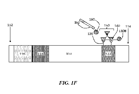
Without wishing to be bound by any particular theory, it is believed that an
analyte-capture reagent complex (e.g., an antigen-capture antibody complex)
may bind
additional analytes and/or capture reagents (e.g., covalently bond, bind via
non-covalent
interactions) as it flows through the flow assay, or the analyte-capture
reagent complex
CA 03228664 2024- 2-9
WO 2023/017319
PCT/IB2022/000468
¨ 24 ¨
may be immobilized within the binding region and bind additional analytes
and/or
capture reagents as they move towards the binding region to form the
interconnected
network. As the analyte-capture reagent complex binds additional analytes
and/or
capture reagents, the interconnected network (comprising the analyte-capture
reagent
complex) may increase in size. In some embodiments, the analyte-capture
reagent
complex or interconnected may also bind (or be configured to bind) one or more
detection reagents, which may also increase the size of the complex or
interconnected
network. In some embodiments, increasing the size of the network comprises an
addition
of a salt or a buffer to the flow assay.
In some embodiments, a method describes forming an interconnected network
comprising the plurality of capture reagents, the plurality of detection
reagents, and/or
the plurality of analytes. In some embodiments, the interconnected network
comprises a
precipitate or a lattice comprising a plurality of capture reagents, detection
reagents,
and/or analytes. In some embodiments, the interconnected network comprises
agglomerates of the analyte and the capture reagent. In some embodiments, the
interconnected network forms a precipitate within the substrate, for example,
within a
binding region of the substrate. In some embodiments, the plurality of capture
reagents
and/or the plurality of detection reagents are configured to form an
interconnected
network with the plurality of analytes at a downstream position of the
substrate. In some
embodiments, the interconnected network comprises a mixture of the plurality
of capture
reagents and the plurality of analytes interconnected with one another. In
some
embodiments, the plurality of capture reagents and the plurality of detection
reagents are
configured to form the interconnected network with at least some of a
plurality of
analytes. In some embodiments, the interconnected network comprises a mixture
of the
plurality of capture reagents, the plurality of detection reagents, and/or the
plurality of
analytes interconnected with one another.
In some embodiments, the interconnected network comprises one or more
connections (e.g., bonds) connecting the components of the interconnected
network (e.g.,
analytes, capture reagents, and/or detection reagents) to one another. In some
embodiments, the interconnected network comprises one or more covalent bonds
between an analyte, a capture reagent, and/or a detection reagent (e.g., at
least some of a
plurality of analytes, at least some of a plurality of capture reagents,
and/or at least some
of a plurality of detection reagents). In some embodiments, the interconnected
network
CA 03228664 2024- 2-9
WO 2023/017319
PCT/IB2022/000468
¨ 25 ¨
comprises one or more non-covalent interactions between an analyte, a capture
reagent,
and/or a detection reagent. Non-limiting examples of non-covalent interactions
include
ionic interactions (e.g., salt bridges), hydrogen bonding, Van der Waals
interactions.
and/or hydrophobic interactions. For example, in some embodiments, the
interconnected
network comprises hydrogen bonding between the plurality of analytes, the
plurality of
detection reagents, and/or the plurality of capture reagents.
In some embodiments, one or more salts (or ionic species of the one or more
salts) may contribute to the blockage of the substrate flow. In some
embodiments, these
one or more salts (or ionic species of the one or more salts) may contribute
to formation
of the interconnected network formation. The different types of salts and ions
may come
from various sources, for example, the sample, a sample buffer, a sample
solvent, the
substrate (e.g., metallic components of a nitrocellulose membrane). Salts (or
ionic
species of the salt) may influence the fragmentation and/or activation of the
side chains
of an analyte (e.g., analytes comprising amino acids, such as a protein)
during its
movement on the substrate. In some embodiments, the salts may also form at
least a
portion of the interconnected network or may surround at least a portion of
the
interconnected network in conjunction with the binding of analyte to the
capture reagent.
In some embodiments, the analyte (e.g., an antigen) and capture reagent (e.g.,
a capture
antibody) binds and may form individual "seeds," or nucleation sites for the
interconnected network (e.g., due to antigen-antibody chains forming or
agglomeration)
and such seeds may form the basis for salts to accumulate or precipitate
around the
interconnected network, enhancing the network and may further impede the flow
on the
substrate once the interconnected network has formed. Advantageously, this
effect may
lead to an even greater signal generated for positive (target analyte
containing) samples
versus negative samples which do not contain target analytes. Without wishing
to be
bound by any particular theory, when the analyte binds to the capture reagent
there may
be a conformational change and this may lead to the obstruction of pore of the
substrate,
for example, due to the negative or positives charges from salts (e.g., salts
of a buffer
system).
In some embodiments, a change in the percentage or concentration of salts
and/or
other ions may lead to the formation of the interconnected network formation.
In some
such embodiments, the interconnected network may form a solid precipitate,
optionally
comprising one or more salts (e.g., salts of a buffer), which may block pores
of the
CA 03228664 2024- 2-9
WO 2023/017319
PCT/IB2022/000468
¨ 26 ¨
substrate. In some such embodiments, the interconnected network may
precipitate the
formation of salt bridges. In some such embodiments, the salt blockage may
"seed" the
formation of the interconnected network and may form through several routes,
without
wishing to be bound by any particular theory: (A) The agglomeration of
alternating ionic
charges leading to lattice formation comprised of the different types of ions,
when
present (e.g., within a sample buffer, an antibody solution, HRP conjugate
solution). The
ions may be either positive or negatively charged. Without wishing to be bound
by any
particular theory, these charges may play a role in exposing, fragmenting,
and/or
activating of the side chains of the analytes while it moves on the substrate.
Binding of
analytc to the capture reagent may cause a blockage in the pore. Further,
accumulation of
the salts at or within the binding region may change the charge distribution
of capture
reagents and/or analytes within the binding region and there may be
agglomeration; (B)
The counterions leading to salt formation where one or more components of a
buffer and
the substrate contributes, respectively, anions and cations. Some substrates
may include
cationic components, such as Ca2+, Mg2+, silicon-containing cations, and these
ions may
interact with ions of a sample buffer (if present), leading to the formation
of the
interconnected network or salt bridge (on which the interconnected network may
form or
precipitate); (C) formation of Ca-phosphate where the substrate includes
components
like Ca2 , Mg2+, silicon-containing cations. At a higher pH i.e., > 7, there
is a tendency of
interaction of Ca-phosphate which lead to the formation of CaHPO4. The
presence of
NaC1 may also contributes to the supply of carbonates which facilitates the
precipitation
formed by Ca-phosphate.
In some embodiments, a sample comprises a salt and/or a buffer (e.g.,
phosphate
buffer, NaH/PO4/Na/HPO4). In some such embodiments, salts can contribute to
formation of the interconnected network or strengthen the interconnected
network by
forming such structures around the interconnected network. Non-limiting
examples of
salts include NaCl. KC1, NH4C1, KNO3, Al(NO3)3, (NH4)2SO4, Mg2SO4, FeCO3,
CaCO3,
FePO4, KH2PO4,Na2flPO4, (NH4)3PO4, Pb(CH3C00)/. Cu(CH3C00)2. Other salts are
possible.
In some embodiments, the sample (e.g., a sample solvent, a sample buffer)
comprises a surfactant. In some such embodiments, the surfactant may also
attach to
capture reagents and/or analytes in such a way as to help restrict flow in the
substrate. In
some embodiments the surfactants may form at least a portion of the
interconnected
CA 03228664 2024- 2-9
WO 2023/017319
PCT/IB2022/000468
¨27 ¨
network or may contribute to the formation of the interconnected network.
Without
wishing to be bound by any particular theory, the analyte (e.g., an antigen)
and capture
reagent (e.g., a capture antibody) binds, this may form individual "seeds" and
such seeds
may form the basis for surfactants to accumulate or precipitate around the
interconnected
network, enhancing the network and further impeding the flow on the substrate.
In some
embodiments, the surfactant may play a role in exposing, fragmenting, and/or
activating
of the side chains of the analytes while it moves on the substrate. In some
embodiments,
the surfactants may play a role in increasing or decreasing the hydrophilicity
of capture
regents and/or analytes or other parts of the interconnected network, which
can cause or
enhance network formation.
In some embodiments, this effect may lead to an even greater signal generated
for positive (i.e., analyte-containing) samples versus negative samples which
do not
contain target analytes.
In some embodiments, the surfactant comprises an anionic surfactant, such as
ammonium lauryl sulfate, sodium laureth sulfate, sodium lauryl sarcosinate,
sodium
myreth sulfate, sodium pareth sulfate, sodium dodecyl sulfate, sodium stearte,
sodium
deoxycholate, a olefin sulfonate, and/or ammonium laureth sulfate, without
limitation.
In some embodiments, the surfactant comprises a cationic surfactant, such as
In
C8-10 alkyl hydroxyethyl dimethylammonium chloride, C8-10 alkylamidodimethyl
propylaminc, and/or ditallow dimethyl ammonium chloride, without limitation.
In some embodiments, the surfactant comprises a non-ionic surfactant, such as
polysorbates (e.g., tween 20), sorbitan esters, alkyl-phenol polyethylenes
(e.g. tritons),
oligomeric alkyl-ethylene oxides, poly (alkylene-oxide) block copolymers,
without
limitation. In some embodiments, the smfactants is an amphoteric surfactant.
In some embodiments, a change in the percentage or concentration of
surfactants
may lead to the formation of the interconnected network formation. In some
such
embodiments, the interconnected network comprises the surfactant and/or the
surfactant
surrounds at least a portion of the interconnected network and may block at
least some of
the pores of a substrate if the substrate is porous.
In some embodiments, surfactants, such as those described above without
limitation,
may cause a change in the configuration of the analyte (e.g., causing a
protein-containing
analyte to change from a "closed" to "open" configuration), which may also
enable or
enhance binding to the capture reagent.
CA 03228664 2024- 2-9
WO 2023/017319
PCT/IB2022/000468
¨ 28 ¨
As mentioned above, various embodiments describe a flow assay for the
detection of an analyte. Accordingly, a sample (or a sample solvent) may be
flowed. In
general, flow is from an upstream position to a downstream position. For
example, in
some embodiments, a method comprises flowing the interconnected network
towards a
binding region (e.g., a binding region at a downstream position). In some
embodiments,
a method comprises stopping flow of the interconnected network within the
binding
region. In some embodiments, a method comprises, reducing or stopping flow on
at least
a portion of the substrate after forming the interconnected network. In some
embodiments, at least some of the plurality of detection reagents is not
flowed off the
flow assay during any one of the flowing steps, such as after the formation of
the
interconnected network in the flow assay. However, as noted above, in some
embodiments, the formation of an interconnected network (e.g., a precipitate,
a lattice) in
the flow assay may affect the flow properties of the assay, such that flow
from a
downstream position towards an upstream position may occur (i.e.. backflow).
For
example, in some embodiments, a method comprises backflowing the interconnect
network away from the binding region.
In some embodiments, a control indicator may be used to indicate when the
sample has passed through some or all of the substrate. In some embodiments
this
control indicator can be a dye placed in the downstream region of the
substrate or at a
downstream position on substrate (for example, in the absorbent or other pad
at the
downstream position on or near the substrate), so that when the sample liquid
passes
through the dye, the dye spreads downstream and appears in a region downstream
from
its original position. The indicator can be any suitable color, such as a red
or other color
dye placed at a downstream position of the substrate (e.g., the bottom of the
absorbent
pad). In the case of a red color indicator, this indicator may spread
downstream when the
sample passes it so that the red color appears in a window in the cassette
downstream
from the original position of the dye. In some embodiments, the appearance of
the red
color in the cassette can be used by an operator to decide when to add
detection reagents
and/or development reagents to the assay. The indicator may be any
colorimetric reagent
that flows and can be read by eye or with a reading device. In other
embodiments, the
color indicator comprises a liquid or solid containing fluorescent particles
or charged or
magnetic particles. Other color indicators are possible. Use of the indicator,
can show
when the sample (for example a liquid which may contain the sample analyte)
has passed
CA 03228664 2024- 2-9
WO 2023/017319
PCT/IB2022/000468
¨ 29 ¨
through the substrate. This may indicate that the interconnected network has
formed,
when there are one or more analytes in the sample, and/or indicates that the
detection
reagent has flowed off the substrate, when there are no target analytes in the
sample.
Thus, when the indicator shows, then detection reagents may be added and/or
development reagents (for example TMB) may be added, which may differentiate
the
positive (analyte-containing) sample from the negative sample (does not
contain the
analyte). The indicator may be a preferred alternative to timing of the sample
flow since
in many existing lateral flow assays or other systems, the sample can flow at
different
speeds depending on temperature, humidity, and other factors, but in all cases
the
indicator shows when the sample has flowed passed that position of the
substrate.
As mentioned above, a cassette may be used to modify the flow properties of a
flow assay (e.g., a lateral flow assay). Accordingly, some embodiments
comprise a
cassette at least partially enclosing the substrate. As was described above in
view of the
figures, the cassette comprises a first portion and a second portion opposing
the first
portion, and the substrate of the flow assay may be positioned between the
first and
second portions. In some embodiments, the second portion comprises a
protrusion
extending towards the first portion, and wherein the protrusion presses the
substrate
against the first portion. Advantageously, the protrusion may prevent excess
sample flow
on a first portion of the substrate or cassette rather than though the
substrate and also
reduce or prevent backflow of flow assay components (e.g., of a sample
solvent, of at
least a portion of a plurality of detection reagents, of at least a portion of
a development
reagents. The protrusion can press against the substrate so as to make contact
with the
first portion, but without causing any liquids (e.g., a sample solvent) to be
forced out of
the substrate. The protrusion should contact the substrate with sufficient
force to prevent
the flow of liquids through it or the backflow of liquids through it; however,
it also
should not press into the substrate with too much force, which could force
liquids out of
the substrate and/or create an indent or trough in the substrate where liquids
could
accumulate. For example, such a trough may act as a place where development
reagents
accumulate and so such excess development reagents are not drawn out of the
substrate
but instead may remain in the trough. Since certain development reagents may
develop
even in the absence of development reagents, if given sufficient time, and
this may lead
to generation of what may appear to be positive signal, even in the case where
there are
no target analytes in the sample, so this can lead to false positive assays.
In other
CA 03228664 2024- 2-9
WO 2023/017319
PCT/IB2022/000468
¨ 30 ¨
embodiments, false negative assays may be generated due to the indent.
Therefore, the
control of the specific features and fabrication of the protrusion is
essential for proper
operation. The cassette may also include one or more windows, for example, for
sample
deposition and/or viewing a signal for analyte detection. For example, in
FIGS. 2C-2D, a
viewing window 251 is included within the cassette 200. FIGS. 2E-2F show
schematic
illustrations of the protrusion (e.g., bridge) 250 schematically illustrated
in FIGS. 2C-2D.
In some embodiments, the protrusion positioned to align with a particular
position of a substrate. For example, in some embodiments, the protrusion is
positioned
at a midpoint between an upstream position and a downstream position (e.g., a
binding
region at a downstream position). In some embodiments, protrusion is less than
or equal
to 10%, less than equal to 20%, less than or equal to 30%, less than or equal
to 40%, less
than or equal to 50%, less than or equal to 60%, less than or equal to 70%,
less than or
equal to 80%, or less than or equal to 90% of a distance between an upstream
position
and a downstream position. By way of illustration and not limitation, if the
distance
between an upstream position and a downstream position is 3 cm, then the
protrusion
could be positioned at 50% of this distance, or 1.5 cm, from the upstream
position. In
some embodiments, the protrusion is greater than or equal to 10%, greater than
or equal
to 20%, greater than or equal to 30%, greater than or equal to 40%, greater
than or equal
to 50%, greater than or equal to 60%, greater than or equal to 70%, greater
than or equal
to 80%, or greater than or equal to 90% of a distance between the upstream
position and
the downstream position. Combinations of the above-referenced ranges are also
possible
(e.g., the protrusion is positioned greater than 10% of a distance between the
upstream
position and the downstream position and less than or equal to 90% of a
distance
between the upstream position and the downstream position). Other ranges are
possible.
The protrusion may extend from a surface of a second portion towards the first
portion. The extent of this protrusion may affect the degree of force applied
to a
substrate, when present, as it presses against the first portion. In some
embodiments, the
protrusion extends greater than or equal to 0.1 mm, greater than or equal to
0.2 mm,
greater than or equal to 0.3 nana, greater than or equal to 0.5 mm, greater
than or equal to
0.7 mm, or greater than or equal to 1 mm from a surface of the second portion.
In some
embodiments, the protrusion extends less than or equal to 1 mm, less than or
equal to 0.7
mm, less than or equal to 0.5 mm, less than or equal to 0.3 mm, less than or
equal to 0.2
mm, or less than or equal to 0.1 mm from a surface of the second portion.
Combinations
CA 03228664 2024- 2-9
WO 2023/017319
PCT/IB2022/000468
¨ 31 ¨
of the above-referenced ranges are also possible (e.g., the protrusion extends
a distance
from a surface of the second portion by greater than or equal to 0.1 mm and
less than or
equal to 1 nun). Other ranges are possible.
In some embodiments, the protrusion is formed of the same material as another
part of the cassette, for example plastics, including, but not limited, to
polyethylene,
polypropylene, ABS, HIPS. In other embodiments, the protrusion is formed from
a
different material as another part of the cassette.
In some embodiments the protrusion may he formed of a different material from
other parts of the cassette. For example, the protrusion may be formed from a
softer
material relative to other components of the flow assay. In some embodiments,
the
protrusion comprises a soft material, for example rubber (e.g., silicone
rubber) or
thermoplastic elastomer (e.g., polyurethane) either forming the whole of the
protrusion
or over molded onto another material which is the base of the protrusion. In
some
embodiments, the softer material may have a lower shore of 25-40 A or less.
Advantageously, the use of the low shore material is that it may be difficult
to achieve
exactly the depth required for a hard protrusion into the substrate (for
example
nitrocellulose membrane) in order to have exactly the right force (i.e., not
too much force
as to squeeze liquid from the substrate, but not too little force as to
provide an
inappropriate amount of force for pressing the substrate). Such tight
tolerance can be
difficult to achieve in mass manufacturing. However, it has been discovered by
this
disclosure, that the ese of a softer protrusion with shore 25-40 A material
instead can
improve the manufacturing of the protrusion.
The articles and methods disclosed herein are suitable for a variety of
applications. In some embodiments, an article is a flow assay (e.g., a lateral
flow assay).
As mentioned above, for example, the articles and methods disclosed may be
used to
detection of pathogens, such as SARS-CoV-2. The articles and methods disclosed
may
be used to detect other pathogens, such as variants of SARS-CoV-2 or influenza
viruses,
as non-limiting examples. However, other applications are possible. For
example, the
articles and methods described herein may be used to test for the presence of
an
environmental contaminant, such as a pollutant or a toxin suspected of being
present in a
particular environment.
CA 03228664 2024- 2-9
WO 2023/017319
PCT/IB2022/000468
¨ 32 ¨
EXAMPLE 1
The following example shows that the movement and location of the capture
reagent (antibody) and detection reagent (HRP conjugate) when run with a
positive
sample (i.e., containing the analyte) and negative sample (i.e., not
containing the
analyte). Here HRP means horseradish peroxidase and HRP conjugate, means HRP
conjugated to some other molecule or species, either covalently or non-
covalently.
To test the concept that, in negative sample, the HRP conjugate and
biotinylated
antibody detection reagent are being washed out into the absorbent pad of the
substrate
in the case of negative samples and remaining in the nitrocellulose membrane
substrate
when the target protein is contained in the sample, 6 devices were used, 3
with PCR
confirmed positive and 3 with PCR confirmed negative.
The sample was added to the sample pad. Once the sample had flowed through the
strip
and entered the absorbent pad of the substrate. The absorbent pad was removed
and
placed into the well of a 96-well plate. 150 iil TMB staining reagent was
added. No
development was observed for 20 minutes at room temperature.
Since the presence of detection reagent HRP conjugate on the absorbent pad is
known, given the flow of the sample, it was considered highly likely that the
absorbent
pad was preventing the staining reagent TMB from coming into contact with the
detection reagent HRP conjugate and/or the majority of the HRP conjugate was
on the
backing pad of the strip. A second example, Example 2, was designed providing
different method to locate the detection reagent HRP conjugate.
EXAMPLE 2
The following example describes isolating each section of the lateral flow
assay
to determine the presence and amount of the detection reagent HRP conjugate.
12 flow assays were used, 6 with an absorbent pad (FIG. 3) and 6 without. For
each
device type. 6 samples were run 3 with PCR-confirmed positive samples and 3
samples
run with PCR-confirmed negative samples. Sample was added to the sample pad.
Once
the flow reached the absorbent pad, having flowed through the full membrane
substrate,
the lateral flow assay was dismantled into its component parts, and the 2.5cm
membrane
substrate was cut into 0.5 cm sections, creating 8 discrete regions of the
assay, as shown
in FIG. 4. Sections were placed into individual wells of a 96-well plate as
shown in FIG.
CA 03228664 2024- 2-9
WO 2023/017319
PCT/IB2022/000468
¨ 33 ¨
5. 150 L of staining reagent TMB was added. The reaction was stopped after 17
minutes by adding 150 I stop solution. The assay components were removed from
the
wells, and the plate was read at 450 nm.
Results
The results arc shown in FIG. 6. The detection reagent region conjugate pads
in
all lateral flow assays still contained significant amount of IIRP conjugate.
For the
assays on which positive sample were run, the level of enzyme remained at a
constant
level until the next section of the membrane substrate, section 2, which
contained the
antibody with a lower level past the test line. By contrast, the negative
samples had a
much lower level of enzyme remaining in the conjugate pad, and very low levels
in the 2
sections of membrane following, and negligible levels in membrane sections 1-
3. The
lower levels of enzyme in the conjugate pads of negative samples, and low
levels across
the membrane, indicate that the HRP conjugate has been largely washed out of
the
membrane by the sample in the negative sample (no target antigen/protein)
case. This
suggested that for the positive samples, flow was reduced or stopped, while
for the
negative sample, flow was not reduced or stopped, and the HRP conjugate could
be
largely washed off the assay substrate (membrane).
The presence of high levels of enzyme in each section of the devices run with
positive samples, which drops off after the test-line, indicated that the
binding of the
target analyte antigen to the biotinylated antibody capture reagent has
restricted the flow
of the sample, preventing the enzyme from flowing through.
The antigen-HRP conjugate complex (network or mesh) appears to be blocking
the flow, either through physical blocking of the pores of the substrate or by
causing
changes to the nature of the membrane substrate (e.g., pH or charge) or any
other
mechanism, which restrict the complex formed through a cascade or chain
reaction
between the antigen, antibody and/or HRP conjugate. In the absence of antigen
(negative
sample), no restrictive complex (network) is being formed and the HRP
conjugate is able
to be washed away.
CA 03228664 2024- 2-9
WO 2023/017319
PCT/IB2022/000468
¨ 34 ¨
EXAMPLE 3
The following example describe analyzing the flow pattern of the sample on a
lateral flow assay.
In order to further confirm the observation discussed in Example 2, a third
example was design with the aim to trace the flow profile of the sample and in
order to
further analyze the movement of the HRP conjugate detection reagent along the
strip
(substrate) in the presence and the absence of the antigen (positive and
negative
samples).
Lateral flow assays were fabricated with longer nitrocellulose membrane
substrates compared to the assay of Example 2. An additional lcm was added
from the
test line, leaving the same distance between the sample pad and the test line
as in
standard devices (strip used in experiment 2), while moving the absorbent pad
lcm
further from the test line.
Results
The assays were run as previous described above, with the addition of the TMB
staining reagent in the test region once the sample had stopped flowing along
the
membrane. The TMB staining reagent was added, and the TMB development along
the
whole strip was generated to trace the flow profile. As shown in FIG. 7, two
flow
profiles can be distinguished in the test line region, depending on the
sample.
For the negative sample, a Poiseuille-type flow profile is observed,
suggesting a fully
developed velocity flow of the negative sample. While for the positive sample,
a
hydrodynamic entrance region can be distinguished with an irrotational flow
region.
This result is consistent with the previous observation in Example 2 and
demonstrated that, in the negative sample, the complex will flow along with
the sample
fluid and reach the absorbent pad. By contrast, in the positive sample, the
antibody
bound to the target antigen analyte that farms a complex through a cascade or
chain
reaction between the antigen, antibody capture reagent and/or HRP conjugate
detection
reagent. The trapped complex creates an obstructed region, resulting in an
irrotational
flow region.
CA 03228664 2024- 2-9
WO 2023/017319
PCT/IB2022/000468
¨ 35 ¨
EXAMPLE 4
The following example uses IgG as the detection reagent.
An anti-S1 IgM was biotinylated and used in devices in place of the
biotinylated
IgG detection reagent. Positive and negative samples were run using these
samples.
The positive samples gave good development and the negative samples remained
blank. The size of IgG is 150 kDa. The results are shown in FIG. 8. By
contrast, IgM is
970 kDa. Given the large difference in size between the two types of
antibodies, this
result suggested that it is not only the size of the antibody-antigen complex
that is
preventing movement along the membrane substrate, that there is a further
interaction,
either between a single complex and the membrane substrate, or between the two
or
more antibody-antigen complexes and/or the salts or other components of the
sample
buffer. The latter suggests the formation of an interconnected network as the
flow assay
is run.
EXAMPLE 5
The following example demonstrates that other analytes can be used other than
the SARS-CoV-2 spike glycoprotein Si subunit. For this example, Respiratory
Syncytial
Virus (RSV) was selected as the source of the analyte.
A mouse anti-RSV antibody IgG, which targets the F protein, MAB12398-100
was used as a detection reagent. Respiratory Syncytial virus A lysatc was used
as a target
analyte. Initially, dilutions were made at 1 in 10 from 100 pg/mL down to 1
pg/mL. With
positive samples, signal was achieved at 1 pg/mL, and serial 1 in 2 dilutions
were made
from 0.5 pg/mL down to 3.91 fg/mL in an extraction buffer.
Results
The results of each assay are shown in FIGS. 9-10. As can be seen, RSV lysate
gave a positive signal down to 62.5 fg/mL. These results show that the
mechanism
describe in the previous examples can be used to detect other pathogens and
biomarkers,
and is not restricted to the SARS-CoV-2 Spike glycoprotein SI.
Similarly, PSA was also run and tested in the same way and showed detection
down to pg/mL level and no signal in the negative sample case (no target
antigen). Since
there can be a large variation in size between these various antigens (PSA is
very small
CA 03228664 2024- 2-9
WO 2023/017319
PCT/IB2022/000468
¨ 36 ¨
for example), this result suggested that it is not only the size of the
antibody-antigen
complex that is preventing movement along the membrane substrate, that there
is a
further interaction, either between a single complex and the membrane
substrate, or
between the two or more antibody-antigen complexes and/or the salts or other
components of the sample buffer.
EXAMPLE 6
The following example describes determining the position of the capture
antibody after running a positive and a negative sample.
To test the concept that the capture antibody is flowing away from the test
line in
the case of a negative sample and remaining in place in the case of a positive
sample, the
antibody was labelled with a fluorophore to enable imaging of the strip via
microscopy.
Anti-S1 IgG was conjugated with a UV label using a commercially available
conjugation kit.
The antibody was deposited at the test line on the nitrocellulose membrane,
and
test strips were manufactured following a procedure similar to the previous
examples.
A SARS-CoV-2 positive nasal swab was run on one device, and a negative nasal
swab was run on another. The positive and negative test strips were imaged
using a
fluorescent microscope from 6 minutes following the application of the sample
and
imaged at 50 second intervals over a 10 minute period.
Results
The results are shown in FIG. 11. The antibody line in both positive and
negative
samples remains in the same position through the 10-minute period of imaging.
This
indicates that during the manufacturing of the test strip, the antibody binds
strongly to
the nitrocellulose membrane and does not flow off the test line in the case of
positive and
negative samples.
EXAMPLE 7
The following example describes labeling of streptavidin-linked HRP conjugate
to visualize the flow of the enzyme through the test strip during the run of a
positive and
negative nasal swab sample.
CA 03228664 2024- 2-9
WO 2023/017319
PCT/IB2022/000468
¨ 37 ¨
To test the concept that the flow of the enzyme, which acted as a detection
reagent, was restricted in the case of positive samples, and not restricted in
the case of
negative samples the enzyme was labeled to enable time lapse imaging of the
labeled
enzyme during flow through the membrane.
An ATTo 488 as fluorophore was attached to the enzyme. The
streptavidin-linked HRP conjugate was incubated with a biotinylated
fluorophore at a 1:4
ratio, in order to bind all the binding sites on the streptavidin.
The fluorophore-conjugated streptavidin-linked HRP conjugate was deposited
onto the conjugate pads and test strips manufactured as described elsewhere
herein.
A SARS-CoV-2 positive nasal swab was run on one device, and a negative nasal
swab was run on another.
The positive and negative test strips were imaged using a fluorescent
microscope
from 6 minutes following the application of the sample and imaged at 30-second
intervals over a 10-minute period.
Results
The flow of the enzyme in the positive swab sample is clearly delayed when
compared with the negative sample, indicating a restriction of flow. The
enzyme
becomes more concentrated towards the test line and front end of the
absorbance pad in
the positive over the 10-minute period of imaging, while in the negative
sample, the flow
disperses out into the absorbance pad over the 10-minute period of imaging.
These
results are visualized in FIG. 12.
EXAMPLE 8
The following example describes the use of HRP substrate TMB to examine the
relationship to enzymatic development with the fluorophore-enabled imagine of
the
movement of the enzyme, and to determine whether the addition of TMB results
in
further movement of the enzyme within the test strip.
The streptavidin-linked HRP conjugate was incubated with a biotinylated
fluorophore at a 1:4 ratio, in order to bind all the binding sites on the
streptavidin.
The fluorophore-conjugatcd streptavidin linked HRP conjugate was deposited
onto the conjugate pads and test strips manufactured as described elsewhere
herein.
CA 03228664 2024- 2-9
WO 2023/017319
PCT/IB2022/000468
¨ 38 ¨
A SARS-CoV-2 positive nasal swab was run on one later flow assay device, and
a negative nasal swab was run on another.
The positive and negative test strips were imaged using a fluorescent
microscope
from 6 minutes following the application of the sample and imaged at 30 second
intervals over a 10-minute period.
At the end of the 10-minute imaging period, the strips were removed from the
microscope. TMB was deposited onto the test line and allowed to develop for 5
minutes.
The development was imaged, and the test strips were placed back into the
microscope to
image the final position of the enzyme.
Results
The results are shown in FIG. 13 and FIG. 14 in the case of the negative
sample,
following the deposition of TMB on the test line, a single area of development
occurred
upstream of the test line (i.e., region 1 in FIG. 13) as the TMB flowed back
along the
nitrocellulose membrane, demonstrating the presence of enzyme at this
location.
In the case of the positive sample, following the deposition of TMB at the
test
line, a clear area of strong development occurred adjacent to the test line
(i.e., region 2 in
FIG. 13), followed by development in region 1, as seen in the case of the
negative
sample. This indicates 2 distinct regions in which enzyme is present in the
case of a
positive sample, with the greater amount retained adjacent to the test line.
Fluorescent imaging of the strips following the TMB development revealed a
non-specific signal or background (i.e., not related to the enzyme) due to
unbound
fluorophore all along the nitrocellulose membrane. The figure shows dark
regions, which
correspond to the regions of TMB development observed (FIG. 14). This may be
due to
the optical density of the developed TMB restricting the excitation and/or
emission of the
fluorophore.
While several embodiments of the present invention have been described and
illustrated herein, those of ordinary skill in the art will readily envision a
variety of other
means and/or structures for performing the functions and/or obtaining the
results and/or
one or more of the advantages described herein, and each of such variations
and/or
modifications is deemed to be within the scope of the present disclosure. More
generally, those skilled in the art will readily appreciate that all
parameters, dimensions,
CA 03228664 2024- 2-9
WO 2023/017319
PCT/IB2022/000468
¨ 39 ¨
materials, and configurations described herein are meant to be exemplary and
that the
actual parameters, dimensions, materials, and/or configurations will depend
upon the
specific application or applications for which the teachings of the present
disclosure
is/are used. Those skilled in the art will recognize or be able to ascertain
using no more
than routine experimentation, many equivalents to the specific embodiments of
the
invention described herein. It is, therefore, to be understood that the
foregoing
embodiments are presented by way of example only and that, within the scope of
the
appended claims and equivalents thereto, the invention may be practiced
otherwise than
as specifically described and claimed. The present disclosure is directed to
each
individual feature, system, article, material, and/or method described herein.
In addition,
any combination of two or more such features, systems, articles, materials,
and/or
methods, if such features, systems, articles, materials, and/or methods are
not mutually
inconsistent, is included within the scope of the present disclosure.
The indefinite articles "a- and "an,- as used herein in the specification and
in the
claims, unless clearly indicated to the contrary, should be understood to mean
"at least
one."
The phrase "and/or," as used herein in the specification and in the claims,
should
be understood to mean "either or both" of the elements so conjoined. i.e.,
elements that
are conjunctively present in some cases and disjunctively present in other
cases. Other
elements may optionally be present other than the elements specifically
identified by the
"and/or" clause, whether related or unrelated to those elements specifically
identified
unless clearly indicated to the contrary. Thus, as a non-limiting example, a
reference to
"A and/or B," when used in conjunction with open-ended language such as
"comprising"
can refer, in one embodiment, to A without B (optionally including elements
other than
B); in another embodiment, to B without A (optionally including elements other
than A);
in yet another embodiment, to both A and B (optionally including other
elements); etc.
As used herein in the specification and in the claims, "or" should be
understood
to have the same meaning as "and/or" as defined above. For example, when
separating
items in a list, "or" or "and/or" shall be interpreted as being inclusive,
i.e., the inclusion
of at least one, but also including more than one, of a number or list of
elements, and,
optionally, additional unlisted items. Only terms clearly indicated to the
contrary, such
as "only one of' or "exactly one of," or, when used in the claims, "consisting
of," will
refer to the inclusion of exactly one element of a number or list of elements.
In general,
CA 03228664 2024- 2-9
WO 2023/017319
PCT/IB2022/000468
- 40 -
the term "or" as used herein shall only be interpreted as indicating exclusive
alternatives
(i.e. "one or the other but not both") when preceded by terms of exclusivity,
such as
"either," "one of." "only one of," or "exactly one of." "Consisting
essentially of," when
used in the claims, shall have its ordinary meaning as used in the field of
patent law.
As used herein in the specification and in the claims, the phrase -at least
one," in
reference to a list of one or more elements, should be understood to mean at
least one
element selected from any one or more of the elements in the list of elements,
but not
necessarily including at least one of each and every element specifically
listed within the
list of elements and not excluding any combinations of elements in the list of
elements.
This definition also allows that elements may optionally be present other than
the
elements specifically identified within the list of elements to which the
phrase "at least
one" refers, whether related or unrelated to those elements specifically
identified. Thus,
as a non-limiting example, "at least one of A and B" (or, equivalently, "at
least one of A
or
or, equivalently "at least one of A and/or B-) can refer, in one
embodiment, to at
least one, optionally including more than one, A, with no B present (and
optionally
including elements other than B); in another embodiment, to at least one,
optionally
including more than one, B, with no A present (and optionally including
elements other
than A); in yet another embodiment, to at least one, optionally including more
than one,
A, and at least one, optionally including more than one, B (and optionally
including other
elements); etc.
Some embodiments may be embodied as a method, of which various examples
have been described. The acts performed as part of the methods may be ordered
in any
suitable way. Accordingly, embodiments may be constructed in which acts are
performed in an order different than illustrated, which may include different
(e.g., more
or less) acts than those that are described, and/or that may involve
performing some acts
simultaneously, even though the acts are shown as being performed sequentially
in the
embodiments specifically described above.
Use of ordinal terms such as "first," "second," "third," etc., in the claims
to
modify a claim element does not by itself connote any priority, precedence, or
order of
one claim element over another or the temporal order in which acts of a method
are
performed, but are used merely as labels to distinguish one claim element
having a
certain name from another element having a same name (but for use of the
ordinal term)
to distinguish the claim elements.
CA 03228664 2024- 2-9
WO 2023/017319
PCT/IB2022/000468
¨ 41 ¨
In the claims, as well as in the specification above, all transitional phrases
such as
"comprising," "including," "carrying," "having," "containing," "involving,"
"holding,"
and the like are to be understood to be open-ended, i.e., to mean including
but not limited
to. Only the transitional phrases "consisting of' and "consisting essentially
of' shall be
closed or semi-closed transitional phrases, respectively, as set forth in the
United States
Patent Office Manual of Patent Examining Procedures, Section 2111.03.
CA 03228664 2024- 2-9