Note : Les descriptions sont présentées dans la langue officielle dans laquelle elles ont été soumises.
-- 204207)
48168-2
ENDOSCOPIC IMAGING SYSTEM FOR DISEASED TISSUE
This invention relates to an apparatus for imaging
abnormal tissues in the body to locate and identify areas
that are otherwise not recognizable by white light
endoscopy. The invention is particularly suited for imaging
abnormal bronchial tissues to detect conditions such as
inflammation, denudation, dysplasia and non-invasive early
cancer (carcinoma in situ).
At present, the most effective method for examination
of body cavities in human patients is by endoscopes. For
examination of the air passages of the lung, a flexible
endoscope is usually used, commonly referred to as a
bronchoscope. Bronchoscopes, like all endoscopes, employs
visible white light to illuminate the surface under
examination. The illuminating light is brought into the air
passages (bronchi) of the lungs via a fiberoptic
illuminating light guide. The reflected and scattered light
from the bronchial tissues is captured by a projection lens
which focuses the image into the bronchoscope's imaging
bundle. The imaging bundle is composed of several thousand
individually wrapped fibers, which transmit a coherent image
to the exterior of the body. This image is then projected
through the ocular of the bronchoscope for human
observation. A colour video camera can be attached to the
eyepiece of the bronchoscope such that colour images of
scattered/reflected white (broadband) light can be viewed on
a colour video monitor.
Using a conventional bronchoscope, large invasive
cancers can be readily seen. However, focal inflammation,
denudation, dysplasia, and early lung cancers cannot be
readily seen by such an apparatus.
Several methods have been developed to visualize small
early lung cancers which are difficult to detect by ordinary
204207)
- 2 -
white light bronchoscopy. All of these involve the use of
tumour localizing drugs, e.g. Haematoporphyrin derivatives
or Porfimer sodium, which have been shown to be
preferentially retained in tumour tissues. Some of these
drugs also fluoresce and their fluorescence can be detected
by non-imaging and imaging devices (Profio AE et al., Med
Phys 6:532-535, 1979; Profio AE et al., Med Phys 11:516-520,
1984; Profio AE et al., Med Phys 13:717-721, 1986; Hayata Y
et al., Chest 82:10-14, 1982; Kato A, Cortese DA, Clin Chest
Med 6:237-253, 1985; Montan S et al., Opt Letters 10:56-58,
1985). The drawback of these techniques is the use of drugs
which may have serious side effects and therefore may not be
appropriate for diagnostic purposes. In addition, the use
of non-imaging devices such as the ratio fluorometer probe
(Profio et al., Med. Phys 11:516-520, 1984) cannot delineate
the exact site and dimensions of the abnormal areas.
An alternative approach for detecting invasive tumours
has been proposed by Alfano et al in United States Patent
4,930,516 issued June 5, 1990. Alfano discloses a method of
detecting cancers on the basis that the fluorescence spectra
of cancerous tissues is different from normal tissues in
that the maximal fluorescence peak of tumour tissues is blue
shifted to lower wavelengths (from 531nm to 521nm). These
observations were made based on in vitro measurements in
excised, large (invasive) animal and human tumours but have
not been reported on human tumours in vivo. In addition,
there are no reports of other abnormal tissues such as
inflamed or pre-cancerous tissues. We have measured tissue
autofluorescence in human patients in vivo using different
excitation wavelengths including 405nm, 442nm, and 488nm by
a specially designed optical multichannel analyzer which can
be attached to a conventional bronchoscope. Contrary to the
observation by Alfano et al., we did not find any difference
in the shape of the fluorescence spectrum between normal and
tumour tissues using these excitation wavelengths. In
particular, there was no blue shift of the emission peaks.
We observed a significant difference in the overall
204207
- 3 -
fluorescence intensity especially in the green region of the
visible spectrum. A significant but a lesser decrease in
the overall fluorescence intensity was also found in pre-
cancerous and non-cancerous lesions (dysplasia and
metaplasia).
The decreased green fluorescence may be attributed to a
reduced level of oxidized form of riboflavin. Riboflavin
emits strongly in the green region and is believed to be
predominantly responsible for the strong green fluorescence
in normal human lung tissue. In the cancerous tissues, much
less riboflavin was found (Pollack MA et al., Cancer Res
2:739-743, 1942) and/or is present in the reduced state.
This may account for the reduced autofluorescence in pre-
malignant and malignant bronchial tissues.
Tests were conducted revealing examples of such
decreased tissue autofluorescence for dysplastic bronchial
tissue, and carcinoma in situ. It was determined that the
main difference between abnormal and normal tissues is
manifested by a greatly reduced fluorescence intensity in
the region of the spectrum from 480nm - 600nm. At
wavelengths greater than approximately 635nm, the tissue
autofluorescence is approximately the same between abnormal
and normal tissues. Test were conducted using excitation
light of 442nm, 405nm and 488nm and abnormal tissue results
were compared to normal tissue results. All of these data
were obtained in vivo during standard fiberoptic
bronchoscopy using the optical multichannel analyzer.
Because of the observed large decrease in the emitted
fluorescence without a change in the spectral profile in the
abnormal tissues, methods using ratioing of two or more
wavelengths that was originally described by Profio and
coworkers and then studied in patients who have received
fluorescent drugs such as Photofrin (Profio et al., Med.
Phys. 11:516-520, 1984) generally will not differentiate
204Z0'~
- 4 -
abnormal from normal bronchial tissues using
autofluorescence alone.
We have invented and constructed an apparatus which
exploits differences in autofluorescence intensity for the
detection and delineation of the extent of abnormal areas in
the human body, particularly the lung.
The present invention provides an imaging apparatus
that uses autofluorescence characteristics of tissues to
detect and delineate the extent of abnormal tissues in human
patients in vivo. Capture and analysis of the
autofluorescence images is achieved using a highly sensitive
detector such as an image intensified CCD camera. A pseudo
image is generated by sending one image to the red channel
and one image to the green channel of an RGB video monitor.
By capturing the two images simultaneously or sequentially
within a few milliseconds, pseudo image generation in real
time can be achieved. The pseudo images can clearly
delineate the diseased tissue from the surrounding normal
tissue.
Accordingly, the present invention provides an
apparatus for imaging diseases in tissue comprising:
a light source for generating excitation light that
includes wavelengths capable of generating characteristic
autofluorescence for abnormal and normal tissues;
means for illuminating tissue with light that includes
at least said excitation light thereby exciting the tissue
to emit said characteristic autofluorescence;
collecting means for gathering emitted autofluorescence
light from said tissue;
means for filtering said autofluorescence light into
spectral bands in which said autofluorescence intensity for
.~ 204207
- 5 -
abnormal tissue is substantially different from normal
tissue and said autofluorescence intensity for abnormal
tissue is substantially similar to normal tissue;
optical means for intercepting said filtered
autofluorescence light to acquire at least two filtered
emitted autofluorescence images of the tissue; and
display means for displaying said acquired images in
such a manner as to delineate abnormal and normal tissue.
In a preferred embodiment, the apparatus of the present
invention is used with a standard bronchoscope for imaging
abnormal bronchial tissues.
Aspects of the present invention are illustrated,
merely by way of example, in the accompanying drawings in
which:
Figures la to ld provide examples of autofluorescence
spectrums at selected excitation wavelengths which indicate
the difference between abnormal and normal tissue;
Figure 2 is a schematic diagram showing the apparatus
of the present invention useful for imaging abnormal lung
tissue;
Figure 3 shows details of the illumination module;
Figure 4a shows the filtering and optical means of the
present invention in which a single sensitive detector is
used to acquire fluorescence images sequentially;
Figure 4b shows alternative filtering and optical means
in which fluorescence images are acquired simultaneously
using two sensitive cameras;
204207
- 6 -
Figure 4c shows a still further filtering and optical
means in which a prism element is incorporated to allow two
fluorescence images to be acquired simultaneously together
with a reflected/scattered excitation light image.
Figure 1 shows examples of decreased tissue
autofluorescence for dysplastic bronchial tissue and
carcinoma in situ. The main difference between abnormal and
normal tissues is manifested by a greatly reduced
fluorescence intensity in the region of the spectrum from
480nm - 600nm. At wavelengths greater than approximately
635nm, the tissue autofluorescence is approximately the same
between abnormal and normal tissues. For the results in
Figure la and lb, a 442nm Helium Cadmium laser light was
used to excite the tissues. Figure la shows tissue
autofluorescence spectra of normal and dysplastic tissues
and Figure lb shows a carcinoma in situ (CIS) lesion
compared to the normal tissue of a different patient.
Similar results were found when employing other excitation
light, e.g. 405nm, Figure lc and 488nm, Figure ld. In both
cases carcinoma in situ patients are compared to their
normal lung tissue. All of these data were obtained in vivo
during standard fiberoptic bronchoscopy using an optical
multichannel analyzer.
The apparatus of the present invention is designed to
exploit the difference in fluorescence intensity in
different regions of the spectrum to identify and delineate
abnormal tissue.
The apparatus of the present invention adapted for use
in examining bronchial tissues of the lung in patients is
schematically illustrated in Figure 2. As such, the
apparatus is integrated with a conventional bronchoscope
used for examining bronchial tissue of the lung.
There is a light source 1 for generating excitation
light that includes wavelengths capable of generating
_. 204207
_,_
characteristic autofluorescence spectra for abnormal and
normal tissue. The light source 1 is shown in greater
detail in Figure 3 and preferably includes a laser light
source 7 capable of producing excitation light at a selected
desirable wavelength. A white light source such as an
incandescent Xenon light source 8 can be used for white
light illumination when desired. The laser light source 7
is use to generate pseudo images derived from tissue
autofluorescence while the white light source is used to
generate colour images of reflected/scattered white light.
The light from each light source passes through
synchronizing means that allow for alternate illumination of
the tissue by the laser light and the white light source.
In the embodiment illustrated in Figure 3, the synchronizing
means comprises blocking means in the form of electronically
controlled shutters 9 and 13 associated with laser light
source 7 and Xenon light source 8, respectively. When
shutter 9 is open to allow laser light to pass, shutter 13
is closed to prevent passage of white light and vice versa.
The light from the laser light source 7 passes through
shutter 9 when open, a mirror with a pin hole 10, and a lens
11 which focuses the laser light onto means for illuminating
the tissue with light comprising a conventional bronchoscope
light guide 12. Light guide 12 conducts the excitation
light to the tissue area under examination. The tissue,
upon illumination with the laser light, emits its
characteristic autofluorescence for abnormal and normal
tissue. To generate regular white light illumination
images, shutter 9 is closed and previously closed shutter 13
is opened to allow the light from Xenon light source 8 to
pass through shutter 13. The white light is then filtered
by a neutral density filter set 14, reflected by a mirror
15, and passes through a lens 16 which focuses the light
onto bronchoscope light guide 12 after being reflected off
mirror 10 and through lens 11. The neutral density filter
set 14 is used to condition the light from the Xenon source
such that it is of the appropriate intensity for the light
20420~J
_8_
sensors used in the apparatus. Thus the white light
conducted to the tissue illuminates the tissue under
examination. Light guide 12 ensures that the light is
evenly dispersed over the area under examination.
In the present embodiment, the bronchoscope provides
the collecting means to gather images in the form of the
bronchoscope lens (not shown) which collects scattered and
reflected light, or emitted autofluorescence light from
within the lung for transmission out of the body by imaging
bundle 2 of the bronchoscope. This collected light is
transmitted to a focusing lens 21 of the bronchoscope ocular
coupled to the imaging bundle.
From the ocular of the bronchoscope, the collected
light enters the image acquisition module 3 which includes
means for filtering the autofluorescence light and optical
means for intercepting the filtered light. Various
embodiments of image acquisition module 3 are possible.
Figure 4a illustrates an image acquisition module that
includes filtering means and optical means that allow for
acquisition of emitted autofluorescence images sequentially.
In this embodiment, the means for filtering the
autofluorescence light comprises a series of filters that
are sequentially insertable into the path of the emitted
autofluorescence light to generate a sequence of filtered
autofluorescence images. Filter wheel 18 is provided and is
rotatably mounted beneath the optical means of the image
acquisition module. When laser excitation light 7 is used,
it is necessary to filter the autofluorescence light
generated into at least two spectral bands. In one spectral
band, the autofluorescence intensity for abnormal tissue is
substantially different from that of normal tissue and in
the other spectral band, the autofluorescence intensity is
substantially similar to that of normal tissue. For
example, in accordance with the characteristic spectral
bands indicated in Figures la to ld for lung examination,
204~07~
- 9 -
filter wheel 18 would be fitted with two filters. For laser
excitation light of 442nm or 405nm, a green filter of 500+-
20nm and a red 630nm longpass filter would be used. The
green filter would filter the autofluorescence light into a
spectral band in which the autofluorescence intensity for
abnormal tissue is substantially different from that of
normal tissue while the red longpass filter would filter the
light into a spectral band in which the autofluorescence
intensity is substantially similar for abnormal and normal
tissue. The two filters are mounted in filter wheel 18 such
that each covers one half of the filter surface. By
rotating filter wheel 18 at an appropriate speed, red and
green filtered autofluorescence images can be captured
sequentially by optical means in the form of a single highly
sensitive detector 17 such as an image intensified CCD
camera.
The foregoing image acquisition module also includes
additional optical means for capturing reflected/scattered
white light images when white light source 8 is providing
illumination of the tissue. A movable mirror 20 is provided
that is insertable into the path of the collected light
transmitted by ocular lens 21. Mirror 20 is positionable to
deflect white light into a colour video camera 22 for
acquisition of white light images. Necessarily, the
movement of mirror 20 is controlled such that the mirror
deflects the collected light into video camera 22 only when
white light source 8 is providing illumination. Using white
light source 8, colour images can be generated on a colour
monitor in the same way as in conventional bronchoscopy.
When laser light source 7 is illuminating the tissue, mirror
20 is removed from the light pass to allow for filtering of
the autofluorescence light and subsequent acquisition by
detector 17.
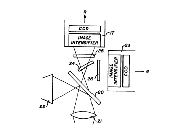
Figure 4b illustrates an alternative arrangement of
image acquisition module 3 in which the optical means
comprises at least two photodetectors that acquire filtered
204207)
- 10 -
autofluorescence images simultaneously. Each photodetector
has associated filtering means. For simultaneous collection
of autofluorescence images, filter wheel 18 of the
embodiment of Figure 4a is replaced by beam splitting means
in the form of a dichroic mirror 24 which allows the red
light >600nm to pass but reflects the shorter wavelengths.
In this case, additional filters 25 and 26 for exact
selection of the desired autofluorescence light can be
employed and the respective images are focused onto two
independent sensitive photodetectors such as image
intensified CCD cameras 17 and 23. In Figure 4, filter 25
is a red 630nm longpass filter to further filter red light
passed by dichroic mirror into a spectral band in which
autofluorescence intensity is substantially similar for
normal and abnormal tissue. Filter 26 is a green filter of
500+-20nm for filtering the autofluorescence light into a
spectral band in which the autofluorescence intensity for
abnormal tissue is substantially different from that of
normal tissue. Images acquired by the image intensified CCD
camera 17 and/or image intensified CCD camera 23 are fed
into red and green input channels of an RGB colour monitor 5
(Figure 1).
As in the arrangement of Figure 4a, reflected/scattered
white light images created by white light source 8 are
captured by a colour camera 22 and are displayed directly
onto the colour monitor for visualization of the examined
site using an identical movable mirror 20 insertable into
the light path whenever white light source 8 is providing
illumination.
Figure 4c illustrates a further embodiment of an image
acquisition module for use with the apparatus of the present
invention. A prism element 27 is provided that
simultaneously splits collected light into a plurality of
directions. By alternating between laser light source 7 and
white light source 8, it is possible to capture sequentially
both autofluorescence images and white light images within a
204207
- 11 -
33 millisecond cycle time, therefore allowing a view of
white (broadband) light colour images and pseudo
fluorescence images at the same time on display means.
A specially developed camera with three photodetectors
28, 29 and 30 is provided. The prism 27 splits the
collected light into three images which are then captured by
the three separate detectors. Photodetectors 28 and 29
comprise CCD imaging devices that are provided with
associated image intensifiers 37 and 38 and photodetector 30
is a regular CCD imaging device. Each photodetector has its
own filter 32, 33 and 34, respectively, as well as an x,y,z
micropositioner 31. Filters 32 and 33 are the same as in
the previous embodiments: a 500+-20nm green filter 33, and a
630nm long pass filter 33. CCD imaging device 30 has an
associated broadband blue filter 34.
As best shown in Figure 2, associated camera control
electronics 4 are such that they generate three image
signals, a red signal produced by red filter 32 and
intensified CCD imaging device 28, a green signal produced
by green filter 33 and intensified CCD imaging device 29 ,
and a blue signal produced by blue filter 34 and non-
intensified CCD imaging device 30.
In all of the above embodiments, one can employ a
specially designed CCD imaging device instead of an image
intensified detector. For example, particularly when a
lesser spatial resolution is required, several pixels of a
sensitive scientific CCD detector can be electronically
combined into a single very large pixel which allows very
low signals to be detected.
All or some of the image signals produced by the
various image acquisition modules of the present invention
may be displayed directly on colour monitor 5 or processed
by image processing means prior to display. The apparatus
of the present invention can switch between white
20420'~~
- 12 -
(broadband) light illumination and laser illumination in one
thirtieth of a second.
Under laser illumination, the image acquisitio module
of Figure 4c can collect autofluorescence images of the
tissue over two selected areas of the spectra and a blue
scattered/reflected excitation light image all
simultaneously. These images can be combined either
visually or mathematically via image processing means to
make distinguishable the various tissue types present in the
image. With white light illumination, the apparatus can
collect red, green and blue reflected/scattered light images
so as to make possible a regular colour image of the
tissues.
Furthermore, the colour image can be combined with the
autofluorescence blue laser illuminated images to enhance
the detection, localization, and delineation of the various
tissues.
For different tissues and/or diseases, a different
combination of filters is employed to enhance the
differences between normal and diseased tissues based on the
characteristic emitted autofluorescence light of the
diseased tissue under study.
As shown in Figure 2, the present invention is
preferably provided with image processing means in the form
of an imaging board 35 associated with a computer 6 that
controls and co-ordinates operation of the apparatus.
Imaging board 35 allows images to be digitally captured if
desired. Board 35 acts to digitize the filtered images
provided by the image acquisition modules and enhance the
digitized images by application of transformational
algorithms to produce pseudo computed images in real time
for display on video monitor 5. Alternatively, the
digitized images can be stored in computer memory.
- 20420'5
- 13 -
The pixel values in the digitized images can be used to
calculate a value for each image pixel, using a mathematical
transformation, so that all pixels covering the diseased
tissue site are clearly different from those of the normal
tissue. This process can be used to enhance the images, to
enable the measurement of the degree of the disease, and
make possible other applications and/or measurements.
Several mathematical algorithms have been developed
that allow the creation of different computed pseudo images
from the digitized emitted autofluorescence images and
scattered/reflected light images, provided the
autofluorescence images are captured over the spectral areas
that are characteristic and appropriate for the specific
tissue disease. Examples of appropriate mathematical
algorithms that can be programmed and applied to the
digitized images include hue, contrast and intensity
functions, principle component decomposition algorithms,
logarithm of differences, and subtraction algorithms, all of
which delineate normal tissues from the diseased tissues.
One transformation which has been reported with tumour
localizing drugs (Profio, Med. Phys. 11:516-520, 1984) was
found by us not to be useful for the imaging method; with
the exception of large invasive cancers, it often fails to
reveal the abnormal areas.
In a preferred embodiment of the present invention,
digitization of images and image processing is not required.
By employing colour monitor 5 and the human visual system,
it is possible to depict differences between the normal and
diseased site as differences in perceived colour.
When using the image acquisition module of Figure 4b
having two sensitive CCD cameras, one camera feeds the Red
channel and the other feeds the Green channel of the RGB
colour monitor 5. The red tissue autofluorescence of the
abnormal and normal bronchial tissues is approximately the
same. The green tissue autofluorescence is dramatically
204207
- 14 -
decreased in the abnormal site compared to normal tissue.
Therefore the abnormal site appears much less green and much
more reddish and/or sandy colour compared to the surrounding
normal tissue which looks bright green as green fluorescence
is much more dominant than red fluorescence in normal
tissue. This preferred embodiment allows visualization of
the diseased sites in real time without any processing of
the images and is therefore very inexpensive.
The same result can be achieved using the single CCD
camera and filter wheel of the image acquisition module of
Figure 4a. In this case, two sequential red and green
fluorescence images must be electronically combined at video
rates to be fed as red and green input signals for an RGB
monitor.
Alternatively, two different spectral bands of tissue
autofluorescence are acquired and interpreted as red and
green signals for colour display on a colour monitor. This
gives excellent pseudo images of inflamed tissue, dysplastic
tissue and non-invasive cancer; clearly delineating these
tissues from normal tissue. The decrease in diseased tissue
autofluorescence, particularly in the green region,
indicates the presence of the disease as well as the
severity of the disease.
If tumour localizing drugs are used, the apparatus of
the present invention can be used to visualize small and
large tumours. For example, for drugs such as Photofrin
(Porfimer sodium), the same filters can be used as the drug
emits fluorescence at peak values of 630nm and 690nm. In
this case all sites where the drug has localized will also
be clearly delineated from the normal tissues.
Although the present invention has been described in
some detail by way of example for purposes of clarity and
understanding, it will be apparent that certain changes and
modifications may be practised within the scope of the
appended claims.