Note : Les descriptions sont présentées dans la langue officielle dans laquelle elles ont été soumises.
20~1~~1
Surgical Apparatus
This invention relates to novel surgical apparatus.
Low back pain syndrome with sciatica secondary to
herniated intervertebral discs represents a major health
problem. An intervertebral disc is a structure which
occupies the space between the vertebrae and acts, among
other things, as a shock absorbing cushion. A normal
intervertebral disc consists of two parts: a central part
known as the "nucleus" and a surrounding part known as the
"annulus fibrosis". The annulus fibrosis degenerates with
age, as does the nucleus. Degeneration of the
intervertebral disc is characterized by collagenation, in
which some of the fluid content of the nucleus is lost and
fragments of collagenized fibrous tissue are formed which
float in the tissue fluid. At this stage of degeneration,
external forces can readily increase the hydrostatic
pressure on the nucleus, causing the fibres of the annulus
fibrosis to rupture. Nucleus fragments protrude. This, in
turn, may cause pressure on the adjacent nerve root with
resultant pain. Degeneration of the disc may also be caused
by other factors, for example, by accidental injury.
Several methods of treatment already exist. One method,
usually referred to as "laminectomy" involves the surgical
excision of the symptomatic portion of the herniated disc.
This method of treatment has been used for many years,
however, typical hospitalization time is nine days.
Microsurgery has also been used in the treatment of
herniated discs, in a procedure known as "microlumbar
discectomy". This microsurgical procedure, although less
invasive, nevertheless carries with it many of the
- 2 -
complications associated with the older procedure, including
injury to the nerve root and ducal sac, perineural scar
formation, reherniation at the site of the surgery, and
instability due to excess bone removal. Another method of
treatment is known as chemonucleolysis, which is carried out
by injection of the enzyme chymopapain into the disc
structure. This procedure has many complications including
severe pain and spasm, which may last up to several weeks
following injection. Sensitivity reactions and anaphylactic
shock occur in limited but significant numbers of patients.
A further method of treatment, automated percutaneous
lumbar discectomy, utilizes a specially designed needle
which is inserted into a ruptured disc space. The nucleus
of the disc is removed by suction instead of open surgery.
Another method of treatment is discussed in US Patent
4,573,448 and involves the percutaneous evacuation of
fragments of the herniated disc through an access cannula
positioned against the annulus of the herniated disc. A
measure of safety and accuracy is added to this operative
procedure by the arthroscopic visualization of the annulus
and other important structures which lie in the path of the
instruments, such as the spinal nerve. While this method is
a considerable improvement over earlier procedures,
nevertheless, this method does not enable a surgeon to
directly view the resection of posterior nuclear Fragments.
That is, the internal diameter of the access cannula as
described in US Patent 4,573,448 limits the design of an
operating discoscope and limits the type and size of
instruments that would allow for the visualization and
~Q~1~~~.
- 3 -
simultaneous suction, irrigation and resection of the
nuclear material.
The introduction of a second portal to the annulus from
the opposite side of a first portal has been reported by
Schreiber and his co-workers in Clinical Orthopaedics and
Related Research, Number 238, page 36, January 1989.
However, this bilateral, biportal procedure has a number of
disadvantages in that it increases the operating room time,
the exposure time to radiation for the physician, the
patient and operating room personnel and also increases
post-operative morbidity by involving both sides of the back
and may cause excessive removal of nuclear material which
increases the possibility for stenosis of the foramen and
nerve root compression.
Thus, there is a need for a percutaneous procedure to
create an accessory unilateral portal in the annulus
adjacent to an already positioned access cannula with a
minimal additional exposure of the patient, physician and
operating roam staff to radiation and without unduly
prolonging time spent in the operating room. A unilateral,
biportal approach would allow for continuous visualization,
identification and extraction of nuclear fragments from the
disc under discoscopic control. Large central herniations
and partially extruded fragments may be visualized and
evacuated. Such a unilateral approach to place more than
one percutaneous portal in, for example, the L5-S1 vertebral
joint, is also highly desirable because this procedure
requires deflection of the patient's spine to enable access
on the one side, causing a corresponding restriction of
z~~~~~.
access an the opposite side. Moreover, by using a
unilateral biportal approach, instruments do not need to
traverse across the disc nucleus from a second portal remote
from the symptomatic side. Therefore, the amount of
non-symptomatic nuclear material removed by the unilateral
approach is decreased as compared to the bilateral, biportal
approach. This is important in preventing collapse of disc
space, which results in nerve compression and stenosis of
the spinal canal. Also, another significant benefit of the
unilateral approach is that the musculature and soft tissue
and disc are traumatized on only one side of the back.
Such a method of surgery is of course highly complex and
we have now found an instrument which is advantageous in
that it may be utilised in a method of surgery for the
percutaneous decompression of a herniated disc by means of
an access cannula as aforementioned.
According to the invention we provide a jig for use in
percutaneous decompression of a herniated disc by means of
an access cannula that is gercutaneously advanced into the
nucleus of the disc, comprising means for removably
attaching said jig to said access cannula which jig is
provided with secondary means adapted to slidingly receive
an accessory member.
The means fox removably attaching said jig to said
access cannula preferably comprises a primary bore through
said jig coupled with a fixing means. A variety of fixing
means may be used eg, a screw protruding into the primary
bore thus urging the access cannula against the wall of the
~o~~~o~
- 5 -
primary bore. Alternatively said fixing means may comprise
spaced apart legs which legs straddle said primary bore,
terminating at free ends and an open slot between said legs
extending from said free end to said primary bore which legs
are provided with means for urging them towards one another
thus constricting the walls of the primary bore against the
access cannula.
The means for urging the legs towards one another may
comprise any conventional means known in the art, eg. a
spring clip, a nut and bolt or they may comprise a resilient
material which permits the legs to be biassed together.
Preferably however, one leg is provided with a first
aperture perpendicular to the primary bore and the second
leg is provided with a second coaxial aperture to said first
aperture which second agerture is screw threaded such that
the legs may be urged together by a screw means.
The accessory member may comprise one or more
conventional surgical instruments, but especially includes a
guidewire and/or an accessory cannula.
In the initial method of surgery the secondary means may
comprise at least one small bore through the jig. By the
term small we mean small relative to the means for attaching
the jig to the access cannula.
The jig preferably has a plurality of small bores, eg.
from 2 to 10, preferably from 2 to 7 and especially 5. The
jig may however be provided with only 1 small bore, When
the jig is provided with a plurality of small bores said
- 6 -
small bores are preferably arranged with their axes parallel
to one another. In addition the axes of the small bores
should be parallel with axis of the access cannula when
attached.
According to a further feature of the invention the
secondary means may comprise an auxiliary bore for guiding
an accessory cannula as it is percutaneously advanced into
the nucleus of the disc. The feature of this secondary
means is particularly useful in the later stages of the
surgical method described herein.
The axis of the auxiliary bore when said jig is attached
to said access cannula is spaced from and parallel to the
axis of said access cannula. The size of the auxilliary
bore may be varied according to the dimensions of the
accessory cannula, but generally we prefer the cannulae and
thus the bore sizes to be substantially the same.
The present invention is useful in that it provides a
percutaneous surgical disc procedure, comprising the steps
of percutaneously entering the back of the patient in a
posterolateral direction with an access cannula, advancing
said access cannula through a first percutaneously created
fenestration of the annulus of the disc, percutaneously
entering the back of the patient in a posterolateral
direction with an accessory cannula, and advancing said
accessory cannula through a second percutaneously created
fenestration of the annulus adjacent to and on the same side
of the disc as the first fenestration. According to the
invention we provide a percutaneous surgical disc procedure
2~8~~~~
as hereinbefore described.
According to the invention we provide a first jig as
herinbefore described in which the secondary member is at
least one small bore and a second jig as herinbefore
described in which the secondary member is an auxilliary
bore which jigs may be used separately or sequentially in
the percutaneous decompression of a herniated disc.
We further provide a kit containing a first and second
jig as hereinbefore described and optionally containing one
or more cannulae and/or one or more guidewires for use in
the percutaneous decompression of a herniated disc.
The present invention may be used to provide a method
for the percutaneous decompression of a herniated
intervertebral disc in a human patient, which comprises
percutaneously entering the back of the patient in a
posterolateral direction with an access cannula, advancing
the access cannula into the disc through a first
percutaneously created fenestration of the annulus of the
disc, percutaneausly entering the back of the patient in a
posterolateral direction with an accessory cannula,
advancing the accessory cannula into the disc through a
second percutaneously created fenestration of the annulus
adjacent to and on the same side of the disc as the first
fenestration, removing nuclear material through one of the
cannulae and observing the removal with an endoscope through
the other cannula.
In a broader sense, the present invention may be used in
_8_
a method of percutaneously emplacing at least two cannulae
in a patient, comprising percutaneously entering the back of
the patient in a posterolateral direction with a first
cannula and advancing the first cannula into the body of the
patient to a position where the distal end of the first
cannula is at a first predetermined location inside the body
and the proximal end thereof projects beyond the outer
surface of the back, securing a guide means to the proximal
end of the first cannula and using the guide means to guide
a second cannula as it percutaneously enters the back of the
patient in a posterolateral direction and is advanced to a
second predetermined location relative to said first
predetermined location.
The method requires only a small incision to place the
cannulae, since this biportal approach utilizes unilateral
placement. The unilateral biportal approach allows for
continuous discoscopic control and visualization and
provides adequate channels for fluid management, which
significantly enhances the visual identification of the
posterior annulus. The method in accordance with the
invention may be carried out under local anesthesia, thus
avoiding the risk of general anesthetics.
The present invention is illustrated in terms of its
preferred embodiments in the accompanying drawings, in
which:
Fig.1 is a plan view of a guide wire useful in the present
invention;
zos~~a.~.
- 9 -
Fig.2 is a plan view, partly in section, of a cannulated
obturator useful in the present invention;
Fig.3 is a plan view, partly in section, of an access
cannula useful in the present invention;
Fig.4 is a plan view of a trephine useful in the present
invention;
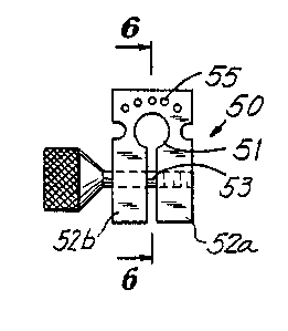
Fig.S is an elevational view of a first jig useful in the
present invention;
Fig.6 is a view in section, taken along lines 6-6 in
Fig.5;
Fig.7 is an elevational view in section of a sealing
adaptor useful in the present invention;
Fig.B is an elevational view of a second jig useful in the
present invention;
Fig.9 is a view in section, taken along the lines 9-9 in
Fig. B;
Fig.lO is a schematic view of a first access cannula
inserted into the herniated disc;
Fig.ll is a view similar to Fig.lO showing the use of the
second jig to index a second access cannula relative
to the first access cannula; and
- 10 -
Fig. l2 is a schematic view showing two access cannulae
placed in the body of the patient.
In the description that follows, instruments are
generally made out of suitable austenitic stainless steel,
unless otherwise specified. While the surgical procedure
described herein refers to decompression of intervertebral
lumbar discs, it is to be understood that the procedure is
not limited to lumbar discectomy and may be used in any
procedure for percutaneously emplacing at least two cannulae
in a patient, such as an intervertebral disc procedure or
operation.
According to the method of the present invention, the
patient is positioned on a radiolucent table in the
appropriate prone or lateral position and guidewire 10
(Fig. l), suitably of about 0.050 in. diameter, is advanced
through the skin of the back posterolaterally under
fluoroscopic observation until the guidewire 10 contacts the
exterior symptomatic side of the annulus fibrosis of the
herniated disc. Thereafter, the cannulated obturator 20
(Fig.2), having a lumen with a diameter slightly larger than
that of the guidewire 10, is passed over the guidewire 10
until the cannulated obturator 20 contacts the external
surface of the annulus fibrosis of the herniated disc. The
removal of the guidewire 10 at this point is optional. An
access cannula 30a (Fig.3), suitably of about 0.25 in. outer
diameter and having external graduations 31 of 10 mm, is
then gassed over the cannulated obturator 20 and advanced to
the external surface of the annulus fibrosis. At this
point, the guidewire 10 is removed if not previously
~OS~S~.
- 11 -
removed. The inner diameter of the access cannula 30a is
sized to closely fit over the cannulated obturator 20. The
cannulated obturator 20 is then removed, and a 3 mm or 5 mm
trephine 40 (Fig.4) is introduced through the access cannula
30a. The trephine 40 has a plurality of saw teeth 40a or
other cutting members. The trephine 40 is advanced into the
annulus of the disc, with rotation, creating an annular
fenestration (that is, a bore) through the annulus fibrosis
into the nucleus. The trephine 40 is then removed.
The cannulated obturator 20 is reintroduced into the
access cannula 30a and passed into the fenestration of the
annulus. Fluoroscopic guidance may be utilized. The access
cannula 30a is then advanced into the fenestration of the
annulus, with rotary movement. After the access cannula 30a
is in the proper position, the cannulated obturator 20 is
removed. The proximal end of cannula 30a projects beyond
the surface of the patient's back (not shown) while the
distal end is in the position shown in Fig.lO. The
procedure described for placement of cannula 30a into the
annulus of the disc follows the procedure described in US
Patent 4,573,448. As is known, suitable local anesthetic is
used as appropriate.
Referring to Fig.lO, the procedure described above
locates the distal end of the access cannula 30a adjacent
the herniation 100 of the disc 101, which protrudes toward
the posterior ligament 102 thus placing pressure on the
nerves 103, which causes the pain characteristic of a
herniated lumbar disc. First jig 50 (Figs.5,6 and 10) is
slid downwardiy over the proximal end of the access cannula
~~8~~~~.
- 12 -
30a by passing the access cannula 30a through the central
bore 51 in the first jig 50. Jig 50 is secured in place
near the proximal end of cannula 30a by tightening the screw
53 thereby clamping the legs 52a and 52b to the access
cannula 30a.
First jig 50 preferably has a plurality of smaller bores
55 each having a diameter substantially the same as the
diameter of the guidewire 10. The axes of the bores 55 are
spaced from and are preferably parallel to the axis of the
large bore 51. Alternatively, jig 50 may have only one
smaller bore 55. Moreover, the bores 55 may be oblique to
the axis of the large bore 51.
Under fluoroscopic observation, the guidewire 10 is slid
through a selected one of the small bores 55 so that the
guidewire 10 will ideally be centred on the annulus
fibrosis. If necessary, a second guidewire 10 is passed
through another of bores 55 and advanced toward the annulus
fibrosis of the disc, while under fluoroscopic observation.
Proper positioning of the guidewire on the annulus is
determined by palpation and, if necessary, by fluoroscopy.
The surgeon can then evaluate the placement of the
guidewires and select the guidewire best positioned to
provide the second fenestration of the annulus of the disc.
having selected the desired guidewire 10, the other
guidewire, if any, is removed, and the guidewire 10 is then
introduced through the fibres of the annulus fibrosis for a
distance of about three to about four millimetres. Jig 50
is removed, leaving the guidewire 10 and access cannula 30a
- 13 -
in place.
Second jig 70 (Figs.8,9 and 11) is secured to access
cannula 30a near the proximal end by passing access cannula
30a through bore 70a, passing the guidewire 10 through bore
70b, and clamping legs 70c together by means of screw 70d.
Cannulated obturator 20 is then advanced over the guidewire
by rotary movement through the bore 70b of the second jig
70 until the cannulated obturator 20 contacts the annulus
fibrosis, as shown in Fig.ll. The guidewire 10 and jig 70
are removed leaving the cannulated obturator 20 in place.
An accessory cannula 30b is passed over the cannulated
obturator 20 and advanced toward the annulus fibrosis.
Accessory cannula 30b is sized to slide in the annulus
between bore 70b and the other surface of cannulated
obturator 20. The cannulated obturator 20 is then removed,
leaving the accessory cannula 30b in place.
Although it is presently preferred to use second jig 70,
it is not necessary to do so. Moreover, while the bores 70a
and 70b are presently preferred to be parallel, in some
cases it may be desired to have one bore oblique to the
other. Also, while it is presently preferred that cannulae
30a, 30b have the same inner and outer diameters, one may
have a smaller inner and/or outer diameter than the other.
The annulus fibrosis is inspected endoscopically through
the accessory cannula 30b, and if satisfactory, a trephine
40 is passed through the accessory cannula 30b and a second
fenestration is cut through the annulus fibrosis into the
nucleus. The trephine 40 is then removed. The accessory
~n8l~d~.~
- 14 -
cannula 30b is advanced into the annulus. Introduction of
both cannulae into the annulus of the disc under
fluoroscopic observation is carried out in a manner known
er se, such as described in US Patent 4,573,488.
Fragments of the herniated disc can be removed through
the desired cannula 30a or 30b by inserting a trephine 40 in
the desired cannula and moving it back and forth within the
nucleus of the herniated disc as suction is applied.
Alternatively, the trephine can be removed and suction may
be applied through the cannula itself. In another method,
forceps, trimmer blades, suction punch forceps, laser
lights, etc. are used to remove such fragments via one of
the cannula.
Preferably, however, before removal of nuclear material,
a sealing adaptor 60 (Fig.7), which is suitably comprised of
silicon rubber, is attached to the proximal extremity of the
access cannula 30a and accessory cannula 30b, as shown in
Fig.l2 with access cannula 30a and accessory cannula 30b
received in boxes 61a and 61b of sealing adaptor 60.
Insertion of access cannula 30a and accessory cannula 30b
into the sealing adaptor will stop when the cannulae contact
shoulders 63 and 64, respectively of bores 61a and 61b.
Nuclear evacuation through one of the cannulae 30a or 30b
and simultaneous arthroscopic observation via the other of
cannulae 30a or 30b is possible by sealingly passing an
arthroscope (not shown) into one of bores 62a and 62b and
thence into one of cannulae 30a or cannula 30b, while a tool
(not shown) is inserted into the other bore and thence into
the other cannula. Nuclear material may then be evacuated
- 15 -
by a conventional powered surgical instrument (not shown)
through the access cannula 30a or accessory cannula 30b
while under arthroscopic observation through the other
cannula. A saline solution may be passed via the
arthroscope through one cannula and excess fluid may be
evacuated through the other cannula. Direct visualization
of the resection of the desired disc material is thus made
possible.
SPG/LC/1357