Note : Les descriptions sont présentées dans la langue officielle dans laquelle elles ont été soumises.
~9753 1
BACKGROUND OF THE lNv~NlION
The present invention relates to an ultrasonic
imaging system for visualizing the structure and
movement in a living body.
Fig. 1 shows a constr~ction of conventional
ultrasonic imaging system. In Fig. l, an ultrasonic probe
51 converts a pulse signal into an ultrasonic signal,
transmits the converted ultrasonic signal to an object
under measurement, receives an echo signal from the object
under measurement, and converts the echo signal into an
electrical signal. Driver means 52 generates the pulse
signal and supplies it to the ultrasonic probe 51. An
amplifier 53 amplifies the echo signal returning from
the object under measurement and received by the
ultrasonic probe 51. Logarithmic conversion (compres-
sion) means 54 performs logarithmic conversion of the
amplitude of the echo signal amplified by the amplifier
53. Detector means 55 performs AM detection of the
logarithmically compressed echo signal and detects an
20 envelope of the echo signal. Scanning conversion means
56 composed of an A/D converter 56a, a memory 56b and a
D/A converter 56c, takes in a detection signal of the
echo signal, which has been AM detected by the detector
means 55, and converts the detection signal to an image
signal according to a TV system represented by the NTSC
_~ .....
_ 209753 1
system to form a two-dimensional tomographic image.
Display means 57 such as a television displays a
tomographic image indicating inner portions of the
object under measurement. Generally, a low-pass filter
for preventing generation of an aliasing noise on the
basis of the sampling theorem is provided at an input
portion of the A/D converter S6a. However, this low-
pass filter is different from filter means of the
present invention, and it is not illustrated here in
order to simplify the eYrl~n~tion of the present
invention.
With respect to the above-described
~o~ L~ction, the operation thereof will be described
herelln~
- The ultrasonic probe Sl is driven by a pulse
signal generated by the driver means 52 to perform
transmitting and receiving operations by transmitting an
ultrasonic pulse, while performing a sC~nni ng operation
with the transmitted ultrasonic pulse, and by receiving
an echo signal returning from a tomographic plane in an
object under measurement. The echo signal thus obtained
is amplified by the amplifier S3 and its amplitude is
subjected to logarithmic conversion by the logarithmic
conversion (compression) means 54. The logarithmically
converted echo signal is subjected to AM detection by
the detector means 55 so that the detection signal
represents an envelope of the echo signal, and the
detection signal is outputted to the scanning conversion
2û97531
1 means 56. The detection signal is converted to a
digital signal by the A/D converter 56a of the scanning
conversion means 56, and the A/D conversion signal is
written in the memory 56b. Since an address of the
memory 56b, in which the A/D conversion signal data is
written, is made to correspond to a position of the
portion of the object under measurement from which the
echo signal has been generated, tomographic image
information of the object under measurement is stored in
the memory 56b, being arranged two-dimensionally
therein. The tomographic image information stored in
the memory 56b is subjected to D/A conversion by the D/A
converter 56c so as to comply with the television
display system for the display means 57 and is outputted
to the display means 57, whereby a tomographic image is
displayed on the display means 57.
As described above, in the conventional
ultrasonic imaging system, the ultrasonic probe 51
performs transmitting and receiving operations of an
ultrasonic pulse, while performing a scanning operation,
- to thereby obtain a tomographic image of portions of an
object under measurement.
In the conventional ultrasonic imaging system,
however, an image displayed on a television display used
as the display means 57 is represented by the scanning
line. Accordingly, the resolution of an image which can
be displayed is determined by the television scanning
line. In addition, even if the resolution of an image
- 2097531
1 is retained within a resolution limit, when displaying a
tomographic image of a fine composition which is near
the resolution limit, a difference in brightness between
adjacent television scanning lines is increased due to
a high spatial frequency of the tomographic image. As a
result, it becomes impossible to represent intermediate
brightness, which raises a problem that the quality of a
diagnostic image is deteriorated significantly.
SUMMARY OF THE INVENTION
The present invention intends to solve the
above-mentioned problem presented in the conventional
ultrasonic imaging system, and it is an object of the
present invention to provide an ultrasonic imaging
system which can represent a tomographic image of high
resolution up to a resolution limit of the television
scanning line of a display means without deteriorating
the quality of the tomographic image.
In order to attain the above-mentioned object,
the ultrasonic imaging system according to the present
invention comprises an ultrasonic probe for transmitting
and receiving an ultrasonic wave, driver means for
driving the ultrasonic probe, detector means for
detecting an echo signal received by the ultrasonic
probe, filter means operative to pass the detection
signal therethrough and having a plurality of passbands
which have respective flat frequency response character-
istics an-d respective gains different from each
" .
. ' 2097531
1 other, scanning conversion means for converting the
detection signal, which has passed through the filter
means, to a display signal for display on a display
system such as a television, and display means for
displaying thereon the display signal converted by the
scanning conversion means in the form of a tomographic
image.
Further, it is preferable to set the gains of
the plurality of passbands of the filter means to
decrease toward higher frequency passbands.
Accordingly, in accordance with the present
invention, an echo signal detected by the detector means
is made to pass through the filter means so that
respective amplitudes of higher frequency components
contained in a detection signal are decreased and a
difference in brightness between adjacent television
scanning lines is reduced. In addition, since
respective frequency response characteristics of a
plurality of passbands of the filter means are
flattened, deterioration of pulse response character-
istics of the detection signal can be prevented.
BRIEF DESCRIPTION OF THE DRAWINGS
Fig. 1 is a schematic block diagram showing a
conventional ultrasonic imaging system.
Fig. 2 is a schematic block diagram showing an
ultrasonic imaging system according to an embodiment of
the present invention.
209753 1
l Fig. 3 is a characteristic diagram showing
frequency response characteristics of the filter means
18 in the system shown in Fig. 2.
Fig. 4 is a circuit diagram showing an example
of the construction of the filter means 18 in the systemshown in Fig. 2.
Fig. 5A is an explanatory drawing illustrating
a change of the gain in the frequency response
characteristics of the filter means 18 in the system
shown in Fig. 2.
Fig. 5B is an explanatory drawing for
explaining a change of the brightness waveform of a
tomographic image caused by the use of the filter means
18 in the system shown in Fig. 2.
DESCRIPTION OF THE PREFERRED EMBODIMENTS
An embodiment of the present invention will be
described hereunder with reference to the accompanying
drawings.
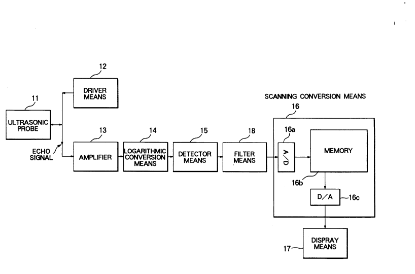
Fig. 2 is a schematic block diagram showing an
ultrasonic imaging system of an embodiment of the
present invention.
Referring to Fig. 2, an ultrasonic probe 11
converts a pulse signal to an ultrasonic signal,
transmits the ultrasonic signal into an object under
measurement, receives an echo signal returning from the
object under measurement, and converts it to an
electrical signal. Driver means 12 generates a pulse
209753 1
signal and sends it to the ultrasonic probe 11. An
amplifier 13 amplifies the echo signal which has
returned from the object and which has been received by
the ultrasonic probe 11. Logarithmic conversion
(compression) means 14 performs logarithmic conversion
of an amplitude of the echo signal amplified by the
amplifier 13. Detector means 15 performs AM detection
of the logari~hmically converted echo signal to thereby
detect an envelope of the echo signal. F;lter means 18
has a plurality of passbands which have respective flat
frequency response characteristics and respective gains
different from each other. The filter means 18 passes
therethrough a detection signal supplied from the
detector means 15. Sc~nni~g conversion means 16 is
composed of an A/D converter 16a, a memory 16b and a D/A
converter 16c, takes in the detection signal outputted
from the filter means 18, and converts the detection
signal to an image signal in compliance with a TV system
represented by the NTSC system, wherein the image signal
represents a two-dimensional tomographic image. Display
means 17 such as a television responds to the image
signal and displays a tomographic image showing inner
portions of the object under measurement.
In addition, in the same way as a conventional
system, a low-pass filter for preventing generation of
an aliasing noise on the basis of the sampling theorem
is provided at an input portion of the A/D converter
16a.
209753 1
1 With the above-described construction of the
system of the present invention, the operation thereof
will be described hereinafter.
The ultrasonic probe 11 is driven by a pulse
signal generated by the driver means 12 to perform
transmitting and receiving operations by transmitting an
ultrasonic pulse, while performing a scanning operation
with the transmitted ultrasonic pulse, and by receiving
an echo signal returning from a tomographic plane in an
object under measurement. The thus obtained echo signal
is amplified by the amplifier 13 and its amplitude is
logarithmically converted by the logarithmic conversion
(compression) means 14. The logarithmically converted
echo signal is subjected to AM detection in the detector
means 15, whereby a detection signal, which represents
an envelope of the echo signal, is obtained, and then
the detection signal is outputted to the filter means
18. The filter means 18 has frequency response
characteristics which are flat in a low frequency region
and a high frequency region, respectively, as shown in
Fig. 3, and which have respective gains in the low
frequency region and the high frequency region, the
gains being relatively different from each other, so
that the amplitudes of components of the detection
signal passing through the filter means 18, which
components are distributed in the high frequency region,
are decreased. The detection signal from the filter
means 18 is subjected to A/D conversion by the A/D
2097531
. .,
1 converter 16a of the scanning conversion means 16 and is
written in the memory 16b. Tomographic image informa-
tion stored in the memory 16b is subjected to D/A
conversion by the D/A converter 16c, that is, it is
S converted in compliance with the television system of
the display means 17, and the D/A conversion signal is
outputted to the display means 17 so that a tomographic
image is displayed on the display means 17.
Next, the filter means 18 will be described in
greater detail. Fig. 4 is a circuit diagram showing an
example of the filter means 18 for realizing the
frequency response characteristics shown in Fig. 3.
Referring to Fig. 4, an inverting amplifier 31 is
connected with a negative feedback circuit formed of
resistors Rfl and Rf2 and a capacitor Cf, and this
circuit of the filter means 18 has frequency response
characteristics represented by the following equations
(1) to (3), in connection with the value of a resistor R
connected to the input end of the inverting amplifier
31:
AL =Rfl/R (1)
A~ =(Rfl-Rf2)/{R-(Rfl + Rf2~ (2)
fb =l/{2~Cf-(Rfl+Rf2)} (3)
In connection with the frequency response
characteristics shown in Fig. 3, the above-mentioned
equations (1) to (3) give the gain AL of the flat
portion of the low frequency region, the gain AH of the
_ - 2U97531
flat portion of the hi~h frequency region, and a
f requency fb at the boundary ~etween the low f requency
region and the high f requency region.
Fig. 5A is an explanatory drawing which
illustrates the distribution of the gain in the
f requency response characteristics of the filter means
18 shown in Fig. 4. Fig. SB is an explanatory drawing
for explaining how a brightness waveform of a tomo-
graphic image displayed on the display means 17 changes
as the gain in the frequency response characteristics of
the filter means 18 changes in accordance with the
values of the resistors Rfl and Rf2 and capacitor Cf.
In the graph of Fig. 5B, the horizontal axis thereof re-
presents a line in a direction perpendicular to respec-
tive television sCAnning lines, and the abscissa re-
presents positions of the respective television scanning
lines which are arranged perpendicularly to the horizon-
tal axis. ~he ordinate of the graph represents bright-
ness of the respective scanning lines on the television
screen. A characteristic curve A shown in Fig. 5A indi-
cates a frequency response characteristic which is flat
over the entire frequency range, that is, a frequency
response characteristic obtained when the resistor Rfl
and the capacitor Cf are not employed, in other words,
this characteristic curve A corresponds to the same con-
struction as that of the conventional system devoid of
the filter means 18. In this case, the brightness wave-
form of a tomographic image is shown by a waveform A in
-- 10 --
A
,..
209753~
Fig. 5B. More particularly, when the detection signal
contains low frequency components, spots indicative of
brightness signal level thereof are distributed with
shorter distance steps on the respective consecutive
television sc~ning lines, as shown at waveform A in the
left half of Fig. SB, and therefore a difference in
brightness level between adjacent television scanning
lines is redu,ced and intermediate brightness tones can
be represented. Also, when the detection signal
contains high frequency components, a large difference
in brightness occurs between adjacent television
scanning lines as shown at waveform A in the right half
of Fig. SB, and therefore the detection signal is
visualized as a binary image devoid of intermediate
brightness, thereby impairing the image quality
significantly. Then, according to the present inven-
tion, values of the resistor Rfl and the capacitor Cf
are selected suitably to decrease the galn AH in the
high frequency region as shown at the characteristic
curve B in Fig. SA, whereby it becomes possible to
reduce the difference in brightness between adjacent
television scanning lines as shown at waveform B in the
right half of Fig. SB, even in the case of the detection
signal containing high frequency components.
Thus, in accordance with the ultrasonic
imaging system of the above-described embodiment of the
present invention, an echo signal detected by the
detector means lS is passed through the filter means 18
A
209753 1
".
1 so that the amplitude of high frequency components
contained in the detected signal can be decreased,
whereby the difference in brightness between adjacent
television scanning lines can be reduced. Further,
since the filter means 18 has flat frequency response
characteristics in a plurality of passbands, deteriora-
tion of pulse response characteristics of the detection
signal can be prevented.
The values of the gain AL in the low frequency
region, the gain AH in the high frequency region, and
the boundary frequency fb are selected in accordance
with the television system of the display means 17.
Therefore, it is necessary to determine the values of
such factors taking into consideration the quality of an
image displayed on the display means 17. In the
standard television system such as the NTSC system, the
boundary frequency fb is set to about 100 to 500kHz, and
the ratio AL: AH between the gain AL in the low
frequency region and the gain AH in the high frequency
region is selected to be about 1:0.6 to 1:0.9.
AS described above, according to the present
invention, by passing an echo signal detected by the
detector means through the filter means, it is possible
to decrease the amplitude of high frequency components
contained in a detection signal and the difference in
brightness between adjacent television scanning lines.
In addition, since the filter means has flat
frequency response characteristics in a plurality of
- 12 -
209753 ~
1 frequency regions, it is possible to avoid degradation
of pulse response characteristics of the detection
signal. As a result, it is possible to produce a
tomographic image of high resolution up to the
resolution limit of the television scanning line of the
display means without impairing the image quality.