Note : Les descriptions sont présentées dans la langue officielle dans laquelle elles ont été soumises.
-` 2 ~ 1 fi ~
ENDOSCOPE WITH ADDITIONAL VIEWING FACILITY
BACKGROUND OF THE INVENTION
This invention relates to an endoscope comprising a
tubular shaft for the introduction thereinto of a viewing
lens and a treatment instrument, the shaft having a distal
end portion which is channel-shaped and provides a viewing
window for the viewing lens.
The present invention proceeds from a pan hystero-
scope such as is marketed, for example by Richard Wolf GmbH
of Knittlingen, Germany, and in which, a viewing lens and a
treatment instrument are arranged adjacent to each other
within the shaft. As the distal end portion of the shaft
is channel-shaped, both the lens and instrument are pro-
tected on one side, while on the other side, that i.s to
say, the open side of the channel-like portion, the lens
and the instrument are exposed in such a way that in use
the instrument can be deflected in the direction of the
open side or displaced axially. Flexible ~orceps, a laser
transmission optical fibre or the like may constitute the
treatment instrument, for the endoscopic examination and
treatment of the uterus, for example.
Silicone implants are commonly used, in particular
for breast reconstruction. Such silicone implants, may,
however, occasionally become encapsulated, within the
breast. There may also be damage to the implant. In such
cases, it is usual to check the implant surgically, release
the encapsulation or, if occasion should arise, exchange
the implant.
It is an object of the present invention to provide
an endoscope which allows of both visual checking of the
implant and release of the encapsulation, as well as,
should occasion arise, the performance of other operations
particularly in the region of the female breast, endoscopi-
cally and with sufficient safety and hence with minimum
invasion.
: , : . , ~
....
'~ :
- 211~
--2--
SUMMARY OF THE INVENTION
According to the invention this is achieved by
providing in the channel-shaped distal end portion of the
shaf~, a recess forming an additional viewing window for
the viewing lens.
This allows of endoscopic operations in the region
of the female breast, involving the inspection of an im-
plant and release of a capsule surround;;ng the implant.
Other operations may, however, also be carried out with
such an endoscope, for example, the removal of tissues, or
for other diagnostic operations. ~he surgeon introduces
the shaft of the endoscope in the region of the nipple and
can then first inspect the implant without open surgery
and, if occasion arises, open an encapsulation surrounding
the implan~ as well. In all such endoscopic operations,
the implant must be protected. To this end the channel-
shaped end portion of the shaft shield~ both the viewing
lens and the treatment instrument, for example a laser
transmission fibre, on one side, since contact between the
fibre and the implant must be avoided. In order to avoid
such contact reliably, however, the duck's bill shape, that
is to say the channel-shape of the distal end portion of
the shaft alone, is insufficient, because when opening the
capsule, the implant must be kept permanently in sight. To
this end the additional viewing window is provided in said
channel~shaped portion of the shaft, in order to give the
implant the necessary protection, and at the sa~e time to
enable visual checking of the implant, or at least of part
of it. The surgeon can therefore observe the implant
permanently during opening of the capsule. The channel-
like shape of the end portion of the shaft also ensures
that the treatment instrument can always be moved and
inserted only in its axial direction or in the direction
away from the implant, that is to say, towaxds the open
side of the channel profile of said end portion of the
shaft, and that the instrument itself slides along the
implant.
' ' ` 2 ~
--3--
Although a laser transmission fibre is preferably
used as the treatment instrument, an HF probe or a
mechanical instrument, for example forceps or a combined
instrument, may also be used. The use of a laser
transmission optical fibre supplied, for example, by means
of a neodymium-YAG laser has proved itself in particular
for the opening of a capsule.
Preferably, the shaft is of essentially oval cross-
section, the channel-shaped end portion being so configured
that it encloses the major semi-axis of said o~al cross-
section. The oval cross-sectional shape of the shaft, in
comparison with a round cross-sectional shape of the same
diameter, affords the advantage of a smaller circumference
and hence less stress on the surgical opening. The viewing
lens and treatment instrument can be arranged ad~acent to
each other in the shaft in such a way that the viewing lens
directly adjoins said channel-shaped shaft portion so that
a clear view through the additional viewing window and a
clear view of the treatment instrument and the tissue
location being treated thereby is ensured.
The distal end of the shaft is, as far as possible,
of a rounded shape in order to allow it to slide inside the
breast with as little friction and injury thereto as possi-
ble. Preferably, however, the channel-shaped end portion
of the shaft is additiona~ly provided with a sloped distal
end face, in order to allow of easier advance of the shaft
in the axial direction within the breast, this being fur-
ther assisted by the supply of flushing liquid.
Particularly during the opening of an implant
capsule, the channel-shaped end portion of the shaft pref-
erably extends not as is usual in hysteroscopes only
through a circumferential angle of about 180, but through
such an angle of at least 200, whereby the distal ends of
viewing lens and treatment instrument are better protected
so that damage to the implant is excluded to a very great
extent. In this case it is preferable that the channel
profile of the distal end portion of the shaft is asymmet-
rical, part of the wall of said profile being extended on
,
. .,
2 1 ~
--4--
one side of the major axis of said oval cross-section.
Preferably, also the recess forming the additional viewin~
window is located in said extended part of the wall. Thus,
during the opening of a capsule not only the implant it-
self, but also the position of the implant relative to the
capsule can be checked by virtue of the aclditional viewing
window.
If the recess has an approximately oval contour as
seen in plan view, it can provide a large enough opening
and the risk of injury can be kept low. The risk of injury
can further be reduced by providing the distal end of the
shaft with an inclined end face at the junction between the
shaft and said channel-shaped distal end portion.
In order to fix the viewing lens reliably within
the shaft and to avoid contact between the lens and the
treatment instrument, the lens may be introduced into, and
fixed in, a tube of approximately D-shaped cross-section
within the shaft. Especially if the treatment instrument
is a laser transmission optical fibre, an additional tube
can be provided in the shaft, for guiding the optical
fibre, the additional tube abutting against the flat side
of the D-cross-section tube within the shaft. If the D~
cross-section tube is arranged eccen~rically in the shaft,
space will remain therein for the introduction of an addi-
tional treatment instrument. In order to avoid collision
between the additional instrument and the sensitive laser
transmission optical fibre, in the distal region of the
shaft, a guide for the additional instrument may be provid-
ed in the distal end of the shaft, the guide being in the
form of a wedge for deflecting the additional instrument
away from the optical fiber as the additional instrument
emerges from the distal end of the shaft. The shaft may be
provided with pipes or conduits for flushing liquid.
A preferred embodiment of the present invention
will now be described by way of example with reference to
the accompanying drawings.
'~ ~" :.
`` s 2 1 1 ~
~RIEF DESCRIP~ION OF THE DRAWINGS
Figure 1 is a schematic side view of an endoscope
according to the preferred embodiment of the invention, in
use in a surgical operation;
Figure 2 is a schematic side view of the endoscope
with the shaft thereof shown in longitudinal section;
Figure 3 is a view taken on the :Lines III-III of
Figure 2;
Figure 4 is an enlarged plan view of the distal end
portion of said shaft;
Figure 5 is a view taken on the lines V-V of Figure
4;
Figure 6 is a view similar to that of Figure 4
showing the distal end portion of the shaft rotated by 90
about its longitudinal axis; and
Figure 7 is a view taken on the lines VII-VII of
Figure 6.
DE~AILED DESCRIPTION OF TH~ PREFERRED EMBODIMENT OF IrHE
INVENTION
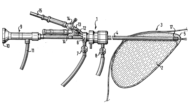
Figure 1, shows an endoscope 1 according to the
preferred embodiment, in use in opening a capsule 5 sur-
rounding an implant 2 within a female human breast 3. A
shaft 4 of the endoscope has been introduced into the
breast 3 through an incision in the region of the nipple
thereof. The endcscope 1 is basically constructed as a
hysteroscope.
~ A flushing connection 7 and a suction connection 8
are provided at the proximal end 6 of the shaft 4, through
which flushing liquid can be conducted to the distal end of
the shaft 4 and conducted away again. A central tube 9 is
provided for the introduction and fixing of a viewing lens
~not shown) in the distal end region of the shaft 4. The
tube 9 has thereon an eyepiece 10 and a lighting connection
11, for the viewing lens. Instrument introduction conduits
12 and 13 extending at an angle from the proximal end 6 of
the shaft 4 are each provided with a closure tap 14. The
conduit 13 receives a first treatment instrument in the
form of an optical fibre 15 for conducting laser light for
-6- 2`~-~.O/~
performing the cutting operation, the distal end of the
fibre 15 being located at the distal end of the shaft 4.
As shown in Figure 3, the shaft 4 is of
substantially oval cross-section and has therein a
D-cross-section tube 16 for receiving and having fixed
therein said viewing lens. The shaft 4 has, as best seen
in Figure 6, a distal end portion 17 formed as a laterally
and distally open channel. The remainder of the shaft 4 is
of fully tubular cross-section. The tube 16 extends over
more than half the cross-section of the shzlft 4 as shown in
Figure 3. The tube 16 is located on the side of the shaft
4 on which the shaft 4 ends in the channel-shaped portion
17. The conduit 13 for the fibre 14 continues as a tube 18
in the shaft 4, which tube is of circular cross-section and
is disposed on one side of the major semi-axis 19 of the
oval cross-section of the shaft 4, as shown in Figure 3, on
the flat side of the D-cross-sQction tube 16. The tube 18
terminates at i-ts distal end in the region of the channel-
shaped poxtion 17. The tube 18 may, however, terminate a
little more proximally of the shaft 4 in order to allow
some elastic deformation of the end of the fibre 15 to
protect it from breaking.
The instrument conduit 12 is defined in the shaft 4
by the flat side of the tube 16, part of the shaft 4 itself
and one side of the tube 18. Within the shaft 4, proximate
to its distal end, is a wedge-shaped guide 20 which tapers
in the proximal direction of the shaft 4 as shown in Figure
2. The guide 20 ensures that a second instrument intro~
duced into the conduit 12 is deflected away from the fibre
15, as the second instrument emerges from the distal end of
the shaft 4, so that the instrument does not damage the
fibre 15.
As shown in Figures 4 to 7, the channel-shaped end
portion 17 of the shaft 4, which extends distally beyond
the fully tubular cross-section of the shaft 4, has a
sloped back distal end 21, as best seen in Figure 6 for
ease in guiding the shaft 4 between the capsule 6 and the
implant 2. As shown in Figures 5 and 7, the channel-shaped
- . . . . .. . . . .
" ~7~ 2110~69
portion 17 of the shaft 4 is asymetrical, as seen in
cross-section with respect to the major cross-sectional
axis 22 of the shaft 4. The profile of the portion 17
encloses the semi-axis 19 and extends on one side thereof
as far as the minor cross-sectional axis 23 of the shaft 4.
A wall of the portion 17 of the shaft 4 on the opposite
side of the semi-axis 19, extends beyond t:he axis 23. The
profile of the portion 17 thus extends over a circumferen-
tial angle of about 200 of the cross-sect:ion of the shaft
4. Said angle may be exceeded according to requirements.
The channel-shaped portion 17 also protects the
implant 2 from accidental collision with, and damage by,
the distal end of the fibre 15 as well as by the distal end
of the viewing lens. Such protection alone, however, is
insufficient, for example, for the release of the capsule
5, which surrounds the implant 2. An additional viewing
window 24 is, thercfore, provided in the portion 17. The
window 24 is in the form of a recess in the wall o the
portion 17, which is approximately oval as seen in Figure
6. The major axis of the window 24 extends axially of the
endoscope. The window 24 is form~d in the flat side of the
portion 17 which extends beyond the minor axis 23 of the
shaft 4. The window 24 could, however, should the occasion
arise, be formed in the bottom of the channel provided by
~he portion 17. The endoscope 1 according to the preferred
embodiment, has, however, proved to be particularly advan-
tageous for opening a capsule 5, that is to say, for cut-
ting through the capsule, because the viewing window 24,
under the protection of the portion 17 enables the implant
2 to be viewed in relation to the capsule 5 during treat-
ment, the point of treatment being viewed as usual through
the viewing lens.
A further sloped end surface 25 is provided in the
region of the distal end of the shaft 4, as shown in Figure
4, at its junction with the channel-shaped portion 17,
further to reduce the risk of injury during the axial
advance of the shaft 4 into the breast 3.
,. , . , . ~ .