Note : Les descriptions sont présentées dans la langue officielle dans laquelle elles ont été soumises.
CA 02206394 1997-0~-27
RADIATION THERAPY METHOD AND DEVICE
BACKGROUND OF THE INVENTION
The present invention generally relates to the use of
radiation therapy to treat a condition such as restenosis and,
more particularly, pertains to the use of an implantable device
to deliver a dose of radiation.
A variety of conditions have been found to be
amenable to treatment by the local irradiation of tissue. In
order to appropriately limit the amount of tissue that is
irradiated, it sometimes is necessary to implant a small source
of radiation and, in order to expose the tissue to a sufficient
dosage of radiation, it has been found advantageous to implant
such device for an extended period of time.
Percutaneoustransluminal coronaryangioplasty (PTCA)
is an established treatment for coronary artery disease. The
procedure involves inserting a balloon catheter through the
vasculature to a position where atherosclerotic plaque has
collected on the vessel wall. The plaque is compressed against
the vessel wall by inflating the balloon located at the distal
end of the catheter in order to increase the diameter of the
vessel and thereby reduce the restriction to blood flow. After
sufficient expansion has been achieved, the balloon is deflated
and removed, and the area of disruption begins to heal.
While this procedure is very widely used, one problem
associated with PTCA is a condition known as restenosis.
Restenosis is the development of further blockage in the
intravascular structure, following an otherwise successful
angioplasty procedure. Restenosis is believed to be an
exaggerated form of the normal healing process of the stretched
tissue. Restenosis is thought to be caused by fibrointimal
proliferation of the stretched wall in which the injured cells
lining the vascular structure multiply and form fibrous tissue.
Such growth at the vascular wall is an almost malignant
phenomenon in which normal cells multiply at a high rate,
thereby creating a new obstruction to flow through the vascular
structure. It occurs in the range of approximately 15-50
percent of the PTCA cases and typically presents within the
CA 02206394 1997-0~-27
first six months following PTCA. Stents have been implanted in
expanded vessels in an effort to maintain patency but do not
appear to have much of an effect on the restenosis rate. In
the event a stent has been implanted, the growth tends to occur
around its ends and through any openings in its walls.
Localized irradiation of the vessel from within the
vessel has been found to be effective in reducing the incidence
of restenosis. To date, such radiation has been delivered via
a number of different vehicles, including by guide wire,
balloon, temporarily implantable wire or permanently-
implantable stent. The delivery device either is partially or
wholly formed of radioactive material or alternatively, is
coated with a radioactive substance. Material giving off high
levels of radiation may be introduced briefly into the body and
then removed. Alternatively, material giving off a relatively
lower level of radiation and with an appropriately short half-
life may be introduced temporarily or, in some instances, left
in place.
A number of shortcomings or disadvantages are
associated with the prior art devices and techniques. With
respect to temporarily implanted devices, implantation time is
limited and therefore the radiation dose necessarily must be
very high. At such high dosage rates, local radiation burns
may be caused on one side of the vessel while the opposite side
may receive a suboptimum dose. Moreover, due to the tendency
of restenosis to occur throughout a six month period, repeated
irradiation procedures would be necessary in order to
adequately address the vagaries of onset.
In the case of permanently implanted devices, a
compromise be made between the shelf life of the device and its
in-vivo efficacious lifetime. If materials with short half-
lives are used, in order to reduce the long term exposure of
the patient to radiation, then the shelf life of the device
necessarily must be short and therefore is undesirable. If, on
the other hand, an isotope is used which will permit a
substantial shelf life, i.e., an isotope having a long half-
life, then the exposure of the patient to radiation will be
CA 02206394 1997-0~-27
long term and may be excessive. Moreover, in view of the fact
that the development of restenosis typically occurs within the
first six months, it has been recognized that it is desirable
to limit irradiation to such a time frame. Of course,
attempting to substantially restrict the release of radiation
from a permanently implantable device to such a limited period
of time imposes further constraints on the shelf life of the
devlce .
Another disadvantage inherent in the heretofore known
delivery devices is related to the need to adequately protect
from exposure to unreasonable radiation dosages all who handle
the device, including the manufacturing, stocking, and shipping
personnel, catheter laboratory personnel, and physicians.
This requires the use of large and cumbersome containers that
further complicate handling and disposal concerns. Some of the
radioisotopes being considered in the industry require ion
implantation into the device or transmutation of the metal in
the device. The complexity of such processes greatly increases
the cost of the devices.
A new approach is necessary that would overcome the
shortcomings of the prior art. It would be desirable to
provide a system by which a very predictable dosage of
radiation can be delivered via a permanently implantable
device. Moreover, it would be most desirable for such device
to be producible at minimal cost, to have a substantial shelf
life and present a minimal risk of exposure to radiation.
SUMMARY OF THE INVENTION
The present invention overcomes the shortcomings of
the techniques and devices heretofore employed to deliver a
dose of radiation to a vascular site. A method is provided for
precisely controlling the dosage that is delivered to the
patient, while concerns relating to shelf-life of the device
are obviated. Moreover, the hazards with respect to the
handling of radioactive devices are substantially mitigated.
Additionally, the present invention provides a method for
CA 02206394 1997-0~-27
quickly and easily rendering an implantable device radioactive.
More particularly, an implantable device is prepared so as to
readily adsorb a preselected amount of radioactive material and
to form a sufficiently strong bond therewith so as to
substantially minimize any subsequent loss thereof upon contact
with bodily fluids. The present invention further provides a
stent or other implantable device which facilitates the
practice of such method.
These advantages generally are achieved by
maintaining separate the implantable hardware and the
radioactive material until just prior to implantation. By
loading a precisely known quantity of material with a known
half-life onto the device and immediately proceeding with the
implantation procedure, a very precise dose of radiation can be
delivered to the patient over a desired period of time.
Particular embodiments of present invention provide
a stent that facilitates the adsorption of a predictable amount
of radioactive material thereon in the surgery room. More
particularly, a stent is provided that is coated with a
chelating agent. A base material and, optionally, a spacer
material first is coated onto the device, after which the
chelator is applied. This approach obviates any shelf life
concerns related to the stent itself and obviates the need for
special handling of the stent prior to loading. The base
material is selected to both form a strong bond with the
surface of the stent as well as with the spacer or chelator
applied thereover. The spacer is selected to form a strong
bond with the underlying base layer as well as with the
chelator and serves to impart a degree of mobility to the
chelator or to increase the number of active sites. Finally,
the chelator is selected to form a strong bond with the base
layer or spacer layer therebelow and of course ultimately
adsorb the radioactive isotope. Such combinations of coatings
are fairly tenacious, are substantially unaffected by the
disinfection processes the stent is normally subjected to and
have no effect on the shelf life of the stent.
CA 02206394 1997-0~-27
Just prior to implantation, the chelator-coated
device is immersed in a solution containing the appropriate
radioactive material to adsorb the radioisotope. The chelator-
isotope combination can be chosen such that the loading is
quantitative with virtually no subsequent release of the
radioactive material from the implanted stent. Knowing the
activity of the material along with the half-life of the
radioisotope renders precisely calculable the total dosage of
radiation that will be delivered. Precautions relating to the
radiation must only be taken when handling the vial containing
the radioactive material and when handling the stent during and
after the loading step.
These and other features and advantages of the
present invention will become apparent from the following
detailed description of preferred embodiments which, taken in
conjunction with the accompanying drawing, illustrate by way of
example the principles of the invention.
BRIEF DESCRIPTION OF THE DRAWINGS
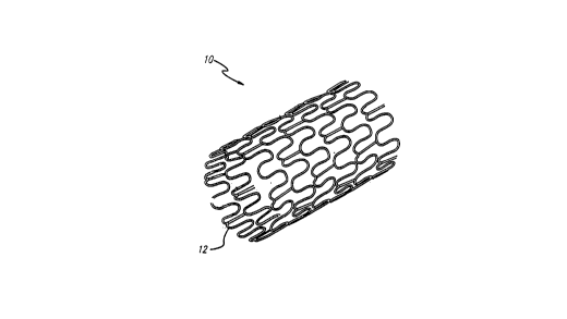
FIGURE 1 is a perspective view of a typical stent
having an open lattice structure and embodying features of the
present invention.
FIG. 2 is a cross-section of one wire strut of the
stent of FIG. 1 depicting the various layers attached to the
stent.
DETAILED DESCRIPTION OF THE PREFERRED EMBODIMENTS
Particular embodiments of the present invention
provide a system for delivering a precise dose of radiation to
a vascular site via an implantable device. The device for
example may be used to prevent restenosis in a blood vessel
that had been subjected to an angioplastic procedure.
The device of the present invention preferably takes
the form of a permanently implantable stent 10. The stent 10
CA 02206394 1997-0~-27
initially is provided in a collapsed state and positioned about
an inflatable balloon on the distal end of a catheter. Upon
maneuvering the balloon into place within the target blood
vessel, the balloon is inflated, which causes the stent to
radially expand. Any of various mechanisms well known in the
art may be incorporated in the stent in order to lock the stent
into its expanded state. Subsequent deflation of the balloon
and extraction of the catheter leaves the expanded stent in
place to maintain the patency of the blood vessel. Further
details of expandable stents and a balloon catheter delivery
system are found in U.S. Patent No. 5,569,295, which issued
October 29, 1996 to Advanced Cardiovascular Systems, Inc. on an
application filed May 3, 1995.
A stent 10 is prepared in accordance with the present
invention to deliver a preselected dose of radiation. FIGS. 1
and 2 depict the stent 10 embodying features of the present
invention. The exterior surface 12 of the stent 10 first is
selectively coated with a base layer 14 that serves as a primer
or foundation. The base material is selected for its ability
to adhere or bond to the surface of the stent while providing
a surface to which the next layer readily bonds. An
intermediate spacer layer 16 optionally is bonded to the base
layer for the purpose of providing sufficient mobility to the
chelating functionality that subsequently is applied thereto
and/or to increase the number of active sites available to the
chelating moiety thereby serving as a chemical amplifier. The
chelator 18 is attached covalently to either the spacer
material 16 or directly to the base layer 14. The chelator is
selected to form a strong bond with the underlying material and
to have a strong affinity for the particular radioisotope to be
used. The top layer is applled just prior to use and comprises
the radioisotope that is adsorbed by the chelator. The
radioisotope is selected based on the type of radiation it
emits and its half-life.
The stent may be constructed of metal or a polymer.
Stainless steel is the preferred material of construction.
CA 02206394 1997-0~-27
The base layer may comprise gold or any organic
coating that contains a nucleophile, or potential nucleophile.
These sites potentially could be aliphatic or benzylic carbons
alpha to an ester, ketone or nitrile (i.e., aliphatic or
benzylic carbons in the alpha position of an ester, ketone or
nitrile). Alternatively, they could be alcohols, amines, ureas
or thiols. Possible base layers include polyurethane, poly
(ethylene-vinyl alcohols), poly (vinyl alcohols), most
hydrogels and polyarcylates.
The spacer layer preferably is attached to the base
layer by nucleophilic substitution due to the degree of control
afforded by such reaction. Alternatively, radical grafting
processes may be employed. Possible spacer materials include
~, ~- mercaptoalkylamines, diisocyanates, diacid chlorides,
dialkylamines, ~, ~- hydroxyalkylamines, dihydroxyalkanes (PE0)
and dimercaptoalkanes.
The chelator is selected to form a covalent bond with
the underlying layer, i.e., either the spacer or the base, and
for a very high binding affinity for the radioisotope.
Possible chelator functionalities include acetates
(monocarboxylic acids), acetylacetone, benzoylacetone, citric
acid, 1,2-diaminocyclohexane-N,N,N',N'-tetraacetic acid,
ethylenediamine-N,N,N',N'-tetraacetic acid, and pyridine-2,6-
dicarboxylic acid.
The radioisotope is selected based on the type of
emission, its half-life and the strength of its bond to the
chelator, which must be sufficient so as not to be displaced by
ions present in the blood. The preferred isotope is a beta
emitter, because gamma radiation penetrates too deeply into
tissue and the energy of alpha particles is insufficient. The
half-life of the radioisotope should be between 24 hours and 2
months, preferably between 2-18 days. The shorter the half-
life, the more problematic becomes the shipping and storage of
the radioactive material, while the longer the half-life, the
more excessive becomes the delivered the dosage in view of the
biological process currently understood to be involved in the
processes of restenosis.
CA 02206394 1997-0~-27
The most preferred combination of materials is a
stainless steel stent, a gold base layer, alpha, omega
mercaptoalkylamine as a spacer, Nl-(2-hydroxethyl)-
ethylenedramine - N,N,Nl - triacetic acid as a chelator and Irl92
as the radioisotope.
In the practice of the invention, the stent first is
prepared by applying the base layer, then optionally the spacer
layer and finally the chelator. The coated stent subsequently
is sterilized and processed along with the stent and associated
devices. The subsequent shelf life and handling constraints
substantially are dictated by the base stent and catheter
rather than by the coating.
The radioisotope, suspended in a solution contained
in a vial, is handled separately according to the general
methods with which hospitals are acquainted. Just prior to
implantation, the stent is immersed in the vial in order to
allow the chelator to adsorb the radioisotope. The loaded
stent subsequently is maneuvered into position within the
patient and expanded to be permanently left in place. The
radiation emitted by stent gradually diminishes as a function
of its half-life but is sufficient during the critical six
month time frame to preclude or at least minimize the chance of
restenosis. Radiation subsequently continues to subside to
insignificant levels obviating the need to remove the device.
While a particular form of the invention has been
illustrated and described, it also will be apparent to those
skilled in the art that various modifications can be made
without departing from the scope of the invention. More
particularly, any type of implantable device may be prepared in
accordance with the invention and the method may b-e practiced
to treat any type of condition that has been found to respond
to the localized irradiation of tissue. Accordingly, it is not
intended that the invention be limited except by the appended
claims.
