Note : Les descriptions sont présentées dans la langue officielle dans laquelle elles ont été soumises.
CA 02250081 1998-09-21
W O97~5~18 PCTnUS97104825
--1--
FOCUSSED ULTRASOUND TISSUE TREATMENT METHOD
Technical Field
This invention relates ~o ultrasound tissue
5 ablation. It is disclosed in the context of prostate
ablation, but it is believed to be useful in other
applications as well.
Background Art
The efficacy of ultrasound as a medium for non-
invasive or minimally invasive tissue removal has been
established. There are, for example the disclosures of
U.S. Patents: 4, 586, 512; 4, 620,546; 4,658,828; 4, 858, 613;
4,9~1,653; 4,955,365; 5,036,855; 5, 054,470; 5,149, 319;
5,215, 680; and, 5,219,401. No representation is intended
hereby that a thorough search of all material prior art has
been conducted or that no more material prior art exists.
Nor should any such representation be inferred.
In some applications, however, some portion o~
20 the transmitted ultrasound energy is not applied to optimal
effect. For example, it is known that in the transrectal
ultrasound ablation of prostate tissue, such as in the
treatment of benign prostatic hyperplasia (BPH), the
posterior lesion (that is, the lesion that forms between
25 the depth at which the urethra passes through the prostate
and the posterior surface of the prostate) is much more
effective in relieving the symptoms of BPH than the
anterior lesion (the lesion that forms between the depth at
which the urethra passes through the prostate and the
30 anterior surface of the prostate). In this sense, the
ultrasound energy that results in the anterior lesion is
"wasted," although the anterior lesion is formed. I~ this
energy could be reflected back posteriorly for integration
with the energy that is absorbed by the posterior prostate,
35 the effectiveness of the ultrasound ablation treatment at
relieving BPH sympto~s would be enhanced.
CA 022~0081 1998-09-21
W O 9713SS18 PCTnUS97/04825
--2--
Disclosure of Invention
- - Accordingly it is an ob~ect of this invention to
provide methods and apparatus by which the effectiveness of
ultrasound as a medium for tissue ablation is enhanced.
According to an aspect of the invention, a method
of treatment of tissue with focussed ultrasound comprises
placing adjacent the tissue to be treated a reflector of
ultrasound, orienting an ultrasound transducer with its
focal point adjacent the reflector and then irradiating the
tissue with focussed ultrasound while the reflector is in
place.
According to another aspect of the invention, a
method of treatment of tissue with focussed ultrasound
comprises placing adjacent the tissue to be treated an
ultrasound energy conversion device which converts received
ultrasound energy to heat, stores the heat and then
releases the heat over time into the tissue to be treated.
An ultrasound transducer is oriented with its focal point
adjacent the ultrasound energy conversion device. The
tissue is then irradiated with focussed ultrasound while
the ultrasound energy conversion device is in place.
Illustratively, the step of placing adjacent the
tissue to be treated a reflector of ultrasound or an
ultrasound energy conversion device comprises the step of
inserting a catheter comprising an ultrasound reflective
material or ultrasound energy converting and heat storage
material into a body lumen or orifice which lies adjacent
the tissue to be treated.
Additionally illustratively, the step of
inserting a catheter comprising an ultrasound reflective
material or an ultrasound energy converting and heat
storage material into a body lumen or orifice comprises the
step of inserting a catheter containing red rubber into the
body lumen or orifice.
Further illustratively, the step of inserting a
catheter comprising an ultrasound reflective material or
CA 022~0081 1998-09-21
WO 9713~518 PCT~US97~825
--3--
ultrasound energy converting and heat storage material into
- a body lumen or orifice which lies adjacent the tissue to
be treated comprises the steps of inserting a balloon
catheter into the urethra to the depth of the bladder and
- 5 inflating the balloon in the bladder.
Additionally illustratively, the step of
irradiating the tissue with ultrasound comprises
irradiating the prostate with the high intensity focussed
ultrasound.
Further illustratively, the step of orienting the
ultrasound transducer with its focal point adjacent the
reflector comprises the step of coupling the transducer
through the rectal wall.
Brief Description of the Drawings
The invention may best be understood by referring
to the following description and accompanying drawings
which illustrate the invention. In the drawings:
Fig. 1 illustrates a step in the prior art
~0 transrectal high intensity focussed ultrasound (HIFU)
ablation of a diseased prostate;
Fig. 2 illustrates a corresponding step to the
one illustrated in Fig. 1, but performed according to the
present invention;
Fig. 3 illustrates a much enlarged view of a
detail of the step illustrated in Fig. 2;
Fig. 4 illustrates a view of the detailed
illustration in Fig. 3 taken generally along section lines
4-4 of Fig. 3; and
Fig. S illustrates temperature profiles obtained
with a system constructed and operated in accordance with
the teachings of Figs. 2-4.
Modes of Practicing the Invention
A limitation on the effectiveness of HIFU as a
treatment for BPH can best be appreciated by referring to
CA 02250081 1998-09-21
W 097/3SS18 PCTAUS97~4825
--4--
Fig. 1. An ultrasound transducer 20 in a coupling fluid 22
- is inserted into the colon 24 of a sufferer of BPH directly
behind the affected prostate 26. Numerous techniques for
visualization of this region are known. Many systems for
visualization employ the same transducer 20 in a low power,
visualization mode for visuallzation of the prostate 26,
and in a HIFU mode for subsequent ablation therapy. The
visualization is usually accomplished with the aid of a
urethral catheter (not shown~ having a balloon end and an
inflating lumen for inflating the balloon in the neck 32 of
the bladder 34 to anchor the catheter. The catheter is
visible in the visualizing intensity ultrasound generated
by transducer 20.
After the visualization of the neck 32 of the
bladder 34, the urethra 35 and surrounding diseased
prostate 26 has been completed and the transducer 20 is
oriented for treatment of the prostate 26, the balloon is
deflated, the catheter is removed and the HIFU treatment of
the prostate 26 for BPH commences according to a treatment
format established by the treating physician. The lesion
36 which results from the application of the HIFU is
somewhat elongated, wlth a somewhat larger posterior
portion 38 of the lesion 36 lying between the rectal wall
40 and the focal zone 42 of the ultrasound beam 44 in the
vicinity of (indeed here at) the urethra 35. A somewhat
smaller portion 4~ of the lesion 36 lies anterior to the
urethra 35 toward the lower front of the abdomen. Although
the HIFU energy which is absorbed by the prostate 26
anterior to the urethra 35 does result in an anterior
lesion 48, the relief of BPH symptoms afforded by the
anterior lesion 48 is not as great as the relief afforded
by the posterior lesion 38.
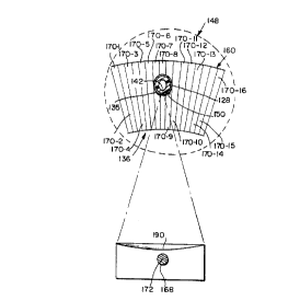
Referring now to Figs. 2, 3 and 4, according to
the invention, an ultrasound transducer }20 in a coupling
fluid 122 is inserted into the colon 1~4 of a sufferer of
BPH directly behind the affected prostate 126.
CA 02250081 1998-09-21
W 09713SS18 PCTrUS97/~nS
--5--
Visualization of the treatment field is accomplished with
- the aid of a urethral catheter 128 having a balloon end 130
and an inflating lumen for inflating the balloon 130 in the
neck 132 of the bladder 134 and to anchor the catheter 128.
The catheter 128 is visible in the visualizing intensity
ultrasound for transducer 120 After the visualizat~on of
- the urethra 135 and surrounding diseased prostate 126 has
been completed and the transducer 120 is oriented for
treatment of the prostate 126, the catheter 128 is not
removed. Rather the HIFU treatment of the prostate 126 for
BPH commences with the catheter 128 in place in the urethra
135 according to a treatment format established by the
treating physician.
The lesion 136 which results from the application
of the HIFU is limited almost exclusively by the presence
of catheter 128 to between the rectal wall 140 and the
focal zone 142 of the ultrasound beam 144 in the vicinity
of the surface of catheter 128. In this way, the HIFU
incident on the catheter 128 is reflected posteriorly and
combines with the direct HIFU to achieve an effective
lesion 136, generally with lower input power to transducer
120. Almost no HIFU energy is transmitted to, or absorbed
by, the prostate 126 anterior to the urethra 135.
Therefore, there is no "wasted" lesion in region 148. The
relief of BPH symptoms is delivered more effectively and
with lower input power by limiting the lesion to region
136. This effect is further enhanced by what appears to be
cavitation bubble "seeding'l of region 136 adjacent catheter
128 and urethra 135. As the HIFU is applied, ca~itation
bubbles 150 appear readily in this area. The cavitation
bubbles trap both some HIFU energy incident on catheter 128
and some HIFU energy reflected from catheter 128, and
release this trapped energy back into the posterior lesion
prostate tissue as they rupture, enhancing the efficiency
of the HIFU at producing the posterior lesion 136.
Microscopic imperfections in the outer wall of catheter 128
CA 022~0081 1998-09-21
W 097/35518 PCTnUSg7104825
--6--
are believed to contribute to this cavitation bubble
- enhancement phenomenon.
Various kinds of catheters 128 have been
employed. Suitable catheters include Surgicot~ red rubber
catheters, Dow Corning Silastic~ Foley catheters, Olbert
urological catheters, Bardex Foley catheters and Baxter
urological catheters.
A suitable treatment format can best be
appreciated by referring to Figs. 2-5. A region of the
prostate 126 to be treated is divided into a grid 160, with
sections 162-1, 162-2 . . . 162-12 spaced uniformly along
the adjacent rectal wall 140 capable of being individually
addressed for HIFU treatment by moving the transducer 120
in the directions indicated by double ended arrow 166 into
and out of the colon 124. Uniformly angularly spaced
sectors 170-1, 170-2 . . . 170-16 are capable of being
individually addressed by rotation of the treatment
transducer 120 about its axis 172 on drive shaft 168, as
indicated by double ended arrow 174. A code wheel (not
shown) can be fixed on the shaft 168 for reading the
angular orientation of the transducer 120 in accordance
with known principles. See, for example, U.S. Patent
4,664,121. In this manner, the tissue of the prostate 126
in the treatment region 160 can be treated, one
longitudinal 162-1, . . . 162-12 and angular 170-1, . . .
170-16 sector at a time. Such a treatment format will
result in temperature profiles illustrated in Fig. 5. Body
temperature is illustrated by curve 180. As will be
appreciated body temperature remote from the treatment site
160 is unaffected by the treatment. The temperature of the
coupling liquid, in this case, deionized, degassed water in
the transducer probe 182 is illustrated by curve 184. The
reason for the discontinuity at about 39 minutes of the
treatment regimen is that an external coupling liquid
circulation circuit ~not shown) was activated at that time,
resulting in circulation of the coupling liquid through the
*rB
. . ~
CA 022~0081 1998-09-21
W O 97/35518 PCTnUSg7/04825
--7--
external circuit and the resultant cooling of the coupling
- liquid. Curve 188 is a temperature profile of the rectal
wall 140. The rectal wall 140 lies in the near field of
the treatment transducer 120, that is, between the emitting
surface 190 of the treatment transducer 120 and the focal
zone 142 of the treatment transducer 120. Curve 196 is a
temperature profile of the surface of the catheter 128.
The spikes in the various temperature profiles occur when
the focal zone 142 is very close to the various
thermocouples used to generate the temperature profiles.
Curve 196 clearly establishes the efficacy of the treatment
method of the present invention employing the HIFU energy
conversion, heat retention and heat radiation capability of
catheter 128 when catheter 128 is left in place during the
HIFU treatment.
Because the treatment thermal dosage is
proportional to ~T~t where T is any elevated temperature at
which tissue is maintained and ~t is the time during which
the tissue is maintained at that temperature, raising the
temperature of the tissue-catheter 128 surface interface,
both by ultrasound absorption and reradiation, and by
reflection of ultrasound, will assure more complete tissue
destruction.
The above-noted materials are illustrative of
materials that absorb ultrasound, convert it into heat and
store and radiate the heat into the surrounding tissue.
Polymers generally are noted for exhibiting these same
characteristics, that is, enhanced effects of HIFU exposure
of tissue. These effects can be further augmented by the
use of Albunex or other microbubble or microbubble
production-enhancing materials. These materials can be
used to enhance cavitation of liquids in the tissue, making
the benefits of cavitation as a tissue ablation mechanism
more accessible at lower applied HIFU power densities.
Additionally, the presence of the red rubber, Silastic~,
and other polymer material catheters 128 promotes
CA 022~0081 1998-09-21
WO9713~S18 PCT~S97/0~2s
--8--
cavitation even without the presence of other cavitation-
- inducing agents at much lower applied HIFU powers
(sometimes reduced by half or more). The mismatched
foreign material and tissue acoustic impedances produce
larger reflected energy as well, accounting for some of the
enhanced tissue ablation demonstrated by the presence of
these foreign material catheters 128 in the urethra 135.
The foreign material provides an enhanced environment for
cavitation at the foreign material-tissue lnterface. The
increased energy released at this interface gives rise to
higher pressure and shock waves, disintegrating the tissue
and mechanically destroying the tissue in a predictable
manner. This promotes sloughing off of the tissue in the
treatment zone 136, providing more immediate and lasting
relief of BPH symptoms.
Also, when the transducer 120 is being used in
the visualization mode, because lower applied power can
induce cavitation and because cavitation bubbles are
extremely echogenic, the treatment site 136 is much more
readily visualized with the foreign object 128 in place.
With the foreign object 128 in place in the body,
differences in ultrasound tissue absorption coefficients
become much less critical in the application of sufficient
HIFU to achieve ablation. Thus, the effects of variations
in the tissues encountered in the near field between the
transducer 120 and the focal zone 142 in a patient, or from
patient to patient, on patient treatment regimens and
formats are reduced.