Note : Les descriptions sont présentées dans la langue officielle dans laquelle elles ont été soumises.
CA 02268007 1999-04-09
WO 98I16643 PCTIUS96/16484
Brain-Associated Inhibitor of
Tissue-Type Plasminogen Activator
Field of the Invention
The present invention relates to a novel human gene encoding a polypeptide
expressed in human brain tissue which is a member of the serine protease
inhibitor
("serpin") superfamily and appears to be a human homolog of "neuroserpin," a
serpin
recently identified in the chicken. More specifically, isolated nucleic acid
molecules
are provided encoding a human polypeptide named Brain-Associated Inhibitor of
Tissue-Type Plasminogen Activator, hereinafter referred to as "BAIT." BAIT
polypeptides are also provided) as are vectors, host cells and recombinant
methods for
producing the same. The invention further relates to screening methods for
identifying agonists and antagonists of BAIT ac~:ivity. Also provided are
diagnostic
methods for detecting disorders related to the central and peripheral nervous
system
and the circulatory system, and therapeutic methods for treating such
disorders.
Background of the' Invention
Localized proteolytic activity through the; action of proteases plays a
critical
regulatory role in a variety of important biological processes. For instance,
the
enzyme plasmin plays such a role in hemostasis, angiogenesis, tumor
metastisis,
cellular migration and ovulation. Plasmin is generated from its precursor
zymogen
plasminogen by the action of plasminogen activators (PAs) such as tissue-type
PA (t-
PA) and urokinase-type (u-PA), both of which au-e serine proteases. The
activity of
the PA system is precisely regulated by several mechanisms) one of which
involves
the interaction of t-PA and u-PA with specific pl asminogen activator
inhibitors.
Among these serine protease inhibitors (i.e.) serpins), plasminogen activator
inhibitor
type 1 (PAI-1 ) is unique in its ability to efficiently inhibit u-PA as well
as the single
and two-chain forms of t-PA. High PAI-1 levels are associated with an
increased risk
of thromboembolic disease, while PAI-I deficiency may represent an inherited
autosomal recessive bleeding disorder. See, for- instance, Reilly, T. M., et
al.)
Recombinant plasminogen activator inhibitor type 1: a review of structural,
functional,
and biological aspects, Blood Coag. And Fibrirrolysis 5:73-81 ( 1994).
Serpin Mechanism
The serpins are a gene family that encompasses a wide variety of protein
products, including many of the proteinase inhit~itors in plasma (Huber &
Carrell,
1989; full citations of references cited in this section on Serpin Mechanism
are listed at
SUBSTITUTE SHEEI' (RULE 26)
CA 02268007 1999-04-09
WO 98/16643 PCT/US96I16484
2
the end of this section). However, in spite of their name, not all serpins are
proteinase
inhibitors. They include steroid binding globulins, the prohormone
angiotensinogen,
the egg white protein ovalbumin, and barley protein Z, a major constituent of
beer.
The serpins are thought to share a common tertiary structure (Doolittle. 1983)
and to
have evolved from a common ancestor (Hunt & Dayhoff. 1980). Proteins with
recognizable sequence homology have been identified in vertebrates, plants,
insects
and viruses but not, thus far, in prokaryotes (Huber & Carrell. 1989; Sasaki.
1991;
Komiyama, Ray, Pickup, et al. 1994). Current models of serpin structure are
based
largely on seminal X-ray crystallographic studies of one member of the family)
a-1-antitrypsin (aIAT), also called a-1-proteinase inhibitor (Huber & Carrell.
l989).
The structure of a modified form of aIAT, cleaved in its reactive center, was
solved
by Loebermann and coworkers in 1984 (Loebermann, Tokuoka, Deisenhofer, &
Huber. I984). An interesting feature of this structure was that the two
residues
normally comprising the reactive center (Met-Ser), were found on opposite ends
of the
0
I S molecule, separated by almost 70 A. Loebermann and coworkers proposed that
a
relaxation of a strained configuration takes place upon cleavage of the
reactive center
peptide bond, rather than a major rearrangement of the inhibitor structure. In
this
model, the native reactive center is part of an exposed loop, also called the
strained
loop (Loebermann, ~Tokuoka, Deisenhofer, & Huber. 1984; Carrell & Boswell.
1986;
Sprang. l992). Upon cleavage, this loop moves or "snaps back", becoming one of
the central strands in a major ~3-sheet structure ((3-sheet A). This
transformation is
accompanied by a large increase in thermal stability (Carrell & Owen. l985;
Gettins &
Harten. l988; Bruch, Weiss, & Engel. l988; Lawrence, Olson, Palaniappan, &
Ginsburg. 1994b).
Recent crystallographic structures of several native serpins, with intact
reactive
center loops, have confirmed Loebermann's hypothesis that the overall native
serpin
structure is very similar to cleaved aIAT, but that the reactive center loop
is exposed
above the plane of the molecule (Schreuder, de Boer, Dijkema, et al. 1994;
Carrell,
Stein, Fermi, & Wardell. 1994; Stein) Leslie, Finch, Turnell, McLaughlin, &
Carrell.
1990; Wei, Rubin, Cooperman, & Christianson. 1994). Additional evidence for
this
model has come from studies where synthetic peptides, homologous to the
reactive
center loops of a 1 AT, antithrombin III (ATIII), or PAI-1 when added in
trans,
incorporate into their respective molecules, presumably as a central strand of
~i-sheet
A (Bjiirk, Ylinenjarvi, Olson, & Bock. l992; Bjork, Nordling, Larsson, &
Olson.
SUBSTITUTE SHEET (RULE 26)
CA 02268007 1999-04-09
WO 98/16643 PCTIUS96/16484
3
1992; Schulze, Baumann, Knof, Jaeger, Huber, & Laurell. 1990; Carrell, Evans,
&
Stein. 1991; Kvassman, Lawrence) & Shore. 1995). This leads to an increase in
thermal stability similar to that observed following cleavage of a serpin at
its reactive
center, and converts the serpin from an inhibitor to a substrate for its
target proteinase.
A third serpin structural form has also been identified, the so-called latent
conformation. In this structure the reactive cen~:er loop is intact, but
instead of being
exposed, the entire amino-terminal side of the reactive center loop is
inserted as the
central strand into ~i-sheet A (Mottonen, Strand, Symersky, et al. 1992). This
accounts for the increased stability of latent PAl(-1 (Lawrence, Olson,
Palaniappan, &
Ginsburg. 1994a) as well as its lack of inhibitory activity (Hekman &
Loskutoff.
1985). The ability to adopt this conformation is not unique to PAI-1, but has
also
now been shown for ATIII and a 1 AT (Carrell, Stein, Fermi, & Warden. l 994;
Lomas, Elliot) Chang, Warden, & Carrell. 1995). Together, these data have led
to the
hypothesis that active serpins have mobile reactive center loops, and that
this mobility
is essential for inhibitor function (Lawrence) Strandberg, Ericson, & Ny.
1990;
CarreIl, Evans, & Stein. I991; Carrell & Evans. 1992; Lawrence, Olson)
Palaniappan) & Ginsburg. 1994b; Shore, Day, Francis-Chmura, et a1. 1994;
Lawrence, Ginsburg) Day, et al. l995; Fa, Karolin, Aleshkov) Strandberg)
Johansson) & Ny. 1995; Olson, Bock) Kvassman, et al. 1995). The large increase
in
thermal stability observed with loop insertion, is presumably due to
reorganization of
the five stranded ~-sheet A from a mixed parallel-antiparallel arrangement to
a six
stranded, predominantly antiparallel ~i-sheet (Carrell & Owen. l985; Gettins &
Harten. 1988; Bruch, Weiss, & Engel. 1988; Lawrence, Olson, Palaniappan, &
Ginsburg. 1994a). This dramatic stabilization has led to the suggestion that
native
inhibitory serpins may be metastable structures, kinetically trapped in a
state of higher
free energy than their most stable thermodynamic; state (Lawrence, Ginsburg,
Day, et
a1. 1995; Lee, Park, & Yu. l996). Such an energetically unfavorable structure
would
almost certainly be subject to negative selection, and thus its retention in
all inhibitory
serpins implies that it has been conserved for functional reasons.
The serpins act as "suicide inhibitors" that react only once with a target
proteinase forming an SDS-stable complex. They interact by presenting a "bait"
amino acid residue, in their reactive center) to the enzyme. This bait residue
is thought
to mimic the normal substrate of the enzyme and to associate with the
specificity
crevice, or S 1 site, of the enzyme (Carrell & Boswell. 1986; Huber & Carrell.
1989;
Bode & Huber. 1994}. The bait amino acid is callled the P 1 residue, with the
amino
SUBSTITUTE SHEET' (RULE 26)
CA 02268007 1999-04-09
WO 98l16643 PCTlUS96116484
4
acids toward the N-terminal side of the scissile reactive center bond labeled
in order
P 1 P2 P3 etc. and the amino acids on the carboxyl side labeled P 1' P2' etc.
(Carrell &
Boswell. l986). The reactive center PI-PI' residues, appear to play a major
role in
determining target specificity. This point was dramatically illustrated by the
identification of a unique human mutation, alAT "Pittsburgh", in which a
single
amino acid substitution of Arg for Met at the P 1 residue converted a 1 AT
from an
inhibitor of elastase to an efficient inhibitor of thrombin, resulting in a
unique and
ultimately fatal bleeding disorder (Owen) Brennan, Lewis, & Carrell. 1983).
Numerous mutant serpins have been constructed, demonstrating a wide range of
changes in target specificity, particularly with substitutions at P 1 (York,
Li, &
Garden. l991; Strandberg, Lawrence, Johansson, & Ny. 1991; Shubeita, Cottey,
Franke, & Gerard. 1990; Lawrence, Strandberg, Ericson, & Ny. 1990; Sherman,
Lawrence, Yang, et al. 1992).
The exact structure of the complex between serpins and their target
proteinases
has been controversial. Originally it was thought that the complex was
covalently
linked via an ester bond between the active site serine residue of the
proteinase and the
new carboxyl-terminal end of the P1 residue, forming an acyl-enzyme complex
(Moroi & Yamasaki, 1974; Owen, 1975; Cohen, Gruenke, Craig, & Geczy. l977;
Nilsson & Wiman. 1982). However, in the late 1980s and early 1990s it was
suggested that this interpretation was incorrect, and that the serpin-
proteinase complex
is instead trapped in a tight non-covalent association similar to the so
called standard
mechanism inhibitors of the Kazal and Kunitz family (Longstaff & Gaffney) J.
1991;
Shieh, Potempa, & Travis. 1989; Potempa, Korzus, & Travis. l994).
Alternatively;
one study suggested a hybrid of these two models where the complex was frozen
in a
covalent but un-cleaved tetrahedral transition state configuration (Matheson,
van
Halbeek, & Travis. l991 ). Recently however, new data by several groups have
suggested that the debate has come full circle, with various studies using
independent
methods indicating that the inhibitor is indeed cleaved in its reactive-center
and that the
complex is most likely trapped as a covalent acyl-enzyme complex (Lawrence,
Ginsburg, Day, et al. 1995; Olson, Bock, Kvassman, et al. l995; Fa, Karolin,
Aleshkov, Strandberg, Johansson, & Ny. l995; Wilczynska, Fa, Ohlsson, & Ny.
1995; Lawrence, Olson, Palaniappan, & Ginsburg. 1994b; Shore, Day, Francis-
Chmura, et al. 1994; Plotnick, Mayne, Schechter, & Rubin. l996).
Recently, three groups have almost simultaneously proposed similar
mechanisms for serpin inhibition (Lawrence, Ginsburg, Day, et al. 1995;
Wilczynska,
Fa, Ohlsson, & Ny. 1995; Wright & Scarsdale. 1995). This model suggests that
SUBSTITUTE SHEET (RULE 26)
CA 02268007 1999-04-09
WO 98I15643 PCT/US95/16484
upon encountering a target proteinase, a serpin binds to the enzyme forming a
reversible complex that is similar to a Michaelis complex between an enzyme
and
substrate. Next, the proteinase cleaves the P 1-P 1 ' peptide bond resulting
in formation
of a covalent acyl-enzyme intermediate. This cleavage is coupled to a rapid
insertion
of the reactive center loop (RCL) into (3-sheet A. at least up to the P9
position. Since
the RCL is covalently linked to the enzyme via the active-site Ser, this
transition
should also affect the proteinase, significantly changing its position
relative to the
inhibitor. If, during this transition, the RCL is prevented from attaining
full insertion
because of its association with the enzyme) and i'.he complex becomes locked,
with the
RCL only partially inserted, then the resulting stress might be sufficient to
distort the
active site of the enzyme. This distortion would then prevent efficient
deacylation of
the acyl-enzyme intermediate, thus trapping the ~:.omplex. However, if RCL
insertion
is prevented, or if deacylation occurs before RCI. insertion then the cleaved
serpin is
turned over as a substrate and the active enzyme released. This means that
what
determines whether a serpin is an inhibitor or a 4;ubstrate is the ratio of
kd;S' to ks~;,b. If
deacylation (k~;ss) is faster than RCL insertion (kst;,b) then the substrate
reaction
predominates. However, if RCL insertion and distortion of the active site can
occur
before deacylation then the complex is frozen as si covalent acyl-enzyme. A
similar
model was first proposed in 1990 (Lawrence, Strandberg, Ericson, & Ny. 1990}
and
is consistent with studies demonstrating that RCI, insertion is not required
for
proteinase binding but is necessary for stable inhiibition (Lawrence) Olson,
Palaniappan, & Ginsburg. 1994b) as well as the observation that only an active
enzyme can induce RCL insertion (Olson, Back) Kvassman, et al. l995). Very
recently, direct evidence for this model was provided by Plotnick et al., who
by NMR
observed an apparent distortion of an enzyme's catalytic site in a serpin-
enzyme
complex (Plotnick, Mayne, Schechter, & Rubin. 1996). In conclusion, these data
suggest that serpins act as molecular springs where the native structure is
kinetically
trapped in a high energy state. Upon association with an enzyme some of the
energy
liberated by RCL insertion is used to distort the active site of the enzyme,
preventing
deacylation and trapping the complex.
References Cited in Serpin Mechanism Section
Bjork, L, Nordling, K., Larsson, L, & Olson, S. 'C. ( 1992). Kinetic
characterization
of the substrate reaction between a complex of antithrombin with a synthetic
reactive-
bond loop tetradecapeptide and four target proteinases of the inhibitor. The
Journal of
Biological Chemistry, 267, 19047-19050.
SUBSTITUTE SHEET (RULE 26)
CA 02268007 1999-04-09
WO 98I16643 PCT/US96/16484
6
Bjork, L, Ylinenjarvi, K., Olson, S. T., & Bock, P. E. ( l992). Conversion of
antithrombin from an inhibitor of thrombin to a substrate with reduced heparin
affinity
and enhanced conformational stability by binding of a tetradecapeptide con
esponding
to the P1 to P14 region of the putative reactive bond loop of the inhibitor.
The Journal
of Biological Chemistry, 267, 1976-1982.
Bode, W., & Huber, R. ( 1994). Proteinase - Protein Inhibitor Interactions.
Fibrinolysis, 8, 161-171.
Bruch, M., Weiss, V., & Engel, J. ( 1988). Plasma serine proteinase inhibitors
(serpins) exhibit major conformational changes and a large increase in
conformational
stability upon cleavage at their reactive sites. The Journal of Biological
Chemistry,
263) 16626-16630.
Carrell, R.W., & Boswell, D.R. ( 1986). Serpins: the superfamily of plasma
serine
proteinase inhibitors. In A.J. Barrett & G. Salvesen (Eds.), Proteinase
Inhibitors.
(pp. 403-420). Amsterdam: Elsevier Science Publishers (Biomedical Division).
Carrell) R.W., Evans, D.L., & Stein, P.E. ( 1991 ). Mobile reactive centre of
serpins
and the control of thrombosis. Nature, 353, 576-578.
Carrell, R.W., & Evans, D.L.I. { l992). Serpins: mobile conformations in a
family of
proteinase inhibitors. Curr Opin Struct Biol, 2) 438-446.
Carrell, R.W., & Owen, M.C. ( l985). Plakalbumin, alpha-1-antitrypsin,
antithrombin and the mechanism of inflammatory thrombosis. Nature, 317, 730-
732.
Carrell) R.W., Stein, P.E., Fermi, G., & Warden, M.R. ( l994). Biological
implications of a 3 A structure of dimeric antithrombin. Structure, 2, 257-
270.
Cohen, A.B., Gruenke, L.D., Craig, J.C., & Geczy, D. ( l977). Specific lysine
labeling by 180H- during alkaline cleavage of the a-1-antitrypsin-trypsin
complex.
Proceedings of the National Academy of Sciences,USA, 74, 43l 1-4314.
Doolittle) R.F. ( 1983). Angiotensinogen is related to the antitrypsin-
antithrombin-
ovalbumin family. Science, 222, 4l7-419.
SUBSTITUTE SHEET (RULE 26)
CA 02268007 1999-04-09
WO 98l16643 PCT/US96/16484
7
Fa, M., Karolin, J., Aleshkov, S., 5trandberg, L., Johansson, L.B.-A., & Ny,
T.
( 1995). Time-Resolved Polarized Fluorescence Spectroscopy Studies of
Plasminogen
Activator Inhibitor Type 1: Conformational Changes of the Reactive Center upon
Interactions with Target proteases, Vitronectin ,and Heparin. Biochemistry,
34)
13833-13840.
Gettins, P.) & Harten) B. ( I988). Properties of thrombin- and elastase-
modified
human antithrombin III. Biochemistry, 27, 3634-3639.
Hekman, C.M., & Loskutoff, D.J. ( 1985). Endothelial cells produce a latent
inhibitor
of plasminogen activators that can be activated by denaturants. The Journal of
Biological Chemistry, 260, 1 I581-l1587.
Huber, R., & Carrell) R.W. (l989). Implications of the three-dimensional
structure of
alpha 1-antitrypsin for structure and function of serpins. Biochemistry, 28)
8951-
8966.
Hunt, L.T., & Dayhoff) M.O. ( 1980). A surprising new protein superfamily
containing ovalbumin, antithrombin III, and alphal-proteinase inhibitor.
Biochemical
and Biophysical Research Communications, 95, 864-871.
Komiyama, T., Ray) C.A., Pickup) D.J., Howard, A.D., Thornberry, N.A.,
Peterson, E.P., & Sal vesen. G. ( 1994). Inhibition of interleukin-1 b
converting
enzyme by the cowpox virus serpin CrmA. An e:~cample of cross-class
inhibition. The
Journal of Biological Chemistry) 269, 19331-19:337.
Kvassman, J., Lawrence, D.) & Shore, J. ( 199S). The acid stabilization of
plasminogen activator inhibitor-1 depends on protonation of a single group
that affects
loop insertion into b-sheet A. J Biol Chem, 270, 27942-27947.
Lawrence, D.A., Ginsburg, D., Day, D.E., Berk:enpas, M.B., Verhamme) LM.,
Kvassman, J.-O., & Shore, J.D. ( 1995). Serpin-Protease Complexes are Trapped
as
Stable Acyl-Enzyme Intermediates. J Biol Chem, 270, 25309-25312.
Lawrence, D.A., Olson, S.T., Palaniappan, S., & Ginsburg, D. ( 1994a).
Engineering plasminogen activator inhibitor-1 (P~~I-1) mutants with increased
functional stability. Biochemistry, 33) 3643-3648.
SUBSTITUTE SHEET (RULE 26)
CA 02268007 1999-04-09
WO 98/16643 PCT/US96/16484
8
Lawrence, D.A., Olson, S.T., Palaniappan) S., & Ginsburg, D. ( 1994b). Serpin
reactive-center loop mobility is required for inhibitor function but not for
enzyme
recognition. The Journal of Biological Chemistry) 269, 27657-27662.
Lawrence, D.A., Strandberg, L., Ericson, J., & Ny, T. ( l990). Structure-
function
studies of the SERPIN plasminogen activator inhibitor type 1: analysis of
chimeric
strained loop mutants. The Journal of Biological Chemistry, 265, 20293-20301.
Lee, K.N., Park, S.D., & Yu, M.-H. ( 1996). Probing the native strain in al-
antitrypsin. Nature Structural Biology) 3) 497-500.
Loebermann, H., Tokuoka, R., Deisenhofer, J., & Huber, R. ( 1984). Human al-
proteinase inhibitor. Crystal structure analysis of two crystal modifications,
molecular
model and preliminary analysis of the implications for function. J Mol Biol,
177, 531-
557.
Lomas, D.A., Elliot, P.R., Chang, W.-S.W., Wardell, M.R., & Carrell, R.W.
( 1995). Preparation and characterization of latent a 1-antitrypsin. J Biol
Chem, 270,
5282-5288.
Longstaff, C., & Gaffney, P., J. ( 1991 ). Serpin-serine protease binding
kinetics:
alpha-2-antiplasmin as a model inhibitor. Biochemistry, 30, 979-986.
Matheson, N.R., van Halbeek, H.) & Travis, J. ( 199l ). Evidence for a
tetrahedral
intermediate complex during serpin-proteinase interactions. The Journal of
Biological
Chemistry, 266, 13489-1349l.
Moroi, M., & Yamasaki, M. ( l974). Mechanism of the interaction of bovine
trypsin
with human al-antitrypsin. Biochim Biophys Acta, 359, 130-141.
Mottonen, J., Strand, A., Symersky, J., Sweet, R.M., Danley, D.E., Geoghegan,
K.F., Gerard, R.D., & Goldsmith, E.J. ( 1992). Structural basis of latency in
plasminogen activator inhibitor-1. Nature, 355, 270-273.
Nilsson, T., & Wiman, B. ( l982). On the structure of the stable complex
between
plasmin and a2-antiplasmin. FEBS Lett, 142, 111-114.
SUBSTITUTE SHEET (RULE 26)
CA 02268007 t999-04-09
WO 98/16643 PCTIUS96/16484
9
Olson, S.T., Bock, P.E., Kvassman, J., Shore) J.D., Lawrence, D.A., Ginsburg,
D., & Bjorl, I. (1995). Role of the catalytic serine in the interactions of
serine
proteinases with protein inhibitors of the serpin family. J Biol Chem, 270,
30007-
30017.
Owen, M.C., Brennan, S.O.) Lewis) 3.H., & (:arrell, R.W. ( l983). Mutation of
antitrypsin to antithrombin: alphal-antitrypsin Pittsburgh (358 Met-Arg), a
fatal
bleeding disorder. N Engl J Med, 309, 694-69,i.
Owen, W.G. ( 1975). Evidence for the formation of an ester between thrombin
and
heparin cofactor. Biochim Biophys Acta, 405, 380-387.
Plotnick, M.L, Mayne, L., Schechter, N.M., & Rubin, H. ( l996). Distortion of
the
active site of chymotrypsin complexed with a s~~rpin. Biochemistry, 35, 7586-
7590.
Potempa, 3., Korzus, E.) & Travis, J. ( 1994). The serpin superfamily of
proteinase
inhibitors: structure, function, and regulation. The Journal of Biological
Chemistry,
269,I5957-l5960.
Sasaki, T. ( 1991 ). Patchwork-structure serpins from silkworm (Bombyx mori)
larval
hemolymph. Eur J Biochem, 202, 255-261.
Schreuder, H.A., de Boer, B., Dijkema, R., Mulders) J., Theunissen, H.J.M.)
Grootenhuis, P.D.J., & Hol, W.G.J. ( 1994). The intact and cleaved human
antithrombin III complex as a model for serpin-proteinase interactions. Nature
Structural Biology, 1, 48-54.
Schulze, A.J., Baumann, U., Knof, S., Jaeger, E., Huber, R., & Lauren, C. (
1990).
Structural transition of al-antitrypsin by a peptide sequentially similar to b-
strand s4A.
Eur J Biochem, 194, 51-56.
Sherman, P.M., Lawrence, D.A., Yang, A.Y., ~~andenberg, E.T., Paielli, D.,
Olson,
S.T., Shore, J.D., & Ginsburg) D. ( l992). Satur;ition mutagenesis of the
plasminogen activator inhibitor-1 reactive center. The Journal of Biological
Chemistry, 267, 7588-7595.
SUBSTITUTE SHEET (RULE 26)
CA 02268007 1999-04-09
WO 98l16643 PCT/US96/1b484
Shieh, B.H., Potempa, J., & Travis, J. ( 1989). The use of alpha 2-antiplasmin
as a
model for the demonstration of complex reversibility in serpins. J Biol Chem,
264,
13420-l3423.
5 Shore, J.D., Day, D.E., Francis-Chmura, A.M., Verhamme, L, Kvassman, J.,
Lawrence, D.A., & Ginsburg, D. ( l994). A fluorescent probe study of
plasminogen
activator inhibitor-1: Evidence for reactive center loop insertion and its
role in the
inhibitory mechanism. The Journal of Biological Chemistry, 270, 5395-5398.
10 Shubeita, H.E., Cottey, T.L., Franke, A.E., & Gerard, R.D. ( 1990).
Mutational and
immunochemical analysis of plasminogen activator inhibitor 1. The Journal of
Biological Chemistry, 265, l8379-18385.
Sprang, S.R. (1992). The latent tendencies of PAI-1. Trends Biochem Sci, 17,
49-
50.
Stein, P.E., Leslie, A.G.W., Finch, J.T., Turnell, W.G., McLaughlin, P.J., &
Carrell, R.W. (l990). Crystal structure of ovalbumin as a model for the
reactive
centre of serpins. Nature, 347, 99-102.
Strandberg, L., Lawrence, D.A., Johansson, L.B., & Ny, T. ( 199l ). The
oxidative
inactivation of plasminogen activator inhibitor type 1 results from a
conformational
change in the molecule and does not require the involvement of the P 1'
methionine.
The Journal of Biological Chemistry, 266, l3852-13858.
Wei, A.) Rubin, H., Cooperman, B.S., & Christianson, D.W. ( 1994). Crystal
structure of an uncleaved serpin reveals the conformation of an inhibitory
reactive
loop. Nature Structural Biology, 1, 251-258.
Wilczynska) M., Fa, M., Ohlsson, P.-L, & Ny, T. ( 1995). The Inhibition
Mechanism of Serpins: Evidence that the mobile reactive center loop is cleaved
in the
native protease-inhibitor complex. The Journal of Biological Chemistry, 270,
29652-
29655.
Wright, H.T., & Scarsdale, J.N. ( l995). Structural basis for serpin inhibitor
activity.
Proteins, 22, 210-225.
SUBSTITUTE SHEET (RULE 26)
CA 02268007 1999-04-09
WO 98/16643 PCT/US96/16484
York) J.D., Li, P., & Garden, S.J. ( 1991 ). Combinatorial mutagenesis of the
reactive
site region in plasminogen activator inhibitor I. The Journal of Biological
Chemistry,
266) 8495-8500.
***
During the development of the nervous, system, neurons form axons which
extend along a prespecif ed path into the target area, where they engage in
the
formation and refinement of synaptic connections. These stages depend
critically on
the capability of the axonal growth cones to interact with a variety of
structures which
they encounter along their way and at their destination. These structures
include cell
surfaces of neuronal and non-neuronal origin and the extracellular matrix.
Along their
trajectory and at their target sites, growth cones. not only receive and
respond to
signals from their Local environment, but also actively secrete
macromolecules. In
particular, secreted proteases have been implicated in supporting the growth
cone
advancement through the tissue. Mare than a decade ago, it was demonstrated
that
I S pIasminogen activators are axonally secreted b~~ neurons in culture.
Recently, their
occurrence in the developing rat nervous systenn during the period of axon
outgrowth
has been revealed. Moreover, several pieces of evidence were presented which
indicated that serine proteases, such as plasminogen activators or thrombin,
are
involved in restructuring of the synaptic connectivity during development and
regeneration. Such processes include elimination during development and
synaptic
plasticity associated with learning and memory in the adult. See) for
instance,
Osterwalder, T., et al., "Neuroserpin, an axonally secreted serine protease
inhibitor,"
EMBO J. l5:2944-2953 ( I 996).
During normal development of the nervous system, about 50% of postmitotic
lumbosacral motoneurons undergo naturally occurring (programmed) cell death
during
a period when these cells are forming synaptic connections with their target
muscles.
Naturally occurring motoneuron death has been described in many vertebrate
species,
including chicken, mouse, rat, and human embryos or fetuses. For example,
programmed motoneuron death occurs between embryonic day (E)6 and E10 in the
chicken. This system has been used as a biological model for testing different
neuratrophic agents an motoneuron survival in vivo. See, for instance,
Houenou) L.
J., et al., "A serine protease inhibitor, protease n.exin I, rescues
motoneurons from
naturally occurring and axotomy-induced cell death," Proc. Natl. Acad. Sci.
USA
92:895-899 ( l995).
Although programmed cell death is completed before birth in mammals, the
maintenance of motoneurons continues to be dependent on support from the
target for
some time after birth. Thus, if transection of motor axons is performed in
neonatal
SUBSTITUTE SHEE1' (RULE 26)
CA 02268007 1999-04-09
WO 98l16643 PCT/US96/16484
12
mammals and reinnervation is prevented, a large number of motoneurons
degenerate
and die. Axotomy-induced death of motoneurons has also been extensively used
as a
model for testing the survival effects of various agents, including
neurotrophic and
growth factors on motoneurons.
Protease nexin I (PNI), also known as glia-derived nexin, is a 43-47-kDa
protein that was first found secreted by cultured fibroblasts but is also
produced by
glial (glioma and primary) and skeletal muscle cells. PNI has been shown to
promote
neurite outgrowth from different neuronal cell types. These include
neuroblastoma
cells, as well as primary hippocampal and sympathetic neurons. The neurite-
promoting activity of PNI in vitro is mediated by inhibition of thrombin, a
potent
serine protease. PNI (mRNA and protein) is transiently up-regulated in rat
sciatic
nerve after axotomy, and PNI-producing cells are localized distal to the
lesion site.
This up-regulation of PNI occurs 2-3 days after a similar up-regulation of
prothrombin and thrombin in the distal stump. Free PNI protein is
significantly
decreased, while endogenous PNI-thrombin complexes are increased, in various
anatomical brain regions, including hippocampus of patients with Alzheimer
disease.
When considered together with the recent demonstration that PNI can promote
the in
vitro survival of mixed mouse spinal chord neurons and that PNI is released
from giia
cells by neuropeptides such as vasoactive intestinal polypeptide, these
observations
suggest that PNI may play a physiological role in neuronal survival)
differentiation,
and/or axonal regeneration in vivo.
Recently, it has been reported that PNI rescues spinal motoneuron death in the
neonatal mouse. Houenou, L. J. et al., l995, supra. The survival effect of PNI
on
motoneurons during the period of programmed cell death was not associated with
increased intramuscular nerve branching. PNI also significantly increased the
nuclear
size of motoneurons during the period of programmed cell death and prevented
axotomy-induced atrophy of surviving motoneurons. These results indicate a
possible
role of PNI as a neurotrophic agent. They also support the idea that serine
proteases
or, more precisely, the balance of proteases and serpins may be involved in
regulating
the fate of neuronal cells during development.
More recently, a cDNA encoding an axonally secreted glycoprotein of central
nervous system (CNS) and peripheral nervous system (PNS) neurons of the
chicken
has been cloned and sequenced. Osterwalder, T.) et al., l996) supra. Analysis
of the
primary structural features characterized this protein as a novel member of
the serpin
superfamily which was therefore called "neuroserpin." No demonstration of
inhibition of any protease was included in this report, however. In situ
hybridization
revealed a predominately neuronal expression during the late stages of
neurogenesis
SUBSTITUTE SHEET (RULE 26)
CA 02268007 1999-04-09
WO 98116643 PCT/US96/16484
13
and in the adult brain in regions which exhibit synaptic plasticity. Thus, it
has been
suggested that neuroserpin may function as an axonally secreted regulator of
the local
extracellular proteolysis involved in the reorg~~nization of the synaptic
connectivity
during development and synapse plasticity in the adult. A role for serine
proteases
and serpins in neuronal remodeling is further supported by the finding that
elevated
tPA mRNA and protein levels are found in cerebellar Purkinje neurons of rats
undergoing motor learning (Seeds NW; Williams BL; Bickford P.C., "Tissue
plasminogen activator induction in Purkinje neurons after cerebellar motor
learning."
Science 270:1992-4 (1995)).
The amplification of a human cDNA fragment of about 450 by corresponding
to the region of the chicken cDNA encoding th.e putative reactive site loop of
the so-
called neuroserpin, using a polymerase chain reaction with two pairs of nested
primers
flanking that region, has also been reported. Osterwalder, T., et aL) 1996,
supra)
page 2946. The authors also reported that the deduced amino acid sequences of
the
human and corresponding mouse cDNA exhibited a sequence identity of 88% and
87% respectively, with chicken neuroserpin. rJo nucleotide or amino acid
sequence
was reported for this human cDNA. However, the present inventors are not aware
of
any other public disclosure of full length cDNA sequence data for a human
counterpart
of the chicken neuroserpin cDNA or polypeptide.
Thus, there is a need for human polype,ptides that function as serpins in the
regulation of various serine proteases) particul~~rly in the nervous system,
since
disturbances of such regulation may be involved in disorders relating to
hemostasis,
angiogenesis, tumor metastisis) cellular migration and ovulation, as well as
neurogenesis; and, therefore, there is a need for identification and
characterization of
such human polypeptides which can play a role in preventing, ameliorating or
correcting such disorders.
Summary of the Invention
The present invention provides isolated nucleic acid molecules comprising a
polynucleotide encoding the human BAIT polypeptide having the amino acid
sequence
shown in Figure 1 (SEQ ID N0:2) or the amino acid sequence encoded by the cDNA
clone deposited in a bacterial host as ATCC Deposit Number 97722 on September
18,
1996. The nucleotide sequence determined by sequencing the deposited BAIT
clone,
which is shown in Figure 1 (SEQ ID NO:1 )) contains an open reading frame
encoding
a complete polypeptide of 410 amino acid residues, including an initiation
codon at
positions 89-91, and a predicted molecular weight of about 46.4 kDa. The
encoded
SUBSTITUTE SHEET (RULE 26)
CA 02268007 1999-04-09
WO 98I16643 PCT/US96/16484
14
polypeptide has a leader sequence of 18 amino acids, underlined in Figure 1;
and the
amino acid sequence of the expressed mature BAIT protein is also shown in
Figure 1,
as amino acid residues 19-410 (SEQ ID N0:2).
The human BAIT protein of the present invention has been shown to exhibit
selective inhibition of tissue-type plasminogen activator (t-PA) with
relatively little
inhibition of trypsin, thrombin or urokinase-type plasminogen activator (u-
PA). The
human BAIT polypeptide also shares extensive sequence homology with the
translation product of the mRNA for a serpin-related protein isolated from
brain cDNA
library which has been named "neuroserpin" (SEQ ID N0:3) (see Figure 2). As
noted
above, neuroserpin in the chicken is thought to play an important an important
role in
regulation of local extracellular proteolysis involved in the reorganization
of the
synaptic connectivity during development and synapse plasticity in the adult.
The
homology between neuroserpin and BATT (90% amino acid similarity) indicates
that
BAIT also may play a similar role in neurogenesis in humans.
Thus, one aspect of the invention provides an isolated nucleic acid molecule
comprising a polynucleotide having a nucleotide sequence selected from the
group
consisting of: (a) a nucleotide sequence encoding the BAIT polypeptide having
the
complete amino acid sequence in Figure 1 (SEQ ID N0:2); (b) a nucleotide
sequence
encoding the expressed mature BAIT polypeptide having the amino acid sequence
at
positions 19-410 in Figure 1 (SEQ ID N0:2); (c) a nucleotide sequence encoding
the
BATT polypeptide having the complete amino acid sequence encoded by the cDNA
clone contained in ATCC Deposit No. DEPOSIT; (d) a nucleotide sequence
encoding
the mature BAIT polypeptide having the amino acid sequence encoded by the cDNA
clone contained in ATCC Deposit No. DEPOSIT; and (e) a nucleotide sequence
complementary to any of the nucleotide sequences in (a), (b), (c) or (d)
above.
Further embodiments of the invention include isolated nucleic acid molecules
that comprise a polynucleotide having a nucleotide sequence at least 90%
identical,
and more preferably at least 95%, 96%, 97%, 98% or 99% identical, to any of
the
nucleotide sequences in (a), (b), (c), (d) or (e), above, or a polynucleotide
which
hybridizes under stringent hybridization conditions to a polynucleotide in
(a}, (b), (c),
(d) or (e), above. This polynucleotide which hybridizes does not hybridize
under
stringent hybridization conditions to a polynucleotide having a nucleotide
sequence
consisting of only A residues or of only T residues. An additional nucleic
acid
embodiment of the invention relates to an isolated nucleic acid molecule
comprising a
polynucleotide which encodes the amino acid sequence of an epitope-bearing
portion
of a BAIT polypeptide having an amino acid sequence in (a), (b), (c) or (d),
above.
SUBSTITUTE SHEET (RULE 26)
CA 02268007 1999-04-09
WO 98I16643 PCT/US96/16484
The present invention also relates to recombinant vectors, which include the
isolated nucleic acid molecules of the present invention, and to host cells
containing
the recombinant vectors) as well as to methods of making such vectors and host
cells
and for using them for production of BAIT polypeptides or peptides by
recombinant
5 techniques.
The invention further provides an isolated BAIT polypeptide having an amino
acid sequence selected from the group consisting of: (a) the amino acid
sequence of
the BAIT polypeptide having the complete amino acid sequence including the
leader
sequence shown in Figure 1 (SEQ ID N0:2); (b) the amino acid sequence of the
10 mature BAIT polypeptide (without the leader) '.having the amino acid
sequence at
positions 19-410 in Figure 1 {SEQ ID N0:2); (c) the amino acid sequence of the
BAIT polypeptide having the complete amino .acid sequence, including the
leader,
encoded by the cDNA clone contained in ATCC Deposit No. 9?722; and (d) the
amino acid sequence of the mature BAIT polypeptide having the amino acid
sequence
15 encoded by the cDNA clone contained in ATCC Deposit No. 97722 The
polypeptides
of the present invention also include polypeptiales having an amino acid
sequence at
least 80% identical, more preferably at least 90~'lo identical, and still more
preferably
95%) 96%, 97%, 98% or 99% identical to tho~;e described in (a), (b), (c) or
(d)
above, as well as polypeptides having an amino acid sequence with at least 90%
similarity, and more preferably at least 95%, 9i5%, 97%, 98% or 99%
similarity, to
those above.
An additional embodiment of this aspect of the invention relates to a peptide
or
polypeptide which has the amino acid sequence of an epitope-bearing portion of
a
BAIT polypeptide having an amino acid sequence described in (a), (b), (c) or
(d),
above. Peptides or polypeptides having the amino acid sequence of an
epitope-bearing portion of a BATT polypeptide of the invention include
portions of
such polypeptides with at least six or seven, preferably at least nine, and
more
preferably at least about 30 amino acids to about 50 amino acids, although
epitope-bearing polypeptides of any length up to~ and including the complete
amino
acid sequence of a polypeptide of the invention <iescribed above also are
included in
the invention.
In another embodiment, the invention provides an isolated antibody that binds
specifically to a BAIT polypeptide having an amino acid sequence described in
(a),
(b), (c) or (d) above. The invention further provides methods for isolating
antibodies
that bind specifically to a BAIT polypeptide having an amino acid sequence as
described herein. Such antibodies are useful diagnostically or therapeutically
as
described below.
SUBSTITUTE SHEET (RULE 26j
CA 02268007 1999-04-09
WO 98I16643 PCT/US96/16484
16
The present invention also provides a screening method for identifying
compounds capable of enhancing or inhibiting a biological activity of the BAIT
polypeptide, which involves contacting a protease which is inhibited by the
BAIT
polypeptide with the candidate compound in the presence gf a partially
inhibitory
amount of BAIT polypeptide, assaying proteolytic activity of the protease on a
susceptible substrate in the presence of the candidate compound and partially
inhibitory amount of BAIT polypeptide, and comparing the proteolytic activity
to a
standard level of activity, the standard being assayed when contact is made
between
the protease and its substrate in the presence of the partially inhibitory
amount of
BAIT polypeptide and the absence of the candidate compound In this assay, an
increase in inhibition of proteolytic activity over the standard indicates
that the
candidate compound is an agonist of BAIT inhibitory activity and a decrease in
inhibition of proteoiytic activity compared to the standard indicates that the
compound
is an antagonist of BAIT inhibitory activity.
In another aspect, a screening assay for agonists and antagonists is provided
which involves determining the effect a candidate compound has on BATT binding
to
the active site of a susceptible protease. In particular) the method involves
contacting
the BATT-susceptible protease with a BAIT polypeptide and a candidate compound
and determining whether BAIT polypeptide binding to the BAIT-susceptible
protease
is increased or decreased due to the presence of the candidate compound.
The present inventor has discovered that BAIT is expressed in whole human
brain, and to a much lesser extent in adult pancreas and adult heart. For a
number of
disorders of the central or peripheral nervous system, significantly higher or
lower
levels of BAIT gene expression may be detected in certain tissues (e.g., adult
brain,
embryonic retina, cerebellum and spinal chord) or bodily fluids (e.g., serum,
plasma,
urine, synovial fluid or spinal fluid) taken from an individual having such a
disorder,
relative to a "standard" BAIT gene expression level, i.e., the BATT expression
level in
healthy tissue from an individual not having the nervous system disorder.
Thus, the
invention provides a diagnostic method useful during diagnosis of nervous
system
disorders, which involves: (a) assaying BAIT gene expression level in cells or
body
fluid of an individual; (b) comparing the BATT gene expression level with a
standard
BATT gene expression level, whereby an increase or decrease in the assayed
BAIT
gene expression level compared to the standard expression level is indicative
of
disorder in the nervous system.
An additional aspect of the invention is related to a method for treating an
individual in need of an increased level of BAIT activity in the body (i_e.,
insufficient
protease inhibitory activity of BATT and/or excessive protease activity of a
protease
SUBSTITUTE SHEET (RULE 26)
CA 02268007 1999-04-09
WO 98I16643 PCT/US96116484
17
inhabited by BAIT, particularly t-PA), which method comprises administering to
such
an individual a composition comprising a therapeutically effective amount of
an
isolated BAIT polypeptide of the invention or ~~n agonist thereof.
A still further aspect of the invention is related to a method for treating an
individual in need of a decreased level of BAl7f activity in the body (i.e.,
less
inhibition of a protease susceptible to BAIT) comprising, administering to
such an
individual a composition comprising a therapeutically effective amount of a
BATT
antagonist. Preferred antagonists for use in the present invention are BAIT-
specific
antibodies.
Brief Description of the Figures
Figure I shows the nucleotide sequence (SEQ ID NO:1 ) and deduced amino
acid sequence (SEQ ID N0:2) of the human B~~IT polypeptide. The leader
sequence
of 18 amino acids is underlined.
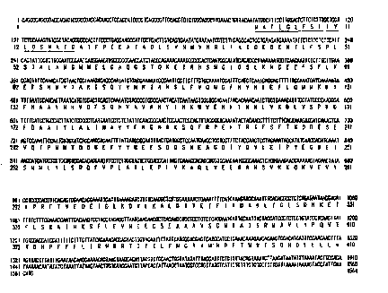
Figure 2 shows the regions of identity between the amino acid sequences of
the human BAIT protein and other indicated serpins with which the human BATT
polypeptide shares significant homology, as foll'~.ows: bovine plasminogen
activator
inhibitor-1 (BovPAI 1; SEQ ID N0:4); rat glial-cjerived nexin I (RatGDNI; SEQ
ID
N0:5); mouse antithrombin III (MusATIII; SE~1 ID N0:6); chicken neuroserpin
(ChkNSP;SEQ ID N0:3). The sequence alignment was generated with the Pileup
module of the Genetics Computer Graup (Wisconsin Package, Version 8, using the
parameters GapWeight = 3.000) GapLengthWe:ight = 0. l00). The reactive site
loops
(from positions 415-452 in Figure 2 (corresponding to BAIT residues 342-378 in
Figure 1; SEQ ID N0:2) are double-underlined, and critical positions in this
sequence are labeled P,~ to P, and P,' according to Schechter and Berger,
Biochem.
Biopys. Res. Commun. 27:157- l 62 ( 1967). The putative reactive site (cleaved
by a
target protease), between Arg at BAIT position ~~62 and Met at BAIT position
363, is
marked with an arrow ('~).
Figure 3 shows an analysis of the BAIT amino acid sequence. Alpha, beta,
turn and coil regions; hydrophilicity and hydrophobicity; amphipathic regions;
flexible
regions; antigenic index and surface probability acre shown. In the "Antigenic
Index -
Jameson-Wolf' graph, the location of the highly antigenic regions of the BAIT
protein, i.e., regions from which epitope-bearing peptides of the invention
may be
obtained.
Figure 4 shows the relationship between the deposited cDNA clone (identified
as clone HSDFB5501X; SEQ ID NO:1) and three: related cDNA clones of the
SUBSTITUTE SHEET (RULE 26)
CA 02268007 1999-04-09
WO 98/16643 PCT/US96/16484
18
invention, designated HPBCT06R (SEQ ID N0:7), HBPDG64R (SEQ ID N0:8),
and HPBCR79R (SEQ ID N0:9).
Figure 5 shows the results of tests for inhibitory activity of purified human
BAIT polypeptide on several proteolytic enzymes including thrombin (2 nM; -O-
);
tissue-type plasminogen activator (tPA, 5 nM; -O-), urokinase-type plasminogen
activator (uPA, 2 nM; -D-), plasmin (5 nM; -O-), and trypsin (2 nM; -~-)
Detailed Description
The present invention provides isolated nucleic acid molecules comprising a
polynucleotide encoding a human BAIT polypeptide having the amino acid
sequence
shown in Figure 1 (SEQ ID N0:2), which was determined by sequencing a cloned
cDNA. The nucleotide sequence shown in Figure 1 (SEQ ID NO:1 ) was obtained by
sequencing the HSDFBSSS01 clone, which was deposited on September 18, 1996 at
the American Type Culture Collection, 1230I Park Lawn Drive, Rockville,
Maryland
20852, and given accession number ATCC 97722. The deposited clone is contained
in the pBluescript SK(-) plasmid (Stratagene, La Jolla, CA).
Nucleic Acid Molecules
Unless otherwise indicated, all nucleotide sequences determined by
sequencing a DNA molecule herein were determined using an automated DNA
sequencer (such as the Model 373 from Applied Biosystems, Inc., Foster City,
CA),
and all amino acid sequences of polypeptides encoded by DNA molecules
determined
herein were predicted by translation of a DNA sequence determined as above.
Therefore, as is known in the art for any DNA sequence determined by this
automated
approach, any nucleotide sequence determined herein may contain some errors.
Nucleotide sequences determined by automation are typically at least about 90%
identical, more typically at least about 95% to at least about 99.9% identical
to the
actual nucleotide sequence of the sequenced DNA molecule. The actual sequence
can
be more precisely determined by other approaches including manual DNA
sequencing
methods well known in the art. As is also known in the art, a single insertion
or
deletion in a determined nucleotide sequence compared to the actual sequence
will
cause a frame shift in translation of the nucleotide sequence such that the
predicted
amino acid sequence encoded by a determined nucleotide sequence will be
completely
different from the amino acid sequence actually encoded by the sequenced DNA
molecule, beginning at the point of such an insertion or deletion.
SUBSTITUTE SHEET (RULE 26)
CA 02268007 1999-04-09
WO 98/16643 PCT/US96/16484
19
Unless otherwise indicated, each "nucleotide sequence" set forth herein is
presented as a sequence of deoxyribonucleotidea (abbreviated A) G) C and T).
However, by "nucleotide sequence" of a nucleic acid molecule or polynucleotide
is
intended, for a DNA molecule or polynucleotidn, a sequence of
deoxyribonucleotides)
and for an RNA molecule or polynucleotide) the; corresponding sequence of
ribonucleotides (A, G, C and U), where each th:ymidine deoxyribonucleotide (T)
in
the specified deoxyribonucleotide sequence is replaced by the ribonucleotide
uridine
(U). For instance, reference to an RNA molecule having the sequence of SEQ ID
NO: l set forth using deoxyribonucleotide abbre~riations is intended to
indicate an
RNA molecule having a sequence in which each deoxyribonucleotide A, G or C of
SEQ ID NO:1 has been replaced by the corresponding ribonucleotide A, G or C,
and
each deoxyribonucleotide T has been replaced b,y a ribonucleotide U. Using the
information provided herein, such as the nucleotide sequence in Figure 1, a
nucleic
acid molecule of the present invention encoding ;~ BATT polypeptide may be
obtained
using standard cloning and screening procedures, such as those for cloning
cDNAs
using mRNA as starting material. Illustrative of the invention, the nucleic
acid
molecule described in Figure 1 (SEQ ID NO:1 ) was discovered in a cDNA library
derived from whole human brain. Additional cD~NA clones of the BAIT gene were
also identified in cDNA libraries from the following tissuesapinal cord,
pineal gland
and adrenal gland tumor.
The determined nucleotide sequence of the BAIT cDNA of Figure 1 (SEQ ID
NO:1 ) contains an open reading frame encoding a protein of 410 amino acid
residues,
with an initiation codon at positions 89-91, and a predicted molecular weight
of about
46.4 kDa. The encoded polypeptide has a leader sequence of 18 amino acids,
underlined in Figure 1; and the amino acid sequence of the expressed mature
BAIT
protein is also shown in Figure 1, as amino acid residues 19-410 (SEQ ID
N0:2).
The amino acid sequence of the BAIT protein shown in Figure 1 (SEQ ID N0:2) is
about 80 % identical to the published mRNA for chicken neuroserpin
(Osterwalder,
T., et al., l996, supra) as shown in Figure 2. Fil;ure 2 shows the regions of
identity
between the amino acid sequences of the human 1=3ATT protein and other
indicated
serpins with which the human BAIT polypeptide shares significant homology) as
follows: bovine plasminogen activator inhibitor-1 (BovPAII; SEQ ID N0:4); rat
glial-derived nexin I (RatGDNI; SEQ ID NO:S); mouse antithrombin III
(MusATIII;
SEQ ID N0:6); chicken neuroserpin (ChkNSP;SEQ ID N0:3).
Sequence comparisons suggest that the chicken neuroserpin and BAIT are
orthologs of one another and are distantly related to the better characterized
mammalian serpins seen in figure 2. There is 77 i~ homology at the DNA level
SUBSTITUTE SHEET (RULE 26)
CA 02268007 1999-04-09
WO 98I16643 PCT/US96/16484
between BAIT and neuroserpin which translates into 90% and 80% amino acid
similarity and identity, respectively. Amino acid identities between the
mammalian
serpins and BAIT drop to about 30%. Moreover, within the functionally
important
reactive site loop, there is only one conservative amino acid change between
BAIT and
5 neuroserpin. There are 7 non-conservative changes between BAIT and PAI-1 in
the
same 38 amino acid region. The active site Pl-P1' residues, however, are
perfectly
conserved between BAIT, neuroserpin, and PAI-1. The BAIT region corresponding
to the ATIII heparin-binding site has 4 acidic amino acids which implies that
heparin is
not a co-factor as it is with ATIII. One potentially significant difference
between
10 BAIT and neuroserpin is the presence of 3 consensus N-linked glycosylation
sites in
the former versus 2 in the latter. Thus, BATT and neuroserpin are likely to
have
similar enzymatic properties which may not overlap those of the related
serpins.
Leader and Mature Sequences
15 The amino acid sequence of the complete BATT protein includes a leader
sequence and a mature protein, as shown in Figure 1 (SEQ ID N0:2). More in
particular, the present invention provides nucleic acid molecules encoding one
or more
mature forms) of the BAIT protein. Thus, according to the signal hypothesis,
proteins secreted by mammalian cells have a signal or secretory leader
sequence which
20 is cleaved from the mature protein once export of the growing protein chain
across the
rough endoplasmic reticulum has been initiated. Most mammalian cells and even
insect cells cleave secreted proteins with the same specificity. However, in
some
cases) cleavage of a secreted protein is not entirely uniform, which results
in two or
more mature species of the protein. Further, it has long been known that the
cleavage
specificity of a secreted protein is ultimately determined by the primary
structure of the
complete protein, that is, it is inherent in the amino acid sequence of the
polypeptide.
Therefore, the present invention provides a nucleotide sequence encoding the
mature
BAIT polypeptide having the amino acid sequence encoded by the cDNA clone
contained in the host identified as ATCC Deposit No. 97722. By the "mature
BAIT
polypeptide having the amino acid sequence encoded by the cDNA clone in ATCC
Deposit No. 97722" is meant the mature forms) of the BATT protein produced by
expression in a mammalian cell (e.g., COS cells, as described below) of the
complete
open reading frame encoded by the human DNA sequence of the clone contained in
the vector in the deposited host.
In the present case, the deposited cDNA has been expressed in insect cells
using a baculovirus expression vector, as described hereinbelow; and amino
acid
sequencing of the amino terminus of the secreted species indicated that the N-
terminus
SUBSTITUTE SHEET (RULE 26)
CA 02268007 1999-04-09
WO 98/16643 PCT/US96/16484
21
of the mature BAIT protein comprises the amino acid sequence beginning at
amino
acid 19 of Figure 1 (SEQ ID N0:2). Thus, the leader sequence of the BAIT
protein in
the amino acid sequence of Figure 1 is 18 amino acids, from position 1 to 18
in Figure
1 (SEQ ID N0:2).
The predicted 410 amino acids of the complete BAIT (prepro) polypeptide is
expected to yield a 46.4 kDa band. The observed doublet band of 45 and 46 kDa
upon expression in the baculovirus system was within the expected size range
when
the putative 18 amino acid signal peptide is removed. The approximate 1 kDa
difference in the observed doublet bands may be: explained by differential
glycosylation. Evidence to support this includes the three consensus N-linked
glycosylation site present in the nucleotide sequence (Figure 1 ) and the
presence of
oligosaccharide moieties on the purified protein determined experimentally.
N Termii:al and C terminal Deletion Mutants
In addition to the mature form of a protein being biologically active, it is
known in the art for many proteins, including thc: mature forms) of a secreted
protein,
that one or more amino acids may be deleted from the N-terminus without
substantial
loss of biological function. In the present case) deletions of at least up to
30 N-
terminal amino acids from the end of the mature (aecreted) polypeptide may
retain
some biological activity such as binding to the active site of at least one
protease.
However, even if deletion of one or more amino acids from the N-terminus of a
protein results in modification of loss of one or more biological functions of
the
protein, other biological activities may still be ret;~ined. Thus, the ability
of the
shortened protein to induce and/or binding to antilbodies which recognize the
complete
or mature protein generally will be retained when less than the majority of
the residues
of the complete or mature protein are removed from the N-terminus. Whether a
particular polypeptide lacking N-terminal residue<. of a complete protein
retains such
immunologic activities can readily be determined by routine methods described
herein
and otherwise known in the art. Similarly, deletion of one or more amino acids
from
the C-terminus of a protein also may provide shortened polypeptides which
retain
some or a11 biological activities.
Accordingly, the present invention further provides polypeptides having one
or more residues from the amino terminus of the amino acid sequence of the
complete
BAIT polypeptide in SEQ ID N0:2, up to 30 residues from the amino terminus
after
the leader cleavage site described above, and polynucleotides encoding such
polypeptides. In particular, the present invention provides polypeptides
having the
amino acid sequence of residues n-410 of the amino acid sequence in SEQ ID
N0:2,
SUBSTITUTE SHEET (RULE 26)
CA 02268007 1999-04-09
WO 98/16b43 PCTlUS9b116484
22
where n is any integer in the range of 2-49 specified range and 49 is the
position of the
30th residue from the N-terminus of the mature polypeptide, after the above
leader
cleavage site, as shown in the amino acid sequence in SEQ ID N0:2. More in
particular, the invention provides polypeptides having the amino acid sequence
of
residues 2-4l0, 3-4l0, 4-410, 5-410, 6-410, 7-410, 8-410, 9-4I 0, 10-410, 11-
410,
12-410, 13-4 I 0, 14-4 I 0, 15-4 I 0, 16-410, 17-410, 18-410, 19-4 I 0, 20-4 I
0, 21-4 I 0,
22-410, 23-410, 24-410, 25-410, 26-4 I0, 27-410, 28-410, 29-4 I0, 30-410, 31-
410,
32-410, 33-410, 34-410, 35-410 , 36-410, 37-410, 38-410, 39-410, 40-410, 41-
4I0, 42-410, 43-4I0, 44-410, 45-410, 46-410, 47-410, 48-410 and 49-410 of SEQ
ID N0:2. Polynucleotides encoding these polypeptides also are provided.
Similarly, the present invention further provides polypeptides having one or
more residues from the carboxyl terminus of the amino acid sequence of the
complete
BAIT polypeptide in SEQ ID N0:2, up to 30 residues from the carboxyl terminus,
and polynucleotides encoding such polypeptides. In particular, the present
invention
provides polypeptides having the amino acid sequence of residues I-m of the
amino
acid sequence in SEQ ID N0:2, where m is any integer in the range of 381-409,
as
shown in the amino acid sequence in SEQ ID N0:2. More in particular, the
invention
provides polypeptides having the amino acid sequence of residues I-38l, 1-382,
1-
383, I-384, I-385, I-386, I-387, etc. up to I-408 of SEQ ID N0:2.
Polynucleotides encoding these polypeptides also are provided. In addition,
polypeptides (and polynucleotides encoding these) having both N-terminal and C-
terminal deletions together, of the general formula n-m of SEQ ID N0:2 are
included,
where n and m are integers as defined above.
As indicated, nucleic acid molecules of the present invention may be in the
form of RNA, such as mRNA, or in the form of DNA, including, for instance,
cDNA
and genomic DNA obtained by cloning or produced synthetically. The DNA may be
double-stranded or single-stranded. Single-stranded DNA or RNA may be the
coding
strand, also known as the sense strand, or it may be the non-coding strand,
also
referred to as the anti-sense strand.
By "isolated" nucleic acid molecules) is intended a nucleic acid molecule,
DNA or RNA, which has been removed from its native environment For example,
recombinant DNA molecules contained in a vector are considered isolated for
the
purposes of the present invention. Further examples of isolated DNA molecules
include recombinant DNA molecules maintained in heterologous host cells or
purified
(partially or substantially) DNA molecules in solution. Isolated RNA molecules
include in vivo or in vitro RNA transcripts of the DNA molecules of the
present
SUBSTITUTE SHEET (RULE 26)
CA 02268007 1999-04-09
WO 98/16643 PCT/US96/16484
23
invention. Isolated nucleic acid molecules according to the present invention
further
include such molecules produced synthetically.
Isolated nucleic acid molecules of the present invention include DNA
molecules comprising an open reading frame (ORF) with an initiation codon at
positions 89-91 of the nucleotide sequence shown in Figure 1 (SEQ ID NO:1 );
DNA
molecules comprising the coding sequence for the mature BATT protein shown in
Figure 1 (amino acids 19-410) (SEQ ID N0:2); and DNA molecules which comprise
a
sequence substantially different from those described above but which, due to
the
degeneracy of the genetic code, still encode the BAIT protein. Of course, the
genetic
code is well known in the art. Thus, it would bc: routine for one skilled in
the art to
generate the degenerate variants described above.
In another aspect, the invention provides isolated nucleic acid molecules
encoding the BAIT polypeptide having an amino acid sequence encoded by the
cDNA
clone contained in the plasmid deposited as ATCC Deposit No. 97722.
Preferably,
this nucleic acid molecule will encode the maturf: polypeptide encoded by the
above-described deposited cDNA clone. The invention further provides an
isolated
nucleic acid molecule having the nucleotide sequence shown in Figure 1 (SEQ ID
NO:1 ) or the nucleotide sequence of the BAIT cI~NA contained in the above-
described
deposited clone, or a nucleic acid molecule having a sequence complementary to
one
of the above sequences. Such isolated molecule;>, particularly DNA molecules,
are
useful as probes for gene mapping, by in situ hylbridization with chromosomes,
and
for detecting expression of the BAIT gene in human tissue, for instance, by
Northern
blot analysis.
The present invention is further directed to nucleic acid molecules encoding
portions of the nucleotide sequences described herein as well as to fragments
of the
isolated nucleic acid molecules described herein. In particular, the invention
provides
a polynucleotide having a nucleotide sequence representing the portion of SEQ
ID
NO:1 which consists of positions 1-410 of SEQ ID NO:1. In addition, the
invention
provides nucleic acid molecules having related nucleotide sequences determined
from
the following related cDNA clones: HPBCT06R ~(SEQ ID N0:7), HBPDG64R (SEQ
ID N0:8), and HPBCR79R (SEQ ID N0:9); see Figure 4. More generally, by a
fragment of an isolated nucleic acid molecule having the nucleotide sequence
of the
deposited cDNA or the nucleotide sequence shown in Figure 1 (SEQ ID NO:1 ) is
intended fragments at least about 15 nt, and more preferably at least about 20
nt, still
more preferably at least about 30 nt) and even more preferably, at least about
40 nt in
length which are useful as diagnostic probes and primers as discussed herein.
Of
course, larger fragments ~0-300 nt in length are also useful according to the
present
SU8ST1TUTE SHEET (RULE 26)
CA 02268007 1999-04-09
WO 98I16643 PCT/US96/16484
24
invention as are fragments corresponding to most, if not all, of the
nucleotide
sequence of the deposited cDNA or as shown in Figure I (SEQ ID NO:1). By a
fragment at least 20 nt in length, for example) is intended fragments which
include 20
or more contiguous bases from the nucleotide sequence of the deposited cDNA or
the
nucleotide sequence as shown in Figure 1 (SEQ ID NO:1 ). Since the gene has
been
deposited and the nucleotide sequence shown in Figure 1 (SEQ ID NO: I ) is
provided,
generating such DNA fragments would be routine to the skilled artisan. For
example,
restriction endonuclease cleavage or shearing by sonication could easily be
used to
generate fragments of various sizes. Alternatively, such fragments could be
generated
synthetically.
Preferred nucleic acid fragments of the present invention include nucleic acid
molecules encoding epitope-bearing portions of the BAIT polypeptide as
identified in
Figure 3 and described in more detail below.
In another aspect, the invention provides an isolated nucleic acid molecule
comprising a polynucleotide which hybridizes under stringent hybridization
conditions
to a portion of the polynucleotide in a nucleic acid molecule of the invention
described
above, for instance, the cDNA clone contained in ATCC Deposit 97722. By
"stringent hybridization conditions" is intended overnight incubation at 42 C
in a
solution comprising: 50% formamide) Sx SSC ( 150 mM NaCI, 15 mM trisodium
citrate), 50 mM sodium phosphate {pH 7.6)) Sx Denhardt's solution, 10% dextran
sulfate, and 20 g/ml denatured, sheared salmon sperm DNA) followed by washing
the
filters in 0.1x SSC at about 65 C.
By a polynucleotide which hybridizes to a "portion" of a polynucleotide is
intended a polynucleotide (either DNA or RNA) hybridizing to at least about 15
nucleotides (nt), and more preferably at least about 20 nt, still more
preferably at least
about 30 nt, and even more preferably about 50-70 nt of the reference
polynucleotide.
These are useful as diagnostic probes and primers as discussed above and in
more
detail below.
Of course, polynucIeotides hybridizing to a larger portion of the reference
polynucleotide (e.g., the deposited cDNA clone), for instance, a portion 50-
300 nt in
length, or even to the entire length of the reference polynucleotide, are also
useful as
probes according to the present invention) as are polynucleotides
corresponding to
most, if not a11, of the nucleotide sequence of the deposited cDNA or the
nucleotide
sequence as shown in Figure 1 (SEQ ID NO:1 ). By a portion of a polynucleotide
of
"at least 20 nt in length," for example, is intended 20 or more contiguous
nucleotides
from the nucleotide sequence of the reference polynucleotide (e.g., the
deposited
cDNA or the nucleotide sequence as shown in Figure 1 (SEQ ID NO:1)). As
SUBSTITUTE SHEET (RULE 26)
CA 02268007 1999-04-09
WO 98/16643 PCT/US96/16484
indicated, such portions are useful diagnostically either as a probe according
to
conventional DNA hybridization techniques or its primers for amplification of
a target
sequence by the polymerase chain reaction (PCR), as described, for instance,
in
Molecular Cloning, A Laboratory Manual, 2nd, edition, Sambrook, J., Fritsch,
E. F.
5 and Maniatis, T., eds., Cold Spring Harbor Laboratory Press, Cold Spring
Harbor,
N.Y. (1989), the entire disclosure of which is hereby incorporated herein by
reference.
Since a BAIT cDNA clone has been deposited and its determined nucleotide
sequence is provided in Figure 1 (SEQ ID NO: l ), generating polynucleotides
which
10 hybridize to a portion of the BAIT cDNA molecrale would be routine to the
skilled
artisan. For example, restriction endonuclease cleavage or shearing by
sonication of
the BAIT cDNA clone could easily be used to generate DNA portions of various
sizes
which are polynucleotides that hybridize to a portion of the BAIT cDNA
molecule.
Alternatively, the hybridizing polynucleotides of the present invention could
be
15 generated synthetically according to known techniques. Of course, a
polynucleatide
which hybridizes only to a poly A sequence (sucih as the 3 terminal poly(A)
tract of
the BAIT cDNA shown in Figure 1 (SEQ ID NO:1 )), or to a complementary stretch
of
T (or U) residues, would not be included in a polynucleotide of the invention
used to
hybridize to a portion of a nucleic acid of the invention, since such a
polynucleotide
20 would hybridize to any nucleic acid molecule containing a poly (A) stretch
or the
complement thereof (e.g., practically any double-stranded cDNA clone).
As indicated, nucleic acid molecules of the present invention which encode a
BATT polypeptide may include, but are not limited to those encoding the amino
acid
sequence of the mature polypeptide, by itself; the coding sequence for the
mature
25 polypeptide and additional sequences, such as those encoding the about 18
amino acid
leader or secretory sequence, such as a pre-, or pro- or prepro- protein
sequence; the
coding sequence of the mature polypeptide) with or without the aforementioned
additional coding sequences, together with additional, non-coding sequences,
including for example, but not limited to introns ~~nd non-coding 5' and 3'
sequences,
such as the transcribed, non-translated sequences that play a role in
transcription,
mRNA processing, including splicing and polyad~~nylation signals, for example -
ribosome binding and stability of mRNA; an additional coding sequence which
codes
for additional amino acids, such as those which provide additional
functionalities.
Thus, the sequence encoding the polypeptide may be fused to a marker
sequence, such as a sequence encoding a peptide which facilitates purification
of the
fused polypeptide. In certain preferred embodiments of this aspect of the
invention,
the marker amino acid sequence is a hexa-histidine: peptide) such as the tag
provided in
SUBSTITUTE SttEET (RULE 26)
CA 02268007 1999-04-09
WO 98I16643 PCT/US96/16484
26
a pQE vector (QIAGEN, Inc.), among others, many of which are commercially
available. As described in Gentz et al., Proc. Natl. Acad. Sci. USA 86:821-824
( 1989)) for instance) hexa-histidine provides for convenient purification of
the fusion
protein. The "HA" tag is another peptide useful for purification which
corresponds to
an epitope derived from the influenza hemagglutinin protein, which has been
described by Wilson et al., Cell 37:767 ( 1984). As discussed below, other
such
fusion proteins include the BAIT fused to Fc at the N- or C-terminus.
The present invention further relates to variants of the nucleic acid
molecules
of the present invention, which encode portions, analogs or derivatives of the
BAIT
protein. Variants may occur naturally, such as a natural allelic variant. By
an "allelic
variant" is intended one of several alternate forms of a gene occupying a
given locus
on a chromosome of an organism. Genes Il, Lewin, B., ed., John Wiley & Sons,
New York ( l 985). Non-naturally occurring variants may be produced using
art-known mutagenesis techniques.
Such variants include those produced by nucleotide substitutions, deletions or
additions. The substitutions, deletions or additions may involve one or more
nucleotides. The variants may be altered in coding regions, non-coding
regions, or
both. Alterations in the coding regions may produce conservative or non-
conservative
amino acid substitutions, deletions or additions. Especially preferred among
these are
silent substitutions) 'additions and deletions, which do not alter the
properties and
activities of the BAIT protein or portions thereof. Also especially preferred
in this
regard are conservative substitutions. Most highly preferred are nucleic acid
molecules encoding the mature protein having the amino acid sequence shown in
Figure 1 (SEQ ID N0:2) or the mature BATT amino acid sequence encoded by the
deposited cDNA clone.
Further embodiments of the invention include isolated nucleic acid molecules
comprising a polynucleotide having a nucleotide sequence at least 90%
identical, and
more preferably at least 95%, 96%, 97%, 98% or 99% identical to (a) a
nucleotide
sequence encoding the full-length BAIT polypeptide having the complete amino
acid
sequence in Figure 1 (SEQ ID N0:2)) including the leader sequence; (b) a
nucleotide
sequence encoding the mature BAIT polypeptide (full-length polypeptide with
the
leader removed) having the amino acid sequence at positions 19-94 in Figure 1
(SEQ
ID N0:2); (c) a nucleotide sequence encoding the full-length BAIT polypeptide
having the complete amino acid sequence including the leader encoded by the
cDNA
clone contained in ATCC Deposit No. 97722; (d) a nucleotide sequence encoding
the
mature BAIT polypeptide having the amino.acid sequence encoded by the cDNA
clone
SUBSTITUTE SHEET (RULE 26)
CA 02268007 1999-04-09
WO 98/16643 PCT/US96/16484
27
contained in ATCC Deposit No. .97722; or (e) a nucleotide sequence
complementary
to any of the nucleotide sequences in (a), (b)) (c ) or (d).
By a polynucleotide having a nucleotide sequence at least, for example, 95%
"identical" to a reference nucleotide sequence encoding a BAIT polypeptide is
intended
S that the nucleotide sequence of the polynucleotide; is identical to the
reference sequence
except that the polynucleotide sequence may include up to five point mutations
per
each I00 nucleotides of the reference nucleotide sequence encoding the BAIT
polypeptide. In other words, to obtain a polynucleotide having a nucleotide
sequence
at least 95% identical to a reference nucleotide se~auence, up to 5% of the
nucleotides
in the reference sequence may be deleted or substituted with another
nucleotide, or a
number of nucleotides up to 5% of the total nucleotides in the reference
sequence may
be inserted into the reference sequence. These mutations of the reference
sequence
may occur at the 5 or 3 terminal positions of the reference nucleotide
sequence or
anywhere between those terminal positions, interspersed either individually
among
nucleotides in the reference sequence or in one or more contiguous groups
within the
reference sequence.
As a practical matter, whether any particul;~r nucleic acid molecule is at
least
90%) 95%, 96%, 97%, 98% or 99% identical to, for instance, the nucleotide
sequence shown in Figure 1 or to the nucleotides sequence of the deposited
cDNA
clone can be determined conventionally using known computer programs such as
the
Bestfit program (Wisconsin Sequence Analysis P;~ckage, Version 8 for Unix,
Genetics Computer Group, University Research F'ark, 575 Science Drive,
Madison,
WI S3711 ). Bestfit uses the local homology algorithm of Smith and Waterman,
Advances in Applied Mathematics 2:482-489 ( 1981 ), to find the best segment
of
homology between two sequences. When using F~estfit or any other sequence
alignment program to determine whether a particular sequence is, for instance,
95%
identical to a reference sequence according to the present invention, the
parameters are
set, of course, such that the percentage of identity is calculated over the
full length of
the reference nucleotide sequence and that gaps in :homology of up to 5% of
the total
number of nucleotides in the reference sequence are allowed.
The present application is directed to nucleic acid molecules at least 90%,
95%, 96%, 97%, 98% or 99% identical to the nucleic acid sequence shown in
Figure
1 (SEQ ID NO:1 ) or to the nucleic acid sequence oi' the deposited cDNA,
irrespective
of whether they encode a polypeptide having BAIT' activity. This is because
even
where a particular nucleic acid molecule does not encode a polypeptide having
BAIT
activity, one of skill in the art would still know hove to use the nucleic
acid molecule,
for instance, as a hybridization probe or a polymerise chain reaction (PCR)
primer.
SUBSTITUTE SHEET (RULE 26)
CA 02268007 1999-04-09
WO 98J1(643 PCT/US96J16484
28
Uses of the nucleic acid molecules of the present invention that do not encode
a
polypeptide having BAIT activity include, inter alia, ( 1 ) isolating the BAIT
gene or
allelic variants thereof in a cDNA library; (2) in situ hybridization (e.g.,
"FISH") to
metaphase chromosomal spreads to provide precise chromosomal location of the
BAIT gene, as described in Verma et al., Human Chromosomes: A Manual of Basic
Techniques, Pergamon Press, New York ( 1988); and Northern Blot analysis for
detecting BATT mRNA expression in specific tissues.
Preferred, however, are nucleic acid molecules having sequences at least 90%,
95%, 96%, 97%, 98% or 99% identical to the nucleic acid sequence shown in
Figure
I 0 I (SEQ ID NO: l ) or to the nucleic acid sequence of the deposited cDNA
which do, in
fact, encode a polypeptide having BAIT protein activity. By "a polypeptide
having
BATT activity" is intended polypeptides exhibiting activity similar, but not
necessarily
identical, to an activity of the BAIT protein of the invention (either the
full-length
protein or) preferably, the mature protein), as measured in a particular
biological
1 S assay. For example, the BAIT protein of the present invention inhibits the
proteolytic
activity of tissue-type plasminogen activator (t-PA). Briefly, the assay
involves
measuring the inhibitory activity against various proteases, particularly tPA,
using a
single step chromogenic assay essentially as described (Lawrence, Strandberg,
Ericson, & Ny, "Structure-function studies of the SERPIN plasminogen activator
20 inhibitor type 1: analysis of chimeric strained loop mutants." J. Biol.
Chem. 265.'
20293-20301 ).
BATT protein inhibits proteolytic activity of t-PA in a dose-dependent manner
in the above-described assay. Thus, "a polypeptide having BATT protein
activity"
includes polypeptides that also exhibit any of the same t-PA-inhibiting
activities in the
25 above-described assay in a dose-dependent manner. Although the degree of
dose-
dependent activity need not be identical to that of the BAIT protein,
preferably, "a
polypeptide having BAIT protein activity" will exhibit substantially similar
dose-
dependence in a given activity as compared to the BAIT protein (i.e., the
candidate
polypeptide will exhibit greater activity or not more than about 25-fold less
and,
30 preferably, not more than about tenfold less activity relative to the
reference BAIT
protein))
Of course, due to the degeneracy of the genetic code, one of ordinary skill in
the art will immediately recognize that a large number of the nucleic acid
molecules
having a sequence at least 90%, 95%, 96%, 97%, 98%, or 99% identical to the
35 nucleic acid sequence of the deposited cDNA or the nucleic acid sequence
shown in
Figure I (SEQ ID NO:1 ) will encode a polypeptide "having BAIT protein
activity." In
fact, since degenerate variants of these nucleotide sequences all encode the
same
SUBSTITUTE SHEET (RULE 26)
CA 02268007 1999-04-09
WO 98116643 PCT/US96/16484
29
polypeptide, this will be clear to the skilled artisan even without performing
the above
described comparison assay. It will be further recognized in the art that, for
such
nucleic acid molecules that are not degenerate variants) a reasonable number
will also
encode a polypeptide having BAIT protein activity. This is because the skilled
artisan
S is fully aware of amino acid substitutions that are either less likely or
not likely to
significantly effect protein function (e.g.) replacing one aliphatic amino
acid with a
second aliphatic amino acid), as further described below.
Vectors and Host Cells
The present invention also relates to vectors which include the isolated DNA
molecules of the present invention, host cells which are genetically
engineered with
the recombinant vectors, and the production of B~,IT polypeptides or fragments
thereof by recombinant techniques. The vector may be, for example) a phage,
plasmid, viral or retroviral vector. Retroviral vectors may be replication
competent or
replication defective. In the latter case) viral propagation generally will
occur only in
complementing host cells.
The polynucleotides may be joined to a vector containing a selectable marker
for propagation in a host. Generally, a plasmid vector is introduced in a
precipitate)
such as a calcium phosphate precipitate, or in a complex with a charged lipid.
If the
vector is a virus, it may be packaged in vitro using an appropriate packaging
cell line
and then transduced into host cells.
Preferred are vectors comprising cis-acting control regions to the
polynucleotide of interest. Appropriate traps-acting; factors may be supplied
by the
host, supplied by a complementing vector or supplied by the vector itself upon
introduction into the host.
In certain preferred embodiments in this regard, the vectors provide for
specific expression, which may be inducible andlor cell type-specific.
Particularly
preferred among such vectors are those inducible by environmental factors that
are
easy to manipulate, such as temperature and nutrient additives.
Expression vectors useful in the present invention include chromosomal-,
episomal- and virus-derived vectors, e.g., vectors derived from bacterial
plasmids,
bacteriophage, yeast episomes, yeast chromosomal elements, viruses such as
baculoviruses, papova viruses, vaccinia viruses, adenoviruses, fowl pox
viruses,
pseudorabies viruses and retroviruses, and vectors derived from combinations
thereof, such as cosmids and phagemids.
The DNA insert should be operatively linked to an appropriate. Among
known bacterial promoters suitable for use in the present invention include
the E. coli
SUBSTITUTE SHEET (RULE 26)
CA 02268007 1999-04-09
WO 98I16643 PCT/US96/16484
lacI and lacZ promoters, the T3 and T7 promoters, the gpt promoter, the phage
lambda PR and PL promoters, the phoA promoter and the trp promoter. Suitable
eukaryotic promoters include the CMV immediate early promoter, the HSV
thymidine
kinase promoter, the early and late SV40 promoters, the promoters of
retroviral LTRs,
5 such as those of the Rous sarcoma virus (RSV), and metallothionein
promoters, such
as the mouse metallothionein-I promoter. Other suitable promoters will be
known to
the skilled artisan.
The expression constructs will further contain sites for transcription
initiation,
termination and, in the transcribed region, a ribosome binding site for
translation. The
IO coding portion of the mature transcripts expressed by the constructs will
preferably
include a translation initiating at the beginning and a termination codon
(UAA, UGA
or UAG) appropriately positioned at the end of the polypeptide to be
translated.
As indicated, the expression vectors will preferably include at least one
selectable marker. Such markers include dihydrofolate reductase, G418 or
neomycin
15 resistance for eukaryotic cell culture and tetracycline, kanamycin or
ampicillin
resistance genes for culturing in E. coli and other bacteria. Representative
examples
of appropriate hosts include, but are not limited to, bacterial cells, such as
E. coil,
Streptomyces and Salmonella typhimurium cells; fungal cells, such as yeast
cells;
insect cells such as Drosophila S2 and Spodoptera Sf9 cells; animal cells such
as
20 CHO, COS, 293 and Bowes melanoma cells; and plant cells. Appropriate
culture
mediums and conditions for the above-described host cells are known in the
art.
Among vectors preferred for use in bacteria include pQE70, pQE60 and
pQE-9, available from QIAGEN; pBS vectors, Phagescript vectors, Bluescript
vectors, pNHBA, pNH 16a, pNH 18A, pNH46A, available from Stratagene; and
25 ptrc99a, pKK223-3, pKK233-3, pDR540, pRITS available from Pharmacia. Among
preferred eukaryotic vectors are pWLNEO) pSV2CAT) pOG44, pXTI and pSG
available from Stratagene; and pS VK3, pBPV, pMSG and pS VL available from
Pharmacia. Other suitable vectors will be readily apparent to the skilled
artisan.
Recombinant constructs may be introduced into host cells using well known
30 techniques such as infection, transduction, transfection, transvection,
electroporation
and transformation. For instance, introduction of the construct into the host
cell can
be effected by calcium phosphate transfection, DEAF-dextran mediated
transfection)
cationic lipid-mediated transfection, electroporation, transduction, infection
or other
methods. Such methods are described in many standard laboratory manuals, such
as
Davis et al., Basic Methods In Molecular Biology ( 1986).
Transcription of the DNA encoding the polypeptides of the present invention
by higher eukaryotes may be increased by inserting an enhancer sequence into
the
SUBSTITUTE SHEET (RULE 26)
CA 02268007 1999-04-09
WO 98/16643 PCTlUS96/16484
31
vector. Enhancers are cis-acting elements of DNA, usually about from 10 to 300
by
in length that act to increase transcriptional activity of a promoter in a
given host
cell-type. Examples of enhancers include the SV40 enhancer, which is located
on the
late side of the replication origin at by l00 to 2'l0) the cytomegalovirus
early promoter
enhancer, the polyoma enhancer on the late side; of the replication origin)
immunoglobulin enhancer and adenovirus enhancers.
For secretion of the translated protein into the lumen of the endoplasmic
reticulum, into the periplasmic space or into the extracellular environment,
appropriate
secretion signals may be incorporated into the expressed polypeptide. The
signals
may be endogenous to the polypeptide or they naay be heterologous signals.
The polypeptide may be expressed in a modified form, such as a fusion
protein, and may include not only secretion signals, but also additional
heterologous
functional regions. For instance, a region of additional amino acids,
particularly
charged amino acids, may be added to the N-terminus of the polypeptide to
improve
stability and persistence in the host cell, during purification, or during
subsequent
handling and storage. Also, peptide moieties ma.y be added to the polypeptide
to
facilitate purification. Such regions may be removed prior to final
preparation of the
polypeptide. The addition of peptide moieties to polypeptides to engender
secretion or
excretion, to improve stability and to facilitate purification) among others,
are familiar
and routine techniques in the art. A preferred fusion protein comprises a
heterologous
region from immunoglobulin that is useful to stabilize and purify proteins.
For
example, EP-A-O 464 533 (Canadian counterpart 2045869) discloses fusion
proteins
comprising various portions of constant region of immunoglobulin molecules
together
with another human protein or part thereof. In many cases, the Fc part in a
fusion
protein is thoroughly advantageous for use in therapy and diagnosis and thus
results,
for example) in improved pharnacokinetic properties (EP-A 0232 262). On the
other
hand, for some uses it would be desirable to be at~le to delete the Fc part
after the
fusion protein has been expressed, detected and purified in the advantageous
manner
described. This is the case when Fc portion proves to be a hindrance to use in
therapy
and diagnosis, for example when the fusion protein is to be used as antigen
for
immunizations. In drug discovery, for example, :human proteins, such as hIL-5
has
been fused with Fc portions for the purpose of hil;h-throughput screening
assays to
identify antagonists of hIL-5. See, D. Bennett et ~~1., Journal of Molecular
Recognition 8:52-58 ( l995) and K. Johanson et al., The Journal of Biological
Chemistry 270:9459-947l (1995).
Peptides and polypeptides of the present invention can be produced by
chemical synthetic procedures known to those of ordinary skill in the art. For
SUBSTITUTE SHEET (RULE 26)
CA 02268007 1999-04-09
WO 98I16643 PCT/US96/16484
32
example, polypeptides up to about 80-90 amino acid residues in length may be
produced on a commercially available peptide synthesizer model 433A (Applied
Biosystems, Inc., Foster City, CA). Thus, as will be readily appreciated, the
full-
length mature BAIT polypeptide can be produced synthetically.
The BATT protein can be recovered and purified from recombinant cell cultures
by well-known methods including ammonium sulfate or ethanol precipitation,
acid
extraction, anion or cation exchange chromatography, phosphocellulose
chromatography, hydrophobic interaction chromatography, affinity
chromatography,
hydroxylapatite chromatography and lectin chromatography. Most preferably)
high
performance liquid chromatography ("HPLC") is employed for purification.
Polypeptides of the present invention include naturally purified products,
products of
chemical synthetic procedures, and products produced by recombinant techniques
from a prokaryotic or eukaryotic host, including, for example, bacterial,
yeast, higher
plant, insect and mammalian cells. Depending upon the host employed in a
recombinant production procedure, the polypeptides of the present invention
may be
glycosylated or may be non-glycosylated. In addition, polypeptides of the
invention
may also include an initial modified methionine residue, in some cases as a
result of
host-mediated processes.
BAIT Polypeptides and 1G'ragments
The invention further provides an isolated BATT polypeptide having the amino
acid sequence encoded by the deposited cDNA, or the amino acid sequence in
Figure
1 (SEQ ID N0:2), or a peptide or polypeptide comprising a portion of the above
polypeptides. The terms "peptide" and "oligopeptide" are considered synonymous
(as
is commonly recognized) and each term can be used interchangeably as the
context
requires to indicate a chain of at least two amino acids coupled by peptidyl
linkages.
The word "polypeptide" is used herein for chains containing more than ten
amino acid
residues. All oligopeptide and polypeptide formulas or sequences herein are
written
from left to right and in the direction from amino terminus to carboxy
terminus.
In addition to mature and N-terminal deletion forms of the protein discussed
above, it will be recognized by one of ordinary skill in the art that some
amino acid
sequences of the BAIT polypeptide can be varied without significant effect of
the
structure or function of the protein. If such differences in sequence are
contemplated,
it should be remembered that there will be critical areas on the protein which
determine
activity. In general, it is possible to replace residues which form the
tertiary structure,
provided that residues performing a similar function are used. In other
instances, the
SUBSTITUTE SHEET (RULE 26)
CA 02268007 1999-04-09
WO 98/16643 PCTIUS96/16484
33
type of residue may be completely unimportant if the alteration occurs at a
non-critical
region of the protein.
Thus, the invention further includes variations of the BATT polypeptide Which
show substantial BAIT polypeptide activity or which include regions of BAIT
protein
such as the protein portions discussed below. S uch mutants include deletions,
insertions, inversions, repeats, and type substitutions selected according to
general
rules known in the art so as have little effect on activity. For example,
guidance
concerning how to make phenotypically silent akmino acid substitutions is
provided in
Bowie, J. U. et al., "Deciphering the Message in Protein Sequences: Tolerance
to
Amino Acid Substitutions," Science 247:1306-1310 (1990), wherein the authors
indicate that there are two main approaches for studying the tolerance of an
amino acid
sequence to change. The first method relies on I:he process of evolution) in
which
mutations are either accepted or rejected by natural selection. The second
approach
uses genetic engineering Eo introduce amino acid changes at specific positions
of a
cloned gene and selections or screens to identify sequences that maintain
functionality.
As the authors state) these studies have revealed that proteins are
surprisingly
tolerant of amino acid substitutions. The authors further indicate which amino
acid
changes are likely to be permissive at a certain position of the protein. For
example,
most buried amino acid residues require nonpolar side chains, whereas few
features of
surface side chains are generally conserved. Other such phenotypically silent
substitutions are described in Bowie, J.U. et aL, supra, and the references
cited
therein. Typically seen as conservative substitutions are the replacements,
one for
another, among the aliphatic amino acids Ala, Val, Leu and Ile; interchange of
the
hydroxyl residues Ser and Thr, exchange of the acidic residues Asp and Glu)
substitution between the amide residues Asn and Gln, exchange of the basic
residues
Lys and Arg and replacements among the aromatic residues Phe) Tyr.
As described above, the BAIT polypeptide includes a reactive center loop
(RCL) which interacts with its target proteinase. Short peptides (e.g., 8-30
residues)
containing this loop sequence will bind to BAIT and convert it to a substrate
for the
target proteinase. Such peptides are therefore antagonists of BAIT and also
form part
of the present invention. Further, mutants of BAIT with enhanced function are
also
provided by the invention, including : RCL replacements to increase inhibitory
activity
with tPA> trypsin or thrombin; mutations that increase structural stability or
clearance
half life ; and mutations which enhance or block association with cofactors.
One of
ordinary skill would appreciate that such mutants can be designed and tested
using,
for instance, the methods described for other serpins in the references cited
in the
section above on "Serpin Mechanism."
SUBSTITUTE SHEET (RULE 26)
CA 02268007 1999-04-09
WO 98I16643 PCT/I1S96/16484
34
The polypeptides of the present invention are preferably provided in an
isolated form, and preferably are substantially purified. A recombinantiy
produced
version of the BAIT polypeptide can be substantially purified by the method
described
in Osterwalder et aL, 1996, supra
The polypeptides of the present invention include the polypeptide encoded by
the deposited cDNA including the leader, the mature polypeptide encoded by the
deposited cDNA minus the leader (i.e., the mature protein), the polypeptide of
Figure
1 (SEQ ID N0:2) including the leader, the polypeptide of Figure 1 (SEQ ID
N0:2)
minus the leader, as well as polypeptides which have at least 90% similarity,
more
preferably at least 95% similarity, and still more preferably at least 96%,
97%, 98% or
99% similarity to those described above. Further polypeptides of the present
invention
include polypeptides at least 80% identical, more preferably at least 90% or
95%
identical, stiI! more preferably at least 96%, 97%, 98% or 99% identical to
the
polypeptide encoded by the deposited cDNA, to the polypeptide of Figure 1 (SEQ
ID
N0:2), and also include portions of such polypeptides with at least 30 amino
acids
and more preferably at least 50 amino acids.
By "% similarity" for two polypeptides is intended a similarity score produced
by comparing the amino acid sequences of the two polypeptides using the
Bestfit
program (Wisconsin Sequence Analysis Package, Version 8 for Unix, Genetics
Computer Group, University Research Park, 575 Science Drive) Madison, WI
53711 ) and the default settings for determining similarity. Bestfit uses the
local
homology algorithm of Smith and Waterman (Advances in Applied Mathematics
2:482-489, 1981 ) to find the best segment of similarity between two
sequences.
By a polypeptide having an amino acid sequence at least, for example, 95%
"identical" to a reference amino acid sequence of a BAIT polypeptide is
intended that
the amino acid sequence of the polypeptide is identical to the reference
sequence
except that the polypeptide sequence may include up to five amino acid
alterations per
each 100 amino acids of the reference amino acid of the BAIT polypeptide. In
other
words, to obtain a polypeptide having an amino acid sequence at least 95%
identical to
a reference amino acid sequence, up to 5% of the amino acid residues in the
reference
sequence may be deleted or substituted with another amino acid, or a number of
amino
acids up to 5% of the total amino acid residues in the reference sequence may
be
inserted into the reference sequence. These alterations of the reference
sequence may
occur at the amino or carboxy terminal positions of the reference amino acid
sequence
or anywhere between those terminal positions, interspersed either individually
among
residues in the reference sequence or in one or more contiguous groups within
the
reference sequence.
SUBSTITUTE SHEET (RULE 26)
CA 02268007 1999-04-09
WO 98I16643 PCT/US96/16484
As a practical matter, whether any particular polypeptide is at least 90%,
95%,
96%, 97%, 98% or 99% identical to, for instance, the amino acid sequence shown
in
Figure 1 (SEQ ID N0:2) or to the amino acid sequence encoded by deposited cDNA
clone can be determined conventionally using kr,~own computer programs such
the
5 Bestfit program (Wisconsin Sequence Analysis :Package, Version 8 for Unix)
Genetics Computer Group, University Research Park) 575 Science Drive, Madison,
WI 537l 1 ). When using Bestfit or any other sequence alignment program to
determine whether a particular sequence is, for instance, 95% identical to a
reference
sequence according to the present invention, the parameters are set, of
course) such
10 that the percentage of identity is calculated over the full length of the
reference amino
acid sequence and that gaps in homology of up to 5% of the total number of
amino
acid residues in the reference sequence are allowed.
The polypeptide of the present invention could be used as a molecular weight
marker on SDS-PAGE gels or on molecular sieve: gel filtration columns using
15 methods well known to those of skill in the art. ~~s described in detail
below, the
polypeptides of the present invention can also be used to raise polyclonal and
monoclonal antibodies, which are useful in assay, for detecting BAIT protein
expression as described below or as antagonists capable of enhancing or
inhibiting
BAIT protein function. Further, such polypeptides can be used in the yeast
20 two-hybrid system to "capture" BAIT protein binding proteins which are
candidate
target proteins for BAIT inhibition, according to the present invention. The
yeast two
hybrid system is described in Fields and Song, Nature 340:24S-246 ( I989).
Epitope-bearing portions of BAIT Polypeptides
25 In another aspect, the invention provides a peptide or polypeptide
comprising
an epitope-bearing portion of a polypeptide of the; invention. The epitope of
this
polypeptide portion is an immunogenic or antigeniic epitope of a polypeptide
of the
invention. An "immunogenic epitope" is defined His a part of a protein that
elicits an
antibody response when the whole protein is the immunogen. These immunogenic
30 epitopes are believed to be confined to a few loci on the molecule. On the
other hand,
a region of a protein molecule to which an antibody can bind is defined as an
"antigenic epitope." The number of immunogenic epitopes of a protein generally
is
less than the number of antigenic epitopes. See, for instance, Geysen et al.,
Proc.
Natl. Acad. Sci. USA 81:3998- 4002 ( l983).
35 Non-limiting examples of antigenic polypeptides or peptides that can be
used
to generate BATT-specific antibodies include amino acid sequences shown in
Figure 1,
as follows: a polypeptide comprising amino acid residues from about Val 31 to
about
SUBSTITUTE SHEET (RULE 26)
CA 02268007 1999-04-09
WO 98I16643 PCT/ITS96/16484
36
Leu 47 (SEQ ID N0:2); a polypeptide comprising amino acid residues from about
Leu
62 to about Ser 88 (SEQ ID N0:2); a polypeptide comprising amino acid residues
from about Val 155 to about Ala 175 (SEQ ID N0:2); a polypeptide comprising
amino
acid residues from about Phe 186 to about Pro 2l5 (SEQ ID N0:2); a poIypeptide
comprising amino acid residues from about Tyr 225 to about Ile 239 (SEQ ID
N0:2);
a polypeptide comprising amino acid residues from about Leu 243 to about Leu
255
(SEQ ID N0:2); a polypeptide comprising amino acid residues from about Arg 380
to
about Gly 386 (SEQ ID N0:2); and a polypeptide comprising amino acid residues
from about Met 395 to about Leu 410. (SEQ ID N0:2). As indicated above, the
inventor has determined that the above polypeptide fragments are antigenic
regions of
the BATT protein based on an analysis of the BAIT amino acid sequence using
the
Jameson-Wolf "Antigenic Index" (Figure 3). Methods for determining other such
epitope-bearing portions of the BAIT protein are described in detail below.
As to the selection of peptides or polypeptides bearing an antigenic epitope
(i.e., that contain a region of a protein molecule to which an antibody can
bind), it is
well known in that art that relatively short synthetic peptides that mimic
part of a
protein sequence are routinely capable of eliciting an antiserum that reacts
with the
partially mimicked protein. See, for instance, Sutcliffe, J. G., Shinnick, T.
M.,
Green, N. and Learner, R.A. ( 1983) "Antibodies that react with predetermined
sites
on proteins", Science, 2l9:660-666. Peptides capable of eliciting protein-
reactive sera
are frequently represented in the primary sequence of a protein, can be
characterized
by a set of simple chemical rules, and are confined neither to immunodominant
regions of intact proteins (i.e., immunogenic epitopes) nor to the amino or
carboxyl
terminals. Peptides that are extremely hydrophobic and those of six or fewer
residues
generally are ineffective at inducing antibodies that bind to the mimicked
protein;
longer, peptides, especially those containing proline residues, usually are
effective.
Sutcliffe et al., supra, at 661. For instance, 18 of 20 peptides designed
according to
these guidelines, containing 8-39 residues covering 75% of the sequence of the
influenza virus hemagglutinin HA I polypeptide chain, induced antibodies that
reacted
with the HA 1 protein or intact virus; and l2/12 peptides from the MuLV
polymerise
and 18/ I 8 from the rabies glycoprotein induced antibodies that precipitated
the
respective proteins.
Antigenic epitope-bearing peptides and polypeptides of the invention are
therefore useful to raise antibodies, including monoclonal antibodies, that
bind
specifically to a polypeptide of the invention. Thus, a high proportion of
hybridomas
obtained by fusion of spleen cells from donors immunized with an antigen
epitope-bearing peptide generally secrete antibody reactive with the native
protein.
SUBSTITUTE SHEET (RULE 26)
CA 02268007 1999-04-09
WO 98/16643 PCT/IJS96/16484
37
Sutcliffe et al., supra, at 663. The antibodies raised by antigenic epitope-
bearing
peptides or polypeptides are useful to detect the mimicked protein, and
antibodies to
different peptides may be used for tracking the fate of various regions of a
protein
precursor which undergoes post-translational processing. The peptides and
S anti-peptide antibodies may be used in a variety of qualitative or
quantitative assays for
the mimicked protein, for instance in competition assays since it has been
shown that
even short peptides (e.g., about 9 amino acids) can bind and displace the
larger
peptides in immunoprecipitation assays. See, for instance, Wilson et al., Cell
37:767-778 ( 1984) at 777. The anti- peptide antilbodies of the invention also
are
useful for purification of the mimicked protein, for instance, by adsorption
chromatography using methods well known in the art.
Antigenic epitope-bearing peptides and polypeptides of the invention designed
according to the above guidelines preferably contain a sequence of at least
seven,
more preferably at least nine and most preferably between about 15 to about 30
amino
acids contained within the amino acid sequence oo a polypeptide of the
invention.
However, peptides or polypeptides comprising a larger portion of an amino acid
sequence of a polypeptide of the invention, containing about 30 to about 50
amino
acids, or any length up to and including the entire amino acid sequence of a
polypeptide of the invention, also are considered e;pitope-bearing peptides or
polypeptides of the invention and also are useful for inducing antibodies that
react
with the mimicked protein. Preferably, the amino acid sequence of the
epitope-bearing peptide is selected to provide sub~;tantial solubility in
aqueous solvents
(i.e., the sequence includes relatively hydrophilic residues and highly
hydrophobic
sequences are preferably avoided); and sequences containing proline residues
are
particularly preferred.
The epitope-bearing peptides and polypeptides of the invention may be
produced by any conventional means for making peptides or polypeptides
including
recombinant means using nucleic acid molecules of the invention. For instance,
a
short epitope-bearing amino acid sequence may be fused to a larger polypeptide
which
acts as a carrier during recombinant production and purification, as well as
during
immunization to produce anti-peptide antibodies. l~pitope-bearing peptides
also may
be synthesized using known methods of chemical synthesis. For instance,
Houghten
has described a simple method for synthesis of large numbers of peptides, such
as
10-20 mg of 248 different 13 residue peptides representing single amino acid
variants
of a segment of the HA 1 polypeptide which were prepared and characterized (by
ELISA-type binding studies) in less than four weeks. Houghten, R. A. ( I985)
"General method for the rapid solid-phase synthesis of large numbers of
peptides:
SUBSTITUTE SHEET (RULE 26)
CA 02268007 1999-04-09
WO 98/16643 PCT/US96/16484
38
specificity of antigen-antibody interaction at the level of individual amino
acids."
Proc. Natl. Acad. Sci. USA 82:513l-5l35. This "Simultaneous Multiple Peptide
Synthesis (SMPS)" process is further described in U.S. Patent No. 4,631,211 to
Houghten et al. ( 1986). In this procedure the individual resins for the solid-
phase
synthesis of various peptides are contained in separate solvent-permeable
packets,
enabling the optimal use of the many identical repetitive steps involved in
solid-phase
methods. A completely manual procedure allows 500-1000 or more syntheses to be
conducted simultaneously. Houghten et al., supra, at 5134.
Epitope-bearing peptides and polypeptides of the invention are used to induce
antibodies according to methods well known in the art. See, for instance,
Sutcliffe et
al., supra; Wilson et al., supra; Chow, M. et al., Proc. Natl. Acad. Sci. USA
82:910-9l4; and Bittle, F. J. et al., J. Gen. Virol. 66:2347-2354 ( 198S).
Generally,
animals may be immunized with free peptide; however, anti-peptide antibody
titer may
be boosted by coupling of the peptide to a macromolecular carrier, such as
keyhole
limpet hemacyanin (KLH) or tetanus toxoid. For instance, peptides containing
cysteine may be coupled to a carrier using a linker such as
m-maleimidobenzoyl-N-hydroxysuccinimide ester (MBS), while other peptides may
be coupled to a carrier using a more general linking agent such as
glutaraldehyde.
Animals such as rabbits, rats and mice are immunized with either free or
carrier-coupled peptides, for instance, by~intraperitoneal and/or intradermal
injection
of emulsions containing about 100 g peptide or carrier protein and Freund's
adjuvant.
Several booster injections may be needed, for instance, at intervals of about
two
weeks, to provide a useful titer of anti-peptide antibody which can be
detected, for
example, by ELISA assay using free peptide adsorbed to a solid surface. The
titer of
anti-peptide antibodies in serum from an immunized animal may be increased by
selection of anti-peptide antibodies, for instance, by adsorption to the
peptide on a
solid support and elution of the selected antibodies according to methods well
known
in the art.
Immunogenic epitope-bearing peptides of the invention, i.e., those parts of a
protein that elicit an antibody response when the whole protein is the
immunogen, are
identified according to methods known in the art. For instance, Geysen et al.,
supra,
discloses a procedure for rapid concurrent synthesis on solid supports of
hundreds of
peptides of sufficient purity to react in an enzyme-linked immunosorbent
assay.
Interaction of synthesized peptides with antibodies is then easily detected
without
removing them from the support. In this manner a peptide bearing an
immunogenic
epitope of a desired protein may be identified routinely by one of ordinary
skill in the
art. For instance, the immunologicaily important epitope in the coat protein
of
SUBSTITUTE SHEET (RULE 26)
CA 02268007 1999-04-09
WO 98I16643 PCTIUS96/16484
39
foot-and-mouth disease virus was located by Geysen et al. with a resolution of
seven
amino acids by synthesis of an overlapping set of a11208 possible hexapeptides
covering the entire 213 amino acid sequence of the protein. Then, a complete
replacement set of peptides in which all 20 amino acids were substituted in
turn at
every position within the epitope were synthesize~3, and the particular amino
acids
conferring specificity for the reaction with antibody were determined. Thus)
peptide
analogs of the epitope-bearing peptides of the invt~ntion can be made
routinely by this
method. U.S. Patent No. 4,708,781 to Geysen ( 1'.987) further describes this
method
of identifying a peptide bearing an immunogenic c:pitope of a desired protein.
Further still, U.S. Patent No. S,194,392 to Geysen ( l990) describes a
general method of detecting or determining the se~luence of monomers (amino
acids or
other compounds) which is a topological equivalent of the epitope (i.e., a
"mimotope") which is complementary to a particular paratope (antigen binding
site) of
an antibody of interest. More generally, U.S. Patent No. 4,433,092 to Geysen
( 1989) describes a method of detecting or determining a sequence of monomers
which
is a topographical equivalent of a ligand which is complementary to the ligand
binding
site of a particular receptor of interest. Similarly, U.S. Patent No.
5,480,971 to
Houghten, R. A. et al. ( l996) on Peralkylated Oli~;opeptide Mixtures
discloses linear
C 1-C7-alkyl peralkylated oligopeptides and sets and libraries of such
peptides, as well
as methods for using such oligopeptide sets and libraries for determining the
sequence
of a peralkylated oligopeptide that preferentially binds to an acceptor
molecule of
interest. Thus, non-peptide analogs of the epitope-bearing peptides of the
invention
also can be made routinely by these methods.
As one of skill in the art will appreciate, BAIT polypeptides of the present
invention and the epitope-bearing fragments thereof described above can be
combined
with parts of the constant domain of immunoglobuiins (IgG), resulting in
chimeric
polypeptides. These fusion proteins facilitate purification and show an
increased
half life in vivo. This has been shown, e.g., for chimeric proteins consisting
of the
first two domains of the human CD4-polypeptide and various domains of the
constant
regions of the heavy or light chains of mammalian .immunoglobulins (EP A
394,827;
Traunecker et al., Nature 331:84-86 ( l988)). Fusion proteins that have a
disulfide-linked dimeric structure due to the IgG part can also be more
efficient in
binding and neutralizing other molecules than the m.onomeric BAIT protein or
protein
fragment alone (Fountoulakis et al., J. Biochem. 27t?:3958-3964 ( 1995)).
SUBSTITUTE SHEET 'RULE 26)
CA 02268007 1999-04-09
WO 98/16643 PCT/US96/16484
Diagnosis of Nervous System-Related Disorders
The present inventors have discovered that BAIT is expressed in whole human
brain, and to a much lesser extent in adult pancreas and adult heart. More
5 particularly, by Northern blotting a 2 kb mRNA was expressed mostly in adult
brain
(at a relative level of ~SX) and to a much lesser extent in adult pancreas
{~1X) and
adult heart (~O.SX). Adult tissues not expressing significant amounts of mRNA
include placenta, lung, liver, skeletal muscle, kidney, spleen, thymus,
prostate, testis,
ovary, small intestine, colon, and peripheral blood leukocytes. In addition,
in the
10 nervous system a 2 kb mRNA was seen in cerebral cortex, medulla, occipital
lobe,
frontal lobe, temporal lobe, putamen, and spinal cord but not in cerebellum.
In the
chicken, neuroserpin, the presumptive ortholog of the human BAIT protein, was
found to be secreted from axons of both CNS and PNS neurons. Osterwalder et
al.,
supra. The most prominant expression of neuroserpin in adult chickens is found
in
15 the hyperstriatum accessorium, the neostriaum and the hippocampus, plastic
regions
of the adult brain involved in processes of learning and memory where a subtle
balance between and anti-proteolytic activities seems to be required for
appropriate
synaptic function. Id. at 2951. Further, transgenic mice with an enhanced
proteolytic activity in the cortex and hippocampus due to overexpression of
20 urokinase-type plasminogen activator (u-PA) have been found to exhibit
impaired
spatial, olfactory and taste-aversion learning. Id. Further still, elimination
of a
serpin inhibitor of u-PA, PNI (described above) by homologous recombination
leads
to reduced long-term potentiation (LTP) of learning, whereas overexpression of
PNI
results in enhanced LTP of hippocampal neurons. Id. The available observations
25 on temporal-spatial patterns of expression of neuroserpin the chicken and
BAIT
polypeptide in human tissues implicate BAIT as a regulator for synaptogenesis
and
the subsequent remodelling processes including synapse elimination rather than
neurite outgrowth. Id.
Accordingly, for a number of disorders of the central or peripheral nervous
30 system, significantly higher or Iower levels of BAIT gene expression may be
detected
in certain tissues (e.g., adult brain, embryonic retina, cerebellum and spinal
chord). or
bodily fluids (e.g., serum, plasma, urine, synovial fluid or spinal fluid)
taken from an
individual having such a disorder, relative to a "standard" BAIT gene
expression
level, i.e., the BAIT expression level in healthy tissue from an individual
not having
35 the nervous system disorder. Thus, the invention provides a diagnostic
method useful
during diagnosis of nervous system disorders, which involves: (a) assaying
BAIT
gene expression level in cells or body fluid of an individual; (b) comparing
the BAIT
SUBSTITUTE SHEET (RULE 26)
CA 02268007 1999-04-09
WO 98I16643 PCT/US96/16484
41
gene expression level with a standard BAIT gene expression level, whereby an
increase or decrease in the assayed BAIT gene expression level compared to the
standard expression level is indicative of disorder in the nervous system.
By individual is intended mammalian individuals, preferably humans,
including adults, children, babies and embryos or fetuses at all stages of
development
of the nervous system. By "measuring the expression level of the gene encoding
the
BATT protein" is intended qualitatively or quantiv:atively measuring or
estimating the
level of the BAIT protein or the level of the mRrfA encoding the BATT protein
in a
first biological sample either directly (e.g., by determining or estimating
absolute
protein level or mRNA level) or relatively (e.g., by comparing to the BAIT
protein
level or mRNA level in a second biological sample). Preferably, the BAIT
protein
level or mRNA level in the first biological sample: is measured or estimated
and
compared to a standard BATT protein level or mhNA level, the standard being
taken
from a second biological sample obtained from an individual not having the
disorder
or being determined by averaging levels from a population of individuals not
having a
disorder of the immune system. As will be appreciated in the art, once a
standard
BAIT protein level or mRNA level is known, it c~~n be used repeatedly as a
standard
for comparison.
By "biological sample" is intended any biological sample obtained from an
individual, body fluid, cell line, tissue culture, or other source which
contains BAIT
protein or mRNA. As indicated, biological samplLes include body fluids (such
as sera,
plasma, urine, synovial fluid and spinal fluid) which contain secreted mature
BAIT
protein, nervous system tissue, and other tissue sources found to express BAIT
or a
BAIT receptor. Methods for obtaining tissue bioFrsies and body fluids from
mammals
are well known in the art. Where the biological sample is to include mRNA, a
tissue
biopsy is the preferred source.
The present invention is useful for diagnosis of various nervous system-
related disorders in mammals, preferably humans. Such disorders include
impaired
processes of learning and memory, including impaired spatial, olfactory and
taste-
aversion learning, learning and memory impairments associated with
Alzherimer's
disease, and the like.
Total cellular RNA can be isolated from a biological sample using any suitable
technique such as the single-step guanidinium-thioc:yanate-phenol-chloroform
method
described in Chomczynski and Sacchi, Anal. Bioclzem. l62:156-159 (1987).
Levels
of mRNA encoding the BAIT protein are then assayed using any appropriate
method.
These include Northern blot analysis, S 1 nuclease mapping, the polymerase
chain
reaction (PCR), reverse transcription in combination with the polymerase chain
SUBSTITUTE SHEET I;RULE 26)
CA 02268007 1999-04-09
WO 98/16643 PCT/US96/16484
42
reaction (RT-PCR), and reverse transcription in combination with the ligase
chain
reaction (RT-LCR).
Northern blot analysis can be performed as described in Harada et al., Cell
63: 303-312 { l990). Briefly, total RNA is prepared from a biological sample
as
described above. For the Northern blot, the RNA is denatured in an appropriate
buffer (such as glyoxal/dimethyl sulfoxide/sodium phosphate buffer), subjected
to
agarose gel electrophoresis, and transferred onto a nitrocellulose filter.
After the
RNAs have been linked to the filter by a UV linker, the filter is
prehybridized in a
solution containing formamide, SSC, Denhardt's solution, denatured salmon
sperm,
SDS, and sodium phosphate buffer. BATf protein cDNA labeled according to any
appropriate method (such as the 3zP-multiprimed DNA labeling system
(Amersham))
is used as probe. After hybridization overnight, the filter is washed and
exposed to
x-ray film. cDNA for use as probe according to the present invention is
described in
the sections above and will preferably be at least 15 by in length.
1 S S 1 mapping can be performed as described in Fujita et al., Cell 49.'357-
367
( I987). To prepare probe DNA for use in S 1 mapping, the sense strand of
above-described cDNA is used as a template to synthesize labeled antisense
DNA.
The antisense DNA can then be digested using an appropriate restriction
endonuclease
to generate further DNA probes of a desired length. Such antisense probes are
useful
for visualizing protected bands corresponding to the target mRNA (i.e., mRNA
encoding the BAIT protein). Northern blot analysis can be performed as
described
above.
Preferably, levels of mRNA encoding the BAIT protein are assayed using the
RT-PCR method described in Makino et al., Technique 2: 295-30I ( l990). By
this
method) the radioactivities of the "amplicons" in the poIyacrylamide gel bands
are
linearly related to the initial concentration of the target mRNA. Briefly,
this method
involves adding total RNA isolated from a biological sample in a reaction
mixture
containing a RT primer and appropriate buffer. After incubating for primer
annealing,
the mixture can be supplemented with a RT buffer, dNTPs, DTT, RNase inhibitor
and
reverse transcriptase. After incubation to achieve reverse transcription of
the RNA)
the RT products are then subject to PCR using labeled primers. Alternatively,
rather
than labeling the primers, a labeled dNTP can be included in the PCR reaction
mixture. PCR amplification can be performed in a DNA thermal cycler according
to
conventional techniques. After a suitable number of rounds to achieve
amplification,
the-PCR reaction mixture is electrophoresed on a polyacrylamide gel. After
drying the
gel, the radioactivity of the appropriate bands (corresponding to the mRNA
encoding
the BAIT protein)) is quantified using an imaging analyzer. RT and PCR
reaction
SUBSTITUTE SHEET (RULE 26)
CA 02268007 1999-04-09
WO 98I16643 PCT/US96/16484
43
ingredients and conditions, reagent and gel concentrations, and labeling
methods are
well known in the art. Variations on the RT-PC:R method will be apparent to
the
skilled artisan. Any set of oligonucleotide primers which will amplify reverse
transcribed target mRNA can be used and can be: designed as described in the
sections
above.
Assaying BAIT protein levels in a biological sample can occur using any
art-known method. Preferred for assaying BATI' protein levels in a biological
sample
are antibody-based techniques. For example, BE1IT protein expression in
tissues can
be studied with classical immunohistological methods. In these, the specific
recognition is provided by the primary antibody (polyclonal or monoclonal) but
the
secondary detection system can utilize fluorescer,~t, enzyme, or other
conjugated
secondary antibodies. As a result, an immunohistological staining of tissue
section
for pathological examination is obtained. Tissues can also be extracted, e.g.,
with
urea and neutral detergent) for the liberation of B,AFT protein for Western-
blot or
dotlslot assay (Jalkanen, M., et al., J. Cell. Biol. 10l: 976-985 ( 1985);
Jalkanen, M.,
et al., J. Cell . Bial. 105: 3087-3096 ( 1987)). In this technique, which is
based on the
use of cationic solid phases, quantitation of BAIT' protein can be
accomplished using
isolated BATT protein as a standard. This technique can also be applied to
body
fluids. With these samples, a molar concentration of BATT protein will aid to
set
standard values of BAIT protein content for different body fluids) like serum,
plasma,
urine, spinal fluid, etc. The normal appearance of BAIT protein amounts can
then be
set using values from healthy individuals, which can be compared to those
obtained
from a test subject.
Other antibody-based methods useful for dletecting BAIT protein gene
expression include immunoassays, such as the en~;yme linked immunosorbent
assay
(ELISA) and the radioimmunoassay (RIA). For e.rcample) a BAIT protein-specific
monoclonal antibody can be used both as an immunoadsorbent and as an
enzyme-labeled probe to detect and quantify the BAIT protein. The amount of
BAIT
protein present in the sample can be calculated by reference to the amount
present in a
standard preparation using a linear regression computer algorithm. Such an
ELISA
for detecting a tumor antigen is described in Iacobe;lli et al.) Breast Cancer
Research
and Treatment 1l:19-30 ( l988). In another ELISf~ assay, two distinct specific
monoclonal antibodies can be used to detect BAIT protein in a body fluid. In
this
assay, one of the antibodies is used as the immunoadsorbent and the other as
the
enzyme-labeled probe.
The above techniques may be conducted essentially as a "one-step" or
"two-step" assay. The "one-step" assay involves contacting BAIT protein with
SUBSTITUTE SHEET (RULE 26)
CA 02268007 1999-04-09
WO 98/16643 PCT/US96/16484
44
immobilized antibody and, without washing, contacting the mixture with the
labeled
antibody. The "two-step" assay involves washing before contacting the mixture
with
the labeled antibody. Other conventional methods may also be employed as
suitable.
It is usually desirable to immobilize one component of the assay system on a
support,
thereby allowing other components of the system to be brought into contact
with the
component and readily removed from the sample.
Suitable enzyme labels include, for example, those from the oxidase group,
which catalyze the production of hydrogen peroxide by reacting with substrate.
Glucose oxidase is particularly preferred as it has good stability and its
substrate
(glucose) is readily available. Activity of an oxidase label may be assayed by
measuring the concentration of hydrogen peroxide formed by the enzyme-labeled
antibody/substrate reaction. Besides enzymes, other suitable labels include
radioisotopes, such as iodine ('zSI, '2'I), carbon ("C), sulfur (35S), tritium
(3H),
indium ("zIn), and technetium (99mTc), and fluorescent labels, such as
fluorescein and
I S rhodamine, and biotin.
In addition to assaying BAIT protein levels in a biological sample obtained
from an individual, BAIT protein can also be detected in vivo by imaging.
Antibody
labels or markers for in vivo imaging of BAIT protein include those detectable
by
X-radiography, NMR or ESR. For X-radiography, suitable labels include
radioisotopes such as barium or cesium, which emit detectable radiation but
are not
overtly harmful to the subject. Suitable markers for NMR and ESR include those
with
a detectable characteristic spin, such as deuterium, which may be incorporated
into the
antibody by labeling of nutrients for the relevant hybridoma.
A BAIT protein-specific antibody or antibody fragment which has been labeled
with an appropriate detectable imaging moiety, such as a radioisotope (for
example,
'3'I; "ZIn, 99"'Tc}, a radio-opaque substance, or a material detectable by
nuclear
magnetic resonance, is introduced (for example, parenterally, subcutaneously
or
intraperitoneally) into the mammal to be examined for immune system disorder.
It
will be understood in the art that the size of the subject and the imaging
system used
will determine the quantity of imaging moiety needed to produce diagnostic
images.
In the case of a radioisotope moiety, for a human subject, the quantity of
radioactivity
injected will normally range from about 5 to 20 millicuries of ~"'Tc. The
labeled
antibody or antibody fragment will then preferentially accumulate at the
location of
cells which contain BAIT protein. In vivo tumor imaging is described in S.W.
Burchiel et al., "Immunopharmaco-kinetics of Radiolabeled Antibodies and Their
Fragments" (Chapter 13 in Tumor Imaging: The Radiochemical Detection of
Cancer,
S.W. Burchiel and B. A. Rhodes, eds., Masson Publishing Inc. ( l982)).
SUBSTITUTE SHEET (RULE 26)
CA 02268007 1999-04-09
WO 98/16643 PCTlUS96/16484
BAIT-protein specific antibodies for use in the present invention can be
raised
against the intact BAIT protein or an antigenic polypeptide fragment thereof,
which
may be presented together with a Garner protein, such as an albumin, to an
animal
system (such as rabbit or mouse) or, if it is long enough (at least about 25
amino
5 acids), without a carrier.
As used herein, the term "antibody" (Ab;l or "monoclonal antibody" (Mab) is
meant to include intact molecules as well as antibody fragments (such as) for
example,
Fab and F(ab')2 fragments) which are capable of specifically binding to BAIT
protein.
Fab and F(ab')2 fragments lack the Fc fragment of intact antibody, clear more
rapidly
10 from the circulation, and may have less non-specific tissue binding of an
intact
antibody (Wahl et al., J. Nucl. Med. 24:3l6-325 ( 1983)). Thus, these
fragments are
preferred.
The antibodies of the present invention may be prepared by any of a variety of
methods. For example) cells expressing the BAfT protein or an antigenic
fragment
15 thereof can be administered to an animal in order to induce the production
of sera
containing polyclonal antibodies. In a preferred method, a preparation of BAIT
protein is prepared and purified to render it substantially free of natural
contaminants.
Such a preparation is then introduced into an animal in order to produce
polyclonal
antisera of greater specific activity.
20 In the most preferred method, the antibodies of the present invention are
monoclonal antibodies (or BAIT protein binding fragments thereof). Such
monoclonal antibodies can be prepared using hybridoma technology (Kohler et
al.,
Nature 256:495 ( 1975); Kohler et al., Eur. J. Imrnunol. 6:5 I 1 ( 1976);
Kohler et al.,
Eur. J. Immunol. 6:292 ( 1976); Hammerling et al'., in: Monoclonal Antibodies
and
25 T-Cell Hybridomas) Elsevier, N.Y., ( l981) pp. 563-68l ). In general, such
procedures involve immunizing an animal (preferably a mouse) with a BAIT
protein
antigen or, more preferably, with a BAIT protein-.expressing cell. Suitable
cells can
be recognized by their capacity to bind anti-BAIT protein antibody. Such cells
may be
cultured in any suitable tissue culture medium; however, it is preferable to
culture cells
30 in Earle's modified Eagle's medium supplemented with 10~1o fetal bovine
serum
(inactivated at about 56~C)) and supplemented with about 10 g/I of
nonessential amino
acids, about 1,000 U/ml of penicillin, and about 11J0 g/ml of streptomycin.
The
splenocytes of such mice are extracted and fused v~~ith a suitable myeloma
cell line.
Any suitable myeloma cell line may be employed i:n accordance with the present
35 invention; however, it is preferable to employ the parent myeloma cell line
(SPzO),
available from the American Type Culture Collection, Rockville) Maryland.
After
fusion, the resulting hybridoma cells are selectivel~~ maintained in HAT
medium, and
SUBSTITUTE SHEET (RULE 26)
CA 02268007 1999-04-09
WO 98l16643 PCT/US96l16484
46
then cloned by limiting dilution as described by Wands et al.
(Gastroenterology
80:225-232 ( 1981 )). The hybridoma cells obtained through such a selection
are then
assayed to identify clones which secrete antibodies capable of binding the
BATT
protein antigen.
Alternatively, additional antibodies capable of binding to the BATT protein
antigen may be produced in a two-step procedure through the use of anti-
idiotypic
antibodies. Such a method makes use of the fact that antibodies are themselves
antigens, and that, therefore, it is possible to obtain an antibody which
binds to a
second antibody. In accordance with this method, BAIT-protein specific
antibodies
are used to immunize an animal, preferably a mouse. The splenocytes of such an
animal are then used to produce hybridoma cells, and the hybridoma cells are
screened
to identify clones which produce an antibody whose ability to bind to the BAIT
protein-specific antibody can be blocked by the BAIT protein antigen. Such
antibodies comprise anti-idiotypic antibodies to the BATT protein-specific
antibody
and can be used to immunize an animal to induce formation of further BATT
protein-specific antibodies.
It will be appreciated that Fab and F(ab')2 and other fragments of the
antibodies of the present invention may be used according to the methods
disclosed
herein. Such fragments are typically produced by proteolytic cleavage, using
enzymes
such as papain (to produce Fab fragments) or pepsin (to produce F(ab')2
fragments).
Alternatively, BAIT protein-binding fragments can be produced through the
application of recombinant DNA technology or through synthetic chemistry.
Where in vivo imaging is used to detect enhanced levels of BATT protein for
diagnosis in humans, it may be preferable to use "humanized" chimeric
monoclonal
antibodies. Such antibodies can be produced using genetic constructs derived
from
hybridoma cells producing the monoclonal antibodies described above. Methods
for
producing chimeric antibodies are known in the art. See, for review, Mornson,
Science 229:1202 ( 1985); Oi et al., BioTechniques 4: 214 ( 1986); Cabilly et
al.) U.S.
Patent No. 4,8l6,567; Taniguchi et al., EP 171496; Morrison et al., EP l73494;
Neuberger et al., WO 8601533; Robinson et al., WO 8702671; Boulianne et al.,
Nature 3l2:643 ( 1984}; Neuberger et al., Nature 3l4: 268 ( 1985 ).
Further suitable labels for the BAIT protein-specific antibodies of the
present
invention are provided below. Examples of suitable enzyme labels include
malate
dehydrogenase) staphylococcal nuclease, delta-5-steroid isomerase, yeast-
alcohol
dehydrogenase, alpha-glycerol phosphate dehydrogenase, triose phosphate
isomerase,
peroxidase, alkaline phosphatase, asparaginase, glucose oxidase, beta-
galactosidase,
SUBSTITUTE SHEET (RULE 26)
CA 02268007 1999-04-09
WO 98/16643 PCT/US96/16484
47
ribonuclease, urease, catalase, glucose-6-phosphate dehydrogenase,
glucoamylase,
and acetylcholine esterase.
Examples of suitable radioisotopic labels include 3H, "'In, 'zsI, "'I, 3zP,
355,
~aC~ s~Cr~ sy.o) ssCo~ s9Fe~ 7s5e~ tszEu~ 9oI7~ 67(:u, 217G' 211M' 212Pb'
475C' ~o9Pd~
etc. "'In is a preferred isotope where in vivo imaging is used since it avoids
the
problem of dehalogenation of the'zsI or'3'I-labeled monoclonal antibody by the
liver.
In addition, this radionucleotide has a more favorable gamma emission energy
for
imaging (Perkins et al., Eur. J. Nucl. Med. I t7:2!6-30l ( 1985); Carasquillo
et al., J.
Nucl. Med. 28:281-287 ( 1987)). For example) "'In coupled to monoclonal
antibodies with 1-(P-isothiocyanatobenzyl)-DPTA has shown little uptake in
non-tumorous tissues, particularly the liver, and therefore enhances
specificity of
tumor localization (Esteban et al., J. Nucl. Med. 28:86l-870 ( l987)).
Examples of
suitable non-radioactive isotopic labels include 's7Gd, SsMn, '6zDy) szTr, and
s6Fe.
Examples of suitable fluorescent labels include an 'szEu label, a fluorescein
l5 label) an isothiocyanate label, a rhodamine label, a phycoerythrin label, a
phycocyanin
label, an allophycocyanin label, an o-phthaldehyd~e label, and a fluorescamine
label.
Examples of suitable toxin labels include diphtheria toxin, ricin, and cholera
toxin.
Examples of chemiluminescent labels include a luminal label, an isoluminal
label, an
aromatic acridinium ester label, an imidazole labei~, an acridinium salt
label, an oxalate
ester label, a luciferin label, a luciferase label, anf~, an aequorin label.
Examples of
nuclear magnetic resonance contrasting agents include heavy metal nuclei such
as Gd,
Mn, and iron.
Typical techniques for binding the above-described labels to antibodies are
provided by Kennedy et al., Clin. Chim. Acta 70:1-31 ( 1976), and Schurs et
al.,
Clin. Chim. Acta 81:1-40 ( 1977). Coupling techniques mentioned in the latter
are the
glutaraldehyde method, the periodate method, the ~~imaleimide method) the
m-maleimidobenzyl-N-hydroxy-succinimide ester method, all of which methods are
incorporated by reference herein.
Treatment of Nervous System-Related and Other Disorders
As noted above) BAIT polynucleotides, polypeptides and other aspects of this
invention are useful for diagnosis of various nervous system-related disorders
in
mammals, including impaired processes of learning and memory, including
impaired
spatial, olfactory and taste-aversion learning, learning and memory
impairments
associated with Alzherimer's disease, and the like. Given the activities
modulated by
BAIT, it is readily apparent that a substantially alteoed (increased or
decreased) level of
SUBSTITUTE SHEET (RULE 26)
CA 02268007 1999-04-09
WO 98/16643 PCTlUS96/16484
48
expression of BAIT in an individual compared to the standard or "normal" level
produces pathological conditions such as those described above in relation to
diagnosis of nervous system-related disorders. It will also be appreciated by
one of
ordinary skill that, since the BAIT protein of the invention is translated
with a leader
peptide suitable for secretion of the mature protein from the cells which
express BAIT,
when BAIT protein (particularly the mature form) is added from an exogenous
source
to cells, tissues or the body of an individual, the protein will exert its
modulating
activities on any of its target cells of that individual. Therefore, it will
be appreciated
that conditions caused by a decrease in the standard or normal level of BAIT
activity
in an individual, or an increase in a protease susceptible to inhibition by
BAIT,
particularly disorders of the nervous system, can be treated by administration
of BATT
protein.
The human BAIT protein of the present invention has been shown to exhibit
selective inhibition of tissue-type plasminogen activator (t-PA) with a lesser
degree of
inhibition of trypsin, thrombin or urokinase-type plasminogen activator (u-
PA). More
in particular, in vitro enzymatic activity has been demonstrated for the
baculovirus-
expressed purified protein. Figure 5 shows the inhibition of t-PA, u-PA,
plasmin,
trypsin, and thrombin proteolytic activity with increasing amounts of purified
protein
expressed and purified as described below. t-PA was inhibited with a half
maximal
inhibitory concentration (ICso) of 200 nM. u-PA and trypsin were inhibited at
an ICso
of 1 p.M and 0.7 p.M, respectively. No other protease was inhibited to 50% of
control. The rate constant for BAIT reaction with tPA is about 7.8~1.5 x 10⁴
mol/sec.
More in particular, the inhibitory activity against various tPA {Genentech),
uPA (Serono)) plasmin (a gift of Dr. D. Strickland), thrombin (a gift of Dr.
S.T.
Olson), and (3-trypsin (a gift of Dr. S.T. Olson), was determined in a single
step
chromogenic assay essentially as described (Lawrence, Strandberg) Ericson) &
Ny,
1990, supra). Briefly, BAIT containing samples were serially diluted in
microtiter
plates into 0.15 M NaCI, 0.05 M Tris-HCI, pH 7.5 containing 100 ~g/ml bovine
serum albumin, and 0.01 % Tween 80, 100 ~1 final volume. Enzyme was added (5
nM for tPA and plasmin, and 2 nM for thrombin, uPA, and trypsin), and the
samples
incubated for 30 minutes at 23~ C. Next, l00 p.l of the same buffer containing
0.5
mM substrate, (Spectrozyme tPA (BioPool) for tPA, S2444 (Chromogenix) for uPA,
S2390 (Chromogenix) for plasmin, and chromozym TRY (Boehringer Mannheim) for
trypsin and thrombin. The plates were then were incubated at 37~C in a
ThermoMax
plate reader and the change in absorbance at 405 nM monitored for 30 minutes.
The
amount of inhibition was calculated from the residual enzyme activity. These
results
SUBSTITUTE SHEET (RULE 26)
CA 02268007 1999-04-09
WO 98I16643 PCT/LTS96/16484
49
of these assays are shown in Figure 5 where the ~r'o inhibition of each enzyme
is
plotted against the concentration of BAIT ("neural serpin").
Thus, the invention also provides a method of treatment of an individual in
need of an increased level of BAIT activity (or of decreased proteolytic
activity of a
BAIT-susceptible protease, particularly t-PA) comprising administering to such
an
individual a pharmaceutical composition comprising an amount of an isolated
BAIT
polypeptide of the invention, particularly a mature form of the BAIT protein
of the
invention, effective to increase the BAIT activity level (and, thereby
decrease the
BAIT-susceptible protease activity) in such an indiividual.
As noted above, one member in the serpin family closely related to BAIT is
protease nexin I (PNI) or glia-derived nexin (GDI\f ) which has been shown to
inhibit
thrombin specifically and to promote, in vitro, neunite extension in
neuroblastoma cell
lines as well as primary hippocampal, and sympathetic neurons. The PNI gene is
induced transcriptionally and protein levels are increased following rat
sciatic nerve
axotomy. Other neurotrophic factors like nerve growth factor, brain-derived
neurotrophic factor, and insulin-like growth factor I respond likewise to
peripheral
nerve damage. Treatment of chick developing mo~:oneurons, i.e. E6-E9
lumbrosacral
motoneurons which normally undergo apoptosis) with PNI results in increased
survival of motoneurons. Motoneuron death experimentally induced by sciatic
nerve
lesioning in mouse is also decreased by PNI addition. Alzheimer-diseased brain
regions contain higher PNIlthrombin complexes compared with free PNI than do
normal brains suggesting that PNI may have a role in CNS pathology.
Thus, due to the similarities in amino acid sc;quence and tissue localization
between BAIT and PNI) BAIT can be used for treating peripheral neuropathies
such
as ALS or multiple sclerosis. Motoneuron or sensory neuron damage resulting
from
spinal cord injury also my be prevented by treatment with BATT . In addition,
central
nervous system diseases like Alzheimer's disease m.ay be treated with BAIT or,
preferably, a small molecule analog capable of crossing the blood-brain
barrier) which
analog can be identified according to the methods of the present invention.
Aside from the nervous system-related disorders described above, under
diagnostic uses of the invention based on detecting 13ATT expression, the
protease
inhibitory activity of BAIT protein of the present invention also indicates
that this
protein may be used for therapeutic treatment of othc;r conditions where
excessive
proteolytic activity of a BATT susceptible protease may be involved,
particularly t-PA.
Thus, BAIT may be used to modulate the process of clot breakdown, for
instance, in
combination with Activase (recombinant t-PA) which Genentec is marketing for
clot
dissolution after stoke. A major problem with the pmsent Activase therapy is
that
SUBSTITUTE SHEET pRULE 26)
CA 02268007 1999-04-09
WO 98I16643 PCT/US96/16484
frequently excessive hemorrhaging occurs. BAIT provides a specific inhibitor
of t-PA
which would fine tune the treatment process and not interact with other serine
proteases in the nervous system. Similarly, a product called Trasylol
(aprotinin), a
protease inhibitor, is being marketed by Bayer for bleeding disorders. The
beneficial
5 action of this serine protease inhibitor in limiting blood loss after
cardiopulmonary
bypass has been widely reported.
PNI has been shown to inhibit breakdown of extracellular matrix in a
fibroblast tumor cell line . Such breakdown is thought to enable tumor cells
to
metastasize by weakening of extracellular matrix which normally prevents
penetration
10 of unrelated cells through a tissue. BAIT also may be used to inhibit
extracellular
matrix destruction associated with tumors secreting a BAIT-susceptible
protease, for
instance) neural tissue tumors secreting t-PA.
The BAIT polypeptide composition will be formulated and dosed in a fashion
consistent with good medical practice, taking into account the clinical
condition of the
15 individual patient (especially the side effects of treatment with BATT
polypeptide
alone), the site of delivery of the BAIT polypeptide composition, the method
of
administration, the scheduling of administration, and other factors known to
practitioners. The "effective amount" of BATT polypeptide for purposes herein
is
thus determined by such considerations.
20 As a general proposition, the total pharmaceutically effective amount of
BAIT
polypeptide administered parenterally per dose will be in the range of about 1
~,g/kg/day to 10 mg/kg/day of patient body weight, although, as noted above,
this will
be subject to therapeutic discretion. More preferably, this dose is at least
0.01
mg/kg/day, and most preferably for humans between about 0.01 and 1 mg/kg/day
for
25 the hormone. If given continuously, the BAIT polypeptide is typically
administered
at a dose rate of about 1 pg/kg/hour to about SO p,g/kg/hour, either by 1-4
injections
per day or by continuous subcutaneous infusions, for example, using a mini-
pump.
An intravenous bag solution may also be employed. The length of treatment
needed to
observe changes and the interval following treatment for responses to occur
appears to
30 vary depending on the desired effect.
Pharmaceutical compositions containing the BAIT of the invention may be
administered orally, rectally, parenterally, intracistemally, intravaginally,
intraperitoneally, topically (as by powders, ointments, drops or transdermal
patch),
bucally, or as an oral or nasal spray. By "pharmaceutically acceptable
carrier" is
35 meant a non-toxic solid, semisolid or liquid filler, diluent, encapsulating
material or
formulation auxiliary of any type. The term "parenteral" as used herein refers
to
SUBSTITUTE SHEET (RULE 26)
CA 02268007 1999-04-09
WO 98/16643 PCT/US96I16484
51
modes of administration which include intravenous, intramuscular,
intraperitoneal,
intrasternal, subcutaneous and intraarticular injection and infusion.
The BAIT polypeptide is also suitably .administered by sustained-release
systems. Suitable examples of sustained-release compositions include semi-
permeable polymer matrices in the form of shaped articles, e.g., films, or
mirocapsules. Sustained-release matrices include polylactides (U.S. Pat. No.
3,773,919, EP 58,481 ), copolymers of L-glutamic acid and gamma-ethyl-L-
glutamate
(Sidman, U. et al., Biopolymers 22:547-556 (a983)), poly (2- hydroxyethyl
methacrylate) (R. Langer et al., J. Biomed. Mater. Res. I5:167-277 ( 1981 ),
and R.
Langer, C'hem. Tech. 12:98- l05 ( 1982)), ethylene vinyl acetate (R. Langer et
al., Id. )
or poly-D- (-}-3-hydroxybutyric acid (EP 133,988). Sustained-release BAIT
polypeptide compositions also include liposom,ally entrapped BAIT polypeptide.
Liposomes containing BAIT polypeptide are prepared by methods known per se: DE
3,2l 8,121; Epstein et al., Proc. Natl. Acad. Sci. (USA) 82:3688-3692 ( 1985);
Hwang et al., Proc. Natl. Acad. Sci. (USA) 77:4030-4034 ( 1980); EP 52,322; EP
36,676; EP 88,046; EP 143,949; EP 142,641; Japanese Pat. Appl. 83-1 l8008;
U.S.
Pat. Nos. 4,485,045 and 4,544,545; and EP 102,324. Ordinarily, the liposomes
are
of the small (about 200-800 Angstroms) unilameIlar type in which the lipid
content is
greater than about 30 mol. percent cholesterol) the selected proportion being
adjusted
for the optimal BAIT polypeptide therapy.
For parenteral administration, in one embodiment, the BAIT polypeptide is
formulated generally by mixing it at the desired degree of purity, in a unit
dosage
injectable form (solution, suspension, or emulsion), with a pharmaceutically
acceptable carrier, i.e., one that is non-toxic to r~:cipients at the dosages
and
concentrations employed and is compatible with other ingredients of the
formulation.
For example, the formulation preferably does not include oxidizing agents and
other
compounds that are known to be deleterious to polypeptides.
Generally, the formulations are prepared by contacting the BAIT polypeptide
(and, optionally, any cofactor which may enhance its activity) uniformly and
intimately with liquid carriers or finely divided solid carriers or both.
Then, if
necessary, the product is shaped into the desired formulation. Preferably the
carrier is
a parenteral carrier, more preferably a solution that is isotonic with the
blood of the
recipient. Examples of such carrier vehicles incilude water, saline, Ringer's
solution)
and dextrose solution. Non-aqueous vehicles such as fixed oils and ethyl
oleate are
also useful herein, as well as liposomes.
The carrier suitably contains minor amounts of additives such as substances
that enhance isotonicity and chemical stability. Such materials are non-toxic
to
SUBSTITUTE SHEET' (RULE 26)
CA 02268007 1999-04-09
WO 98I16643 PCT/US96/16484
52
recipients at the dosages and concentrations employed, and include buffers
such as
phosphate, citrate, succinate, acetic acid, and other organic acids or their
salts;
antioxidants such as ascorbic acid; low molecular weight (less than about ten
residues)
polypeptides, e.g., polyarginine or tripeptides; proteins, such as serum
albumin,
gelatin, or immunoglobulins; hydrophilic polymers such as
polyvinylpyrrolidone;
amino acids, such as glycine, glutamic acid, aspartic acid, or arginine;
monosaccharides, disaccharides, and other carbohydrates including cellulose or
its
derivatives, glucose, manose, or dextrins; chelating agents such as EDTA;
sugar
alcohols such as mannitol or sorbitol; counterions such as sodium; and/or
nonionic
surfactants such as polysorbates, poloxamers, or PEG.
The BATT polypeptide is typically formulated in such vehicles at a
concentration of about 0.1 mg/ml to 100 mg/ml, preferably I -10 mg/ml, at a pH
of
about 3 to 8. It will be understood that the use of certain of the foregoing
excipients,
carriers, or stabilizers will result in the formation of BAIT polypeptide
salts.
BAIT polypeptide to be used for therapeutic administration must be sterile.
Sterility is readily accomplished by filtration through sterile filtration
membranes
(e.g., 0.2 micron membranes). Therapeutic BAIT polypeptide compositions
generally
are placed into a container having a sterile access port, for example, an
intravenous
solution bag or vial having a stopper pierceable by a hypodermic injection
needle.
BAIT polypeptide ordinarily will be stored in unit or multi-dose containers,
for
example, sealed ampoules or vials, as an aqueous solution or as a iyophiiized
formulation for reconstitution. As an example of a lyophilized formulation, 10-
ml
vials are filled with 5 ml of sterile-filtered 1 % (w/v) aqueous BATT
polypeptide
solution, and the resulting mixture is lyophilized. The infusion solution is
prepared
by reconstituting the lyophilized BAIT polypeptide using bacteriostatic Water-
for-
Injection.
The invention also provides a pharmaceutical pack or kit comprising one or
more containers filled with one or more of the ingredients of the
pharmaceutical
compositions of the invention. Associated with such containers) can be a
notice in the
form prescribed by a governmental agency regulating the manufacture, use or
sale of
pharmaceuticals or biological products, which notice reflects approval by the
agency
of manufacture, use or sale for human administration. In addition, the
polypeptides of
the present invention may be employed in conjunction with other therapeutic
compounds.
SUBSTITUTE SHEET (RULE 26)
CA 02268007 1999-04-09
WO 98I16643 PCT/US96J16484
53
Agonists and Antagonists - Assays and ltloleceiles
The invention also provides a method of screening compounds to identify
those which enhance or block the action of BATT on proteases) such as its
interaction
with proteases or with protein cofactors such as extracellular matrix
proteins. Thus)
protease -inhibiting activity of another serpin, plasminogen activator
inhibitor-I (PAI-
1 ), is known to be modulated by its protein cofactor, vitronectin, which
binds to
active PAI-1 and prevents its spontaneous conversion to a latent form. See,
for
instance, Reilly, T. M., et al., supra. Similarly, heparin is known to enhance
the
activity of antithrombin III and several other ser~~ins. The present invention
provides
an assay for identifying such a protein or other cofactor which binds to BAIT
and
thereby modulates its anti-proteolytic activity. In general) therefore, an
agonist in the
present context is a compound which increases the natural biological functions
of
BATT or which functions in a manner similar to 1BATT, while antagonists
decrease or
eliminate such functions.
1 S For example, a cellular compartment, such as a membrane or a preparation
thereof) such as a membrane-preparation, may be: prepared from a cell that
expresses a
molecule that binds BAIT, such as a molecule of a signaling or regulatory
pathway
modulated by BATT. The preparation is incubated with labeled BATT in the
absence
or the presence of a candidate molecule which may be a BAIT agonist or
antagonist.
The ability of the candidate molecule to bind the Minding molecule is
reflected in
decreased binding of the labeled ligand. Molecules which bind gratuitously)
i.e.,
without inducing the effects of BATT on binding t:he BAIT binding molecule,
are most
likely to be good antagonists. Molecules that bind well and elicit effects
that are the
same as or closely related to BAIT are agonists.
BAIT-like effects of potential agonists and antagonists may be measured, for
instance, by determining activity of a second messenger system following
interaction
of the candidate molecule with a cell or appropriate cell preparation, and
comparing the
effect with that of BATT or molecules that elicit the same effects as BAIT.
Second
messenger systems that may be useful in this rega~~d include but are not
limited to
AMP guanylate cyclase, ion channel or phosphoinositide hydrolysis second
messenger systems.
Another example of an assay for BAIT antagonists is a competitive assay that
combines BAIT and a potential antagonist BAIT-susceptible protease,
particularly t
PA, under appropriate conditions for a competitive: inhibition assay. BAIT can
be
labeled, such as by radioactivity, such that the number of BAIT molecules
bound to
SUBSTITUTE SHEET (RULE 26)
CA 02268007 1999-04-09
WO 98I16643 PCT/US96/16484
54
protease molecules can be determined accurately to assess the effectiveness of
the
potential antagonist.
Potential antagonists include small organic molecules, peptides, polypeptides
and antibodies that bind to a polypeptide of the invention and thereby inhibit
or
extinguish its activity. Potential antagonists also may be small organic
molecules, a
peptide, a polypeptide such as a closely related protein or antibody that
binds the same
sites on a binding molecule, such as BAIT susceptible protease molecule,
without
inducing BAIT-induced activities, thereby preventing the action of BAIT by
excluding
BAIT from binding.
Other potential antagonists include antisense molecules. Antisense technology
can be used to control gene expression through antisense DNA or RNA or through
triple-helix formation. Antisense techniques are discussed, for example, in
Okano) J.
Neurochem. 56: 560 ( 199l ); "Oligodeoxynucleotides as Antisense Inhibitors of
Gene
Expression, CRC Press, Boca Raton, FL ( 1988). Triple helix formation is
discussed
in, for instance Lee et al., Nucleic Acids Research 6: 3073 ( 1979); Cooney et
al.,
Science 24l: 456 ( l988); and Dervan et al., Science 251: 1360 ( 199l ). The
methods
are based on binding of a polynucleotide to a complementary DNA or RNA. For
example, the 5' coding portion of a polynucleotide that encodes the mature
polypeptide of the present invention may be used to design an antisense RNA
oligonucleotide of from about 10 to 40 base pairs in length. A DNA
oligonucleotide is
designed to be complementary to a region of the gene involved in transcription
thereby
preventing transcription and the production of BAIT. The antisense RNA
, oligonucleotide hybridizes to the mRNA in vivo and blocks translation of the
mRNA
molecule into BAIT polypeptide. The oligonucleotides described above can also
be
delivered to cells such that the antisense RNA or DNA may be expressed in vivo
to
inhibit production of BAIT.
The agonists and antagonists may be employed in a composition with a
pharmaceutically acceptable carrier, e.g., as described above.
The BAIT agonists may be employed in place of a BAIT polypeptide, for
instance, for treating peripheral neuropathies such as ALS or multiple
sclerosis.
Motoneuron or sensory neuron damage resulting from spinal cord injury also may
be
prevented by treatment with BAIT agonists. In addition, central nervous system
diseases like Alzheimer's disease may be treated a small molecule agonist
capable of
crossing the blood-brain barrier, which analog can be identified according to
the
methods of the present invention. BAIT agonists also may be used for
therapeutic
treatment of other conditions where excessive proteolytic activity of a BATT
susceptible protease may be involved, particularly t-PA. Thus, BAIT may be
used to
SUBSTITUTE SHEET (RULE 26)
CA 02268007 1999-04-09
WO 98I16643 PCT/US96/16484
modulate the process of clot breakdown, for instance) in combination with
Activase
(recombinant t-PA) for clot dissolution after stoke. Further, BAIT agonists
also may
be used to inhibit extracellular matrix destruction associated with tumors
secreting a
BAIT-susceptible protease, for instance, neural tissue tumors secreting t-PA.
5 The BAIT antagonists may be used in a method for treating an individual in
need of a decreased level of BATT activity in the body (i.e., less inhibition
of a
protease susceptible to BAIT) comprising administering to such an individual a
composition comprising a therapeutically effective amount of a BAIT
antagonist. As
noted above, elimination of a serpin inhibitor of u-PA, PNI (described above)
by
10 homologous recombination leads to reduced long;-term potentiation (LTP) of
learning, whereas overexpression of PNI results i.n enhanced LTP of
hippocampal
neurons. Id. Similarly, antagonists of BAIT activity capable of passing the
blood-
brain barner, by mimicking overexpression of BAIT, can be used to enhance LTP
of
hippocampal neurons in nervous system conditions characterized by excessive
15 BAIT expression.
Chromosome Assays
Chromosome mapping studies have shown that the BAIT gene maps in the
human genome to the location 4q31.2-31.3. Thus, the nucleic acid molecules of
the
20 present invention are also valuable for chromosome identification. The
sequence is
specifically targeted to and can hybridize with the above particular location
on an
individual human chromosome. Moreover, there is a current need for identifying
particular sites on the chromosome. Few chromosome marking reagents based on
actual sequence data (repeat polymorphisms) are presently available for
marking
25 chromosomal location. The mapping of DNAs to chromosomes according to the
present invention is an important first step in correlating those sequences
with genes
associated with disease.
In certain preferred embodiments in this regard, the cDNA herein disclosed is
used to clone genomic DNA of a BATT protein gene. This can be accomplished
using
30 a variety of well known techniques and libraries, wlhich generally are
available
commercially. The genomic DNA then is used for in situ chromosome mapping
using
well known techniques for this purpose. Typically" in accordance with routine
procedures for chromosome mapping, some trial and error may be necessary to
identify a genomic probe that gives a good in situ hybridization signal.
35 In addition, in some cases, sequences can be mapped to chromosomes by
preparing PCR primers (preferably 15-25 bp) from the cDNA. Computer analysis
of
SUBSTITUTE SHEET (RULE 26)
CA 02268007 1999-04-09
WO 98I16643 PCT/US96/16484
56
the 3 untranslated region of the gene is used to rapidly select primers that
do not span
more than one exon in the genomic DNA, thus complicating the amplification
process.
These primers are then used for PCR screening of somatic cell hybrids
containing
individual human chromosomes. Only those hybrids containing the human gene
corresponding to the primer will yield an amplified portion.
PCR mapping of somatic cell hybrids is a rapid procedure for assigning a
particular DNA to a particular chromosome. Using the present invention with
the same
oligonucleotide primers, sublocalization can be achieved with panels of
portions from
specific chromosomes or pools of large genomic clones in an analogous manner.
Other mapping strategies that can similarly be used to map to its chromosome
include
in situ hybridization, prescreening with labeled flow- sorted chromosomes and
preselection by hybridization to construct chromosome specific-cDNA libraries.
Fluorescence in situ hybridization ("FISH") of a cDNA clone to a metaphase
chromosomal spread can be used to provide a precise chromosomal location in
one
step. This technique can be used with probes from the cDNA as short as SO or
60 bp.
For a review of this technique, see Verma et al., Human Chromosomes: A Manual
Of
Basic Techniques, Pergamon Press, New York ( 1988).
Once a sequence has been mapped to a precise chromosomal location, the
physical position of the sequence on the chromosome can be correlated with
genetic
map data. Such data are found, for example, in V. McKusick, Mendelian
Inheritance
In Man, available on-line through Johns Hopkins University, Welch Medical
Library.
The relationship between genes and diseases that have been mapped to the same
chromosomal region are then identified through linkage analysis (coinheritance
of
physically adjacent genes).
Next) it is necessary to determine the differences in the cDNA or genomic
sequence between affected and unaffected individuals. If a mutation is
observed in
some or all of the affected individuals but not in any normal individuals,
then the
mutation is likely to be the causative agent of the disease.
With current resolution of physical mapping and genetic mapping techniques,
a cDNA precisely localized to a chromosomal region associated with the disease
could
be one of between 50 and 500 potential causative genes. This assumes 1
megabase
mapping resolution and one gene per 20 kb.
Having generally described the invention, the same will be more readily
understood by reference to the following examples, which are provided by way
of
illustration and are not intended as limiting.
SUBSTITUTE SHEET (RULE 26)
CA 02268007 1999-04-09
WO 98I16643 PCTlUS96/16484
5?
Examples
Example l: Expression and Purification of BAIT in E. toll
The bacterial expression vector pQE9 (pDlO) is used for bacterial expression
in this example. (QIAGEN, Inc., 9259 Eton Avenue, Chatsworth, CA, 91311 ).
pQE9 encodes ampicillin antibiotic resistance (" Ampr") and contains a
bacterial origin
of replication ("ori"), an IPTG inducible promoter, a ribosome binding site
("RBS"),
six codons encoding histidine residues that allovv affinity purification using
nickel-
nitrilo-tri-acetic acid ("Ni-NTA") affinity resin sold by QIAGEN, Inc., supra,
and
suitable single restriction enzyme cleavage sites. These elements are arranged
such
that an inserted DNA fragment encoding a polypeptide expresses that
polypeptide with
the six His residues (i.e., a "6 X His tag") covalently linked to the amino
terminus of
that polypeptide.
The DNA sequence encoding the desired portion BAIT protein lacking the
hydrophobic leader sequence is amplified from the deposited cDNA clone using
PCR
oligonucleotide primers which anneal to the amino terminal sequences of the
desired
portion of the BAIT protein and to sequences in the deposited construct 3' to
the
cDNA coding sequence. Additional nucleotides containing restriction sites to
facilitate
cloning in the pQE9 vector are added to the 5' arid 3' primer sequences,
respectively.
For cloning the mature protein, the 5' primer has the sequence
5' GAGCATGGATCCGCCACTTTCCCTGA.GGAA 3' (SEQ ID NO:10)
containing the underlined BamHI restriction site followed by 18 nucleotides of
the
amino terminal coding sequence of the mature B~~IT sequence in Figure 1. One
of
ordinary skill in the art would appreciate, of course, that the point in the
protein
coding sequence where the 5' primer begins may be varied to amplify a DNA
segment
encoding any desired portion of the complete BAIT protein shorter or longer
than the
mature form. The 3' primer has the sequence
5' GCACATG_ GATCCTTAAAGTTCTTCGAA.ATCATG 3' (SEQ ID NO:11 )
containing the underlined BamHI restriction site followed by 21 nucleotides
complementary to the 3' end of the coding sequence of the BAIT DNA sequence in
Figure 1.
The amplified BAIT DNA fragment and the vector pQE9 are digested with
BamHI and the digested DNAs are then ligated together. Insertion of the BAIT
DNA
into the restricted pQE9 vector places the BATT protein coding region
downstream
SUBSTITUTE SHEET (RULE 26)
CA 02268007 1999-04-09
WO 98I16643 PCT/US96/16484
58
from the IPTG-inducible promoter and in-frame with an initiating AUG and the
six
histidine codons.
The ligation mixture is transformed into competent E. coli cells using
standard
procedures such as those described in Sambrook et al., Molecular Cloning: a
Laboratory Manual, 2nd Ed.; Cold Spring Harbor Laboratory Press, Cold Spring
Harbor, NY ( 1989). E. coli strain M 15/rep4, containing multiple copies of
the
plasmid pREP4, which expresses the lac repressor and confers kanamycin
resistance
("Kanr"), is used in carrying out the illustrative example described herein.
This
strain, which is only one of many that are suitable for expressing BAIT
protein, is
available commercially from QIAGEN, Inc., supra. Transformants are identified
by
their ability to grow on LB plates in the presence of ampicillin and
kanamycin.
Plasmid DNA is isolated from resistant colonies and the identity of the cloned
DNA
confirmed by restriction analysis, PCR and DNA sequencing.
Clones containing the desired constructs are grown overnight ("O/N") in liquid
culture in LB media supplemented with both ampicilIin ( I00 p,g/ml) and
kanamycin
(25 pg/ml). The O/N culture is used to inoculate a large culture, at a
dilution of
approximately 1:25 to 1:250. The cells are grown to an optical density at 600
nm
("0D600") of between 0.4 and 0.6. isopropyl-b-D-thiogalactopyranoside ("IPTG")
is
then added to a final concentration of 1 mM to induce transcription from the
lac
repressor sensitive promoter, by inactivating the IacI repressor. Cells
subsequently
are incubated further for 3 to 4 hours. Cells then are harvested by
centrifugation.
The cells are then stirred for 3-4 hours at 4~ C in 6M guanidine-HCI, pH 8.
The cell debris is removed by centrifugation) and the supernatant containing
the BAIT
is loaded onto a nickel-nitrilo-tri-acetic acid ("Ni-NTA") affinity resin
column
(available from QIAGEN, Inc., supra). Proteins with a 6 x His tag bind to the
Ni-
NTA resin with high affinity and can be purified in a simple one-step
procedure (for
details see: The QIAexpressionist, i 995, QIAGEN, Inc., supra). Briefly the
supernatant is loaded onto the column in 6 M guanidine-HCI, pH 8, the column
is first
washed with 10 volumes of 6 M guanidine-HCI, pH 8, then washed with 10 volumes
of 6 M guanidine-HCl pH 6, and finally the BAIT is eluted with 6 M guanidine-
HCI,
pH 5.
The purified protein is then renatured by dialyzing it against phosphate-
buffered saline (PBS) or 50 mM Na-acetate, pH 6 buffer plus 200 mM NaCI.
Alternatively, the protein can be successfully refolded while immobilized on
the Ni-
NTA column. The recommended conditions are as follows: renature using a linear
SUBSTITUTE SHEET (RULE 26)
CA 02268007 1999-04-09
WO 98I16643 PCT/US96/16484
59
6M-1M urea gradient in 500 mM NaCI) 20% glycerol) 20 mM TrislHCl pH 7.4,
containing protease inhibitors. The renaturation should be performed over a
period of
1.5 hours or more. After renaturation the proteins can be eluted by the
addition of 250
mM immidazole. Immidazole is removed by a :Final dialyzing step against PBS or
50
mM sodium acetate pH 6 buffer plus 200 mM TTaCI. The purified protein is
stored at
4~ C or frozen at -80~ C.
Example 2: Cloning, Expression and Purif:cartion of BAIT protein in a
Baculovirus Expression System
In this illustrative example, the plasmid ~;huttle vector pA2 is used to
insert the
cloned DNA encoding the complete protein, including its naturally associated
secretory signal (leader) sequence, into a baculo~rirus to express the mature
BAIT
protein) using standard methods as described in Summers et al.) A Manual of
Methods far Baculovirus Vectors and Insect Cell Culture Procedures, Texas
Agricultural Experimental Station Bulletin No. 1555 ( l987). This expression
vector
contains the strong polyhedrin promoter of the Acctographa californica nuclear
polyhedrosis virus (AcMNPV) followed by convenient restriction sites such as
BamHI, Xba I and Asp718. The polyadenylation site of the simian virus 40
("SV40")
is used for efficient polyadenylation. For easy se~.lection of recombinant
virus, the
plasmid contains the beta-galactosidase gene from E. coli under control of a
weak
Drosophila promoter in the same orientation, follnwed by the polyadenylation
signal
of the polyhedrin gene. The inserted genes are flanked on both sides by viral
sequences for cell-mediated homologous recombination with wild-type viral DNA
to
generate viable virus that express the cloned polynucleotide.
Many other baculovirus vectors could be used in place of the vector above,
such as pAc373, pVL941 and pAcIMl, as one skiilled in the art would readily
appreciate, as long as the construct provides appropriately located signals
for
transcription, translation, secretion and the like, including a signal peptide
and an in-
frame AUG as required. Such vectors are described, for instance, in Luckow et
al.,
Virology 17D:31-39 ( 1989).
The cDNA sequence encoding the full length BAIT protein in the deposited
clone, including the AUG initiation codon and the naturally associated leader
sequence
shown in Figure 1 (SEQ ID N0:2), is amplified u~~ing PCR oligonucleotide
primers
corresponding to the 5' and 3' sequences of the gene. The 5' primer has the
sequence
5' GAGCATGGATCCGCCATCATGGCTTTC('.TTGGACTC 3' (5EQ ID N0:12)
SUBSTITUTE SHEET (RULE 26)
CA 02268007 1999-04-09
WO 98/16643 PCT/US96/16484
containing the underlined BamHI restriction enzyme site, an efficient signal
for
initiation of translation in eukaryotic cells, as described by Kozak, M., J.
Mol. Biol.
l96:947-950 ( 1987), followed by I8 nucleotides of the sequence of the
complete
BAIT protein shown in Figure 1, beginning with the AUG initiation codon. The
3'
primer has the sequence 5'-GAGCATTCTAGAGTTGCAAACATAATGTGC-3'
(SEQ ID N0:13) containing the underlined XbaI restriction site followed by 18
nucleotides complementary to the 3' nvncoding sequence in Figure I.
The amplified fragment was isolated from a I % agarose gel using a
commercially available kit ("Geneclean," BIO 101 Inc., La Jolla, Ca.). The
fragment
10 then is digested with BamHI and XbaI and again was purified on a 1 %
agarose gel.
This fragment is designated herein F1.
The plasmid was digested with the restriction enzymes BamHI and XbaI using
routine procedures known in the art. The DNA was then isolated from a 1 %
agarose
gel using a commercially available kit ("Geneclean" BIO 101 Inc., La Jolla,
Ca.).
I5 This vector DNA is designated herein "V 1 ".
Fragment F1 and the plasmid V 1 were ligated together with T4 DNA ligase.
Competent E. coli cells were transformed with the ligation mixture and spread
on
culture plates. Bacteria were identified that contain the plasmid with the
human BAIT
gene by digesting DNA from individual colonies using BamHI and XbaI and then
20 ~alyzing the digestion product by gel electrophoresis. The sequence of the
cloned
fragment was confirmed by DNA sequencing. This plasmid is designated herein
pA2BAIT.
Five pg of the plasmid pA2BAIT was co-transfected with 1.0 pg of a
commercially available linearized baculovirus DNA ("BaculoGoldTM baculovirus
25 DNA", Pharmingen, San Diego, CA), using the lipofection method described by
Felgner et al., Proc. Natl. Acad. Sci. USA 84: 7413-7417 (1987). One p.g of
BaculoGoldTM virus DNA and S pg of the plasmid pA2BATT were mixed in a sterile
well of a microtiter plate containing 50 p.l of serum-free Grace's medium
(Life
Technologies Inc., Gaithersburg, MD). Afterwards, 10 pl Lipofectin plus 90 p,l
30 Grace's medium were added, mixed and incubated for 15 minutes at room
temperature. Then the transfection mixture was added drop-wise to Sh7 insect
cells
(ATCC CRL 1711 ) seeded in a 35 mm tissue culture plate with 1 m1 Grace's
medium
without serum. The plate was then incubated for 5 hours at 27~ C. After 5
hours the
transfection solution was removed from the plate and 1 m1 of Grace's insect
medium
SUBSTITUTE SHEET (RULE 26)
CA 02268007 1999-04-09
WO 98I16643 PCT/US96/16484
6l
supplemented with 10% fetal calf serum was added. The plate was put back into
an
incubator and cultivation was continued at 27~ (J for four days.
After four days the supernatant was collected and a plaque assay was
performed, as described by Summers and Smitri, supra. An agarose gel with
"Blue
Gal" (Life Technologies Inc., Gaithersburg) wa:; used to allow easy
identification and
isolation of gal-expressing clones, which produce blue-stained plaques. (A
detailed
description of a "plaque assay" of this type can ;also be found in the user's
guide for
insect cell culture and baculovirology distributed by Life Technologies Inc.,
Gaithersburg, page 9-10). After appropriate incubation, blue stained plaques
were
picked with the tip of a micropipettor (e.g., EppE;ndorf). The agar containing
the
recombinant viruses were then resuspended in a :microcentrifuge tube
containing 200
~,1 of Grace's medium and the suspension containing the recombinant
baculovirus was
used to infect Sf9 cells seeded in 35 mm dishes. Four days later the
supernatants of
these culture dishes were harvested and then they were stored at 4~ C. The
recombinant virus is called V-BAIT.
To verify the expression of the BAIT gene Sf9 cells were grown in Grace's
medium supplemented with 10% heat-inactivatedf FBS. The cells were infected
with
the recombinant baculovirus V-BAIT at a multiplicity of infection ("MOI") of
about 2.
Six hours later the medium was removed and replaced with SF900 II medium minus
methionine and cysteine (available from Life Technologies Inc. > Rockville,
MD). 42
hours later) 5 p,Ci of 'SS-methionine and 5 ~,Ci 35;i-cysteine (available from
Amersham) were added. The cells were further incubated for 16 hours and then
harvested by centrifugation. The proteins in the supernatant as well as the
intracellular
proteins were analyzed by SDS-PAGE followed by autoradiography (if
radiolabeled).
For production of unlabeled BAIT polype:ptide, Sfi7 cells were seeded in
serum-free media at a density of 1.5x10⁶ cells/ml in 200 ml spinner flasks.
They
were infected at an multiplicity of infection (moi) of 1 with the recombinant
baculovirus encoding BAIT. At 96 hrs post-infection (pi), the cells were
removed by
centrifugation, and the conditioned media used as starting material.
Medium was diluted 1:1 (vol:vol) with 50 mM Na-Acetate pH 6.0 (Buffer A}.
The sample was applied to an HQ-50 column (Poros Resins, Perseptive
Biosystems)
at a flow rate of 30 mls/min. Bound protein was step-eluted with Buffer A
containing
0.l5, 0.35, 0.6 and 1.0 M NaCI and the fractions analyzed by SDS-PAGE. BATT-
containing fraction (350 mM step) were pooled, anal diluted with Buffer A to a
final
NaCI concentration of 50 mM. This sample was applied to an HS-50 column (Poros
SUBSTITUTE SHEET (RULE 26)
CA 02268007 1999-04-09
WO 98I16643 PCT/US96/16484
62
Resins, Perseptive Biosystems) previously equilibrated with Buffer A plus 50
mM
NaCI at a flow rate of 10 mls/min. Bound proteins were step eluted with Buffer
A
containing 1.0 M NaCI and fractions analyzed by SDS-PAGE. Finally, the pooled
fractions were applied to an S-200 (Pharmacia) gel filtration column
previously
equilibrated with 50 mM Na-Acetate pH 6.5; 250 mM NaCI. BAIT-containing
fractions eluted as a single peak which were pooled.
Protein concentration was determined using the Bio-Rad Protein Assay with
BSA as a standard. Alternatively, the BCA Assay (Pierce) was used. The protein
was -90% pure as judged by SDS-PAGE. The baculovirus produced protein was
shown to be glycosylated and the isolectric point (pI} of the protein was
determined to
be 5Ø This protein was used for in vitro activity assays described
hereinabove.
Microsequencing of the amino acid sequence of the amino terminus of the
purified
protein immediately after purification was used to determine the amino
terminal
sequence of the mature protein and thus the cleavage point and length ( 18
amino acids)
of the secretory signal peptide, as shown in Figure 1 (SEQ ID N0:2). However,
subsequent sequencing of the same preparation in another laboratory following
storage at -80~ C for several weeks revealed an approximately equal molar
mixture of
the original mature species and a second species lacking one additional
residue, i.e.,
with the N terminus ending with Thr at position 19 (and thus comprising amino
acids
19-410 of SEQ ID N0:2). Both species appeared to be efficiently cleaved upon
interaction with tPA.
Example 3: Cloning and Expression in Mammalian Cells
A typical mammalian expression vector contains the promoter element, which
mediates the initiation of transcription of mRNA, the protein coding sequence,
and
signals required for the termination of transcription and polyadenylation of
the
transcript. Additional elements include enhancers, Kozak sequences and
intervening
sequences flanked by donor and acceptor sites for RNA splicing. Highly
efficient
transcription can be achieved with the early and late promoters from S V40,
the long
terminal repeats (LTRs) from Retroviruses, e.g., RSV, HTLVI, HIVI and the
early
promoter of the cytomegalovirus (CMV). However, cellular elements can also be
used (e.g., the human actin promoter). Suitable expression vectors for use in
practicing the present invention include, for example, vectors such as pS VL
and
pMSG (Pharmacia, Uppsala, Sweden), pRSVcat (ATCC 37152), pSV2dhfr (ATCC
37146) and pBC 12MI (ATCC 67109). Mammalian host cells that could be used
include, human Hela, 293, H9 and Jurkat cells, mouse NIH3T3 and C 127 cells,
Cos
SUBSTITUTE SHEET (RULE 26)
CA 02268007 1999-04-09
WO 98/16643 PCT/US96/16484
63
l, Cos 7 and CV1, quail QCI-3 cells, mouse L c:ells and Chinese hamster ovary
(CHO) cells.
Alternatively, the gene can be expressed in stable cell lines that contain the
gene integrated into a chromosome. The co-transfection with a selectable
marker such
as dhfr, gpt) neomycin) hygromycin allows the identification and isolation of
the
tramsfected cells.
The transfected gene can also be amplified to express large amounts of the
encoded protein. The DHFR (dihydrofolate redu.ctase) marker is useful to
develop
cell lines that carry several hundred or even several thousand copies of the
gene of
interest. Another useful selection marker is the enzyme glutamine synthase
(GS)
(Murphy et al., Biochem J. 227:277-279 ( 1991 ); Bebbington et al.,
BiolTechnology
1 D:169-175 ( 1992)). Using these markers) the mammalian cells are grown in
selective medium and the cells with the highest resistance are selected. These
cell lines
contain the amplified genes) integrated into a chromosome. Chinese hamster
ovary
(CHO) and NSO cells are often used for the production of proteins.
The expression vectors pC 1 and pC4 contain the strong promoter (LTR) of the
Rous Sarcoma Virus (Cullen et al., Molecular and Cellular Biology, 438-4.47
(March,
1985)) plus a fragment of the CMV-enhancer (Boshart et al., Cell 4I:521-530
( 1985)). Multiple cloning sites, e.g., with the restriction enzyme cleavage
sites
BarnIiI) XbaI and Asp718, facilitate the cloning of the gene of interest. The
vectors
contain in addition the 3' intron, the polyadenylation and termination signal
of the rat
preproinsulin gene.
Example 3(a): Cloning and Expression in COS' Cells
The expression plasmid, pBAIT HA, is made by cloning a cDNA encoding
BAIT into the expression vector pcDNA1/Amp or ~~cDNAIII (which can be obtained
from Invitrogen, Inc.).
The expression vector pcDNAIlamp contains: ( 1 ) an E. coli origin of
replication effective for propagation in E. coli and other prokaryotic cells;
(2) an
ampicillin resistance gene for selection of plasmid-containing prokaryotic
cells; (3) an
SV40 origin of replication for propagation in eukaryotic cells; (4) a CMV
promoter, a
polylinker, an SV40 intron; (5) several codons encoding a hemagglutinin
fragment
(i.e., an "HA" tag to facilitate purification) followed by a termination codon
and
polyadenylation signal arranged so that a cDNA can be conveniently placed
under
expression control of the CMV promoter and operai5ly linked to the SV40 intron
and
SUBSTITUTE SHEET (RULE 26)
CA 02268007 1999-04-09
WO 98I16643 PCT/US96/16484
64
the polyadenylation signal by means of restriction sites in the polylinker.
The HA tag
corresponds to an epitope derived from the influenza hemagglutinin protein
described
by Wilson et al., Cell 37: 767 ( l984). The fusion of the HA tag to the target
protein
allows easy detection and recovery of the recombinant protein with an antibody
that
recognizes the HA epitope. pcDNAIII contains, in addition, the selectable
neomycin
marker.
A DNA fragment encoding the BATT is cloned into the polylinker region of the
vector so that recombinant protein expression is directed by the CMV promoter.
The
plasmid construction strategy is as follows. The BAIT cDNA of the deposited
clone
is amplified using primers that contain convenient restriction sites, much as
described
above for construction of vectors for expression of BAIT in E. coli. Suitable
primers
include the following, which are used in this example. The 5' primer,
containing the
underlined BamHI site, a Kozak sequence, an AUG start codon and 18 nucleotides
of
the 5' coding region of the complete BAIT has the following sequence:
5' GAGCATGGATCCGCCATCATGGCTTTCCTTGGACTC 3' (SEQ ID
N0:14). The 3' primer, containing the underlined BamHI site and 15 nucleotides
complementary to the 3' coding sequence, has the following sequence:
5' GCACATGGATCCAAGTTCTTCGAAATCATG 3' (SEQ ID NO:15).
The PCR amplified DNA fragment and the vector, pcDNAI/Amp, are digested
with BamHI, the vector is dephosphorylated and then the vector and amplified
DNA
are ligated. The ligation mixture is transformed into E. coli strain SURE
(available
from Stratagene Cloning Systems, 11099 North Torrey Pines Road, La Jolla, CA
92037}, and the transformed culture is plated on ampicillin media plates which
then
are incubated to allow growth of ampicillin resistant colonies. Plasmid DNA is
isolated from resistant colonies and examined by restriction analysis or other
means
for the presence of the BAIT-encoding fragment.
For expression of recombinant BAIT, COS cells are transfected with an
expression vector, as described above, using DEAF-DEXTRAN, as described, for
instance, in Sambrook et al., Molecular Cloning: a Laboratory Manual, Cold
Spring
Laboratory Press, Cold Spring Harbor, New York ( l989). Cells are incubated
under
conditions for expression of BAIT by the vector.
Expression of the BAIT-HA fusion protein is detected by radiolabeling and
immunoprecipitation, using methods described in, for example Harlow et al.,
Antibodies: A Laboratory Manual, 2nd Ed.; Cold Spring Harbor Laboratory Press,
Cold Spring Harbor, New York ( 1988). To this end, two days after
transfection, the
SUBSTITUTE SHEET (RULE 26)
CA 02268007 1999-04-09
WO 98/16643 PCT/US96l16484
cells are labeled by incubation in media containing 35S-cysteine for 8 hours.
The cells
and the media are collected, and the cells are washed and the lysed with
detergent-
containing RIPA buffer: 150 mM NaCI, 1 % NP-40, 0.1 % SDS, 1 % NP-40, 0.5%
DOC, 50 mM TRIS, pH 7.5, as described by Wilson et al. cited above. Proteins
are
precipitated from the cell lysate and from the culture media using an HA-
specific
monoclonal antibody. The precipitated proteins then are analyzed by SDS-PAGE
and
autoradiography. An expression product of the expected size is seen in the
cell lysate,
which is not seen in negative controls.
Example 3(b): Cloning and Expression in CI~~O Cells
10 The vector pC4 is used for the expression of BAIT protein. Plasmid pC4 is a
derivative of the plasmid pS V2-dhfr (ATCC Accession No. 37146) The plasmid
contains the mouse DHFR gene under control of the S V40 early promoter.
Chinese
hamster ovary- or other cells lacking dihydrofolate activity that are
transfected with
these plasmids can be selected by growing the cells in a selective medium
(alpha
15 gnus MEM, Life Technologies) supplemented with the chemotherapeutic agent
methotrexate. The amplification of the DHFR genes in cells resistant to
methotrexate
(MTX) has been well documented (see, e.g., Alt) F. W., Kellems, R. M.,
Bertino, J.
R., and Schimke, R. T., 1978) J. Biol. Chem. 2:i3:1357-1370, Hamlin, J. L. and
Ma, C, l990, Biochem. et Biophys. Acta, l097:107-l43) Page, M. J. and
20 Sydenham, M. A. 1991, Biotechnology 9:64-68). Cells grown in increasing
concentrations of MTX develop resistance to the drug by overproducing the
target
enzyme, DHFR, as a result of amplification of the: DHFR gene. If a second gene
is
linked to the DHFR gene, it is usually co-amplified and over-expressed. It is
known
in the art that this approach may be used to develop cell lines carrying more
than 1,000
25 copies of the amplified gene(s). Subsequently, when the methotrexate is
withdrawn,
cell lines are obtained which contain the amplified gene integrated into one
or more
chromosomes) of the host cell.
Plasmid pC4 contains for expressing the gene of interest the strong promoter
of the long terminal repeat (LTR) of the Rouse Sarcoma Virus (Cullen, et al.,
30 Molecular and Cellular Biology, March 1985:438-447) plus a fragment
isolated from
the enhancer of the immediate early gene of human cytomegalovirus (CMV)
(Boshart
et al., Cell 41:52l-530 ( 1985)). Downstream of th.e promoter are the
following single
restriction enzyme cleavage sites that allow the intf:gration of the genes:
BamHI, Xba
I, and Asp718. Behind these cloning sites the plasmid contains the 3' intron
and
SUBSTITUTE SHEET' (RULE 26)
CA 02268007 1999-04-09
WO 98/16643 PCT/US96/16484
66
polyadenylation site of the rat preproinsulin gene. Other high efficiency
promoters
can also be used for the expression, e.g., the human 13-actin promoter, the S
V40 early
or late promoters or the long terminal repeats from other retroviruses, e.g.,
HIV and
HTLVI. Clontech's Tet-Off and Tet-On gene expression systems and similar
systems can be used to express the BATT in a regulated way in mammalian cells
(Gossen, M., & Bujard, H. 1992) Proc. Natl. Acad. Sci. USA 89: 5S47-5551). For
the polyadenylation of the mRNA other signals, e.g., from the human growth
hormone or globin genes can be used as well. Stable cell lines carrying a gene
of
interest integrated into the chromosomes can also be selected upon co-
transfection
with a selectable marker such as gpt, G418 or hygromycin. It is advantageous
to use
more than one selectable marker in the beginning, e.g.) G418 plus
methotrexate.
The plasmid pC4 is digested with the restriction enzymes BamHI and XbaI
and then dephosphorylated using calf intestinal phosphates by procedures known
in
the art. The vector is then isolated from a 1 ~lo agarose gel.
The DNA sequence encoding the complete BAIT protein including its leader
sequence is amplified using PCR oligonucleotide primers corresponding to the
S' and
3' sequences of the gene. The 5' primer has the sequence
5' GAGCATGGATCCGCCATCATGGCTTTCCTTGGACTC 3' (SEQ ID
N0:16) containing the underlined BamHI restriction enzyme site followed by an
efficient signal for initiation of translation in eukaryotes, as described by
Kozak, M.,
J. Mol. Biol. l96:94?-950 ( l987), and 24 bases of the coding sequence of BAIT
o shown in Figure 1 (SEQ ID NO:1 ). The 3' primer has the sequence
5' GAGCATTC'TAGAGTTGCAAACATAATGTGC 3' (SEQ ID N0:17) containing
the underlined XbaI restriction site followed by 18 nucleotides complementary
to the
non-translated region of the BAIT gene shown in Figure 1 (SEQ ID NO:1 ).
The amplified fragment is digested with the endonucleases BamHI and XbaI
and then purified again on a 1 ~lo agarose gel. The isolated fragment and the
dephosphorylated vector are then ligated with T4 DNA ligase. E. coli HB 101 or
XL-
1 Blue cells are then transformed and bacteria are identified that contain the
fragment
inserted into plasmid pC4 using, for instance, restriction enzyme analysis.
Chinese hamster ovary cells lacking an active DHFR gene are used for
transfection. Five El.g of the expression plasmid pC4 is cotransfected with
0.5 ~.g of
the plasmid pSVneo using lipofectin (Felgner et al., supra). The plasmid pSV2-
neo
contains a dominant selectable marker, the neo gene from Tn5 encoding an
enzyme
that confers resistance to a group of antibiotics including G418. The cells
are seeded
SUBSTITUTE SHEET (RULE 26)
CA 02268007 1999-04-09
WO 98/16643 PCT/US96/16484
67
in alpha minus MEM supplemented with 1 mg/:ml G418. After 2 days, the cells
are
trypsinized and seeded in hybridoma cloning plates (Greiner, Germany) in alpha
minus MEM supplemented with 10, 25, or 50 ng/ml of metothrexate plus 1 mg/ml
G418. After about 10-14 days single clones am trypsinized and then seeded in 6-
well
petri dishes or 10 ml flasks using different concentrations of methotrexate
(50 nM,
l00 nM, 200 nM) 400 nM, 800 nM). Clones growing at the highest concentrations
of
methotrexate are then transferred to new 6-well vplates containing even higher
concentrations of methotrexate ( 1 p.M, 2 ~.M, 5 p,M, I 0 mM, 20 mM). The same
procedure is repeated until clones are obtained v~rhich grow at a
concentration of 100 -
200 ~M. Expression of the desired gene product is analyzed, for instance, by
SDS-
PAGE and Western blot or by reversed phase HPLC analysis.
Example 4: Tissue distribution of BAIT protean expression
Northern blot analysis is carried out to e~:amine BAIT gene expression in
human tissues, using methods described by, among others, Sambrook et al.,
cited
above. A cDNA probe containing the entire nucleotide sequence of the BAIT
protein
(SEQ ID NO:1 ) is labeled with'ZP using the rediprimeTM DNA labeling system
(Amersham Life Science), according to manufacturer's instructions. After
labeling,
the probe is purified using a CHROMA SPIN-100TM column (Clontech Laboratories,
Iric. ), according to manufacturer's protocol number PT 1200-1. The purified
labeled
probe is then used to examine various human tissues for BAIT mRNA.
Multiple Tissue Northern (MTN) blots containing various human tissues (H)
or human immune system tissues (IM) are obtained from Clontech and are
examined
with the labeled probe using ExpressHybTM hybridization solution (Clontech)
according to manufacturer's protocol number PT 1 l90-1. Following
hybridization and
washing, the blots are mounted and exposed to film at -70 C overnight, and
films
developed according to standard procedures.
It will be clear that the invention may be F~racticed otherwise than as
particularly described in the foregoing description and examples.
Numerous modifications and variations oi" the present invention are possible
in
light of the above teachings and, therefore, are within the scope of the
appended
claims.
The entire disclosure of all publications (including patents, patent
applications,
journal articles) laboratory manuals, books) or other documents) cited herein
are
hereby incorporated by reference.
SUBSTITUTE SHEEP (RULE 26)
CA 02268007 1999-04-09
WO 98l16643 PCT/US96/16484
68
SEQUENCE LISTING
( 1 ) GEI~TERAL INFORMATION
(i) APPLICANT: HASTINGS, GREGG
COLEMAN, TIM
LAWRENCE, DAN
(ii) TITLE OF INVENTION: BRAIN-ASSOCIATED INHIBITOR OF
TISSUE-TYPE PLASMINOGEN ACTIVATOR
(iii) NUMBER OF SEQUENCES: 17
(iv) CORRESPONDENCE ADDRESS:
(A) ADDRESSEE: HUMAN GENOME SCIENCES, INC.
(B) STREET: 9410 KEY WEST AVENUE
(C) CITY: ROCKVILLE
(D) STATE: MD
(E) COUNTRY: USA
(F) ZIP: 20850
(v) COMPUTER READABLE FORM:
(A) MEDIUM TYPE: Floppy disk
(B) COMPUTER: IBM PC compatible
(C) OPERATING SYSTEM: PC-DOS/MS-DOS
(D) SOFTWARE: PatentIn Release #1.0, Version #1.30
(vi) CURRENT APPLICATION DATA:
(A) APPLICATION NUMBER: US
(B) FILING DATE:
(C) CLASSIFICATION:
(viii) ATTORNEY/AGENT INFORMATION:
(A) NAME: KIMBALL, PAUL C
(B) REGISTRATION NUMBER: 34,610
(C) REFERENCE/DOCKET NUMBER: PF336
(ix) TELECOMMUNICATION INFORMATION:
(A) TELEPHONE: (301) 309-8504
(B) TELEFAX: (301) 309-8512
(2) INFORMATION FOR SEQ ID NO:1:
(i) SEQUENCE CHARACTERISTICS:
(A) LENGTH: 1564 base pairs
(B) TYPE: nucleic acid
(C) STRANDEDNESS: single
(D) TOPOLOGY: linear
(ii) MOLECULE TYPE: DNA (genomic)
(ix) FEATURE:
SUBSTITUTE SHEET (RULE 26)
CA 02268007 1999-04-09
WO 98/16643 PCTNS96i16484
69
(A) NAME/KEY: CDS
(B} LOCATION: 89..l318
(ix) FEATURE:
(A) NAME/KEY: sig peptide
(B) LOCATION: 89..140
(ix) FEATURE:
(A) NAME/KEY: mat_peptide
(B) LOCATION: l43..1318
(xi) SEQUENCE DESCRIPTION: SEQ ID NO:1:
GAGCGGAGCG GAGCACAGTC CGCCGAGCAC AAGCTCCAGC ATCCCGTCAG GGGTTGCAGG 60
TGTGTGGGAG GCTTGAAACT GTTACAAT ATG GCT 'TTC CTT GGA CTC TTC TCT 112
Met Ala Phe Leu Gly Leu Phe Ser
-18 -15
TTG CTG GTT CTG CAA AGT ATG GCT ACA GGG GCC ACT TTC CCT GAG GAA 160
Leu Leu Val Leu Gln Ser Met Ala Thr Gly Ala Thr Phe Pro Glu Glu
-10 -5 1 5
GCC ATT GCT GAC TTG TCA GTG AAT ATG TAT AAT CGT CTT AGA GCC ACT 208
Ala Ile Ala Asp Leu Ser Val Asn Met Tyr Asn Arg Leu Arg Ala Thr
to is 20
GGT GAA GAT GAA AAT ATT CTC TTC TCT CCA TTG AGT ATT GCT CTT GCA 256
Gly Glu Asp Glu Asn Ile Leu Phe Ser Pro Leu Ser Ile Ala Leu Ala
25 30 35
ATG GGA ATG ATG GAA CTT GGG GCC CAA GGA TCT ACC CAG AAA GAA ATC 304
Met Gly Met Met Glu Leu Gly Ala Gln Gly Ser Thr Gln Lys Glu Ile
40 45 50
CGC CAC TCA ATG GGA TAT GAC AGC CTA AAA .AAT GGT GAA GAA TTT TCT 352
Arg His Ser Met Gly Tyr Asp Ser Leu Lys :~sn Gly Glu Glu Phe Ser
55 60 65 70
TTC TTG AAG GAG TTT TCA AAC ATG GTA ACT (~CT AAA GAG AGC CAA TAT 400
Phe Leu Lys Glu Phe Ser Asn Met Val Thr ~~la Lys Glu Ser Gln Tyr
75 80 85
GTG ATG AAA ATT GCC AAT TCC TTG TTT GTG C'AA AAT GGA TTT CAT GTC 448
Val Met Lys Ile Ala Asn Ser Leu Phe Val Gln Asn Gly Phe His val
90 95 100
AAT GAG GAG TTT TTG CAA ATG ATG AAA AAA T'AT TTT AAT GCA GCA GTA 496
Asn Glu Glu Phe Leu Gln Met Met Lys Lys T'yr Phe Asn Ala Ala Val
105 1l0 l15
AAT CAT GTG GAC TTC AGT CAA AAT GTA GCC GTG GCC AAC TAC ATC AAT 544
Asn His Val Asp Phe Ser Gln Asn Val Ala Val Ala Asn Tyr Ile Aan
120 125 l30
SUBSTITUTE SHEET' (RULE 26)
CA 02268007 1999-04-09
WO 98I16643 PCT/ITS96l16484
AAG TGG GTG GAG AAT AAC ACA AAC AAT CTG GTG AAA GAT TTG GTA TCC 592
Lys Trp Val Glu Asn Asn Thr Asn Asn Leu Val Lys Asp Leu Val Ser
135 140 145 150
CCA AGG GAT TTT GAT GCT GCC ACT TAT CTG GCC CTC ATT AAT GCT GTC 640
Pro Arg Asp Phe Asp Ala Ala Thr Tyr Leu Ala Leu Ile Asn Ala Val
155 160 165
TAT TTC AAG GGG AAC TGG AAG TCG CAG TTT AGG CCT GAA AAT ACT AGA 688
Tyr Phe Lys.Gly Asn Trp Lys Ser Gln Phe Arg Pro Glu Asn Thr Arg
l70 175 180
ACC TTT TCT TTC ACT AAA GAT GAT GAA AGT GAA GTC CAA ATT CCA ATG 736
Thr Phe Ser Phe Thr Lys Asp Asp Glu Ser Glu Val Gln Ile Pro Met
185 190 195
ATG TAT CAG CAA GGA GAA TTT TAT TAT GGG GAA TTT AGT GAT GGC TCC 784
Met Tyr Gln Gln Gly Glu Phe Tyr Tyr Gly Glu Phe Ser Asp Gly Ser
200 205 210
AAT GAA GCT GGT GGT ATC TAC CAA GTC CTA GAA ATA CCA TAT GAA GGA 832
Asn Glu Ala Gly Gly Ile Tyr Gln Val Leu Glu Ile Pro Tyr Glu Gly
215 220 225 230
GAT GAA ATA AGC ATG ATG CTG GTG CTG TCC AGA CAG GAA GTT CCT CTT 880
Asp Glu Ile Ser Met Met Leu Val Leu Ser Arg Gln Glu Val Pro Leu
235 240 245
GCT ACT CTG GAG CCA/TTA GTC AAA GCA CAG CTG GTT GAA GAA TGG GCA 928
Ala Thr Leu Glu Pro Leu Val Lys Ala Gln Leu Val Glu Glu Trp Ala
250 255 260
AAC TCT GTG AAG AAG CAA AAA GTA GAA GTA TAC CTG CCC AGG TTC ACA 976
Asn Ser Val Lys Lys Gln Lys Val Glu Val Tyr Leu Pro Arg Phe Thr
265 270 275
GTG GAA CAG GAA ATT GAT TTA AAA GAT GTT TTG AAG GCT CTT GGA ATA 1024
Val Glu Gln Glu Ile Asp Leu Lys Asp Val Leu Lys Ala Leu Gly Ile
280 285 290
ACT GAA ATT TTC ATC AAA GAT GCA AAT TTG ACA GGC CTC TCT GAT AAT 1072
Thr G1u Ile Phe Ile Lys Asp Ala Asn Leu Thr Gly Leu Ser Asp Asn
295 300 305 310
AAG GAG ATT TTT CTT TCC AAA GCA ATT CAC AAG TCC TTC CTA GAG GTT 1120
Lys Glu Ile Phe Leu Ser Lys Ala Ile His Lys Ser Phe Leu Glu Val
315 320 325
AAT GAA GAA GGC TCA GAA GCT GCT GCT GTC TCA GGA ATG ATT GCA ATT 1168
Asn Glu Glu Gly Ser Glu Ala Ala Ala Val Ser Gly Met Ile Ala Ile
330 335 340
AGT AGG ATG GCT GTG CTG TAT CCT CAA GTT ATT GTC GAC CAT CCA TTT 1216
Ser Arg Met Ala Val Leu Tyr Pro Gln Val Ile Val Asp His Pro Phe
SUBSTITUTE SHEET (RULE 26)
CA 02268007 1999-04-09
WO 98/16643 PCT/US96/16484
71
345 350 355
TTC TTT CTT ATC AGA AAC AGG AGA ACT GG'C ACA ATT CTA TTC ATG GGA l264
Phe Phe Leu Ile Arg Asn Arg Arg Thr G1!r Thr Ile Leu Phe Met Gly
360 365 370
CGA GTC ATG CAT CCT GAA ACA ATG AAC AC~~ AGT GGA CAT GAT TTC GAA 13l2
Arg Val Met His Pro Glu Thr Met Asn Thr Ser Gly His Asp Phe Glu
375 380 385 390
GAA CTT TAAGTTACTT TATTTGAATA ACAAGGAAF~A CAGTAACTAA GCACATTATG 1368
Glu Leu
TTTGCAACTG GTATATATTT AGGATTTGTG TTTTAC'AGTA TATCTTAAGA TAATATTTAA 1428
AATAGTTCCA GATAAAAACA ATATATGTAA ATTATAAGTA ACTTGTCAAG GAATGTTATC l488
AGTATTAAGC TAATGGTCCT GTTATGTCAT TGTGTTTGTG TGCTGTTGTT TAAAATAAAA 1548
GTACCTATTG AACATG 1564
(2) INFORMATION FOR SEQ ID N0:2:
(i) SEQUENCE CHARACTERISTICS:
(A) LENGTH: 410 amino acids
(B) TYPE: amino acid
(D) TOPOLOGY: linear
(ii) MOLECULE TYPE: protein
(xi) SEQUENCE DESCRIPTION: SEQ ID N0:2:
Met Ala Phe Leu Gly Leu Phe Ser Leu Leu Val Leu Gln Ser Met Ala
-18 -15 -10 -5
Thr Gly Ala Thr Phe Pro Glu Glu Ala Ile Ala Asp Leu Ser Val Asn
1 5 10
Met Tyr Asn Arg Leu Arg Ala Thr Gly Glu Asp Glu Asn Ile Leu Phe
15 20 25 30
Ser Pro Leu Ser Ile Ala Leu Ala Met Gly Met Met Glu Leu Gly Ala
35 40 45
Gln Gly Ser Thr Gln Lys Glu Ile Arg His Ser Met Gly Tyr Asp Ser
50 55 60
Leu Lys Asn Gly Glu Glu Phe Ser Phe Leu Lys Glu Phe Ser Asn Met
65 70 75
Val Thr Ala Lys Glu Ser Gln Tyr Val Met Lys Ile Ala Asn Ser Leu
80 85 90
SUBSTITUTE SHEET' (RULE 2S)
CA 02268007 1999-04-09
WO 98I16643 PCTIUS96/16484
72
Phe Val Gln Asn Gly Phe His Val Asn Glu Glu Phe Leu Gln Met Met
95 100 105 110
Lys Lys Tyr Phe Asn Ala Ala Val Asn His Val Asp Phe Ser Gln Asn
115 120 125
Val Ala Val Ala Asn Tyr Ile Asn Lys Trp Val Glu Asn Asn Thr Asn
l30 135 140
Asn Leu Val Lys Asp Leu Val Ser Pro Arg Asp Phe Asp Ala Ala Thr
145 150 l55
Tyr Leu Ala Leu Ile Asn Ala Val Tyr Phe Lys Gly Asn Trp Lys Ser
160 165 170
Gln Phe Arg Pro Glu Asn Thr Arg Thr Phe Ser Phe Thr Lys Asp Asp
175 180 1B5 190
Glu Ser Glu Val Gln Ile Pro Met Met Tyr Gln Gln Gly Glu Phe Tyr
195 200 20S
Tyr Gly Glu Phe Ser Asp Gly Ser Asn Glu Ala Gly Gly Ile Tyr Gln
210 215 220
Val Leu Glu Ile Pro Tyr Glu Gly Asp Glu Ile Ser Met Met Leu Val
225 230 235
Leu Ser Arg Gln Glu Val Pro Leu Ala Thr Leu Glu Pro Leu Val Lys
240 245 250
Ala Gln Leu Val Glu Glu Trp Ala Asn Ser Val Lys Lys Gln Lys Val
255 260 265 270
Glu Val Tyr Leu Pro Arg Phe Thr Val Glu Gln Glu Ile Asp Leu Lys
275 280 285
Asp Val Leu Lys Ala Leu Gly Ile Thr Glu Ile Phe Ile Lys Asp Ala
290 295 300
Asn Leu Thr Gly Leu Ser Asp Asn Lys Glu Ile Phe Leu Ser Lys Ala
305 310 315
Ile His Lys Ser Phe Leu Glu Val Asn Glu Glu Gly Ser Glu Ala Ala
320 325 330
Ala Val Ser Gly Met Ile Ala Ile Ser Arg Met Ala Val Leu Tyr Pro
335 340 345 350
Gln Val Ile Val Asp His Pro Phe Phe Phe Leu Ile Arg Asn Arg Arg
355 360 365
Thr Gly Thr Ile Leu Phe Met Gly Arg Val Met His Pro Glu Thr Met
370 375 380
Asn Thr Ser Gly His Asp Phe Glu Glu Leu
SUBSTITUTE SHEET (RULE 26)
CA 02268007 1999-04-09
WO 98/16643 PCTIUS96116484
73
385 390
(2) INFORMATION FOR SEQ ID N0:3:
(i) SEQUENCE CHARACTERISTICS:
(A) LENGTH: 407 amino acids
(B) TYPE: amino acid
(C) STRANDEDNESS: single
(D) TOPOLOGY: linear
(ii) MOLECULE TYPE: protein
(xi) SEQUENCE DESCRIPTION: SEQ ID N0:3:
Met Tyr Phe Leu Gly Leu Leu Ser Leu Leu Val Leu Pro Ser Lys Ala
1 5 10 15
Phe Lys Thr Asn Phe Pro Asp Glu Thr Ile Ala Glu Leu Ser Asn Val
20 25 30
Tyr Asn Leu Arg Ala Ala Arg Glu Asp Glu Asn Ile Leu Phe Cys Pro
35 40 45
Leu Ser Ile Ala Ile Ala Met Gly Met Ile Glu Leu Gly Ala His Gly
50 55 60
Thr Thr Leu Lys Glu Ile Arg His Ser Leu Gly Phe Asp Ser Leu Lys
65 70 75 80
Asn Gly Glu Glu Phe Thr Phe Leu Lys Asp Leu Ser Asp Met Ala Thr
B5 90 95
Thr Glu Glu Ser His Tyr Val Leu Asn Met Ala Asn Ser Leu Tyr Val
l00 105 1l0
Gln Asn Gly Phe His Val Ser Glu Lys Phe Leu Gln Leu Val Lys Lys
1l5 120 125
Tyr Phe Lys Ala Glu Val Glu Asn Ile Asp Phe Ser Gln Ser Ala Ala
130 135 140
Val Ala Thr His Ile Asn Lys Trp Val Glu Asn His Thr Asn Asn Met
145 150 155 160
Ile Lys Asp Phe Val Ser Ser Arg Asp Phe Ser Ala Leu Thr His Val
165 170 175
Leu Ile Asn Ala Ile Tyr Phe Lys Gly Asn Trp Lys Ser Gln Phe Arg
180 185 l90
Pro Glu Asn Thr Arg Thr Phe Ser Phe Thr Lys Asp Asp Glu Thr Glu
195 20Q 205
SUBSTITUTE SHEET (RULE 26)
CA 02268007 1999-04-09
WO 98/16643 PCT/US96/16484
74
Val Gln Ile Pro Met Met Tyr Gln Gln Gly Glu Pro Tyr Tyr Gly Glu
2l0 215 220
Phe Ser Asp Gly Ser Asn Glu Ala Gly Gly Ile Tyr Gln Val Leu Glu
225 230 235 240
Ile Pro Tyr Glu Gly Asp Glu Ile Ser Met Met Ile Val Leu Ser Arg
245 250 255
Gln Glu Val Pro Leu Val Thr Leu Glu Pro Leu Val Lys Ala Ser Leu
260 265 270
Ile Asn Glu Trp Ala Asn Ser Val Lys Lys Gln Lys Val Glu Val Tyr
27S 2B0 2B5
Leu Pro Arg Phe Thr Val Glu Gln Glu Ile Asp Leu Lys Asp Val Leu
290 29S 300
Lys Gly Leu Gly Ile Thr Glu Val Phe Ser Arg Ser Ala Asp Leu Thr
305 310 3l5 320
Ala Met Ser Asp Asn Lys Glu Leu Tyr Leu Ala Lys Ala Phe His Lys
325 330 335
Ala Phe Leu Glu Val Asn Glu Glu Gly Ser Glu Ala Ala Ala Ala Ser
340 345 350
Gly Met Ile Ala Ile Ser Arg.Met Ala Val Leu Tyr Pro Gln Val Ile
355 360 365
Val Asp His Pro Phe Phe Phe Leu Val Arg Asn Arg Arg Thr Gly Thr
370 375 380
Val Leu Phe Met Gly Arg Val Met His Pro Glu Ala Met Asn Thr Ser
385 390 395 400
Gly His Asp Phe Glu Glu Leu
405
{2} INFORMATION FOR SEQ ID N0:4:
(i) SEQUENCE CHARACTERISTICS:
(A) LENGTH: 402 amino acids
{B) TYPE: amino acid
(C) STRANDEDNESS: single
(D) TOPOLOGY: linear
(ii) MOLECULE TYPE: protein
(xi) SEQUENCE DESCRIPTION: SEQ ID N0:4:
SUBSTITUTE SHEET (RULE 26)
CA 02268007 1999-04-09
WO 98I16643 PCT/US96/16484
Met Arg Met Ser Pro Val Phe Ala Cys Leu A1a Leu Gly Leu Ala Leu
1 5 10 15
Ile Phe Gly Glu Gly Ser Ala Ser Tyr Gln Pro Gln Ser Ala Ala Ala
20 25 30
Ser Leu Ala Thr Asp Phe Gly Val Lyys Val Phe Gln Gln Val Val Arg
35 40 45
Ala Ser Lys Asp Arg Asn Val Val Plze Ser Pro Tyr Gly Val Ala Ser
50 55 60
Val Leu Ala Met Leu Gln Leu Thr Thr Gly Gly Glu Thr Arg Gln Gln
65 70 75 80
Ile Gln Glu Ala Met Gln Phe Lys I:Le Glu Glu Lys Gly Met Ala Pro
90 95
A1a Phe His Arg Leu Tyr Lys Glu Le~u Met Gly Pro Trp Asn Lys Asp
l00 105 110
Glu Ile Ser Thr Ala Asp Ala Ile Phe Val Gln Arg Asp Leu Glu Leu
115 120 125
Val His Gly Phe Met Pro Asn Phe Phe Arg Leu Phe Arg Thr Thr Val
130 l35 140
Lys Gln Val Asp Phe Ser Glu VaI Glu Arg Ala Arg Phe Ile Val Asn
145 l50 155 160
Asp Trp Val Lys Arg His Thr Lys Gly Met Ile Ser Asp Leu Leu Gly
165 170 175
Glu Gly Ala Val Asp Gln Leu Thr Arg Leu Val Leu Val Asn Ala Leu
' 180 185 190
Tyr Phe Asn Gly Gln Trp Lys Met Pro Phe Pro Glu Ser Asn Thr His
195 200 205
His Arg Leu Phe His Lys Ser Asp Gly Ser Thr Ile Ser Val Pro Met
210 21S 220
Met Ala Gln Thr Asn Lys Phe Asn Ty:r Thr Glu Phe Thr Thr Pro Asp
225 230 235 240
Gly Arg Tyr Tyr Asp Ile Leu Glu Leu Pro Tyr His Gly Asn Thr Leu
245 250 2S5
Ser Met Leu Ile Ala Ala Pro Tyr Glu Lys Glu Val Pro Leu Ser Ala
26Q 26!i 270
Leu Thr Ser Ile Leu Asp Ala Glu Leu Ile Ser Gln Trp Lys Gly Asn
275 280 285
Met Thr Arg Leu Thr Arg Leu Leu Va7_ Leu Pro Lys Phe Ser Leu Glu
SUBSTITUTE SHEET (RULE 26j
CA 02268007 1999-04-09
WO 98I16643 PCT/US96/16484
76
290 295 300
Thr Glu Ile Asp Leu Arg Arg Pro Leu Glu Asn Leu Gly Met Thr Asp
305 310 315 320
Met Phe Arg Pro Ser Gln Ala Asp Phe Ser Ser Phe Ser Asp Gln Glu
325 330 335
Phe Leu Tyr Val Ser Gln Ala Leu Gln Lys Val Lys Ile Glu Val Asn
340 345 350
Glu Ser Gly Thr Leu Ala Ser Ser Ser Thr Ala Leu Val Val Ser Ala
355 360 365
Arg Met Ala Pro Glu Glu Ile Ile Met Asp Arg Pro Phe Leu Phe Val
370 375 380
Val Arg His Asn Pro Thr Gly Thr Val Leu Phe Met Gly Gln Val Met
385 390 395 400
Glu Pro
(2) INFORMATION FOR SEQ ID N0:5:
(i) SEQUENCE CHARACTERISTICS:
(A) LENGTH: 397 amino acids
(B) TYPE: amino acid
(C) STRANDEDNESS: single
(D) TOPOLOGY: linear
(ii) MOLECULE TYPE: protein
(xi) SEQUENCE DESCRIPTION: SEQ ID N0:5:
Met Asn Trp His Phe Pro Phe Phe Ile Leu Thr Thr Val Thr Leu Ser
1 5 10 15
Ser Val Tyr Ser Gln Leu Asn Ser Leu Ser Leu Glu Glu Leu Gly Ser
20 25 30
Asp Thr Gly Ile Gln Val Phe Asn Gln Ile Ile Lys Ser Gln Pro His
35 40 45
Glu Asn Val Val Ile Ser Pro His Gly Ile Ala Ser Ile Leu Gly Met
50 55 60
Leu Gln Leu Gly Ala Asp Gly Arg Thr Lys Lys Gln Leu Ser Thr Val
65 70 75 80
Met Arg Tyr Asn Val Asn Gly Val Gly Lys Val Leu Lys Lys Ile Asn
85 90 95
SUBSTITUTE SHEET (RULE 26)
CA 02268007 1999-04-09
WO 98I16643 PCT/US96/16484
77
Lys Ala Ile Val Ser Lys Lys Asn L~ys Asp Ile VaI Thr Val Ala Asn
100 l05 l10
Ala Val Phe Val Arg Asn Gly Phe Lys Val Glu Val Pro Phe Ala Ala
1l5 120 125
Arg Asn Lys Glu Val Phe Gln Cys Glu Val Gln Ser Val Asn Phe Gln
130 l35 140
Asp Pro Ala Ser Ala Cys Asp Ala Ile Asn Phe Trp Val Lys Asn Glu
145 l50 155 160
Thr Arg Gly Met Ile Asp Asn Leu L..a Ser Pro Asn Leu Ile Asp Ser
165 170 l75
Ala Leu Thr Lys Leu Val Leu Val Aan Ala Val Tyr Phe Lys Gly Leu
l80 185 190
Trp Lys Ser Arg Phe Gln Pro Glu Asn Thr Lys Lys Arg Thr Phe Val
195 200 205
Ala Gly Asp Gly Lys Ser Tyr Gln Val Pro Met Leu Ala Gln Leu Ser
210 215 220
Val Phe Arg Ser Gly Ser Thr Lys Thr Pro Asn Gly Leu Trp Tyr Asn
225 230 235 240
Phe Ile Glu Leu Pro Tyr His Gly Glu Sex Ile Ser Met Leu Ile Ala
245 250 255
Leu Pro Thr Glu Ser Ser Thr Pro Leu Ser Ala Ile Ile Pro His Ile
260 265 270
Ser Thr Lys Thr Ile Asn Ser Trp Met Asn Thr Met Val Pro Lys Arg
275 280 285
Met Gln Leu Val Leu Pro Lys Phe Thr Ala Leu Ala Gln Thr Asp Leu
290 295 300
Lys Glu Pro Leu Lys Ala Leu Gly Il~e Thr Glu Met Phe Glu Pro Ser
305 3l0 315 320
Lys Ala Asn Phe Ala Lys Ile Thr Ar<3 Ser Glu Ser Leu His Val Ser
325 ~ 330 335
His Ile Leu Gln Lys Ala Lys Ile Glu Val Ser Glu Asp Gly Thr Lys
340 345 350
Ala Ala Val Val Thr Thr Ala Ile Leu Ile Ala Arg Ser Ser Pro Pro
355 360 365
Trp Phe Ile Val Asp Arg Pro Phe Leu Phe Cys Ile Arg His Asn Pro
370 375 380
SUBSTITUTE SHEET (RULE 26)
CA 02268007 1999-04-09
WO 98/16643 PCT/US96/16484
78
Thr Gly Ala Ile Leu Phe Leu Gly Gln Val Asn Lys Pro
385 390 395
(2) INFORMATION FOR SEQ ID N0:6:
(i) SEQUENCE CHARACTERISTICS:
(A) LENGTH: 465 amino acids
(B) TYPE: amino acid
(C) STRANDEDNESS: single
(D) TOPOLOGY: linear
(ii) MOLECULE TYPE: protein
(xi) SEQUENCE DESCRIPTION: SEQ ID N0:6:
Met Tyr Ser Pro Gly Ala Gly Ser Gly Ala Ala Gly Glu Arg Lys Leu
1 5 10 15
Cys Leu Leu Ser Leu Leu Leu Ile Gly Ala Leu Gly Cys Ala Ile Cys
20 25 30
His Gly Asn Pro Val Asp Asp Ile Cys Ile Ala Lys Pro Arg Asp Ile
35 40 45
Pro Val Asn Pro Leu Cys Ile Tyr Arg Ser Pro Gly Lys Lys Ala Thr
50 55 60
Glu Glu Asp Gly Ser Glu Gln Lys Val Pro Glu Ala Thr Asn Arg Arg
65 70 75 80
Val Trp Glu Leu Ser Lys Ala Asn Ser Arg Phe Ala Thr Asn Phe Tyr
~ 85 90 95
Gln His Leu Ala Asp Ser Lys Asn Asp Asn Asp Asn Ile Phe Leu Ser
100' 105 110
Pro Leu Ser Ile Ser Thr Ala Phe Ala Met Thr Lys Leu Gly Ala Cys
115 l20 125
Asn Asp Thr Leu Lys Gln Leu Met Glu Val Phe Lys Phe Asp Thr Ile
130 135 140
Ser Glu Lys Thr Ser Asp Gln Ile His Phe Phe Phe Ala Lys Leu Asn
145 150 155 160
Cys Arg Leu Tyr Arg Lys Ala Asn Lys Ser Ser Asp Leu Val Ser Ala
165 170 175
Asn Arg Leu Phe Gly Asp Lys Ser Leu Thr Phe Asn Glu Ser Tyr Gln
180 185 190
Asp Val Ser Glu Val Val Tyr Gly Ala Lys Leu Gln Pro Leu Asp Phe
SUBSTITUTE SHEET (RULE 26)
CA 02268007 1999-04-09
WO 98I16643 PCT/US96/16484
79
195 200 205
Lys Glu.Asn Pro Glu Gln Ser Arg Val Thr Ile Asn Asn Trp Val Ala
210 215 220
Asn Lys Thr Glu Gly Arg Ile Lys Aap Val Ile Pro Gln Gly Ala Ile
225 230 235 240
Asn Glu Leu Thr Ala Leu Val Leu Va~l Asn Thr Ile Tyr Phe Lys Gly
245 250 255
Leu Trp Lys Ser Lys Phe Ser Pro Glu Asn Thr Arg Lys Glu Pro Phe
260 265 270
Tyr Lys Val Asp Gly Gln Ser Cys Pro Val Pro Met Met Tyr Gln Glu
275 280 2B5
Gly Lys Phe Lys Tyr Arg Arg Val Ala Glu Gly Thr Gln Val Leu Glu
290 295 300
Leu Pro Phe Lys Gly Asp Asp Ile Thr Met Val Leu Ile Leu Pro Lys
305 310 3l5 320
Pro Glu Lys Sex Leu Ala Lys Val Glu Gln Glu Leu Thr Pro Glu Leu
325 330 335
Leu Gln Glu Trp Leu Asp Glu Leu Ser Glu Thr Met Leu Val Val His
340 34.5 350
Met Pro Arg Phe Arg Thr Glu Asp Gly Phe Ser Leu Lys Glu Gln Leu
355 360 365
Gln Asp Met Gly Leu Ile Asp Leu Ph~~ Ser Pro Glu Lys Ser Gln Leu
370 375 380
Pro Gly Ile Val Ala Gly Gly Arg Asp Asp Leu Tyr Val Ser Asp Ala
385 390 395 400
Phe His Lys Ala Phe Leu Glu Val Asn Glu Glu Gly Ser Glu Ala Ala
405 410 415
Ala Ser Thr Ser Val Val Ile Thr G11 Arg Ser Leu Asn Pro Asn Arg
420 42~_. 430
Val Thr Phe Lys Ala Asn Arg Pro Phe: Leu Val Leu Ile Arg Glu Val
43S 440 44S
Ala Leu Asn Thr Ile Ile Phe Met Gly Arg Val Ala Asn Pro Cys Val
450 45S 460
Asn
465
(2) INFORMATION FOR SEQ ID N0:7:
SUBSTITUTE SHEET (RULE 26)
CA 02268007 1999-04-09
WO 98I16643 PCT/US96/16484
(i) SEQUENCE CHARACTERISTICS:
(A) LENGTH: 353 base pairs
(B) TYPE: nucleic acid
(C) STRANDEDNESS: single
(D) TOPOLOGY: linear
(ii) MOLECULE TYPE: DNA (genomic)
(xi) SEQUENCE DESCRIPTION: SEQ ID
N0:7:
GGAAGTTCCT CTTGCTACTC TGGAGCCATT CAGCTGGTTG AAGAATGGGC60
AGTCAAAGCA
AAACTCTGTG AAGAAGCAAA AAGTAGAAGT AGGTTCACAG TGGAACAGGA120
ATACCTGCCC
AATTGATTTA AAAGATGTTT TGAAGGCTCT GAAATTTTCA TCAAAGATGC180
TGGAATAACT
AAATTTGACA GGCCTCTCTG ATAATAAGGA TCCAAAGCAA TTCACAAGTC240
GATTTTTCTT
CTTCCTAGAG GTTAAATGAA GGAAGGCTCC GCTGGTCTTC AGGAATGATT300
AGAAGCTGCT
TGCAATTAGT AGGGTTGGCT GTCTGTATCC TTGTCGGCCA TCC 353
CTCAAGGTTA
(2) INFORMATION FOR SEQ ID NO: B:
(i) SEQUENCE CHARACTERISTICS:
(A) LENGTH: 352 base pairs
(B) TYPE: nucleic acid
(C) STRANDEDNESS: single
(D) TOPOLOGY: linear
(ii) MOLECULE TYPE: DNA (genomic)
(xi) SEQUENCE DESCRIPTION: SEQ ID NO: B:
AGACAGGAAG TTCCTCTTGC TACTCTGGAG CCATTAGTCA AAGCACAGCT GGTTGAAGAN 60
TGGGCAAACT CTGTNAAGAA GCAAAAAGTA GAAGTATACC TGCCCAGGTT CACAGTGGAA 120
CAGGAAATTN ATTTAAAAGA TGTTTTGAAG GCTCTTGGAA TAACTGAAAT TTTCATCAAA 180
GATGCAAATT TGACAGGCCT CTCTGATAAT AAGGAGATTT TCNTTTCCAA AGCAATTCAC 240
AAGTCCTTCC TAGAGGTTAA TGNAGGAGGC TCCAGAAGCT GCTGCTGTCT CAGGGATGAT 300
TTGCAATTTA NGTAGGNTGG GCTGTGCTGG TATCCNCAAG GTTATTTTTC GG 352
(2) INFORMATION FOR SEQ ID N0:9:
(i) SEQUENCE CHARACTERISTICS:
SUBSTITUTE SHEET (RULE 26)
CA 02268007 1999-04-09
WO 98/16643 PCT/LTS96/16484
81
(A) LENGTH: 399 base pairs
(B) TYPE: nucleic acid
(C) STRANDEDNESS: single
(D) TOPOLOGY: linear
(ii) MOLECULE TYPE: DNA (genomic)
(xi) SEQUENCE DESCRIPTION: SEQ ID
Pd0:9:
GGAAGTTCCT CTTGCTACTC TGGAGCCATT CAGCTGGTTG AAGAATGGGC60
AGTCA~,AGCA
AAACTCTGTG AAGAAGCAAA AAGTAGAAGT AGGTTCACAG TGGAACAGGA120
ATACC7.'GCCC
AATTGATTTA AAAGATGTTT TGAAGGCTCT GAAATTTTCA TCAAAGATGC180
TGGAATAACT
AAATTTGACA GGCCTCTCTG ATAATAAGGA TCCAAAGCAA TTCACAAGTC240
GATTTT'TCTT
CTTCCTAGAG GTTAATGAAG AAGGCTCAGA TGTCTCAGGA ATGATTGCAA300
AGCTGC'TGCT
TTAGTAGGAT GGCTGTGCTG TATCCTCAAG ACCATCCATT TTTCCTTTCT360
GTTATTGTCG
TATCAGAACC AGGGGACCTG GTACAATTCT 399
ATTCATGGG
(2) INFORMATION FOR SEQ ID NO:10:
(i) SEQUENCE CHARACTERISTICS:
(A) LENGTH: 30 base pairs
(B) TYPE: nucleic acid
(C) STRANDEDNESS: single
(D) TOPOLOGY: linear
(ii) MOLECULE TYPE: DNA (genomic)
(xi) SEQUENCE DESCRIPTION: SEQ ID NO:10:
GAGCATGGAT CCGCCACTTT CCCTGAGGAA 30
(2) INFORMATION FOR SEQ ID NO:11:
(i) SEQUENCE CHARACTERISTICS:
(A) LENGTH: 33 base pairs
(B) TYPE: nucleic acid
(C) STRANDEDNESS: single
(D) TOPOLOGY: linear
(ii) MOLECULE TYPE: DNA (genomic)
SUBSTITUTE SHEET (RULE 26)
CA 02268007 1999-04-09
WO 98/16643 PCT/US96/16484
82
(xi) SEQUENCE DESCRIPTION: SEQ ID NO:11:
GCACATGGAT CCTTAAAGTT CTTCGAAATC ATG 33
(2) INFORMATION FOR SEQ ID N0:12:
(i) SEQUENCE CHARACTERISTICS:
(A) LENGTH: 36 base pairs
(B) TYPE: nucleic acid
(C) STRANDEDNESS: single
(D) TOPOLOGY: linear
(ii) MOLECULE TYPE: DNA (genomic}
(xi) SEQUENCE DESCRIPTION: SEQ ID N0:12:
GAGCATGGAT CCGCCATCAT GGCTTTCCTT GGACTC 36
(2) INFORMATION FOR SEQ ID N0:13:
(i) SEQUENCE CHARACTERISTICS:
(A) LENGTH: 30 base pairs
(B) TYPE: nucleic acid
(C) STRANDEDNESS: single
(D) TOPOLOGY: linear
(ii) MOLECULE TYPE: DNA (genomic)
(xi) SEQUENCE DESCRIPTION: SEQ ID N0:13:
GAGCATTCTA GAGTTGCAAA CATAATGTGC 30
(2) INFORMATION FOR SEQ ID N0:14:
(i) SEQUENCE CHARACTERISTICS:
(A) LENGTH: 36 base pairs
(B) TYPE: nucleic acid
(C) STRANDEDNESS: single
(D) TOPOLOGY: linear
(ii) MOLECULE TYPE: DNA (genomic)
(xi) SEQUENCE DESCRIPTION: SEQ ID N0:14:
GAGCATGGAT CCGCCATCAT GGCTTTCCTT GGACTC 36
SUBSTITUTE SHEET (RULE 26)
CA 02268007 1999-04-09
WO 98/16643 PCT/US96/16484
83
(2) INFORMATION FOR SEQ ID NO:15:
(i) SEQUENCE CHARACTERISTICS:
(Ay LENGTH: 30 base pairs
(B) TYPE: nucleic acid
(C) STRANDEDNESS: single
(D) TOPOLOGY: linear
(ii) MOLECULE TYPE: DNA (genomic)
(xi) SEQUENCE DESCRIPTION: SEQ ID DlO:i5:
GCACATGGAT CCAAGTTCTT CGAAATCATG 30
(2) INFORMATION FOR SEQ ID N0:16:
(i) SEQUENCE CHARACTERISTICS:
(A) LENGTH: 36 base pairs
(B) TYPE: nucleic acid
(C) STRANDEDNESS: single
(D) TOPOLOGY: linear
(ii) MOLECULE TYPE: DNA (genomic)
(xi) SEQUENCE DESCRIPTION: SEQ ID N0:16:
GAGCATGGAT CCGCCATCAT GGCTTTCCTT GGACTC 36
(2) INFORMATION FOR SEQ ID N0:17:
(i) SEQUENCE CHARACTERISTICS:
(A) LENGTH: 30 base pairs
(B) TYPE: nucleic acid
(C) STRANDEDNESS: single
(D) TOPOLOGY: linear
(ii) MOLECULE TYPE: DNA (genomic)
(xi) SEQUENCE DESCRIPTION: SEQ ID Nt>:17:
GAGCATTCTA GAGTTGCAAA CATAATGTGC 30
SUBSTITUTE SHEET (RULE 26)