Note : Les descriptions sont présentées dans la langue officielle dans laquelle elles ont été soumises.
CA 02272290 2005-05-03
METHOD FOR MEASUREMENTOF SKIN HISTOLOGY
This invention relates to a method for the non-invasive measurement of
skin histology and is particularly, but not exclusively, concerned with a
method for identifying and measuring the presence and depth of dermal
invasion of melanin. The presence and extent of dermal invasion within a
skin cancer is considered to be the most important factor governing a
patient's prognosis. The present invention is considered to be potentially
useful for the preliminary screening of patients to identify those who
should be referred to an appropriate clinician for diagnosis and further to
assist the clinician in diagnosis.
The present invention is based on the findings reported by Symon D'O
Cotton in "Do all human skin colours lie on a defined surface within LMS
space?", University of Birmingham Technical Report, 30 December 1995.
In this Technical Report, the relation between healthy skin and the colour
of the skin represented in LMS, a particular colour space, is reported, and
it discloses that, for healthy skin, the coloration, regardless of race or
amount of tanning, lies on a defined curved surface within a three-
dimensional colour space. This, if used with a correct colour
measurement system, can measure and quantify the amount of melanin
and blood at any particular paint at which this measurement is made. If
the skin is sampled as an image, then corresponding images showing the
variation of blood and melanin across the skin can be obtained. In the
above Technical Report, it is disclosed that melanin can sometimes
penetrate into the dermis producing the characteristic hues of melanoma
and that this melanocytic descent has been quantified by Clark et al ("The
CA 02272290 1999-OS-18
WO 98/22023 PCT/GB97/03177
2
Histogenesis and Biological Behaviour of Primary Human Malignant
Melanomas of the Skin", Cancer Research, 29, 1989) into five levels of
tumour invasion, in which level 1 corresponds to confinement within the
epidermis, level 2 corresponds to invasion into the papillary dermis, etc.
In an alternative system, the extent of tumour invasion in mm from the
cornified layer is expressed as the Breslow thickness. The above
Technical Report also acknowledges that, in the case of melanoma, CD
Neville ("Melanoma: Issues of Importance to the Clinician", British Journal
of Hospital Medicine, March 1985) discloses the existence of a strong
relationship between this level of invasion and prognosis. However, the
above Technical Report does not disclose in detail any method suitable for
taking the necessary measurements.
According to the present invention, there is provided a method of non-
invasively analysing skin structure, comprising the steps of:
(i) measuring infrared radiation from a plurality of locations over an area of
skin under investigation so as to give an indication of the variation in
papillary dermis thickness over said area;
(ii) measuring the skin colour coordinates at a plurality of locations over
said area of skin;
(iii) using data obtained in measuring steps (i) and (ii) to calculate
corrected skin colour coordinates over said area which corresponds to a
predetermined papillary dermis thickness, and;
(iv) comparing the corrected skin colour coordinates obtained in step (iii)
with a reference colour coordinate range for healthy skin of the same
predetermined papillary dermis thickness.
The method can be used for locating and measuring the properties of a
skin abnormality, in which case the method further comprises the steps of;
CA 02272290 1999-OS-18
WO 98!22023 PCT/GB97/03177
3
(v) identifying an abnormal location (i.e. a region where melanin exists
within the dermis) within said area of skin where the corrected skin colour
coordinates lie outside the reference colour coordinate range;
(vi) calibrating the corrected skin colour coordinates of said abnormal
location with the corrected skin colour coordinates of at least one skin
location having colour coordinates lying within said reference colour
coordinate range for normal skin, and;
(vii) using the skin colour coordinates to assess the degree of abnormality
of said abnormal skin location.
It is to be understood that using this method, it is possible to reconstruct a
full 3D model of the skin architecture which conveys information grossly
comparable to that available through microscopical examination of
biopsied skin tissue.
It has been found that the papillary dermal skin thickness can change
markedly with some skin lesions which are not otherwise of concern. This
throws the coloration of the skin off the surface of predicted coloration
and so can give rise to false measurements of the histology of such skin
lesions. It is for this reason that papillary dermis thickness is measured
first, and subsequent calculations are based on the skin colour coordinates
corrected to a predetermined papillary dermis thickness. Any arbitrary
value for this thickness may be chosen, such as 2.Ox10~ m which is the
average value for healthy human skin.
The thickness of the papillary dermis may be obtained by utilising the
property of human skin to vary its absorption of infrared radiation with
varying papillary dermis thickness. In general, there is an inverse
CA 02272290 1999-OS-18
WO 98/22023 PCT/GB97/03177
4
relationship between absorption and thickness. The fact that infrared
radiation is also absorbed by other materials within the skin, particularly
melanin and blood, is a complicating factor. However the effect on
absorption of varying blood and melanin content is far smaller than the
effect of papillary dermis thickness, and so the latter may still be
measured. This can be done by obtaining two infrared images, each at a
different wavelength. The chosen wavelengths are not important, but one
should be further into the infrared (ie at longer wavelength) than the other.
Suitable wavelength bands are 800-1000nm and 600-800nm, in that
readily available infrared films and filters may be used. The brightness of
points within the image obtained at the longer wavelength is affected to a
greater extent by variations in the papillary dermis thickness. Conversely,
the image obtained at shorter wavelength will be affected to a greater
extent by other materials such as melanin and blood. By predicting the
brightnesses of points of differing papillary dermis thickness and amounts
of epidermal melanin which absorb near-infrared radiation at the two
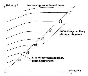
different infrared wavelengths, a reference graph (Fag 1 ) can be obtained
which consists of lines of constant papillary dermis thickness, wherein
Primary 1 is the measurement made at the longer (800-1000nm)
wavelength and Primary 2 is the measurement made at the shorter (b00-
800nm) wavelength. The absorption of blood within these wavelengths is
very small (a hundredth of its peak value for visible wavelengths at 600-
800nm and even less for 800-1000nm) and to a first approximation may
be ignored. The presence of dermal melanin does introduce a small error
in the range of low values for both primaries, but this is insignificant in
practice. Thus, by comparing values obtained at these wavelengths with
this graph, it is possible to ascertain the papillary dermis thickness.
However it is within the scope of the present invention to measure
CA 02272290 1999-OS-18
WO 98/22023 PCT/GB97/03177
brightness at such a long infra-red wavelength eg. 1100nm that the
brightness would vary to such a negligible extent with melanin and blood
content that it would effectively depend solely on the papillary dermis
thickness. This would also reduce the error introduced by the presence of
dermal melanin. In such a case only one set of brightness measurements
would be required. Furthermore, a transformation can be calculated
which allows an image of the skin to be created which represents how the
skin would appear if it had a papillary dermis thickness of any
predetermined value.
In a preferred embodiment, the reference colour coordinate range for
normal skin at the predetermined papillary dermis thickness is obtained as
disclosed in the above-mentioned Technical Report as a curved surface
lying within a three-dimensional colour space, with one of the bounding
axes relating to the amount of melanin within the epidermis and the other
relating to the amount of blood within the dermis. When an area
containing dermal melanin is located, i.e. points do not lie on the normal
colour surface, the epidermal melanin value within this area is estimated
by either reference to the reference colour coordinate range for normal
skin within regions identified as normal, or by reference to the epidermal
melanin levels calculated within normal regions adjacent to said area
containing dermal melanin. This value is then used with the corrected
colour coordinates of the abnormal region at the same predetermined
papillary dermis thickness to compute invasion depth and concentration of
dermal melanin. The corrected skin colour coordinates for the area of skin
under investigation may be calibrated to values equivalent to zero
epidermal melanin. Instead of using LMS colour space, it is possible to
CA 02272290 2005-05-03
6
use any other colour space, for example, the RGB colour space or a UV G
IR colour space.
The dermis contrasts strongly in structure to that of the epidermis, being
highly vascular, containing many sensory receptors and being made
largely from collagen fibres to provide the essential structure of the skin.
Between the epidermis and the dermis, the junction presents an extremely
uneven boundary with finger-like dermal protrusions called dermal
papillae projecting towards the skin surface. The dermis can be split into
two further histologically distinct layers, the papillary dermis and the
reticular dermis within which the structure of the collagen fibres differs
significantly. The papillary dermis is situated directly below the epidermis
and within which the collagen exists as a fine network of fibres. This is in
contrast with the reticular dermis where the collagen fibres are aggregated
into thick bundles which are arranged nearly parallel to the skin surface.
In the case of melanin invasion of the papillary dermis, there is a layer
containing blood, melanin and collagen, a layer containing either blood
and collagen or melanin and collagen, depending upon whether melanin
has passed the blood layer; and a layer containing just collagen. The
different thicknesses of these layers, the amount of blood and the
concentration of dermal melanin along with the amount of melanin in the
overlying epidermis affect the remitted light. This can be modelled by
calculating the net effect of these three layers for the differing parameters
outlined.
A mathematical model describing the optics of the skin has been
described in the above mentioned Symon D'O Cotton's Technical Report,
and this model
CA 02272290 1999-OS-18
WO 98/22023 PCTIGB97/03177
7
can be extended to predict coloration of skin containing dermal descent of
melanin.
As can be seen from Fig 2, there are now four distinct layers within the
dermis which can combine to construct a simple model, 1) a layer within
the upper papillary dermis containing no melanin, 2) a layer within the
upper papillary dermis containing melanin, 3) a layer within the lower
papillary dermis containing melanin, 4) a layer within the lower papillary
dermis containing no melanin.
It should also be noted that the condition of melanin existing up to the
dermo-epidermal junction is facilitated by allowing the thickness of layer 1
to be zero and likewise melanin can exist up to the papillary-reticular
dermis boundary by setting the thickness of layer 4 to be zero.
- In computing a model to predict this coloration it is useful to make note of
the fact that, as discussed in section 2.1 of the Technical Report, the
amount of back scatter due to melanin can be considered negligible.
Therefore, in the same manner that it was possible to apply the Kubelka-
Munk theory to the papillary dermis (section 3.2.2 of the Technical
Report), to compute the coloration of sections of papillary dermis
containing blood, where the back scattering component of blood was
considered negligible, it is possible to compute the coloration of sections
containing melanin. In this situation S(~,) (scattering coefficient) remains
dependent only on wavelength whilst a (fraction of radiation absorbed per
unit path length) becomes a(~,,p,c~) where d~ represents the density of
dermal melanin within that layer. Further, following the proof given in
equation (17) of the Technical Report, a (~,,p,c~) can be shown to be the
CA 02272290 1999-OS-18
WO 98/22023 PCT/GB97/03177
8
sum of a;,,(~,), ab(7~) and am(~,), where am(~,) is the absorption coefficient
of
melanin. From the above it is possible to calculate R and T (diffuse
radiation and transmission respectively). For simplicity of notation it is
helpful to consider R, & T, where,
R! ~~ ~ P~ ~~ d,r ) = R( ~ (k(a ( ~~ P~ ~))~ sfS f ~ )))~ K(k~a (~ ~ P~ ~))~
s~S (~ )))~ d,. )
and
T (~~ P~ ~~ d,~ ) = T(~(k(aO~ P~ ~))~s(S (~)))~ K(k(a (~~ P~ ~))~ s(S (~))),
d" )
where d" is the layer thickness.
As was shown in section 3.2.3 of the Technical Report, two-layer systems
can be combined to produce the total remitted and transmitted sight for
the dermis resulting in equation (20) of the Technical Report.
This can be simplified using the geometric series
a+ar+arz +arj+~~~- a if-1 < r < I
1-r
to
'' z
~.7 '' T uJ (~ ~ P ud ~ urul ) RlIr1 (~ ~ PIrl ~ ~frl )
RIIalal~~~PIrd~Plrl~~rrd~ulrl) - Rlud(~~Parl~uud)+ 1 '
1 -~~uO~~PiaW~url)R~rJ(~~Prd~~rJ
Similarly, T,tocal can be shown to be
_ ~rrd(~~P..d~~ne) ~y(~~Prn~~rd)
~roral ( ~ ~ P ud ~ P Id ~ dud ~ ~Id ) - ( /1
1 - ~lud~~~Prrd~~ud)RndO~Prn~dld)
These equations can be extended, as is shown by Wan et al. [1981], to an
n layered system resulting in values for R~2..." and T~z,.." of
z
Tlz...rr-! R.,
R! z...r~ = R! z...,~-! +
1- Rlz...rr-! R"
T z...n-! T
Tz...rr =
1 - R! z...rr-! Rr
CA 02272290 1999-OS-18
WO 98/22023 PCT/GB97/03177
9
This system of equations can therefore compute the total remitted and
transmitted light from an n layered system of arbitrary complexity
provided that the thickness and composition of the layers is specified.
For the tour-layer system shown in Fig 2, this results in a value for the
total
light remitted and transmitted from the dermis dependent on ~,, P"d, Pld
dud, drd~ drz, ~r2, dr3 and ~r3 where drZ and dr3 are the thickness of layers
2
and 3 whilst ~,2 and ~,3 are their corresponding melanin densities. The
thickness of layer 1 and layer 2 do not need to be explicitly defined as
they are simply d"~-dr2 and d,~- dr3 respectively; similarly ~j, and ~r4 are
zero by definition. A further simplification is possible if it is assumed that
~,2 = ~r3 leading to a single value of cb for the dermis.
The results of these equations can be combined with the predicted light
transmitted by the epidermis in the same manner as that discussed in
section 3.3 of the Technical Report, thus leading to the following
description of total remitted, S~d, and transmitted S~~.
Sra(~~Pu~r~Prn~d~,a,dr~~d,2~y~~ ~~d,~~)=
Rzmrpr(~~P»a~Prn~~"a~~nr~drz~~r~~~)g(~~d".)zS(~)
Sid(a'~Pu~l~Pr~l~uurl~urd~ur2~~I3~ ~~dnr) _
Tzrorar(~~Pud ~Prd ~dua~a'rd ~dr2,dr3~~~(~~dm)ZS(~)
These can be used to predict the value of the corresponding LMS
primaries
CA 02272290 1999-OS-18
WO 98/22023 PCT/GB97/03177
L(Prml~Pld~durl~dlol> dl2~dll~~,d,n) -
w
JR2lorol ( ~, P r,d , P In ~ d r,rr , d rrr ~ d,7 , d r3 ~ ~)~ ( ~, d"i ) Z
.S(~ )S,. (~ )~
0
M(Pr,r~~Prn,dr,a~dm~ drz,dr~,~.d"r
JRzn,rr,r(~.Prm,Prrr,dr,n,drrl,drz,dr~,~)e(~,d"r)zS(~)Shr(~)~
0
.S(Pr.~,Prrr~dr,rr,dm, d,z,dr;,~,d",)=
JRzrr,mr(~,Pr,rr,Prrr,drm,drrr,drz,dr"~~(~,d,rr)zS(~)Ss(~)d7v,
0
A further generalisation can be made to any primary, P", leading to the
following equation where S" defines the spectral response of that primary.
p.(Pr,~~Pm,dr,n,drn~ dlz,d",~,dra) _
f IZzrrrn,r(~~~Pr,a,Pm,dr,rr,drn,drz,dry,~)e(~,d"r)z.S'(~,)5,,,(~,)d~,
o ,
This equation can then be used~to generate the expected coloration of
human skin exhibiting dermal descent of melanin.
The result of this analysis is that it is possible for the same coloration to
result from different combinations of the above parameters. This
complicates the measurement of the dermal invasion of melanin, (but not
identifying the presence of any dermal melanin). Indeed, to obtain this
measurement, it is necessary to know the amount of melanin in the
overlying epidermis. However, at points where dermal invasion has taken
place, this parameter is difficult to determine simply by comparing colour
coordinates of the abnormal location with colour coordinates for healthy
skin. It is for this reason that, in the present invention, regions where
dermal melanin exists are identified by reference to a reference colour
coordinate range for healthy skin, and then the colour coordinates of these
regions are compared with the colour coordinates at one or more normal
CA 02272290 1999-OS-18
WO 98/22023 PCT/GB97/03177
11
skin locations. If said normal skin locations are adjacent to the region
where dermal melanin exists, it is sufficient to use the epidermal melanin
levels calculated for such normal skin locations to estimate the epidermal
melanin levels at the region where dermal melanin exists Alternatively, it
is possible to perform a measurement of the epidermal melanin levels
within areas of the skin where the presence of dermal melanin has been
identified, by assessing the deviation in coloration at the blue end of the
spectrum, from the reference colour coordinate range for normal skin due
to the presence of such dermal melanin. At the blue end of the spectrum,
the increase in such deviation quickly slows with increasing depth of
melanin penetration until a "saturation point" is reached. 8y assuming
that the depth of melanin penetration within the dermis is large enough for
such saturation to have occurred, an estimate of the deviation from the
reference colour coordinate range for normal skin can be made. This
estimate allows a calculation to be made of the skin coloration assuming
no dermal melanin, and therefore by reference to the colour coordinate
range for normal skin, of the level of epidermal melanin. It is within the
scope of the present invention to measure the epidermal melanin levels
directly, for example using polarised light, and to incorporate such
measurements in the measuring step (ii) above.
By any of the above methods, the effect of what would have been the
normal epidermal melanin level in the abnormal skin location can be
taken into account, thereby enabling a more accurate determination of
melanin descent
By comparing the values of the skin image represented in a certain colour
space with theoretically calculated values covering all possible amounts of
CA 02272290 1999-OS-18
WO 98/22023 PCT/GB97/03177
12
blood, dermal melanin penetration and melanin concentration within the
same colour space, the values of those three parameters can be obtained
for every point in the image. Since the papillary dermis thickness and
epidermal melanin content are known, it is possible to compute a detailed
three-dimensional reconstruction of the top layers of human skin. This is
of great potential interest to the medical profession and enables routine
examination of the internal structure of living skin just as X-rays, NMR and
ultrasound are used for examining other parts of the body. It is also within
the scope of the invention to acquire the infra-red and/ or visible images
using lasers of different wavelengths or by using spectral analysis.
It is possible to use a computer programmed with the above algorithms to
perform the actual calculations. However, before these calculations can
be performed, an image of the area of skin under investigation must be
represented in the same colour space as for the normal skin reference
colour coordinate range. This can be done in a number of ways. In one
way, the skin colour coordinates are acquired from an image using the
same lighting conditions and a CCD camera calibrated in the same way as
that used to produce the healthy skin reference colour coordinate range.
Alternatively, if exactly the same lighting conditions are not used, a white
standard or other appropriate correction factor can be used to allow
calibration of the image within the software. As a further alternative, a
colour image can be acquired using a colour photographic film which is
then digitised. This can be performed using either exactly the same
lighting conditions and a calibrated set-up or again with the inclusion of a
white standard or other appropriate correction factor. It is within the
scope of this invention to obtain both the infra-red and visible images with
CA 02272290 1999-OS-18
WO 98/22023 PCTJGB97/03177
13
a single digital camera or to calculate the value of the necessary primaries
through the use of spectroscopy.
The present invention will now be described in further detail and with
reference to the accompanying drawings, in which:-
Fig 1 is a graph showing variation of brightness with papillary dermis
thickness for primaries 1 and 2. as described hereinabove;
Fig 2 is a schematic cross-sectional view through a section of skin
illustrating melanin descent into the papillary dermis;
Fig 3 is a schematic cross-sectional view through a section of skin
illustrating normal, healthy regions and an abnormal region where, in this
case, melanin descent into the papillary dermis and the reticular dermis
has taken place;
Fig 4 is a block diagram showing the steps involved in one embodiment of
the method of the present invention;
Fig 5 is a diagram showing the predicted surface of normal skin coloration
within a three-dimensional colour space;
Fig 6 is a diagram showing coloration within the skin cancer that is shown
in Fig 7 in the same 3-D colour space as depicted in Fig 5, wherein areas
of normal and abnormal coloration are shown; and
Fig 7 is a photographic image of the skin cancer.
CA 02272290 2005-05-03
Referring now to Fig 3 of the drawings, a schematic skin section is shown
wherein melanin (indicated by the black circles in Fig 3) in normal healthy
skin are present in the lower part of epidermis 10 adjacent but above the
dermo-epidermal junction 12 between the epidermis and the papillary
dermis 14. The Breslow thickness referred to above is the depth of
melanin invasion in millimetres measured from granular layer 16 which is
a layer in the epidermis 10 where he skin goes scaly and forms the tough
outer cornified layer 18. 1n the abnormal region of the skin, the melanin is
shown as having descended not only into the papillary dermis 14, but also
into the underlying reticular dermis 20 lying above the subcutaneous fat
layer 22. It is to be appreciated that, in other cases, melanin decent can
be into any layer of the skin and may even be into the subcutaneous fat
layer 22.
Referring now to Fig 4, there is shown a block diagram illustrating the
steps involved in a typical method of measurement in accordance with the
present invention. fn Fig 4,
Block 40 exemplifies method step (ii) above- the acquisition of
an image at visible wavelengths of the same skin area. This can be by
CCD camera, digitised film or any other convenient means. Block 42
exemplifies method step (iii) above- the transformation of the image into
corrected colour space of the skin model at a predetermined papillary
dermis thickness. Block 44 exemplifies method steps (iv and v) above- the
identification of regions containing dermal melanin, by comparing the
CA 02272290 1999-OS-18
WO 98/22023 PCT/GB97/03177
corrected skin colour coordinates with the reference colour coordinate
range. Block 46 exemplifies method step (vi) above- use of the corrected
colour space to calculate the amounts of epidermal melanin within normal
regions adjacent to the regions containing dermal melanin and use thereof
to give an indication of the amounts thereof which exist in the regions
containing dermal melanin. Block 48 exemplifies a first part of method
step (vii) above- calculation of dermal invasion using the measured
coloration of the abnormal regions and the calculated amount of
epidermal melanin from 46. Block 50 exemplifies a second part of
method step (vii) above- transformation of the calculated dermal invasion
of melanin into either the Breslow thickness or the Clark's level of
invasion. This can be reported as either representing the maximum
invasion or as an image showing invasion over the skin.
Referring now to Fig 5, the shaded surface indicates the range of
colorations which can exist in normal healthy skin corrected to the
predetermined papillary dermis thickness. Skin colorations which depart
from this surface are indicative of dermal melanin.
Referring now to Figs. 6 and 7, it can be seen that a region of the skin
which is shown in Fig 7 and which is indicated by arrow H in Fig 6 lies at
a position corresponding to part of the shaded surface illustrated in Fig 5
and is indicative of normal healthy skin, whereas an adjacent region
indicated by arrow U in Fig 6 lies outside such surface and is indicative of
skin containing dermal melanin. Comparison of the coloration of these
two adjacent regions H and U enables the depth of melanin invasion in
the abnormal region of the skin in Fig 7 to be computed.