Note : Les descriptions sont présentées dans la langue officielle dans laquelle elles ont été soumises.
CA 02283300 1999-08-31
WO 98/39033 PCT/EP98/01009
Costimulation of T-cell proliferation by a chimeric bispecific costimulatory
protein
This invention pertains to nucleic acids encoding novel chimeric proteins,
their corresponding
gene products, and their use, whereby the proteins contain two binding domains
one of which
specifically recognizes a surface molecule on target cells and one of which is
derived from
the extracellular domain of costimulatory ligands or the counter receptors of
such
costimulatory ligands naturally expressed on the surface of B lymphocytes, T
lymphocytes,
or professional antigen presenting cells.
The most effective mechanism for tumor rejection is mediated by tumor-specific
T
lymphocytes (Greenberg, P.D., 1991, Melief, C.J., 1992). This might favor the
progression of
tumors which escape immune surveillance by a variety of strategies like the
prevention of
efficient antigen presentation through the loss of major histocompatibilty
complex (MHC)
molecules (Doyle, A., et al., 1985, Lassam, N., and Jay, G., 1989) or defects
in antigen
processing (Rcstifo, N.P., et al., 1993, Cromme, F.V., et al., 1994). Another
mechanism for
tumor cells to evade the immune system is the absence of costimulatory
molecules
(Lundberg, A., et al., 1993). For activation and clonal expansion T cells
require costimulatory
signals in addition to the primary signal provided by the T-cell receptor
{TCR) which
interacts with peptide-bearing MHC molecules (Rudd, C.E., et al., 1994). TCR
stimulation in
the absence of costimulation can result in unresponsiveness and the induction
of clonal
anergy (Harding, F.A., et al., 1992: Gimmi, C.D., et al., 1993; Tan, P.C., et
al.. 1993). CD28
is the major costimulatory signal receptor for CD4+ and CD8+ T cells . Members
of the B7
family of proteins including B7-1 (CD80) and B7-2 (CD8C) are its natural
ligands on antigen
presenting cells (APC) (Gimmi, C.D., et al., 1991; Linsley, P.V4'., et al.,
1991; Galvin, F., et
al., 1992: Hathcock. K.S., et al., 1993; Freeman, G.J., et al., Science, 1993;
Azuma, M., et al.,
1993).
Many attempts have been made to increase tumor immunogenicity. Thereby
strategies to
provide tumor cells with members of the B7 family of costimulatory molecules
have led to
promising results. Recently it has been shown that the transmembrane and
intracellular
domains of B7 molecules are not required for their activity as costimulators
of T-cell
activation. Expression of the extracellular domain of murine B7-1 or B7-2 on
the tumor-cell
surface and insertion in the cell membrane via a GPI anchor is sufficient to
provide T-cell
costimulation in vitro and in vivo (Brunschwig, E.B., et al., 1995).
Costimulation of T-cell
CA 02283300 1999-08-31
WO 98/39033 PCT/EP98/01009
-2-
proliferation in vitro was also achieved upon incorporating in the membrane of
tumor cells a
recombinant GPI-linked form of human B7-1 which was expressed in CHO cells and
purified
from cell lysates (McHugh et al., 1995). While this strategy allows to insert
a B7 molecule in
the cell membrane without the need to transfect the tumor cells with foreign
genes, the
applicability of such molecules in vivo are limited by their lack of target
cell specificity. --
Independent from the availability of professional APCs, T-cell dependent
rejection of tumors
can be achieved by presenting costimulatory signals directly on the tumor cell
surface.
Transfection of several murine tumor cells with B7-1 (Chen, L., et al., 1992;
Townsend, S.E.,
and Allison, J.P., 1993; Li, Y., et al., 1994) or B7-2 genes (Hodge, J.W., et
al., 1994; Yang,
G., et al., 1995) induces T-cell dependent rejection of B7 expressing tumors
in mice and
protects against subsequent challenge with parental B7-negative tumor cells
(Chen et al.,
1992; Yang et al., 1995; Baskar, S., et al., 1993).
Alvarez-Vallina et al. describe, in Eur. J. Immunol. 26 (1996) 2304-2309, an
scFv-CD28
fusion gene, its construction, and its functional characterization. The gene
product is
produced in T cells after gene transfer and is inserted into the cell membrane
as a
transmembrane protein. It is therefore an immobilized insoluble protein. The
fusion protein
described by Alvarez-Vallina et al. is a chimeric signal transduction molecule
which is
produced by the T cell itself.
The invention comprises a novel approach to direct a costimulatory molecule to
the surface of
target cells. This approach is based on a chimeric fusion protein which
consists preferably of
the extracellular domain (thus without the transmembrane or intracellular
domain) of a
costimulatory molecule fused to a single-chain antibody domain (scFv) specific
for a tumor-
specific antigen, preferably a type 1 growth factor receptor overexpressed in
a high percentage
of human adenocarcinomas. Such a molecule is functionally active, soluble and
not
membrane-located due to the lack of intracellular domain, and binds, for
example, to B7
counter-receptors and to ErbB2. The fusion protein localizes specifically to
the surface of
target cells expressing a tumor-specific antigen, thereby providing a
costimulatory signal
which results in enhanced proliferation of T cells. The invention shows that
effective tumor
vaccines for cancer immunotherapy could be created by targeting such chimeric
ligands to the
surface of tumor cells.
- _ r fi
CA 02283300 1999-08-31
WO 98/39033 PCT/EP98/01009
-3-
The invention comprises the use of a soluble bispecific fusion protein
consisting of
a) a binding domain which recognizes a specific surface molecule on a target
cell,
covalently linked to
b) a domain capable of costimulation of T cell proliferation, --
for a specific costimulation of a T cell directed against said target cell.
The invention further comprises a method of manufacturing a therapeutic agent
comprising
said fusion protein for a specific costimulation of a T cell directed against
said target cell of a
patient. The therapeutic agent can be administered locally or systemically.
It has surprisingly been found that with the chimeric fusion proteins
according to the
invention, a very specific cell activation is possible. In contrast to known
methods of cell
stimulation (e.g., expression of B7 domains on the cell surface), according to
the invention,
essentially no stimulation of cells in the presence of target cells not
carrying the tumor-
specific surface antigen is achieved. Therefore, when using methods according
to the
invention, no background stimulation of cells is found.
The fusion proteins according to the invention consist of two binding domains
which do not
have any signal transduction function themselves because they contain no
intracellular
domains, but are in a soluble state when being located outside of cells, and
will activate the
wild-typical CD28 of a T cell after binding of both the antigen via the scFv
domain and of
CD28 via the B7 domain, so that said CD28 can generate a signal and transmit
it then. Thus.
the molecules of the invention provide an antigen-dependent activation of the
signal
transduction. The fusion proteins according to the invention are typical,
because they produce
an effect that leads to the stimulation of a specific immune response. The
fusion proteins of
the invention therefore also are bispecific.
"Target cell" according to the invention preferably means a syngeneic cell, a
tumor cell or a
cell infected by a pathogen (e.g., virus, bacterium, yeast, fungi).
Therefore, in another embodiment of the invention, a specific activation of,
e.g., cytotoxic
T cells from a T cell population can also be achieved with the fusion proteins
according to the
invention, when the fusion protein binds to the specific counter-receptor of
the costimulatory
domain on the T cell. In such case, it is suitable to carry out a pre-
treatment using IL-2.
CA 02283300 1999-08-31
WO 98/39033 PCT/EP98/01009
-4-
In another preferred embodiment of the invention, the costimulation can be
coupled to ex
vivo or in vivo transfection of T cells. Hera T cells are transduced with a
viral or non-viral
gene therapy vector containing a desired gene, the transfected cells being
selected, e.g.,
through a positive or negative selection system. Thereafter, the T cells,
which are still at rest,
are stimulated through a costimulatory signal, preferably in accordance with
the invention;
and their proliferation in vitro or in vivo is initiated.
According to the invention a costimulatory molecule is directed to the surface
of target cells,
which are preferably based on providing the extracellular, CD28-binding domain
of human
B7-2 with a target-cell specific recognition domain. Consistent alterations of
cell surface
antigens have been identified in human cancer cells. The erbB2 gene encodes a
185-kDa
transmembrane glycoprotein that is a member of the type 1 family of receptor
tyrosine kinases
(RTK) which also includes epidermal growth factor (EGF) receptor, ErbB-3 and
ErbB-4
(Peles, E., and Yarden, Y., 1993). Overcxpression of ErbB2 is frequently
observed in human
adenocarcinomas arising at numerous sites including breast, ovary, lung,
stomach and
salivary gland where it correlates with an unfavorable patient prognosis
(Hypes, N.E., 1993).
Its role in cancer development and its accessible location on the cell surface
make ErbB2 a
target for directed therapy. From the mRNA of hybridoma cells producing a
monoclonal
antibody specific for the extracellular domain of human ErbB2 previously a
recombinant
single chain (scFv) antibody domain consisting of the variable domains of the
antibody heavy
and light chains connected via a synthetic linker sequence was constructed
(Wets, W., et al.,
1992). This recombinant binding domain was incorporated in several fusion
proteins and has
been used to target heterologous effector functions such as enzymes or toxins
or gene-
transduced cytotoxic T cells (Moritz, D., et al., 1994) specifically to ErbB2
expressing tumor
cells (Wets, W., et al., Bio/Technology, 1992; Wels, W.. et al., Cancer Res.,
1992).
While these strategies are independent of an existing anti-tumor immune
response and can he
effective in eliminating an established tumor (Wels et al., Cancer Res.,
1992), they do not
provide long term protection or systemic immunity which could prevent possible
tumor
recurrence. In contrast, molecules according to the invention might support
the generation of
a specific T-cell dependent anti-tumor immune response. The invention shows
that such a
chimeric protein is bifunctional: it localizes specifically to the surface of
ErbB2 expressing
target cells via the scFv domain and interacts with soluble or cell-surface
CTLA-4. Likewise
the fusion protein bound to the surface of Jurkat cells which express low
levels of CD28 as
determined by FACS analysis. In the in vitro experiments with ErbB2 expressing
target cells
CA 02283300 1999-08-31
WO 98/39033 PCT/EP98/01009
-5-
the chimeric proteins according to the invention provided costimulation of PMA-
activated
syngeneic T cells via the B7 domain.
Cell-surface targeted fusion proteins according to the invention were able to
costimulate pre-
activated T cells. Previously a soluble B7-1-Ig fusion protein at
concentrations of 1 to 10=
pg/ml showed only modest enhancement of T-cell proliferation in combination
with an anti-
CD3 antibody, but was more active when immobilized on a plastic surface
(Linsley et al.,
1991 ). The most likely explanation for these findings is that CD28 molecules
have to be
clustered on the surface of the T cell to reach a certain threshold for T-cell
activation
(Ledbetter, J.A., et al., 1990). Recently it has been demonstrated that a
disulphide-linked
CTLA-4 homodimer binds to two molecules of monomeric B7-1 or B7-2 with a very
fast off
rate (Linsley, P.S., et al., 1995). It is very likely that this is also the
case for the binding of
CD28 to members of the B7 family, which occurs with an even faster off rate
for B7-2
compared to B7-1 (Linsley, P.S. et al., 1994). B7 fusion proteins anchored on
the target cell
surface via the antibody domain or membrane-anchored B7 molecules in general
simultaneously provide multiple contacts with CD28 molecules which could
stabilize the
interaction and result in CD28 crosslinking.
The invention shows that the extracellular domain of a costimulatory molecule,
preferably
human B7-2, targeted to the surface of cells via an antibody domain is able to
provide a
costimulatory signal for the activation of T cells. Recent work in murine
model systems
suggests that B7-1 transfected tumor cells might be more effective than those
transfectcd with
the B7-2 gene in activating T cells (Gajcwski, T.F., et al., 1996; Matulonis,
U., et al., 1996).
In a recent report where the extracellular domains of murine B7-1 and B7-2
were expressed
as GPI-anchored proteins on the surface of the T-lymphoma line EL-4, B7-?
expressing EL-4
transfectants appeared to be at least as potent in enhancing the proliferation
of PMA-
stimulated primary T cells as B7-1 transfectants (Brunschwig et al., 1995).
A recombinant B7-2225 protein (amino acids 1-225) in an E. coli expression
system has also
been provided which failed to bind to B7 counter-receptors. In contrast,
according to the
invention, biologically active, soluble lymphocyte receptors and their ligands
can be
produced in the yeast Pichia pastoris. The B7-2225 and B7-2225-scFv(FRPS)
proteins as well
as a truncated human CTLA-4 molecule purified from Pichia pastoris culture
supernatants
showed specific binding to their respective receptors.
CA 02283300 1999-08-31
WO 98/39033 PCT/EP98/01009
-G-
According to the invention, a functionally active B7-2 fusion protein can be
targeted to the
surface of tumor cells via a specific binding domain. Due to the modular
structure of this
fusion protein also similar molecules with altered target cell specificity or
containing a
different immunomodulatory domain could be obtained. Such molecules are
therefore useful
reagents for cancer immunotherapy. --
Preferred binding domains which recognize surface antigens on target cells
include growth
factor domains or recombinant antibody domains (e.g., single chain Fv domains;
disulphide
bridged Fv domains) specific for members of the ErbB family of receptor
tyrosine kinases
such as EGF receptor, variant EGF receptor (EGFRvIII), ErbB2 (HER2, Neu),
ErbB3
(HER3), ErbB4 (HER4}, which are overexpressed on a variety of tumor cells of
epithelial
origin. Alternatively, these binding domains can bind to different molecules
with enhanced or
exclusive expression on the surface of target cells such as tumor cells, or
cells infected by
pathogens. Such binding domains include binding domains which bind to other
growth factor
and cytokine receptors, or domains which bind to receptors for peptide ligands
such as alpha-
MSH expressed on the surface of melanoma cells, or domains which bind to
surface
molecules which are not receptors for growth factors, cytokines or peptide
ligands such as
EGP-2, a 38 kDa pancarcinoma antigen recognized by the monoclonal antibody MOC-
31, or
domains which bind to antigens of pathogens expressed on the surface of
infected host cells.
Preferred domains of costimulatory ligands and counter receptors of
costimulatory ligands
are derived from B7-1 (CD80), B7-2 (CD86), B7-3, CD40, CDlla/18 (LFA-1), CD19,
CD22, CD58 (LFA-3), CD59, CD54, CD10G (VCAM), CD72, CTLA-4, CD28, CD40 ligand
(CD40L), CD54 (ICAM-1 ), CD45R0, CD43, CD49d/29, CDS which are expressed on
the
surface of B lymphocytes, professional antigen presenting cells, or T
lymphocytes.
Recombinant chimeric molecules containing at least two binding domains are
derived by
isolating nucleic acids encoding binding domains recognizing a surface
molecule on target
cells, and nucleic acids encoding binding domains derived from costimulatory
ligands or their
counter receptors, and fusing such nucleic acids in a single open reading
frame.
Chimeric molecules according to the invention, containing binding domains
which recognize
a surface molecule on target cells such as tumor cells, or cells infected by a
pathogen, and
binding domains derived from costimulatory ligands (e.g., B7-l, B7-2), may act
as a vaccine
and specifically localize to the surface of a target cell thereby providing
the target cell with
the costimulatory activity and facilitating a target-cell specific cellular
immune response
CA 02283300 1999-08-31
WO 98/39033 PCT/EP98/01009
(e.g., by costimulating the activation of T lymphocytes). This could be
achieved by systemic
treatment of a mammal (patient; farm animal; pet animal) with such a chimeric
fusion
protein, or by injection of the chimeric fusion protein into a tumor, or by
injection of the
chimeric fusion protein into tissue infected by a pathogen. Alternatively such
chimeric fusion
proteins could be used ex vivo for the activation of patient-derived tumor
infiltrating
lymphocytes (TILs), or lymphokine-activated killer cells (LAK), or other
patient-derived
lymphocyte preparations in the presence of cells expressing the target
antigen, possibly in
addition to the presence of activating cytokines such as interleukin 2,
interleukin 12, etc,
followed by adoptive transfer of such activated lymphocytes into a patient.
Alternatively such
patient-derived lymphocytes may consist of lymphocytes transduced with
chimeric antigen
receptors (e.g., nucleic acids encoding chimeric proteins which consist of a
binding domain
specific fo the same antigen on the surface of the target cells recognized by
the chimeric
costimulatory molecule, or a binding domain recognizing a different antigen on
the surface of
the target cells, and an intracellular domain derived from molecules such as
the zeta-chain or
other molecules of the CD3 complex).
The following examples, references, sequence listing and drawings are provided
to aid the
understanding of the present invention, the true scope of which is set forth
in the appended
claims. It is understood that modifications can be made in the procedures set
forth without
departing from the spirit of the invention.
Description of the Figures
Figure 1 Construction and expression in yeast of recombinant B7-2 proteins.
(A)
Schematic representation of B7-? genes in the yeast expression plasmids
pPIC9-B7-2225 (upper panel) encoding amino acids 1 to 225 of human B7-
2 (B7-2225) and pPIC9-B7-2225-scFv(FRPS) (lower panel) encoding the
B7-2 fragment fused to the scFv(FRPS) single chain antibody domain
specific for ErbB2. Expression in the yeast Pichia astoris is regulated by
the alcohol oxidase promoter (AOX1) and is directed to the extracellular
space via the yeast a-factor secretion signal (a). M, c-Myc tag; H,
polyhistidine tag. (B) SDS-PAGE analysis of B7-2225-scFv(FRPS) fusion
protein. Lane 1, Coomassie-stained B7-2225-scFv{FRPS) protein purified
from Pichia astoris culture supernatants; lanes 2 and 3, immunoblot
analysis of purified B7-2225-scFv(FRPS) (lane 2) and B7-2225-
scFv(FRPS) after treatment with protein N-gycosidase F (lane 3) with
CA 02283300 1999-08-31
WO 98/39033 PCT/EP98/01009
_g-
monoclonal antibody 9E10 specific for the C-terminal c-Myc tag of the
fusion protein. M, molecular weight standards (kDa).
Figure 2 Binding of B7-2225-scFv(FRPS) to CTLA-4. The binding of B7-2225-
scFv(FRPS) to CHO cells (A) and CHO-CTLA-4 cells stably transfected=
with a human CTLA-4 cDNA (A, B, C) in the absence (A, B) or presence
of a 50-fold molar excess of soluble CTLA-4 protein (C) was detected by
FACS analysis with monoclonal antibody 9E10 and FITC-labeled (A) or
PE-labeled (B, C) goat anti-mouse IgG.
Figure 3 Binding of B7-2225-scFv(FRPS) to ErbB2. (A) Immunoblot analysis with
monoclonal antibody 9E10 of B7-2225-scFv(FRPS) protein precipitated
with glutathione-coupled agarose beads after incubation with a bacterially
expressed glutathione S-transferase (GST) - ErbB2 fusion protein (lane 2)
or a GST control protein (lane 3). M, molecular weight standards (kDa). (B)
The binding of B7-2225-scFv(FRPS) to murine HC11 cells and HC11-
ErbB2 cells stably transfected with a human ErbB2 cDNA was detected by
FACS analysis with monoclonal antibody 9E10 and FITC-labeled goat anti-
mouse IgG.
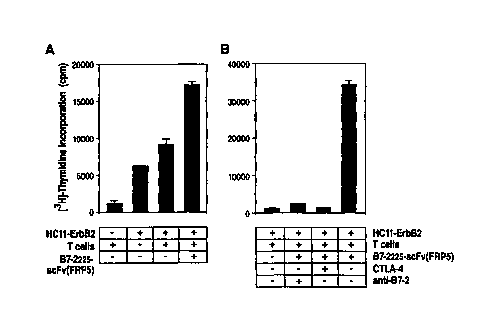
Figure 4 Costimulation of syngeneic T cells by B7-2225-scFv(FRPS). (A) HC1 1-
ErbB2 cells and primary T cells from Balb/c mice pre-stimulated with PMA
and 1L-2 were incubated in the presence or absence of lU ng/ml o>"purified
B7-2225-scFv(FRPS) as indicated. Proliferation of cells was measured by
[3H]-thymidine incorporation. (B) Cells were treated as described in (A)
with or without the addition of 2.5 ug/ml soluble CTLA-4 protein or 0.5
pg/ml anti-B7-2 antibody as indicated. Each value was determined in
triplicates. The standard deviation is represented by error bars.
Figure 5 Costimulation by B7-2225-scFv(FRPS) is dependent on the binding to
cell-
surface ErbB2. (A) HC11-ErbB2 cells were incubated with pre-stimulated
T cells as described in the legend of Figure 4 in the presence of increasing
concentrations of purified B7-2225-scFv(FRPS) or B7-2225 as indicated.
Control cells were treated in the absence of recombinant proteins.
Proliferation of cells was measured by [3HJ-thymidine incorporation.
(B) HC11-ErbB2 cells or parental HC11 cells were treated with 1 pg/ml
CA 02283300 1999-08-31
WO 98/39033 PCT/EP98/01009
-9-
B7-2225-scFv(FRPS), incubated with a 5-fold excess of pre-stimulated
syngeneic T cells as indicated and proliferation was measured by [3H]-
thymidine incorporation. Controls show the background of [3H]-thymidine
incorporation in the absence of T cells or target cells. Each value was
determined in triplicates. The standard deviation is represented by error=
bars.
Example 1
Construction of the B7-2225-scFv(FRfS) fusion gene
A fusion gene encoding the extracellular domain of human B7-2 (amino acids 1
to 225,
referred to as B7-2225), the ErbB2 specific scFv(FRPS), and synthetic sequence
tags
facilitating immunological detection and affinity purification was inserted
into the AvrII and
Notl restriction sites of the yeast expression vector pPIC9 (Invitrogen). In
this vector
expression of the gene is controlled by the methanol-inducible alcohol oxidase
1 (AOX1)
promoter and the gene product is secreted into the medium via an N-terminal a-
factor
secretion signal. pPIC9 also contains a functional histidinol dehydrogenase
(HIS4) gene for
positive selection in the Pichia asp toris HIS4 mutant strain GS115
(Invitrogen). The B7-2225
cDNA was derived from total RNA of human peripheral blood mononuclear cells
(PBMC)
by reverse transcription followed by PCR using the oligonucleotides B7-2-sense
5'-AAAAG-
TCGACGCTAGCGCTGCTCCTCTG-3' (SEQ ID NO:1 ) and B7-2-antisense
5'-AAAACTCTAGAGATCTATCGATAGGAATGTGGTCTGG-3' (SEQ ID N0:2), which
introduce Salt and NheI restriction sites at the 5'-end and CIaI, BglIl, and
Xba1 restriction
sites at the 3'-end of the PCR product. The B7-225 cDNA fragment, the cDNA
encoding the
scFv(FRPS) (Wels et al., Bio;Technology, 1992), and a synthetic sequence
encoding the Myc
tag recognized by the monoclonal antibody (Mab) 9ElU (Evan, G.L, et al., 1985)
as well as a
polyhistidine tag were assembled into a single open reading frame and
subsequently inserted
into the expression vector pPIC9. As a control a similar B7-2225 gene lacking
the
scFv(FRPS) domain was constructed. The integrity of the constructs was
confirmed by
restriction analysis and DNA sequencing.
CA 02283300 1999-08-31
WO 98/39033 PCT/EP98/01009
- 10-
Example 2
a) Cell lines and cell culture conditions
CHO cells and CHO-CTLA-4 cells stably transfected with a human CTLA-4 cDNA
were'
maintained in MEMa with deoxyribonucleosides (Gibco BRL), containing 2 mM
glutamine,
50 pM (3-mercaptoethanol, 10% heat-inactivated fetal bovine serum (FBS), and 1
mg/ml
6418 {CHO-CTLA-4). Balb/c derived HC11 mouse mammary epithelial cells and HC11-
ErbB2 cells (HC11 R1#11) stably transfected with a human erbB2 cDNA were grown
in
RPMI 1640 supplemented with 8% FBS and 5 pg/ml bovine insulin as described
(Hynes,
N.E., et al., 1990). The Pichia asp toris GS 115 yeast cells (Invitrogen) were
propagated in
buffered minimal glycerol-complex medium (BMGY) and expression of recombinant
proteins was induced in buffered minimal methanol-complex medium (BMMY)
according to
the distributor's recommendation.
b) Expression and purification of recombinant proteins
Construction and expression in yeast of the chimeric fusion protein B7-2225-
scFv(FRPS)
A chimeric gene encoding the extracellular domain of human B7-2 (amino acids 1
to 225,
referred to as B7-2225) fused to the ErbB2 specific scFv(FRPS) antibody domain
(Wets et
al., Bio/Technology, 1992) was constructed and inserted into the yeast
expression vector
pPIC9 shown in Fig. lA. The resulting piasmid pPIC9-B7-2225-scFv(FRPS) encodes
under
the control of the methanol inducible alcohol oxidase I (AOX1) promoter a
chimeric fusion
protein termed B7-2225-scFv(FRPS), which consists of an N-terminal a-factor
secretion
signal from yeast, amino acids 1 to 225 of human B7-2, the scFv(FRPS) antibody
domain, the
Myc epitope recognized by Mab 9E10 (Evan et al., 1985), and a polyhistidine
cluster
facilitating the purification of the recombinant protein via Ni2+ affinity
chromatography.
The B7-2225-scFv(FRPS) protein was expressed in the Pichia astoris strain
GS115, and
purified via Ni2+-affinity chromatography and gel filtration. The yield of
soluble
recombinant protein purified from 1 1 of yeast culture supernatant was
typically 0.5 mg. SDS-
PAGE and Mab 9E10 immunoblot analysis of the purified material revealed a
purity of
greater than 90% after two purification steps (Fig. 1B). The B7-2225-
scFv(FRPS) molecule is
present as a monomer in yeast culture supernatants and in purified fractions
as determined by
CA 02283300 1999-08-31
WO 98/39033 PCT/EP98/01009
-11-
SDS-PAGE under non-reducing conditions (data not shown). In contrast to the
calculated
molecular weight of 55.95 kDa the protein migrates as a smear of bands with
apparent
molecular masses of approximately 80 to 110 kDa in SDS-PAGE under reducing
conditions
(Fig. 1B). N-glycosidase F treatment of the protein reduced the apparent
molecular mass to
approximately 60 kDa indicating that the higher apparent molecular mass of
yeast expressed-
B7-2225-scFv(FRPS) protein is mainly due to post-translational modification by
N-linked
glycosylation.
Linearized pPIC9-B7-2225 and pPIC9-B7-2225-scFv(FRPS) plasmid DNAs were used
for
the transformation of Pichia asp torts GS115 cells by electroporation (Scorer,
C.A., et al.,
1994). His4+/methanol-utilization+ (mut+) yeast colonies were isolated on
selection media
following established protocols (Ban, K.A., et al., 1992) and upon induction
with methanol
B7-2225 or B7-2225-scFv(FRPS) expressing clones were identified by immunoblot
analysis
of culture supernatants using Mab 9E 10. For expression at a larger scale a
single colony each
was grown to an OD600 of 3 in BMGY medium, pH 8, the medium was exchanged with
methanol-containing BMMY medium, pH 8, and protein expression was induced for
72 h at
30°C. Yeast cells were removed by centrifugation at 20,000 g.
Supernatants containing the
soluble B7-2225 and B7-2225-scFv(FRPS) fusion proteins were passed through a
45 pm
filter, applied onto a Ni2+-saturated chelating sepharose column (Pharmacia
Biotech) and the
recombinant proteins specifically bound via their C-terminal polyhistidine tag
were eluted
with 250 mM imidazole in PBS. The B7-2225-scFv(FRPS) protein was purified
further by
gel filtration using a Superdex 200 column (Pharmacia Biotech), fractions
containing the
fusion protein were identified by SDS-PAGE and immunoblotting with Mab 9E10,
pooled,
concentrated by ultrafiltration, and dialyzed against PBS. Post-translational
modification of
B7-225-scFv(FRPS) protein from yeast was analyzed in a dcglycosylation
reaction. 0.2 pg
of purified fusion protein were heated to 100°C for I 0 min in PBS
containing 0.1 °~o SDS.
Triton X-100 was added to a final concentration of 1 % and the protein was
incubated with 1
U of N-glycosidase F (Boehringer Mannheim GmbH, DE) for I 6 h at 37°C
in a total reaction
volume of 100 gl. The sample was then analyzed by SDS-PAGE and immunoblotting
with
Mab 9E10.
CA 02283300 1999-08-31
WO 98/39033 PCT/EP98/01009
-12-
Example 3
Binding assays
a) Purified B7-2225-scFv(FItPS) speciCcally binds to CTLA-4 expressing cells
B7-2 binds to the B7 counter-receptors CD28 and CTLA-4 on the surface of T
cells
(Hathcock et al., 1993; Freeman et al., Science, 1993; Azuma et al., 1993), To
investigate the
functionality of the B7-2 domain the binding of the recombinant B7-2225-
scFv(FRPS) to
CTLA-4 on the surface of cells was tested by FACS analysis. CHO-CTLA-4 cells
stably
transfected with a human CTLA-4 cDNA were incubated with B7-2225-scFv(FRPS)
and
specifically bound fusion protein was detected with Mab 9E10 and FITC-labeled
goat anti-
mouse IgG. The results are shown in Fig. 2A. Significant binding of B7-2225-
scFv(FRPS) to
CHO-CTLA-4 cells but not to CHO control cells could be detected. Comparable
results were
obtained with the B7-2225 protein lacking the scFv domain and an anti-CTLA-4
antibody.
The specificity of the B7-2225-scFv(FRPS) binding to CTLA-4 was further
confirmed in a
competition assay. Similar to the soluble B7-2 protein a recombinant protein
comprising
amino acids 1 to 125 of human CTLA-4 was expressed in Pichia pastoris and
purified from
culture supernatants. CHO-CTLA-4 cells were incubated with B7-2225-scFv(FRPS)
in the
presence or absence of a 50-fold molar excess of soluble CTLA-4 protein as a
specific
competitor and the binding of B7-2225-scFv(FRPS) was investigated by FACS
analysis with
Mab 9E10 and PE-labeled goat anti-mouse IgG. As shown in Fig. 2B and C soluble
CTLA-4
protein almost completely blocked the binding of B7-2225-scFv(FRPS) to CHO-
CTLA-4
cells. These data indicate that the B7-2 domain of B7-2225-scFv(FRPS) is
functionally active
and interacts specifically with a B7 counter-receptor.
The binding of B7-2225-scFv(FRPS) to the B7 counter-receptor CTLA-4 was
determined by
FACS analysis using CHO-CTLA-4 cells and parental CHO cells as a control. 5 x
105
trypsinized cells were incubated for 45 min at 4°C with 0.1 or 1 pg of
B7-2225-scFv(FRPS)
protein, followed by incubation with 3 pg of Mab 9E10 and FITC- or PE-labeled
goat anti-
mouse IgG (PharMingen) for 30 min. Binding of B7-2225-scFv(FRPS) was detected
using a
FACScan (Becton-Dickinson). Similarly, the binding of B7-2225-seFv(FRPS) to
ErbB2
expressing HC11-ErbB2 mouse mammary epithelial cells was determined by FACS
analysis.
Binding of B7-2225-scFv(FRPS) to ErbB2 was also tested using a recombinant
glutathione
S-transferase (GST) fusion protein which contains an N-terminal portion of the
ErbB2 protein
and is recognized by the ErbB2 specific Mab FRPS. Bacterially expressed GST or
GST-
ErbB2 fusion proteins (10 pg) were bound to 200 pl each of glutathione-coupled
agarose
r
CA 02283300 1999-08-31
WO 98/39033 PCT/EP98/01009
-13-
beads (Sigma). The beads were incubated with 4 pg B7-2225-scFv(FRPS), washed
with PBS,
and specifically bound proteins were analyzed by SDS-PAGE and immunoblotting
with a
Mab binding specifically to the B7-2225-scFv(FRPS) protein.
Example 4 _
Cell proliferation assays
a) B7-2225-scFv(FRPS) provides costimulation for the proliferation of
syngeneic T cells
The chimeric B7-2225-scFv(FRPS) protein is bispecific since it binds to CTLA-4
and to
ErbB2 on the surface of cells. Several reports have demonstrated that human B7-
1 or B7-2
can interact functionally with the murine B7 counter-receptors CD28 or CTLA-4,
and vice
versa (Freeman, G.J., et al., J. Exp. Med., 1993; Cai, Y.C., et al., 1995). In
order to determine
whether purified B7-2225-scFv(FRPS) presented on the surface of cells can
provide
costimulation for T-cell proliferation, a syngeneic lymphocyte reaction (SLR)
was performed
using primary T cells from Balb/c mice pre-stimulated with PMA and IL-2, and
murine
HC11-ErbB2 cells which are of Balb/c origin and express human ErbB2 on their
surface.
HC11-ErbB2 cells were treated with B7-2225-scFv(FRPS) (10 ng/ml) and
subsequently
incubated with a 5-fold excess of pre-stimulated T cells. B7-2225-scFv(FRPS)
treatment
resulted in a strong increase in T-cell proliferation in comparison to cells
treated in the
absence of the fusion protein (Fig. 4A). The addition of a 500-fold molar
excess of soluble
CTLA-4 or a 30-fold molar excess of the inhibitory anti-B7-2 antibody FUN-1
(PharMingen)
reduced completely the stimulatory effect of B7-2225-scFv(FRPS) (Fig. 4B).
This indicates
that the obsewed stimulation of T-cell proliferation is due to specific
interaction of the B7-2
domain with its cognate counter-receptor on the T cells.
b) Binding of B7-2225-scFv(FRPS) to cell-surface ErbB2 is required for
costimulation of
T-cell proliferation
Cell-surface bound B7-2225-scFv(FRPS) provides a costimulatory signal for T-
cell
proliferation. To investigate whether the presentation on the cell surface is
necessary for the
costimulatory activity of B7-2225-seFv(FRPS) a SLR experiment with HC11-ErbB2
cells
and syngeneic T cells was performed as described above either in the presence
of increasing
concentrations (1 to 1000 ng/ml) of purified B7-2225-scFv(FRPS) or a similar
B7-2225
protein lacking the ErbB2 specific antibody domain. As shown in Fig. SA, B7-
2225 which
was present in the incubation but is unable to bind to the surface of HC11-
ErbB2 cells did not
CA 02283300 1999-08-31
WO 98139033 PCT/EP98/01009
- 14-
stimulate T-cell proliferation, whereas the B7-2225-seFv(FRPS) molecule at
concentrations
of 10 ng/ml or higher strongly enhanced T-cell proliferation. The dependency
of the
costimulatory activity of B7-2225-scFv(FRPS) on the binding to cell-surface
ErbB2 was
confirmed in a similar SLR experiment with HC 11-ErbB2 and parental HC 11
cells. HC 11-
ErbB2 but not HC11 cells in the presence of B7-2225-scFv(FRPS) (1 p.g/ml) led
to a strong
stimulation of T-cell proliferation indicating that presentation of the
chimeric protein on the
cell surface of ErbB2 expressing cells is required for its costimulatory
activity (Fig. SB).
These data show that the B7-2225-scFv(FRPS) molecule is highly specific for
ErbB2
expressing target cells and does not enhance T-cell proliferation in a
reaction with ErbB2-
negative cells when it is present only in soluble form.
Spleen cells from Balb/c mice were depleted of red blood cells by hypotonic
lysis with
NH4C1 and subsequently passed through a nylon-wool syringe as described
(Coligan, J.E., et
al., 1993). The enriched cell preparation contained more than 85% T cells
(TCR+), less than
S% B cells (Ig+), and about 10 % other cells as determined by FACS analysis.
Enriched
primary T cells were pre-stimulated for 48 h in medium containing 10 ng/ml PMA
(Sigma)
and 50 IU/ml recombinant human IL-2 (Boehringer Mannheim GmbH, DE). 2 x 10⁴
cells/well of mitomycin-treated HC11 or HC11-ErbB2 cells were incubated for 2
h with 1,
10, 100, or 1000 ng/ml of the B7-2225-scFv(FRPS) fusion protein in 96 well
plates. Control
cells were treated with the B7-2225 protein lacking the scFv(FRPS) domain or
left untreated.
1 x I05 pre-stimulated T cells were added to each well and the cells were
incubated further
for 2 h in a total volume of 200 pl/well of RPMI medium supplemented with 8%
FBS and 20
IU/ml recombinant human IL-2. The cells were pulsed with 0.?5 ~Ci/well [3H]-
thvmidine
(Du Pont) for 20 h, and the incorporation of [3H]-thymidine was measured with
a liquid
scintillation counter (Beckman).
List of References
Alvarez-Vallina, L., et al., Eur. J. Immunol. 26 ( 1996) 2304-2309
Azuma, M., et al., Nature 366 (1993) 76
Barr, K.A., et al., Pharm. Eng. 12 ( 1992) 48
Baskar, S., et al., Proc. Natl. Acad. Sci. USA 90 (1993) 5687
Brunschwig, E.B., et al., J. Immunol. 155 (1995) 5498
Cai, Y.C., et al., Immunity 3 {1995) 417
Chen, L., et al., Cell 71 ( 1992) 1093
Coligan, J.E., et al., Curr. Prot. Immunol. 2 (1993) 3.2.1
CA 02283300 1999-08-31
WO 98/39033 PCT/EP98/01009
-15-
Cromme, F.V., et al., J. Exp. Med. 179 (1994) 335
Doyle, A., et al., J. Exp. Med, 161 {1985) 1135
Evan, G.I., et al., Mol. Cell. Biol. 5 (1985) 3610
Freeman, G.J., et al., J. Exp. Med. 178 (1993) 2185
Freeman, G.J., et al., Science 262 {1993) 909 --
Gajewski, T.F., et al., J. lmmunol. 156 (1996) 2909
Galvin, F., et al., J. Immunol. 149 (1992) 3802
Gimmi, C.D., et al., Proc. Natl. Acad. Sci. USA 88 ( 1991 ) 6575
Gimmi, C.D., et al., Proc. Natl. Acad. Sci. USA 90 (1993) 6586
Greenbcrg, P.D., Adv. Immunol. 49 (1991) 281
Harding, F.A., et al., Nature 356 (1992) 607
Hathcock, K.S., et al., Science 262 (1993) 905
Hodge, J.W., et al., Cancer Res. 54 (1994) 5552
Hynes, N.E., et al., Mol. Cell. Biol. 10 ( 1990) 4027
Hynes, N.E., Semin. Cancer Biol. 4 (1993) 19
Lassam, N., and Jay, G., J. Immunol. 143 (1989) 3792
Ledbetter, J.A., et al., Blood 75 (1990) 1531
Li, Y., et al., J. Immunol. 153 (1994) 421
Linsley, P.S., et al., J. Biol. Chem. 270 (1995) 15417
Linsley, P.S., et al., J. Exp. Med. 173 ( 1991 ) 721
Linsley, P.S. et al., Immunity 1 (1994) 793
Lundberg, A., et al., Blood 82 (1993) 123a
Matulonis, U., ct al., J. Immunol. 156 ( 1996) 1126
McHugh, R.S., ct al., Proc. Natl. Acad. Sci. USA 92 (1995) 8059
Mclief, C.J., Adv. Cancer Rcs. 58 ( 1992) 143
Moritz, D., et al., Proc. Natl. Acad. Sci. USA 91 ( 1994) 4318
Peles, E., and Yarden, ~'., Bioessays 15 (1993) 815
Restifo, N.P., et al., J. Exp. Med. 177 (1993) 265
Rudd, C.E., et al., Immunol. Today 15 ( 1994) 225
Scorer, C.A., et al., Bio/Technology 12 ( 1994) 181
Tan, P.C., et al., J. Exp. Med. 177 (1993) 165
Townsend, S.E., and Allison, J.P., Science 259 (1993) 368
Wels, W., et al., Bio/Technology 10 (1992) 1128
Wels, W., et al., Cancer Res. 52 (1992) 6310
Yang, G., et al., J. Immunol. 154 (1995) 2794
CA 02283300 1999-08-31
WO 98139033 PCT/EP98/01009
- i6 -
SEQUENCE LISTING -
(1) GENERAL INFORMATION:
(i) APPLICANT:
(A) NAME: BOEHRINGER MANNHEIM GMBH
(B) STREET: Sandhofer Str. 116 '-
(C) CITY: Mannheim
(E) COUNTRY: Germany
(F) POSTAL CODE (ZIP): D-68305
(G) TELEPHONE: 08856/60-3446
(H) TELEFAX: 08856/60-3451
(ii) TITLE OF INVENTION: Costimulation of T-cell proliferation by a
chimeric bispecific costimulatory protein
(iii) NUMBER OF SEQUENCES: 2
(iv) COMPUTER READABLE FORM:
(A) MEDIUM TYPE: Floppy disk
(B) COMPUTER: IBM PC compatible
(C) OPERATING SYSTEM: PC-DOS/MS-DOS
(D) SOFTWARE: PatentIn Release #1.0, Version #1.30B (EPO)
(2) INFORMATION FOR SEQ ID NO: 1:
(i) SEQUENCE CHARACTERISTICS:
(A) LENGTH: 28 base pairs
(B) TYPE: nucleic acid
(C) STRANDEDNESS: single
(D) TOPOLOGY: linear
(ii) MOLECULE TYPE: other nucleic acid
(A) DESCRIPTION: /desc = "primer"
(xi) SEQUENCE DESCRIPTION: SEQ ID NO: 1:
AAAAGTCGAC GCTAGCGCTG CTCCTCTG 28
(2) INFORMATION FOR SEQ ID NO: 2:
(i) SEQUENCE CHARACTERISTICS:
(A) LENGTH: 37 base pairs
(B) TYPE: nucleic acid
(C} STRANDEDNESS: single
(D) TOPOLOGY: linear
CA 02283300 1999-08-31
WO 98/39033 PCT/EP98/01009
-17-
(ii) MOLECULE TYPE: other nucleic acid
(A) DESCRIPTION: /desc = "primer"
(xi) SEQUENCE DESCRIPTION: SEQ ID NO: 2:
AAAACTCTAG AGATCTATCG ATAGGAATGT GGTCTGG 3~ -