Note : Les descriptions sont présentées dans la langue officielle dans laquelle elles ont été soumises.
CA 02319147 2013-09-20
YEAST CELL SURFACE OF PROTEINS
HAVING ENHANCED PROPERTIES
BACKGROUND OF THE INVENTION
Field of the Invention
The present invention relates generally to the fields of immunology and
protein chemistry. More specifically, the present invention relates to the
display of peptides
and proteins on the yeast cell surface for selection of sequences with
desirable binding
properties from combinatorial libraries.
Description of the Related Art
Antibody combining site structure can be predicted with reasonable accuracy
from polypeptide sequence data, but the ability to rationally engineer
improvements in
binding affinity and specificity has proven more elusive, despite some
successes (e.g.,
Roberts et al., '87; Riechmann et al., '92). As a result, mutagenesis and
screening of libraries
currently represents the most fruitful approach to directed affinity
maturation of antibodies.
Combinatorial library screening and selection methods have become a common
tool for
altering the recognition properties of proteins (Ellman et al., 1997, Phizicky
& Fields, 1995).
In particular, the construction and screening of antibody immune repertoires
in vitro promises
improved control over the strength and specificity of antibody-antigen
interactions.
The most widespread technique is phage display, whereby the protein of
interest is expressed as a polypeptide fusion to a bacteriophage coat protein
and
CA 02319147 2001-03-09
W099/36569
PCT/US99/01188
subsequently screened by binding to immobilized or soluble biotinylated ligand
(e.g..
Huse et al., '89; Clackson et al.,'91; Marks et al.,*92). Fusions are made
most
commonly to a minor coat protein, called the gene III protein (pIII), which is
present
in three to five copies at the tip of the phage. A phage constructed in this
way can be
considered a compact genetic "unit", possessing both the phenotype (binding
activity
of the displayed antibody) and genotype (the gene coding for that antibody) in
one
package. Phage display has been successfully applied to antibodies, DNA
binding
proteins, protease inhibitors, short peptides, and enzymes.
Antibodies possessing desirable binding properties are selected by
binding to immobilized antigen in a process called "panning." Phage bearing
nonspecific antibodies are removed by washing, and then the bound phage are
eluted
and amplified by infection of E. coll. This approach has been applied to
generate
antibodies against many antigens, including: hepatitis B surface antigen
(Zebedee et at.,
'92); polysaccharides (Deng et at., '94), insulin-like growth factor 1
(Garrard &
Henner,193), 2-phenyloxazol-5-one (Rieclunann & Well, '93), and 4-hydroxy-5-
iodo-
3-nitro-phenacetyl-(NIP)-caproic acid (Hawkins et al., '92).
Nevertheless, phage display possesses several shortcomings. Although
panning of antibody phage display libraries is a powerful technology, it
possesses
several intrinsic difficulties that limit its wide-spread successful
application. For
example, some eucaryotic secreted proteins and cell surface proteins require
post-
translational modifications such as glyc,osylation or extensive disulfide
isomerization
which are unavailable in bacterial cells. Furthermore, the nature of phage
display
precludes quantitative and direct discrimination of ligand binding parameters.
For
example, very high affinity antibodies (KD nM) are difficult to isolate by
panning,
since the elution conditions required to break a very strong antibody-antigen
interaction are generally harsh enough (e.g., low pH, high salt) to denature
the phage
particle sufficiently to render it non-infective. Additionally, the
requirement for
physical immobilization of an antigen to a solid surface produces many
artifactual
difficulties. For example, high antigen surface density introduces avidity
effects which
mask true affinity. Also, physical tethering reduces the translational and
rotational
entropy of the antigen, resulting in a smaller DS upon antibody binding and a
resultant
overestimate of binding affinity relative to that for soluble antigen and
large effects
2
_
CA 02319147 2001-03-09
WO 99/36369 PCIIUS99/01183
from variability in mixing and washing procedures lead to difficulties with
reproducibility. Furthermore, the presence of only one to a few antibodies per
phage
particle introduces substantial stochastic variation, and discrimination
between
antibodies of similar affinity becomes impossible. For example, affinity
differences of
6-fold or greater are often required for efficient discrimination (Riechmann &
Weill,
'93). Finally, populations can be overtaken by more rapidly growing wildtype
phage.
In particular, since pIII is involved directly in the phage life cycle, the
presence of
some antibodies or bound antigens will prevent or retard amplification of the
associated phage.
Several bacterial cell surface display methods have been developed.
However, use of a procaryotic expression system occasionally introduces
unpredictable expression biases and bacterial capsular polysaccharide layers
present a
diffusion banier that restricts such systems to small molecule ligands
(Roberts, 1996).
E. coil possesses a lipopolysaccharide layer or capsule that may interfere
sterically
with macromolecular binding reactions. In fact, a presumed physiological
function of
the bacterial capsule is restriction of macromolecular diffusion to the cell
membrane, in
order to shield the cell from the immune system (DiRienzo et al, '78). Since
the
periplasm of E. coil has not evolved as a compartment for the folding and
assembly of
antibody fragments, expression of antibodies in E coil has typically been very
clone
dependent, with some clones expressing well and others not at all. Such
variability
introduces concerns about equivalent representation of all possible sequences
in an
antibody library expressed on the surface of E. coil.
The discovery of novel therapeutics would be facilitated by the
development of yeast selection systems. The structural similarities between B-
cells
displaying antibodies and yeast cells displaying antibodies provide a closer
analogy to
in vivo affinity maturation than is available with filamentous phage.
Moreover, the
ease of growth culture and facility of genetic manipulation available with
yeast will
enable large populations to be mutagenized and screened rapidly. By contrast
with
conditions in the mammalian body, the physicochemical conditions of binding
and
selection can be altered for a yeast culture within a broad range of pH,
temperature,
and ionic strength to provide additional degrees of freedom in antibody
engineering
experiments. The development of a yeast surface display system for screening
3
CA 02319147 2001-03-09
WO 99/36569
PCT/US99/01188
combinatorial antibody libraries and a screen based on antibody-antigen
dissociation
kinetics has been described herein.
The potential applications of engineering monoclonal antibodies for the
diagnosis and treatment of htunan disease are far-reaching. Applications to
cancer
therapy and tumor imaging in particular have been pursued actively. Antibody
therapies for Gram-negative sepsis still hold promise despite discouraging
preliminary
results. In vitro applications to inununohistochemistry, imrnunoassay, and
immunoaffinity chromatography are already well-developed. For each of these
applications, antibodies with high affinity (i.e., KD 5 10 nM) and high
specificity are
desirable. Anecdotal evidence suggest that phage display or bacterial display
systems
are unlikely to consistently produce antibodies of sub-nanomolar affinity. To
date,
yeast display will fill this gap and as such, should be a key technology of
tremendous
commercial and medical significance.
The importance of T cell receptors to cell-mediated inununity has been
known since the 1980's, but no method for engineering higher affinity T cell
receptors
has been developed. Although several groups have produced single-chain T cell
receptor constructs, these expression systems have allowed biochemical
analysis of T
cell receptor binding, but have not enabled library methods for altering those
binding
properties in a directed fashion. The T-cell receptor has been particularly
difficult to
produce in soluble form. In its endogenous form, it is a heterodimeric (all)
membrane
protein that associates non-covalently with subunits of the CD3 complex on the
surface of T-Iymphocytes. The extracellular a and 13 domains are composed of
both
constant regions (Ca and Cl), and variable regions (Va and Vll) which directly
function in binding of a peptide/MHC antigen. Several different methods for
production have been developed: secretion of VaVll from E. coil, production of
chimeric molecules in myeloma cells consisting of VaCa VllCll fused to the
constant
regions of inununoglobulin k light chain, production of VccCa VllCi3 in
thymoma cells
by cleavage from a fusion with cell surface lipids (Slanetz et al., 1991),
cleavage of the
rat basophilic leukemia cell line produced VaCa VOCll-z complex before the
transmembrane neon with thrombin digestion, production of VaCa voco in
4
CA 02319147 2001-03-09
WO 99/36569
PCT/US99/01188
baculovirus infected insect cells with and without leucine zipper association,
and
secretion of immtmoglobulin Cs-gd TCR chimeras from COS cells (Eilat et at.,
1992).
Smaller single-chain TCR fragments (scTCR) analogous to single-chain antibody
fragments which contain the minimum binding subunit of the full TCR have been
constructed and produced in E. coil refolded from inclusion bodies or folded
in the
periplasm at levels of 0.5-1.0 mg/L.
The prior art is deficient in the lack of effective means of displaying
cell surface peptides and proteins for selection of sequences with desirable
binding
properties. The prior art is also deficient in the lack of effective means of
engineering
the T cell receptor for improved binding properties. More specifically, no
technology
has been available to engineer soluble T cell receptors to produce therapeutic
intervention of cell-mediated immunity. The present invention fulfills this
longstanding need and desire in the art.
SUMMARY OF THE INVENTION
In one embodiment of the present invention, there is provided a genetic
method for tethering polypeptides to the yeast cell wall in a form accessible
for
protein-protein binding. Combining this method with fluorescence-activated
cell
sorting provides a means of selecting proteins with increased or decreased
affinity for
another molecule, altered specificity, or conditional binding.
In another embodiment of the present invention, there is provided a
method of genetic fusion of a polypeptide of interest to the C-terminus of the
yeast
Aga2p cell wall protein. Under mating conditions, the outer wall of each yeast
cell
contains about 104 protein molecules called agglutinins. The agglutinins serve
as
specific contacts to mediate adhesion of yeast cells of opposite mating type
during
mating. In effect, yeast has evolved a platform for protein-protein binding
without
steric hindrance from cell wall components. By attaching an antibody to the
agglutinin, one effectively can mimic the cell surface display of antibodies
by B cells in
the immune system.
In another embodiment, there is provided a method of fusing a nine
residue epitope (HA) tag to the C-terminus of the AGA2 protein. This short
peptide
, _
CA 02319147 2001-03-09
WO 99/36569
PCT/US99/01188
is accessible on the cell surface to an antibody in solution without any
fixation or
digestion of the cells, and can be detected by flow cytometry or fluorescence
microscopy. Thus, yeast can be used to display peptides.
In yet another embodiment, there is provided a method of fining an
scFv fragment of the 4-4-20 monoclonal antibody to the C-terminus of the AGA2
protein. This fragment is accessible on the cell surface and binds the
fluorescein
antigen without any fixation or digestion of the cells, and can be detected by
flow
cytometry or fluorescence microscopy. Thus, yeast can be used to display
antibody
fragments.
One aspect of the present invention provides a method for selecting
proteins with desirable binding properties comprising: transforming yeast
cells with a
vector expressing a protein to be tested fused at its N-terminus to a yeast
cell wall
binding protein; labeling the yeast cells with a first label, wherein the
first label
associates with yeast expressing the protein to be tested and does not
associate with
yeast which do not express the protein to be tested; selecting for the yeast
cells with
which the first label is associated; and quantitating the first label, wherein
a high
occurrence of the first label indicates the protein to be tested has desirable
binding
properties and wherein a low occurrence of the first label indicates the
protein to be
tested does not have desirable binding properties.
A preferred embodiment of the present invention further includes the
steps of labeling the yeast cells with a second label, wherein the second
label
associates with yeast expressing an epitope tag fused to the protein to be
tested and
encoded by the vector and does not associate with yeast which do not express
the
epitope tag encoded by the vector; quantitating the second label, wherein an
occurrence of the second label indicates a number of expressed copies of the
epitope-
tagged protein to be tested on the yeast cell surface; and comparing the
quantitation of
the first label to the quantitation of the second label to determine the
occurrence of the
first label normalized for the occurrence of the second label, wherein a high
occurrence
of the first label relative to the occurrence of the second label indicates
the protein to
be tested has desirable binding properties.
Another preferred embodiment of the present invention includes the
steps of: labeling the yeast cells with a third label that competes with the
first label
6
CA 02319147 2001-03-09
WO 99/36569
PCT/US99/01188
for binding to the protein to be tested; labeling the yeast cells with the
first label;
quantitating the first label; labeling the yeast cells with the second label;
quantitating
the second label; and comparing the quantitation of the first label to the
quantitation of
the second label to detennine the occurrence of the first label normalized for
the
occurrence of the second label, wherein a low occurrence of the first label
relative to
the occurrence of the second label indicates the protein to be tested has
desirable
binding properties.
In one embodiment of the present invention, the first label is a
fluorescent label attached to a ligand and the second label is a fluorescent
label attached
to an antibody. When the labels are fluorescent, the quantitation step is
performed by
flow cytometry or confocal fluorescence microscopy.
Another aspect of the present invention provides a vector for
performing the method of the present invention, comprising a cell wall binding
protein
fused to an N-terminus of a protein of interest. Preferred embodiments of this
aspect
of the present invention include means for expressing a polypeptide epitope
tag fused
to the protein of interest in the yeast cells. A more preferred embodiment
provides
that the cell wall binding protein is the binding subunit of a yeast
agglutinin protein,
even more preferably yeast agglutinin binding subunit is Aga2p.
Another preferred embodiment of the present aspect of the invention
provides that the epitope tag amino acid sequence is selected from the group
of
YPYDVPDYA (HA) (SEQ ID No. 1), EQKLISEEDL (c-myc) (SEQ ID No. 2),
DTYRYI (SEQ ID No. 3), TDFYLK (SEQ ID No. 4), EEEEYMPME (SEQ ID No.
5), KPPTPPPEPET (SEQ ID No. 6), HHHHHH (SEQ ID No. 7), RYIRS (SEQ ID
No. 8), or DYKDDDDK (SEQ ID No. 9), and that the N-terminus of the protein of
interest is fused to a C-tenninus of the cell wall binding protein.
In yet another preferred embodiment of the present invention, there is
provided a method for selecting proteins with enhanced phenotypic properties
relative to those of the wild-type protein, comprising the steps of:
transforming yeast
cells with a vector expressing a protein to be tested fused to a yeast cell
wall protein,
wherein mutagenesis is used to a generate a variegated population of mutants
of the
protein to be tested; labeling the yeast cells with a first label, wherein the
first label
associates with yeast expressing the protein to be tested and does not
associate with
7
CA 02319147 2001-03-09
WO 99/36569 PC7/US99/01188
yeast which do not express the protein to be tested; isolating the yeast cells
with
which the first label is associated; and analyzing and comparing the
properties of the
mutant protein expressed by yeast with properties of the wild-type protein,
wherein
yeast cells exhibiting mutant proteins with enhanced properties over the wild-
type
protein are selected. As described above, a second and/or third label may be
employed
with this embodiment, and selection of mutated proteins of interest with
enhanced
phenotypic properties may involve iterative cycles of the enrichment and
labeling
steps..
In preferred embodiments, the yeast cell wall protein is an agglutinin;
the protein to be tested is fused by its N terminus to the C terminus of a
binding
subunit an agglutinin; the yeast strain is of a genus selected from the group
consisting
of Saccharomyces, Pichia, Hansenula, Schizosaccharomyces, Kluyveromyces,
Yarrowia, and Candida; the protein to be tested is an antibody, Fab, Fv, or
scFv
antibody fragment, more prefereably the protein to be tested is the ligand
binding
domain of a cell surface receptor, even more prefereably the cell surface
receptor is a T
cell receptor.
An object of the present invention is to provide a method to select for
mutant proteins exhibiting enhanced phenotypic properties selected from the
group
consisting of surface expression, stability, binding constant, dissociation
constant,
level of secretion, and solubility, wherein the mutants of the protein to be
tested may
contain single mutations or multiple mutations.
In yet another embodiment, a second label may be used to
quantitatively determine cell surface expression levels, which may be used as
an assay
to select for other desirable phenotypic properties, such as intracellular
expression,
stability, binding constant, dissociation constant, level of secretion, and
solubility.
In another embodiment, the mutant proteins selected by the methods of the
present invention may be further characterized by cloning the gene encoding
the
selected mutant proteins into a vector adapted for expression in a eukaryote;
and
expressing the mutant protein in the eukaryote, wherein the enhanced
properties of
the mutant protein are confirmed by comparing the phenotypic properties of the
enhanced properties of the mutant protein with the properties of the wild-type
protein. Preferably, the eukaryote is selected from the group consisting of
8
CA 02319147 2001-03-09
WO 99/36569 PCT/US99/01188
mammalian, insect, or yeast. BeeRillge the phenotype of the selected mutant is
an
inherent property of the "new" (i.e. mutated) protein, this approach is also
applicable
to expressing the mutant in other non-eulcaryotic expression systems.
Yeast surface display and sorting by flow cytometry have been used to
isolate mutants of a scFv that is specific for the Vb8 region of the T cell
receptor.
Selection was based on equilibrium binding by two fluorescently-labeled
probes, a
soluble Vb8 domain and an antibody to the c-myc epitope tag present at the
carboxy-
terminus of the scFv. The mutants that were selected in this screen included a
scFv
with three-fold increased affinity for the Vb8 and scFv clones that were bound
with
reduced affinities by the anti-c-myc antibody. The latter finding indicates
that the
yeast display system may be used to map conformational epitopes, which can not
be
revealed by standard peptide screens. Equilibrium antigen binding constants
were
estimated within the surface display format, allowing screening of isolated
mutants
without necessitating subcloning and soluble expression. Only a relatively
small
library of yeast cells (3 x 10⁵) displaying randomly mutagenized scFv was
screened to
identify these mutants, indicating that this system will provide a powerful
tool for
engineering the binding properties of eucaryotic secreted and cell surface
proteins.
Another preferred embodiment of the present aspect of the invention
provides a method for displaying proteins that are not displayed as their
normal
("wild type") sequence. In the example shown, the T cell receptor for antigen
was not
expressed as its "wild type" sequence. However, after random mutagenesis and
selection by flow cytometry with appropriate confonnationally-specific
antibodies,
the mutant receptors were expressed on the yeast cell surface. This strategy
will allow
the discovery of novel T cell receptors and it provides a method for the
display of
virtually any polypeptide. Thus, the present invention also provides a method
for
selecting proteins for displayability on a yeast cell surface, comprising the
step of:
transforming yeast cells with a vector expressing a protein to be tested fused
to a
yeast cell wall protein, wherein mutagenesis is used to a generate a
variegated
population of mutants of the protein to be tested; labeling the yeast cells
with a first
label, wherein the first label associates with yeast expressing the protein to
be tested
and does not associate with yeast which do not express the protein to be
tested;
isolating the yeast cells with which the first label is associated, by
quantitating the
9
CA 02319147 2001-03-09
WO 99/36569 PCT/US99/01188
first label, wherein a high occurrence of the first label indicates the
protein to be tested
has desirable display properties and wherein a low occurrence of the first
label
indicates the protein to be tested does not have desirable display properties.
Preferably, the protein tested is an antibody, Fab, Fv, or scFv antibody
fragment or
the ligand binding domain of a cell surface receptor. A representative example
of a cell
surface receptor is a T cell receptor.
Other and further aspects, features, and advantages of the present
invention will be apparent from the following description of the presently
preferred
embodiments of the invention given for the purpose of disclosure.
BRIEF DESCRIPTION OF THE DRAWINGS
So that the matter in which the above-recited features, advantages and
objects of the invention are attained and can be understood in detail, more
particular
descriptions of the invention may be had by reference to certain embodiments
thereof
which are illustrated in the appended drawings. These drawings form a part of
the
specification. It is to be noted that the appended drawings illustrate
preferred
embodiments of the invention and therefore are not to be considered limiting
in their
scope.
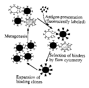
Figure 1 is a schematic, showing in vitro affinity maturation by yeast
display.
Figure 2 shows the schematic illustration of surface display on yeast.
A nine amino acid peptide epitope from the hemagglutinin antigen (HA) was
fused to
the C-tenninus of the Aga2p subunit of a-agglutinin, followed by the 4-4-20
anti-
fluorescein scFv sequence. An additional ten residue epitope tag (c-myc) was
fused at
the C-terminus of the scFv, allowing quantitation of fusion display
independent of
antigen binding by either the HA or c-myc tags. The HA or c-myc tag can be
used to
normalize for variation in the number of displayed fusion proteins in double-
label flow
cytometry.
Figure 3 shows a vector for yeast surface display. Figure 3A shows
the construction of the vector pCT202. Figure 3B shows the specific
restriction
CA 02319147 2001-03-09
WO 99/36569
PCT/US99/01188
sites and the transcriptional regulation by galactose, the N-terminal HA and C-
terminal c-myc epitope tags and the Factor Xa protease cleavage site.
Figure 4 demonstrates that the displayed fusions can be detected by
fluorescence techniques, showing a flow cytometric histogram of yeast labeled
with a-
c-myc/a-mouse-PE.
Figure 5 shows that antigen binding by 4-4-20 scFv can be detected
by fluorescence, showing a flow cytometric histogram of yeast labeled with
FITC-
dextran (2 x 10⁶ Da).
Figure 6 shows that 4-4-20 activity and c-myc can be detected
simultaneously, and demonstrate a 1:1 correlation of fluorescence signals;
therefore,
variation in intensity signal 1 (FITC) can be normalized for cell-to-cell
variation in
expression of the protein of interest by the intensity of signal 2 (PE).
Figure 7 shows the sequence of the AGA2-HA-4-4-20-c-myc gene
cassette.
Figure 8 shows confocal microscopic images of yeast displaying scFv.
Yeast containing plasmid directing surface expression of the HA peptide
(Figure 8A)
or the scFv fusion (Figure 8B) were labeled with InAb 9E10, followed by a
secondary anti-mouse IgG-R-phycoerythrin (PE) conjugate and FITC-dextran. DIC
(upper panels), red PE fluorescence (middle panels), and green FITC
fluorescence
(lower panels) images were collected.
Figure 9 shows flow cytometric analyses of yeast displaying scFv.
Yeast strains displaying either (Figure 9A) an irrelevant peptide or (Figure
9B) the
4-4-20 scFv were labeled with mAb 9E10 and FITC-dextran. Cells displaying scFv
were also treated with 5 mM DTT prior to labeling (Figure 9C). (i) Univariate
histograms of PE fluorescence associated with labeling by 9E10; (ii)
univariate
histograms of FITC fluorescence; (iii) bivariate histograms showing
correlation
between PE and FITC fluorescence.
Figure 10 demonstrates the enrichment of yeast displaying improved
scFv variants by kinetic selection and flow cytometric cell sorting. Yeast
expressing a
mutagenized 4-4-20 scFv library (Figure 10A) and a yeast pool resulting from
three
rounds of kinetic selection and amplification (Figure 10B) were subjected to
competitive dissociation of fluorescent antigen with 5-aminofluorescein,
leaving cells
11
CA 02319147 2001-03-09
= = I.1,14.1,,,IVMJ=JR1
displaying the tightest binding mutants with the highest ratio of FITC
intensity/PE
intensity.
Figure 11 shows dissociation kinetics of the interaction between
fluorescein and surface displayed scFv. Yeast displaying 4-4-20 scFv
(circles),
mutant 4M1.1 (squares) isolated from the library, and mutant 4M1.2 (triangles)
were
labeled with niAb 9E10 and FITC-dextran. 5-aminofluorescein was added as a
competitor. Mean intensity of FITC fluorescence of the 9E10 positive
population of
cells was followed as a function of time. The slope of the line is equal to
the kinetic
dissociation rate, and the extrapolated value at time t = 0 sec is equal to
the valency of
the interaction. MFI; = relative mean fluorescence intensity of yeast at time
t
Figure 12 shows the expression levels and antigen binding properties
of yeast surface displayed scFv-KJ16 (shaded) and control Aga2p/HA (unshaded).
Yeast strain EBY100 was transformed with scFv-KJ16 cloned into the yeast
display
vector pCT202 or the pCT202 vector alone. After induction in galactose medium
at
20 C overnight, cells were stained with fluorescent antibodies and analyzed by
flow
cytometry. (Figure 12A) scFv-KJ16/yeast or Aga2p/HA/yeast stained with mouse
anti-HA Mab (12CA5) followed by FITC-labeled goat anti-mouse IgG, (Figure 128)
scFv-KJ16/yeast or Aga2p/HA/yeast stained with mouse anti-c-myc Mab (9E10)
followed by FITC-labeled goat anti-mouse IgG, (Figure 12C) scFv-KJ16/yeast or
Aga2p/HA/yeast stained with biotinylated-scTCR at ¨10 nM followed
by a
streptavidin-phycoerythrin conjugate, and (Figure I2D) scFv-KJ16/yeast stained
with
biotinylated-scTCR followed by a streptavidin-phycoerythrin conjugate in the
presence (shaded) or absence (unshaded) of intact IgG ICJ16 at 100 mg/nil.
Figure 13 shows the equilibrium antigen binding isotherm of cell wall
displayed scFv-KJ16, determined by flow cytometry. Yeast strain
EBY100
displaying surface scFv-KJ16 was incubated with varying concentrations of
biotinylated-scTCR, labeled with a streptavidin-phycoerythrin conjugate, and
detected by flow cytometry. Data was plotted as a Scatchard diagram or as a
titration
(inset) and an effective KD ¨500 nM was determined. MFU refers to mean
fluorescence units.
Figure 14 shows the two dimensional fluorescence histograms and
sorting window used to select scFv-KJ16 mutants. scFv-KJ16 cloned into the
12
_______________________________________________________________________ _
CA 02319147 2001-03-09
WO 99/36569
PCT/US99/01188
display vector pCT202 was transformed into the E. coil mutator strain XL I-Red
(Stratagene) and propagated for six overnight growth cycles. Plasmids of the
mutant
library were purified and used in LiAc transformation (Gietz et al., 1995) of
EBY100
yeast. After induction at 30 C, yeast were sorted using a fluorescence-
activated cell
sorter. (Figure 14A) Representative histogram from the first round of cell
sorting,
with the sorting window indicated, and (Figure 14B) representative histogram
from
the fourth (final) round of sorting, illustrating an enrichment of the
population in the
sorting window.
Figure 15 shows the mean levels of binding to anti-HA Mab, anti-c-
myc Mab, or biotinylated-scTCR for ten randomly selected clones from the final
sort
shown in Figure I4B. Ten mutants and wt scFv-KJ16/yeast were induced in
galactose
medium at 30 C overnight. Cells were analyzed by flow cytometry after staining
with mouse anti-HA Mab followed by FITC-labeled goat anti-mouse IgG (open
bars),
mouse anti-c-myc followed by FITC-labeled goat anti-mouse IgG (gray bars), or
biotinylated-scTCR at -40 nM followed by a streptavidin-phycoerythrin
conjugate
(black bars).
Figure 16 shows the fluorescent labeling distributions for anti-c-myc
or scTCR binding of three selected mutants shown in Figure 4. Three classes of
scFv-
KJ16/yeast mutants were double-stained with anti-c-myc and biotinylated-scTCR
followed by FiTC-labeled goat anti-mouse IgG and a streptavidin-phycoerythrin
conjugate, then analyzed by flow cytometry as described in Figure 4. The
fluorescent
distributions for each scFv-KJ16/yeast mutant (shaded) and wt scFv-KJ16/yeast
(unshaded) are shown. Figures 16A and 16B, mut4; Figures 16C and 16D, mut7;
Figures 16E and 16F, mut10.
Figure 17 shows the equilibrium antigen binding isotherms for three
mutants shown in Figure 16. Aga2p/HA/yeast, wt scFv-KJ16/yeast, and three
mutant scFv-KJ16/yeast characterized in Figure 16 were stained with various
dilutions of biotinylated-scTCR followed by a streptavidin-phycoerythrin
conjugate.
After analysis by flow cytometry, binding isotherms were graphed with MFU as a
function of scTCR dilution.
Figure 18 shows the sequence analysis of wild-type scFv-KJ16,
mut4, and mut7. Plasmids from wt scFv-K.116/yeast and two mutants (mut4 and
13
CA 02319147 2001-03-09
WO 99/36569
PCT/US99/01188
mut7) were recovered by plasmid rescue and transformed into E. con DH5a
competent cells to produce plasrnids for sequencing, as described below.
Sequence
analysis was performed using primers that flank the scFv of the display
vector.
Mutations are indicated in bold.
Figure 19 shows the flow cytometry profiles of antibody binding to
yeast that have been transformed with a plasmid that contains a T cell
receptor single-
chain (VaV13) gene. The normal or wild type ( wt) sequence is compared with
several
mutants (mTCR7, mTCR15, mTCR16) that were selected after random mutagenesis
of the scTCR plasmid. Selection involved binding of the antibody 1B2, which
recognizes a conformational epitope on the T cell receptor, followed by
several round
of fluorescent-activated cell sorting. In the first panel, the yeast cells
were stained
with an antibody (12CA5) to the HA tag. In the second panel, the yeast cells
were
stained with an antibody (182) to the T cell receptor. Although the HA epitope
is
expressed on the surface in each case, only those cells that express a
mutagenized
plasmid are capable of expressing the native T cell receptor (182 positive).
Figure 20 shows the flow cytometry profiles of antibody binding to
yeast that have been transformed with double mutants from the selection shown
in
Figure 19. Cells were stained for flow cytometry as described in Figure 19.
Double
mutants expressed an increase in level of the T cell receptor (Le. 1132-
reactive
material). The results show that by combining single mutations it is possible
to
enhance the level of cell surface expression of the T cell receptor.
Figure 21 shows the sequence of mutations that lead to the enhanced
expression of the cell surface T cell receptor. These included residues 17 of
the V(3,
43 of the Va, and 104 of the Va.
Figure 22 shows relative secretion levels of scTCR. Soluble
expression levels (arbitrary units) of scTCR produced using low copy yeast
expression system. Triplicate cultures from independent clones were analyzed
for
1B2 ELISA activity.
Figure 23A shows flow histograms of scTCR displaying yeast. The
cell sorted populations for wild-type and representative single, double, and
triple
mutants are presented. The mean fluorescence units (FITC fluorescence: anti-
HA, PE
14
_
CA 02319147 2001-03-09
WO 99/36569 PCT/US99/01188
fluorescence: 1B2) are indicated on each histogram. Anti-HA indicates number
of
surface fusions, and 1B2 indicates the number of cells displaying properly
folded
scTCR. Figure 23B shows the correlation between surface expression and soluble
secretion. 1B2-active surface scTCR determined by flow is compared to 1B2
activity
of soluble secreted material by ELISA assay. At minimum, duplicate flow
experiments were done to determine the mean fluorescence units of a particular
clone.
Figure 24A shows the temperature stabilities of scTCR. Yeast
supernatant samples containing scTCR were subjected to the indicated
temperatures
for one hour. Triplicate samples were analyzed for 1B2-active fractions by
ELISA.
The fractions were normalized independently to unity by the highest intensity
ELISA
signal having no activity loss. Figure 248 shows the kinetics of scTCR thermal
denaturation. 1B2 ELISA activity of supernatant samples was monitored as a
function of the time each sample was incubated at 46 C. Observed kinetic
thermal
denaturation rates (1c,,,bs) at 46 C are indicated. In both Figure 24A and
248,
comparisons are made to TRX-TCR rather than wild-type scTCR because wild-type
scTCR was not detected in yeast supernatants.
Figure 25 shows the correlation between thermal stability and soluble
expression. The scTCR 1B2 ELISA activity remaining after being incubated at 48
C
for one hour is compared to relative secretion levels (1B2 ELISA). Triplicate
samples
were analyzed for the secretion levels and for the thermal denaturation
analyses.
DETAILED DESCRIPTION OF THE INVENTION
As used herein, the term "affinity maturation" shall refer to a process
of successive mutation and selection by which antibodies of higher affinity
are
selected. As used herein, the term "agglutinin" shall refer to a yeast surface
adhesion
protein which binds two yeast cells together during mating. As used herein,
the term
"antibody" shall refer to a protein produced by mammalian immune systems which
binds tightly and specifically to particular molecules. As used herein, the
term
"ligand" shall refer to a molecule that is bound specifically by a particular
protein. As
used herein, the term "antigen" shall refer to a ligand that is bound
specifically by an
antibody. As used herein, the term "Complementarity Determining Region" or
CA 02319147 2001-03-09
WO 99/36569 PCT/US99/01188
"CDR" shall refer to the portion of an antibody which directly contacts the
bound
antigen. As used herein, the term "Fluorescence Activated Cell Sorting" or
"flow
cytometry" shall refer to a method for sorting cell populations on the basis
of
differential fluorescent labeling. As used herein, the term "hapten" shall
refer to a
small antigen which cannot stimulate an immune response without being
conjugated to
a carrier. As used herein, the term "single chain antibody" or "SCA" shall
refer to a
fusion of portions of the heavy and light chains of an antibody which retains
a single
active binding site. The term scFv is used interchangeably to refer to a
single chain
antibody. As used herein, the term "epitope tag" shall refer to a
contiguous
sequence of amino acids specifically bound by an antibody when fused to
another
protein. As used herein, the term "HA" refers to the epitope tag sequence
YPYDVPDYA (SEQ ID No. 1). As used herein, the term "c-myc" refers to the
epitope tag sequence EQICLISEEDL (SEQ ID No. 2). As used herein, the term
"scFv
4-4-20" refers to an scFv which binds specifically to fluorescein and
fluorescein
conjugated to other molecules such as biotin or dextran. As used herein, the
term
"AGA2p" refers to the protein product of the yeast AGA2 mating type a
agglutinin
gene. The term "displayability" will be used to describe a combination of
biophysical
characteristics allowing a protein to escape the secretory "quality control"
apparatus
that retains and degrades misfolded proteins (Hammond & Helenius, 1995.)
Proteins
displayed on the yeast cell surface must first pass successfully through the
quality
control step. Protein folding kinetics and thermodynamic stability together
are
believed to determine the efficiency of escape from the quality control
apparatus.
In accordance with the present invention there may be employed
conventional molecular biology, microbiology, and recombinant DNA techniques
within the skill of the art. Such techniques are explained fully in the
literature. See,
e.g., Maniatis, Fritsch & Sambrook, "Molecular Cloning: A Laboratory Manual
(1982); "DNA Cloning: A Practical Approach," Volumes I and II (D.N. Glover ed.
1985); "Oligcmucleotide Synthesis" (Mi. Gait ed. 1984); "Nucleic Acid
Hybridization" (B.D. Harnes & S.J. Higgins eds. (1985)); "Transcription and
Translation" (B.D. Hames & S.J. Higgins eds. (1984)); "Animal Cell Culture"
(R.I.
Freshney, ed. (1986)); "Immobilized Cells And Enzymes" (IRL Press, (1986)); B.
Perbal, "A Practical Guide To Molecular Cloning" (1984).
16
CA 02319147 2001-03-09
WO 99/36569
PCT/US99/01188
A "vector" is a replicon, such as plasmid, phage or cosmid, to which
another DNA segment may be attached so as to bring about the replication of
the
attached segment
A DNA "coding sequence" is a double-stranded DNA sequence which
is transcribed and translated into a polypeptide in vivo when placed under the
control
of appropriate regulatory sequences. The boundaries of the coding sequence are
determined by a start codon at the 5' (amino) terminus and a translation stop
codon at
the 3' (carboxyl) terminus. A coding sequence can include prokaryotic
sequences,
cDNA from eukaryotic mRNA, genomic DNA sequences from eukaryotic (e.g.,
mammalian) DNA, and even synthetic DNA sequences. A polyadenylation signal and
transcription termination sequence will usually be located 3' to the coding
sequence.
Transcriptional and translational control sequences are DNA regulatory
sequences, such as promoters, enhancers, polyadenylation signals, terminators,
and
the like, that provide for the expression of a coding sequence in a host cell.
A "promoter sequence" is a DNA regulatory region capable of binding
RNA polymerase in a cell and initiating transcription of a downstream (3'
direction)
coding sequence. For purposes of defining the present invention, the promoter
sequence is bounded at its 3' terminus by the transcription initiation site
and extends
upstream (5' direction) to include the minimum number of bases or elements
necessary
to initiate transcription at levels detectable above background. Within the
promoter
sequence will be found a transcription initiation site (conveniently defined
by
mapping with nuclease Si), as well as protein binding domains (consensus
sequences)
responsible for the binding of RNA polymerase. Eulcaryotic promoters will
often, but
not always, contain "TATA" boxes and "CAT" boxes. Prokaryotic promoters
contain Shine-Dalgarno sequences in addition to the -10 and -35 consensus
sequences.
An "expression control sequence" is a DNA sequence that controls and
regulates the transcription and translation of another DNA sequence. A coding
sequence is "under the control" of transcriptional and translational control
sequences
in a cell when RNA polymerase transcribes the coding sequence into mRNA, which
is
then translated into the protein encoded by the coding sequence.
A "selection gene" refers to a gene that enables the discrimination of
cells displaying a required phenotype upon implementation of certain
conditions. For=
17
CA 02319147 2001-03-09
WO 99/36569
PCT/US99/01188
example, the growth of bacteria in medium containing antibiotics to select for
the
bacterial cells containing antibiotic resistance genes.
The term "primer" as used herein refers to an oligonucleotide, whether
occurring naturally as in a purified restriction digest or produced
synthetically, which
is capable of acting as a point of initiation of synthesis when placed under
conditions
in which synthesis of a primer extension product, which is complementary to a
nucleic acid strand, is induced, i.e., in the presence of nucleotides and an
inducing agent
such as a DNA polymerase and at a suitable temperature and pH. The primer may
be
either single-stranded or double-stranded and must be sufficiently long to
prime the
synthesis of the desired extension product in the presence of the inducing
agent. The
exact length of the primer will depend upon many factors, including
temperature, the
source of primer and the method used. For example, for diagnostic
applications,
depending on the complexity of the target sequence, the oligonucleotide primer
typically contains 15-25 or more nucleotides, although it may contain fewer
nucleotides.
The primers herein are selected to be "substantially" complementary to
different strands of a particular target DNA sequence. This means that the
primers
must be sufficiently complementary to hybridize with their respective strands.
Therefore, the primer sequence need not reflect the exact sequence of the
template.
For example, a non-complementary nucleotide fragment may be attached to the 5'
end
of the primer, with the remainder of the primer sequence being complementary
to the
strand. Alternatively, non-complementary bases or longer sequences can be
interspersed into the primer, provided that the primer sequence has sufficient
complementarity with the sequence or hybridize therewith and thereby form the
template for the synthesis of the extension product.
A cell has been "transformed" by exogenous or heterologous DNA
when such DNA has been introduced inside the cell. The transforming DNA may or
may not be integrated (covalently linked) into the genome of the cell. In
prokaryotes,
yeast, and mammalian cells for example, the transforming DNA may be maintained
on
an episomal element such as a plasmid. With respect to eulcaryotic cells, a
stably
transformed cell is one in which the transforming DNA has become integrated
into a
chromosome so that it is inherited by daughter cells through chromosome
replication.
18
CA 02319147 2001-03-09
WO 99/36569
PCT/US99/01188
This stability is demonstrated by the ability of the eukaryotic cell to
establish cell
lines or clones comprised of a population of daughter cells containing the
transforming
DNA. A "clone" is a population of cells derived from a single cell or common
ancestor by mitosis. A "cell line" is a clone of a primary cell that is
capable of stable
growth in vitro for many generations.
A "heterologous" region of the DNA construct is an identifiable
segment of DNA within a larger DNA molecule that is not found in association
with
the larger molecule in nature. Thus, when the heterologous region encodes a
mammalian gene, the gene will usually be flanked by DNA that does not flank
the
mammalian genomic DNA in the genome of the source organism. In another
example,
coding sequence is a construct where the coding sequence itself is not found
in nature
(e.g., a cDNA where the genornic coding sequence contains introns, or
synthetic
sequences having codons different than the native gene). Allelic variations or
naturally-occurring mutational events do not give rise to a heterologous
region of DNA
as defined herein.
A number of polypeptide sequences that can be fused to proteins and
bound specifically by antibodies are known and can be utilized as epitope
tags. These
include, for example, HA (SEQ ID No. 1), c-myc (SEQ ID No. 2), DTYRYI (SEQ ID
No. 3), TDFYLK (SEQ ID No. 4), EEEEYMPME (SEQ ID No. 5), KPFTPPPEPET
(SEQ ID No. 6), HHHHHH (SEQ ID No. 7), RYIRS (SEQ ID No. 8), and
DYICDDDDK (SEQ ID No. 9).
Antibodies are protein molecules produced by the human immune
system to recognize, bind to, and mediate the clearance of foreign substances
from the
body. Technologies have been developed to take advantage of antibodies for
highly-
specific cancer diagnosis and therapy. For example, by tethering radioisotopes
or
toxins to an antibody which binds to tumor cells, it is possible to deliver a
focused
dosage of such cell-killing agents to the diseased tissue while leaving
surrounding
tissue comparatively unharmed. Antibodies are also critical tools in
biotechnology,
and are used extensively for analytical purposes, e.g., to quantify trace
quantities of
substances and separations, and to purify desired biological products from
complex
mixtures.
19
CA 02319147 2001-03-09
W099/36569
PCT/U599/01188
In these applications, both the strength of the antibody bond with its
target (affinity) and the selectivity with which an antibody binds to only its
particular
target (specificity) are crucial. For this reason, protein engineers seek to
alter and
improve the binding characteristics of particular antibodies. Rational
approaches to
antibody structural design have met with limited success, and available
methods for
random screening possess significant limitations.
The mammalian immune system's approach to the problem of fine
tuning antibody affinity is by a process called "affinity maturation," wherein
cycles of
mutation and evolutionary selection produce antibodies which bind their
targets more
tightly. The present invention discloses a powerful new system for engineering
antibody affinity and specificity, by constructing a microbial analog of the
mammalian
immune system's B cell repertoire. Antibodies were displayed on the surface of
yeast
cells by genetic fusion with cell wall proteins. After mutation, variants were
selected
on the basis of improved binding characteristics with fluorescently labeled
targets.
The yeast antibody display method was tested by studying model
antibodies whose physical and chemical properties are already well
characterized.
These methods are then straightforwardly applied to antibodies of practical
interest.
The genetic malleability of yeast, the ease of growth of this microbe, and the
ability to
modify antibody binding conditions in the test tube combine to produce
unprecedented control over the engineering of antibody affinity and
specificity.
The advantage of the library method of the present invention is that it
is particularly suited for proteins such as antibodies. The most widely used
method
currently consists of "panning" for antibodies displayed on the surface of
bacteriophage. Yeast display has several advantages over phage display. First,
the
antibody-antigen bond need not be broken to recover tightly-bound variants.
The
harsh conditions required for disrupting this bond in prior art methods can
reduce
infectivity of phage. Secondly, increased library diversity due to decreased
clonal
deletion is an advantage. It is well known that many antibody structures
cannot be
correctly processed by the bacterial secretory apparatus. Yeast cells are
eucaryotic
and possess secretory pathways very similar to mammalian cells. Thirdly, the
present invention provides a more accurate and precise determination of
antibody-
antigen affinity. The presence of 104 molecules per cell eliminates the
stochastic
CA 02319147 2001-03-09
WO 99/36569 PCMJS99/01188
variation that results with only a few molecules per phage. Finally,
quantitation of
fluorescence by flow cytometry provides a continuous measure of surface-bound
antigen without a priori knowledge of affinity in by comparison to the binary
bound/released dichotomy with panning of phage. Also, bacteria possess a
lipopolysaccharide layer which acts as a macromolecular permeability barrier
preventing antibody or protein access to displayed molecules.
The present invention discloses a surface display system for the in
vitro expression and selection of peptide and protein libraries on yeast. A
nine
residue peptide epitope (HA) has been fused to the binding subunit of a yeast
cell
wall protein (AGA2), followed by the 4-4-20 anti-fluorescein single-chain Fv.
Selection was performed by flow cytometry on mixtures of cells with and
without the
displayed fusion. 600-fold enrichments were achieved in one pass of sorting.
The
system of the present invention illustrates a process for the in vitro
affinity
maturation of antibodies as well as a process for the directed evolution of
other
proteins and peptides, with the advantages of (i) a double-label flow
cytometry
selection scheme allowing finer affinity discrimination than panning; (ii) as
many as
104 copies of the displayed sequence per cell, eliminating stochastic
variations in the
selection; and (iii) library expression in yeast, with an altered or
potentially improved
expression bias which could yield clones that would be deleted from a library
expressed in E. coil.
One object of the present invention is the engineering of antibodies for
improved affinity and specificity. Toward this end, antibody-hapten binding
was
studied via mutagenesis and screening of antibodies expressed on the external
cell wall
of the yeast Saccharomyces cerevisiae. As an experimentally facile and
genetically
pliable eucaryote, yeast presents significant advantages over filamentous
phage
display as a platform for antibody expression and engineering. In essence, a
microbial
analog of the mammalian immune system B-cell repertoire was constructed in
vitro,
allowing antibody affinity maturation to be performed under strictly
controlled
conditions of mutagenesis and selection. As a result, antibodies of
significantly
improved affinity and specificity were attainable.
One aspect of the present invention provides a method for selecting
proteins with desirable binding properties comprising: transforming yeast
cells with a
21
CA 02319147 2001-03-09
WO 99/36569
PC11US99/01188
vector expressing a protein to be tested fused at its N-terminus to a yeast
cell wall
binding protein; labeling the yeast cells with a first label, wherein the
first label
associates with yeast expressing the protein to be tested and does not
associate with
yeast which do not express the protein to be tested; selecting for the yeast
cells with
which the first label is associated; quantitating the first label, wherein a
high
occurrence of the first label indicates the protein to be tested has desirable
binding
properties and wherein a low occurrence of the first label indicates the
protein to be
tested does not have desirable binding properties.
A preferred embodiment of the present invention further includes the
steps of: labeling the yeast cells with a second label, wherein the second
label
associates with yeast expressing an epitope tag fused to the protein to be
tested and
encoded by the vector and does not associate with yeast which do not express
the
epitope tag encoded by the vector; quantitating the second label, wherein an
occurrence of the second label indicates a number of expressed copies of the
epitope
tagged protein to be tested on the yeast cell surface; and comparing the
quantitation of
the first label to the quantitation of the second label to determine the
occurrence of the
first label normalized for the occurrence of the second label, wherein a high
occurrence
of the first label relative to the occurrence of the second label indicates
the protein to
be tested has desirable binding properties.
Another preferred embodiment of the present invention includes the
steps of: labeling the yeast cells with a third label that competes with the
first label
for binding to the protein to be tested; labeling the yeast cells with the
first label;
quantitating the first label; labeling the yeast cells with the second label;
quantitating
the second label; and comparing the quantitation of the first label to the
quantitation of
the second label to determine the occurrence of the first label normalized for
the
occurrence of the second label, wherein a low occurrence of the first label
relative to
the occurrence of the second label indicates the protein to be tested has
desirable
binding properties.
Another aspect of the present invention provides a vector for
performing the method of the present invention, comprising a cell wall binding
protein
fused to an N-terminus of a protein of interest. Preferred embodiments of this
aspect
of the present invention include means for expressing a polypeptide epitope
tag in the
22
CA 02319147 2001-03-09
WO 99/36569 PCT/US99/01188
yeast cells. A more preferred embodiment provides that the cell wall binding
protein
is a yeast agglutinin protein binding subunit, even more preferably, the yeast
agglutinin protein is Aga2p.
Another aspect of the present invention provides for a method of
selecting proteins with enhanced phenotypic properties relative to those of
the wild-
type protein, comprising the steps of: transforming yeast cells with a vector
expressing a protein to be tested fused to a yeast cell wall protein, wherein
mutagenesis is used to a generate a variegated population of mutants of the
protein to
be tested; labeling the yeast cells with a first label, wherein the first
label associates
with yeast expressing the protein to be tested and does not associate with
yeast which
do not express the protein to be tested; isolating the yeast cells with which
the first
label is associated; and analyzing and comparing the properties of the mutant
protein
expressed by yeast with properties of the wild-type protein, wherein yeast
cells
exhibiting mutant proteins with enhanced properties over the wild-type protein
are
selected. As described above, a second and/or third label may be employed with
this
embodiment, and selection of mutated proteins of interest with enhanced
phenotypic
properties may involve iterative cycles of the enrichment and labeling steps..
In preferred embodiments, the yeast cell wall protein is an agglutinin;
the protein to be tested is fused by its N terminus to the C terminus of a
binding
subunit an agglutinin; the yeast strain is of a genus selected from the group
consisting
of Saccharomyces, Pichia, Hansenula, Schizosaccharomyces, Kluyveromyces,
Yarrowia, and Candida; the protein to be tested is an antibody, Fab, Fv, or
scFv
antibody fragment, more prefereably the protein to be tested is the ligand
binding
domain of a cell surface receptor, even more preferably, the cell surface
receptor is a T
cell receptor.
=
An object of the present invention is to provide a method to select for
mutant proteins exhibiting enhanced phenotypic properties selected from the
group
consisting of surface expression, stability, binding constant, dissociation
constant,
level of secretion, and solubility, wherein the mutants of the protein to be
tested may
contain single mutations or multiple mutations.
In yet another aspect of the present invention, a second label may be
used to quantitatively determine cell surface expression levels, which may be
used as
23
CA 02319147 2001-03-09
WO 99/36569
PCT/US99/01188
an assay to select for other desirable phenotypic properties, such as
intracellular
expression, stability, binding constant, dissociation constant, level of
secretion, and
solubility.
The mutant proteins selected by the methods of the present invention
may be further characterized by cloning the gene encoding the selected mutant
proteins into a vector adapted for expression in a eukaryote; and expressing
the
mutant protein in the eukaryote, wherein the enhanced properties of the mutant
protein are confirmed by comparing the phenotypic properties of the enhanced
properties of the mutant protein with the properties of the wild-type protein.
Preferably, the eukaryote is selected from the group consisting of mammalian,
insect,
or yeast. Because the phenotype of the selected mutant is an inherent property
of the
"new" (i.e. mutated) protein, this approach is also applicable to expressing
the mutant
in other non-eukaryotic expression systems.
Another preferred aspect of the present invention provides a method
for displaying proteins that are not displayed as their normal ("wild type")
sequence.
In the example shown, the T cell receptor for antigen was not expressed as its
"wild
type" sequence. However, after random mutagenesis and selection by flow
cytometry
with appropriate conformationally-specific antibodies, the mutant receptors
were
expressed on the yeast cell surface. This strategy will allow the discovery of
novel T
cell receptors and it provides a method for the display of virtually any
polypeptide.
Thus, the present invention also provides a method for selecting proteins for
displayability on a yeast cell surface, comprising the step of: transforming
yeast cells
with a vector expressing a protein to be tested fused to a yeast cell wall
protein,
wherein mutagenesis is used to a generate a variegated population of mutants
of the
protein to be tested; labeling the yeast cells with a first label, wherein the
first label
associates with yeast expressing the protein to be tested and does not
associate with
yeast which do not express the protein to be tested; isolating the yeast cells
with
which the first label is associated, by quantitating the first label, wherein
a high
occurrence of the first label indicates the protein to be tested has desirable
display
properties and wherein a low occurrence of the first label indicates the
protein to be
tested does not have desirable display properties. Preferably, the protein
tested is an
antibody, Fab, Fv, or scFv antibody fragment or the ligand binding domain of a
cell
24
CA 02319147 2001-03-09
WO 99/36569 PCT/US99/01188
surface receptor. A representative example of a cell surface receptor is a T
cell
receptor.
The following examples are given for the purpose of illustrating various
embodiments of the invention and are not meant to limit the present invention
in any
fashion.
EXAMPLE 1
Media/Buffers
The following media/buffers were used herein:
4 coil
LB media (IX): Bacto tryptone (Difco, Detroit, MI): 10.0 g; Bacto
yeast extract (Difco): 5.0 g; NaCI: 10.0 g. Make up to 1 L, autoclave. For
plates, add
15g/L agar and autoclave.
Ampicigin Stock: 25 mg/ml of sodium salt of ampicillin in water.
Filter sterilize and store in aliquots of 4 mls at -20 C. Working [ 1 = 35-50
nighnl; 4
ml of aliquot in 1 L ¨> 100 mg/int; 2 ml of aliquot in 1 L ---) 50 mg/ml. Add
to
autoclaved LB only after it has cooled to ¨ 55 C.
OC media (100 mL): 2% Bacto tryptone: 2.0 g, 0.5% Yeast Extract
(Difco): 0.5 g; 10 mM NaCI: 0.2 ml 5 M; 10 mM MgC12: 1.0 ml I M; 10 mM
MgSO4: 1.0 ml 1 M; 20 mM Dextrose: 0.36 g. Autoclave or filter sterilize.
Yeast
Synthetic Minimal + Casamino acids (SD-CAA) 500 mL
Dextrose(Glucose) 10.00 g
Yeast Nitrogen Base w/o Amino Acids (Difco) 3.35 g
Na2HPO4 = 7H20 5.1 g
NaH2PO4 = H20 4.28 g
Casamino Acids (Trp-, Um-) (Difco) 2.5 g
Add dH20 to final volume. Filter sterilize and refrigerate. For plates,
dissolve sodium phosphates and sorbitol to 1 M final concentration in 400 ml
dH20.
Add 7.5 g agar and autoclave. Dissolve dextrose, N2 base, and amino acids in
100 ml
CA 02319147 2001-03-09
WO 99/36569 PCT/US99/01188
dH20 and filter sterilize. Add the filtered reagents after the autoclaved
salts have
cooled enough to touch.
SG-CAA (jnduction Medium) 500 nt
Galactose 10.00 g
Yeast Nitrogen Base w/o Amino Acids (Difco) 3.35 g
Na2HPO4 = 7H20 5.1 g
NaH2PO4 = H20 4.28 g
Casamino Acids (Trp-, Ura-) (Difco) 2.5 g
Add dH20 to final volume. Filter sterilize and refrigerate.
Rich (YPD1(1000 mL); Yeast extract: 10 g Peptone (Difco): 20 g
Dextrose: 20 g. Add dH20 to 1 L. Autoclave.
TAE (Tris-Acetate): Working solution: 0.04 M Tris-acetate and 0.001
M EDTA
Stock (50X): in IL
Tris base 242 g
glacial acetic acid 57.1 ml
0.5 M EDTA (pH 8.0) 100 ml
TBE (Tris-Borate): Working solution: 0.09 M Tris-borate and
0.001M EDTA
Stock (5X): in _LL
Tris base 54g
boric acid 27.5g
0.5 M EDTA (pH 8.0) 20.0 ml
Stop buffer 10X (Restriction): 50% v/v glycerol; 0.1 M EDTA (pH
7.5); 1% w/v SDS; 0.1% w/v bromophenol blue. Combine all components except for
the dye and pH to 7.5 before dye addition.
Staining ¨ Ethidium bromide: 0.5 mg/nil in water; Stock: 10 mg/ml.
Dilutions of Stock: 1/10 in TBE. Add 100 ml dilution in 100 ml buffer.
TBS Working solution: 10 mM Tris-HCI, 140 mM NaC1 and 5
mM EDTA. Filter sterilize.
26
CA 02319147 2001-03-09
WO 99/36569
PCT/US99/01188
EXAMPLE 2
Protocol: Replica Plating
1. Choose a material suitable for colony lifts, making sure it is washed,
dried and sterile.
2. Mark the bottom of each fresh replica plate with an arrow to line up
the plate. Also mark the starting plate.
3. Take the top off the starting plate, turn it upside down and line up
the arrow on the bottom with the mark on the colony lift material. Lay the
plate
down onto the surface of the material and gently put pressure on the entire
plate.
Make sure the plate doesn't move around after it has touched the material.
Remove
the plate and replace the lid. Portions of the colonies that transferred to
the material
can be seen.
4. Repeat this procedure with one of the fresh replica plates. Make
sure the arrow lines up with the mark also to make an exact replica. Hold up
to the
light to see the tiny colonies that transferred.
5. Repeat the entire procedure for each replica plate to be made.
6. Incubate the replica plates at the appropriate conditions for
selective growth. Colonies will usually grow up within a day or so.
EXAMPLE 3
Protocol: Electrotransformation of Yeast
Cell Preparation:
1. Inoculate 50 ml of YPD with an overnight culture to an OD of 0.1.
2. Grow cells at 30 C with vigorous shaking to an OD600 of 1.3 to 1.5
(approximately 6 hours).
3. Harvest in cold rotor at 3500 rpm for 5 minutes at 4 C. Discard
supernatant.
4. Thoroughly wash the cells by resuspending in 50 ml cold sterile
water. Centrifuge as above and discard supernatant.
5. Repeat step 4 with 25 ml cold water.
27
CA 02319147 2001-03-09
WO 99/36569
PCT/US99/01188
6. Resuspend in 2 ml of ice-cold sterile 1 M sorbitol. Centrifuge as
above and discard supernatant.
7. Resuspend in 50 ml ice-cold 1 M sorbitol. Final volume of cells is
about 150 ml (enough for 3 transformations).
Electrotransformation.,
1. Place 0.2 cm cuvettes and white slide chamber on ice.
2. In an eppendorf tube, add 50 ml of yeast suspension and gently mix
in <5 ml (0.1 mg) of plasmid DNA in TE. Make sure to add DNA to yeast already
in eppendorf. Place on ice for 5 minutes (This time frame is pretty critical).
3. Set GENE PULSER at 1.5 kV and 25 mF. Set the Pulse Controller
to 200 W. The time constant for this pulse should be 4.5 to 5.0 msec.
4. Transfer 40 ml of cell/DNA mixture to pre-chilled electroporation
cuvette. Tap contents to bottom, making sure the sample is in contact with
both
aluminum sides of the cuvette. Place the cuvette in chilled safety chamber
slide. Push
slide into the chamber until the cuvette makes contact with the electrodes in
the base
of the chamber.
5. Apply one pulse at the settings above.
6. Remove the slide with the cuvette, and immediately add 1 ml of cold
1M sorbitol to the cuvette. Mix and return cuvette to ice. Spread 200 ml onto
selective plates containing 1 M sorbitol.
EXAMPLE 4
Protocol: E. Coli Transformation
1. Thaw aliquot of competent subcloning efficiency HB101 (-70 C
storage) on ice, keeping all reagents on ice. Use DH5a, cells for lac z
complementation; DM-1 for non-methylation.
2. Dispense 50 ml 1-IB101 to required # of eppendorf tubes, one for
each DNA sample, one for pBR322 (positive control) (pUC19 for DH5a and DM-1),
one for no DNA (negative control).
3. Aliquot unused cells into 50 nth portions and refreeze in a dry
ice/Et0H bath; store at-70 C.
28
CA 02319147 2001-03-09
WO 99/36569 PCT/US99/01188
4. Add 1 ml DNA (1 mg) to eppendorfs (1 ml of pBR322 or pUC19),
tap tubes to mix, then incubate on ice for 30 min. For ligations, use 1-2 ml
of ligation
mixture (too much sours the transformation).
5. Heat shock at 37 C for 20 seconds. (45 sec at 42 C for DM-1
cells).
6. Place on ice for 2 minutes, then add 0.95 rnL of room temp SOC
media. Incubate at 37 C for 1 hour in bath (shaking optional) or on shaker in
37 C
room.
7. Plate 100-200 mL of cells onto LB, 100 mg/mL Ampicillin, and
incubate overnight at 37 C.
EXAMPLE 5
Protocol: GELasee DNA Purification
Wizards PCR prep (Promega; Madison, WI) is an alternative protocol
for DNA purification from a gel. GELasee is recommended if the Wizard prep
yield
is low. Low yields happen if the desired fragments are less than 200 kb or
more than
kb long.
1. Separate DNA fragments on a 1% low melting agarose gel in fresh lx
TAE buffer.
2. Stain the gel with ethidium bromide in water. Using the hand-held
UV lamp, cut out the fragments of interest with a new razor blade.
3. Place the gel slice in a pre-weighed eppendorf tube. Weigh both
again to determine weight of gel slice. If the gel slice weight is more than
300 mg, split
samples into two tubes after step 6.
4. Add 2 ml of 50x GELasee buffer per 100 mg of gel slice.
5. Incubate the tube containing the gel slice at 70 C until the gel is
completely molten. This will take at least 20 minutes. A good technique is to
wait 30
minutes, pipette the mixture up and down a couple of times, then wait another
10
minutes. Be sure gel is completely melted.
6. Equilibrate the molten gel at 45 C for at least 30 minutes.
7. Add 1 U of GELase per 150 mg of molten agarose. Incubate for 4
hours. For >600 mg add 2 U of GELasee.
29
CA 02319147 2001-03-09
WO 99/36569 PCT/U599/01188
8. Add 1 volume (1 volume = mg of gel slice) 5 M ammonium acetate
to the solution. If using >300 mg of gel, a larger tube is needed for the
ethanol
precipitation.
9. Add 2 volumes (1 volume = mg of gel slice + ammonium acetate) of
room temperature 100% ethanol and invert several times.
10. Pellet the DNA by centrifuging for at least 30 min at room
temperature in 19 RAL. If the DNA concentration is very low, wait for 30
minutes
after adding the ethanol and then centrifuge for 30 minutes.
11. Remove supernatant with a pipette and discard.
12. Wash the pellet with room temperature 70% ethanol.
13. Dissolve the DNA in water or TE. DNA can then be stored at -
20 C.
EXAMPLE 6
Protocol; Ligation
Materials: T4 DNA ligase and 2x T4 DNA ligase buffer
For phosphatasing: Calf intestine phosphatase (CIP) and buffer (if
needed). For blunt ending: dNTP mix (0.5 mM). Klenow fragment of E. coli DNA
polymerase I or T4 DNA polymerase. For linking: Oligonucleotide linkers- 0.2
mM
D TT.
1. In a 20 ml reaction mixture, cleave the individual DNA components
with appropriate restriction endonuclease. After the reaction is complete,
inactivate
the enzymes by heating 15 minutes to 65 C. If no further enzymatic treatments
are
necessary, proceed to step 6.
2. If the 5' phosphates of one of the DNAs are to be removed, add 2
ml of 10x CIP buffer and 1 U CIP; incubate 30 to 60 minutes at 370 C. After
the
reaction is complete, inactivate CIP by heating 15 minutes to 75 C. If no
further
enzymatic treatments are necessary, proceed to step 6.
3. For blunt ending, add 1 ml of a solution containing all 4 dNTPs (0.5
mM each) and an appropriate amount of the Klenow fragment of E. coli DNA
polymerase I or T4 DNA polymerase; carry out the filling in or trinuning
reaction.
__________________________________________________________________ _
CA 02319147 2001-03-09
WO 99/36569 PCT/US99/01188
After complete, inactivate the enzymes by heating 15 minutes to 75 C. If
oligonucleotide linkers are to be added, proceed to step 4. If a DNA fragment
containing only one blunt end is desired, cleave the reaction products with an
appropriate restriction endonuclease. If no further enzymatic treatments are
necessary, proceed to step 6.
4. Add 0.1 to 1.0 mg of an appropriate oligonucleotide linker, 1 ml of
mM ATP, 1 ml of 0.2 M DTT, and 20 to 100 cohesive - end units of T4 DNA
ligase; incubate overnight at 15 C. Inactivate the ligase by heating 15
minutes to
75 C.
5. Cleave the products from step 4 with the restriction enzyme
recognizing the oligonucleotide linker, adjusting the buffer conditions if
necessary. If
only one of the two ends is to contain a linker, cleave the products with an
additional
restriction enzyme.
6. Isolate the desired DNA segments by gel electrophoresis, if
necessary. Then purify (GeneClean II or GELase4).
7. Ligation: 9 ml component DNAs (0.1 to 5 mg), 4 ml 5x ligase buffer,
1 mL (cohesive end) T4 DNA ligase
(BRL: 1 unit = 300 cohesive end units, want 20 to 500 cohesive end units)
water to 20
mL. Incubate 1 to 24 hours at 16 C.
8. Introduce 1 ml of the ligatecl products into competent E. con cells
and select for transfonnants. Then do miniprep and restriction mapping to
screen for
desired product.
EXAMPLE 7
All transformations were into E coli strain DH5ct following the
manufacturer's protocol.
PCR
= Ampliwax PCR gem-mediated hot start PCR (Perkin-Elmer-Cetus, Norwalk, CT)
-
manufacturer's protocol for thin-walled tubes.
= GeneAmp PCR Core Reagents (Perkin-Elmer-Cetus)
31
CA 02319147 2001-03-09
WO 99/36569
PCT/US99/01188
= DNA Thermal Cycler 480 (Perkin-Elmer-Cetus)
AGA2
Cloned by PCR.
= Template for PCR was CEN BANK S. cerewsiae genomic library (American Type
Culture Collection, Rockville, MD).
= Primers: 5'-ATTAGAATTCCCTACTTCATACA 1111 CAA-3' (SEQ ID No. 10)
and 5'-
ATTACTCGAGCTATTACTGCAGagcgtagtctggaacgtcgtatgggtaAAAAACATACT
GTGTGTTTATGGG-3' (SEQ ID No. 11).
= Thermal profile:
Denaturation 1 minute at 94 C
Annealing 2 minutes at 41 C (first 5 cycles), 2 minutes at 45 C (25 additional
cycles)
Extension 25 seconds at 72 C
Final polishing step 10 minutes at 72 C
The PCR product was cloned into plasmid pCR-Script using the pCR-
Script SK(+) Cloning Kit (Stratagene, La Jolla, CA) following the
manufacturer's
protocol. The 342 bp AGA2 fragment was excised with EcoRI and Xhol, purified
on
a 1% agarose gel (protocol 6.1.7.2) and subcloned into pCR-Script containing
the
CUP1 promoter as a KpnUEcoR1 fragment
HA peptide
The HA peptide was inserted by cassette mutagenesis.
Complementary oligonucleotide strands encoding the Factor Xa recognition
sequence
and HA epitope were synthesized with cohesive overhangs allowing ligation to
the 3'
Xhol site of the AGA2 clone while at the same time destroying this site; a
downstream Sad site in pCR-Script annealed and ligated to the CUP I -AGA2
construct in pCR-Script. The insert included a new Xhol site at the 3' end of
the HA
sequence. CUP 1-AGA2-HA was excised as a KpnlIXhol fragment, purified on a 1%
agarose gel, and subcloned into yeast shuttle vector pRS314 (1) already
containing the
alpha factor terminator sequence, to form surface display vector pCT101. Oligo
sequences: 5'-
TCGACGATTGAAGGTAGAT A CC CATAC G AC G"I'T CC AGAC T AC GCT C TG
32
CA 02319147 2001-03-09
WO 99/36569
PCI1US99/01188
CAGTAATAGATTATCCTCGAGCT-3' (SEQ ID No. 12) and 5'-
CGAGGATAATCTATTACTGCAGAGCGTAGTCTGGAACGTCGTATGGGT
ATCTACCTTCAATCG-3' (SEQ ID No. 13).
The GAL promoter was excised from vector YCplac22-GAL. 12 bp
palindromic linkers with appropriate cohesive overhangs were first cloned into
this
vector to alter restriction sites at both ends: EcoRI -4 KpnI (E/ICLINK) and
BamHI
-4 EcoRI (B/ELINK). The resulting KpnII EcoRI fragment was cloned into pCT101
to
form vector pCT201. Oligonucleotide sequences: E/LLINK 5'-AATTGGTACC-3'
(SEQ ID No. 14); B/ELINK 5'-GATCGAATTC-3' (SEQ ID No. 15).
The 4-4-20 scFv was amplified by PCR as above:
= Template: 4-4-20 in GeneX vector (obtained from D. Kranz, UIUC Dept. of
Biochem.)
= Primers: 5'-ggttggccaagctagcGACGTCGTTATGACTCAA-3' (SEQ ID No. 16) and
5'-
ggccggccaactcgagctattacaagtcttettcagaaataagcttttgtteTGAGGAGACGGTGACTGA-3'
(SEQ ID No. 17).
= Thermal profile:
Denaturation 1 minute at 94 C
Annealing 2 minutes at 40 C (first 5 cycles), 2 minutes at 48 C (30 additional
cycles)
Extension 50 sec at 72 C
Final polishing step 10 minutes at 72 C
The PCR product was cloned into pCR-Script and subcloned into
pCT201 as a NheIl XhoI fragment using methods as above, creating vector
pCT202.
Vector pCT302 was created by inserting a synthetic oligonucleotide (UIUC
Biotechnology Center) encoding a (Gly4-Ser)3 linker in frame between the AGA2
and
4-4-20 open reading frames of pCT202.
AGA I
Amplified by PCR as above:
= Template: CEN BANK
33
CA 02319147 2001-03-09
WO 99/36569 PCT/US99/01188
= Primers: 5'-A1TAGAA1TCAGCTAAAAAAACCAAAAAAT-3" (SEQ ID No.
18) and 5'-ATTACTCGAGctaTTAACTGAAAATTACA'FTGC-3 (SEQ ID No.
19).
= Thermal profile:
Denaturation 1 minute at 94 C
Annealing 2 minutes at 41 C (first 5 cycles), 2 minutes at 45 C (25 additional
cycles)
Extension 2 minutes, 20 sec at 72 C
Final polishing step 10 minutes at 72 C
The PCR product was gel purified using the GELase. kit. The
KpnlISstl fragment of pCT201 was cloned into vector pRS316. This was then
digested with EcoRI and Xhol, the excised AGA2 fragment removed by gel
purifying
the remaining vector fragment, and ligated to purified AGA1 PCR product
digested
with EcoRI and Xhol. The resulting clone was pCT211. The KpnUSstl fragment of
pCT211 was subsequently cloned into vector YIplac211 to form vector pIU211.
EXAMPLE 8
Expression in yeast
Yeast strain S. cerevisiae BJ5465 (a ura3-52 trp 1 leu2D1 his3D200
pep4:HIS2 prbl D1.6R canl GAL). This strain is a pep4 and prbl mutant, making
it
deficient in proteases. Three nutritional markers have been deleted and may be
used
for plasmid selection: URA3, TRP1, and LEU2. HIS3 has been deleted, but the
PEP4 deletion is covered with a HIS marker.
Transformation;
Vector pIU211 was cut with BsiWI which occurs uniquely within the
AGA1 sequence. Approximately 100 ng of this linearized vector and 200 ng
pCT202
were transformed simultaneously into yeast strain BJ5465 by electroporation
(protocol 2.6.1). Transformants were selected on SD-CAA plates.
Experimental induction conditions:
A single colony of yeast transformed with pIU211 and pCT202 was
inoculated into 3 ml SD-CAA and grown ¨24 hours at 30 C. Cell density at this
point was ¨107-108 cells/m1 (i.e., OD600 ¨1-3). Sufficient cells were
collected by
34
CA 02319147 2001-03-09
WO 99/36569 PCT/US99/01188
centrifugation to inoculate 3 ml SG-CAA to a starting OD600 of ¨0.5. This
culture
was grown ¨20 hours at 30 C.
EXAMPLE 9
Fluorescent Labeling of Yeast Cells
The following method was used for the fluorescent labeling of yeast
cells:
I. Collect 0.2 OD-ml (at 600 nrn) of cells following growth for 20
hours in SG-CAA by centrifuging for ¨10-30 sec at 14,000g.
2. Wash cell pellet by resuspending in TBS and spinning down.
3. Resuspend pellet in appropriate volume of TBS to make 100 ml
final volume for incubation. Add the following amounts of labeling reagents,
as
appropriate: 1 ml of 25 mg/ml FITC-dextran (MW 2,000,000) (Sigma); 1 ml 9E10
Mab ascites fluid (Babco) or 10 ml 9E10 at 100 mg/ml (Santa Cruz
Biotechnology);
100 ml 12CA5 Mab (Boehringer-Mannheim) at 10 mg/m1 in TBS. Mix by vortexing
or pipeting up and down.
4. Incubate 1 hour at room temperature, mixing cells approximately
every 20 min by flicking tube, vortexing, or pipeting.
5. Spin down the cells and resuspend in appropriate volume of TBS to
make 100 ml final volume. Add secondary reagents as appropriate in the
following
amounts: 4 ml a-mouse-PE (Sigma); 2 ml a-mouse-FITC (Sigma); 1 ml FITC-
dextran.
6. Incubate 30 min at room temp.
7. Spin down and wash as in step 2.
8. Resuspend pellet in ¨100 ml 10 mM Tris base, pH 8.3 (for anything
labeled with FITC) or TBS for microscopy. For flow cytometry, resuspend in 500
ml
mM Tris (final cell density ¨106/m1 or more). Samples need to be in 0.5 ml
microcentrifuge tubes for flow cytometry. For experiments using biotin-
fluorescein,
cells were grown, induced, harvested, and labeled as described with 10 mM
biotin-
fluorescein in place of FITC-dextran as the primary label, and a mixture of 3
mg of
streptavidin-PE and 1 mg of RED613-conjugated goat anti-mouse F(ab')2 (Life
Technologies, Grand Island, NY) as the secondary labeling reagents.
CA 02319147 2001-03-09
WO 99/36569 PCT/US99/01188
EXAMPLE 10
Confocal fluorescence microscopy.
Yeast containing plasmid-directing surface expression of the HA
peptide (pCT201) or the scFv fusion (pCT202) were grown for 20 hr in medium
containing 2% galactose as the only carbon source and subsequently labeled
with mAb
9E10, followed by a secondary anti-mouse IgG-R-phycoerythrin (PE) conjugate
and
FITC-dextran, as described. The labeled cells were mounted on polylysine-
coated
slides in 90% glycerol mounting medium containing 1 mg/m1 p-phenylenediamine
as an
anti-bleaching reagent and analyzed with a laser scanning confocal microscope
(UIUC
Beckman Institute Microscopy Suite) at a rate of 8 seconds with a 63x power
objective. Images from DIC, red PE fluorescence, and green FITC fluorescence
were
collected.
EXAMPLE 11
Flow cytometric analysis and sorting,
Labeled yeast cell suspensions were analyzed on a Coulter Epics XL
flow cytometer at the Flow Cytometry Center of the UIUC Biotechnology Center.
Event rate was maintained near 500 cells/sec. The population was gated by
light
scatter to avoid examination of clumped cells, and data for 100,000 events
were
collected. For initial cell sorting experiments, yeast carrying the pCT202
vector were
mixed with the untransformed parent strain BJ5465 and sorted based upon FITC
signal on a Coulter 753 cell sorting bench modified with CICERO sorting
electronics
(UIUC Flow Cytometry Center). Presort and sorted samples were plated on non-
selective medium, then replica plated onto medium selective for the pCT202
vector.
Purity was determined as the fraction of non-selective colonies which are
viable on
selective plates.
EXAMPLE 12
Quantitation of surface antibody expression level.
Cells bearing vector pCT202 and Quantum Simply Cellular beads
(Sigma, St Louis, MO) were labeled with FITC conjugated inAb 12CA5 (Boehringer
Mannheim, Indianapolis, IN) at 10 (g/m1 in TBS as described and analyzed on a
36
CA 02319147 2001-03-09
WO 99/36569
PCT/US99/01188
Coulter Epics XL flow cytometer. Comparison of the fluorescence intensity of
the
yeast sample with the standard beads allowed determination of antibody binding
capacity of the displaying yeast cells by linear regression using QuickCal for
Quantum
Simply Cellular (Sigma).
EXAMPLE 13
1Cinetic analysis of antigen dissociation from cells displaying scFv.
Yeast cells bearing plasmid pCT202 were grown and labeled with anti-
c-myc inAb 9E10 and FITC-dextran or biotin-fluorescein, as described. A
fraction of
the labeled population was analyzed flow cytometrically to determine the
initial level
of fluorescence. Non-fluorescent competitor (5-aminofluorescein) was added to
a final
concentration of approximately 10 mM (-1000-fold excess) and the FITC or PE
fluorescence of the c-myc positive cell population was followed as a function
of time
at room temperature (21-23 C) on a Coulter Epics XL. Data were fitted as an
exponential decay. The probability that a polyvalent antigen is bound to the
cell as a
function of time is given by P = 1 - (1 - e-Ict)N, where N is the valency, k
is the kinetic
rate constant for dissociation, and t is time. For long times t, this reduces
to P = Ne-
kt. Thus, extrapolation of data for long t to time zero yields P = N, or a
fluorescence
intensity of Fe xt = N Fo, where Fe xt is the extrapolated fluorescence at the
time of
competitor addition and Fo is the actual initial fluorescence. The valency of
the
interaction of surface displayed scFv 4-4-20 and polyvalent FITC-dextran was
therefore determined as the y-intercept of the curves in Figure 11.
Binding to soluble fluorescein (FDS) was assayed by observing
fluorescence quenching by whole cells displaying scFv. Cells were suspended at
2 x
107 cells/m1 in TBS + 0.1% BSA in a quartz cuvette thermostatted at 23 C and
titrated with FDS over a range of 0-7.5 nM. Fluorescence at 520 nm was
observed
with an SLM Amine SPF-500 spectrofluorometer using 488 nm excitation. Control
cells displaying an irrelevant scFv were titrated to obtain a slope for a two-
parameter
fit of an equilibrium binding model to the data, yielding equilibrium
constants and
effective scFv concentrations. Following the equilibrium titration, 5-
aminofluorescein
37
CA 02319147 2001-03-09
WO 99/36569 Pcrius99/01188
was added to 1 rnM and the change in fluorescence of the sample followed with
time
to determine koff for FDS.
EXAMPLE 14
Mutagenesis of scFv gene
Approximately 100 ng of pCT302 were transformed in duplicate into
E. coil strain XL1-Red (Stratagene, La Jolla, CA) according to the
manufacturer's
protocol. Following 1 hr induction in SOC medium, the two transformant groups
were pooled and 1/2000 of the pool plated on LB medium containing 100 mg/m1
ampicillin to determine transformation efficiency. 5 ml of liquid LB medium
containing 50 mg/nil ampicillin plus 100 mg/m1 carbenkillin (LB-AMP5O-CARB100)
were inoculated with the remainder of the transforrnants and grown overnight
at 37 C
(OD 600 ¨1.0). A sufficient volume of this culture was collected to inoculate
50 ml LB-
AMPSO-CARB100 to OD = 0.01 in a baffled shake flask and grown to Do ¨1.0
- 1.1 at 37 C. Cells were collected by centrifugation and used to inoculate
200 ml LB-
AMP5O-CARB100 to Do = 0.001, and the culture was grown at 37 C to No
¨1Ø Plasmid DNA was isolated by the QIAGEN Maxiprep kit (QIAGEN4), Santa
Clarita, CA). The recovered DNA was retransformed into XL 1-Red and the growth
cycle repeated three times, yielding a final product subjected to
approximately 90
generations of growth in the mutator strain.
EXAMPLE 15
Library expression and kinetic screen
50 mg of mutagenized pCT302 DNA were transformed into yeast
strain EBY100 by the method of Gietz and Schiestl in ten separate reactions.
The
products were pooled, and 1/2000 of the total plated on selective medium to
determine the total number of transformants. The remainder were inoculated
into 50
ml of selective glucose medium, grown overnight at 30 C, passaged to 01)600 =
0.1,
and expanded 10-fold. Selective galactose medium (5 ml) was inoculated to Do
=
0.5 and grown overnight at 30 C to 013600 = 1.0-2Ø Samples of 107 cells (1
D600-
m!) were labeled with FITC-dextran as described. Following labeling, cells
were
resuspended in 10 mM 5-aminofluorescein and 9E10 mAb at room temperature for
20
38
_________________________________________________________________ _ _
CA 02319147 2001-03-09
WO 99/36569 PCT/US99/01188
min, at which time samples were rinsed with ice cold buffer to stop
competitive
dissociation of FITC-dextran and labeled with anti-mouse-PE secondary antibody
as
described. Samples were sorted on a Coulter 753 bench with a sort window as
shown
in Fig. 4 and event rate of 4000/sec. 6 x 10⁷ cells were examined during
sorting round
1 and the window was set to collect 0.2% of the population. The collected
cells were
regrown in glucose medium and switched to galactose as described prior to
repeating
the competition and sorting. A total of four rounds of sorting and
amplification were
performed. 4 x 10⁷ cells were examined in round 2, and 2 x 10⁷ cells in each
of rounds
3 and 4. Rounds 1 and 2 were performed in enrichment mode to provide a high
recovery of all positive clones, and rounds 3 and 4 were performed in purify
mode to
reject coincident negative cells and achieve larger enrichment factors. The
products of
round 4 were plated to isolate individual clones.
EXAMPLE 16
establishment of Fusion Display System
A gene coding for a peptide epitope tag fusion with a yeast cell wall
protein has been constructed and surface expression of the epitope verified.
This cell
wall protein, a-agglutinin, is involved in cell-cell adhesion during yeast
mating and
therefore is exposed on the external cell surface as a receptor for
agglutinins on cells of
the opposite mating type. Trial mixing and sorting experiments were performed
to
determine the one-pass and two-pass purification yields and purity for
affinity
screening by flow cytometry.
EXAMPLE 17
Yeast mating agglutinins
In the yeast life cycle, haploid cells occur as one of two possible
mating types, a or a. When an a haploid cell and an a haploid cell come into
physical
contact, they adhere to one another through strong, specific interactions
between cell
surface adhesion proteins called "agglutinins." Once bound in this fashion,
the cells
fuse to form a diploid cell. As a platform for antibody display on the yeast
cell wall,
polypeptide fusions to a subunit of a-agglutinin were constructed. Since the
physiological role of agglutinins is to display protein binding sites on the
exterior of
39
CA 02319147 2001-03-09
WO 99/36569 PCT/US99/01188
the cell for specific, high-affinity interaction with other proteins,
artifactual steric
hindrance from yeast cell wall components was minimal.
As a eucaryote, yeast possesses a secretory apparatus which is highly
homologous to that of antibody-secreting B lymphocytes. As a result,
artifactual
clone-dependent inefficient secretion should be minimized with this host.
Numerous
studies have revealed striking homology between the yeast and mammalian
secretory
pathways, such that particular molecules can be exchanged without significant
loss of
fimction, both in vivo and in vitro. Expression of mouse BiP functionally
replaces
yeast BiP, whose expression is essential for growth (Norrnington, 1989).
Expression
of mammalian PDI functionally replaces yeast PDI, another essential yeast ER
lumenal protein (Gunther et al., 1993). Given the extensive similarities
between yeast
and mammalian secretion, clonal variability in antibody expression due to
misfolding
should be substantially reduced in yeast, compared to bacterial hosts. In
fact, folding,
assembly, and secretion of active antibodies in yeast has been demonstrated
previously (Wood et al., '85; Horwitz et al., '88).
Yeast a-agglutinin is synthesized as two subunits: ,Agalp, which
possesses a phosphatidyl-inositol-glycan tail for anchorage to the cell wall
and Aga2p,
which binds to a-agglutinin with high affinity (KD I nM) (Lipke & Kurjan,
1992).
Agalp and Aga2p are linked by intersubunit disulfide bonds, and Aga2p is
released to
the growth medium after incubation with reducing agents such as DTT. Although
phosphatidyl-inositol-glycan tails are generally localized to a membrane,
substantial
evidence has accumulated that Agalp is linked to the fibrous glucan in the
cell wall by
transglycosylation (de Nobel & Lipke, 1994).
EXAMPLE 18
Fusion construction
In order to establish the feasibility of using agglutinin fusions to
display polypeptides on the yeast cell surface, an "epitope tag" peptide was
first
genetically fused to Aga2p. It is straightforward to extend this approach to
antibody
fusions. Passage of scFv molecules through the yeast secretory pathway is
efficient,
since folding, assembly, and secretion of active IgG's and Fab's has been
demonstrated
in yeast (Horwitz et al., 1988; Wood et al., 1985).
CA 02319147 2001-03-09
WO 99/36569 PCT/US99/01188
The AGA2 gene was cloned by PCR from a yeast genomic library and
subcloned into an expression vector containing the strong copper-inducible
CUP1
promoter which allows 25-fold variation of expression level. Coding sequence
for the
influenza HA epitope tag (Tyr-Pro-Tyr-Asp-Val-Pro-Asp-Tyr-Ala) (SEQ ID No. 1)
was fused to the 3' end of the AGA2 open reading frame, preceded by a Factor
Xa
site-specific protease cleavage site (11e-Glu-Gly-Arg') (SEQ ID No. 26). The
DNA
sequence of this construct is shown in Figure 7. Convenient restriction sites
have
been included for in-frame fusion of single-chain antibody genes.
Although the Aga2p-HA fusion is simply a construction intermediate
towards the final Aga2p-HA-antibody fusion, it does provide a means for
confirming
that a fusion peptide is anchored and accessible on the cell surface in this
system.
Anchorage of the HA peptide to the external cell wall by the fusion has been
verified
by immtmofluorescent staining of whole unfixed cells with the 12CA5 mAb
(Boehringer Mannheim, Indianapolis, IN), detected by flow cytometry and
fluorescence microscopy (Figure 9). Since whole 12CA5 antibody molecules bind
to
the HA epitope without any disruptive biochemical treatment of the cell wall,
the
Aga2p is accessible to the cell exterior for macromolecular recognition.
As described previously, the Aga2p binding subunit is attached to the
cell wall through disulfide bonds to Agalp, which is covalently anchored to
other cell
wall components. Treatment with DTT abolishes labeling with 12CA5, indicating
that the Aga2p-HA fusion is attached to the cell surface by disulfide bonds.
The
AGA1 gene was cloned by PCR and subcloned downstream of the GAL! promoter.
Expression of AGA I was induced by switching to galactose growth media.
The HA epitope tag was included in antibody fusions, to enable double
fluorescence labeling for both surface antibody levels and binding of
fluorescently-
labeled antigens. This approach decouples cell-to-cell variations in antibody
expression level from single-cell measurements of antigen affinity. For
example,
indirect immunofluorescence with phycoerythrin-labeled secondary IgG against
the a-
HA monoclonal antibody provides a measure of surface antibody numbers, while
fluorescein-labeled antigen bound to the antibodies provides a measure of
binding
affinity. Because 104 copies of a-agglutinin are displayed per cell,
stochastic effects
on binding measurements are minimal. Since commercial flow cytometers can
detect
41
CA 02319147 2001-03-09
WO 99/36569
PCT/US99/01188
under 103 fluorophore molecules, signal-to-noise ratio should not be
problematic. The
ratio of green fluorescence (fluorescein, i.e., antigen binding) to red
fluorescence
(phycoerythrin, i.e., antibody number) is proportional to the fraction of
antibodies
bound by antigen.
In order to test the separation factors possible by this method, cells
expressing the HA epitope tag were mixed at defined ratios with wild-type
cells. The
mixture of cells was labeled with fluorescein-labeled a-HA IgG and the most
highly
fluorescent subpopulation was sorted by flow cytometry. The sorted fraction
was
recultured and the fraction of cells bearing the Aga2p-HA fusion were
determined by
replica plating for a genetic marker associated with the expression vector.
From this
information, the single-pass purification possible by this method was
estimated.
EXAMPLE 19
Fusion of a single-chain anti-fluorescein antibody to a-agglutinin
An anti-fluorescein single chain antibody based on the monoclonal
antibody 4-4-20 has been constructed and characterized (Bird, '88; Davis et
al., 1991).
This single chain antibody is known to fold stably and retain affinity
comparable to
Fab fragments. The gene for monoclonal antibody 4-4-20 was fused to the
existing
AGA2-HA fusion gene for expression on the yeast cell surface.
Monoclonal antibodies against fluorescein are a useful model system
for physicochemical studies of antibody-hapten interaction. The kinetics of
antibody-
antigen binding (Kranz et al., 1982), thermodynamic analysis of complexation
(Herron
et al., 1986), the role of electrostatic interactions (Omelyanenko et al.,
1993), and site-
directed mutagenesis of an antibody binding site (Denzin et al., 1993) have
been
studied using this system.
Functionality of the displayed 4-4-20 fusion was determined by
binding to multiply fluorescein-labeled dextran (Sigma). Since antibody
binding
quenches fluorescein emissions, the detected fluorescein represents moieties
tethered
to dextran which is bound to yeast via 4-4-20 bound fluorescein.
EXAMPLE 20
Development of dialaay scaffold
42
CA 02319147 2001-03-09
WO 99/36569 PCT/US99/01188
Yeast possesses two related cell surface receptors known as a- and a-
agglutinin that function to mediate cell-cell adhesion between a and a haploid
cells as a
prelude to fusion to form the diploid (Lu, 1995). a-agglutinin has been shown
to be
covalently linked to cell wall glucan by the C-terminus (Lu, 1995; Schreuder,
1993),
and a-agglutinin is believed to be anchored by a similar linkage (Lu, 1995).
Fusion to
the C-terminal portion of a-agglutinin has been used previously to anchor
enzymes
and viral antigens on the yeast surface (Schreuder, 1993).
As a model system for development of the yeast surface display
library screening method, we have displayed a functional anti-fluorescein scFv
and c-
myc epitope tag on the cell wall of yeast by fusion to a-agglutinin, which
unlike a-
agglutinin is a two-subunit glycoprotein (Fig. 2). The 725 residue Aga1 p
subunit
anchors the assembly to the cell wall (Roy, 1991) via 13-g,1ucan covalent
linkage (Lu,
1995); the 69 amino acid binding subunit Aga2p is linked to Aga lp by two
disulfide
bonds (Cappellaro, 1994). The native a-agglutinin binding activity is
localized to the
c-terminus of Aga2p (Cappellaro, 1994); thus, this represents a molecular
domain
with accessibility to extracellular macromolecules and a useful site for
tethering
proteins for display. A vector for displaying proteins as C-terminal fusions
to Aga2p
was constructed (Figure 3).
EXAMPLE 21
Verification of expression and surface localization of scFv
Expression of the Aga2p-scFv fusion is directed by the inducible
GAL I promoter (Johnston, 1984). Growth of yeast on glucose medium allows
essentially complete repression of transcription from the GAL1 promoter, an
important consideration for avoiding counterselection against sequences which
negatively influence host growth. Switching cells to medium containing
galactose
induces production of the Agalp and Aga2p fusion gene products, which
associate
within the secretory pathway and are exported to the cell surface. Surface
localization
of the Aga2p-scFv fusion has been verified by confocal fluorescence microscopy
and
flow cytometry. Cells labeled simultaneously with an anti-c-myc mAb and
fluorescein-conjugated dextran (F1TC-dextran) were examined by laser scanning
43
CA 02319147 2001-03-09
WO 99/36569 PCT/US99/01188
confocal microscopy (Figure 8). Control cells bearing a vector which directs
display
of an irrelevant peptide (i.e., a hemagglutinin (HA) epitope tag only) are not
labeled
by mAb specific for the c-myc epitope or FITC-dextran (Figure 8A).
In contrast, cells bearing the surface display vector pCT202 expressing
the Aga2p-scFv-c-myc fusion are co-labeled by both the anti c-myc antibody and
FITC-dextran (Figure 8B), demonstrating that the antigen binding site is
accessible to
very large macromolecules. Both of these strains are positively stained by mAb
12CA5 directed against the HA epitope tag. Accessibility of the fusion for
binding to
both an intact IgG (150 kDa) and a 2 x 10⁶ Da dextran polymer indicates an
absence of
significant steric hindrance from cell wall components, a significant
advantage relative
to E. coli surface displayed proteins which are buried within a
lipopolysaccharide
layer that forms a barrier to macromolecular diffusion.
Two-color flow cytometric analysis of these yeast strains likewise
demonstrates accessibly-displayed scFv on the cell surface. Negative control
and
scFv displaying strains were labeled with the anti-c-myc mAb 9E10 and FITC-
dextran
simultaneously. Bivariate histograms demonstrate a linear relationship between
the
intensity of phycoerythrin fluorescence (level of mAb 9E10 binding) and FITC
fluorescence (antigen binding) for the cell population carrying the 4-4-20
display
plasmid, while the control population exhibits background fluorescence
(Figures 9A
and B). The distribution of fluorescence intensity within the positive
fraction
illustrates the importance of correcting the antigen binding signal for cell-
to-cell
variability in the number of displayed fusions, as determined by epitope tag
labeling.
Quantitation of the display efficiency by comparison of an scFv-
displaying cell population with calibration standards of known antibody
binding
capacities yields an average value of greater than 3x10⁴ fusions per cell.
Treatment of
cells displaying the Aga2p-scFv fusion with dithiothreitol prior to labeling
eliminated
staining of the cell surface by both FITC-dextran and mAb 9E10 (Figure 9C),
consistent with adherence of the fusion protein to the cell surface by a
specific
disulfide bonding interaction between the recombinant Aga2p subunit and Agalp.
This property illustrates another important feature of the yeast display
system,
namely that proteins can be simply released from the cell surface by reduction
for
further characterization.
44
_ _
CA 02319147 2001-03-09
WO 99/36569
PCT/US99/01188
To examine further the specificity of the 4-4-20/fluorescein interaction,
a competitive dissociation assay was performed using a non-fluorescent analog
of
fluorescein, 5-aminofluorescein. Analysis of these data yields a monovalent
dissociation rate constant (koff) at 21 C of 3.7 x see for FITC-Dextran,
and 3.9
x 1 0-3 sec-1 for fluorescein-biotin. Extrapolation of the exponential fit to
t = 0 sec
shows that the average valency of the interaction of a FITC-dextran molecule
with
scFv is less than 1.5. Similar results were obtained using fluoresceinated
inulin,
fluorescein-conjugated bovine serum albumin, and fluorescein-biotin as the
competitor,
indicating that the labeling of cells by FITC-dextran or fluorescein-biotin is
due to a
specific interaction between the displayed fusion and the fluorescein moiety.
Furthermore, dissociation kinetics of fluorescein disodium salt (FDS) from
surface
displayed 4-4-20 scFv matched those from yeast-produced soluble 4-4-20 scFv as
observed by spectrofluorometry.
EXAMPLE 22
enrichment of displaying cells by flow cytometric cell sorting
To determine the effectiveness of flow cytometric sorting with yeast
surface display, mixtures of yeast bearing the surface display vector with
those
lacking the associated selectable marker were sorted and purities
independently
determined by replica plating. Significant enrichment factors (up to 600-fold)
are
obtained (TABLE I). Thus, rare clones may be selected from yeast displayed
libraries
by initially enriching positive cells at relaxed stringency and high yield to
provide a
smaller population which can then be subjected to several passes of more
stringent
sorting to isolate rare clones.
CA 02319147 2001-03-09
WO 99/36569 PCT/US99/01188
TABLE 1
Cells displaying 4-4-20 can be selectively enriched by flow cytometry
Initial Fraction Replica Sorted Fraction Enrichment
w/ pCT202 Colonies w/ pCT202 Factor
14% 48/58 83% 6x
0.5% 87/92 95% 200x
1!--0. 1%* 34/58 59% 600x
*Initial fraction was estimated by flow cytometry.
EXAMPLE 23
Isolation of mutant scFv with lower kid from a yeast displayed mutagenized
library
Selection of scFv genes randomly mutagenized by propagation in a
"mutator" strain of E. coli has been described (Low, 1996). A library of ¨5 x
105 4-4-
20 scFv mutants created by propagation of the yeast surface display vector in
such a
strain was expressed in yeast. The pool of cells displaying the scFv library
were
subjected to kinetic selection by competition of FITC-dextran labeled cells
with 5-
aminoflumescein. c-myc positive cells exhibiting the highest ratio of FITC to
PE
fluorescence were collected by flow cytometric sorting (Figure WA), amplified
by
regrowth under fusion repressing conditions (glucose carbon source), induced
for
surface fusion display, and resorted. Cells demonstrating a substantially
increased
persistence time of labeling by FITC-dextran were dramatically enriched
following
three rounds of sorting and amplification (Figure 10).
FITC-dextran dissociation kinetics for two individual clones selected
from the scFv library differed by 2.9-fold compared to wild-type 4-4-20 scFv
(Figure
11). Rate constants for the mutants were 1.9 x 10⁴ see (mutant 4M1.1) and 2.0
x
104 sect (4M1.2) at 23 C, compared to 5.6 x i0 sec-I for wild-type; similar
experiments yielded koff values for fluorescein-biotin of 2.4 x 1104 sec'',
2.8 x 10⁴
see, and 5.0 x 10⁴ sec-1, respectively. Additionally, soluble fluorescein
dissociation
kinetics determined by spectrofluorometry demonstrated a 2.2-fold improvement
for
both mutants relative to wild-type, and initial equilibrium fluorescence
quenching
46
CA 02319147 2001-03-09
WO 99/36569
PCT/US99/01188
experiments suggest a similar improvement in the affinity constant of the
binding
reaction. Isolation of clones with only threefold reduced off-rate
demonstrates the
capability of this screening method to achieve precise quantitative
distinctions.
Of 26 selected clones individually analyzed, two were identically
improved in koff (4M1.1 and 4M1.2, described above); two demonstrated wild-
type
koff with a decrease in c-myc label* skewing the linear expression
level/activity
relationship; one exhibited wild-type koff and c-myc labeling; and 21 bound
with an
apparent lcoff approximately 10-fold lower than wild-type only to polyvalent 2
x 106
Da FITC-dextran, but not to monovalent FITC-dextran or fluorescein-biotin.
Enrichment for clones with increased avidity resulted from use of polyvalent
antigen
(approximately 90 fluoresceins per dextran); avidity effects can be
effectively avoided
by appropriate design of screening conditions to ensure monovalent antigen
binding.
Furthermore, selection of epitope tag mutants can be eliminated by alternately
detecting expression level by c-myc and HA tag labeling in sequential sorting
rounds,
or by alternative mutagenesis strategies targeting changes only to the scFv
gene.
These results show that scFv fragments can be displayed on the
surface of yeast in a manner accessible for macromolecular recognition and
amenable to
combinatorial library construction and screening. The displayed scFv
specifically
binds antigen--the first demonstration of a functional antibody fragment
displayed on
the yeast cell surface. The application of this display system to library
methods for
in vitro antibody affinity maturation and for display of other mammalian
proteins is a
significant complementary alternative to existing technologies such as phage
display,
bacterial surface display, and the yeast two-hybrid method. Indeed, the
literal first-
attempt success of the yeast display system in recovery of improved
fluorescein-
binding scFv mutants from a relatively small library under non-optimized
screening
conditions clearly demonstrates the robustness of this technology. The
demonstrated
highly quantitative kinetic analysis of surface-tethered scFv and fine
discrimination of
clones with similar binding characteristics further attests to the great
potential of yeast
display for combinatorial optimization of proteins.
47
CA 02319147 2001-03-09
WO 99/36569
PCT/US99/01188
EXAMPLE 24
Display of an antibody to the T cell receptor in the yeast display system
Herein, a scFv (KJ16) specific for the Vb8 region of the T cell receptor
(Roehm et at., 1985) was expressed in the yeast display system. This scFv-KJ16
inhibited the activity of T cells by competitively blocking the recognition of
a TCR
ligand such as the superantigen staphylococcal enterotoxin B (Cho et al.,
1995). Since
the affinity variants of this scFv may show enhanced T cell inhibition, the
use of the
yeast display system in engineering higher affinity forms of scFv-KJ16 were
examined.
A screen based on equilibrium binding of cell surface scFv to
fluorescently-labeled antigen, a Vb8 single-chain TCR (Schlueter et al., 1996)
was
developed. Using two-channel flow sorting, selection was also based on the
binding of
a fluorescently-labeled anti-c-myc antibody to a ten-residue c-myc tag at the
carboxy-
terminus of the scFv. Variant scFv with a higher affinity for the TCR or with
a lower
affinity for the anti-c-myc antibody were isolated. As expected, the former
had a
mutation in a CDR (VI, CDR1) and the latter had a mutation in the c-myc
epitope.
Thus, these findings demonstrate that the yeast display approach can be used
either
to isolate higher affinity scFv or to identify the epitopes of a displayed
protein
recognized by a particular Mab.
Plasmids and Swains
The scFv-KJ16 VL and VH genes joined by a modified 205 linker (Cho
et at., 1995) were subcloned by PCR into the vector pCR-Script (Stmtagene, La
Jolla,
CA) following the manufacturer's protocol. A c-myc epitope tag was included at
the
carboxy-terminus of the scFv. The ¨800-bp NhellXhol fragment containing the
scFv
was excised from pCR-Script and ligated into the yeast surface display vector
pCT202 containing a nine-residue epitope tag (HA) and the AGA2 open reading
frame downstream of the inducible GAL1 promoter. The resultant construct was
transformed by the lithium acetate (LiAc) transformation method of Gietz and
Schiestl (Gietz et al., 1995) into the S. cerevisiae strain BJ5465 (a ura3-52
trp 1
leu2D1 his3D200 pep4::HIS2 prbD1.6 canl GAL; Yeast Genetic Stock Center,
48
CA 02319147 2001-03-09
WO 99/36569
PCT/US99/01185
Berkeley, CA) containing a chromosomally integrated AGA1 controlled by the
GAL1
promoter (strain EBY 100).
EXAMPLE 25
Induction and detection of scFv-K,IL6 on the yeast surface
Yeast cells transformed with pCT202/scFv-K.I16 were grown
overnight at 30 C with shaking in 3 ml selective glucose medium SD-CAA
(glucose 2
wt %, Difco yeast nitrogen base 0.67 wt %, casarnmo acids 0.5 wt %). After ¨18-
20
hours, recombinant AGAI + AGA2-scFv expression was induced at 20 C with
shaking in 5 ml selective galactose medium (SG-CAA, where 2% galactose
replaces the
glucose in SD-CAA). Cultures were harvested after ¨20-24 hours (1-2 doublings)
by
centrifiigation, washed with PBS (10 mM NaPO4, 150 mM NaC1, pH 7.3) containing
0.1% bovine serum albumin and 0.05% azide, and incubated 45 minutes on ice
with 25
mL of 10 mg/ml anti-HA Mab 12CA5 (Boehringer Mannheim, Indianapolis, IN), anti-
c-myc Mab 9E10 (1:100 dilution of raw ascites fluid; Berkeley Antibody Co.,
Richmond, CA), or biotinylated-scTCR [-360 n114] prepared from inclusion
bodies
expressed in E. coli (Schodin et al., 1996). Cells were washed with PBS and
incubated
30 minutes on ice with either FITC-labeled F(ab')2 goat anti-mouse IgG (1:50;
Kirkegaard and Perry Labs, Inc., Gaithersburg, MD) or a streptavidin-
phycoerythrin
(SA-PE) conjugate (1:100; PharMingen, San Diego, CA). Labeled yeast cells were
analyzed on a Coulter Epics XL flow cytometer at the Flow Cytometry Center of
the
UITJC Biotechnology Center. Event rate was ¨250 cells/sec. Data for 10,000
events
was collected, and the population was gated according to light scatter (size)
to prevent
analysis of cell clumps. These conditions were also used to generate
equilibrium
antigen binding isotherms after incubation of scFv-KJ16 yeast with various
dilutions
of scTCR. Scatchard analysis was performed to determine the KD values, using
the
estimated concentration of the biotinylated-scTCR and mean fluorescence units
taken
directly from flow data.
49
CA 02319147 2001-03-09
WO 99/36569
PCT/US99/01188
EXAMPLE 26
Production of a scFv-KJ16 random mutant library
Approximately 50 ng of pCT202/scFv-KJ16 were transformed into E.
coli XL1-Red cells (Stratagene, La Jolla, CA) according to the manufacturer's
protocol. Following a 1 hour induction in SOC medium, the recovery was
centrifuged
at 2000 rpm for 5 minutes and resuspended in 500 ml of liquid LB medium
containing
100 Ing/m1 ampicillin plus 50 mg/nil carbenicillin (LB-AMP100-CARB50). The
resuspension was added to 15-ml LB-AMP100-CARB50 in a 50-ml Erlenmeyer flask
and grown at 37 C with shaking. The culture was replenished with a fresh 15-ml
LB-
AMP100-CARB50 at mid-log phase (0D600 - 0.2-0.4), then grown to saturation
(01)500 - 1.0-1.1; this was considered one "cycle" or round of mutation). A
small
fraction of this culture (0.75 ml) was added to the next cycle (15-ml LB-
AMP100-
CARB50). After six cycles of growth, Wizard Miniprep (Prornega, Madison, WI)
DNA plasmid preparations were performed on the 15-ml culture. Approximately
4.5
mg of pCT202/scFv-KJ16 DNA from cycle six were transformed into each of 3
tubes
of yeast strain EBY100 using the LiAc method (Gietz et at., 1995). The 3
reactions
were pooled and after resuspension in 1-tn1 ddH20, 1/2000 of the pool plated
on
selective plates to determine transformation efficiency, Fifty milliliters of
SD-CAA
were inoculated with the remainder of the culture, grown overnight at 30 C
with
shaking, passaged to OD 600 = 0.05, and grown overnight at 30 C to OD 600
>1Ø Five
milliliters of SG-CAA were then inoculated to OD 600 - 0.5 and grown overnight
at
30 C with shaking to OD600 = 1.0-2Ø
EXAMPLE 27
Selection sif scFv-KJ16 mutant library by FACS
Cells were double-labeled as described above with anti-c-myc Mab and
biotinylated-scTCR (used at a concentration -10 nM). The reaction volume was
adjusted to maintain -10-fold molar excess of antigen (scTCR) over surface
scFv.
Samples were sorted on a Coulter 753 bench with a sort window as shown in
Figure 3
and an event rate of 4,000 cells/sec. A total of 8 x 10⁷ cells were examined
during the
first sorting round, with 0.1-0.4% of the population collected. The collected
cells
were regrown at 30 C in SD-CAA and switched to SG-CAA prior to the next round
CA 02319147 2001-03-09
WO 99/36569 PCT/US99/01188
of sorting. A total of 4 rounds of sorting was performed, with the first 2
sorts in
enrichment mode (high recovery of all positive clones) and the last 2 sorts in
purification mode (coincident negative cells rejected). Immediately following
the last
sort, the collected cells were re-sorted and plated on selective plates to
isolate
individual clones.
EXAMPLE 28
Rescue and sequencing of mutant scFv-KJI6 zegei
Plasmids from scFv-KJ16 yeast (wt and 2 mutants) were rescued
according to the protocol described by Ward (Ward, 1991), except that cells
were
disrupted with a bead beater (BioSpec Products, Inc., Bartlesville, OK) for 2
minutes
instead of vortexing. Cells were centrifuged for 1 minute and the upper
(aqueous)
phase collected. A Wizards miniprep kit (Promega, Madison, WI) was used to
prepare the plasmid DNA and E coil DH5a competent cells (GibcoBRL,
Gaithersburg, MD) were transformed with 1 ml of the DNA preparation using the
CaCl2 method. Transformations were plated on LB-AMP50. Sequencing of wt scFv-
KJ16 and two mutants (mut4 and mut7) was performed using primers that flank
the
scFv of the display vector and fluorescence automated sequencing (Genetic
Engineering Facility of the UIUC Biotechnology Center).
EXAMPLE 29
TCR Binding by Yeast Cell Surface. scFv
The monoclonal anti-TCR antibody KJI6 recognizes a conformational
epitope on the Vb8 chain of the TCR (Bmdnicki et al., 1996). KJI6 has been
used for
many in vivo studies in mice, including efforts to target and delete the Vb8
population
of T cells (Born et al., 1987, McDuffie et al., 1986, Roehm et al., 1985). To
evaluate
the possible effects of varying antibody affinity in mediating these effects,
the use of a
yeast display system to identify KJ16 variants with increased affinity for TCR
was
examined. The scFv gene from the anti-TCR antibody KJ16 has been cloned
previously and the scFv protein exhibited approximately the same affinity, KD
¨ 120
nM, as KJ16 Fab fragments (Cho et al., 1995).
51
CA 02319147 2001-03-09
WO 99/36569 PCT/US99/01188
The scFv-KJ16 coding sequence was subcloned so as to be expressed
as a fusion polypeptide with the Aga2p agglutinin subunit expressed on the
yeast cell
surface. The fusion polypeptide includes a hemagglutinin (HA) epitope tag N
terminal to the scFv and a c-myc epitope tag at the carboxy-terminus. The
inclusion
of these epitopes allows monoclonal anti-HA (12CA5) and anti-c-myc (9E10)
antibodies to be used in flow cytometry to quantify surface expression of the
full
length scFv independently of antigen-binding activity. Such normalization
helps
account for the effects of cell-to-cell variability in surface levels of the
fusion
polypeptide. As discussed below, the availability of two independent epitope
tags
can also control for the selection of individual epitope mutants that might
not be
desired in screening for ligand binding mutants. To evaluate the binding
properties of
cell surface scFv, a soluble single-chain Vb8-Va3 TCR (Schodin et al., 1996)
was
biotinylated and the bound ligand was detected with a phycoerythrin-
streptavidin
con.illPte=
Figure 12 shows that yeast transformed with the scFv-KJ16/Aga2
plasmid expressed the HA epitope (Figure 12A) and the c-myc epitope (Figure
12B).
Control yeast transfected with only the Aga2p/HA expression vector were
positive
for the anti-HA Mab but not for the anti-c-myc antibody. The fraction of cells
in the
non-fluorescent population has been found to depend on plasmid stability and
culture
growth phase, but the physiological processes that are involved are unknown.
Nevertheless, decreasing the induction temperature to 20 C and decreasing the
induction time to less than two culture doublings produces populations with
>75% of
the cells displaying the scFv-KJ16. scFv-4-4-20 was displayed with this system
with
approximately the same proportion of positive cells.
Binding of biotinylated scTCR to cell surface scFv was also detected
by flow cytometry (Figure 12C). The fraction of cells that expressed active
scFv was
similar to that detected with anti-HA and c-myc antibodies, consistent with
the
expression of full length, properly folded scFv. Furthermore, two-color
histograms
demonstrated a tight correlation of scTCR binding with both HA and c-myc
epitope
display. Biotinylated-scTCR binding is specific to yeast displaying the scFv-
KJ16,
and was completely inhibited by excess soluble 1(316 IgG (Figure 12D).
52
CA 02319147 2001-03-09
WO 99/36569 PCT/US99/01188
The approximate affinity of the surface displayed scFv-KJ16 was
determined in situ on the cell wall by titrating whole cells with varying
concentrations
of biotinylated scTCR. Equilibrium binding was measured by analyzing cell-
bound
scTCR by flow cytometry. Scatchard analysis of the binding data (Figure 13)
yielded
a KD of 500 nM, within five fold of that observed for soluble scFv-KJ16. Such
agreement is reasonable, since KD was calculated under the assumption that
100% of
the scTCR was active, likely to be an overestimate (i.e. if only 20% were
correctly
folded, then the surface scFv would have a KD 100 nM). Previously, a
substantial
fraction of the scTCR purified from solubilized E. coil inclusion bodies is
incorrectly
folded was found (Schodin et al., 1996).
EXAMPLE 30
Selection of Mutagenized scFv-KJ16/Yeast by Fluorescence-Activated Cell
Sorting
An E. coil mutator strain has been used to mutagenize an scFv for
affinity maturation by phage display (Low et al., 1996). This approach was
successful in identifying a mutant of scFv-4-4-20 with higher affinity for
fluorescein
using yeast display. A strength of this mutagenesis approach is its
simplicity,
requiring only E. con transformation and cell growth. Furthermore, the E. coil
mutator
strain introduces mutations throughout the expression plasmid, and therefore
does not
bias changes to portions of the scFv believed to be important for determining
binding
characteristics. Whether this aspect of mutator strain mutagenesis is
advantageous
depends on the ability to identify key residues that might influence antigen
binding,
based on available structural information. Examination of published affinity
maturation studies suggest that the location of such residues, generally in
non-contact
residues, is not yet predictable a priori (Hawkins et al., 1993, Patten et
al., 1996,
Schier et al., 1996, Thompson et al., 1996, Yang et al., 1995, Yelton et al.,
1995).
To apply this strategy to scFv-KJ16, the scFv-KJ16/Aga2 plasmid
was propagated in the E. coil mutator strain XL1-Red (Stratagene) for six
cycles of
growth. This procedure was predicted to introduce an average of two to three
point
mutations in the scFv coding sequence, based on a mutation rate per cycle of 1
in 2000
bps. The resultant plasmid preparation was transformed into yeast yielding a
library
size of approximately 3 X 10⁵ transformants. In other work, larger libraries
(107) have
53
CA 02319147 2001-03-09
W099/36569 PCI7US99/01188
been obtained by further optimization of transformation procedures and by
pooling
independent transfonnations. This number does not represent an upper size
limit for
library construction, as further efforts at optimization and scaleup could be
straightforwardly applied.
The mutagenizzd yeast library was subjected to four successive cycles
of sorting and amplification, using a double stain for anti-c-myc antibody
binding
(FITC) and biotinylated-scTCR binding (PE). Biotinylated TCR was used at a
1:5000 dilution (-10 nM) that yielded just below the detectable threshold of
binding
by wt scFv-KJ16/yeast (Figure 13). The two channel fluorescence profiles of
the
mutated scFv-KJ16 sample after one sorting cycle (Figure 14A) and after four
sorting
cycles (Figure14B) are shown. Cells that exhibited fluorescence above the
diagonal
window shown in Figure 14 were collected for regrowth. The rationale for this
diagonal window was that in any given round the sort criteria were based on
antigen
binding per displayed polypeptide fusion. For example, selection based only on
higher PE fluorescence levels (i.e. scTCR binding) would include not only
those
mutants with higher affinity scFv, but those that display a higher density of
scFv per
yeast cell. The latter mutants would in principle be eliminated by including
the anti-c-
myc antibody as one of the two parameters to normalize for surfs= expression
variability. The first two sorting rounds were performed in enrichment mode,
isolating the -0.5% of the cell population with the highest fluorescence and
not setting
the sort software to reject coincidences (two cells in the same sorted
droplet). The
final two sorting rounds were performed for purity, with high coincidence
rejection.
After the fourth cycle, cells were resorted immediately and plated. Ten
colonies
(mutl - 10) were selected for further analysis.
EXAMPLE 3i
Characterization of Mutant scFv-Yeast
Each of the 10 selected mutants were labeled with anti-HA antibody,
anti-c-myc antibody, and biotinylated-TCR and was analyzed by flow cytometry
(Figure 15). As might be expected, one clone (mut6) appeared phenotypically
similar
to wt scFv-KJ16/yeast. Another clone (mut7) was found to exhibit higher TCR
binding levels, a result confirmed by several independent titrations. Finally,
a number
54
CA 02319147 2001-03-09
WO 99/36569 PCT/US99/01188
of the mutants (mut1-5, 8, 9) consistently showed reduced binding to the anti-
c-myc
antibody compared to binding of the anti-HA antibody or the biotinylated
scTCR.
The presence of this class of mutants could be explained by the diagonal sort
window
specification: as shown in Figure 14, cells can "move" into the sort window
either by
increasing scTCR (PE) binding at constant c-myc (FITC) signal, or
alternatively by
decreasing c-myc (FITC) binding at constant scTCR (PE) signal. The selection
of
these mutants could be easily circumvented by using both epitope tags in the
fusion,
HA and c-myc. Thus, by alternating labeling of each of these epitope tags in
each
round of sorting, diminished binding to one of the epitope tags would not be
enriched
in consecutive sorting rounds as in this case.
Fluorescence histograms of the presumptive c-myc epitope mutant
(mut4), the scTCR binding mutant (mut7) and another mutant (mut10) were
compared with the wt scFv (Figure 16). Mut4 (Figures 16A and 16B) showed a
reduction in anti-c-myc labeling, mut7 showed enhanced scTCR binding (Figures
16C
and 16D), and mull did not show a shift in either, but the fraction of cells
that were
positive was higher than with the wt scFv (Figures 16E and 16F). As shown in
Figures 16E and 16F, close to 100% of mut10 cells were positive for each of
the
agents tested. This contrasts with each of the other mutants (e.g. see mut4
and mut7)
which resembled the wt scFv-KJ16 yeast in exhibiting two distinct populations
of
cells, one with reduced levels of cell surface scFv. Enhanced plasmid
stability of
mutl 0 and repeated failures to rescue the expression plasmid from mut10 into
E. coli
suggest that chromosomal integration has occurred with this mutant plasmid.
Thus,
the altered surface expression characteristics of mut 1 0 appear to be a
consequence of
integration of the expression plasmid.
Binding affinity to scTCR was estimated for the mutants shown in
Figure 16 by titration with soluble biotinylated scTCR (Figure 17). Nonlinear
curve
fitting of this data indicate unaltered KD for mut4 and mut10, but a threefold
increased affinity for mut7. The increase in mean fluorescence of mut 1 0 is
due to the
absence of a nonfluorescent tail in the distribution rather than increased
scTCR
binding, as is evident in Figures 16E and 16F.
CA 02319147 2001-03-09
WO 99/36569 PCT/US99/01188
EXAMPLE 32
Sequences of Mutant scFv
The nucleotide sequences of the wt-scFv-KJ16 cloned into the yeast
display plasmid, and mut4 and mut7 following rescue of the plasmids from yeast
was
determined (Figure 18). The wt scFv-KJ16 contained two silent changes from the
originally published scFv sequence (Cho et al., 1995). These may have been
introduced by PCR prior to cloning of the scFv into the yeast display plasmid.
The
mut4 sequence contained one mutation and mut7 contained two mutations. The
only
mutation in mut4 was present in the c-myc epitope (Lys to Glu), consistent
with its
reduced binding by anti-c-myc antibody as described above. Mut7 contained a
change
from Arg to Lys in a framework region of the VL region and a change from Ser
to Arg
in CDRI of the VL chain. The latter mutation is consistent with the higher
binding
affinity observed for mut7.
Plage display has been used for the selection of scFv with higher
antigen binding affinity, as well as isolation of new scFv's from naive
libraries
(Hoogenboom, 1997). However, there have been difficulties in the expression of
some
mammalian proteins in E coil, in part because of toxicity, codon bias, or
folding
problems (e.g. ICriappik & Pluckthun, 1995, Ulrich et at., 1995, Walker &
Gilbert,
1994). Yeast expression can potentially obviate some of these problems, by
offering
the advantage that proteins can be expressed with eucaryotic post-
translational
modifications (e.g., glycosylation and efficient disulfide isomerization).
Furthermore,
phage display does not generally possess the quantitative precision to
discriminate
between mutants with binding affinity differing by less than five-fold
(Kretzschmar et
al., 1995). By contrast, fluorescence labeling and sorting allowed the
isolation of 4-4-
20 scFv clones with only 3 fold increased affinity. Since most large changes
in antigen
binding affinity result from directed combination of point mutations, each
with smaller
effects (Hawkins et al., 1993, Schier et al., 1996, Yang et al., 1995), the
capability to
identify subtle improvements in affinity could be of significant value. With
these
advantages in mind, the use of a yeast display system for the affinity
maturation of an
anti-T cell receptor scFv was developed.
A scFv that is specific for the Vb8 region of a mouse TCR was used in
order to generate anti-TCR reagents that may ultimately have enhanced T cell
targeting
56
CA 02319147 2001-03-09
WO 99/36569
PCT/U599/01188
properties in vivo (Cho et al., 1997, Cho et al., 1995). The active scFv was
expressed
as an Aga2p fusion protein on the surface of yeast, with an affinity that was
similar to
the native scFv (-500 nM compared to 120 nM for the scFv). To select higher
affinity scFv, random mutagenesis with a DNA-repair deficient strain of E.
con yielded a mutation frequency of ¨2 to 3 per 1000 base pairs after six
growth
cycles. Flow cytometry with fluorescendy labeled scTCR and anti-c-myc
antibodies
was used to sort cells displaying scFv's with increased scTCR affinity. The
anti-c-
myc antibody was included as a second criteria for selection to control for
mutants
with increased TCR binding due not to higher affinity but because of higher
cell
surface expression of the scFv-c-myc fusion. After multiple rounds of
selection, three
mutant phenotypic classes were observed: 1) reduced binding to the c-myc
antibody
but unaltered scTCR binding (mut1-5, 8, 9); 2) enhanced binding to the scTCR
with
unaltered c-myc labeling (mut7); and 3) higher efficiency surface expression
due to
chromosomal vector integration (mut10).
The isolation of classes of mutants that are represented by mut4 and
mut7 could be predicted from the selection criteria illustrated in Figure 14.
That is,
any mutant cell that was identified above the diagonal sort window boundary
could be
accounted for by either of the properties described for mut4 and mut7, since
either an
increase in scTCR (PE) signal or a decrease in c-myc (FITC) signal places a
cell in the
sorting window. This does not represent a substantial problem for this
approach,
however, because of the availability of two independent epitope tags. By
utilizing the
HA and c-myc tags in alternating sorting cycles, progressive enrichment for
diminished labeling of one of the epitope tags should not occur.
The isolation of epitope tag mutants highlights an additional
application for yeast surface display: mapping of epitopes recognized by
monoclonal
antibodies. Although alternative strategies that use peptide libraries have
been
successful in this regard for linear epitopes, the approach described here can
be
extended to conformational epitopes. Accordingly, a properly folded protein
can be
displayed on the yeast cell surface and straightforward random mutagenesis as
described herein can be applied to identify epitope residues from non-
contiguous
polypeptide sequence. Since nonfolded proteins are retained and degraded by
the
eucaryotic secretory quality control apparatus and varied expression levels
are
57
CA 02319147 2001-03-09
WO 99/36569 PCT/US99/01188
identified by HA or c-myc labeling, false identification of epitope residues
should be
minimized by this procedure. The described approach is substantially easier
than
alanine scanning mutagenesis.
It is not clear why mut 1 0 was enriched in this screen, since its average
single chain T cell receptor labeling per c-myc labeling was unaltered. It is
possible
that the higher fraction of positively labeled cells biased this clone for
enrichment due
to random spillover into the sort window. In any case, neither scTCR or c-myc
labeling were different for this clone, and structural rearrangements of the
expression
plasmid indicate that it had integrated into a chromosome.
The identification of a single unique CDR mutation in mut7 is
consistent with the finding that this mutant scFv has enhanced binding to the
T cell
receptor. Future efforts to obtain only scFv with higher affinity for the T
cell
receptor (and not c-myc mutants) involves alternate selection with anti-HA and
anti-
c-myc antibodies to control for cell surface levels of the scFv. This
strategy,
combined with DNA shuffling techniques among selected mutants (Stemmer, 1994),
should allow the isolation of scFv-KJ16 with considerably higher affinity than
the wt
scFv (KD ¨ 120 nM). Such mutant KJ16 scFv's can be used to test T cell
signaling
kinetic phenomena, as well as targeting of T cell-mediated killing via
bispecific
antibodies (Cho etal., 1997).
The present invention demonstrates the purposeful isolation of affinity
matured antibodies via cell surface display. As described above, off-rate
selection was
employed to identify mutants with decreased dissociation rates, whereas in the
expression of scFv-KJ16, equilibrated antigen binding was used. These two
approaches are complementary, and depend on the affinity of the starting scFv.
For
KD >1 nM, it is reasonable to pursue the strategy of equilibration with
soluble labeled
antigen as dissociation rates would be too rapid to allow effective
discrimination of
kinetic variation. Furthermore, at these lower dimities bulk soluble antigen
is not
substantially depleted from the labeling reaction mix, given that displayed
scFv is
present at effective concentrations of approximately 1-10 nM. By contrast,
tightly
binding antibodies such as 4-4-20 (KD =0.4 nM) would deplete soluble labeled
antigens at concentrations below KD unless inconveniently large labeling
volumes were
employed. However, dissociation kinetics for such tightly binding antibodies
are slow
58
CA 02319147 2001-03-09
WO 99/36569
PCT/US99/011118
enough to enable quenching, sorting, and analysis via manual mixing
procedures.
Thus, one could employ a strategy whereby scFv's would be affinity matured via
cycles of equilibrium-based screening and mutagenesis to reach KD -1 nM,
followed
by cycles of off-rate screening and mutagenesis to obtain still further
improvement.
Cell surface display and flow cytometric screening allows selection of
clones from a library based on kinetic binding parameters such as KD and the
dissociation rate constant (kdiss). Binding parameters of selected mutants may
then
be quantitatively estimated in situ in the display format without a need for
subcloning
or soluble expression, as shown in Figure 17. By contrast, selection of phage
displayed antibodies often involves increasingly stringent wash and elution
conditions,
even to the extent of pH 2 and 8 M GuHCI. Such stringency selection has poor
quantitative precision and may not always relate directly to binding
parameters such
as KD or lcdiss under ambient or physiological conditions.
Bacterial cell surface display systems have been described
(Gtumeriusson et al., 1996) for engineering of antibodies and other proteins.
These
systems possess some of the advantages of the present yeast display system,
although they do not provide the post-translational processing capabilities of
the
eucaryotic secretory pathway. Access of macromolecules to the displayed
protein on
bacteria may also be restricted by the diffusion barrier presented by the
lipopolysaccharide layer (Roberts, 1996). For this reason, binding to soluble
protein
antigens or epitope tag labeling with monoclonal antibodies is not possible.
Surface
display systems in cultured mammalian cells are also available but
construction and
screening of combinatorial libraries for these systems are not as rapid or as
versatile as
for yeast.
A fairly small library (3 x 10⁵) was screened to isolate the mutants
described herein. This does not represent an upper limit on yeast library
size. Yeast
libraries with 107 clones have been constructed and further increases in
library size, if
necessary, would be attainable. The present invention shows that yeast surface
display can be used to isolate a mutant scFv with increased affinity and that
mutants
with altered znAb epitopes can be enriched or excluded as desired. Further,
the KD can
be estimated in situ in the display format without necessitating subcloning
and soluble
expression. Quantitative optimization of the screening conditions will enable
further
59
CA 02319147 2001-03-09
WO 99/36569
PCT/US99/01188
improvements in this method. Applications of yeast surface display extend
beyond
antibody affinity maturation, to the isolation of binding domains from cDNA
expression libraries, or isolation of mutant receptors or ligands on the
direct basis of
kinetic and equilibrium binding parameters.
EXAMPLE 33
DisplavabilitN and expression of the T cell receptor in the yeast display
system
The present invention is also directed to a new process for engineering
the T cell receptor for improved binding properties, e.g., to peptide-MHC
complexes
or superantigens. This invention establishes a method for displaying a T cell
receptor
in a yeast surface display library format. This method can be used: 1) in
general, to
express polypeptides that are not normally expressed on the surface or yeast,
and 2)
more specifically, to engineer higher affinity T cell receptors for a ligand
of choice.
Protein engineering has not reached a level of development that allows
rational and directed engineering of increased affinity binding. As a result,
approaches
have been developed that identify improved mutants from large mutant
populations.
The most widely used approach is "phage display", which has used to engineer
antibodies, especially in the form of linked, "single-chain" antibodies.
However phage
display methodology has been unable to display single-chain T cell receptors
(scTCRs) successfully. This is most likely because folding of isolated single-
chain T
cell receptors is very inefficient in the absence of the other components of
the CD3
complex and the protein folding machinery of the eucaryotic endoplasmic
retictdtun;
the bacterial periplasm is unable to effectively fold these fragments.
The establishment of a yeast surface displayed T cell receptor is
illustrated in Figures 19 through 21. A key improvement has been to isolate a
mutant
T cell receptor which can be displayed in this system. The wild-type T cell
receptor
is not functionally displayed, as shown by the absence of binding by an
antibody
(1B2) that is specific for the native conformation of the T cell receptor
(Figure 19).
By mutating the T cell receptor and screening a library for 1B2 binding, a
mutant
single chain T cell receptor displayed in yeast was identified. This
establishes a
system which can now be used to isolate mutant single chain T cell receptors
with
improved binding properties.
CA 02319147 2001-03-09
WO 99/36569
PCT/US99/01188
The present invention provides a yeast cell-surface display system
successful in expressing the T cell receptor. Second, expression of the full
length T
cell receptor could only be achieved after randomly mutagenizing the T cell
receptor
gene and then selecting by flow cytometry for surface expression. This method
exploited an evolutionary approach to "correcting" the expression defect in
the T cell
receptor.
This same approach could be applied to any polypeptide which in its
wild-type form is not displayed efficiently. Selection for "displayability"
has been
reduced to practice for the T cell receptor, as described in examples 33-37.
Once
displayable mutant versions of the polypeptide are obtained, these versions
can then
be subjected to the screening processes for improved binding properties that
are
described in examples 1-32.
Improved T cell receptor molecules are useful in therapies for cancer,
sepsis, and autoimmune diseases such as arthritis, diabetes, or multiple
sclerosis. For
example, soluble forms of high affinity T cell receptors would act as
antagonists of
detrimental T-cell mediated autoirnmune diseases and thereby provide potential
treatments for these diseases. Analogous strategies have been successfully
employed
with a soluble tumor necrosis factor receptor (TNF-R) and forms of this
receptor are
in clinical trials for septic shock and rheumatoid arthritis (Moosmayer et
al., 1995).
In the methods of the present invention, yeast surface display allows
single chain T cell receptors to be engineered to bind with high affinity to
MHC-
peptide complexes or superantigens. Such molecules would find a variety of
medical
uses. Examples include, but are not limited to: 1) interfering with
inappropriate T cell
attacks on healthy tissue in autoimmune diseases such as arthritis, diabetes,
and
multiple sclerosis; 2) interfering with septic shock due to bacterial
superantigen that
interact with T cells, leading to massive inflammatory reactions; and 3)
destruction of
tumor cells that bear I cell receptor ligands (e.g. specific tumor peptide/MHC
complexes) by using high affinity T cell receptor together with anti-CD3
bispecific
agents to redirect T cells to attack the cancerous cells.
Plasmidtand strains
61
CA 02319147 2001-03-09
W099/36569
PCT/US99/01188
The single-chain TCR gene (V(8.2-linker-V(3.1) gene joined by a
modified 205 linker (Cho et al., 1995) was subcloned by PCR into the vector
pCR-
Script (Stratagene, La Jolla, CA) following the manufacturer's protocol. A 6-
His
epitope tag was included at the carboxy-terminus of the scTCR for purification
purposes. The -.800-bp NhellXhol fragment containing the scTCR was excised
from
pCR-Script and ligated into the yeast surface display vector pCT202 containing
a
nine-residue epitope tag (HA) and the AGA2 open reading frame downstream of
the
inducible GAL1 promoter. The resultant construct was transformed by the
lithium
acetate (LiAc) transformation method of Gietz and Schiestl (Gietz et al.,
1995) into
the S. cerevisiae strain BJ5465 (aura3-52 trpl leu2D1 his3D200 pep4::HIS2
prbD1.6
canl GAL; Yeast Genetic Stock Center, Berkeley, CA) containing a chromosomally
integrated AGA1 controlled by the GAL1 promoter (strain EBY100).
EXAMPLE 34
Production of an scTCR random mutant library
Approximately 50 ng of pCT202/scTCR were transformed into E. coil
XL1-Red cells (Stratagene, La Jolla, CA) according to the manufacturer's
protocol.
Following a 1 hour induction in SOC medium, the recovery was centrifuged at
2000
rpm for 5 min. and resuspended in 500 ml of liquid LB medium containing 100
mg/ml
ampicillin plus 50 mg/ml carbenicillin (LB-AMP100-CARB50). The resuspension
was added to 15-ml LB-AMP100-CARB50 in a 50-ml Erlenmeyer flask and grown at
37 C with shaking. The culture was replenished with a fresh 15-ml LB-AMP100-
CARB50 at mid-log phase (0D600 (0.2-0.4), then grown to saturation (0D600
1.1; this was considered one "cycle" or round of mutation). A small fraction
of this
culture (0.75 ml) was added to the next cycle (15-ml LB-AMP100-CARB50). After
six cycles of growth, Wizards miniprep (Promega, Madison, WI) DNA plasmid
preparations were performed on the 15-ml culture. Approximately 10 mg of
pCT202/scTCR DNA from cycle six were transformed into each of 10 tubes of
yeast
strain EBY100 using the LiAc method. The 10 reactions were pooled after
resuspension in 1-ml ddH20/tube, 1/10,000 of the pool plated on selective
plates to
determine transformation efficiency. The library size was approximately 7 x
106. A
50 ml volume of SD-CAA (glucose 2 wt %, Difco yeast nitrogen base 0.67 wt %,
62
CA 02319147 2001-03-09
WO 99/36569 PCT/US99/01188
casamino acids 0.5 wt %) was inoculated with the remainder of the culture,
grown
overnight at 30 C with shaking, passaged to OD6,00 = 0.05, and grown overnight
at
30 C to OD600 >1Ø Five milliliters of selective galactose medium SG-CAA
(where
2% galactose replaces the glucose in SD-CAA) were then inoculated to OD600 =
0.5
and grown overnight at 20 C with shaking for ¨20-24 h (1-2 doublings).
EXAMPLE 35
Selection of scTCR mutant library by fluorescence-activated cell sorting
Cells were labeled with 25 mL Mab 182 (anti-Vb8.2Va3.1; prepared
from ascites fluid and conjugated to biotin) at a concentration of 20 mg/nil.
Samples
were sorted on a Coulter 753 bench with an event rate of -4,000 cells/sec
(Flow
Cytometry Center, UIUC Biotechnology Center). A total of 6 x 10⁷ cells were
examined dining the first sorting round, with ¨5% of the population collected.
The
collected cells were regrown between sorts at 30 C in 4 ml selective glucose
medium
SD-CAA. After ¨18-20 hours, recombinant AGA1 + AGA2-scFv expression was
induced at 20 C with shaking in 5 ml SG-CAA. A total of 3 rounds of sorting
was
performed, with the first sort in enrichment mode (high recovery of all
positive
clones) and the last 2 sorts in purification mode (coincident negative cells
rejected).
Immediately following the last sort, the collected cells were re-sorted,
collected as two
separate populations ("high expression" and "low expression"), and plated on
selective plates to isolate individual clones. Twenty clones were examined by
flow
cytometry.
EXAMPLE 36
Induction and detection of mutant scTCR on the yeast surface
Individual clones from the pCT202/seTCR library sorting were grown
overnight at 30 C with shaking in 3 ml SD-CAA followed by induction in SG-CAA
as
described above. Cultures were harvested after (20-24 hours (1-2 doublings) by
centrifugation, washed with PBS (10 mM NaPO4, 150 inM NaCI, pH 7.3) containing
0.1% bovine serum albumin and 0.05% azide, and incubated 45 minutes on ice
with 25
(L of 10 mg/m1 anti-HA Mab 12CA5 (Boehringer Mannheim, Indianapolis, IN), or
biotinylated-182 Mab (20 mg/m1) prepared from ascites fluid. Cells were washed
63
CA 02319147 2001-03-09
WO 99/36569
PCT/US99/01188
with PBS and incubated 30 minutes on ice with either FITC-labeled F(abl2 goat
anti-
mouse IgG (1:50; Kirkegaard and Perry Labs, Inc., Gaithersburg, MD) or a
streptavidin-phycoerythrin (SA-PE) conjugate (1:100; PharMingen, San Diego,
CA).
Labeled yeast cells were analyzed on a Coulter Epics XL flow cytometer. Event
rate
was ¨250 cells/sec. Data for 10,000 events was collected, and the population
was
gated according to light scatter (size) to prevent analysis of cell clumps.
Results from
the wild type (wt) TCR and several representative TCR mutants are shown in
Figure
19. Double mutants containing the combined mutations from several of these
isolates
were also constructed and the results of flow cytometry of these are shown in
Figure
20.
EXAMPLE 37
Rescue and sequencing of mutant scTCR genes
Plasmids from scTCR yeast (wt and 20 mutants) were rescued
according to the protocol described by Ward (Ward, 1991), except that cells
were
disrupted with a bead beater (BioSpec Products, Inc., Bartlesville, OK) for 2
minutes
instead of vortodng. Cells were centrifuged for 1 minute and the upper
(aqueous)
phase collected. A Wizards DNA Clean-Up kit (Promega, Madison, WI) was used
to prepare the plasmid DNA and E. coil DH5ot ElectroMAX competent cells
(GibcoBRL, Gaithersburg, MD) were transformed via electroporation with 1 ml of
the
DNA preparation. Transformations were plated on LB-AMPS . Sequencing of wt
scTCR and 20 mutants (mTCRI-mTCR20) was performed using primers that flank
the scTCR of the display vector and fluorescence automated sequencing (Genetic
Engineering Facility of the UIUC Biotechnology Center). Single mutations were
found in the TCR for each of the isolates shown (Figure 21).
EXAMPLE 38
Surface display and ,soluble expression of single. double and triple TCR
mutants
The single-site mutants included the following residues: VaL43P
(mTCR7), VaL104P (mTCR16), and VbG17E (mTCR15). Combination of these
mutations into double or triple mutants yielded surface display of the scTCR
at even
higher levels.
64
_ _ __________
CA 02319147 2001-03-09
W099/36569
PCT/U599/01188
To examine the mutant scTCRs selected by the yeast surface display
in more detail, the single (mTCR7, mTCR15, mTCR16), double (mTCR7/15,
mTCR7/16, mTCRI 5/16) and triple (mTCR7/15/16) mutants were cloned into a
yeast
secretion plasmid, with a synthetic pre pro region based on a consensus signal
sequence (Clements et al., 1991) under the control of the inducible GAL 1-10
promoter. The constructs were expressed in yeast and the resultant
supernatants
were monitored in a quantitative 1B2 binding assay as a measure of properly
folded
TCR protein. The expression levels of 1B2 active scTCR mutants varied from
undetectable levels for the wild type scTCR to the triple mutant, that
expressed the
highest levels (Figure 22). The order of secretion levels was: triple mutant >
double
mutants > single mutants > wild type. Selected scTCR double mutants were
affinity
purified using an anti-TCR antibody column (KJ16-Affigel) and the absolute
secretion
levels using the low copy expression system were found to be approximately 100
EXAMPLE 39
Comparison of TCR Secretion and Surface Display Lpvels
In order to directly compare the secreted levels of the scTCR in this
system with the levels of TCR expressed in the surface display system, the
same
mutants were examined as Aga-2 fusions on yeast, using flow cytometry. The
scTCR's displayed on the surface of yeast were labeled with the anti-HA
antibody
followed by a fluorescein-labeled secondary antibody and the biotinylated 1B2,
followed by streptavidin-phycomythrin. The resulting fluorescence intensity of
the
yeast populations were monitored by flow cytometry and determined as a mean
fluorescence units (Figure 23A). The level of fluorescence varied among the
single,
double and triple mutants, but the relative levels were exactly the same as
with the
yeast secretion system (Figure 23B)(i.e. triple mutant > double mutants >
single
mutants > wild type). Thus, the yeast display system was capable of
identifying
those TCR mutants that would be produced in a secretable expression system at
high
levels, by simply selecting for those that were expressed at higher levels on
the surface
of yeast.
CA 02319147 2001-03-09
WO 99/36569 PCT/US99/01188
EXAMPLE 40
Thermal Stability of Soluble TCR,s
In order to explore the protein property that might govern the secretion
(and display) levels for the mutant scTCR's, the stability of mutant scTCR
were
investigated by performing thermal denaturation. Yeast scTCR supernatants were
incubated at various temperatures for one hour and the protein activity
monitored as a
percent of 1B2 activity that remained. Because the wild-type scTCR was not
expressed in the yeast system, a wild-type scTCR refolded from E. coli
inclusion
bodies was used for comparison. This scTCR was used as a thioredoxin fusion
protein (TRX-TCR), which is predicted to increase the stability and solubility
of a
protein. The results showed that the single mutants had higher temperatures of
thermal denaturation than the TRX-TCR (Figure 24A). In addition, the double
and
triple mutants were even more stable, having even higher temeperatures of
denaturation than either the wild type TCR or the single mutants (Figure 24A).
The kinetics of thermal denaturation for the mutant scTCR's were
determined at 46 C in order to compare the rates of denaturation with each
mutant
(Figure 24B). Again, yeast scTCR supernatants and their 182 binding activity
were
monitored for TCR activity that remained after incubation for various times.
The rate
for thermal denaturation was highest for the TRX-TCR and lowest for the triple
mutant, with the single and double mutants lying in the intermediate range
(Figure
24B). Affinity purified mTCR15/16 had similar thermal denaturation kinetics as
that
determined for mTCR15/16 measured directly in yeast culture supernatants. This
indicates that endogenous yeast proteins present in supernatants did not
affect the
measured denaturation kinetics.
A direct correlation was observed when the amounts of native TCR
remaining after one hour at 48 C were plotted against secretion levels for the
mutant
scTCRs (need Figure 4 from Shusta et al. = Figure 25). Thus, in the case of
the TCR,
the intrinsic property of stability was a reliable predictor of secretion
efficiency and
was directly correlated with yeast cell surface levels of the TCR. Because of
this
correlation, the display system can be used, by selecting for the higher
surface levels
of a protein, to isolate mutant proteins that exhibit increased stability.
66
CA 02319147 2001-03-09
WO 99/36569
PCT/US99/01188
EliAMPLE 41
Post-translational modification and yeast disolay
The methods of the present invention have identified variants of the 2C
scTCR which are readily expressed in yeast and display significantly enhanced
thermal stability. In fact, the single-site mutants identified converted what
is normally
an unstable scTCR into a scTCR that appears to be as stable as covalently,
disulfide
"stabilized" or chemically crosslinked single-chain antibody fragments (scFv).
This is
especially important for the TCR, which has potential as an antagonist in the
treatment of autoinunune diseases. As heterologously expressed wild-type TCR
has
previously been very unstable, pharmacokinetics at physiological temperature
will be
important.
It has been shown that glycosylation can result in increased secretion
and thermal stability of proteins. For example, glucoamylase was less stable
as the
non-glycosylated form produced in E. coil than the glycosylated form produced
in the
yeast S. cerevisiae. Therefore, it was possible that the stabilities of the
scTCRs
produced in yeast, which have one N-linked glycosylation site in the Va
region,
would be greater than that of the non-glycosylated TRX-TCR produced in E.
co/i.
However, this is unlikely to be the sole explanation, as the double and triple
mutants
have increased stability over the single mutants and both of these species are
glycosylated. In addition, expression of the wild-type scTCR in yeast was not
detected, even though it also would be expected to be glycosylated.
The expression levels of the various mutant scTCRs in the secretion
system correlated completely with the level of TCR on the surface in the yeast
display system. This relationship may arise from the fact that both syste,ms
require
the same secretory apparatus. Accordingly, mutations that affect protein
folding and
stability can affect the same steps of protein transport to the outside (i.e.
cell surface
or secreted). This is a distinct advantage of expressing eukaryotic proteins
in yeast
(compared to bacteriophage or bacteria), as there are quality control
mechanisms to
ensure that only properly folded proteins traverse the entire exocytotic
pathway.
Mutant scTCR expression levels also correlated with thermal
denaturation rates. This effect is similar to those seen with bovine
pancreatic trypsin
inhibitor, in which mutations that resulted in increased stabilities also led
to increases
67
CA 02319147 2001-03-09
WO 99/36569 PCT/US99/01188
in secretion levels in yeast. This correlation supports the theory that a
protein which
has increased stability in folded form is more likely to be properly packaged
and
exported from the cell rather than being retained and degraded by the
eulcaryotic
quality control machinery. Of course, many other factors such as forward
folding
rate, disulfide bond formation, and association with endoplastic reticulum
protein
folding assistants also play important roles. However, as this invention
demonstrates,
thermal stability appears to be a good measure of secretion competence for a
previously unstable and poorly secreted molecule such as the TCR.
Based on the findings presented herein, one could envision a
methodology through which a protein which is poorly expressed and/or unstable
can
be systematically evolved to express at higher levels and/or as a more stable
molecule.
The gene encoding the protein can be randomly mutated and the library
expressed on
the surface of yeast. Once displayed, the population can be screened for
clones which
exhibit high surface concentrations (high mean fluorescence due to a specific
probe).
As demonstrated with the seTCR, those variants with improved surface
expression
have the potential to be more stable molecules which express at high levels.
Accordingly, yeast surface display can be a powerful tool for increasing the
stability
of proteins for crystallization, industrial, and medical applications.
The following references were cited herein:
Bassolino-Klimas, D., et al., Protein Science 1:1465-1476, 1992.
Baumgartner, J-D, et al., Immunobiol. 187:464-477,1993.
Berelc, C., etal., ImrnunoL Rev. 96:23-41, 1987.
Bird, ILE., etal., Science 242:423-426,1988.
Born, etal., (1987) J. Immunol. 138, 999.
Brodnicki, etal., (1996) Mol. Immunol. 33, 253-263.
Bnunmell, D.A., et al., Biochemis. 32:1180-1187, 1993.
Cappellaro, C., etal., EMBO Journal 13:4737-4744, 1994.
Chang etal., (1994) PNAS USA 91:11408-11412.
Cho, et al., (1997) Bioconj. Chem. 8, 338-346.
Cho, etal., (1995) J. Biol. Chem. 270, 25819-25826.
Choo, Y., & Klug, A. (1995) Curr. Opin. Bioteclmol. 6, 431-436.
Clackson, T., et al., Nature 352:624-628, 1991.
68
CA 02319147 2001-03-09
WO 99/36569
PCT/US99/011188
Clements, et al., (1991) Gene 106:267-271.
Daniels, D. A., 8c Lane, D. P. (1996) Methods 9, 494-507.
Davis, G.T., et al., Biotechnology 9:165-169,1991.
de Nobel, H., et al., Trends in Cell Biol. 4:42-45,1994.
Deng, S., et al., J. Biol. Chem. 269:9533-9538, 1994.
Denzin, L.K., etal., Mol. Immol. 30:1331-1345, 1993.
DiRienzo, J.M., et al., Ann. Rev. Blochem. 47: 481-532, 1978.
Droupadi, P.R., et al., J. Mol. Recog. 5:173-179,1993.
Eilat et al., (1992) PNAS USA 89:6871-6875.
Ellman, et al., (1997) Proc. Natl. Acad. Sci. USA 94,2779-2782.
Engel et al., (1992) Science 256:1318-1321.
Fisclunan, A.J., et al., J. Nucl. Med.. 34:2253-2263, 1993.
Foote, J., etal., Nature 352: 530-532, 1991.
Francisco, S.A., et al., PNAS 90:10444-10448,1993.
Garrard, L.J., etal., Gene 128: 103-109, 1993.
Georgiou, et al., (1997) Nat. Biotecluiol. 15, 29-34.
Gilli, P., et al., J. Phys. Chem. 98:1515-1518, 1994.
Gilson, M.K., et al., Proteins:Struc., Func., Genet. 3:32-52, 1988.
Goldenberg, D.M. et al., Am. J. Med. 94:297312,1993.
Goldenberg, D.M., et al., Int. J Oncol. 3:5-11, 1993.
Greener, A., et al., Stat. in MoL Biol. 7: 32-34, 1994.
Guddat, L.W., et al., J. Mol. Biol. 236:247-274, 1994.
Gunther, R., et al., J. BioL Chem. 268: 7728-7732, 1993.
Hammond, C., Helenius, A. Curr. Opin. Cell Biol. 5:523-529, 1995.
Hand, P.H., et al., Cancer 73:1105-1113,1994.
Hawkins, RE., et al., J. MoL BioL 234; 958-964,1993.
Hawkins, RE., et al., Mol. Rio!. 226: 889-896, 1992.
Herron, JN., et al., Biochemistry 25:4602-4609, 1986.
Hibbits, K.A., et at., Biochemistry 33:3584-3590,1994-
Hilzernann, R. TIPS 9:408-411, 1988.
Holland, J.I. Adaptation in Natural and Artificial Systems , MIT Press,
Cambridge,
1992.
69
CA 02319147 2001-03-09
WO 99/36569 PCT/US99/01188
Hoist, M., et al., Proteins: Struct, Func., & Gen. 18:231-245, 1994.
Horwitz, A.H., et al., PNAS 85: 8678-8682, 1988.
Huse, W.D., et al., Science 246: 1275-1291, 1989.
Huston, J.S., et al., PNAS 85:5879, 1988.
Ishikawa, E., et al., J. Clin. Lab. Anal. 7:376-393, 1993.
Johnsson, N., et al., Cell, in press, 1994.
Johnston, M., etal., Mol. Cell. Biol. 4:1440-1448, 1984.
Kang, A.S., et al., PNAS 88:11120-11123, 1991.
Kappler et al., (1994) PNAS USA 91:8462-8466.
Kelley, R.F., et al., Biochemistry 32;6828-6835, 1993.
Kozack, R.E., et al., Protein Sci. 2:915-926, 1993.
Kranz, D.M., et al., J. Biol. Chem. 257:6987-6995, 1982.
Kricka, L.J. J, Clin. Inununoassay 16:267-271, 1993.
Lipke, P.N., etal., Microbiol, Rev. 56:180-194, 1992.
Low, N. M., et al., J. Mol. Biol. 260:359-368, 1996.
Lu, C.-F., et al., J. Cell Biol. 128:333-340, 1995.
Mallender, W.D., etal., J. BioL Chem. 269: 199-206, 1994.
Marks, J.D., Griffiths, A.D., Malmq ' vist, M., Clackson, T.P., Bye, J.M.,
Ward, A.C. Nucl. Acids. Res. 18:5319, '1991.
Winter, G. "By-Passing Immunization: Building High Affinity Human Antibodies
by
Chain Shuffling." Biotechnology 10:779-783,1992.
Marks, J.D., etal., J. BioL Chem. 267: 16007-16010, 1992.
McKearn, T.J. Cancer 71:4302-4313, 1993,
Miettinen, M. Ann. Med. 25:221-233, 1993.
Moks, T., et at., Biochemistry 26:5239-5244,1987.
Muklcur, T.K.S. CRC Crit. Rev. Biochem. 16:133-167,1984.
Muller, G.W., et al., J. Med. Chem. 35:740-743, 1992.
Near, R.I., etal., Mol Immunol, 30: 369-377, 1993.
Necker. et al., (1991) Eur. J. Inununol. 21:3035-3040.
Nell, L.J., etal., Biopolymers 32i 1 1-21, 1992
Normington, K., etal., Cell 57:1223-1236,1999.
Novotny et al., (1991) PNAS USA 88:8646-8650.
CA 02319147 2001-03-09
WO 99/36569
PCT/US99/01188
Omelyanenko, V.G., et al., Biochemistry 32:10423-10429, 1993.
Riechmann, L., et al., Biochemistry 32:8848-8855, 1993,
Riectunarui, L., et al., MoL Biol. 224:913-918, 1992.
Roberets, S., et at., Nature 328:731-734,1987.
Roy, A., et al., Mol. Cell. Biol. 11:4196-4206, 1991.
Rumbley, C.A., et al., J. Biol. Chem. 268: 13667-13674,1993.
Schreuder, M. P., et al., Yeast 9:399-409, 1993.
Schreuder, M. P.,et al., Trends Biotechnol. 14:115-120, 1996.
Searle, M.S., etal., Anals de Quimica 89:17-26, 1993.
Serafini, A.N. J. Nucl. Med. 34:533-536, 1993.
Sigurslcjold, B.W., et al., Eur. J. Biochem. 197:239-246,1991.
Slanetz et al., (1991) Eur. J. Inununol. 21:179-183.
Soo Hoo et al., (1992) PNAS USA 89:4759-4763.
Stemmer, W.P.C. Pro. Nat. Acad. Sci. U.S.A. 91:10747-10751, 1994 a.
Stemmer, W.P.C. Nature .370:389-391, 1994 b.
Stemmer, W.P.C.et al., BioTechniques14:256-265,1993.
Ward, M., et al., Biotechnology 8:435-440, 1990.
Ward et al. (1991) Scand. J. Immunol. 34:215-220.
Williams, D.H., et al., PNAS 90:1172-1178,1993.
Wood, Cit., etal., Nature 314:446-449, 1985.
Wulfing etal. (1994) J. Mol. Biol. 242:655-669.
Yaimush, M., etal., Crit Rev. Ther. Drug Carr. Sys. 10:197-252, 1993.
Yarmush, Ml., et al., Biotech. Adv. 10:413-446, 1992.
Zaccolo, M., etal., hit. J. Clin. Lab. Res. 23:192-198, 1993.
Zebedee, S.L., etal., PNAS 89-.3175-3179, 1992.
Gietz, et al., (1995) Yeast 11, 355-360.
Gtumeriusson, et al., (1996) J. Bacterial. 178, 1341-1346.
Hoogenboom, H. R. (1997) Trends Biotechnol. 15, 62-70.
Knappik, A., & Plucicthun, A. (1995) Prot. Eng. 8, 81-89.
Kretzschmar, etal., (1995) Anal. Biochem. 224,413-419.
Ladner, R. C. (1995) Trends Biotechriol. 13, 426-430.
Lowman, et al., (1991) Biochemistry 30, 10832-10838.
71
CA 02319147 2012-06-27
WO 99/36569
PCT/U599/01188
Marldand, et al., (1996) Methods Enzymol. 267, 28-51.
Matthews, D. J., & Wells, J. A. (1993) Science 260, 1113-1117.
McDuffle, et al., (1986) Proc. Nail. Acad. Sci. USA 83, 8728.
Patten, et al., (1996) Science 271, 1086-1091.
Petsko, G. (1996) Nature 384 (Supplement), 7-9.
Phizicky, E. M., & Fields, S. (1995) Microbiol. Rev. 59, 94-123.
Rabinowitz, et al., (1996) Proc. Natl. Acad. Sci. 93, 1401-1405.
Roberts,!. S. (1996) Annu. Rev. Microbiol. 50,285-315.
Rode, et al., (1996) Biotecluliques 21, 650.
Roam), et al., (1985) J. Immunol. 135, 2176.
Schier, et at., (1996) J. Mol. Biol. 263, 551-567.
Schlueter, et al., (1996) J. Mol. Biol. 256, 859-869.
Schodin, et al., (1996) Mol. Immunol. 33, 819-829.
Thompson, et al., (1996) J. Mol. Biol. 256, 77-88.
Ulrich, H. D., Patten, P. A., Yang, P. L., Romesberg, F. E., & Schultz, P. G.
(1995)
Proc. Natl. Acad. Sci. USA 92, 11907-11911.
Walker, K. W., 8c Gilbert, H. F. (1994) J. Biol. Chem 269,28487-28493.
Wang, C. 1., Yang, Q., etal. (1996) Methods Enzymol. 267, 52-68.
Yang, et al., (1995) J. Mol. Biol. 254, 392-403.
Yelton, etal., (1995) J. Immunol. 155,1994-2004.
Any patents or publications mentioned in this specification are
indicative of the levels of those skilled in the art to which the invention
pertains.
One skilled in the art will readily appreciate that the present invention
is well adapted to carry out the objects and obtain the ends and advantages
mentioned,
as well as those inherent therein. The present examples along with the
methods,
procedures, molecules, and specific compounds described herein are presently
representative of preferred embodiments, are exemplary. Changes therein and
other
uses will occur to those skilled in the art which are encompassed within the
invention as defined by the scope of the claims.
72
CA 02319147 2001-03-09
WO 99136569 PCT/US99/01188
SEQUENCE LISTING
<110> Wittrup et al.
<120> Yeast Cell Surface Display of Proteins and Uses
Thereof
<130> D6061PCT
<141> 1999-01-20
<150> 09/009,388
<151> 1998-01-20
<160> 26
<170> WORD 6Ø1 for Macintosh
<210> 1
<211> 9
<212> PRT
<213> Unknown
<220>
<223> Epitope tag
<400> 1
Tyr Pro Tyr Asp Val Pro Asp Tyr Ala
<210> 2
<211> 10
<212> PRT
<213> Unknown
<220>
<223> Epitope tag
<400> 2
Glu Gin Lys Leu Ile Ser Glu Glu Asp Leu
5 10
<210> 3
<211> 6
<212> PRT
<213> Unknown
<220>
<223> Epitope tag
SE(1419
CA 02319147 2001-03-09
WO 99/36569 PCT/US99/01188
<400> 3
Asp Thr Tyr Arg Tyr Ile
<210> 4
<211> 6
<212> PRT
<213> Unknown
<220>
<223> Epitope tag
<400> 4
Thr Asp Phe Tyr Leu Lys
5
<210> 5
<211> 9
<212> PRT
<213> Unknown
<220>
<223> Epitope tag
<400> 5
Glu Glu Glu Glu Tyr Met Pro Met Glu
5
<210> 6
<211> 11
<212> PRT
<213> Unknown
<220>
<223> Epitope tag
<400> 6
Lys Pro Pro Thr Pro Pro Pro Glu Pro Glu Thr
5 10
<210> 7
<211> 6
<212> PRT
<213> Unknown
<220>
SEQ-2/9
CA 02319147 2001-03-09
VI4399/35569 PC17US99/01188
<223> Epitope tag
<400> 7
His His His His His His
<210> 8
<211> 5
<212> PRT
<213> Unknown
<220>
<223> Epitope tag
<400> 8
Arg Tyr Ile Arg Ser
5
<210> 9
<211> 8
<212> PRT
<213> Unknown
<220>
<223> Epitope tag
<400> 9
Asp Tyr Lys Asp Asp Asp Asp Lys
5
<210> 10
<211> 30
<212> DNA
<213> Artificial sequence
<220>
<223> PCR primer towards AGA2 gene of S. cerevisiae
<400> 10
attagaattc cctacttcat acattttcaa 30
<210> 11
<211> 73
<212> DNA
<213> Artificial sequence
<220>
SEQ-3/9
CA 02319147 2001-03-09
W099/36569 PCT/US99/01188
<223> PCR primer towards AGA2 gene of S. cerevisiae
<400> 11
attactcgag ctattactgc agagcgtagt ctggaacgtc gtatgggtaa aaaacatact 60
gtgtgtttat ggg 73
<210> 12
<211> 71
<212> DNA
<213> Artificial sequence
<220>
<223> PCR primer towards the factor Xa recognition
sequence
<400> 12
tcgacgattg aaggtagata cccatacgac gttccagact acgctctgca gtaatagatt 60
atcctcgagc t 71
<210> 13
<211> 63
<212> DNA
<213> Artificial sequence
<220>
<223> PCR primer towards the factor Xa recognition
sequence
<400> 13
cgaggataat ctattactgc agagcgtagt ctggaacgtc gtatgggtat ctaccttcaa 60
tcg 63
<210> 14
<211> 10
<212> DNA
<213> Artificial sequence
<220>
<223> PCR primer towards Gal promoter
<400> 14
aattggtacc 10
<210> 15
<211> 10
<212> DNA
SEQ-4/9
CA 02319147 2001-03-09
WO 99/36569 PCT/US99/01188
<213> Artificial sequence
<220>
<223> PCR primer towards Gal promoter
<400> 15
gatcgaattc 10
<210> 16
<211> 34
<212> DNA
<213> Artificial sequence
<220>
<223> PCR primer towards 4-4-20 scPv
<400> 16
ggttggccaa gctagcgacg tcgttatgac tcaa 34
<210> 17
<211> 70
<212> DNA
<213> Artificial sequence
<220>
<223> PCR primer towards 4-4-20 scFcr
<400> 17
ggccggccaa ctcgagctat tacaagtctt cttcagaaat aagcttttgt tctgaggaga 60
cggtgactga 70
<210> 18
<211> 30
<212> DNA
<213> Artificial sequence
<220>
<223> PCR primer towards AGA1 gene of S. cerevisiae
<400> 18
attagaattc agctaaaaaa accaaaaaat 30
<210> 19
<211> 33
<212> DNA
<213> Artificial sequence
<220>
SEQ-5/9
CA 02319147 2001-03-09
MK)99M569 PCIYUS99/01188
<223> PCR primer towards AGA1 gene of S. cerevisiae
<400> 19
attactcgag ctattaactg aaaattacat tgc 33
<210> 20
<211> 15
<212> PRT
<213> Artificial sequence
<220>
<223> Linker between PCR products to maintain correct
reading frame
<400> 20
Gly Gly Gly Gly Ser Gly Gly Gly Gly Ser Gly Gly Gly Gly Ser
10 15
<210> 21
<211> 1172
<212> DNA
<213> Artificial sequence
<220>
<223> Cloned PCR products to produce AFA2-HA-4-4-20 gene
cassette
<400> 21
attagaattc cctacttcat acattttcaa ttaagatgca gttacttcgc tgtttttcaa 60
tattttctgt tattgcttca gttttagcac aggaactgac aactatatgc gagcaaatcc 120
cctcaccaac tttagaatcg acgccgtact ctttgtcaac gactactatt ttggccaacg 180
ggaaggcaat gcaaggagtt tttgaatatt acaaatcagt aacgtttgtc agtaattgcg 240
gttctcaccc ctcaacaact agcaaaggca gccccataaa cacacagtat gtttttaagg 300
acaatagctc gacgattgaa ggtagatacc catacgacgt tccagactac gctctgcagg 360
ctagcgacgt cgttatgact caaacaccac tatcacttcc tgttagtcta ggagatcaag 420
cctccatctc ttgcagatct agtcagagcc ttgtacacag taatggaaac acctatttac 480
gttggtacct gcagaagcca ggccagtctc caaaggtcct gatctacaaa gtttccaacc 540
gattttctgg ggtcccagac aggttcagtg gcagtggatc agggacagat ttcacactca 600
agatcagcag agtggaggct gaggatctgg gagtttattt ctgctctcaa agtacacatg 660
ttccgtggac gttcggtgga ggcaccaagc ttgaaattaa gtcctctgct gatgatgcta 720
agaaggatgc tgctaagaag gatgatgcta agaaagatga tgctaagaaa gatggtgacg 780
tcaaactgga tgagactgga ggaggcttgg tgcaacctgg gaggcccatg aaactctcct 840
gtgttgcctc tggattcact tttagtgact actggatgaa ctgggtccgc cagtctccag 900
SEQ-6/9
CA 02319147 2001-03-09
M4)99/36569 PCIYUS99/01188
agaaaggact ggagtgggta gcacaaatta gaaacaaacc ttataattat gaaacatatt 960
attcagattc tgtgaaaggc agattcacca tgtcaagaga tgattccaaa agtagtgtct 1020
acctgcaaat gaacaactta agagttgaag acatgggtat ctattactgt acgggttctt 1080
actatggtat ggactactgg ggtcaaggaa cctcagtcac cgtctcctca gaacaaaagc 1140
ttatttctga agaagacttg taatagctcg ag 1172
<210> 22
<211> 366
<212> DNA
<213> Artificial sequence
<220>
<223> PCR products of wild type scFv-KJ16
<400> 22
gacgtcctgg tgacccaaac tcctgcctcc ctgtctgcat ctccggatga atctgtcacc 60
atcacatgcc aggcaagcca ggacattggt acttcgttag tttggtatca gcagaaacca 120
gggaaatctc ctcagctcct ggtctatagt gcaactatct tggcagatgg ggtcccatca 180
aggttcagtg gcagtagatc tggcacacag tattctctta agatcaacag actacaggtt 240
gaagatattg gaacctatta ctgtctacag gtttctagtt ctccgtacac gtttggagct 300
ggcaccaagc tggagctcaa acggtcctca gaacaaaagc ttatttccga agaagatttg 360
tagtaa 366
<210> 23
<211> 366
<212> DNA
<213> Artificial sequence
<220>
<223> PCR products of KJ16-mut4
<400> 23
gacgtcctgg tgacccaaac tcctgcctcc ctgtctgcat ctccggatga atctgtcacc 60
atcacatgcc aggcaagcca ggacattggt acttcgttag tttggtatca gcagaaacca 120
gggaaatctc ctcagctcct ggtctatagt gcaactatct tggcagatgg ggtcccatca 180
aggttcagtg gcagtagatc tggcacacag tattctctta agatcaacag actacaggtt 240
gaagatattg gaacctatta ctgtctacag gtttctagtt ctccgtacac gtttggagct 300
ggcaccaagc tggagctcaa acggtcctca gaacaagagc ttatttccga agaagatttg 360
tagtaa 366
<210> 24
<211> 366
<212> DNA
SEQ-7/9
CA 02319147 2001-03-09
WO 99/36569 PICT/US99/01184
<213> Artificial sequence
<220>
<223> PCR products of KJ16-mut7
<400> 24
gacgtcctgg tgacccaaac tcctgcctcc ctgtctgcat ctccggatga atctgtcacc 60
atcacatgcc aggcacgcca ggacattggt acttcgttag tttggtatca gcagaaacca 120
gggaaatctc ctcagctcct ggtctatagt gcaactatct tggcagatgg ggtcccatca 180
aggttcagtg gcagtaaatc tggcacacag tattctctta agatcaacag actacaggtt 240
gaagatattg gaacctatta ctgtctacag gtttctagtt ctccgtacac gtttggagct 300
ggcaccaagc tggagctcaa acggtcctca gaacaaaagc ttatttccga agaagatttg 360
tagtaa 366
<210> 25
<211> 747
<212> DNA
<213> Artificial sequence
<220>
<223> Sequence showing mutations in T-cell receptor
<400> 25
gacgtcgcag tcacccaaag cccaagaaac aaggtggcag taacaggagg aaaggtgaca 60
ttgagctgta atcagactaa taaccacaac aacatgtact ggtatcggca ggacacgggg 120
catgggctga ggctgatcca ttattcatat ggtgctggca gcactgagaa aggagatatc 180
cctgatggat acaaggcctc cagaccaagc caagagaact tctccctcat tctggagttg 240
gctaccccct ctcagacatc agtgtacttc tgtgccagcg gtgggggggg caccttgtac 300
tttggtgcgg gcacccgact atcggtgcta tcctccgcgg atgatgctaa gaaggatgct 360
gctaagaagg atgatgctaa gaaagatcat gctaagaaag atgcacagtc agtgacacag 420
cccgatgctc gcgtcactgt ctctgaagga gcctctctgc agctgagatg caagtattcc 480
tactctgcga caccttatct gttctggtat gtccagtacc cgcggcaggg gctgcagctg 540
ctcctcaagt actattccgg agacccagtg gttcaaggag tgaatggctt tgaggctgag 600
ttcagcaaga gcaactcttc cttccacctg cggaaagcct ccgtgcactg gagcgactcg 660
gctgtgtact tctgtgctgt gagcggcttt gcaagtgcgc tgacatttgg atctggcaca 720
aaagtcattg ttctaccata catctag 747
<210> 26
<211> 4
<212> PR?
<213> Unknown
<220>
<223> Epitope tag
SEC-8/9
CA 02319147 2001-03-09
WO 99/36569 PCT/US99/01188
<400> 26
Ile Glu Gly Arg
SEQ-9/9