Note : Les descriptions sont présentées dans la langue officielle dans laquelle elles ont été soumises.
CA 02330538 2000-10-27
WO 99/56132 PCT/GB99/01341
- 1 -
ANTENATAL SCREENING FOR DOWN'S SYNDROME
This invention relates to a method and apparatus for
determining for screening purposes whether a pregnant woman is
at an increased risk of fetal Down's syndrome.
The risk of Down's syndrome in a fetus is known to
increase with the age of the mother. In addition, abnormally
high or low concentrations of certain substances in the
maternal serum (biochemical markers), and abnormally large or
small measurements of certain ultrasonographic signs
(ultrasound markers), are known to be associated with an
increased risk of Down's syndrome in the fetus.
Information on one or more of these biochemical or
ultrasound markers (collectively called screening markers) can
be combined with the age-related risk of Down's syndrome, to
form the basis of a screening test.
The aim of a screening test is to identify women who are
at a sufficiently high risk of Down's syndrome to justify a
further test which is diagnostic of Down's syndrome. Such
further diagnostic tests, eg. chorionic villus sampling or
amniocentesis, involve sampling procedures that carry a
certain risk to the mother and/or fetus, the induction of
miscarriage and fetal limb defects being among the recognised
hazards. There is, therefore, a need for screening tests that
maximise the chance of identifying those pregnancies at
highest risk of Down's syndrome, so as to justify further
diagnostic tests with their attendant risks.
The effectiveness of a screening test depends on its
ability to discriminate between pregnancies with Down's
syndrome and unaffected pregnancies. The discriminatory power
of a test is usually specified in terms of the detection rate
achieved for a given false-positive rate, or in terms of the
false-positive rate required to achieve a given detection
rate. The detection rate is the proportion of Down's syndrome
pregnancies with a positive result. The false-positive rate
is the propartion of unaffected pregnancies with a positive
CA 02330538 2003-08-27
- 2 -
result.
Different screening markers generally impart more
discriminatory power to a screening test at one stage of
the pregnancy than at other stages. Currently employed
screening tests rely on certain combinations of biochemical
and ultrasound markers that have been identified as being
effective when used together at a specific, single stage of
pregnancy.
For example, the "combined test" carried out in the
first trimester using nuchal translucency and free a-hCG
and PAPP-A as screening markers can achieve an 80%
detection rate with a 5% false-positive rate. The "triple
test" carried out in the second trimester uses AFP, uE3 and
hCG as screening markers. The "quadruple test" carried out
in the second trimester uses the screening markers of the
"triple test" plus inhibin-A. The "triple test" and the
"quadruple test" can achieve an 80% detection rate with a
false positive rate of 10% and 6.6%, respectively.
However, a screening test with greater discriminatory power
would be desirable. A high false-positive rate means that
a large number of women with screen-positive results in
fact have unaffected pregnancies. For these unaffected
women the screen-positive result, quite apart from causing
considerable anxiety, might lead to a diagnostic procedure
such as amniocentesis or chorionic villus sampling which
have a risk of miscarriage of about 1 in 100.
The present invention relies on screening markers
obtained from two or more different stages of pregnancy.
In particular, according to the first aspect of the present
invention, there is provided a method of determining
whether a pregnant woman is at an increased risk of having
a fetus with Down's syndrome, the method comprising the
steps of:
measuring the level of at least one screening marker
from a first trimester of pregnancy by:
CA 02330538 2005-02-07
- 3 -
(i) assaying a sample obtained from the pregnant
woman at said first trimester of pregnancy for
at least one biochemical screening marker;
and/or
(ii) measuring at least one screening marker from an
ultrasound scan taken at said first trimester
of pregnancy;
measuring the level of at least one screening marker
from a second trimester of pregnancy by:
(i) assaying a sample obtained from the pregnant
woman at said second trimester of pregnancy for
at least one biochemical screening marker;
and/or
(ii) measuring at least one screening marker from an
ultrasound scan taken at said second trimester
of pregnancy; and
calculating an estimate of the risk of Down's
syndrome by comparing the measured levels of both the at
least one screening marker from the first trimester of
pregnancy and the at least one screening marker from the
second trimester of pregnancy with observed relative
frequency distributions of marker levels in Down's
syndrome pregnancies and in unaffected pregnancies.
In a further aspect of the invention, there is
provided a method of determining whether pregnant women
are at an increased risk of having a fetus with Down's
syndrome, the method comprising the following steps
performed for each individual woman:
measuring the level of at least one screening marker
from a first trimester of pregnancy by:
(i) assaying a sample obtained from the pregnant
woman at said first trimester of pregnancy for
at least one biochemical screening marker;
and/or
CA 02330538 2005-02-07
- 4 -
(ii) measuring at least one screening marker from an
ultrasound scan taken at said first trimester
of pregnancy;
calculating a first estimate of the risk of Down's
syndrome using the measured levels of the at least one
screening marker from the first trimester of pregnancy;
comparing the first estimate of the risk of Down's
syndrome with a predetermined cut-off level to initially
classify the pregnant woman as screen-positive or screen-
negative based on the comparison; and
if the pregnant woman is initially classified as
screen-negative:
measuring the level of at least one screening marker
from a second trimester of pregnancy by:
(i) assaying a sample obtained from the pregnant
woman at said second trimester of.pregnancy for
at least one biochemical screening marker;
and/or
(ii) measuring at least one screening marker from an
ultrasound scan taken at said second trimester
of pregnancy; and
calculating a second estimate of the risk of Down's
syndrome by comparing the measured levels of both the at
least one screening marker from the first trimester of
pregnancy and the at least one screening marker from the
second trimester of pregnancy with observed relative
frequency distributions of marker levels in Down's
syndrome pregnancies and in unaffected pregnancies.
The risk of Down's syndrome may be determined by a
statistical analysis of the screening marker levels based
on reference data which may be derived from existing or
future studies. Preferably the step of determining the
risk of Down's syndrome comprises deriving the likelihood
ratio of Down's syndrome using a multivariate analysis
based on distribution parameters derived from a set of
reference data.
CA 02330538 2005-02-07
- 5 -
Such a method can provide a single integrated
screening test that is more effective at identifying
affected pregnancies than tests which are based on
samples collected at a single stage of pregnancy, that
is, it yields a higher detection rate at the same false-
positive rate or a lower false-positive rate at the same
detection rate. For example, if the risk of Down's
syndrome is determined by a method integrating nuchal
translucency measurement and PAPP-A in the first
trimester and the "quadruple test" using AFP, uE3, hCG and
inhibin-A as markers in the second trimester, it is
estimated that at a detection rate of 80%, the false-
positive rate will be brought below 1%. This is a
considerable improvement over the 5% false positive rate
for the "combined test" alone. This means fewer
unaffected pregnancies will be classified as screen-
positive. Furthermore, at an 80% detection rate, if the
expense of the additional screening measurements amounts
to, say, US$100, there would be no overall extra expense
because the extra screening costs would be offset by
savings from performing substantially fewer invasive
diagnostic tests.
The present invention utilizes the fact that the
ability of different screening markers to discriminate
between Down's syndrome pregnancies and unaffected
pregnancies varies according to the stage of pregnancy.
For example, the screening marker PAPP-A is most useful
before 14 weeks, but not afterwards, and vice versa with
the screening marker inhibin-A, as summarized in Wald NJ,
Kennard A, Hackshaw A, McGuire A. (1997); Antenatal
screening for Down's syndrome, J Med Screen 4, 181-246.
The present invention can also provide the important
advantage of permitting the use of the maternal serum AFP
for screening for open neural tube defects (which is best
done after 15 weeks of pregnancy) as well as using the
earlier test results for Down's syndrome screening.
CA 02330538 2005-02-18
- 5a -
According to a second aspect of the present
invention, there is provided a method as defined
hereinbefore, further comprising: determining a first
risk estimate of Down's syndrome using the measured
screening marker levels from the first stage of
pregnancy; comparing the first risk estimate with a
predetermined cut-off level to initially classify the
pregnant woman as screen-positive or screen-negative
based on the comparison; and performing said steps of
measuring at least one screening marker level from a
second stage of pregnancy and determining the risk of
Down's syndrome using the measured screening marker
levels from both the first and second stages of pregnancy
if the pregnant woman is initially classified as screen-
negative.
The processing of the measurements of the screening
marker levels may be implemented by a data processing
system, suitably a general purpose computer executing an
appropriate program. Therefore, according to a third
aspect of the present invention, there is provided an
apparatus for determining whether a pregnant woman is at
an increased risk of having a fe---us with Down's syndrome,
the apparatus comprising:
data input means arranged to input a measurement of
the level of at least one screen:Lng marker from a first
trimester of pregnancy obtained by:
(i) assaying a sample obta_Lned from the pregnant
woman at said first tr=_mester of pregnancy for
at least one biochemical screening marker;
and/or
(ii) measuring at least one screening marker from an
ultrasound scan taken at said first trimester
of pregnancy;
data input means arranged tc> input a measurement of
the level of at least one screening marker from a second
trimester of pregnancy obtained by
DOCSMTL: 1708333\ I
CA 02330538 2006-06-29
- 5b -
(i) assaying a sample obtained from the pregnant
woman at said second trimester of pregnancy for
at least one biochemical screening marker;
and/or
(ii) measuring at least one screening marker from an
ultrasound scan taken at said second trimester
of pregnancy; and
calculation means arranged to calculate an estimate
of the risk of Down's syndrome by comparing the input
levels of both the at least one screening marker from the
first trimester of pregnancy and the at least one
screening marker from the second trimester of pregnancy
with observed relative frequency distributions of marker
levels in Down's syndrome pregnancies and in unaffected
pregnancies.
According to a fourth aspect of the invention, there
is provided a computer readable memory having recorded
thereon a computer program which when executed on a
computer causes the computer to perform a process for
determining a pregnant woman's risk of having a fetus
with Down's syndrome, the process comprising the steps
of: inputting a measurement ofthe level of at least one
screening marker from a first trimester of pregnancy
obtained by: (i) assaying a sample obtained from the
pregnant woman at the first trimester of pregnancy for at
least one biochemical screening marker; and/or (ii)
measuring at least one screening marker from an
ultrasound scan taken at the first trimester of
pregnancy; inputting a measurement of the level of at
least one screening marker from a second trimester of
pregnancy obtained by: (i) assaying a sample obtained
from the pregnant woman at the second trimester of
pregnancy for at least one biochemical screening marker;
and/or (ii) measuring at least one screening marker from
an ultrasound scan taken at the second trimester of
pregnancy; and calculating a quantitative estimate of the
CA 02330538 2006-06-29
- 5c -
risk of Down's syndrome by comparing the input level of
both the at least one screening marker from the first
trimester of pregnancy and the at least one screening
marker from the second trimester of pregnancy with
observed relative frequency distributions of marker
levels in Down's syndrome pregnancies and in unaffected
pregnancies.
According to a fifth aspect of the present
invention, there is provided a computer readable memory
having recorded thereon a computer program which when
executed on a computer causes the computer to perform a
process for determining a pregnant woman's risk of having
a fetus with Down's syndrome, the process comprising the
steps of:
receiving an input of a measurement of at least one
screening marker level from a first stage of pregnancy
obtained by:
(i) assaying a sample obtained from the pregnant
woman at said first stage of pregnancy for at
least one biochemical screening marker; and/or
(ii) measuring at least one screening marker from an
CA 02330538 2000-10-27
WO 99/56132 PCT/GB99/01341
- 6 -
ultrasound scan taken at said first stage of
pregnancy;
receiving an input of a measurement of at least one
screening marker level from a second stage of pregnancy
obtained by
(i) assaying a sample obtained from the pregnant woman
at said second stage of pregnancy for at least one
biochemical screening marker; and/or
(ii) measuring at least one screening marker from an
ultrasound scan taken at said second stage of
pregnancy; and
determining the risk of Down's syndrome using the input
screening marker levels from both the first and second stages
of pregnancy.
To allow better understanding the following description
of a method and apparatus for screening for fetal Down's
syndrome according to the present invention is given by wav of
non-limitative example with reference to the drawings in
which:
Figs. 1, 2, 3 and 4 show the distributions of risk in (a)
Down's syndrome and (b) unaffected pregnancies using different
sets of markers at two stages in pregnancy;
Fig. 5 is a flowchart illustrating a specific method
according to the present invention, in particular, a screening
test that involves deriving a risk estimate from measurements
made on biochemical samples and/or ultrasound images collected
at different stages of pregnancy;
Fig. 6 is a flowchart illustrating the procedure for
calculating multiples of the median (MoM) for biochemical and
ultrasound markers;
Fig. 7 is a flowchart illustrating the procedure for
adjusting MoM values to allow for various factors, other than
gestational age, that may affect biochemical marker levels;
Fig. 8 is a flowchart illustrating the procedure for
selecting the appropriate parameters of the distributions of
screening markers in affected and unaffected pregnancies; and
CA 02330538 2000-10-27
WO 99/56132 PCT/GB99/01341
- 7 -
Fig. 9 is a flowchart illustrating the procedure for
calculating the age-specific risk of Down's syndrome.
Measurements carried out on biochemical samples may
include assaying one or more of the following biochemical
markers of Down's syndrome in maternal serum or plasma, among
others :-
- alpha feto-protein (AFP)
- unconjugated oestriol (uE3)
- human chorionic gonadotrophin (hCG)
- free alpha sub-unit of hCG (free (Y-hCG)
- free beta sub-unit of hCG (free 8-hCG)
- inhibin , preferably dimeric inhibin-A (inhibin A)
- pregnancy-associated plasma protein A (PAPP-A)
Measurements carried out on biochemical samples may also
include assaying one or more of the following biochemical
markers of Down's syndrome in maternal urine, among others:-
- beta-core hCG
- total oestriol
Measurements carried out on ultrasound images may include
one or more of the following ultrasound markers of Down's
svndrome, among others :-
- nuchal translucency (NT) thickness, nuchal fold
thickness
- femur length
- humerus length
- hyperechogenic bowel
- renal pyelectasis
- fetal heart rate
- certain cardiac abnormalities
Use of the above and other screening markers at a single
stage of pregnancy is known, so the specific techniques by
which measurements are obtained need not be described in
detail here. In the known methods the biochemical and
ultrasound markers levels are interpreted in combination with
maternal age, to derive a risk estimate. The estimation of
risk is conducted using standard statistical techniques. For
CA 02330538 2000-10-27
WO 99/56132 PCT/GB99/01341
- 8 -
example, known methods are described in Wald NJ, Cuckle HS,
Densem JW, et al (1988); Maternal serum screening for Down's
syndrome in early pregnancy. BMJ 297, 883-887 and in Royston
P, Thompson SG (1992); Model-based screening by risk with
application to Down's syndrome. Stat Med 11, 256-268.
In the present method, a single risk estimate is derived
from measurements of marker levels carried out on biochemical
samples (eg. serum or plasma or urine or cells) and/or
ultrasound images which are obtained sequentially at two or
more different stages of pregnancy. Thus the calculation can
be integrated as a single test at one stage. The individual
measurements are obtained by using known methods. One or more
screening markers from each of the stages of pregnancy may be
used. Any markers which are effective at each particular
stage may be selected. For example, in one embodiment of this
invention, the markers from the first trimester between 8 to
13 weeks of pregnancy are the "combined test" markers (NT,
free 13-hCG and PAPP-A) and the markers from the second
trimester between 14 to 22 weeks are the "quadruple test"
markers AFP, uE,, total hCG and inhibin-A. Preferably, one
would not use both free R-hCG from the first trimester and
total hCG from the second trimester because of an expected
high correlation between these markers. Therefore the
preferred embodiment is to use NT and PAPP-A from the first
trimester and the "quadruple test" markers from the second
trimester. Other possible marker combinations are set out in
Tables 4a and 4b below. In practice one might need to omit
the use of NT at some test centres which are not experienced
in its measurement or to omit the use of inhibin-A at some
test centres which prefer to retain their current use of the
"triple test" markers instead of the "quadruple test" markers.
The measured marker levels are used in combination,
preferably together with maternal age, to derive a risk
estimate of having an affected pregnancy.
Most screening markers levels are known to vary with
gestational age. To take account of this variation each
CA 02330538 2000-10-27
WO 99/56132 PCT/GB99/01341
- 9 -
marker level may be expressed as a multiple of the median
level (MoM) for unaffected pregnancies of the same gestational
age. Especially, for markers derived from ultrasound scans,
crown-rump length (CRL) or biparietal diameter (BPD)
measurement are alternative measures of gestational age. MoMs
may be adjusted in a known way to take account of factors
which are known to affect marker levels, such as maternal
weight, ethnic group, diabetic status and the number of
fetuses carried.
When using several markers in combination to screen for a
particular disorder, it is desirable to take account of
correlation between the markers. If two markers are perfectly
correlated, one adds nothing to the other in assessing the
risk of having the disorder, whereas if they are completely
uncorrelated, each provides an independent measure of risk.
To the extent that they may be partially correlated, each will
provide some independent information. The correlations
between markers known to be suitable for use at the same stage
of pregnancv are known, for example as summarised in Table 1
below for the preferred markers.
In the present method, the markers from different stages
of pregnancy are assumed to be independent of each other among
affected and unaffected pregnancies. There may be some degree
of correlation between these markers but this is unlikely to
have a material effect on the estimated screening
performances. In any case, if required, such correlation
coefficients can be incorporated into the calculation of risk
estimates in the same way as correlation coefficients are
already used in the present method.
Calculation of risk from the measured marker levels is
based on the observed relative frequency distribution of
marker level in (a) Down's syndrome and (b) unaffected
pregnancies. Any of the known statistical techniques may be
used. Preferably the multivariate Gaussian model is used,
which is appropriate where the observed distributions are
reasonably Gaussian. Such multivariate Gaussian analysis is
CA 02330538 2000-10-27
WO 99/56132 PCT/GB99/01341
- 10 -
in itself known, for example from Wald NJ, Cuckle HS, Densem
JW, et al (1988) and Royston P, Thompson SG (1992) referred to
above. Thus no detailed discussion is necessary, but a
summary is given as follows.
If a matrix representation is used, the height H of the
Gaussian distribution for a given set of measured levels is
given by the equation:
H= p,z 1,2 exp(-1/2.ZT.R-'.Z)
fl (a~). (21z) . det(R)
where p is the number of markers, II(6) is the product of
the standard deviations for each distribution, Z is a matrix
containing the measured level of each marker expressed in
standard deviation units, namely ((measured level - mean) /
standard deviation), and R is a matrix containing the
correlations between the tests.
For each test two Gaussian heights are calculated, (a)
one for the Down's syndrome distribution and (b) the other for
the unaffected distribution. For this calculation the
necessary statistical distribution parameters which specify
the Gaussian distribution are the mean, standard deviation and
correlations for the two distributions. These are known,
being derivable from observed distributions and are given for
some markers for example in Wald NJ, Hackshaw AK (1997);
Combining ultrasound and biochemistry in first-trimester
screening for pown's syndrome. Prenat Diagn 17,821-829; in
Wald NJ, Densem JW, George L, Muttukrishna S, Knight PG
(1996); Prenatal screening for Down's syndrome using inhibin-A
as a serum marker. Prenat Diagn 16,143-153; and in Wald NJ,
Densem JW, George L, Muttukrishna S, Knight PG (1997) Inhibin-
A in Down's syndrome pregnancies: revised estimate of standard
deviation. Prenat Diagn 17,285-290, as summarised in Table 1
below for the preferred markers. The distribution parameters
are stored as reference data for use in the analysis.
Table 1: Standard deviations, correlation coefficients, and
CA 02330538 2000-10-27
WO 99/56132 PCT/GB99/01341
- 11 -
means (loglo MoM) for unaffected and Down's syndrome
pregnancies for screening markers (based on the gestational
age estimate using an ultrasound scan examination, with
maternal weight adjustment of serum markers).
Unaffected Down's
pregnancies syndrome
pregnancies
STANDARD DEVIATIONS
Nuchal translucency 0.1717 0.2396
PAPP-A 0.2659 0.3471
Free (3-hCG 0.2833 0.2870
AFP 0.1789 0.1821
uE, 0.1102 0.1210
Total hCG 0.2239 0.2520
Inhibin-A 0.2154 0.1986
CORRELATION COEFFICIENTS
Nuchal translucency PAPP-A 0.0000 0.0000
PAPP-A Free-(i-hCG 0.1407 0.0648
Free (.3-hCG Nuchal 0.0000 0.0000
translucency
AFP uE3 0.0901 0.1770
AFP Total hCG 0.0596 0.2148
AFP Inhibin-A 0.0780 0.1045
uE; Total hCG -0.0586 -0.0474
uE3 Inhibin-A 0.0175 -0.1024
Total hCG Inhibin-A 0.1882 0.2493
MEANS
Nuchal translucency 0.0000 0.3118
PAPP-A 0.0000 -0.3704
Free p-hCG 0.0000 0.2540
AFP 0.0000 -0.1427
uE3 0.0000 -0.1411
Total hCG 0.0000 0.3023
Inhibin-A 0.0000 0.2522
The ratio of the two Gaussian heights gives the
likelihood ratio. The likelihood ratio is a measure of the
increased risk of having a disorder, given a particular
combination of test results, compared to the background risk
CA 02330538 2000-10-27
WO 99/56132 PCT/GB99/01341
- 12 -
(that is, the risk in the absence of the test results).
The likelihood ratio is multiplied by the known
background risk, which is preferably the age-specific risk, to
calculate the improved estimate of risk. The age-specific
risk can be calculated using the maternal age distribution of
England and Wales for 1984-1988 (taken from Office of
Population Censuses and Surveys (1985-1990); Birth Statistics,
Series FM1, Nos, 11, 12, 15-17, London: HMSO) and the birth
rate of Down's syndrome in live births (taken from Cuckle HS,
Wald NJ, Thompson SG (1987); Estimating a woman's risk of
having a pregnancy associated with Down's syndrome using her
age and serum alpha-fetoprotein level, Br J Obstet Gynaecol
94, 387-402).
The estimated risk is classified as screen-positive or
screen-negative based on a comparison with a predetermined
cut-off. The value of the cut-off may be altered to vary the
detection rate and false-positive rate.
Expected Down's syndrome detection rates and false-
positive rates using the present invention have been
estimated. They show an improved performance over the tests
from a singie stage of pregnancy. Tables 2, 3 and 4
illustrate this improved performance. Performance is shown in
tables 2a, 3a and 4a in terms of the detection rate achieved
at specified false-positive rates and in tables 2b, 3b and 4b
in terms of the false-positive rate achieved at specified
detection rates. The estimates are based on a gestational age
estimate using an ultrasound scan, with maternal weight
adjustment of serum markers. Tables 2a and 2b show the
performance of different screening tests currently performed
between 10 and 13 weeks of pregnancy. Tables 3a and 3b show
the performance of different screening tests currently
performed between 14 and 22 weeks of pregnancy. Tables 4a and
4b show the performance of four different integrated screening
tests according to the present invention. Tables 5a and 5b
show the performance of the preferred embodiment (using NT and
PAPP-A from the first trimester and the "quadruple test"
CA 02330538 2000-10-27
WO 99/56132 PCT/GB99/01341
- 13 -
markers from the second trimester), and also with the omission
of inhibin-A, NT and both.
The performance of the integrated tests of the present
method can be seen to be superior, because at each false-
positive rate the detection rate of the present method is
higher than that of each currently available tests based on a
single stage of pregnancy, and at each detection rate the
false-positive rate of the present method is lower than that
of the currently available tests. As shown in Tables 5a and
Sb, even omitting inhibin-A, NT or both it is of benefit to
integrate the markers from the first and second trimesters
into a single screening test.
Table 2a
Detection rate (a-). Maternal age with:
False AFP and AFP, uE3 and AFP, uE3, AFP, uE
positive rate total hCG total hCG free a-hCG total hCG
( ~ ) and free (3- and
hCG inhibin-A
1 35 46 53 54
2 44 55 61 64
3 50 62 66 69
4 55 66 70 73
5 59 69 73 76
Table 2b
False-positive rate (}). Maternal age with:
Detection AFP and AFP, uE3 and AFP, uE3, AFP, uE3,
rate (~) total hCG total hCG free a-hCG total hCG
and free p- and
hCG inhibin-A
55 4.0 1.9 1.2 1.1
60 5.4 2.7 1.8 1.6
65 7.1 3.8 2.7 2.2
70 9.4 5.2 4.0 3.2
75 12 7.3 5.9 4.5
80 17 10 8.9 6.6
85 22 15 14 9.8
90 30 22 22 15
95 45 35 37 26
CA 02330538 2000-10-27
WO 99/56132 PCT/GB99/01341
- 14 -
Table 3a
Detection rate ($). Maternal age with:
False positive NT Free P-hCG and NT, free P-hCG
rate M PAPP-A and PAPP-A
1 43 40 62
2 51 49 70
3 56 55 75
4 59 59 78
1 5 63 62 80
Table 3b
False-positive rate (%). Maternal age with:
Detection rate NT Free P-hCG and NT, free P-hCG
(~) PAPP-A and PAPP-A
55 2.8 3.1 0.5
60 4.2 4.4 0.8
65 5.9 6.2 1.3
70 8.7 8.6 2.0
75 13 12 3.1
80 18 17 5.0
85 26 23 8.1
90 37 34 14
95 58 51 27
Table 4a
Detection rate (%). Maternal age with:
False At 10-13 PAPP-A and NT and PAPP- NT, PAPP-A
positive rate weeks: PAPP- free P-hCG A and free
(~) A P-hCG
At 14-22 AFP, uE3 and AFP, uE3, AFP, uE3r
weeks: AFP, inhibin-A total hCG and
uE3 total hCG and inhibin- inhibin-A
and inhibin- A
A
1 66 65 81 80
2 75 74 86 86
3 79 78 89 89
4 82 81 91 91
5 85 84 92 92
CA 02330538 2000-10-27
WO 99/56132 PCT/GB99/01341
- 15 -
Table 4b
False-positive rate (~). Maternal age with:
Detection At 10-13 PAPP-A and NT and PAPP- NT, PAPP-A
rate (*) weeks: PAPP- free p-hCG A and free
A (3-hCG
At 14-22 AFP, uE3 and AFP, uE3, AFP, uEõ
weeks: AFP, inhibin-A total hCG and
uE3, total and inhibin- inhibin-A
hCG and A
inhibin-A
55 0.4 0.4 0.1 0.1
60 0.6 0.6 0.1 0.1
65 0.9 1.0 0.2 0.2
70 1.4 1.5 0.3 0.3
75 2.1 2.3 0.5 0.6
80 3.2 3.5 0.9 1.0
85 5.1 5.6 1.7 1.9
90 8.8 9.5 3.4 3.7
95 17 18 8.2 8.8
Table 5a
Detection Rate Maternal age with:
False Preferred Omitting Omitting NT Omitting
positive rate embodiment inhibin-A NT and
(~) inhibin-A
1 81 76 66 60
3 89 86 79 74
5 92 90 85 80
Q Q R4
Table 5b
False-positive rate ($). Maternal age with:
Detection Preferred Omitting Omitting NT Omitting
rate (~) embodiment inhibin-A NT and
inhibin-A
60 0.1 0.2 0.6 1.0
70 0.3 0.5 1.4 2.2
CA 02330538 2000-10-27
WO 99/56132 PCT/GB99/01341
- 16 -
80 0.9 1.5 3.2 5.0
~
an 5-0 1.4 8.8
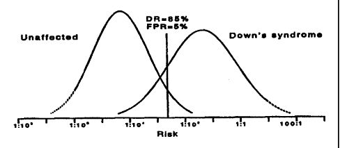
Figs. 1 to 4 show the distributions of estimated risk of
a term pregnancy with Down's syndrome in unaffected
pregnancies and in Down's syndrome pregnancies using different
markers in accordance with the present invention. In these
figures, the vertical lines illustrate the detection rate
(corresponding to the area under the Down's syndrome
distribution curve to the right of the vertical line)
achievable at a 5% false-positive rate (corresponding to the
area under the unaffected distribution curve to the right of
the vertical line). The dotted lines indicate uncertainties
in the precise risk estimates.
Fig. 1 shows the distributions when using PAPP-A between
10 and 13 weeks and AFP,uE, and inhibin-A between 14 and 22
weeks.
Fig. 2 shows the distributions when using PAPP-A and free
(3-hCG between 10 and 13 weeks and AFP, uEz and inhibin-A
between 14 and 22 weeks.
Fig. 3 shows the distributions when using NT and PAPP-A
between 10 and 13 weeks and AFP, uE, inhibin A and total hCG
between 14 and 22 weeks.
Fig. 4 shows the distributions when using NT, PAPP-A and
free (3-hCG between 10 and 13 weeks and AFP, uE3 and inhibin-A
between 14 and 22 weeks.
As an alternative, a sequeiitial test can be performed.
In this case the risk is initially determined based on only
the marker levels from the first stage of pregnancy. This
first estimate of risk is compared with a predetermined cut-
off risk as is known for initial classification as screen-
positive or screen-negative. Women having a screen-positive
result are referred for a diagnostic test and might not be
tested for screening marker levels at the second stage of
pregnancy.
Women initially classified as screen-negative are
CA 02330538 2005-02-07
- 17 -
retested for markers measured at the second stage of
pregnancy. The risk of Down's syndrome is determined
again using the markers from both the first and second
stages of pregnancy. In determining the risk, the
likelihood ratio can be calculated in the same way as in
the non-sequential test described first above. Again, it
is desirable to take account of any correlation between
the markers.
Figs. 5 to 9 are flowcharts illustrating a specific
method according to the present invention which is
explained in detail below.
In the first trimester at around 8 to 13 weeks, or
preferably around 10 to 13 weeks, an ultrasound scan is
taken in step 1 and the nuchal translucency (NT) marker
and the crown-rump length (CRL) are measured and recorded
in step 2. At the same stage, a blood sample is drawn in
step 3, and the separated serum is refrigerated in step
4, whereupon no action is taken during a wait in step 5
until after a secQnd sample is drawn in the second
trimester. The ultrasound scan 1 and the blood sample 3
may be performed as alternatives or together depending
whether it is desired to use ultrasound markers,
biochemical markers or both.
In the second trimester at aroumd 14 to 22 weeks, a
second blood sample is drawn in step 6. Subsequently in
step 7, the first and second samples are assayed for the
respective biochemical markers selected.
The processing of the measurements taken in steps 2
and 7 is described below and illustrated in the blocks
numbered 8 and above in Figs. 5 to 8. This processing
may be implemented in a data processing apparatus, most
suitably an appropriately programmed computer. Thus the
blocks numbered 8 and above also illustrate elements of
the computer program or programming methods which
performs the processing. In particular, the process
CA 02330538 2005-02-07
- 18 -
blocks represent processing performed by the computer
processor. The data entry blocks represent data entry
processing which may be implemented by use of appropriate
data entry fields shown on a display into which data may
be entered from the computer's keyboard. The data item
blocks represent data used by the program. The stored
data blocks represent stored reference data which may be
stored in the memory of the computer in files referenced
by the computer program.
Data input means are used to input the
concentrations (levels) of the serum markers in step 8
and the NT marker level and CRL measurement in step 9.
If the levels from the first trimester are input
immediately after measurement, a message may be
automatically generated and displayed at an appropriate
time in the second trimester to remind the user that
measurements from a second sample are due.
In step 10, each marker level is re-expressed as a
multiple of the median (MoM) level for unaffected
pregnancies of the same gestational age and output as
data item 11.
Step 10 is illustrated in more detail in Fig. 6.
Stored data LMP 27 and scan 28 specific to respective
methods of estimating gestational age are used to select an
equation which estimates the expected median concentrations
for different gestational ages for each marker in step 29.
Data LMP 27 is specific to estimation of gestational age
based on the first day of the last menstrual period. Data
scan 28 is specific to estimation of gestational age from
an ultrasound measure of the fetus, usually a BPD or a CRL.
The equations selected based on stored data 27 or 88 may be
simple linear equations or may be more complicated, for
example, in the case of inhibin-A in the second trimester.
Since inhibin-A levels decline at the start of the second
trimester, and start to rise again after 17 weeks
CA 02330538 2005-02-07
- 19 -
gestation, it is preferable-to use a log-quadratic
regression to calculate the median inhibin-A level at
different gestational ages. The following equation is
suggested in Watt HC, Wald NJ, Huttly WJ (1998). The
pattern of maternal serum inhibin-A concentrations in the
second trimester of pregnancy. Pregnat Diagn 18, 846-848:
loglo I = k + 0.0001864 x (a - 120)2
where I is the inhibin-A concentration, a is the
gestational age in days and the coefficient k is
separately derived for each screening centre.
Based on an input in step 30 of the gestational age
at the date of the sample, for each marker in step 31 the
expected median levels in unaffected pregnancies of the
same gestational age is calculated using the equation
selected in step 29. In step 32, each marker level input
in step 8 is divided by the expected median for that
marker to output the MoM as data item 11.
In step 12, the NT marker is re-expressed as a MoM
and output as data item 13. The specific calculation of
step 12 is illustrated in Fig. 6 and corresponds to the
MoM calculation for the biochemical markers, except that
the CRL measurement input in step 9 is used as the
estimate of gestational age. Stored data 33 represents
the NT medians for different CRL measurements, preferably
as an equation.
There can be considerable systematic variation in
nuchal translucency (NT) measurements from one
ultrasonographer to another. Therefore, the stored data
33 may, optionally, represent NT medians which are
ultrasonographer-specific in cases where it has been
possible to base this data on sufficiently large numbers
of measurements taken by individual ultrasonographers.
In step 34 stored data 33 is used to calculate the
expected median NT levels in unaffected pregnancies of
the same CRL, i.e. the same age. In step 35, the NT
CA 02330538 2005-02-07
- 20 -
measurement input in step 9 is divided by the expected
median NT to give the NT MoM which is output as data item
13.
Optionally, the MoMs 11 for the biochemical markers
may be adjusted in step 14 which is illustrated in detail
in Fig. 7. Based on an input of any one or more of
maternal weight, ethnic group, diabetic status and the
number of fetuses in steps 36 to 39, respectively, stored
weight adjustment equations 40, ethnic group adjustments
41, diabetes correction factors 42 and multiple birth
correction factors 43 are used in step 44 to adjust the
MoMs 11. The adjusted MoMs are output as data item 15.
In step 16, a multivariate Gaussian analysis of the
MoM for all the markers from each stage of pregnancy is
performed. For use in this analysis, distribution
parameters 18 are selected in step 17 which is described
in more detail in Fig. 8. For each marker the
distribution parameters are stored as reference data 45
to 48 for different methods of estimating gestational age
(LMP or scan) and based on whether or not the MoM has
been adjusted for maternal weight. In step 49, the
appropriate distribution parameters are selected and
output as data item 18.
The multivariate Gaussian analysis 16 outputs a
likelihood ratio as data item 19. This needs to be
multiplied by a background risk to derive the estimated
risk of Down's syndrome. Whilst an overall population
risk may be used, the present method uses age-specific
risks calculated in step 20 which is described in more
detail in Fig. 9. The gestational age of the sample
input in step 50 (or 30) and the date of the sample input
in step 51 are used to calculate the expected date of
delivery (EDD) in step 52. The maternal date of birth is
input in step 53 and is combined with the EDD to
calculate the age at EDD as data item 56. This is used
CA 02330538 2005-02-07
- 21 -
calculate the age at EDD as data item 56. This is used
in the stored age-specific risk equation 57 to output the
age-specific risk as data item 21. The likelihood ratio
19 and age-specific risk 21 are multiplied in step 22 to
output the estimated risk of Down's syndrome as data item
23. The estimated risk 23 is compared with a
predetermined cut-off in step 24 to produce a screen-
positive result 25 when the risk is equal to or greater
than the cut-off, or a screen-negative result 26
otherwise.
The apparatus may be arranged to provide estimates
of the expected screen performance (i.e. the detection
rate, false-positive rate and odds of being affected
given a positive result), taking into account the age
distribution of the screened population, the combination
of screening markers used, the risk cut-off used, and
other factors. The performance observed in practice can
then be compared to the expected performance as an aid to
monitoring.
The values of the stored data used in the method
described above depends on which markers from the two
stages of pregnancy are selected to be used. Appropriate
data values for each marker are known, for example from
the references.