Note : Les descriptions sont présentées dans la langue officielle dans laquelle elles ont été soumises.
CA 02344715 2001-03-20
WO 00/21604 PCT/US99/23528 "
1
DESCRIPTION
Percutaneous Filtration Catheter For Valve
Repair Surg_ery And Methods Of Use
Field of the Invention
The present invention relates generally to filter devices for placement in a
blood
vessel to capture embolic material, and more particularly to a catheter system
having an
associated filter for percutaneous placement in an aorta to entrap embolic
material from
the aorta and heart during cardiac surgery.
Background of the Invention
Stroke has become a major source of morbidity following coronary artery bypass
and other cardiovascular surgeries, including valvular repair, septal defect
repair, removal
of atrial myxoma, aneurysm repair, and myocardial drilling. Classic factors
associated
with an increased post-operative stroke rate are advanced age, severe left
ventricular
dysfunction, long standing diabetes, protracted cardiopulmonary bypass time,
severe
perioperative hypotension, history of previous stroke, and bilateral carotid
disease.
Possible mechanisms of perioperative stroke include a reduction in cerebral
blood flow
through a stenotic extracranial or intracranial vessel, embolization of
atherosclerotic debris
from an ulcerated carotid artery plaque or aortic plaque, embolization of post-
infarction
left ventricular mural thrombus or atrial thrombus, and embolization of air
inadequately
evacuated from the heart or aorta. In valvular repair surgery, manipulation of
the heavily
calcific aortic or mitral valve may result in calcium dislodgment in the left
coronary artery
or left ventricle, with subsequent embolization. Although atheromatous debris
most
frequently embolizes to the brain, other affected body sites include the
spleen, kidney,
pancreas, and gastrointestinal tract. Embolization of these peripheral organs
can lead to
tissue ischemia or death.
In addition to stroke, other factors, e.g., chest wall trauma, contributing to
morbidity in cardiac surgeries often arise from the use of cardiopulmonary
bypass for
circulatory support and median sternotomy. Minimally invasive procedures using
beating-heart and port-access approach have been developed to achieve aortic
occlusion,
cardioplegia delivery, and left ventricular decompression to allow coronary
revascularization and other cardiac procedures to be performed in a less
invasive fashion.
CA 02344715 2009-04-21
2
A need therefore exists for less invasive devices and methods which facilitate
aortic occlusion and/or cardioplegia delivery in cardiac surgeries and provide
an arterial
filter for reducing a patient's risk of perioperative stroke.
Summary of the Invention
The present invention provides a percutaneous filtration catheter having the
ability
to capture emboli, including atheromatous fragments, fat, myocardial tissue
debris, and
air. The catheter further includes capabilities to provide aortic occlusion
and cardioplegia
delivery in cardiac surgeries, especially in heart valve repair.
In one embodiment, the catheter comprises an elongate member having proximal
and distal ends. The distal end has (1) a balloon occluder which communicates
with a
lumen carried by the elongate member, and (2) an expandable filter mounted on
the
elongate member distal to the balloon occluder. The balloon occluder and the
expandable
filter are operated independently at the proximal end of the elongate member.
The
expandable filter typically has a proximal edge bonded circumferentially and
continuously to the elongate member, and a distal edge which expands radially
outward
on activation.
In another embodiment, the elongate member has a second lumen for infusing
fluid, such as cardioplegia solution. The expandable filter may comprise an
expansion
frame, which may have an umbrella frame in one embodiment (for construction,
see
Barbut et al., U.S. Pat. No. 5,769,816, and an inflation seal in another
embodiment (for
construction, see Barbut et al., U.S. Pat. No. 5,769,816). Furthermore, in
certain
embodiments, the expandable filter is operable by manipulating at least one
pull string at
the proximal end of the elongate member.
In accordance with a particular aspect the present invention provides
a percutaneous filtration catheter, comprising:
an elongate member having a proximal end and a distal end;
a balloon occluder mounted on the distal end of the elongate member, the
balloon
occluder defining a chamber which communicates with a lumen carried by the
elongate
member;
and
CA 02344715 2009-04-21
2a
an expandable filter mounted on the elongate member distal the balloon
occluder,
the expandable filter operable independently of the balloon occluder, the
expandable
filter having a proximal edge that is fixed to the elongate member and a
distal edge that
opens when the filter is operated.
The present invention also provides methods for capturing embolic material in
cardiac surgeries, thereby protecting a patient from neurologic complication
due to
embolization. The methods employ a percutaneous filtration catheter having an
elongate
member with proximal and distal ends, a balloon occluder mounted on the distal
end of
the elongate member, and an expandable filter mounted on the elongate member
distal
the balloon occluder. A percutaneous incision in a patient's peripheral
artery, such as a
femoral or brachial artery, is made followed by insertion of the elongate
member through
the incision. In minimally invasive cardiac procedures, the percutaneous
filtration
catheter can be introduced percutaneously through a peripheral artery, or
alternatively,
through a minimal access port, often located in a patient's intercostal space,
to the
ascending aorta. The distal end of the catheter is then advanced into the
ascending aorta.
The filter is expanded and positioned above the aortic valve to entrap embolic
material
from flowing
CA 02344715 2001-03-20
WO 00/21604 PCT/US99/23528
3
downstream to peripheral organs. The balloon occluder is inflated to provide
circulatory
isolation of the heart and coronary blood vessels from the peripheral vascular
system. In
the embodiment which includes a second lumen, the second lumen can be used to
(1)
deliver cardioplegia solution upstream to the heart to arrest cardiac
function, or (2) to carry
a pressure monitor. After cardiac arrest is achieved and cardiopulmonary
bypass is
initiated for circulatory support, a variety of cardiothoracic surgeries can
then be
performed, including coronary artery bypass grafting, heart valve repair,
septal defect
repair, removal of atrial myxoma, aneurysm repair, and myocardial drilling.
It will be understood that are many advantages to using a percutaneous
filtration
catheter as disclosed herein. For example, the catheter provides (1) a
percutaneous access
for catheter insertion, obviating the need for an extensive tissue incision,
(2) aortic
occlusion through inflating a balloon occluder, thereby minimizing damage to
the aortic
wall and reducing the risk of emboli dislodgment as compared to traditional
clamping, (3)
a filter which entraps embolic material during cardiac surgery, thereby
reducing a patient's
risk of stroke perioperatively, (4) cardioplegia delivery upstream to the
heart for cardiac
arrest, and (5) access for devices to be introduced through an intercostal
incision in
minimally invasive cardiac procedures.
Brief Description of the Drawings
Fig. 1 depicts a percutaneous filtration catheter according a first
embodiment.
Fig. 2 depicts a percutaneous filtration catheter according to another
embodiment,
the catheter having a second lumen.
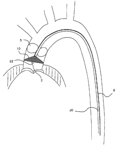
Fig. 3 depicts a percutaneous filtration catheter positioned in an ascending
aorta.
Fig. 4 depicts different access routes for entry of the percutaneous
filtration
catheter for use in a patient.
Detailed Description
The devices and methods disclosed herein can be used in patients who have been
identified as being at risk for embolization during cardiothoracic surgeries,
thereby
reducing their perioperative complications and length of hospital stay. Fig. 1
depicts a
percutaneous filtration catheter according to one embodiment. The catheter has
elongate
member 1, distal end 2, and proximal end 3. Balloon occluder 5, which may
comprise an
elastomeric balloon, is mounted on elongate member 1 and communicates. with
inflation
lumen 6. Expandable filter 10 is mounted on elongate member 1 distal the
balloon
occluder and can be operated by actuating mechanism 12 at the proximal end of
the
catheter.
CA 02344715 2001-03-20
WO 00/21604 PCT/US99/23528
4
Fig. 2 depicts another embodiment of a percutaneous filtration catheter having
a
second lumen. The catheter carries second lumen 20 in addition to lumen 6
which
communicates with balloon occluder 5 and inflation port 7 for inflating the
balloon
occluder. Second lumen 20 communicates with port 22 and can be used to infuse
cardioplegia solution, aspirate fluid or air, and/or to house a pressure
monitor. Filter 10,
mounted on elongate member 1, has expansion frame 15 and can be actuated by
mechanism 12 at proximal end 3 of the catheter.
The length of a percutaneous filtration catheter is generally between 20 and
90
centimeters, preferably approximately 50 centimeters. The outer diameter of
the catheter
is generally between 0.1 and 0.4 centimeters, preferably approximately 0.2
centimeters.
The balloon occluder, when inflated, will generally have a diameter between
1.0 and 5.0
centimeters, more preferably between 2.0 and 4.0 centimeters. The filter, when
expanded,
will generally have a diameter between 1.0 and 5.0 centimeters, more
preferably between
2.0 and 4.0 centimeters. The foregoing ranges are set forth solely for the
purpose of
illustrating typical device dimensions. The actual dimensions of a device
constructed
according to the principles of the present invention may obviously vary
outside of the
listed ranges without departing from those basic principles.
Methods of using the devices disclosed herein are illustrated in Fig. 3. After
a
small percutaneous incision is made in a patient's femoral artery, distal end
2 of the
percutaneous catheter is introduced through the incision and advanced into the
ascending
aorta. Expandable filter 10 is then expanded to entrap embolic material
originating from
the heart or the aorta, including air, atheromatous plague, tissue debris,
fat, or thrombi.
Balloon occluder 5 is inflated through its communicating inflation lumen 6 to
provide
aortic occlusion for cardiopulmonary bypass. Cardiac arrest can be achieved by
delivering
cardioplegia solution upstream to the heart through lumen 20 and port 22.
Lumen 20 and
port 22 can also be used to place a pressure monitor or to aspirate fluid,
blood, air, tissue,
or plaque debris from the heart and the aorta. A surgeon then can proceed with
various
cardiothoracic surgeries.
Fig. 4 depicts different percutaneous entry sites for a percutaneous
filtration
catheter. The catheter is generally inserted through a patient's femoral
arteries. Right
groin 30 and left groin 32 are common percutaneous incision sites for
introducing elongate
member 1 of the catheter, shown here entering through left groin 32 in the
left femoral
artery and advanced to the ascending aorta. In some patients, however, the
femoral
arteries are not suitable for catheter manipulation due to severe
atherosclerosis.
Alternatively, the catheter can be inserted througli right antecubital area 34
or left
antecubital area 36. Elongate member 1 of the catheter is shown inserted
through
CA 02344715 2007-04-30
antecubital area 34 and advanced through the right brachial artery and
brachiocephalic
trunk to enter the ascending aorta. After final placement of the catheter in
the ascending
aorta, expandable filter 10 is expanded to entrap emboli and balloon occluder
5 is inflated
to provide circulatory isolation of the heart and coronary blood vessels from
the peripheral
5 vascular system.
It will be understood that the devices disclosed herein are particular well
suited to
application for valve repair surgeries because these surgeries are recognized
to generate
embolic material upstream of the site of aortic blockage. According to McBride
et al.,
Glenn's Thoracic and Cardiovascular Surgery, 6d' Ed., Vol. II (1986),
=, median.sternotomy is often the incision used for replacing the aortic valve
or
any combination of valves in which the aortic valve is included. After venous
cannulation
of the right atrium, inferior vena cava, or superior vena cava, arterial
cannulation of the
aorta is established for cardiopulmonary bypass using the. percutaneous
filtration catheter
disclosed herein. The catheter is positioned in the ascending aorta via
femoral artery or
brachial artery access. The balloon occluder, or other impermeable dam, is
deployed to
isolate the heart from peripheral circulation. The filter is deployed upstream
of the
occiuder in order to capture calcified plaque from the aortic or mitral valve.
Oxygenated
blood from a bypass machine is infused through the catheter downstream of the
occluder.
Cardioplegia solution is administered to the aortic root while the aortic
valve is manually
closed by external pressure on the root of the aorta or directly into the
coronary ostia.
In aortic valve repair, the aorta is opened through a transverse incision
approximately 1 to 1.5 centimeters above the right coronary artery. The
standard approach
to mitral valve repair is often through an incision parallel to the intra-
atrial groove into the
left atrium. After the heavily calcific or fibrotic valve is resected
sufficiently to permit
visualization of the left ventricular chamber, a sponge is oftenplaced in this
cavity to
enmesh any calcium that may fragment fromthe annulus during decalcification. A
culture
stick placed in the left coronary orifice prevents embolization of calcium
into this vessel,
and the retractor providing exposure of the valve usually blocks the right
coronary orifice.
A prosthetic valve is sutured into the valvular annulus after the diseased
native valve is
removed. The aorta is then closed with two layers of sutures. Aortic occlusion
and
filtration is removed, and the patient is taken off cardiopulmonary bypass.
Although the foregoing invention has, for purposes of clarity of
understanding,
been described in some detail by way of illustration and example, it will be
obvious that
certain changes and modifications may be practiced which-will still fall
within the scope
of the appended claim.
