Note : Les descriptions sont présentées dans la langue officielle dans laquelle elles ont été soumises.
CA 02355850 2001-06-19
_ WO 00./36975 PCT/GB99/04371
- 1 -
Device for reducing signal noise in a fetal ECG signal
The present invention relates to a method and apparatus
for reducing signal noise in a fetal ECG signal,
typically one obtained by using a unipolar ECG lead
configuration which detects a predominant T wave vector
whilst avoiding changes in ECG waveform shape due to
fetal rotation through the birth canal.
Fetal surveillance during labour is standard clinical
practice. The purpose is to identify abnormal events
and fetal oxygen deficiency in particular. Since its
introduction in the sixties it has been evident that
electronic fetal monitoring by fetal heart rate analysis
alone does not provide all the information required for
an optimal identification of a fetus suffering from lack
of oxygen.
During the last 20 years work has been ongoing to
clarify what fetal signals could be made use of to
provide additional information. Since the early
seventies, waveform analysis of the fetal
electrocardiogram has been studied from both
physiological, signal processing and clinical aspects
(Rosen KG: Fetal ECG waveform analysis in labour. Fetal
monitoring. Physiology and techniques of antenatal and
intrapartum assessment. ad. Spencer JAD). Castle House
Publications. pp. 18~-187,1989). It was found that the
ST interval and T-wave amplitude were of particular
interest.
Figure 1 depicts two consecutive heart beats with the
different ECG components of interest during foetal
surveillance being identified. It has been found that
changes in the ST interval of the Fetal Electro
CA 02355850 2001-06-19
WO 00136975 PCT/GB99/04371
- 2 -
CardioGram (ECG) are part of the components that reflect
the stress of the fetal heart during the labour.
Basically, the changes that appear in the ST interval
due to lack of oxygen, can be divided into 3 classes:
1. ST rise with increased ST segment and T wave
amplitude;
2. Appearance of so called biphasic ST changes,
with an ST segment with a negative slope;
3. Appearance of negative T waves.
These discoveries have been applied in a clinical trial
in which the ST-waveform (ie. the ST segment plus the T
wave) of the fetal electrocardiogram was shown to
provide more useful information than the mere detection
of RR-intervals (fetal heart rate) (Westgate J, M
Harris, JSH Curnow, RR Greene: Plymouth randomised trial
of cardiotocogram only versus ST waveform plus
cardiotocogram for intrapartum monitoring; 2400 cases.
Am J Obstet Gynaecol 169(1993)1151).
Several problems regarding the fetal ECG-signal quality
have been identified aver the years. Clearly, it is a
prerequisite to be able to detect the ST waveform, and
so one of the main requirements for ST-waveform analysis
of the fetal electrocardiogram is a fetal ECG lead
configuration that is consistent and allows the
identification of the T vector during labour.
The conventional ECG level configuration used for fetal
monitoring is the bipolar fetal ECG lead configuration.
Here, both exploring electrodes are located close to
each other on the presenting part of the fetal body, ie.
the head or buttock. As a consequence of the location
of the electrodes, there is a maximum sensitivity to ECG
CA 02355850 2001-06-19
y WO 0736975 PCT/GB99/04371 .
-
waveform changes with a main vectorial distribution in
the horizontal plane of the fetus. However,
experimental data have shown a maximal representation of
T wave vector along the longitudinal axis of the fetus.
Thus, the standard fetal ECG lead, well suited when only
using the R wave for fetal heart rate detection, will
not enable the accurate detection of changes in T wave
amplitude.
This can only be done by constructing an ECG lead that
is sensitive to ECG waveform changes appearing in the
longitudinal axis of the fetus. It is known from the
literature that the use of a unipolar fetal ECG lead
configuration enables the detection of the main T-wave
vector more accurately then the standard bipolar ECG
lead configuration (Lindecrantz K, Lilja H, Widmark C,
Rosen KG: The fetal ECG during labour. A suggested
standard. J. Biomed. Eng. 1988; 10: 351-353). In this
configuration, one of the exploring electrodes is
located well away from the fetus, e.g. on the maternal
skin. The maternal thigh has been. found to be a
suitable place. The other exploring electrode is the
standard scalp electrode needle placed under the skin of
the presenting fetal part.
A further problem is the existence of signal noise which
is far more significant when the S-T waveform is being
studied than is the case with conventional fetal ECG
monitoring. An illustration of progressive changes in
the ST segment of the foetal ECG recorded during labour
is presented in Figures 2a-c. The ECG baseline as
indicated by the present invention is depicted as well.
The appearance of biphasic changes in the ST segment
follows a pattern, which is exemplified in Figures 2a-c.
This is a sequential recording showing 30-beat ECG
averages. As seen in Figures 2a-c, the ST segments are
classified in a 3-level scale that reflects the relation
CA 02355850 2001-06-19
'- PCT/GB99/04371
WO 00136975
- 4 -
between the negative slope of the ST segment compared to
the baseline of the ECG. As will be appreciated, to be
able to perform this type of analysis, a very high
signal quality regarding low frequency noise is
required.
Although the unipolar fetal ECG electrode configuration
discussed above enables the T vector to be identified, a
signal noise problem is generated at the same time. The
maternal skin electrode is sensitive to maternal
movements causing both low frequency (movement
artifacts) and high frequency (muscular activity) noise.
Another source of noise is the interference from mains
frequencies.
Thus, the sources of signal noise may be summarised as:-
A. High frequency components related to muscle
activity.
B. Interference from mains frequencies.
C. Low frequency noise largely generated by fetal
and maternal movements
Any system for assessment of the ST-waveform of the
fetal electrocardiogram has to reduce the interference
from these potential sources of signal noise, but
obviously, any techniques applied to reduce signal noise
should not significantly interfere with the ST waveform.
Furthermore, the signal processing should be done
continuously as the state of oxygen delivery to the
fetus can change from one minute to another and any
delay in the presentation of ECG-waveform data would be
disadvantageous.
The technique used in the Plymouth trial (Westgate et
CA 02355850 2001-06-19
WO 00%36975 PCTIGB99I04371
_ 5 _
a1, 1993) used analogue filtering signal processing
undoubtedly with some success. However, there were
limitations to what can be achieved. The fetal scalp
ECG signal amplitude (QRS complex) varies normally
between 100 and 400 ~.V but the T wave is normally only
1/10 of an amplitude of the peak signal and so great
care has to be taken not to interfere with this low
amplitude part of the signal. The use of analogue high
pass filters to reduce low frequency (ie. below 1 Hz)
baseline shifts carries the risk of markedly affecting
the T wave amplitude and guidelines instituted by the
American Heart Association recommend a low frequency
cut-off of only 0.05 Hz (Electrocardiography
recommendations for the standardization of leads and of
specifications for instruments in ECG/VCG circulation.
American Heart Association Committee, 1975, Pp 1-25).
These guidelines were followed in the Plymouth trial.
As a consequence, the prior art analogue filtering
techniques will, to only a very limited extent reduce
low frequency noise generated by electrode movements and
the data interpretation has therefore been limited to
more robust changes. There is therefore a need to
improve the quality of the fetal electrocardiogram to
enable continuous and detailed assessment of ST-waveform
changes during labour.
According to the present invention there is provided a
method of reducing noise in a fetal ECG signal
comprising connecting electrodes to the fetus and the
maternal skin in a unipolar configuration and feeding
the signal detected by said electrodes through a first
high pass filter, the cut-off frequency of the first
high pass filter being between 0.2 and 2.7 Hz.
The invention also provides an apparatus for obtaining a
fetal ECG signal comprising exploring electrodes for
CA 02355850 2001-06-19
WO 00%36975 PCT/GB99/04371
- 6 -
connection to the fetus and the maternal skin in a
unipolar configuration in order to detect an ECG signal
and a signal noise reducing device linked to the
electrodes by means of a first signalling link, wherein
the signal noise reducing device comprises a first high
pass filter, the cut-off frequency of the first filter
being between 0.2 and 2.7 Hz.
Typically, one electrode is attached to the fetal scalp
and one is attached to the maternal thigh.
The "cut-off frequency" as used herein refers to the
frequency below which a significant degree of signal
attenuation e.g. -3dB, takes place. In preferred forms
of the invention there may be as little as 0.1 dB
attenuation in most of the pass band and around 40 dB
attenuation in most of the stop band.
Thus, it will be seen that the invention provides signal
filtering using a far higher cut-off frequency than that
thought possible in the prior art. This is based upon a
recognition that, although the baseline fluctuations of
the ECG signal (due to movements, breathing, impedance
variations etc.) can have a significantly higher
amplitude than the ST waveform, most of the energy of
the baseline fluctuation is at a lower frequency range
than the frequency range of the ST interval. This is
illustrated in the spectrum shown in Fig. 3. Thus, the
invention provides a signal quality enhancement model
that allows the accurate presentation of ECG waveform
changes within the ST interval frequency.
The signal may be fed directly from the electrodes to
the noise reducing device of the invention, or it may be
pre-filtered, e.g. using the prior art apparatus
discussed above such as an analogue band pass filter
having cut-off frequencies of about 0.05 and 100 Hz.
CA 02355850 2001-06-19
WO 86736975 PCT/GB99/04371
_ 7 _
The former cut-off serves to eliminate DC levels and
very low frequency components that might otherwise
decrease the dynamic range of the signal.
In the device of the invention, a cut-off frequency of
up to 2.7 Hz has been found satisfactory in that the
T/QRS ratio is largely unaffected when the fetal heart
rate is over 100 beats/ min. However, in order to
provide satisfactory performance with lower heart rates,
10 it is preferred that the cut-off frequency is less than
1.7 Hz. To optimise noise reduction, the cut off
frequency is preferably greater than 0.7 Hz and around
1.2 Hz is believed to be the most effective cut-off
frequency overall.
The first high pass filter may be an analogue filter,
but it is highly desirable that this filter should add
the minimum of phase distortion and so it is believed
that this invention may more readily be achieved using
20 digital techniques, in which case the signal is
digitised before being passed through the first high
pass filter.
As discussed above, another source of signal noise is
interference from mains frequencies. In order to
decrease the influence of mains, the device preferably
also comprises a notch mains frequency filter for
attenuating the mains frequency contents of the ECG
signal, the notch mains frequency filter preferably
30 being applied to the ECG signal in connection with the
first high pass filter. The notch mains frequency
filter is arranged to correspond to the local mains
frequency, for example 50 Hz or at 60 Hz. Since modern
digital filters may improve signal/noise ratio
35 substantially without causing unwanted changes in signal
waveform a multitude of digital filters may be used with
very narrow cut-offs to reduce interference from both
CA 02355850 2001-06-19
WO 00/36975 PCT/GB99/04371 _
_ g _
low and high frequencies as well as mains noise.
The noise reducing steps described above may be combined
with further steps. For example, one technique for
performing noise reduction of a repetitive signal is to
use averaging with equal or weighted coefficients.
However, there are limitations and as an example, ECG
complexes with marked baseline shift may corrupt the
averaged complex causing erroneous information to be
generated. It would therefore be advantageous if as
much as possible of signal noise could be eliminated
prior to such signal averaging.
Even modern digital (in this case high-pass) filters,
may leave a part of the low frequency noise in the
signal which, during an R-R interval, can be seen as a
baseline shift or slope. This deviation in the
available signal, compared to the real ECG, may in some
circumstances make a qualified analysis of the ST
segment difficult. Therefore, the invention preferably
also includes a step in which residual low frequency
noise of the continuous ECG signal is attenuated further
using vector subtraction principles. An advantage with
such a filter is the ability to operate immediately
after a possible loss of signal with the ECG signal
exceeding the dynamic range.
Thus, preferably a second high pass filter is provided
for further attenuation of signal noise in a digitised
fetal ECG signal where the signal noise is primarily
constituted by baseline fluctuations of the ECG signal.
The ECG signal typically comprises a sequence of ECG
complexes in the form of uncompensated samples, each ECG
complex including a QRS complex, the second high pass
filter being arranged after the cutoff frequency high
pass filter, the additional high pass filter comprising:
means for identifying ECG complexes of the ECG signal
CA 02355850 2001-06-19
y WO 00/36975 PCT/GB99/04371
- 9 -
and their P-Q points; means for obtaining an
approximating function to a curve between one P-Q point
and a proceeding or a succeeding P-Q points by using a
number of proceeding and succeeding P-Q points, the
number being at least one; and means for forming the
compensated samples to an output signal.
One way of implementing the vector subtraction is that
the means for obtaining an approximating function to the
curve is arranged for determination of slopes of lines
between P-Q points and proceeding or succeeding P-Q
points; and compensated values y[i] 'are obtained
according to: y [i] =x [i] -m-k (i-ipq) , where i, x [i] , m, k
and ipq denote index for each sample, uncompensated
sample with index i, the level of the P-Q point for the
present complex, the slope for the present complex, the
index for the P-Q point sample, respectively.
In case a first degree polynomial is not sufficient, it
is possible that the means for obtaining an
approximating function to the curve is arranged for
determination of polynomials of higher degree than one;
the polynomials being based on a P-Q point and
proceeding and/or succeeding P-Q points.
Tn some circumstances, it may be possible to provide a
signal of such quality that signal averaging would be
unnecessary. However, when this is not the case, it may
be advantageous if the device also comprises an
averaging filter which is preferably applied to the ECG
signal in connection with the second high pass filter.
Preferably averaging takes place over twenty to thirty
cycles. A larger number risks causing appreciable
attenuation to the height of the T wave.
An embodiment of the invention will now be described, by
way of example only, and with reference to the
CA 02355850 2001-06-19
WO 00/36975 FCT/GB99/04371
- 20 -
accompanying drawings:-
Figure 1 depicts two consecutive heart beats with the
different ECG components of interest to the present
invention for foetal surveillance.
Figures 2a-2c present an illustration of progressive
changes in the ST segment of the foetal ECG recorded
during labour. The ST segments are indicated by arrows.
The ECG baseline indicated by the present invention is
also depicted.
Figure 3 presents an exemplary spectrum including
baseline fluctuations and an ST interval frequency
ranges.
Figure 4 presents an illustration of the impact of high
pass filtering on T wave amplitude quantified by the
T/QRS ratio, at different fetal heart rate levels.
Figure 5 presents a block diagram of the noise reducing
device of the embodiment.
Figure 6 presents a graph relating to complete frequency
spectrum of a preferred embodiment of a filter of the
present invention, this filter being a 1.5 Hz high pass
(mufti-notch) filter.
Figure 7 presents a graph relating to a first cut off
region of a preferred embodiment of a filter of the
present invention, this filter being a 1.5 Hz high pass
(mufti-notch) filter.
Figure 8 depicts a subtraction filter to be used in the
embodiment.
CA 02355850 2001-06-19
- WO 00r36975 PCT/GB99/04371
- 11 -
Figure 9 depicts a two stage filter used in the
embodiment.
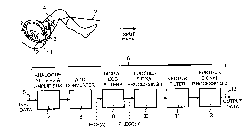
Turning first to Figure 5 there is provided an over-view
of a fetal monitoring apparatus in use. A first
electrode 1 is attached to the head 2 of the fetus and a
second electrode 3 is attached to the maternal thigh 4.
Electrode leads 5 transmit the detected ECG signal to
the noise reducing device (shown generally as &), the
structure of which is described in more detail below. A
further set of leads 13 transmits the output from the
device 4 to dispray apparatus such as a monitor (not
shown? .
The first stage 7 of the noise reducing device contains
conventional analogue filters for reducing DC and low
frequency components of the signal. The cut-off
frequency of this stage is 0.05 Hz. This stage also
contains a 100 Hz low pass filter for removing
comparatively high frequency components.
The first stage serves to reduce the requirements of the
next stage 8 which is an analogue to digital converter,
operating at 500 Hz.
The digitised signal is then fed to a first digital ECG
filter stage 9 which has a 1.2 Hz cut-off frequency (for
3 dB attenuation) and which attenuates the signal by
Less than 0.1 dB above 1.5 Hz. It also contains notch
filters for removing mains supply interference. This
stage is discussed more fully below.
Subsequently, the signal is processed further in stage
10. This serves to detect the QRS complexes in the ECG
signal and to define their PQ points. In combination
with vector filter 11, this enables residual low
frequency noise to be removed by means of the vector
CA 02355850 2001-06-19
- WO 00!36975 PCT/GB99/04371
- 12 -
subtraction process previously described.
The final part of the device 6 is stage 12 which
performs the calculation of HR values, ECG averaging and
ECG waveform analysis in the known manner before the
output data is transmitted via leads 13 to a display
screen and/or a printer.
As previously discussed, the ECG filter section 9
comprises a high-pass filter, with a cut-off frequency
of 1.2 Hz and includes other notch stop bands for mains
supply noise rejection. It is phase-linear (i.e. it has
constant group delay) in the pass band. Figures 6 and 7
illustrate the characteristics of this filter section.
The filter can,be realised in a number of ways. Two
examples axe:
1. A FIR-filter consisting of one or several serial
stages.
OR
2. A 'subtraction filter', where the output signal is
simply the input signal with the noise subtracted in the
time domain. The noise is the result of a filter with
the inverse frequency response compared to the figure
above, see Figure 8.
One example of the first type of filter is a two stage
serial FIR filter with the two following transfer
functions. An example of this kind of filter is
presented in Figure 9.
The hl block presented in Figure 9 is a FIR filter with
the following transfer function:
CA 02355850 2001-06-19
- WO 00136975 PCT/GB99/Q4371
- Z3 -
i<NI
y{n) _ ~ ECG(n-a~.hl (a~
i=v
The h2 block presented in Figure 9 is a FIR filter with
the following transfer function:
i<Na
Filt ECG(n) _ ~ y{n-a).h2(Z)
i=a
Figures 6 and 7 show the high-pass cut-off frequency at
1.2 Hz for 3 dB attenuation. Apart from this cut-off
characteristic, there is a lot of characteristics that
affect the N1 and N2 values and the related
coefficients, such as:
Ripple in the pass band.
Attenuation in the stop band.
Slope of the frequency response from stop band to
pass band, i.e. how wide is the stop band (it can
not be equal to the 1.2 Hz above, would result in
an indefinite number of coefficients for a digital
filter) .
In addition, the transfer functions will be affected
(possibly resulting in simpler implementations) if notch
stop band are used or not, or if the characteristic of
the notches are related to the characteristic of the
first high-pass cut-off region.
Therefore, no absolute setup of coefficients is
relevant, but the main characteristic is the high-pass
cut-off frequency (at some attenuation level), as
illustrated in Figure 7 to 1.2 Hz.
CA 02355850 2001-06-19
_ WO 00/36975 PCT/GB99/04371
- 14 -
An experimental comparison of embodiments of the present
invention with noise reducing devices having different
high pass filters has been carried out. This was done
by applying a series of digital filters to a set of
stored fetal ECG data with the following
characteristics:
The ECG is recorded from a skin and a scalp electrode.
The ECG signal has passed an analogue band pass filter
with cut-off frequencies of 0.05 and 100 Hz.
The analogue ECG is sampled and AD converted with 500
Hz.
Distinct changes in the ST interval with increasing
T/QRS at varying foetal heart rate levels.
The reason for testing at different foetal heart rate
levels is the marked fluctuations that may occur and we
can assume that the frequency range of the ST interval
may change depending on heart rate.
The following filters with minimum phase distortion were
applied:
1. No digital filters used at all (TQRS-OHz).
2. Multi notch N2 with pass band 0-48.5, 51.5-148.5Hz
etc. Additional HP1 mufti notch with pass band 0.5-
124.5Hz, 125.5-249.5Hz (TARS-~ Hz).
3. Mufti notch N2 with pass band 0-48.5, 51.5-148.5Hz
etc. Additional HP1 mufti notch with pass band 1-124Hz,
126-249Hz (TQRS-1Hz).
4. Mufti notch with pass band 1.5-48.5Hz, 51.5-98.5Hz,
CA 02355850 2001-06-19
- WO 00/36975 PCT/GB99/04371
- 15 _
101.5-148.5Hz etc (TQRS-~ Hz).
5. Same as filter no. 4 regarding 50Hz and overtones,
but additional mufti notch pass band 2-123Hz and 127-
248Hz {TARS-2Hz).
5. Same as filter no. 4 regarding 50Hz and overtones,
but additional mufti notch pass band 2.5-122.5Hz, 127.5-
247.5Hz (TQRS-2~ Hz).
7. Same as filter no. 4 regarding 50Hz and overtones,
but additional mufti notch pass band 3-122Hz, 128-247Hz
{TARS-3Hz) .
In this experiment, the pass bands are regarded as those
frequencies where less than 0.1 dB attenuation occurs.
As may be seen from Figure 7, the frequency response
typical of the filters used is such that the cut-off
frequency defined with reference to 3 dB attenuation is
approximately 0.3 Hz lower. In the case of the notch
filters, the upper end of the pass band is approximately
0.3 Hz higher fox 3 dB than for 0.1 dB attenuation.
The following can be found from examining the data
displayed in Figure 4. The frequencies in parentheses
refer to the corresponding cut-off values for 3 dB
attenuation:
1. A filter with a high pass of 3 Hz {2.7 Hz) affects
the T/QRS ratio with a false lowering of the ratio
recorded regardless of fetal heart rate.
2. The T/QRS ratio is largely unaffected by the high
pass filters of < 3.0 Hz {2.7 Hz) when ECG data are
sampled at fetal heart rates> 100 beats/min
approximately.
CA 02355850 2001-06-19
_ WO 00/36975 PCT/GB99/04371
- 16 -
3. When heart rate drops below approximately 100 beats
per minute filter characteristics becomes even more
important and a high pass of < 2 Hz (1.7 Hz) is required
not to affect the T/QRS ratio.
Thus, it may be seen that by means of the invention it
is possible to attenuate fetal ECT signal noise at
higher frequencies than was previously thought possible.
In view of the noise frequency distribution discussed
above, this allows for much greater signal noise
reduction which thereby enables more reliable fetal
monitoring.