Note : Les descriptions sont présentées dans la langue officielle dans laquelle elles ont été soumises.
CA 02367696 2001-09-14
WO 00/65357 PCT/EP00/03605
Method of diagnosing transmissible spongiform
encephalopathies
The invention relates to a method of diagnosing
transmissible spongiform encephalopathies and a diagnostic
test kit using prion-protein- and laminin-receptor-specific
antibodies. The invention also relates to the use of the
method or test kit for diagnosing transmissible spongiform
encephalopathies.
Over 250 years ago a disease of sheep was discovered which
was accompanied by nervousness, itching and ataxia and
finally ended in paralysis and death. This disease is now
known as "Scrapie" in English speaking countries (as the
animals rub against posts and trees in order to control the
itching), "la tremblante" in French and "Traberkrankheit"
in German. These names reflect the variety of the symptoms.
"Scrapie" was investigated as the prototype of a group of
diseases which affect not only animals but also human
beings: the transmissible spongiform encephalopathies (_
TSE). TSEs are fatal neurogenerative diseases which can
attack a number of mammals .
Bovine spongiform encephalopathy (BSE) is a
neurodegenerative disease in cattle and is related to
Scrapie in sheep and goats and Creutzfeldt-Jakob disease in
humans. A host-coded, membrane-associated glycoprotein of
unknown function, the so-called prion protein PrP, plays a
central part in the pathogenesis of these diseases. This
cellular isoform is expressed particularly strongly on
neuronal cells but can also be detected with variable
frequency on non-neuronal cells. This membrane protein is
sensitive (PrP-sen) to digestion with specific enzymes
(proteases). However, the soluble malignant form of PrP
(PrP-res) is protease-resistant and accumulates in the
brains of BSE-infected animals to form amyloid plaques.
CA 02367696 2001-09-14
WO 00/65357 PCT/EP00/03605
2
This PrP-res form is associated exclusively with all TSE
diseases including BSE and can be extracted from TSE/BSE-
infected brain tissue.
The unusual characteristics of the Scrapie/BSE pathogen
gave rise to speculation at an early stage that the
pathogen consists solely of nucleic acid or proteins or
contains neither nucleic acid nor proteins and is a
polysaccharide or a membrane fragment. The scenarios which
are most discussed at present are the " protein only "
hypothesis and the virus/virino hypothesis:
The " protein o_nlv " hypothesis is based on the infectious
prion PrP-res containing no nucleic acid and being self-
replicating. It is speculated that PrP-res binds to PrP-sen
and thereby converts it into the malignant isoform. The
conformation of this malignant isoform is marked by beta-
pleated sheet structures, whereas in the cellular isoform
the alpha helices predominate. The virus/virino hypothesis
is based on the infectious agent consisting of viral
nucleic acid (possible RNA) and the prion protein being a
shell for the virus genome. The host origin of the prion
shell would explain the absence of immunological and
inflammatory reactions. The existence of a nucleic acid
would additionally explain the 20-odd different Scrapie-
mouse strains which have been described hitherto.
The cellular isoform PrP-sen is glycosylated at two
asparagine positions, has a molecular weight of 33-35000 Da
and is anchored to the outer surface of the plasma membrane
by a phosphatidyl-inositol glycolipid which is fixed at its
carboxy-terminal amino acid. The highest expression rate of
PrP-sen is measured in the brain, but the gene is also
expressed in non-neuronal embryonic and adult tissue. The
biological function of the protein is still unclear today.
It is thought, inter alia, that PrP might be a receptor for
neurotrophic differentiation factors. This protein has also
been linked to the sleeping/waking rhythm. However, PrP
could also be a receptor for neurotrophic viruses.
CA 02367696 2001-09-14
WO 00/65357 PCT/EP00/03605
3
The normal cellular isoform of the protein is totally
degraded by proteases (PrP-sen). The malignant isoform
(PrP-res), on the other hand, is degraded by proteases into
a 27-30000 Da fragment which is still completely infectious
(PrP-res). The PrP-res lacks the first ,67 amino acids of
the mature PrP-sen protein. A post-transcription process is
connected with the conversion of PrP-sen to PrP-res, and it
is suspected that the only difference between PrP-sen and
PrP-res is a difference in the three-dimensional structure.
There does not appear to be any biochemical difference
between the normal and abnormal form of the protein. This
also explains why the two isoforms do not display any
antigenic difference. Moreover, PrP-sen and PrP-res have a
common amino acid sequence . PrP is coded by a single copy
of a chromosomal gene and is highly conserved in mammals.
The entire PrP-coding sequence is contained in a single
exon.
In contrast to PrP-sen, which is expressed on the surface,
PrP-res accumulates in cytoplasmic vesicles, many of which
are secondary lysosomes.
TSE diseases are characterised by a long incubation period
during which no clinical symptoms are observed. This is
followed by a short clinical phase which invariably leads
to death.
Scrapie and BSE have hitherto been diagnosed using
histopathological, clinical and epidemiological methods,
since naturally infected animals probably do not react
serologically to PrP-res and diagnosis by inoculation into
laboratory animals can take up to 18 months. Clinical
diagnosis is made post-mortem by histopathological
examination of the brain. The BSE status is subdivided
into: BSE-positive, BSE-negative and BSE-suspected. Animals
regarded as BSE-suspected display the same clinical signs
as BSE-positive animals. However, at the time of
CA 02367696 2001-09-14
WO 00/65357 PCT/EP00/03605
4
examination, the histopathological evidence of BSE is
(still) negative. However, this "BSE-suspected" state may
be a "pre-BSE-positive" state, though in some cases an
alternative diagnosis is made or the animal may not have
BSE.
The complex diagnostic methods mentioned above contain the
microscopic investigation of, for example, BSE-specific
vacuoles in the neurons and neuropil, astrocytosis,
neuronal loss and the depositing of abnormal accumulations
of PrP-res, also known as Scrapie-associated fibrils (SAF).
SAF can be detected in situ by immunohistochemistry of
histoblots and in treated extracts of the affected brain by
Western blotting, dot blots or as typical fibril
accumulations by negative staining in transmission electron
microscopy.
Another approach for diagnosing BSE indirectly is by
analysing cerebrospinal fluid. Neurological diseases are
associated with qualitative and quantitative changes in the
protein metabolism within the central nervous system (CNS)
and these are reflected in an altered composition of the
cerebrospinal fluid (CSF). Using two-dimensional gel
electrophoresis it is possible to find marker proteins
which correlate with the disease. Possible markers of this
kind (only an indirect indication of BSE) were first
detected in a late stage of the incubation period of
experimental BSE. However, it is known from comparative
experiments with samples taken from Creutzfeldt-Jakob
patients that this marker can also be found in Alzheimer's
patients. Alzheimer's disease is regarded as a non-
transmissible spongiform encephalopathy.
Even if simpler methods were developed for detection, it is
unlikely that such tests would be useful for diagnosing
pre-clinical BSE.
CA 02367696 2001-09-14
WO 00/65357 PCT/EP00/03605
The prior art describes methods of preparing synthetic
polypeptides with antigen determinants of prion protein
(WO 93/11155, W093/23432), antibodies specific for native
Scrapie prion protein (W097/10505), and methods of
5 detecting Scrapie in sheep (W097/37227). Korth et a1.
(1997, Nature 390: 74-77) describe a monoclonal antibody
from mice which is able to distinguish between the cellular
isoform (PrPc) and the Scrapie isoform (PrPs c ) .
The aim of the present invention is to provide a method of
diagnosing pre-clinical or clinical transmissible
spongiform encephalopathies.
This objective has been achieved according to the present
invention within the scope of the specification and claims
by means of a method of diagnosing pre-clinical or clinical
transmissible spongiform encephalopathies.
According to the invention, the method is characterised in
that
a) a blood sample is taken from a live mammal
b) cells are concentrated from this blood sample, said
cells are referred to as target cells
c) the expression of a marker protein for transmissible
spongiform encephalopathies is determined in the target
cells
d) the result obtained is compared with a control value.
In a particular embodiment of the method according to the
invention the target cells are homogenised.
The pre-clinical phase of transmissible spongiform
encephalopathies is the long incubation period after
infection with the prion protein with no external clinical
symptoms. The present invention makes it possible, in
particular, to diagnose transmissible spongiform
encephalopathies during this phase with no external
clinical symptoms. Thus, the present invention also makes
it possible to make a diagnosis in mammals in which
CA 02367696 2001-09-14
WO 00/65357 PCT/EP00/03605
6
transmissible spongiform encephalopathies are suspected but
in which the histopathological findings are (still)
negative at the time of the test (TSE-suspected, cf. also
the description for BSE above) . The clinical phase is the
brief phase of clinical symptoms which follows the pre-
clinical phase and has hitherto invariably led to the death
of the infected mammals owing to the absence of any
treatment. Transmissible spongiform encephalopathies can
also be diagnosed during this phase using the technical
teaching of the present invention. The transmissible
spongiform encephalopathies (TSE) include in particular
Scrapie in sheep, bovine spongiform encephalopathy (BSE) in
cattle and Kuru-Kuru disease and Creutzfeldt-Jakob's
disease in humans.
Determining the expression of a marker protein means that
the said marker protein is demonstrably raised or lowered
compared with a control. Demonstrably means, for example,
that the marker protein is expressed 50 to 100 o higher or
lower than in the control or is statistically significantly
raised or lowered. A control value or standard can be
determined, for example, using cells from non-infected
animals and is used to calibrate the method according to
the invention. Methods of doing this are known to those
skilled in the art.
Marker proteins may be any proteins known to the skilled
person which are demonstrably raised or lowered in pre-
clinical or clinical transmissible spongiform
encephalopathies. This is the case, for example, if the
marker protein is undetectable in the control and can
clearly be identified using the methods described below in
infected mammals or mammals which are suspected of having a
transmissible spongiform encephalopathy.
In one particular embodiment the method according to the
invention is characterised in that the marker protein is
the prion protein PrP-sen. The prion protein PrP-sen
CA 02367696 2001-09-14
WO 00/65357 PCT/EP00/03605
7
according to the invention is the cellular isoform of the
prion protein which is often referred to as PrP~. PrP-sen
(sen = sensitive) is totally degraded by proteases.
In one particular embodiment the method is characterised in
that the marker protein is interferon gamma (IFNy) . In yet
another particular embodiment the method is characterised
in that the marker protein is bovine interferon gamma
(IFNy). IFNy (e. g. Vilcek, J. and Oliveira, I.C. Int Arch
Allergy Immunol 1994, 104: 311-316) and bovine IFNy (Keefe,
R.G. et al., Vet Immunol Immunopathol, 1997, 56: 39-51) are
known to those skilled in the art.
In another particular embodiment the method is
characterised in that the marker protein is the laminin
receptor (LR) or the laminin receptor precursor (LRP). The
laminin receptor (e. g. Grosso, L.E. et al., Biochemistry,
1991, 30: 3346-3350) and the laminin receptor precursor
(e. g. Castronovo, V. et al., J Biol Chem 1991, 266:
20440-20446) are known in the art. In another even more
particular embodiment the method is characterised in that
the marker protein is the bovine laminin receptor (LR) or
the bovine laminin receptor precursor(LRP).
The said marker proteins can be detected using any methods
known to the average skilled person.
In a preferred embodiment the marker protein is determi ned
by an immune test. An immune test uses monoclonal
antibodies or polyclonal antisera specific to the marker
protein which are available in the art. For example, the
monoclonal antibody 13 or the monoclonal antibody 142 may
be used for the marker protein prpsen (garmeyer S. et al.,
J Gen Virol 1998, 79, 937-945, see Figure 1) . Immune tests
include the methods of detection known in the art such as
the ELISA test (enzyme-linked immuno-sorbent assay) or the
so-called sandwich-ELISA test, dot blots, immunoblots,
CA 02367696 2001-09-14
WO 00/65357 PCT/EP00/03605
8
radioimmuno tests (radioimmunoassay RIA), diffusion-based
Ouchterlony test or rocket immunofluorescent assays).
Another immune test is the so-called Western blot (also
known as Western transfer procedure or Western blotting).
The purpose of Western blot is to transfer proteins or
polypeptides separated by polyacrylamide gel
electrophoresis onto a nitrocellulose filter or other
suitable carrier and at the same time retain the relative
positions of the proteins or polypeptides obtained from the
gel electrophoresis. The Western blot is then incubated
with an antibody which specifically binds to the protein or
polypeptide under consideration. These methods ~of detection
can be used by the average skilled person to perform the
invention described herein. Literary references in which
the skilled person can find the above-mentioned methods and
other detection methods are listed as follows: An
Introduction to Radioimmunoassay and Related Techniques,
Elsevier Science Publishers, Amsterdam, The Netherlands
(1986); Bullock et al., Techniques in Immunocytochemistry,
Academic Press, Orlando, FL Vol. 1 (1982), Vol. 2 (1983),
Vol. 3 (1985); Tijssen, Practice and Theory of Enzyme
Immunoassays: Laboratory Techniques in Biochemistry and
Molecular Biology, Elsevier Science Publishers, Amsterdam,
The Netherlands (1985).
In another, most particular embodiment, the target cells
are incubated with antibodies which are specific to the
marker protein and the antigen/antibody complex thereby
formed is determined.
In a particularly preferred embodiment of the method
according to the invention, the altered expression of the
marker protein for transmissible spongiform
encephalopathies is determined by molecular biology
methods. Molecular biology methods as used herein means
detection methods which include, for example, polymerase
chain reaction (PCR) or may be Northern or Southern blots
which the skilled person can find in the standard reference
CA 02367696 2001-09-14
WO 00/65357 PCT/EP00/03605
9
books (e.g. Sambrook et al. (1989) Molecular Cloning: A
Laboratory Manual, 2nd ed., Cold Spring Harbor Laboratory
Press, Cold Spring Harbor, New York and Bertram, S. and
Gassen, H.G. Gentechnische Methoden, G. Fischer Verlag,
Stuttgart, New York, 1991).
In another preferred embodiment of the method according to
the invention the marker protein for transmissible
spongiform encephalopathies is determined by a reverse
transcriptase polymerase chain reaction (RT-PCR). In this
special form of the polymerase chain reaction (PCR) first
of all the total RNA is isolated, this is reverse
transcribed using the enzyme "reverse transcriptase" into
cDNA with which the PCR reaction is then carried out. This
detection method is known to those skilled in the art and
is published in standard reference books (e.g. Sambrook et
al. I1989) Molecular Cloning: A Laboratory Manual, 2n d
ed., Cold Spring Harbor Laboratory Press, Cold Spring
Harbor, New York and Bertram, S. and Gassen, H.G.
Gentechnische Methoden, G. Fischer Verlag, Stuttgart, New
York, 1991).
Examples of "live mammals" are known to the average skilled
person and include, for example, human beings as well as
sheep, goats, pigs, cattle, deer, rabbits, hamsters, rats
and mice.
In one particular embodiment the method is characterised in
that the live mammal is a member of all bovidae family,
most preferred a cow or a sheep. Although the application
relates particularly to methods of diagnosing transmissible
spongiform encephalopathies in cattle, the technical
teaching is equally applicable to any animal which can be
afflicted with the pathogen of said encephalopathies and is
therefore included in the present invention.
CA 02367696 2001-09-14
WO 00/65357 PCT/EP00/03605
The cells contained in the blood sample comprise all the
blood cells obtained from a haematopoietic stem cell, e.g.
lymphocytes, thrombocytes, platelets or erythrocytes.
5 In another particular embodiment, the method is
characterised in that the target cells are leukocytes. The
term leukocytes includes, for example, polymorphonuclear
and mononuclear leukocytes, mast cells, B-cells or B
lymphocytes, T-cells or T-lymphocytes and natural killer
10 cells (NK cells).
In a most particular embodiment the method is characterised
in that the target cells are mononuclear leukocytes. The
term mononuclear leukocytes refers in particular to
monocytes and macrophages, dendritic cells and Langerhans
cells.
In another particular embodiment the method is
characterised in that the target cells are
polymorphonuclear leukocytes. The polymorphonuclear
leukocytes include the eosinophilic, neutrophilic and
basophilic granulocytes.
The invention further relates to a diagnostic test kit for
detecting spongiform encephalopathies which contains all
the elements required to detect the altered expression of a
marker protein for transmissible spongiform
encephalopathies using a method according to the invention.
The invention further relates, in particular, to a
diagnostic test kit which contains antibodies specific to a
marker protein for transmissible spongiform
encephalopathies.
The invention further relates, in particular, to a
diagnostic test kit, characterised in that the antibodies
according to the invention are polyclonal.
CA 02367696 2001-09-14
i~VO 00/65357 PCT/EP00/03605
11
The invention further relates, in particular to a
diagnostic test kit, characterised in that the antibodies
according to the invention are monoclonal.
The invention also includes a diagnostic test kit according
to the invention which is characterised in that it contains
all the necessary elements for detecting the altered
expression of the marker protein PrP-sen by a method
according to the invention.
The invention also includes a diagnostic test kit according
to the invention which is characterised in that it contains
all the necessary elements for detecting the altered
expression of the marker protein IFNy by a method according
to the invention.
The invention also includes a diagnostic test kit according
to the invention which is characterised in that it contains
all the necessary elements for detecting the altered
expression of the marker protein bovine IFNy by a method
according to the invention.
The invention also includes a diagnostic test kit according
to the invention which is characterised in that it contains
all the necessary elements for detecting the altered
expression of the marker protein laminin receptor (LR) or
the marker protein laminin receptor precursor (LRP) by a
method according to the invention.
The invention also includes a diagnostic test kit according
to the invention which is characterised in that it contains
all the necessary elements for detecting the altered
expression of the marker protein bovine laminin receptor
(LR) or the marker protein bovine laminin receptor
precursor (LRP) by a method according to the invention.
CA 02367696 2001-09-14
WO 00/65357 PCT/EP00/03605
12
The invention also relates, in particular, to a diagnostic
test kit which is suitable for carrying out an immune test
in si to .
A diagnostic test kit is a collection of all the components
for a method of diagnosis according to the invention. Some
examples (not an exhaustive list) of other elements for
performing a method according to the invention include
containers such as 96-well plates or microtitre plates,
test tubes, other suitable containers, surfaces and
substrates, membranes such as nitrocellulose filter,
washing reagents and buffers. A diagnostic test kit may
also contain reagents which may detect bound antibodies,
such as for example labelled secondary antibodies,
chromophores, enzymes (e.g. conjugated with antibodies) and
the substrates thereof or other substances which are
capable of binding antibodies.
The invention also relates to a diagnostic test kit for
detecting transmissible spongiform encephalopathies, which
contains oligonucleotides capable of hybridising under
stringent conditions to the nucleic acid coding for a
marker protein for transmissible spongiform
encephalopathies, and the other elements needed to carry
out a method according to the invention.
The invention further relates to a diagnostic test kit
according to the invention which is characterized in that
it contains all the necessary elements for carrying out a
reverse transcriptase polymerase chain reaction (RT-PCR).
Said kit may contain, but is not limited to in addition to
test tubes or 96-well plates or microtitre plates, other
suitable containers, surfaces and substrates, membranes
such as nitrocellulose filters, washing reagents and
reaction buffers (which may vary in pH and magnesium
concentrations), sterile water, mineral oil, BSA (bovine
serum albumin), MgCl2, (NH4)2504, DMSO
(dimethylsulphoxide), mercaptoethanol, nucleotides (dNTPs),
CA 02367696 2001-09-14
WO 00/65357 PCT/EP00/03605
13
enzymes such as Taq-polymerase and reverse transcriptase
and, as the DNA matrix, the DNA sequence of the marker
protein or parts thereof, oligonucleotides specific for a
marker protein according to the invention, control
template, DEPC-water, DNAse, RNAse and further compounds
known to the skilled artisan.
Oligonucleotides according to the invention are short
nucleic acid molecules from about 15 to about 100
nucleotides long, which bind under stringent conditions to
the nucleic acid sequence which is complementary to a
marker protein. By stringent conditions the skilled person
means conditions which select for more than 850, preferably
more than 90o homology (cf. Sambrook et al. (1989)
Molecular Cloning: A Laboratory Manual, 2n d ed., Cold
Spring Harbor Laboratory Press, Cold Spring Harbor, New
York and Bertram, S. and Gassen, H.G. Gentechnische
Methoden, G. Fischer Verlag, Stuttgart, New York, 1991).
The invention further relates to a diagnostic test kit
according to the invention containing oligonucleotides
which are capable of hybridising under stringent conditions
with the nucleic acid coding for PrP-sen.
The invention also relates to a diagnostic test kit
according to the invention containing oligonucleotides
which are capable of hybridising under stringent conditions
with the nucleic acid coding for IFNy.
The invention also relates to a diagnostic test kit
according to the invention containing oligonucleotides
which are capable of hybridising under stringent conditions
with the nucleic acid coding for bovine IFNy.
The invention also relates to a diagnostic test kit
according to the invention containing oligonucleotides
which are capable of hybridising under stringent conditions
CA 02367696 2001-09-14
WO 00/65357 PCT/EP00/03605
14
with the nucleic acid coding for the laminin receptor (LR)
or the laminin receptor precursor (LRP).
In another embodiment the present invention relates to the
use of an antibody which is specific for PrP-sen in a
method according to the invention.
In another embodiment the present invention relates to the
use of an antibody which is specific for IFNy in a method
according to the invention.
In another embodiment the present invention relates to the
use of an antibody which is specific for bovine IFNy in a
method according to the invention.
In another embodiment the present invention relates to the
use of an antibody which is specific for the laminin
receptor (LR) or the laminin receptor precursor (LRP) in a
method according to the invention.
In another preferred embodiment the present invention
relates to the use of oligonucleotides which are capable of
hybridising under stringent conditions to the nucleic acid
coding for PrP-sen in a method according to the invention.
In another preferred embodiment the present invention
relates to the use of oligonucleotides which are capable of
hybridising under stringent conditions to the nucleic acid
coding for IFNy in a method according to the invention.
In another preferred embodiment the present invention
relates to the use of oligonucleotides which are capable of
hybridising under stringent conditions to the nucleic acid
coding for bovine IFNy in a method according to the
invention.
CA 02367696 2001-09-14
WO 00/65357 PCT/EP00/03605
In another preferred embodiment the present invention
relates to the use of oligonucleotides which are capable of
hybridising under stringent conditions to the nucleic acid
coding for the laminin receptor (LR) or the laminin
5 receptor precursor (LRP) in a method according to the
invention.
Another preferred embodiment of the invention is the use of
the diagnostic test kit according to the invention for
10 detecting transmissible spongiform encephalopathies in the
diagnosis of human and animal spongiform encephalopathies
or for epidemiological control measures for endemic BSE or
Scrapie.
CA 02367696 2001-09-14
WO 00/65357 PCT/EP00/03605
16
Captions to the Figures
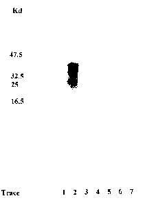
Figure 1: Determining the marker protein prpsen in a
Western blot
The Figure shows the determining of the marker protein
Prpsen in mononuclear (MN) leukocytes of BSE infected
cattle and BSE-negative control animals in a Western blot
using 142 monoclonal antibodies.
The cells were homogenised in 2o sarcosyl. The homogenised
preparation is applied to the gel in a concentration of
60 ~.g/well.
Trace 1: Molecular weight marker
Trace 2: BSE brain (BSE positive, positive control)
Trace 3: Cow No. 058193 (BSE positive)
Trace 4: Cow No. 5061 (BSE positive)
Trace 5: Cow No. 2819 (BSE positive)
Trace 6: Cow No. 279046 (BSE negative, negative control)
Trace 7: Cow No. 4751 (BSE positive)
All the MN samples are obtained from BSE infected cattle,
with the exception of cow no. 279046. The expression of
prpsen is positive in all the BSE infected cattle compared
with the negative control. Cow no. 5061 (trace 4) expresses
prpsen less strongly here but significantly above the
negative control.
Figure 2: Determining the marker protein IFN-y by RT-PCR
The Figure shows the determining of the marker protein
IFN-y by RT-PCR in BSE infected cattle and BSE negative
control animals.
Trace 1: GAPDH control, Cow No. 4372 (BSE positive)
Trace 2: IFN-y, Cow No. 4372 (BSE positive)
CA 02367696 2001-09-14
WO 00/65357 PCT/EP00/03605
17
Trace 3: GAPDH control, Cow No. 441 (BSE negative)
Trace 4: IFN-y, Cow No. 441 (BSE negative)
Figure 3: Determining the marker protein laminin receptor
by RT-PCR
The Figure shows the measurement of the marker protein
laminin receptor (LR) by RT-PCR in BSE-infected cattle,
cattle in which BSE is suspected and in BSE-negative
control animals.
Part A)
Trace 1: Cow No. 4471 (BSE positive)
Trace 2: Cow No. 58193 (BSE positive)
Trace 3: Cow No. 462 (negative control)
Trace 4: Cow No. 5621 (BSE suspected)
Trace 5: Cow No. 5054 (BSE suspected)
Trace 6: Target DNA control
Trace 7: Void
Trace 8: Molecular weight marker
Part B)
For each trace in Part A) there is a corresponding RT-PCR
of D-glyceraldehyde-3-phosphate-dehydrogenase (GAPDH) as a
control for the differential activity of reverse
transcriptase.
The invention is described more fully with reference to the
Example which follows.
CA 02367696 2001-09-14
WO 00/65357 PCT/EP00/03605
18
Example l: Diagnosis of BSE by means of the increased
expression of specific marker proteins in isolated
leukocytes
The Example which follows describes the diagnosis of BSE in
cattle by determining the increased expression of the
marker proteins prpsen or IFN-y or the laminin receptor
(precursor) (LR(P)) on isolated mononuclear (MN) or
polymorphonuclear (PMN) leukocytes.
Isolation of mononuclear (MN) and polymorphonuclear (PMN)
leukocytes from bovine whole blood
These special blood cells are isolated by two
centrifugation steps, the latter being a density gradient
centrifugation in order to obtain the so-called leukocyte
"buffy" coat, followed by lysis of the erythrocytes.
Step 1: Blood samples
The blood samples (about 400 ml in volume) are taken from
the animals and placed directly in a special container.
This container is already provided with a mixture of
glucose and citrate: 68 mM glucose, 37.4 mM tri-sodium
citrate, 17.4 mM citric acid, adjusted to pH 7.3, as
anticoagulant. The blood and anticoagulant are in a ratio
of 6:1. The blood samples are sent immediately to the
laboratory for isolation of the cells.
Step 2: Concentration of leukocytes
1. Centrifugation
Exactly 40 ml of whole bovine blood treated with
anticoagulant is placed in a sealable sterile 50 ml
centrifugal test tube and centrifuged at 800 x g for 20
minutes at room temperature in a rotary centrifuge without
brake. Centrifugation should be extended for 5 minutes for
CA 02367696 2001-09-14
WO 00/65357 PCT/EP00/03605
19
each hour (up to 3 hours) that the blood samples have been
stored after collection.
This first centrifugation leads to the formation of three
separate bands:
Upper band - serum
Middle band - leukocytes (so-called "buffy")
Lower band - erythrocytes
Density centrifugation medium:
Standard commercial medium can be used to isolate the
leukocytes. We used in NYCOMED LymphoprepT M, density
1.077 g/ml.
The serum is carefully removed and deep-frozen for further
analysis at -20°C. The leukocyte layer is taken off and
placed in a new centrifugal test tube.
2nd Centrifugation
Exactly 15 ml of the leukocytes from the first
centrifugation are placed on 35 ml of NYCOMED
LymphoprepTM, density 1.077 g/ml in a sealable sterile
50 ml centrifugal test tube.
Centrifugation: 800 x g/20 min- (RT) in a rotary centrifuge
without a brake.
Centrifugation should be extended for 5 minutes for each
hour (up to 3 hours) that the blood samples have been
stored after collection.
This 2nd centrifugation leads to the formation of four
separate bands:
From top to bottom:
1St band - (residual) serum
2nd band - mononuclear leukocytes (monocytes and
lymphocytes = buffy)
3rd band - medium interface
CA 02367696 2001-09-14
WO 00/65357 PCT/EP00/03605
4th band - polymorphonuclear leukocytes and
(residual) erythocytes
Step 3: Separation of the mononuclear (MN) leukocytes
5
Band 2 contains monocytes and lymphocytes and is carefully
sucked out using a sterile Pasteur pipette and transferred
into a sterile 50 ml centrifugal test tube. The cells are
washed twice with the same volume of sterile PBS (phosphate
10 buffered saline) and centrifuged at 600 x g/15 min/10°C.
The pelleted cells are resuspended in HBSS (Hanks balanced
salt solution with NaHC03, without phenol red). The
vitality and cell number are determined using trypan blue.
15 Step 4: Separation of the polymorphonuclear (PMI~)
leukocytes
After band 2 has been pipetted out, bands 1 and 3 are also
removed by suction. The PMN/erythrocyte mixture is then
20 diluted three times with sterile erythrocyte lysing buffer
(ELB, consisting of 8.9 mM KHC03, 154.9 mM NH4C L and
0.01 mM EDTA), mixed carefully and incubated for 10 min at
RT.
Centrifugation: 800 x g/10 min/10°C
The supernatant is discarded and the pellet is resuspended
in 20 ml of ELB buffer, mixed, incubated and centrifuged
again.
Centrifugation: 800 x g/10 min/10°C
The supernatant is discarded and the pellet is washed with
20 ml HBSS (as above, but with the addition of 1 mM
MgCl2).
Centrifugation: 800 x g/8 min/10°C
CA 02367696 2001-09-14
WO 00/65357 PCT/EP00/03605
21
The supernatant is discarded and the cells are resuspended
in HBSS. The vitality and cell number are determined with
trypan blue.
Results: Example cell numbers
Cell type Cell number/ml of blood Vitality
1. MN 2.25 x 10⁶ >_930
2. PMN 4.03 x 10⁶
?98%
The following measurements were made with the isolated MN-
and PMN-leukocytes of BSE infected animals:
I. Increased expression of the cellular isoform of the
prion protein, prpsen
II. Increased expression of the interferon gamma protein,
IFN-y
III. Increased expression. of the laminin (precursor)
receptor, L (P) R
I. Increased expression of the cellular isoform PrPsen
The expression rate of prpsen in isolated leukocytes from
control animals and BSE infected animals is measured by
Western blot analysis (Harmeyer S. et al., J Gen Virol
1998, 79, 937-945). Chromogenic development is used.
Opening up of the cells:
The isolated leukocytes are homogenised in 2o sarcosyl
solution (Sigma, St. Louis, USA) for 10 min/4°C. The
homogenised preparation thus obtained is then pelleted at
15,000 x g/40 min/4°C. The supernatant is suction filtered
and stored at -20°C.
The protein concentration of the homogenised preparation
was determined and all samples were standardized to 6 mg/ml
CA 02367696 2001-09-14
WO 00/65357 PCT/EP00/03605
22
protein. Exactly 60 ~g of the homogenate was loaded into
each well.
Detection is carried out using either the monoclonal
antibody 13 or the monoclonal antibody 142. The antibodies
are described in detail in the above-mentioned publication.
The dilution of the antibody is 1:10 in each case.
The secondary antibody used is this example were AP-
conjugated antibodies in a dilution of 1:3000.
Resul is
It was shown that the protein expression of prpsen is
significantly raised both in MN- and PMN-leukocytes of BSE
infected animals compared with healthy control animals (cf.
also the Western blot of Figure 1 with samples from other
cattle) .
Case No./Sample BSE Status Increased expression of
PrPsgn
4372 positive yes
4471 positive yes
4401 suspect yes
Negative
Control negative no
negative
II. Increased expression of IFN-y
The increased expression is measured in two ways:
- measurement of the protein by ELISA
- measurement of the specific mRNA by RT-PCR
CA 02367696 2001-09-14
WO 00/65357 PCT/EP00/03605
23
1. IFN-y measurement using ELISA
A commercial ELISA made by CSL Veterinary Ltd. , Melbourne,
Australia is used.
Results:
BSE Status Number of animals IgN_Y ~pg~~l~
BSE positive 9 314.0 78.2
BSE suspected 9 504.0 109.7
BSE negative 13 0.0 0.0
2. IFN-y mRNA measurement using RT-PCR
The isolation of RNA (from the total leukocyte fraction)
and the subsequent RT-PCR are carried out by standard
methods (Yi-Jun Shi and Jing-Zhlong Liu,
Genet. Anal. Tech.Appl., 1992, 9, 149-150; Izraeli S. et al.,
Nucl.Acid.Res. 1991, 21, 6051; Michel U. et al.,
Anal.Biochem. 1997, 249, 246-247) with the modifications
described below.
Isolation of RNA:
The Promega System (Catalogue No. 63191) is used to isolate
the total RNA.
cDNA (RT reaction):
Isolated RNA samples are reverse transcribed using the
Reverse Transcription System of Promega (Catalogue No.
A3500)
PCR Reaction:
In order to determine the specific mRNA a double polymerase
chain reaction (~~nested PCR~~) is used.
The IFN-y primers used for this:
CA 02367696 2001-09-14
WO 00/65357 PCT/EP00/03605
24
FWl (IFNF1) 5'GGAGTATTTTAATGCAAGTAGCCC 3'
FW2 (IFNF2) 5'GTAGCTAAGGGTGGGCCTCT 3'
RV (IFNR1) 5'GCTCTCCGGGCCTCGAAAGAGATT 3'
The PCR product to be expected should be 357 base pairs
(bp) long.
Resul is
The IFNy ELISA is clearly confirmed by this RT-PCR, i.e. in
BSE infected animals IFNy is significantly raised.
Figure 2 shows the RT-PCR of mRNA from total leukocytes in
whole blood with the samples of cattle nos. 4372 and 441.
III. Increased expression of the laminin receptor
(precursor) , LR (P)
The expression was measured by RT-PCR. This reaction also
includes the detection of LRP as well as LR.
The bovine sequence of LRP or LR has not yet been
described. However, this protein is highly conserved in
mammals. The published sequence data of human as well as
murine LR are therefore compared. On the basis of this data
analysis the following primer sequence is established:
Primers
Forwards 5' AAGAGGACCTGGGAGAAGCT 3'
Backwards 5' CCTTCTCAGCAGCAGCCCTGC 3'
Expected product: 517 by
RNA isolation
As described under point II.2.
cDNA (RT reaction)
As described under point II.2.
CA 02367696 2001-09-14
WO 00/65357 PCT/EP00/03605
PCR reaction
Simple reaction corresponding to standard methods.
5
Resul is
Case No./Sample BSE Status Increased expression of
LRP/LR
4471 positive yes
58193 positive yes
5621 suspected (yes)
5054 suspected yes
Control (462) negative no
These results are shown in Figure 3.
IV. Cloning and expression of the bovine Laminin Receptor
(Precursor), LR(P), to generate specific antibodies
against LR.
Development of Primers towards LR:
Primers towards bovine LR are designed to amplify the
entire gene of LR (Genebank Accession No: S 37431). Primers
were designed from the bovine c10 protein gene (Genebank
Accession No: M 64923).
In the LR primers defined below, their restriction sites
appear in a box. Based on these restriction sites a direct
cloning into E.coli is possible.
The LR designed primers will be used to amplify the LR from
total cellular RNA isolated from whole bovine blood.
CA 02367696 2001-09-14
WO 00/65357 PCT/EP00/03605
26
The sequence for the three primers may be seen below:
LRPF1 5' AT CTCGAG GTCCGGAGCCCTTGATGTCC 3'
LRPR1 5' ATTG TTCC ACGACCACTCGGTGGTGGT 3'
LRPR2 5' ATT CTAG CGACCACTCGGTGGTTCC 3'
Restriction sites: LRPF1 primer can be cut by Xho, LRPR1
primer can be cut by Eco R1, while LRPR2 can be cut by Xba
1.
RT-PCR and Cloning of bovine LR gene:
The LR primers are re-suspended in sterile water to a final
concentration of 50 ng/ml.
Bovine leukocyte RNA is isolated from total leukocyte
fractions of whole bovine blood.
5 ~,g of RNA is DNAsed (Gibco BRL) and Reverse Transcription
is carried out using a Reverse Transcription Kit (Promega,
Cat. No; A3500). Polymerase Chain Reactions (PCR) are
carried out using LRPF1, LRPR1, and LRPR2.
PCR: 35 cycles, annealing temperature 50~ C.
The amplified fragments obtained following PCR run on to
Agarose gel to determine fragment size. The resulting bands
are gel purified, checked again for size and the fragments
are removed from agarose and re-suspended in 10,1 steril
water.
The amplified fragments are ligated into pGEM-T vector
(Boehringer Mannheim Ligation Kit).
CA 02367696 2001-09-14
WO 00/65357 PCT/EP00/03605
27
All clones are sequenced and have been demonstrated as
being the LR gene.
Sub-cloning into the expression vectors pBADGIII and
pTrcHis (both obtained from Invitrogen Ltd.) has been
carried out and the clones are currently being examined for
the presence of inserts.
Design of Peptides for the development of antibodies to LR:
Four peptides are designed to be utilised in the
development of antibodies to the Laminin Receptor (LR).
These peptides are designed using the bovine c10 protein
(Genbank Accession No: M 64923; protein Id. AAA62713.1)
A computer program (Antheprot) which predicts a proteins
structure, hydrophobic, hydrophiliy and antigenic sites is
used for designing the petides.
The principle parameters concentrate upon for the selection
of two of the peptides are hydrophilic and antigenic, two
other peptides are chosen due to their location at the C-
and N-Terminal regions of the protein.
Peptide 1141 MSGALDVLQMKEEDVLKFLAGC
(Amino Terminal) Corresponding to amino acid residues 1-20
from the amino-terminal end of the protein.
Isoelectric point (pI) of 4.32.
Peptide 1142 RLLVVTDPRADHQPLTEASYGC
(Antigenic) Corresponding to amino acid residues 120-
140 selected from a antigenic region of the
Protein.
Isoelectric point (pI) of 5.38.
CA 02367696 2001-09-14
WO 00/65357 PCT/EP00/03605
28
Peptide 1143 KEEQAAEKAVTKEEFQGEWGC
(Hydrophilic and antigenic) Corresponding to amino acid
residues 212-231 selected from a hydrophilic and antigenic
region of the protein.
Isoelectric point (pI) of 4.48.
Peptide 1144 FTAAQPEVADWSEGVQVPSVGC
(Carboxy Terminal) Corresponding to amino acid residue 238
257 selected from the caraboxy terminal region of the
protein.
Isoelectric point (pI) of3.58.
Each peptide is conjugated to Imject ~ Nlaleimide Activated
Ovalbumin Carrier Protein (Pierce Warner Ltd.) as follows.
The peptides are dissolved to a final concentration of 10 mg/ml
in 100 mM Na2HP04 (pH 7.2).
Imject ° Nlaleimide Activated Ovalbumin is dissolved to a
final concentration of 10 mg/ml in steril water.
The peptide and Ovalbumin are allowed to conjugate for 2 hours
at room temperature.
Following conjugation the conjugated protein solution will be
dialyzed in 500-fold volume of PBS (pH 7.4) and stored at ~20~
C until required.
Production of polyclonal and monoclonal antibodies
The production of polyclonal and monoclonal antibodies are
carried out in a similar way as described e.g., Harmeyer S. et
al., J Gen Virol, 1998.
Briefly:
Day 0: Pre-immune bleeds are taken prior to injection for
examination of anti-LR-antibodies.
CA 02367696 2001-09-14
WO 00/65357 PCT/EP00/03605
29
Day 0: 0.5 mls of conjugated. peptide suspended in Freund°s
Complete Adjuvant (Sigma-Aldrich, Cat. No: F-5881) are injected
subcutaneous.
Day 14: the first booster injection of 0.5 mls conjugated
peptide in Incomplete Freund's Adjuvant (Sigma-Aldrich, Cat.
No: F-5506)
Day 21: the second booster injection of 1,0 ml conjugated
peptide in the absence of adjuvant.
Day 31: Test Bleed for control of produced antibodies.
ELISA System for the measurement of Marker-proteins
The ELISA technology is especially suitable for the
measurement of BSE marker-proteins. In this case special
blood cells will be separeted which carry these markers on
the cell surface.
Cell separation procedure
Sub-division of MN and PMN leukocyte sub-classes (target
cells) will be achieved using immunomagnetic bead
( Dynabeads ~) depletion. Beads will be coated with antibodies
specific to bovine leukocyte cell types.
Shortly:
- Add cell specific Dynabeads to the blood sample.
- Immuncapture of the target cells.
- Magnetic separation of the target cells.
- Washing and concentration of pure target cells.
CA 02367696 2001-09-14
WO 00/65357 PCT/EP00/03605
ELISA measurement of Marker-proteins
An ELISA system is choosen, based upon isolation of target
5 cells using target cell specific capture antibodies:
- Microtitre plates are coated with primary capture
antibody specific to target cell surface marker.
10 - Incubation with cells expressing target cell surface
marker. Binding to primary capture antibody.
- Following cell capture, incubation with biotinylated
secondary antibody directed against the BSE marker
15 protein.
- Incubation with enzyme-conjugated detection protein.
Addition of substrate and measurement by ELISA-reader.