Note : Les descriptions sont présentées dans la langue officielle dans laquelle elles ont été soumises.
CA 02369743 2001-10-16
WO 00/64524 PCT/iJS00/11194
CAVITY MEASUREMENT DEVICE AND METHOD OF ASSEMBLY
Field of the Invention
The present invention relates generally to a cavity measurement device used
during
surgical procedures and a method of assembling a cavity measurement device.
The present
invention particularly relates to inflation tubes with a balloon used during
surgery and methods
of attaching a balloon to an inflation tube.
Background of the Invention
The main structural support of the human skeleton is the spine which is the
bone
structure that extends from the base of the skull to the pelvis. It includes a
spinal cord which is
approximately eighteen inches in len;th and comprised of nerves that carry
impulses to and
from the brain to the rest of the body.
Surrounding the spinal cord are pairs of rings of bone called vertebrae which
constitute
the spinal column (back bones) and each pair of vertebrae is connected by a
flexible joint that
stabilizes the vertebrae and allows the spine to move.
An "intervertebral disk" - or simply the "disk" - is located between each pair
of
vertebrae within the flexible joint and bears most of the compressive load of
the spinal column.
Each disk is a flat, circular capsule approximately one inch in diameter and
has an outer layer
or membrane which is strong and flexible and comprised of a fibrous cartilage
called the
annulus fibrosis. It also has an inner core which consists of a soft,
gelatinous substance called
the nucleus pulposus. The main function of the disk is to cushion the
vertebrae during
movement.
The structure of the human spine is designed for an upright position, a
typical posture
for humans throughout history, where walking, running, hunting, fathering,
working on farms
or at workbenches were common body motions and positions. Today, a high
proportion of
people lead sedentary lives, spending the better part of each day sitting
behind desks writing
patent applications, at work stations, in automobiles, etc. These changes in
human behavior
overtime, mainly resulting from technological advances, have had a profound
and largely
negative impact on human physiology, and particularly the spine. As a result,
spine or back
problems are the most common physical complaints among adults.
Everyday physical stresses and the normal aging process also adversely affect
the
human spine. In that connection, one of the most common back problems
experienced by
adults results from degenerative disk disease, a general term applied to
degeneration of the
CA 02369743 2001-10-16
WO 00/64524 PCT/US00/11194
intervertebral disks. As the body ages, the disk material loses its elasticity
and hardens,
developing a consistency similar to a piece of hard rubber.
A specific example of degenerative disk disease is a herniated disk which is a
condition
resulting from strain or injury to the disk that causes the inner material of
the disk to swell or
herniate and the outer layer to rupture. When the disk ruptures, the inner
material bulges and
presses against, or pinches, the spinal nerves, resulting in severe pain.
When the disk degenerates to the point where it no longer properly functions,
the disk is
removed during a procedure called a diskectomy. A diskectomy involves removal
of the
ruptured or diseased disk from its location between adjacent vertebrae. By
removing the disk
and any associated disk or bone fragments, the source of the pressure on the
spinal nerve is also
removed, thereby relieving the pain.
Following a diskectomy, the adjacent vertebrae may be fused together,
resulting in
partial loss of spinal flexibility. On the other hand, a bone graft or other
specialized material,
such as a prosthetic intervertebral implant, may be placed in the empty disk
space in order to
stabilize the vertebrae.
Bone grafts and similar prosthetic implants used following diskectomy require
the
implant and surrounding vertebrae to be shaped using precision drilling and
shaving techniques
in order to provide a proper fit with the implant. This type of surgical
reconstruction is
difficult and time-consuming and often still results in limited flexibility of
the spine. As a
result, synthetic intervertebral disk prostheses have been developed, such as
those described in
U.S. Patent No. 4,863,477. These synthetic prostheses are fabricated prior to
performance of
the surgery and are shaped during surgery to conform specifically to the shape
of the disk
space, thereby eliminating the tedious task of precision drilling and shaving
techniques
associated with bone implants. Moreover, these synthetic prostheses provide a
resiliency that
facilitates flexibility of the spine.
In order to ensure that the prosthetic incorporates the proper shape and
volume for the
target space, various measuring techniques have been proposed. These
techniques include X-
rays, magnetic resonance imaging (MRI), computed tomography (CT) scans and
myelography,
a radiological technique for viewing the spinal cord. These techniques,
although quite useful,
are not without certain drawbacks including high costs, potential adverse side
effects and
inherent measuring inaccuracies which result from a variety of factors,
including high signal to
noise ratios, limited two-dimensional images, and potential radiation
exposure. Furthermore,
these devices are expensive and require highly-skilled technicians to operate
them properly.
CA 02369743 2001-10-16
WO 00/64524 PCT/US00/11194
As a result, practitioners and medical institutions have continually sought a
lower cost
and less complex method of obtaining the data necessary to fabricate a quality
prosthetic. In
particular, there is a desire to obtain low-cost yet highly accurate body
cavity measuring device
that can be used with minimal to no side effects. Such a device must be
biocompatible, non-
toxic and simple to use. Finally, such _a device must be fabricated by a
manufacturing method
that is efficient, easy to implement and cost effective.
Objects and Summary of the Invention
In view of the foregoing, it is an object of the present invention to provide
a cavity
measurement device that addresses the drawbacks associated with prior art
cavity measuring
devices, yet meets the needs of the users.
A further object of the present invention is to provide a cavity measurement
device that
is biocompatible, non-toxic and simple to use.
A further object of the present invention is to provide a cavity measurement
device that
provides a leakproof attachment of an inflatable balloon to a tube or cannula.
A further object of the present invention is to provide a cavity measurement
device that
provides a leakproof attachment able to withstand a 45 psi balloon pressure
during use.
A further object of the present invention is to provide a cavity measurement
device that
includes a mechanical attachment with an attachment strength greater than the
balloon material
tensile strength.
A further object of the present invention is to provide a method of making a
cavity
measurement device that is efficient, easy to implement and cost effective.
These and other objects not specifically enumerated herein are believed to be
addressed
by the present invention which contemplates a cavity measurement device that
includes an
inflation tube assembly comprising an elongated tube having a distal end and a
proximal end
and a balloon mounted at the distal end of the elongated tube. The elongated
tube and the
balloon comprise dissimilar materials and include an adhesive free seal
between the elongated
tube and the balloon.
The present invention also contemplates a method of assembling a cavity
measurement
device which may include the steps of attaching a silicone inflatable balloon
having a tubular
stem section and a bulbous section onto an elongated thermoplastic tube by
sliding the stem
section onto an end of the elongated tube and positioning a piece of heat-
shrink tube over an
area of the stem section that overlaps the elongated tube. The next steps may
include shrinking
the heat shrink tube and folding a length of the stem section over the heat-
shrink tube. The
following steps would include overlaying a compression tube onto an overtube
so as to create
-3-
CA 02369743 2001-10-16
WO 00/64524 PCT/US00/11194
an overtube assembly and aligning the overtube assembly onto the stem section
and a portion
of the elongated tube. The next steps would likely include heating and
subsequently cooling
the overtube assembly, so that the compression tube molds the overtube onto
the stem section
and elongated tube. The final steps would include bonding an end of the
overtube onto the
elongated tube, thereby forming a mechanical, leakproof bond, and removing the
compression
tube.
Brief Description of the Drawings
Other features and advantages of the present invention will be seen as the
following
description of particular embodiments progresses in conjunction with the
drawings, in which:
FIG. 1 is a perspective view of a cavity measurement device in accordance with
a
preferred embodiment of the present invention;
FIG. 2 is a second perspective view of a cavity measurement device in
accordance with
a preferred embodiment of the present invention;
FIG. 3 is a side perspective view of an inflatable balloon component of the
cavity
measurement device in accordance with a preferred embodiment of the present
invention;
FIG. 4 is a cross-sectional view of an inflatable balloon taken along the
lines 4-4 in
FIG. 3;
FIG. 5 is a perspective view of an inflatable balloon attachment assembly in
accordance
with a preferred embodiment of the present invention;
FIG. 6 is a cross-sectional side view of a step of a method of assembling a
cavity
measurement device in accordance with a preferred embodiment of the present
invention;
FIG. 7 is a cross-sectional side view of a step of a method of assembling a
cavity
measurement device in accordance with a preferred embodiment of the present
invention;
FIG. 8 is a cross-sectional side view of a step of a method of assembling a
cavity
measurement device in accordance with a preferred embodiment of the present
invention;
FIG. 9 is a cross-sectional side view of a step of a method of assembling a
cavity
measurement device in accordance with a preferred embodiment of the present
invention; and
FIG. 10 is a cross-sectional side view of a step of a method of assembling a
cavity
measurement device in accordance with a preferred embodiment of the present
invention.
Detailed Description of the Invention
Referring to Figure l, an embodiment of a cavity measurement device 10 for use
with a
pistol-grip handle 12 or other similar device in accordance with the present
invention includes
an elongated inflation tube or cannula 14 and an inflatable balloon 16. The
inflatable balloon
-4-
CA 02369743 2001-10-16
WO 00/64524 PCT/US00/11194
16 is mechanically affixed to one end of the elongated tube 14 and provides a
leakproof
attachment that can withstand a 45 psi balloon pressure. The other end of the
elongated tube
14 connects to a balloon mount 18 that couples onto the trigger receptacle 20
of the handle 12.
Referring to Figure 2, an embodiment of the elongated tube 14 includes a
distal end 22
S and a proximal end 24. The distal end 22 of the elongated tube 14 connects
to the inflatable
balloon 16 and the proximal end 24 of the elongated tube 14 connects to the
balloon mount 18
(not shown). The inner diameter of the elongated tube 14 should be large
enough to adequately
support a flow of fluid and is commonly between the range of 3-4 French. Since
a portion of
the elongated tube 14 is inserted into a body space, the outer diameter of the
elongated tube 14
should be large enough to accommodate the flow of fluid in its inner diameter,
yet small
enough so as to be minimally invasive during a surgical procedure. A suitable
outer diameter
for the elongated tube 14 is about 7 French to 8 French.
The elongated tube 14 should be lone enough so that a first section of the
elongated
tube 14 adequately fits into the trigger receptacle 20 ( not shown) and the
remaining section of
the elongated tube 14 extends sufficiently beyond the trigger receptacle 20.
For the cavity
measurement device 10 of the present invention, the length of the elongated
tube 14 is typically
about 38 cm to 40 cm. The section of elongated tube 14 extending beyond the
trigger
receptacle 20 must be of optimal length, such as 20 cm, to allow a portion of
the elongated tube
14 to be inserted into the body between the vertebrae of a spine during a
diskectomy or similar
procedure.
Since a portion of the elongated tube 14 will contact the body, its material
should be
biocompatible and non-toxic. In a preferred embodiment, the material of the
elongated tube 14
is a thermoplastic, such as polyethylene terephthalate (PET). Similar
materials, such as nylon,
may also be used.
The fabrication of the elongated tube 14 typically involves an extrusion
process that
provides precisely controlled inner diameters and wall-thicknesses. The
particular
configuration of the elongated tube 14 provides sufficient rigidity to
withstand the forces and
pressures exerted on it during a surgical procedure.
As shown in Figure 3, the cavity measurement device 10 of the present
invention also
includes an inflatable balloon 16. In a preferred embodiment, the inflatable
balloon 16 is made
of an elastic material, such as silicone, that is capable of being easily
stretched or expanded and
resuming its former shape. The preferred balloon 16 material is silicone
because of its low
durometer and high tear resistance (i.e. elon;ation at break),
biocompatibility and non-toxicity.
Silicone is also preferred due to its high elasticity and low modulus of
elasticity which enables
-5-
CA 02369743 2001-10-16
WO 00/64524 PCT/US00/11194
the balloon to conform substantially to the inner surfaces of the cavity being
measured. An
example of such a silicone is MED10-6640 made by NuSil. Other types of typical
balloon
materials, such as polyurethane, do not have sufficient elasticity to enable
the proper degree of
conformance to the inner surfaces.
The inflatable balloon 16 includes a tubular stem section 26 and a bulbous
portion 28.
The stem section 26 and bulbous portion 28 are located at the proximal and
distal ends,
respectively, of the inflatable balloon 16. In a preferred embodiment, the
length of the stem
section 26 is about 1.9 cm. Additionally, the length of the bulbous portion 28
of the inflatable
balloon 16 is typically 1.2 cm.
In its unassembled state, as shown in Figure 3, the inner diameter of the stem
section 26
of the inflatable balloon 16 is approximately 0.25 mm. The diameter of the
stem section 26,
together with its material elasticity, enable the stem section 26 to be easily
mounted onto the
distal end 22 of the elongated tube 14. This particular configuration ensures
uniform surface
contact between the inner surface of the stem section 26 and the outer surface
of the elongated
tube 14. In addition, the thickness of the inflatable balloon 16 material is
relatively uniform
along its entire length so as to allow uniform inflation when a fluid is
introduced. However, in
an alternate embodiment, the material thickness of the inflatable balloon 16
may be variable
along its length depending on the various desired inflation characteristics
and surgical
procedure to be performed.
To minimize potential damage to surrounding tissues when the cavity
measurement
device 10 is inserted into the body cavity during a surgical procedure, the
outer surface of the
inflatable balloon 16 is relatively smooth. In a preferred embodiment, the
bulbous portion 28
of the inflatable balloon 16 is relatively oblong in shape, allowing for easy
insertion into a
body cavity such as a disk space. Alternative geometries for the bulbous
portion 28 include,
but are not limited to, oval, spherical, tubular and barrel-shaped. The
particular geometry
chosen is typically based upon the body cavity shape and type of surgical
procedure performed.
As shown in Figures 3 and 4, when confi;ured in an oval geometry, the bulbous
portion
28 has a center diameter of approximately 9.5 French. However, the center
diameter of the
bulbous portion 28 can range from 8 to 10.5 French, or any suitable size that
allows the cavity
measurement device 10 to be inserted into a body cavity. Typically, the center
diameter of the
bulbous portion 28 is greater than the inner and/or outer diameters of the
stem section 26 of the
inflatable balloon 16.
Turning next to how the balloon 16 is mounted to the elongated tube 14, it is
important
to note that conventional adhesives are ineffective bonding agents in this
context due to the
-6-
CA 02369743 2001-10-16
WO 00/64524 PCT/US00/11194
dissimilar material characteristics of the inflatable balloon 16 and elongated
tube 14. Such
adhesives are typically unable to withstand the high pressures exerted on the
bond by the
inflatable balloon 16. In addition, the elasticity of the inflatable balloon
16 tends to be
compromised by the rigid bond caused by chemical adhesives. As a result, such
bonds have a
tendency to separate or peel away when subject to various stresses and
pressures encountered
during surgical procedures.
As a result, in the present invention, a mechanical fixation method is used.
The
mechanical fixation provides a durable, leakproof attachment of the inflatable
balloon 16 to the
elongated tube 14 which, in a preferred embodiment is capable of withstanding
a 45 psi balloon
pressure and is greater than the balloon 16 material tensile strength. In
addition, the use of a
mechanical fixation method avoids pre-surface treatments, primers, cure times,
or other
preparatory operations typically associated with adhesive bonding methods.
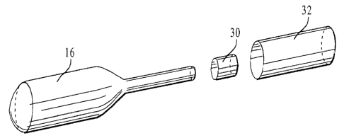
Referring to Figure 5, the structure used to mechanically attach the balloon
16 to the
tube 14 is shown and includes a heat-shrink tube 30 and an overtube 32. The
heat-shrink tube
30 is used to mechanically attach the inflatable balloon 16 to the elongated
tube 14. More
specifically, the heat-shrink tube 30 is positioned onto a portion of the stem
section 26 of the
balloon 16 that is mounted onto the elongated tube 14. Heat is then applied to
the heat-shrink
tube 30 which causes it to shrink and conform to the size and shape of that
portion of the stem
section 26 and the elongated tube 14, thereby securing the stem section 26 to
the elongated
tube 14.
In a preferred embodiment, the heat-shrink tube 30 is made of polyvinylidene
fluoride
(PVDF). Alternatively, the heat-shrink tube 30 may be fabricated from other
suitable heat-
shrink materials, such as polyolefin and teflon
The heat-shrink tube 30 is approximately 0.4 cm long, with an internal
diameter of
about 0.3 cm. The internal diameter of the heat-shrink tube 30 must be of
sufficient size to
allow the heat-shrink tube to fit, prior to heating, over the stem section 26
of the inflatable
balloon 16 when it is mounted onto the elongated tube 14.
In addition to use of the heat shrink, an overtube 32 of the present invention
is also used
to assist in mechanically fixating the inflatable balloon 16 onto the
elongated tube 14. A
description of how the overtube 32 is utilized is set forth below. The
overtube 32 also softens
when subject to heat and is preferably made from polyethylene terephthalate
(PET).
Alternatively, the overtube 32 may be fabricated from nylon or other similar
materials that are
conformable when heat is applied.
_7_
CA 02369743 2001-10-16
WO 00/64524 PCT/LJS00/11194
In addition to the particular material attributes of the overtube 32, the
dimensional
configuration of the overtube 32 is also important in order to obtain the
necessary mechanical
bonding of the balloon 16 to the elongated tube 14. The inner diameter and
length of the
overtube 32 should be appropriately sized to allow the overtube 32 to surround
and overlap the
S assembled heat-shrink tube 30, stem section 26 and a portion of the
elongated tube 14. In a
preferred embodiment, the length and inner diameter of the overtube 32 are 1.9
cm and 0.38
cm respectively.
Prior to use, the cavity measurement device 10 is primed by evacuating the air
from the
inflatable balloon 16 and elongated tube 14 and infusing a fluid therein. The
fluids used during
the priming procedure include, but are not limited to, water, saline and
contrast media. It is
important to note that these same fluids can also be used as inflation fluids
when the device is
inserted into a body cavity during a measurement procedure. The objective of
the priming
procedure is to remove anv and all air from the inflatable balloon 16 and
elongated tube 14 to
ensure accurate volume measurements by the cavity measurement device 10. After
the priming
procedure is completed, the fluid is removed.
In use during a surgical procedure, such as measuring a disk space volume, the
inflatable balloon 16, in a deflated state, is inserted into the disk space.
Fluid is infused into
the balloon 16 causing the balloon 16 to inflate and fill the disk space
volume. The volume of
fluid infused into the cavity measurement device 10 must attain a
predetermined pressure
within the disk space that is substantially equivalent to the normal
anatomical pressures exerted
on a natural intervertebral disk. The amount of fluid volume infused into the
cavity
measurement device is calculated and a prosthetic disk of equivalent volume is
then selected
for insertion into the disk space. A preferred prosthetic disk is a hydrogel
disk, although other
similar prosthetic disks may be used. When inserted into the disk space, the
prosthetic disk
conforms to the configuration of the cavity and fills the cavity with a
sufficient volume of
material to create appropriate pressures in the spine to support the body.
Method of Fabrication
The present invention also contemplates a method of fabricating a cavity
measuring
device and particularly contemplates a method of mechanical fixation of the
inflatable balloon
I6 onto the elongated tube 14 of the cavity measurement device 10, as shown in
Figures 6-10.
To keep the lumen of the elongated tube l4 open during the assembly procedure,
a straight,
rigid mandrel 33, preferably made from Nitinol wire, is inserted into the
lumen. The mandrel
33 is approximately 1.04 mm in diameter and extends along the length, and
slightly beyond the
ends 22,24, of the elongated tube 14. The first step of assembling the device
of the present
_g_
CA 02369743 2001-10-16
WO 00/64524 PCT/US00/11194
invention includes sliding the stem section 26 of the inflatable balloon 16
onto the distal end 22
of the elongated tube 14 such that the entire length of the stem section 26 is
placed onto the
elongated tube 14. In a preferred embodiment, the bulbous portion 28 of the
inflatable balloon
16 does not contact and extends beyond the distal end 22 of the elongated tube
14. Therefore,
the bulbous portion 28 of the inflatable balloon 16 abuts the distal end 22 of
the elongated tube,
thereby forming a junction between the stem section 26 and the bulbous portion
28.
The next step includes mountin<, a heat-shrink tube 30 onto an area of the
stem section
26, as shown in Figure 6. Preferably, the inner diameter of the heat-shrink
tube 30 is smaller
than the outer diameter of the bulbous portion 28 and larger than the outer
diameter of the stem
section 26 of the inflatable balloon 16. The heat-shrink tube 30 is mounted
onto the elongated
tube 14 at the proximal end 24 of the elonvated tube 14, advanced along the
length of the
elongated tube 14 and positioned on the stem section 26 of the inflatable
balloon 16. In
particular, the heat-shrink tube 30 is located on an area of the stem section
26 that allows a
sufficient length of stem section 26 to extend beyond the heat-shrink tube 30
toward the
proximal end 24 of the elongated tube I 4. The length of stem section 26
extending beyond the
heat-shrink tube 30 should preferably be greater than the overall length of
the heat-shrink tube
30.
The heat-shrink tube 30 is secured onto the stem section 26 using heat which
causes the
tube 30 material to contract and conform to the shape of the object it
surrounds namely, the
balloon 16 and the tube 14. After the heat is removed, the heat-shrink tube 30
retains its newly
conformed shape, as shown in Figure 6, and forms a uniform thickness and
contact surface
with the stem section 26. In addition, the diminished inner diameter size of
the heat-shrink
tube 30 presses upon the outer diameter of the stem section 26, forming a
mechanical fixation.
After shrinking the tube 30 onto the elongated tube 14, the length of the stem
section 26
of the balloon 16 that extends past the heat shrink tube 30 toward the
proximal end 24 of the
elongated tube 14 is folded over the heat-shrink tube 30. In a preferred
embodiment, the folded
portion of the stem section 26 completely overlaps and partially extends
beyond the distal end
ofthe heat-shrink tube 30, as shown in Figure 7, so that a secure fixation is
formed.
As shown in Figure 8, an overtube 32 and a compression tube 34 are then
introduced in
the form of an overtube assembly. The compression tube 34 is preferably made
of silicone,
however other similar materials may be used.
In particular, the overtube assembly includes a compression tube 34 with an
overtube
32 located internally of the compression tube 34. The overtube assembly is
made by radially
expanding the compression tube 34 using a flow of fluid, such as air, to
increase the inner
-9_
CA 02369743 2001-10-16
WO 00/64524 PCT/LJS00/11194
diameter of the compression tube 34. The overtube 32 is then inserted in the
lumen of the
compression tube 34 and the flow of fluid is discontinued so that the inner
surface of the
compression tube 34 uniformly contacts the outer surface of the overtube 32
but remains
expanded (due to the overtube 32) beyond its otherwise unstressed
configuration.
The overtube assembly is next positioned over the stem section 26 of the
balloon and a
portion of the elongated tube l4 such that the distal end of the overtube
assembly is aligned
with the junction formed by the elongated tube 14 and the bulbous portion 28
of the inflatable
balloon 16. A portion of the overtube assembly extends beyond the stem section
26 toward the
proximal end of the elongated tube 14.
After the overtube assembly is properly ali,ned onto the stem section 26 and
elongated
tube 14, heat is applied to the assembly which causes the overtube 32 to
soften. In its softened
state, the overtube 32 offers less radial resistance to the compression force
exerted by the
compression tube 34 thereby allowing the compression tube 34 to compress and
conform the
overtube 32 to the configuration of the stem section 26 and elongated tube 14,
as shown in
Figure 9. After the heat is removed and the assembly allowed to cool, the
overtube 32 remains
in its conformed configuration, thereby forming a mechanical fixation that
further secures the
fixation formed by the balloon and the heat shrink tube 30. In a preferred
embodiment, the
inner surface of the overtube 32 uniformly contacts the outer surface of a
portion of the
elongated tube 14 and the entire length of the stem section 24 of the
inflatable balloon 16.
Although the compression tube 34 has now been allowed to return to its
substantially
unexpanded state, it remains on the assembly and is used to mask and protect
the overtube 32
from the hot dyes used during the melt bonding process discussed below.
The final assembly step includes securing a proximal end of the overtube 32
onto the
elongated tube I4 by melt bonding the proximal end of the overtube 32 onto the
elongated tube
14 thereby forming a clamp bond as shown in Figure 10. The clamp bond secures
the overtube
32 to the elongate tube 14 and prevents the overtube 32 from slipping off of
the elongate tube
14 during use of the device. In addition, the clamp bond also creates a
mechanical, leakproof
barrier between the overtube 32 and the elongated tube 14. After the overtube
32 is firmly
secured onto the elongated tube 14 and stem section 26 of the inflatable
balloon 16, the
compression tube 34 and manc.Irel 33 are removed.
Although the invention has been described in terms of particular embodiments
and
applications, one of ordinary skill in the art, in light of this teaching, can
generate additional
embodiments and modifications without departing from the spirit of or
exceeding the scope of
the claimed invention. Accordingly, it is to be understood that the drawings
and descriptions
-10-
CA 02369743 2001-10-16
WO 00/64524 PCT/US00/11194
herein are proffered by way of example to facilitate comprehension of the
invention and should
not be construed to limit the scope thereof.