Note : Les descriptions sont présentées dans la langue officielle dans laquelle elles ont été soumises.
CA 02371788 2001-11-22
WO 00/72354 PCT/US00/14390
DENTAL X-RAY APPARATUS
Technical Filed
The present invention is generally directed toward a dental x-ray apparatus.
More particularly, the invention is directed toward an x-ray apparatus having
a tube
head formulated from a cast zinc material. Further, the invention also
provides tube
head components fabricated from a high molecular weight material, such as
barium
sulfite. More specifically, the tube head components are fabricated from a
barium
sulfite-charged plastic material. Further, the present invention is directed
toward a
DC powered x-ray dental apparatus.
BACKGROUND OF THE INVENTION
X-ray generators of small or moderate power for medical radiological
application, normally use a fixed-anode x-ray tube (verses a rotating-anode x-
ray tube
as used when large power is required). In this case, the x-ray tube is usually
contained in the same oil-filled housing as the high-voltage transformer and
other
components of the high-voltage circuit, and such an assembly is called a
tubehead.
During the last several years, x-ray generators commercially available for
dental
application (whether intraoral, panoramic, or other) have adopted this general
design
almost universally.
Inside the tubehead, the x-ray tube is supported by a mechanical part known
as the tube holder, made out of a high-insulate and high electric tensile
strength
material, which performs essentially two functions:
1) to securely and precisely hold the x-ray tube in position, in relation
to the surrounding construction and in particular to the output windows and
the
CA 02371788 2001-11-22
WO 00/72354 PCT/US00/14390
external Beam-Limiting-Device; it ensures the accurate geometrical position of
the x-
ray source;
2) to generate high-voltage insulation between the x-ray tube (one or
more of whose electrodes are at extremely high electrical potential) and the
surrounding constructive metallic parts (in particular the housing) which are
grounded.
In order to provide near-focus shielding against radiation from the x-ray tube
(primary and extrafocal) in all directions except through a suitable output
windows
(thus greatly reducing the additional radiation shielding required for the
housing as a
whole), the tube holder is usually surrounded by a lead jacket, at least at
the anode
side and except for a small opening in correspondence with the wanted x-ray
beam
path.
The design and construction of this lead jacket may be critical, as any sharp
or pointed detail (e.g., such as the thread of a screw) should be avoided
because they
imply singularities in the electric field and hence may cause high-voltage
discharges.
Other structural components of the tube head, such as the housing or other
such components, have typically been formulated from steel sheet metal, or
cast
aluminum alloys. Fabrication with welded steel sheet metal is relatively
expensive,
and quality might be difficult to control because it depends on the accuracy
of the
individual manufacturing process. Cast aluminum alloys requires special
precautions
to prevent oil leakage due to the fact that the material is often porous.
In both cases, but particularly when using aluminum, additional x-ray
shielding is necessary. Lead plates or foil normally provide this shielding.
Lead is an
undesirahle material to work with because of environmental and health issue
concerns. In addition, lead shielding can either be placed inside the housing
or
outside the housing. In the case of inside shielding, the efficacy of the
shielding
cannot be visually inspected over time to check for example, for positioning
of the
2
CA 02371788 2001-11-22
WO 00/72354 PCT/US00/14390
lead plates. Further, the lead and its associated components, such as lead
protective
paints, are potential contaminants to the dielectric-oil, and potentially can
spoil the
insulating performance of the oil. When placed outside the housing, the
shielding
may be mechanically damaged (lead is a soft material) by improper handling
during
production, installation or service. Further, the harmful lead is exposed. A
need
exists therefore for a dental x-ray apparatus having improved shielding and
structural
components.
DISCLOSURE OF THE INVENTION
It is therefore, an object of the present invention to provide a dental x-ray
apparatus.
It is another object of the invention to provide a dental x-ray apparatus
having
improved structural and shielding components.
It is a further object of the invention to provide dental x-ray apparatus
having
a tube head formed from a cast zinc material.
It is an additional object of the invention to provide a dental x-ray
apparatus
having a tube head with structural and shielding components formed from a
plastic
impregnated with a high molecular weight material.
It is still a further object of the present invention to provide a dental x-
ray
apparatus wherein the structural components are formed from a plastic
impregnated
with barium sulfite.
In general, a dental x-ray apparatus comprises a housing, an x-ray tube, and
structural components to support said x-ray tube. The housing is formulated
from a
cast of zinc material. The structural components, such as the tube carrier,
are
formulated from a plastic or resin material impregnated with a high molecular
weight
radiation absorber. The radiation absorber is preferably barium sulfite. These
and
3
CA 02371788 2001-11-22
WO 00/72354 PCT/US00/14390
other objects of the present invention, which shall become clear from the
following
description, are accomplished by the invention as hereinafter described.
BRIEF DISCUSSION OF THE DRAWINGS
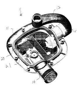
Fig. 1 is a perspective view of a dental x-ray tube head showing the top
portion of the main housing removed.
Fig. 2 is a close-up, perspective view of one portion of the tube head'of Fig.
1.
Fib. 3 is a front perspective view of an x-ray apparatus incorporating the
concepts of the present invention.
PREFERRED EMBODIMENTS FOR CARRYING OUT THE INVENTION
A dental x-ray apparatus according to the concepts of the present invention is
shown by way of example by the number 10 on the attached drawings. Dental x-
ray
apparatus 10 includes a tube head 11 supported on a yoke 12. Yoke 12 is in
turn
supported by support apparatus 13. A control unit 14 is provided and may also
be
supported by yoke 12. A unique aspect of the present invention is that control
operations may also be preformed by control panel 15 located on yoke 12. It
should
be understood that control panel 15 may be placed anywhere in proximity to
tube
head 1 l, and may even be on tube head 11 itself. Any location for control
panel 15 in
close proximity tube head 11 is within the scope of the present invention. As
is
conventional, tube head 11 is provided with collimator 16. The operation of
the x-ray
apparatus is conventional except as otherwise noted herein. A conventional
dental x
ray tube head isshgwn by way of example in US Patent 4,157,476 which is hereby
incorporated by reference for such disclosure.
Tube head housing 11 may be fabricated in any shape or design. An
exemplary such tube had is shown by way of example on the accompanying
4
CA 02371788 2001-11-22
WO 00/72354 PCT/US00/14390
drawings. Tube head 11 is preferably fabricated from a cast of zinc material.
Zinc
has a sufficiently high atomic number, and hence x-ray attenuation
coefficient, as to
provide enough shielding to secondary x-rays, i.e. against radiation leakage,
without
need for additional shielding such as those made from lead. Further, zinc
lends itself
well to casting, so it is suitable for such parts as a housing for tube head
11 with
relatively thin and large walls. As is otherwise conventional, tube head 11
may be
used to contain a dielectric oil. Further, zinc has sufficient mechanical
properties to
make it useful for structural components of the tube head. Zinc is also fairly
inexpensive, is compact and has no inherent porosity. An additional advantage
of
zinc is that it is relatively lightweight.
As shown in Figs. 1 and 2, other components of the tube head 11 such as
carrier 20 which is used to support an x-ray tube 21 is preferably fabricated
from a
plastic material impregnated with a radiation absorber. One preferred
radiation
absorber is barium sulfite. Barium sulfite is known for use as a contrast
medium
swallowed by a patient during gastro-intestinal radiography and as an additive
to
concrete for enhancing the radiation-shielding properties of masonry. It is
generally
considered to be not hazardous to health. Barium sulfite is also used as a
charge
added to certain types of products to enhance or change their mechanical
properties.
In the present invention, barium sulfite is used as an additive to the
plastics of
constructive parts inside the tube head, such as the tube holder or carrier
20, for the
purpose of imparting radiation-shielding properties to otherwise radiation-
transparent
plastics. By making the x-ray tube holder out of a barium sulfite-charged
plastic, the
holder 20, which normally has a lead jacket, is free of such additional
shielding. It is
to appreciated~l~lead, a generally hazardous and polluting material need not
be
used. Further, holder 20 is a one part component, thus, eliminating the need
for a lead
jacket. Further, the potential of a high-voltage dielectric discharge is
reduced because
5
CA 02371788 2001-11-22
WO 00/72354 PCT/US00/14390
of the absence of a metallic conductive part (potentially with sharp edges),
in the
immediate vicinity of the high voltage tube 21.
Although otherwise conventional, is it preferred that the present x-ray
apparatus be powered by a DC power supply.
It should be apparent that the present invention provides a dental x-ray
apparatus carrying out the objects of the invention as set forth hereinabove.
The
invention has been exemplified about an with respect to the attached drawings,
without attempting to show all of the variations that will be readily apparent
to those
skilled in the art. The scope of the invention shall only the determined by
the
attached claims.
6