Note : Les descriptions sont présentées dans la langue officielle dans laquelle elles ont été soumises.
CA 02382848 2002-02-25
WO 01/16377 PCT/US00/23844
Diagnostics and Therapeutics for Osteoporosis
1. Background of the Invention
Osteoporosis
In 1993, osteoporosis was identified as "one of the leading diseases of
women" by Bernadine Healy, MD, then director of the National Institutes of
Health.
Complications following osteoporosis fractures are the fourth leading cause of
death for
women over the age of 65, following heart disease, cancer and stroke. It is
the leading
cause of disability in the United States and the most common cause of hip
fracture.
Twenty-five million Americans suffer from osteoporosis, of which 85% are
women. Type 1 osteoporosis, which is postmenopausal osteoporosis stemming from
loss of
estrogen, affects more than half of all women over 65 and has been detected in
as many as
90 percent of women over age 75. Type II or senile osteoporosis which is
strictly age
related, affects both men and women usually over the age of seventy. Type III,
the newest
classification affecting both sexes, is drug-induced, for example, by long-
term steroid
therapy, known to accelerate bone loss. Patient groups that receive long term
steroid
therapy include asthmatics (7 million over the age of 18 in the United States)
as well a
patients with rheumatoid arthritis or other autoimmune diseases. Type IV is
caused by an
underlying disease such as rheumatoid arthritis (prevalence of 1-2% in the
population).
Osteoporosis is responsible for a majority of the 1.5 million bone fractures
each year leading to disabilities costing 10 billion dollars in medical,
social and nursing-
home costs. Even in the best hands, 40% of patients 65 years of age or older
will not
survive two years following a hip fracture.
In 1991, one in three American women were 50 years or older. The baby
boom generation will begin to enter this age group in 1996. Because the
average woman
lives some thirty years after menopause, with present trends, osteoporosis
threatens to be
one of the biggest health threats of modern times.
Lifestyle can be a factor in onset of osteoporosis and in particular can be an
important factor in building and maintaining healthy bone mass to prevent
osteoporosis.
Currently, persons under 65 are more likely than their parents to have had a
sedentary
lifestyle, bad eating habits, increased alcohol and caffeine intake, and a
history of greater
medication associated with bone loss. It is also clear that there is a genetic
predisposition to
1
CA 02382848 2009-04-09
the development of osteoporosis (see WO 94/03633 for a discussion of genetic
factors in
osteoporosis..
It would therefore be useful to be able to identify early those individuals at
greatest risk for developing osteoporosis so that the individual can be
counseled to make
appropriate life style changes or institute other therapeutic interventions.
For example,
calcium supplements and exercise have been shown to be valuable preventive
factors if
used during a critical early age window. Hormone replacement therapy (HRT) has
also
been used successfully to combat osteoporosis occurring after menopause. HRT
may be of
greatest benefit if used early in the disease process before major bone loss
has occurred.
Since HRT has potentially serious side-effects, it would be useful for women
to known
their personal risk level for osteoporosis when making decisions about the use
of HRT
versus other interventions aimed at reducing the risk of developing
osteoporosis.
The following published patent applications describe a variety of methods
for diagnosing, monitoring and/or treating osteoporosis: WO 94/20615, WO
95/01995, WO
94/14844, EP93113604, W018809457, W093/11149 and W019403633. The following
references describe the association of various IL-I gene polymorphisms in
osteoporosis:
U.S. Patent No. 5,698,399; Eastell, R. et al., (1998) Bone 23 (5S): S375;
Eastell, R. et al.
and Keen, RW et al., (1998) Bone 23: 367-371.
Gen _r the LL- I Gene Chu
t r
The IL-1 gene cluster is on the long arm of chromosome 2 (2g13) and
contains at least the genes for IL- I a (IL- I A), IL-1 P (IL-I B), and the IL-
I receptor
antagonist (IL-IRN), within a region of 430 Kb (Nicklin, et al. (1994)
Genomics, 19: 382-
4). The agonist molecules, IL-la and IL-1(3, have potent pro-inflammatory
activity and are
at the head of many inflammatory cascades. Their actions, often via the
induction of other
cytokines such as IL-6 and IL-8, lead to activation and recruitment of
leukocytes into
damaged tissue, local production of vasoactive agents, fever response in the
brain and
hepatic acute phase response. All three IL-1 molecules bind to type I and to
type II IL-1
receptors, but only the type I receptor transduces a signal to the interior of
the cell. In
contrast, the type II receptor is shed from the cell membrane and acts as a
decoy receptor.
The receptor antagonist and the type II receptor, therefore, are both anti-
inflammatory in
their actions.
Inappropriate production of IL-1 plays a central role in the pathology of
many autoimmune and inflammatory diseases, including rheumatoid arthritis,
inflammatory
2
CA 02382848 2009-04-09
bowel disorder, psoriasis, and the like. In addition, there are stable inter-
individual
differences in the rates of production of IL-1, and some of this variation may
be accounted
for by genetic differences at IL-1 gene loci. Thus, the IL-1 genes are
reasonable candidates
for determining part of the genetic susceptibility to inflammatory diseases,
most of which
have a multifactorial etiology with a polygenic component.
Certain alleles from the IL-I gene cluster are known to be associated with
particular disease states. For example, IL-1RN (VNTR) allele 2 (U.S. Patent
No.
5,698,399) and IL-1RN (VNTR) allele 1 (Keen RW et al., (1998) Bone 23:367-371)
have
been reported to be associated with osteoporosis. Further IL-IRN (VNTR) allele
2 has
been reported to be associated with nephropathy in diabetes mellitus
(Blakemore, et al.
(1996) Hum. Genet. 97(3): 369-74), alopecia areata (Cork, et al., (1995) J.
Invest.
Dermatol. 104(5 Supp.): 15S-16S; Cork et al. (1996) Derinatol Clin 14: 671-8),
Graves
disease (Blakemore, et al. (1995) J. Clin. EndocrinoL 80(1): 111-5), systemic
lupus
erythematosus (Blakemore, et al. (1994) Arthritis Rheum. 37: 1380-85), lichen
sclerosis
(Clay, et al. (1994) Hum. Genet. 94: 407-10), an4 ulcerative colitis
(Mansfield, et al.
(1994) Gastoenterol. 106(3): 637-42)).
It addition; theIL-iA alltle 2 from tnarker -889 iud II,-IB (Tag1) all e 2
from marker +3954 have been found to be associated with periodontal disease
(U.S. Patent
No. 5,686,246; Kornman and diGiovine (1998) Ann Periodont 3: 327-38; Hart and
Kornman (1997) Periodontol 2000 14: 202-15; Newman (1997) Compend Contin Educ
Dent 18: 881-4; Kornman et al. (1997) J. Clin Periodontol 24: 72-77). The IL-
IA allele 2
from marker -889 has also been found to be associated with juvenile chronic
arthritis,
particularly chronic iridocyclitis (McDowell, et al. (1995) Arthritis Rheum.
38: 221-28 ).
The IL-1B (Tagl) allele 2 from marker +3954 of IL-1B has also been found to be
associated
with psoriasis and insulin dependent diabetes in DR3/4 patients (di Giovine,
et al. (1995)
Cytokine 7: 606; Pociot, et al. (1992) Eur J. Clin. Invest. 22: 396-402).
Additionally, the
IL-1RN (VNTR) allele 1 has been found to be associated with diabetic
retinopathy (see
U.S. 6,713,253 B1 and PCT/GB97/02790). Furthermore allele 2 of IL-1RN (VNTR)
has
been found to be associated with ulcerative colitis in Caucasian populations
from North
America and Europe (Mansfield, J. et al., (1994) Gastroenterology 106: 637-
42).
Interestingly, this association is particularly strong within populations of
ethnically related
Ashkenazi Jews (PCT W097/25445).
3
CA 02382848 2002-02-25
WO 01/16377 PCTIUSOO/23844
Genotype Screening
Traditional methods for the screening of heritable diseases have depended on
either the identification of abnormal gene products (e.g., sickle cell anemia)
or an abnormal
phenotype (e.g., mental retardation). These methods are of limited utility for
heritable
diseases with late onset and no easily identifiable phenotypes such as, for
example, vascular
disease. With the development of simple and inexpensive genetic screening
methodology,
it is now possible to identify polymorphisms that indicate a propensity to
develop disease,
even when the disease is of polygenic origin. The number of diseases that can
be screened
by molecular biological methods continues to grow with increased understanding
of the
genetic basis of multifactorial disorders.
Genetic screening (also called genotyping or molecular screening), can be
broadly defined as testing to determine if a patient has mutations (alleles or
polymorphisms) that either cause a disease state or are "linked" to the
mutation causing a
disease state. Linkage refers to the phenomenon th DNA sequences which are
close
together in the genome have a tendency to be inherited together. Two sequences
may be
linked because of some selective advantage of co-inheritance. More typically,
however,
two polymorphic sequences are co-inherited because of the relative infrequency
with which
meiotic recombination events occur within the region between the two
polymorphisms.
The co-inherited polymorphic alleles are said to be in linkage disequilibrium
with one
another because, in a given human population, they tend to either both occur
together or
else not occur at all in any particular member of the population. Indeed,
where multiple
polymorphisms in a given chromosomal region are found to be in linkage
disequilibrium
with one another, they define a quasi-stable genetic "haplotype." In contrast,
recombination
events occurring between two polymorphic loci cause them to become separated
onto
distinct homologous chromosomes. If meiotic recombination between two
physically
linked polymorphisms occurs frequently enough, the two polymorphisms will
appear to
segregate independently and are said to be in linkage equilibrium.
While the frequency of meiotic recombination between two markers is
generally proportional to the physical distance between them on the
chromosome, the
occurrence of "hot spots" as well as regions of repressed chromosomal
recombination can
result in discrepancies between the physical and recombinational distance
between two
4
CA 02382848 2002-02-25
WO 01/16377 PCT/US00/23844
markers. Thus, in certain chromosomal regions, multiple polymorphic loci
spanning a
broad chromosomal domain may be in linkage disequilibrium with one another,
and
thereby define a broad-spanning genetic haplotype. Furthermore, where a
disease-causing
mutation is found within or in linkage with this haplotype, one or more
polymorphic alleles
of the haplotype can be used as a diagnostic or prognostic indicator of the
likelihood of
developing the disease. This association between otherwise benign
polymorphisms and a
disease-causing polymorphism occurs if the disease mutation arose in the
recent past, so
that sufficient time has not elapsed for equilibrium to be achieved through
recombination
events. Therefore identification of a human haplotype which spans or is linked
to a disease-
causing mutational change, serves as a predictive measure of an individual's
likelihood of
having inherited that disease-causing mutation. Importantly, such prognostic
or diagnostic
procedures can be utilized without necessitating the identification and
isolation of the actual
disease-causing lesion. This is significant because the precise determination
of the
molecular defect involved in a disease process can be difficult and laborious,
especially in
the case of multifactorial diseases such as inflammatory disorders.
Indeed, the statistical correlation between an inflammatory disorder and an
IL-1 polymorphism does not necessarily indicate that the polymorphism directly
causes the
disorder. Rather the correlated polymorphism may be a benign allelic variant
which is
linked to (i.e. in linkage disequilibrium with) a disorder-causing mutation
which has
occurred in the recent human evolutionary past, so that sufficient time has
not elapsed for
equilibrium to be achieved through recombination events in the intervening
chromosomal
segment. Thus, for the purposes of diagnostic and prognostic assays for a
particular
disease, detection of a polymorphic allele associated with that disease can be
utilized
without consideration of whether the polymorphism is directly involved in the
etiology of
the disease. Furthermore, where a given benign polymorphic locus is in linkage
disequilibrium with an apparent disease-causing polymorphic locus, still other
polymorphic
loci which are in linkage disequilibrium with the benign polymorphic locus are
also likely
to be in linkage disequilibrium with the disease-causing polymorphic locus.
Thus these
other polymorphic loci will also be prognostic or diagnostic of the likelihood
of having
inherited the disease-causing polymorphic locus. Indeed, a broad-spanning
human
haplotype (describing the typical pattern of co-inheritance of alleles of a
set of linked
polymorphic markers) can be targeted for diagnostic purposes once an
association has been
5
CA 02382848 2002-02-25
WO 01/16377 PCT/US00/23844
drawn between a particular disease or condition and a corresponding human
haplotype.
Thus, the determination of an individual's likelihood for developing a
particular disease of
condition can be made by characterizing one or more disease-associated
polymorphic
alleles (or even one or more disease-associated haplotypes) without
necessarily determining
or characterizing the causative genetic variation.
2. Summary of the Invention
In one aspect, the present invention provides novel methods and kits for
determining whether a female subject is predisposed to developing
osteoporosis,
comprising identifying the IL-1 haplotype pattern of the female, wherein the
presence of
haplotype pattern 1 indicates that the female is susceptible to larger bone
loss and/or
increased risk of fracture during the early menopausal years and the presence
of haplotype
pattern 2 indicates that the female is susceptible to larger bone loss and/or
increased risk of
fracture during post-menopause .
IL-1 haplotype patterns can be identified by detecting any of the component
alleles using any of a variety of available techniques, including: 1)
performing a
hybridization reaction between a nucleic acid sample and a probe that is
capable of
hybridizing to the allele; 2) sequencing at least a portion of the allele; or
3) determining the
electrophoretic mobility of the allele or fragments thereof (e.g., fragments
generated by
endonuclease digestion). The allele can optionally be subjected to an
amplification step
prior to performance of the detection step. Preferred amplification methods
are selected
from the group consisting of: the polymerase chain reaction (PCR), the ligase
chain reaction
(LCR), strand displacement amplification (SDA), cloning, and variations of the
above (e.g.
RT-PCR and allele specific amplification). Oligonucleotides necessary for
amplification
may be selected, for example, from within the IL-1 gene loci, either flanking
the marker of
interest (as required for PCR amplification) or directly overlapping the
marker (as in ASO
hybridization). In a particularly preferred embodiment, the sample is
hybridized with a set
of primers, which hybridize 5' and 3' in a sense or antisense sequence to the
vascular
disease associated allele, and is subjected to a PCR amplification.
An allele may also be detected indirectly, e.g. by analyzing the protein
product encoded by the DNA. For example, where the marker in question results
in the
translation of a mutant protein, the protein can be detected by any of a
variety of protein
6
CA 02382848 2002-02-25
WO 01/16377 PCTIUSOO/23844
detection methods. Such methods include immunodetection and biochemical tests,
such as
size fractionation, where the protein has a change in apparent molecular
weight either
through truncation, elongation, altered folding or altered post-translational
modifications.
In another aspect, the invention features kits for performing the above-
described assays. The kit can include a nucleic acid sample collection means
and a means
for determining whether a subject carries at least one allele comprising an IL-
1 haplotype.
The kit may also contain a control sample either positive or negative or a
standard and/or an
algorithmic device for assessing the results and additional reagents and
components
including: DNA amplification reagents, DNA polymerase, nucleic acid
amplification
reagents, restrictive enzymes, buffers, a nucleic acid sampling device, DNA
purification
device, deoxynucleotides, oligonucleotides (e.g. probes and primers) etc..
As described above, the control may be a positive or negative control.
Further, the control sample may contain the positive (or negative) products of
the allele
detection technique employed. For example, where the allele detection
technique is PCR
amplification, followed by size fractionation, the control sample may comprise
DNA
fragments of the appropriate size. Likewise, where the allele detection
technique involves
detection of a mutated protein, the control sample may comprise a sample of
mutated
protein. However, it is preferred that the control sample comprises the
material to be
tested. For example, the controls may be a sample of genomic DNA or a cloned
portion of
the IL-1 gene cluster. Preferably, however, the control sample is a highly
purified sample
of genomic DNA where the sample to be tested is genomic DNA.
The oligonucleotides present in said kit may be used for amplification of the
region of interest or for direct allele specific oligonucleotide (ASO)
hybridization to the
markers in question. Thus, the oligonucleotides may either flank the marker of
interest (as
required for PCR amplification) or directly overlap the marker (as in ASO
hybridization).
Information obtained using the assays and kits described herein (alone or in
conjunction with information on another genetic defect or environmental
factor, which
contributes to osteoporosis) is useful for determining whether a non-
symptomatic subject
has or is likely to develop the particular disease or condition. In addition,
the information
can allow a more customized approach to preventing the onset or progression of
the disease
or condition. For example, this information can enable a clinician to more
effectively
prescribe a therapy that will address the molecular basis of the disease or
condition.
7
CA 02382848 2002-02-25
WO 01/16377 PCT/US00/23844
In yet a further aspect, the invention features methods for treating or
preventing osteoporosis in a subject by administering to the subject an
appropriate
therapeutic of the invention. In still another aspect, the invention provides
in vitro or in
vivo assays for screening test compounds to identify therapeutics for treating
or preventing
the development of osteoporosis. In one embodiment, the assay comprises
contacting a cell
transfected with a causative mutation that is operably linked to an
appropriate promoter
with a test compound and determining the level of expression of a protein in
the cell in the
presence and in the absence of the test compound. In a preferred embodiment,
the causative
mutation results in decreased production of IL-1 receptor antagonist, and
increased
production of the IL-1 receptor antagonist in the presence of the test
compound indicates
that the compound is an agonist of IL-1 receptor antagonist activity. In
another preferred
embodiment, the causative mutation results in increased production of IL-1 a
or IL-10 , and
decreased production of IL-la or IL-1R in the presence of the test compound
indicates that
the compound is an antagonist of IL-1a or IL-1 3 activity. In another
embodiment, the
invention features transgenic non-human animals and their use in identifying
antagonists of
IL-1 a or IL-1(3 activity or agonists of IL-1 Ra activity.
Other embodiments and advantages of the invention are set forth in the
following detailed description and claims.
3. Brief Description of the Figures
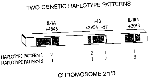
FIG. 1 shows two different genetic haplotype patterns.
FIG. 2 is a graph showing the risk of osteoporotic non-spine fractures.
FIG. 3 is a graph showing the risk of osteoporotic hip fractures
FIG. 4 is a graph showing the risk of osteoporotic wrist fractures
FIG. 5 is a graph showing the risk of non-spine fractures
4. Detailed Description of the Invention
4.1 Definitions
For convenience, the meaning of certain terms and phrases employed in the
8
CA 02382848 2002-02-25
WO 01/16377 PCTIUSOO/23844
specification, examples, and appended claims is provided below.
The term "allele" refers to the different sequence variants found at different
polymorphic regions. For example, IL-1RN (VNTR) has at least five different
alleles. The
sequence variants may be single or multiple base changes, including without
limitation
insertions, deletions, or substitutions, or may be a variable number of
sequence repeats.
The term "allelic pattern" refers to the identity of an allele or alleles at
one or
more polymorphic regions. For example, an allelic pattern may consist of a
single allele at
a polymorphic site, as for IL-1RN (VNTR) allele 1, which is an allelic pattern
having at
least one copy of IL-1RN allele 1 at the VNTR of the IL-1RN gene loci.
Alternatively, an
allelic pattern may consist of either a homozygous or heterozygous state at a
single
polymorphic site. For example, IL1-RN (VNTR) allele 2,2 is an allelic pattern
in which
there are two copies of the second allele at the VNTR marker of IL-1RN that
corresponds to
the homozygous IL-RN (VNTR) allele 2 state. Alternatively, an allelic pattern
may consist
of the identity of alleles at more than one polymorphic site.
The term "antibody " as used herein is intended to refer to a binding agent
including a whole antibody or a binding fragment thereof which is specifically
reactive with
an IL-1 polypeptide. Antibodies can be fragmented using conventional
techniques and the
fragments screened for utility in the same manner as described above for whole
antibodies.
For example, F(ab)2 fragments can be generated by treating an antibody with
pepsin. The
resulting F(ab)2 fragment can be treated to reduce disulfide bridges to
produce Fab
fragments. The antibody of the present invention is further intended to
include bispecific,
single-chain, and chimeric and humanized molecules having affinity for an IL-
lB
polypeptide conferred by at least one CDR region of the antibody.
"Biological activity" or "bioactivity" or "activity" or "biological function",
which are used interchangeably, for the purposes herein means an effector or
antigenic
function that is directly or indirectly performed by an IL-1 polypeptide
(whether in its
native or denatured conformation), or by any subsequence thereof. Biological
activities
include binding to a target peptide, e.g., an IL-1 receptor. An IL-1
bioactivity can be
modulated by directly affecting an IL-1 polypeptide. Alternatively, an IL-1
bioactivity can
be modulated by modulating the level of an IL-1 polypeptide, such as by
modulating
expression of an IL-1 gene.
As used herein the term "bioactive fragment of an IL-1 polypeptide" refers
9
CA 02382848 2002-02-25
WO 01/16377 PCT/US00/23844
to a fragment of a full-length IL-1 polypeptide, wherein the fragment
specifically mimics or
antagonizes the activity of a wild-type IL-1 polypeptide. The bioactive
fragment preferably
is a fragment capable of interacting with an interleukin receptor.
The term "an aberrant activity", as applied to an activity of a polypeptide
such as IL-1, refers to an activity which differs from the activity of the
wild-type or native
polypeptide or which differs from the activity of the polypeptide in a healthy
subject. An
activity of a polypeptide can be aberrant because it is stronger than the
activity of its native
counterpart. Alternatively, an activity can be aberrant because it is weaker
or absent
relative to the activity of its native counterpart. An aberrant activity can
also be a change in
an activity. For example an aberrant polypeptide can interact with a different
target
peptide. A cell can have an aberrant IL-1 activity due to overexpression or
underexpression
of an IL-1 locus gene encoding an IL-1 locus polypeptide.
"Cells", "host cells" or "recombinant host cells" are terms used
interchangeably herein to refer not only to the particular subject cell, but
to the progeny or
potential progeny of such a cell. Because certain modifications may occur in
succeeding
generations due to either mutation or environmental influences, such progeny
may not, in
fact be identical to the parent cell, but are still included within the scope
of the term as used
herein.
A "chimera," "mosaic," "chimeric mammal" and the like, refers to a
transgenic mammal with a knock-out or knock-in construct in at least some of
its genome-
containing cells.
The terms "control" or "control sample" refer to any sample appropriate to
the detection technique employed. The control sample may contain the products
of the
allele detection technique employed or the material to be tested. Further, the
controls may
be positive or negative controls. By way of example, where the allele
detection technique
is PCR amplification, followed by size fractionation, the control sample may
comprise
DNA fragments of an appropriate size. Likewise, where the allele detection
technique
involves detection of a mutated protein, the control sample may comprise a
sample of a
mutant protein. However, it is preferred that the control sample comprises the
material to
be tested. For example, the controls may be a sample of genomic DNA or a
cloned portion
of the IL-1 gene cluster. However, where the sample to be tested is genomic
DNA, the
control sample is preferably a highly purified sample of genomic DNA.
CA 02382848 2002-02-25
WO 01/16377 PCT/US0O/23844
The phrases "disruption of the gene" and "targeted disruption" or any similar
phrase refers to the site specific interruption of a native DNA sequence so as
to prevent
expression of that gene in the cell as compared to the wild-type copy of the
gene. The
interruption may be caused by deletions, insertions or modifications to the
gene, or any
combination thereof.
The term "haplotype" as used herein is intended to refer to a set of alleles
that are inherited together as a group (are in linkage disequilibrium) at
statistically
significant levels (p.o,, < 0.05). As used herein, the phrase "an IL-1
haplotype" refers to a
haplotype in the IL-1 loci. An IL-1 inflammatory or proinflammatory haplotype
refers to a
haplotype that is indicative of increased agonist and/or decreased antagonist
activities.
The terms "IL-1 gene cluster" and "IL-1 loci" as used herein include all the
nucleic acid at or near the 2g13 region of chromosome 2, including at least
the IL-IA, IL-
1B and IL-1RN genes and any other linked sequences. (Nicklin et al., Genomics
19: 382-
84, 1994). The terms "IL- l A", "IL-1 B", and "IL-1 RN" as used herein refer
to the genes
coding for IL-1 , IL-1 , and IL-1 receptor antagonist, respectively. The gene
accession
number for IL-IA, IL-1B, and IL-1RN are X03833, X04500, and X64532,
respectively.
"IL-1 functional mutation" refers to a mutation within the IL-1 gene cluster
that results in an altered phenotype (i.e. effects the function of an IL-1
gene or protein).
Examples include: IL-1A(+4845) allele 2, IL-1B (+3954) allele 2, IL-lB (+6912)
allele 2
and IL-1RN (+2018) allele 2.
"IL-1X (Z) allele Y " refers to a particular allelic form, designated Y,
occurring at an IL-1 locus polymorphic site in gene X, wherein X is IL-lA, B,
or RN and
positioned at or near nucleotide Z, wherein nucleotide Z is numbered relative
to the major
transcriptional start site, which is nucleotide +1, of the particular IL-1
gene X. As further
used herein, the term "IL-1X allele (Z)" refers to all alleles of an IL-1
polymorphic site in
gene X positioned at or near nucleotide Z. For example, the term "IL-lRN
(+2018) allele"
refers to alternative forms of the IL-1RN gene at marker +2018. "IL-lRN
(+2018) allele 1"
refers to a form of the IL-1RN gene which contains a cytosine (C) at position
+2018 of the
sense strand. Clay et al., Hum. Genet. 97:723-26, 1996. "IL-iRN (+2018) allele
2" refers
to a form of the IL-1RN gene which contains a thymine (T) at position +2018 of
the plus
strand. When a subject has two identical IL-1RN alleles, the subject is said
to be
homozygous, or to have the homozygous state. When a subject has two different
IL-1RN
11
CA 02382848 2002-02-25
WO 01/16377 PCT/US00/23844
alleles, the subject is said to be heterozygous, or to have the heterozygous
state. The term
"IL-1RN (+2018) allele 2,2" refers to the homozygous IL-1 RN (+2018) allele 2
state.
Conversely, the term "IL-1RN (+2018) allele 1,1" refers to the homozygous IL-1
RN
(+2018) allele 1 state. The term "IL-1RN (+2018) allele 1,2" refers to the
heterozygous
allele I and 2 state.
"IL-1 related" as used herein is meant to include all genes related to the
human IL-1 locus genes on human chromosome 2 (2q 12-14). These include IL-1
genes of
the human IL-1 gene cluster located at chromosome 2 (2q 13-14) which include:
the IL-IA
gene which encodes interleukin-1 a, the IL-1B gene which encodes interleukin-
1(3, and the
IL-1 RN (or IL-Ira) gene which encodes the interleukin- 1 receptor antagonist.
Furthermore
these IL-i related genes include the type I and type II human IL-1 receptor
genes located on
human chromosome 2 (2g12) and their mouse homologs located on mouse chromosome
1
at position 19.5 cM. Interleukin-1 a, interleukin-113, and interleukin- 1 RN
are related in so
much as they all bind to IL-1 type I receptors, however only interleukin-la
and interleukin-
1(3 are agonist ligands which activate IL-l type I receptors, while
interleukin-1RN is a
naturally occurring antagonist ligand. Where the term "IL-1" is used in
reference to a gene
product or polypeptide, it is meant to refer to all gene products encoded by
the interleukin-1
locus on human chromosome 2 (2q 12-14) and their corresponding homologs from
other
species or functional variants thereof. The term IL-1 thus includes secreted
polypeptides
which promote an inflammatory response, such as IL-la and IL-1(i, as well as a
secreted
polypeptide which antagonize inflammatory responses, such as IL-1 receptor
antagonist and
the IL-1 type II (decoy) receptor.
An "IL-1 receptor" or "IL-1R" refers to various cell membrane bound
protein receptors capable of binding to and/or transducing a signal from an IL-
1 locus-
encoded ligand. The term applies to any of the proteins which are capable of
binding
interleukin- 1 (IL-1) molecules and, in their native configuration as
mammalian plasma
membrane proteins, presumably play a role in transducing the signal provided
by IL-1 to a
cell. As used herein, the term includes analogs of native proteins with IL-1-
binding or
signal transducing activity. Examples include the human and murine IL-1
receptors
described in U.S. Patent No. 4,968,607. The term "IL-1 nucleic acid" refers to
a nucleic
acid encoding an IL-1 protein.
An "IL-1 polypeptide" and "IL-1 protein" are intended to encompass
12
CA 02382848 2002-02-25
WO 01/16377 PCT/US00/23844
polypeptides comprising the amino acid sequence encoded by the IL-1 genomic
DNA
sequences shown in Figures 1, 2, and 3, or fragments thereof, and homologs
thereof and
include agonist and antagonist polypeptides.
"Increased risk" refers to a statistically higher frequency of occurrence of
the
disease or condition in an individual carrying a particular polymorphic allele
in comparison
to the frequency of occurrence of the disease or condition in a member of a
population that
does not carry the particular polymorphic allele.
The term "interact" as used herein is meant to include detectable
relationships or associations (e.g. biochemical interactions) between
molecules, such as
interactions between protein-protein, protein-nucleic acid, nucleic acid-
nucleic acid and
protein-small molecule or nucleic acid-small molecule in nature.
The term "isolated" as used herein with respect to nucleic acids, such as
DNA or RNA, refers to molecules separated from other DNAs, or RNAs,
respectively, that
are present in the natural source of the macromolecule. For example, an
isolated nucleic
acid encoding one of the subject IL-1 polypeptides preferably includes no more
than 10
kilobases (kb) of nucleic acid sequence which naturally immediately flanks the
IL-i gene in
genomic DNA, more preferably no more than 5kb of such naturally occurring
flanking
sequences, and most preferably less than 1.5kb of such naturally occurring
flanking
sequence. The term isolated as used herein also refers to a nucleic acid or
peptide that is
substantially free of cellular material, viral material, or culture medium
when produced by
recombinant DNA techniques, or chemical precursors or other chemicals when
chemically
synthesized. Moreover, an "isolated nucleic acid" is meant to include nucleic
acid
fragments which are not naturally occurring as fragments and would not be
found in the
natural state. The term "isolated" is also used herein to refer to
polypeptides which are
isolated from other cellular proteins and is meant to encompass both purified
and
recombinant polypeptides.
A "knock-in" transgenic animal refers to an animal that has had a modified
gene introduced into its genome and the modified gene can be of exogenous or
endogenous
origin.
A "knock-out" transgenic animal refers to an animal in which there is partial
or complete suppression of the expression of an endogenous gene (e.g, based on
deletion of
at least a portion of the gene, replacement of at least a portion of the gene
with a second
13
CA 02382848 2002-02-25
WO 01/16377 PCTIUS0O/23844
sequence, introduction of stop codons, the mutation of bases encoding critical
amino acids,
or the removal of an intron junction, etc.).
A "knock-out construct" refers to a nucleic acid sequence that can be used to
decrease or suppress expression of a protein encoded by endogenous DNA
sequences in a
cell. In a simple example, the knock-out construct is comprised of a gene,
such as the
IL-1RN gene, with a deletion in a critical portion of the gene, so that active
protein cannot
be expressed therefrom. Alternatively, a number of termination codons can be
added to the
native gene to cause early termination of the protein or an intron junction
can be
inactivated. In a typical knock-out construct, some portion of the gene is
replaced with a
selectable marker (such as the neo gene) so that the gene can be represented
as follows:
IL-1 RN 5'/neo/ IL-1 RN 3', where IL-1 RN5' and IL-1 RN 3', refer to genomic
or cDNA
sequences which are, respectively, upstream and downstream relative to a
portion of the IL-
1RN gene and where neo refers to a neomycin resistance gene. In another knock-
out
construct, a second selectable marker is added in a flanking position so that
the gene can be
represented as: IL- 1 RN/neo/IL- 1 RN/TK, where TK is a thymidine kinase gene
which can
be added to either the IL-1RN5' or the IL-1RN3' sequence of the preceding
construct and
which further can be selected against (i.e. is a negative selectable marker)
in appropriate
media. This two-marker construct allows the selection of homologous
recombination
events, which removes the flanking TK marker, from non-homologous
recombination
events which typically retain the TK sequences. The gene deletion and/or
replacement can
be from the exons, introns, especially intron junctions, and/or the regulatory
regions such as
promoters.
"Linkage disequilibrium" refers to co-inheritance of two alleles at
frequencies greater than would be expected from the separate frequencies of
occurrence of
each allele in a given control population. The expected frequency of
occurrence of two
alleles that are inherited independently is the frequency of the first allele
multiplied by the
frequency of the second allele. Alleles that co-occur at expected frequencies
are said to be
in "linkage disequilibrium". The cause of linkage disequilibrium is often
unclear. It can be
due to selection for certain allele combinations or to recent admixture of
genetically
heterogeneous populations. In addition, in the case of markers that are very
tightly linked
to a disease gene, an association of an allele (or group of linked alleles)
with the disease
gene is expected if the disease mutation occurred in the recent past, so that
sufficient time
14
CA 02382848 2002-02-25
WO 01/16377 PCT/US00/23844
has not elapsed for equilibrium to be achieved through recombination events in
the specific
chromosomal region. When referring to allelic patterns that are comprised of
more than
one allele, a first allelic pattern is in linkage disequilibrium with a second
allelic pattern if
all the alleles that comprise the first allelic pattern are in linkage
disequilibrium with at least
one of the alleles of the second allelic pattern. An example of linkage
disequilibrium is that
which occurs between the alleles at the IL-1RN (+2018) and IL-1RN (VNTR)
polymorphic
sites. The two alleles at IL-1RN (+2018) are 100% in linkage disequilibrium
with the two
most frequent alleles of IL-1RN (VNTR), which are allele 1 and allele 2.
The term "marker" refers to a sequence in the genome that is known to vary
among individuals. For example, the IL-1RN gene has a marker that consists of
a variable
number of tandem repeats (VNTR).
A "mutated gene" or "mutation" or "functional mutation" refers to an allelic
form of a gene, which is capable of altering the phenotype of a subject having
the mutated
gene relative to a subject which does not have the mutated gene. The altered
phenotype
caused by a mutation can be corrected or compensated for by certain agents. If
a subject
must be homozygous for this mutation to have an altered phenotype, the
mutation is said to
be recessive. If one copy of the mutated gene is sufficient to alter the
phenotype of the
subject, the mutation is said to be dominant. If a subject has one copy of the
mutated gene
and has a phenotype that is intermediate between that of a homozygous and that
of a
heterozygous subject (for that gene), the mutation is said to be co-dominant.
A "non-human animal" of the invention includes mammals such as rodents,
non-human primates, sheep, dogs, cows, goats, etc. amphibians, such a s
members of the
Xenopus genus, and transgenic avians (e.g. chickens, birds, etc.). The term
"chimeric
animal" is used herein to refer to animals in which the recombinant gene is
found, or in
which the recombinant gene is expressed in some but not all cells of the
animal. The term
"tissue-specific chimeric animal" indicates that one of the recombinant IL-1
genes is
present and/or expressed or disrupted in some tissues but not others. The term
"non-human
mammal" refers to any member of the class Mammalia, except for humans.
As used herein, the term "nucleic acid" refers to polynucleotides or
oligonucleotides such as deoxyribonucleic acid (DNA), and, where appropriate,
ribonucleic
acid (RNA). The term should also be understood to include, as equivalents,
analogs of
either RNA or DNA made from nucleotide analogs (e.g. peptide nucleic acids)
and as
CA 02382848 2002-02-25
WO 01/16377 PCT/US00/23844
applicable to the embodiment being described, single (sense or antisense) and
double-
stranded polynucleotides.
The term "osteoporosis" is defined by the World Health Organization as "...a
systemic skeletal disease characterized by low bone mass and micro-
architectural
deterioration of bone tissue, with a consequent increase in bone fragility and
susceptibility
to fracture"(WHO Consensus Development Conference 1993). The clinical
definition of
osteoporosis is a condition in which the bone mineral density (BMD) or bone
mineral
concentration (BMC) is greater than about 2.5 standard deviations (SD) below
the mean of
young healthy women. Severe osteoporosis is defined as having a BMD or BMC
greater
than about 2.5 SD below the mean of young healthy women and the presence of
one or
more fragility fractures. Since bone loss is not strictly confined to specific
sites,
osteoporosis can manifest itself in various ways including alveolar, femoral,
radial,
vertebral or wrist bone loss or fracture incidence, postmenopausal bone loss,
severely
reduced bone mass, fracture incidence or rate of bone loss.
The term "polymorphism" refers to the coexistence of more than one form of
a gene or portion (e.g., allelic variant) thereof. A portion of a gene of
which there are at
least two different forms, i.e., two different nucleotide sequences, is
referred to as a
"polymorphic region of a gene". A specific genetic sequence at a polymorphic
region of a
gene is an allele. A polymorphic region can be a single nucleotide, the
identity of which
differs in different alleles. A polymorphic region can also be several
nucleotides long.
The term "propensity to disease," also "predisposition" or "susceptibility" to
disease or any similar phrase, means that certain alleles are hereby
discovered to be
associated with or predictive of a subject's incidence of developing a
particular disease (e.g.
a vascular disease). The alleles are thus over-represented in frequency in
individuals with
disease as compared to healthy individuals. Thus, these alleles can be used to
predict
disease even in pre-symptomatic or pre-diseased individuals.
"Small molecule" as used herein, is meant to refer to a composition, which
has a molecular weight of less than about 5kD and most preferably less than
about 4kD.
Small molecules can be nucleic acids, peptides, peptidomimetics,
carbohydrates, lipids or
other organic or inorganic molecules.
As used herein, the term "specifically hybridizes" or "specifically detects"
refers to the ability of a nucleic acid molecule to hybridize to at least
approximately 6
consecutive nucleotides of a sample nucleic acid.
16
CA 02382848 2002-02-25
WO 01/16377 PCT/US00/23844
"Transcriptional regulatory sequence" is a generic term used throughout the
specification to refer to DNA sequences, such as initiation signals,
enhancers, and
promoters, which induce or control transcription of protein coding sequences
with which
they are operably linked.
As used herein, the term "transgene" means a nucleic acid sequence
(encoding, e.g., one of the IL-1 polypeptides, or an antisense transcript
thereto) which has
been introduced into a cell. A transgene could be partly or entirely
heterologous, i.e.,
foreign, to the transgenic animal or cell into which it is introduced, or, is
homologous to an
endogenous gene of the transgenic animal or cell into which it is introduced,
but which is
designed to be inserted, or is inserted, into the animal's genome in such a
way as to alter the
genome of the cell into which it is inserted (e.g., it is inserted at a
location which differs
from that of the natural gene or its insertion results in a knockout). A
transgene can also be
present in a cell in the form of an episome. A transgene can include one or
more
transcriptional regulatory sequences and any other nucleic acid, such as
introns, that may be
necessary for optimal expression of a selected nucleic acid.
A "transgenic animal" refers to any animal, preferably a non-human
mammal, bird or an amphibian, in which one or more of the cells of the animal
contain
heterologous nucleic acid introduced by way of human intervention, such as by
transgenic
techniques well known in the art. The nucleic acid is introduced into the
cell, directly or
indirectly by introduction into a precursor of the cell, by way of deliberate
genetic
manipulation, such as by microinjection or by infection with a recombinant
virus. The term
genetic manipulation does not include classical cross-breeding, or in vitro
fertilization, but
rather is directed to the introduction of a recombinant DNA molecule. This
molecule may
be integrated within a chromosome, or it may be extrachromosomally replicating
DNA. In
the typical transgenic animals described herein, the transgene causes cells to
express a
recombinant form of one of an IL-1 polypeptide, e.g. either agonistic or
antagonistic forms.
However, transgenic animals in which the recombinant gene is silent are also
contemplated,
as for example, the FLP or CRE recombinase dependent constructs described
below.
Moreover, "transgenic animal" also includes those recombinant animals in which
gene
disruption of one or more genes is caused by human intervention, including
both
recombination and antisense techniques. The term is intended to include all
progeny
generations. Thus, the founder animal and all Fl, F2, F3, and so on, progeny
thereof are
17
CA 02382848 2002-02-25
WO 01/16377 PCT/US00/23844
included.
The term "treating" as used herein is intended to encompass curing as well as
ameliorating at least one symptom of a condition or disease.
The term "vector" refers to a nucleic acid molecule, which is capable of
transporting another nucleic acid to which it has been linked. One type of
preferred vector
is an episome, i.e., a nucleic acid capable of extra-chromosomal replication.
Preferred
vectors are those capable of autonomous replication and/or expression of
nucleic acids to
which they are linked. Vectors capable of directing the expression of genes to
which they
are operatively linked are referred to herein as "expression vectors". In
general, expression
vectors of utility in recombinant DNA techniques are often in the form of
"plasmids" which
refer generally to circular double stranded DNA loops which, in their vector
form are not
bound to the chromosome. In the present specification, "plasmid" and "vector"
are used
interchangeably as the plasmid is the most commonly used form of vector.
However, the
invention is intended to include such other forms of expression vectors which
serve
equivalent functions and which become known in the art subsequently hereto.
The term "wild-type allele" refers to an allele of a gene which, when present
in two copies in a subject results in a wild-type phenotype. There can be
several different
wild-type alleles of a specific gene, since certain nucleotide changes in a
gene may not
affect the phenotype of a subject having two copies of the gene with the
nucleotide changes.
4.2 Predictive Medicine
4.2.1. Identifying IL-2 Alleles and Haplotypes
The present invention is based at least in part, on the identification of
certain
alleles that have been determined to be association (to a statistically
significant extent) to
bone loss, fracture risk or other indicators of osteoporosis. Therefore,
detection of the
alleles can indicate that the subject has or is predisposed to the development
of
osteoporosis. However, because these alleles are in linkage disequilibrium
with other
alleles, the detection of such other linked alleles can also indicate that the
subject has or is
predisposed to the development of a particular disease or condition. For
example, the
44112332 haplotype comprises the following genotype:
18
CA 02382848 2002-02-25
WO 01/16377 PCT/US00/23844
allele 4 of the 222/223 marker of IL-lA
allele 4 of the gz5/gz6 marker of IL-IA
allele I of the -889 marker of IL-IA
allele 1 of the +3954 marker of IL-1B
allele 2 of the -511 marker of IL-1B
allele 3 of the gaat.p33330 marker
allele 3 of the Y31 marker
allele 2 of +2018 of IL-1RN
allele 2 of the VNTR marker of IL-1R
Three other polymorphisms in an IL-1RN alternative exon (Exon lic, which
produces an intracellular form of the gene product) are also in linkage
disequilibrium with
allele 2 of IL-1RN (VNTR) (Clay et al., (1996) Hum Genet 97:723-26). These
include: IL-
IRN exon lic (1812) (GenBank:X77090 at 1812); the IL-1RN exon lic (1868)
polymorphism (GenBank:X77090 at 1868); and the IL-1RN exon lic (1887)
polymorphism (GenBank:X77090 at 1887). Furthermore yet another polymorphism in
the
promoter for the alternatively spliced intracellular form of the gene, the Pic
(1731)
polymorphism (GenBank:X77090 at 1731), is also in linkage disequilibrium with
allele 2 of
the IL-I RN (VNTR) polymorphic locus. For each of these polymorphic loci, the
allele 2
sequence variant has been determined to be in linkage disequilibrium with
allele 2 of the
IL-1RN (VNTR) locus (Clay et al., (1996) Hum Genet 97:723-26).
The 33221461 haplotype comprises the following genotype:
allele 3 of the 222/223 marker of IL-IA
allele 3 of the gz5/gz6 marker of IL-IA
allele 2 of the -889 marker of IL-IA
allele 2 of the +3954 marker of IL-1B
allele 1 of the -511 marker of IL-1B
allele 4 of the gaat.p33330 marker
allele 6 of the Y31 marker
allele 1 of +2018 of IL-1RN
allele 1 of the VNTR marker of IL-1RN
Individuals with the 44112332 haplotype are typically overproducers of both
IL-1 a and IL-I (3 proteins, upon stimulation. In contrast, individuals with
the 33221461
haplotype are typically underproducers of IL-Ira. Each haplotype results in a
net
19
CA 02382848 2002-02-25
WO 01/16377 PCT/US00/23844
IL-la and IL-10 proteins, upon stimulation. In contrast, individuals with the
33221461
haplotype are typically underproducers of IL-lra. Each haplotype results in a
net
proinflammatory response. Each allele within a haplotype may have an effect,
as well as a
composite genotype effect. In addition, particular diseases may be associated
with both
haplotype patterns.
In addition to the allelic patterns described above, as described herein, one
of
skill in the art can readily identify other alleles (including polymorphisms
and mutations)
that are in linkage disequilibrium with an allele associated with
osteoporosis. For example,
a nucleic acid sample from a first group of subjects without osteoporosis can
be collected,
as well as DNA from a second group of subjects with the disorder. The nucleic
acid sample
can then be compared to identify those alleles that are over-represented in
the second group
as compared with the first group, wherein such alleles are presumably
associated with
osteoporosis. Alternatively, alleles that are in linkage disequilibrium with
an allele that is
associated with osteoporosis can be identified, for example, by genotyping a
large
population and performing statistical analysis to determine which alleles
appear more
commonly together than expected. Preferably the group is chosen to be
comprised of
genetically related individuals. Genetically related individuals include
individuals from the
same race, the same ethnic group, or even the same family. As the degree of
genetic
relatedness between a control group and a test group increases, so does the
predictive value
of polymorphic alleles which are ever more distantly linked to a disease-
causing allele.
This is because less evolutionary time has passed to allow polymorphisms which
are linked
along a chromosome in a founder population to redistribute through genetic
cross-over
events. Thus race-specific, ethnic-specific, and even family-specific
diagnostic genotyping
assays can be developed to allow for the detection of disease alleles which
arose at ever
more recent times in human evolution, e.g., after divergence of the major
human races, after
the separation of human populations into distinct ethnic groups, and even
within the recent
history of a particular family line.
Linkage disequilibrium between two polymorphic markers or between one
polymorphic marker and a disease-causing mutation is a meta-stable state.
Absent selective
pressure or the sporadic linked reoccurrence of the underlying mutational
events, the
polymorphisms will eventually become disassociated by chromosomal
recombination
events and will thereby reach linkage equilibrium through the course of human
evolution.
Thus, the likelihood of finding a polymorphic allele in linkage disequilibrium
with a
CA 02382848 2009-04-09
disease or condition may increase with changes in at least two factors:
decreasing physical
distance between the polymorphic marker and the disease-causing mutation, and
decreasing
number of meiotic generations available for the dissociation of the linked
pair.
Consideration of the latter factor suggests that, the more closely related two
individuals are,
the more likely they will share a common parental chromosome or chromosomal
region
containing the linked polymorphisms and the less likely that this linked pair
will have
become unlinked through meiotic cross-over events occurring each generation.
As a result,
the more closely related two individuals are, the more likely it is that
widely spaced
polymorphisms may be co-inherited. Thus, for individuals related by common
race,
ethnicity or family, the reliability of ever more distantly spaced polymorphic
loci can be
relied upon as an indicator of inheritance of a linked disease-causing
mutation.
Appropriate probes may be designed to hybridize to a specific gene of the
IL-1 locus, such as IL-IA, IL-1B or IL-1RN or a related gene. These genomic
DNA
sequences are shown in Figures 3, 4 and 5, respectively, and further
correspond to SEQ ID
Nos. 1, 2 and 3, respectively. Alternatively, these probes may incorporate
other regions of
the relevant genomic locus, including intergenic sequences. Indeed the IL-1
region of
l +omt~srtYe'2`1 'le pawand,'sutidiig average of one
single nucleotide polymorphism every 1,000 base pairs, includes some 400 SNPs
loci
alone. Yet other polymorphisms available for use with the immediate invention
are
obtainable from various public sources. For example, the human genome database
collects
intragenic SNPs, is searchable by sequence and currently contains
approximately 2,700
entries. Also available is a human polymorphism database maintained by the
Massachusetts Institute of Technology (MIT SNP database). From such sources
SNPs
as well as other human polymorphisms may be found.
For example, examination of the IL-1 region of the human genome in any
one of these databases reveals that the IL-1 locus genes are flanked by a
centromere
proximal polymorphic marker designated microsatellite marker AFM220ze3 at
127.4 cM
(centiMorgans) (see GenBank Acc. No. Z17OO8) and a distal polymorphic marker
designated microsatellite anchor marker AFMO87xal at 127.9 cM (see GenBank
Acc. No.
Z16545). These human polymorphic loci are both CA dinucleotide repeat
microsatellite
polymorphisms, and, as such, show a high degree of heterozygosity in human
populations.
21
CA 02382848 2002-02-25
WO 01/16377 PCT/US00/23844
For example, one allele of AFM220ze3 generates a 211 bp PCR amplification
product with
a 5' primer of the sequence TGTACCTAAGCCCACCCTTTAGAGC (SEQ ID No. 4) and a
3' primer of the sequence TGGCCTCCAGAAACCTCCAA (SEQ ID No. 5). Furthermore,
one allele of AFM087xal generates a 177 bp PCR amplification product with a 5'
primer of
the sequence GCTGATATTCTGGTGGGAAA (SEQ IDNo. 6) and a 3' primer of the
sequence GGCAAGAGCAAAACTCTGTC (SEQ ID No. 7). Equivalent primers
corresponding to unique sequences occurring 5' and 3' to these human
chromosome 2 CA
dinucleotide repeat polymorphisms will be apparent to one of skill in the art.
Reasonable
equivalent primers include those which hybridize within about 1 kb of the
designated
primer, and which further are anywhere from about 17 bp to about 27 bp in
length. A
general guideline for designing primers for amplification of unique human
chromosomal
genomic sequences is that they possess a melting temperature of at least about
50 C,
wherein an approximate melting temperature can be estimated using the formula
Tmelt = [2 x
(# ofAorT)+4x(#ofGorC)].
A number of other human polymorphic loci occur between these two CA
dinucleotide repeat polymorphisms and provide additional targets for
determination of a
prognostic allele in a family or other group of genetically related
individuals. For example,
the National Center for Biotechnology Information web site
(www.ncbi.nlm.nih.gov/genemap/) lists a number of polymorphism markers in the
region
of the IL-1 locus and provides guidance in designing appropriate primers for
amplification
and analysis of these markers.
Accordingly, the nucleotide segments of the invention may be used for their
ability to selectively form duplex molecules with complementary stretches of
human
chromosome 2 q 12-13 or cDNAs from that region or to provide primers for
amplification
of DNA or cDNA from this region. The design of appropriate probes for this
purpose
requires consideration of a number of factors. For example, fragments having a
length of
between 10, 15, or 18 nucleotides to about 20, or to about 30 nucleotides,
will find
particular utility. Longer sequences, e.g., 40, 50, 80, 90, 100, even up to
full length, are
even more preferred for certain embodiments. Lengths of oligonucleotides of at
least about
18 to 20 nucleotides are well accepted by those of skill in the art as
sufficient to allow
sufficiently specific hybridization so as to be useful as a molecular probe.
Furthermore,
depending on the application envisioned, one will desire to employ varying
conditions of
22
CA 02382848 2002-02-25
WO 01/16377 PCT/US00/23844
hybridization to achieve varying degrees of selectivity of probe towards
target sequence.
For applications requiring high selectivity, one will typically desire to
employ relatively
stringent conditions to form the hybrids. For example, relatively low salt
and/or high
temperature conditions, such as provided by 0.02 M-0.15M NaCl at temperatures
of about
50 C to about 70 C. Such selective conditions may tolerate little, if any,
mismatch
between the probe and the template or target strand.
Other alleles or other indicia of a disorder can be detected or monitored in a
subject in conjunction with detection of the alleles described above, for
example,
identifying vessel wall thickness (e.g. as measured by ultrasound), or whether
the subject
smokes, drinks is overweight, is under stress or exercises.
4.2.2 Detection of Alleles
Many methods are available for detecting specific alleles at human
polymorphic loci. The preferred method for detecting a specific polymorphic
allele will
depend, in part, upon the molecular nature of the polymorphism. For example,
the various
allelic forms of the polymorphic locus may differ by a single base-pair of the
DNA. Such
single nucleotide polymorphisms (or SNPs) are major contributors to genetic
variation,
comprising some 80% of all known polymorphisms, and their density in the human
genome
is estimated to be on average 1 per 1,000 base pairs. SNPs are most frequently
biallelic-
occurring in only two different forms (although up to four different forms of
an SNP,
corresponding to the four different nucleotide bases occurring in DNA, are
theoretically
possible). Nevertheless, SNPs are mutationally more stable than other
polymorphisms,
making them suitable for association studies in which linkage disequilibrium
between
markers and an unknown variant is used to map disease-causing mutations. In
addition,
because SNPs typically have only two alleles, they can be genotyped by a
simple
plus/minus assay rather than a length measurement, making them more amenable
to
automation.
A variety of methods are available for detecting the presence of a particular
single nucleotide polymorphic allele in an individual. Advancements in this
field have
provided accurate, easy, and inexpensive large-scale SNP genotyping. Most
recently, for
example, several new techniques have been described including dynamic allele-
specific
hybridization (DASH), microplate array diagonal gel electrophoresis (MADGE),
23
CA 02382848 2002-02-25
WO 01/16377 PCT/US00/23844
pyrosequencing, oligonucleotide-specific ligation, the TaqMan system as well
as various
DNA "chip" technologies such as the Affymetrix SNP chips. These methods
require
amplification of the target genetic region, typically by PCR. Still other
newly developed
methods, based on the generation of small signal molecules by invasive
cleavage followed
by mass spectrometry or immobilized padlock probes and rolling-circle
amplification,
might eventually eliminate the need for PCR. Several of the methods known in
the art for
detecting specific single nucleotide polymorphisms are summarized below. The
method of
the present invention is understood to include all available methods.
Several methods have been developed to facilitate analysis of single
nucleotide polymorphisms. In one embodiment, the single base polymorphism can
be
detected by using a specialized exonuclease-resistant nucleotide, as
disclosed, e.g., in
Mundy, C. R. (U.S. Pat. No.4,656,127). According to the method, a primer
complementary
to the allelic sequence immediately 3' to the polymorphic site is permitted to
hybridize to a
target molecule obtained from a particular animal or human. If the polymorphic
site on the
target molecule contains a nucleotide that is complementary to the particular
exonuclease-resistant nucleotide derivative present, then that derivative will
be incorporated
onto the end of the hybridized primer. Such incorporation renders the primer
resistant to
exonuclease, and thereby permits its detection. Since the identity of the
exonuclease-resistant derivative of the sample is known, a finding that the
primer has
become resistant to exonucleases reveals that the nucleotide present in the
polymorphic site
of the target molecule was complementary to that of the nucleotide derivative
used in the
reaction. This method has the advantage that it does not require the
determination of large
amounts of extraneous sequence data.
In another embodiment of the invention, a solution-based method is used for
determining the identity of the nucleotide of a polymorphic site. Cohen, D. et
al. (French
Patent 2,650,840; PCT Appln. No. W091/02087). As in the Mundy method of U.S.
Pat.
No. 4,656,127, a primer is employed that is complementary to allelic sequences
immediately 3' to a polymorphic site. The method determines the identity of
the nucleotide
of that site using labeled dideoxynucleotide derivatives, which, if
complementary to the
nucleotide of the polymorphic site will become incorporated onto the terminus
of the
primer.
An alternative method, known as Genetic Bit Analysis or GBA TM is
24
CA 02382848 2002-02-25
WO 01/16377 PCT/US00/23844
described by Goelet, P. et al. (PCT Appln. No. 92/15712). The method of
Goelet, P. et al.
uses mixtures of labeled terminators and a primer that is complementary to the
sequence 3'
to a polymorphic site. The labeled terminator that is incorporated is thus
determined by, and
complementary to, the nucleotide present in the polymorphic site of the target
molecule
being evaluated. In contrast to the method of Cohen et al. (French Patent
2,650,840; PCT
Appln. No. W091/02087) the method of Goelet, P. et al. is preferably a
heterogeneous
phase assay, in which the primer or the target molecule is immobilized to a
solid phase.
Recently, several primer-guided nucleotide incorporation procedures for
assaying polymorphic sites in DNA have been described (Komher, J. S. et al.,
Nucl. Acids.
Res. 17:7779-7784 (1989); Sokolov, B. P., Nucl. Acids Res. 18:3671 (1990);
Syvanen, A.
-C., et al., Genomics 8:684-692 (1990); Kuppuswamy, M. N. et al., Proc. Natl.
Acad. Sci.
(U.S.A.) 88:1143-1147 (1991); Prezant, T. R. et al., Hum. Mutat. 1:159-164
(1992);
Ugozzoli, L. et al., GATA 9:107-112 (1992); Nyren, P. et al., Anal. Biochem.
208:171-175
(1993)). These methods differ from GBA TM in that they all rely on the
incorporation of
labeled deoxynucleotides to discriminate between bases at a polymorphic site.
In such a
format, since the signal is proportional to the number of deoxynucleotides
incorporated,
polymorphisms that occur in runs of the same nucleotide can result in signals
that are
proportional to the length of the run (Syvanen, A. -C., et al., Amer. J. Hum.
Genet.
52:46-59 (1993)).
For mutations that produce premature termination of protein translation, the
protein truncation test (PTT) offers an efficient diagnostic approach (Roest,
et. al., (1993)
Hum. Mol. Genet. 2:1719-21; van der Luijt, et. al., (1994) Genomics 20:1-4).
For PTT,
RNA is initially isolated from available tissue and reverse-transcribed, and
the segment of
interest is amplified by PCR. The products of reverse transcription PCR are
then used as a
template for nested PCR amplification with a primer that contains an RNA
polymerase
promoter and a sequence for initiating eukaryotic translation. After
amplification of the
region of interest, the unique motifs incorporated into the primer permit
sequential in vitro
transcription and translation of the PCR products. Upon sodium dodecyl sulfate-
polyacrylamide gel electrophoresis of translation products, the appearance of
truncated
polypeptides signals the presence of a mutation that causes premature
termination of
translation. In a variation of this technique, DNA (as opposed to RNA) is used
as a PCR
template when the target region of interest is derived from a single exon.
CA 02382848 2009-04-09
Any cell type or tissue may be utilized to obtain nucleic acid samples for use
in the diagnostics described herein. In a preferred embodiment, the DNA sample
is
obtained from a bodily fluid, e.g, blood, obtained by known techniques (e.g.
venipuncture)
or saliva. Alternatively, nucleic acid tests can be performed on dry samples
(e.g. hair or
skin). When using RNA or protein, the cells or tissues that may be utilized
must express an
IL-1 gene.
Diagnostic procedures may also be performed in situ directly upon tissue
sections (fixed and/or frozen) of patient tissue obtained from biopsies or
resections, such
that no nucleic acid purification is necessary. Nucleic acid reagents may be
used as probes
and/or primers for such in situ procedures (see, for example, Nuovo, G.J.,
1992, PCR in situ
hybridization: protocols and applications, Raven Press, NY).
In addition to methods which focus primarily on the detection of one nucleic
acid sequence, profiles may also be assessed in such detection schemes.
Fingerprint
profiles may be generated, for example, by utilizing a differential display
procedure,
Northern analysis and/or RT-PCR.
A preferred detection method is allele specific hybridization using probes
overlapping a-region of t least one allele of an IL-1 iiroi latnnri atoty
haplotype and having
about 5, 10, 20, 25, or 30 nucleotides around the mutation or polymorphic
region. In a
preferred embodiment of the invention, several probes capable of hybridizing
specifically to
other allelic variants involved in osteoporosis are attached to a solid phase
support, e.g. a
"chip" (which can hold up to about 250,000 oligonucleotides). Oligonucleotides
can be
bound to a solid support by a variety of processes, including lithography.
Mutation
detection analysis using these chips comprising oligonucleotides, also termed
"DNA probe
arrays" is described e.g., in Cronin et al. (1996) Human Mutation 7:244. In
one
embodiment, a chip comprises all the allelic variants of at least one
polymorphic region of
a gene. The solid phase support is then contacted with a test nucleic acid and
hybridization
to the specific probes is detected. Accordingly, the identity of numerous
allelic variants of
one or more genes can be identified in a simple hybridization experiment.
These techniques may also comprise the step of amplifying the nucleic acid
before analysis. Amplification techniques are known to those of skill in the
art and include,
but are not limited to cloning, polymerase chain reaction (PCR), polymerase
chain reaction
of specific alleles (ASA), ligase chain reaction (LCR), nested polymerase
chain reaction,
26
CA 02382848 2002-02-25
WO 01/16377 PCTIUSOO/23844
self sustained sequence replication (Guatelli, J.C. et al., 1990, Proc. Natl.
Acad. Sci. USA
87:1874-1878), transcriptional amplification system (Kwoh, D.Y. et al., 1989,
Proc. Natl.
Acad. Sci. USA 86:1173-1177), and Q- Beta Replicase (Lizardi, P.M. et al.,
1988,
Bio/Technology 6:1197).
Amplification products may be assayed in a variety of ways, including size
analysis, restriction digestion followed by size analysis, detecting specific
tagged
oligonucleotide primers in the reaction products, allele-specific
oligonucleotide (ASO)
hybridization, allele specific 5' exonuclease detection, sequencing,
hybridization, and the
like.
PCR based detection means can include multiplex amplification of a
plurality of markers simultaneously. For example, it is well known in the art
to select PCR
primers to generate PCR products that do not overlap in size and can be
analyzed
simultaneously. Alternatively, it is possible to amplify different markers
with primers that
are differentially labeled and thus can each be differentially detected. Of
course,
hybridization based detection means allow the differential detection of
multiple PCR
products in a sample. Other techniques are known in the art to allow multiplex
analyses of
a plurality of markers.
In a merely illustrative embodiment, the method includes the steps of (i)
collecting a sample of cells from a patient, (ii) isolating nucleic acid
(e.g., genomic, mRNA
or both) from the cells of the sample, (iii) contacting the nucleic acid
sample with one or
more primers which specifically hybridize 5' and 3' to at least one allele of
an IL-1
proinflammatory haplotype under conditions such that hybridization and
amplification of
the allele occurs, and (iv) detecting the amplification product. These
detection schemes are
especially useful for the detection of nucleic acid molecules if such
molecules are present in
very low numbers.
In a preferred embodiment of the subject assay, the allele of an IL-1
proinflammatory haplotype is identified by alterations in restriction enzyme
cleavage
patterns. For example, sample and control DNA is isolated, amplified
(optionally), digested
with one or more restriction endonucleases, and fragment length sizes are
determined by gel
electrophoresis.
In yet another embodiment, any of a variety of sequencing reactions known
in the art can be used to directly sequence the allele. Exemplary sequencing
reactions
27
CA 02382848 2002-02-25
WO 01/16377 PCT/USOO/23844
include those based on techniques developed by Maxim and Gilbert ((1977) Proc.
Natl
Acad Sci USA 74:560) or Sanger (Sanger et al (1977) Proc. Nat. Acad. Sci USA
74:5463).
It is also contemplated that any of a variety of automated sequencing
procedures may be
utilized when performing the subject assays (see, for example Biotechniques
(1995)
19:448), including sequencing by mass spectrometry (see, for example PCT
publication
WO 94/16101; Cohen et al. (1996) Adv Chromatogr 36:127-162; and Griffin et al.
(1993)
Appl Biochem Biotechnol 38:147-159). It will be evident to one of skill in the
art that, for
certain embodiments, the occurrence of only one, two or three of the nucleic
acid bases
need be determined in the sequencing reaction. For instance, A-track or the
like, e.g.,
where only one nucleic acid is detected, can be carried out.
In a further embodiment, protection from cleavage agents (such as a
nuclease, hydroxylamine or osmium tetroxide and with piperidine) can be used
to detect
mismatched bases in RNA/RNA or RNA/DNA or DNA/DNA heteroduplexes (Myers, et
al.
(1985) Science 230:1242). In general, the art technique of "mismatch cleavage"
starts by
providing heteroduplexes formed by hybridizing (labeled) RNA or DNA containing
the
wild-type allele with the sample. The double-stranded duplexes are treated
with an agent
which cleaves single-stranded regions of the duplex such as which will exist
due to base
pair mismatches between the control and sample strands. For instance, RNA/DNA
duplexes can be treated with RNase and DNA/DNA hybrids treated with Si
nuclease to
enzymatically digest the mismatched regions. In other embodiments, either
DNA/DNA or
RNA/DNA duplexes can be treated with hydroxylamine or osmium tetroxide and
with
piperidine in order to digest mismatched regions. After digestion of the
mismatched
regions, the resulting material is then separated by size on denaturing
polyacrylamide gels
to determine the site of mutation. See, for example, Cotton et al (1988) Proc.
Natl Acad Sci
USA 85:4397; and Saleeba et al (1992) Methods Enzymol. 217:286-295. In a
preferred
embodiment, the control DNA or RNA can be labeled for detection.
In still another embodiment, the mismatch cleavage reaction employs one or
more proteins that recognize mismatched base pairs in double-stranded DNA (so
called
"DNA mismatch repair" enzymes). For example, the mutY enzyme of E. coli
cleaves A at
G/A mismatches and the thymidine DNA glycosylase from HeLa cells cleaves T at
G/T
mismatches (Hsu et al. (1994) Carcinogenesis 15:1657-1662). According to an
exemplary
embodiment, a probe based on an allele of an IL-1 locus haplotype is
hybridized to a cDNA
28
CA 02382848 2002-02-25
WO 01/16377 PCT/US00/23844
or other DNA product from a test cell(s). The duplex is treated with a DNA
mismatch
repair enzyme, and the cleavage products, if any, can be detected from
electrophoresis
protocols or the like. See, for example, U.S. Patent No. 5,459,039.
In other embodiments, alterations in electrophoretic mobility will be used to
identify an IL-i locus allele. For example, single strand conformation
polymorphism
(SSCP) may be used to detect differences in electrophoretic mobility between
mutant and
wild type nucleic acids (Orita et al. (1989) Proc Natl. Acad. Sci USA 86:2766,
see also
Cotton (1993) Mutat Res 285:125-144; and Hayashi (1992) Genet Anal Tech Appl
9:73-
79). Single-stranded DNA fragments of sample and control IL-1 locus alleles
are denatured
and allowed to renature. The secondary structure of single-stranded nucleic
acids varies
according to sequence, the resulting alteration in electrophoretic mobility
enables the
detection of even a single base change. The DNA fragments may be labeled or
detected
with labeled probes. The sensitivity of the assay may be enhanced by using RNA
(rather
than DNA), in which the secondary structure is more sensitive to a change in
sequence. In
a preferred embodiment, the subject method utilizes heteroduplex analysis to
separate
double stranded heteroduplex molecules on the basis of changes in
electrophoretic mobility
(Keen et al. (1991) Trends Genet 7:5).
In yet another embodiment, the movement of alleles in polyacrylamide gels
containing a gradient of denaturant is assayed using denaturing gradient gel
electrophoresis
(DGGE) (Myers et al. (1985) Nature 313:495). When DGGE is used as the method
of
analysis, DNA will be modified to insure that it does not completely denature,
for example
by adding a GC clamp of approximately 40 bp of high-melting GC-rich DNA by
PCR. In a
further embodiment, a temperature gradient is used in place of a denaturing
agent gradient
to identify differences in the mobility of control and sample DNA (Rosenbaum
and
Reissner (1987) Biophys Chem 265:12753).
Examples of other techniques for detecting alleles include, but are not
limited to, selective oligonucleotide hybridization, selective amplification,
or selective
primer extension. For example, oligonucleotide primers may be prepared in
which the
known mutation or nucleotide difference (e.g., in allelic variants) is placed
centrally and
then hybridized to target DNA under conditions which permit hybridization only
if a
perfect match is found (Saiki et al. (1986) Nature 324:163); Saiki et al
(1989) Proc. Natl
Acad. Sci USA 86:6230). Such allele specific oligonucleotide hybridization
techniques may
29
CA 02382848 2002-02-25
WO 01/16377 PCTIUSOO/23844
be used to test one mutation or polymorphic region per reaction when
oligonucleotides are
hybridized to PCR amplified target DNA or a number of different mutations or
polymorphic regions when the oligonucleotides are attached to the hybridizing
membrane
and hybridized with labelled target DNA.
Alternatively, allele specific amplification technology which depends on
selective PCR amplification may be used in conjunction with the instant
invention.
Oligonucleotides used as primers for specific amplification may carry the
mutation or
polymorphic region of interest in the center of the molecule (so that
amplification depends
on differential hybridization) (Gibbs et al (1989) Nucleic Acids Res. 17:2437-
2448) or at
the extreme 3' end of one primer where, under appropriate conditions, mismatch
can
prevent, or reduce polymerase extension (Prossner (1993) Tibtech 11:238. In
addition it
may be desirable to introduce a novel restriction site in the region of the
mutation to create
cleavage-based detection (Gasparini et al (1992) Mol. Cell Probes 6:1). It is
anticipated
that in certain embodiments amplification may also be performed using Taq
ligase for
amplification (Barany (1991) Proc. Natl. Acad. Sci USA 88:189). In such cases,
ligation
will occur only if there is a perfect match at the 3' end of the 5' sequence
making it possible
to detect the presence of a known mutation at a specific site by looking for
the presence or
absence of amplification.
In another embodiment, identification of the allelic variant is carried out
using an oligonucleotide ligation assay (OLA), as described, e.g., in U.S.
Pat. No.
4,998,617 and in Landegren, U. et al. ((1988) Science 241:1077-1080). The OLA
protocol
uses two oligonucleotides which are designed to be capable of hybridizing to
abutting
sequences of a single strand of a target. One of the oligonucleotides is
linked to a separation
marker, e.g,. biotinylated, and the other is detectably labeled. If the
precise complementary
sequence is found in a target molecule, the oligonucleotides will hybridize
such that their
termini abut, and create a ligation substrate. Ligation then permits the
labeled
oligonucleotide to be recovered using avidin, or another biotin ligand.
Nickerson, D. A. et
al. have described a nucleic acid detection assay that combines attributes of
PCR and OLA
(Nickerson, D. A. et al. (1990) Proc. Natl. Acad. Sci. USA 87:8923-27). In
this method,
PCR is used to achieve the exponential amplification of target DNA, which is
then detected
using OLA.
Several techniques based on this OLA method have been developed and can
CA 02382848 2002-02-25
WO 01/16377 PCTIUSOO/23844
be used to detect alleles of an IL-1 locus haplotype. For example, U.S. Patent
No.
5,593,826 discloses an OLA using an oligonucleotide having 3'-amino group and
a
5'-phosphorylated oligonucleotide to form a conjugate having a phosphoramidate
linkage.
In another variation of OLA described in Tobe et al. ((1996) Nucleic Acids Res
24: 3728),
OLA combined with PCR permits typing of two alleles in a single microtiter
well. By
marking each of the allele-specific primers with a unique hapten, i.e.
digoxigenin and
fluorescein, each OLA reaction can be detected by using hapten specific
antibodies that are
labeled with different enzyme reporters, alkaline phosphatase or horseradish
peroxidase.
This system permits the detection of the two alleles using a high throughput
format that
leads to the production of two different colors.
Another embodiment of the invention is directed to kits for detecting a
predisposition for developing a restenosis. This kit may contain one or more
oligonucleotides, including 5' and 3' oligonucleotides that hybridize 5' and
3' to at least
one allele of an IL-1 locus haplotype. PCR amplification oligonucleotides
should hybridize
between 25 and 2500 base pairs apart, preferably between about 100 and about
500 bases
apart, in order to produce a PCR product of convenient size for subsequent
analysis.
Particularly preferred primers for use in the diagnostic method of the
invention include SEQ ID Nos. 1-6
The design of additional oligonucleotides for use in the amplification and
detection of IL-1 polymorphic alleles by the method of the invention is
facilitated by the
availability of both updated sequence information from human chromosome 2q13 -
which
contains the human IL-1 locus, and updated human polymorphism information
available for
this locus. For example, the DNA sequence for the IL-lA, IL-lB and IL-1RN is
shown in
Figures 1 (GenBank Accession No. X03833), 2 (GenBank Accession No. X04500) and
3
(GenBank Accession No. X64532) respectively. Suitable primers for the
detection of a
human polymorphism in these genes can be readily designed using this sequence
information and standard techniques known in the art for the design and
optimization of
primers sequences. Optimal design of such primer sequences can be achieved,
for example,
by the use of commercially available primer selection programs such as Primer
2.1, Primer
3 or GeneFisher (See also, Nicklin M.H.J., Weith A. Duff G.W., "A Physical Map
of the
Region Encompassing the Human Interleukin-la, interleukin-l , and Interleukin-
1
Receptor Antagonist Genes" Genomics 19: 382 (1995); Nothwang H.G., et al.
"Molecular
31
CA 02382848 2009-04-09
Cloning of the Interleukin-1 gene Cluster: Construction of an Integrated
YAC/PAC Contig
and a partial transcriptional Map in the Region of Chromosome 2g13" Genomics
41: 370
(1997); Clark, et al. (1986) Nucl. Acids. Res., 14:7897-7914 [published
erratum appears in
Nucleic Acids Res., 15:868 (1987)] and the Genome Database (GDB) project.
For use in a kit, oligonucleotides may be any of a variety of natural and/or
synthetic compositions such as synthetic oligonucleotides, restriction
fragments, cDNAs,
synthetic peptide nucleic acids (PNAs), and the like. The assay kit and method
may also
employ labeled oligonucleotides to allow ease of identification in the assays.
Examples of
labels which may be employed include radio-labels, enzymes, fluorescent
compounds,
streptavidin, avidin, biotin, magnetic moieties, metal binding moieties,
antigen or antibody
moieties, and the like.
The kit may, optionally, also include DNA sampling means. DNA sampling
means are well known to one of skill in the art and can include, but not be
limited to
substrates, such as filter papers, the AmpliCardTM (University of Sheffield,
Sheffield,
England S 10 2JF; Tarlow, JW, et al., J. of Invest. Dermatol. 103:387-389
(1994)) and the
like, 9pitn t;ation retÃgeuts`l uch as Nucleen' i ('b fts, protdlttase sole ii-
i its
and the like; PCR reagents, such as l Ox reaction buffers, thermostable
polymerase, dNTPs,
and the like; and allele detection means such as the Hinfl restriction enzyme,
allele specific
oligonucleotides, degenerate oligonucleotide primers for nested PCR from dried
blood.
4.2.3. Pharmacogenomics
Knowledge of the particular alleles associated with a susceptibility to
developing osteoporosis, alone or in conjunction with information on other
genetic defects
contributing to the same condition allows a customization of the prevention or
treatment in
accordance with the individual's genetic profile, the goal of
"pharmacogenomics". Thus,
comparison of an individual's IL-1 profile to the population profile for
osteoporosis,
permits the selection or design of drugs or other therapeutic regimens that
are expected to
be safe and efficacious for a particular patient or patient population (i.e.,
a group of patients
having the same genetic alteration).
In addition, the ability to target populations expected to show the highest
clinical benefit, based on genetic profile can enable: 1) the repositioning of
already
marketed drugs; 2) the rescue of drug candidates whose clinical development
has been
32
CA 02382848 2002-02-25
WO 01/16377 PCT/USOO/23844
discontinued as a result of safety or efficacy limitations, which are patient
subgroup-
specific; and 3) an accelerated and less costly development for candidate
therapeutics and
more optimal drug labeling (e.g. since measuring the effect of various doses
of an agent on
the causative mutation is useful for optimizing effective dose).
The treatment of an individual with a particular therapeutic can be monitored
by determining protein (e.g. IL-1 a, IL-1P, or IL-1Ra), mRNA and/or
transcriptional level.
Depending on the level detected, the therapeutic regimen can then be
maintained or
adjusted (increased or decreased in dose). In a preferred embodiment, the
effectiveness of
treating a subject with an agent comprises the steps of: (i) obtaining a
preadministration
sample from a subject prior to administration of the agent; (ii) detecting the
level or amount
of a protein, mRNA or genomic DNA in the preadministration sample; (iii)
obtaining one
or more post-administration samples from the subject; (iv) detecting the level
of expression
or activity of the protein, mRNA or genomic DNA in the post-administration
sample; (v)
comparing the level of expression or activity of the protein, mRNA or genomic
DNA in the
preadministration sample with the corresponding protein, mRNA or genomic DNA
in the
postadministration sample, respectively; and (vi) altering the administration
of the agent to
the subject accordingly.
Cells of a subject may also be obtained before and after administration of a
therapeutic to detect the level of expression of genes other than an IL-1 gene
to verify that
the therapeutic does not increase or decrease the expression of genes which
could be
deleterious. This can be done, e.g., by using the method of transcriptional
profiling. Thus,
mRNA from cells exposed in vivo to a therapeutic and mRNA from the same type
of cells
that were not exposed to the therapeutic could be reverse transcribed and
hybridized to a
chip containing DNA from numerous genes, to thereby compare the expression of
genes in
cells treated and not treated with the therapeutic.
4.3 Osteoporosis Therapeutics
Osteoporosis therapeutics refers to any agent or therapeutic regimen
(including pharmaceuticals, nutraceuticals and surgical means) that prevents
or postpones
the development of or alleviates the symptoms of osteoporosis in the subject.
The
therapeutic can be a polypeptide, peptidomimetic, nucleic acid or other
inorganic or organic
molecule, preferably a "small molecule" including vitamins, minerals and other
nutrients.
33
CA 02382848 2002-02-25
WO 01/16377 PCTIUSOO/23844
Preferably the therapeutic can modulate at least one activity of an IL-1
polypeptide, e.g.,
interaction with a receptor, by mimicking or potentiating (agonizing) or
inhibiting
(antagonizing) the effects of a naturally-occurring polypeptide. An agonist
can be a wild-
type protein or derivative thereof having at least one bioactivity of the wild-
type, e.g.,
receptor binding activity. An agonist can also be a compound that upregulates
expression
of a gene or which increases at least one bioactivity of a protein. An agonist
can also be a
compound which increases the interaction of a polypeptide with another
molecule, e.g., a
receptor. An antagonist can be a compound which inhibits or decreases the
interaction
between a protein and another molecule, e.g., a receptor or an agent that
blocks signal
transduction or post-translation processing (e.g., IL-1 converting enzyme
(ICE) inhibitor).
Accordingly, a preferred antagonist is a compound which inhibits or decreases
binding to a
receptor and thereby blocks subsequent activation of the receptor. An
antagonist can also
be a compound that downregulates expression of a gene or which reduces the
amount of a
protein present. The antagonist can be a dominant negative form of a
polypeptide, e.g., a
form of a polypeptide which is capable of interacting with a target peptide,
e.g., a receptor,
but which does not promote the activation of the receptor. The antagonist can
also be a
nucleic acid encoding a dominant negative form of a polypeptide, an antisense
nucleic acid,
or a ribozyme capable of interacting specifically with an RNA. Yet other
antagonists are
molecules which bind to a polypeptide and inhibit its action. Such molecules
include
peptides, e.g., forms of target peptides which do not have biological
activity, and which
inhibit binding to receptors. Thus, such peptides will bind to the active site
of a protein and
prevent it from interacting with target peptides. Yet other antagonists
include antibodies
that specifically interact with an epitope of a molecule, such that binding
interferes with the
biological function of the polypeptide. In yet another preferred embodiment,
the antagonist
is a small molecule, such as a molecule capable of inhibiting the interaction
between a
polypeptide and a target receptor. Alternatively, the small molecule can
function as an
antagonist by interacting with sites other than the receptor binding site.
Modulators of IL-1 (e.g. IL-la, IL-1(3 or IL-1 receptor antagonist) or a
protein encoded by a gene that is in linkage disequilibrium with an IL-i gene
can comprise
any type of compound, including a protein, peptide, peptidomimetic, small
molecule, or
nucleic acid. Preferred agonists include nucleic acids (e.g. encoding an IL-1
protein or a
gene that is up- or down-regulated by an IL-i protein), proteins (e.g. IL-1
proteins or a
protein that is up- or down-regulated thereby) or a small molecule (e.g. that
regulates
34
CA 02382848 2002-02-25
WO 01/16377 PCT/US00/23844
expression or binding of an IL-1 protein). Preferred antagonists, which can be
identified,
for example, using the assays described herein, include nucleic acids (e.g.
single (antisense)
or double stranded (triplex) DNA or PNA and ribozymes), protein (e.g.
antibodies) and
small molecules that act to suppress or inhibit IL-1 transcription and/or
protein activity.
4.3.1. Effective Dose
Toxicity and therapeutic efficacy of such compounds can be determined by
standard pharmaceutical procedures in cell cultures or experimental animals,
e.g., for
determining The LD50 (the dose lethal to 50% of the population) and the Ed50
(the dose
therapeutically effective in 50% of the population). The dose ratio between
toxic and
therapeutic effects is the therapeutic index and it can be expressed as the
ratio LD50/ED50=
Compounds which exhibit large therapeutic indices are preferred. While
compounds that
exhibit toxic side effects may be used, care should be taken to design a
delivery system that
targets such compounds to the site of affected tissues in order to minimize
potential damage
to uninfected cells and, thereby, reduce side effects.
The data obtained from the cell culture assays and animal studies can be
used in formulating a range of dosage for use in humans. The dosage of such
compounds
lies preferably within a range of circulating concentrations that include the
ED50 with little
or no toxicity. The dosage may vary within this range depending upon the
dosage form
employed and the route of administration utilized. For any compound used in
the method
of the invention, the therapeutically effective dose can be estimated
initially from cell
culture assays. A dose may be formulated in animal models to achieve a
circulating plasma
concentration range that includes the IC50 (i.e., the concentration of the
test compound
which achieves a half-maximal inhibition of symptoms) as determined in cell
culture. Such
information can be used to more accurately determine useful doses in humans.
Levels in
plasma may be measured, for example, by high performance liquid
chromatography.
4.3.2. Formulation and Use
Compositions for use in accordance with the present invention may be
formulated in a conventional manner using one or more physiologically
acceptable carriers
or excipients. Thus, the compounds and their physiologically acceptable salts
and solvates
may be formulated for administration by, for example, injection, inhalation or
insufflation
(either through the mouth or the nose) or oral, buccal, parenteral or rectal
administration.
CA 02382848 2002-02-25
WO 01/16377 PCTIUSOO/23844
For such therapy, the compounds of the invention can be formulated for a
variety of loads of administration, including systemic and topical or
localized
administration. Techniques and formulations generally may be found in
Remmington's
Pharmaceutical Sciences, Meade Publishing Co., Easton, PA. For systemic
administration,
injection is preferred, including intramuscular, intravenous, intraperitoneal,
and
subcutaneous. For injection, the compounds of the invention can be formulated
in liquid
solutions, preferably in physiologically compatible buffers such as Hank's
solution or
Ringer's solution. In addition, the compounds may be formulated in solid form
and
redissolved or suspended immediately prior to use. Lyophilized forms are also
included.
For oral administration, the compositions may take the form of, for example,
tablets or capsules prepared by conventional means with pharmaceutically
acceptable
excipients such as binding agents (e.g., pregelatinised maize starch,
polyvinylpyrrolidone or
hydroxypropyl methylcellulose); fillers (e.g., lactose, microcrystalline
cellulose or calcium
hydrogen phosphate); lubricants (e.g., magnesium stearate, talc or silica);
disintegrants
(e.g., potato starch or sodium starch glycolate); or wetting agents (e.g.,
sodium lauryl
sulfate). The tablets may be coated by methods well known in the art. Liquid
preparations
for oral administration may take the form of, for example, solutions, syrups
or suspensions,
or they may be presented as a dry product for constitution with water or other
suitable
vehicle before use. Such liquid preparations may be prepared by conventional
means with
pharmaceutically acceptable additives such as suspending agents (e.g.,
sorbitol syrup,
cellulose derivatives or hydrogenated edible fats); emulsifying agents (e.g.,
lecithin or
acacia); non-aqueous vehicles (e.g., ationd oil, oily esters, ethyl alcohol or
fractionated
vegetable oils); and preservatives (e.g., methyl or propyl-p-hydroxybenzoates
or sorbic
acid). The preparations may also contain buffer salts, flavoring, coloring and
sweetening
agents as appropriate.
Preparations for oral administration may be suitably formulated to give
controlled release of the active compound. For buccal administration the
compositions may
take the form of tablets or lozenges formulated in conventional manner. For
administration
by inhalation, the compounds for use according to the present invention are
conveniently
delivered in the form of an aerosol spray presentation from pressurized packs
or a nebuliser,
with the use of a suitable propellant, e.g., dichlorodifluoromethane,
trichlorofluoromethane,
dichlorotetrafluoroethane, carbon dioxide or other suitable gas. In the case
of a pressurized
36
CA 02382848 2002-02-25
WO 01/16377 PCT/US00/23844
aerosol the dosage unit may be determined by providing a valve to deliver a
metered
amount. Capsules and cartridges of e.g., gelatin for use in an inhaler or
insufflator may be
formulated containing a powder mix of the compound and a suitable powder base
such as
lactose or starch.
The compounds may be formulated for parenteral administration by
injection, e.g., by bolus injection or continuous infusion. Formulations for
injection may be
presented in unit dosage form, e.g., in ampoules or in multi-dose containers,
with an added
preservative. The compositions may take such forms as suspensions, solutions
or
emulsions in oily or aqueous vehicles, and may contain formulating agents such
as
suspending, stabilizing and/or dispersing agents. Alternatively, the active
ingredient may
be in powder form for constitution with a suitable vehicle, e.g., sterile
pyrogen-free water,
before use.
The compounds may also be formulated in rectal compositions such as
suppositories or retention enemas, e.g., containing conventional suppository
bases such as
cocoa butter or other glycerides.
In addition to the formulations described previously, the compounds may
also be formulated as a depot preparation. Such long acting formulations may
be
administered by implantation (for example subcutaneously or intramuscularly)
or by
intramuscular injection. Thus, for example, the compounds may be formulated
with
suitable polymeric or hydrophobic materials (for example as an emulsion in an
acceptable
oil) or ion exchange resins, or as sparingly soluble derivatives, for example,
as a sparingly
soluble salt. Other suitable delivery systems include microspheres which offer
the
possibility of local noninvasive delivery of drugs over an extended period of
time. This
technology utilizes microspheres of precapillary size which can be injected
via a coronary
catheter into any selected part of the e.g. heart or other organs without
causing
inflammation or ischemia. The administered therapeutic is slowly released from
these
microspheres and taken up by surrounding tissue cells (e.g. endothelial
cells).
Systemic administration can also be by transmucosal or transdermal means.
For transmucosal or transdermal administration, penetrants appropriate to the
barrier to be
permeated are used in the formulation. Such penetrants are generally known in
the art, and
include, for example, for transmucosal administration bile salts and fusidic
acid derivatives.
In addition, detergents may be used to facilitate permeation. Transmucosal
administration
37
CA 02382848 2002-02-25
WO 01/16377 PCT/US00/23844
may be through nasal sprays or using suppositories. For topical
administration, the
oligomers of the invention are formulated into ointments, salves, gels, or
creams as
generally known in the art. A wash solution can be used locally to treat an
injury or
inflammation to accelerate healing.
The compositions may, if desired, be presented in a pack or dispenser device
which may contain one or more unit dosage forms containing the active
ingredient. The
pack may for example comprise metal or plastic foil, such as a blister pack.
The pack or
dispenser device may be accompanied by instructions for administration.
4.4 Assays to Identify Therapeutics
Based on the identification of mutations that cause or contribute to the
development of osteoporosis, the invention further features cell-based or cell
free assays for
identifying therapeutics. In one embodiment, a cell expressing an IL-1
receptor, or a
receptor for a protein that is encoded by a gene which is in linkage
disequilibrium with an
IL-1 gene, on the outer surface of its cellular membrane is incubated in the
presence of a
test compound alone or in the presence of a test compound and another protein
and the
interaction between the test compound and the receptor or between the protein
(preferably a
tagged protein) and the receptor is detected, e.g., by using a
microphysiometer (McConnell
et al. (1992) Science 257:1906). An interaction between the receptor and
either the test
compound or the protein is detected by the microphysiometer as a change in the
acidification of the medium. This assay system thus provides a means of
identifying
molecular antagonists which, for example, function by interfering with protein-
receptor
interactions, as well as molecular agonist which, for example, function by
activating a
receptor.
Cellular or cell-free assays can also be used to identify compounds which
modulate expression of an IL-1 gene or a gene in linkage disequilibrium
therewith,
modulate translation of an mRNA, or which modulate the stability of an mRNA or
protein.
Accordingly, in one embodiment, a cell which is capable of producing an IL-1,
or other
protein is incubated with a test compound and the amount of protein produced
in the cell
medium is measured and compared to that produced from a cell which has not
been
contacted with the test compound. The specificity of the compound vis a vis
the protein
can be confirmed by various control analysis, e.g., measuring the expression
of one or more
38
CA 02382848 2009-04-09
control gene. In particular, this assay can be used to determine the efficacy
of antisense,
ribozyme and triplex compounds.
Cell-free assays can also be used to identify compounds which are capable
of interacting with a protein, to thereby modify the activity of the protein.
Such a
compound can, e.g., modify the structure of a protein thereby effecting its
ability to bind to
a receptor. In a preferred embodiment, cell-free assays for identifying such
compounds
consist essentially in a reaction mixture containing a protein and a test
compound or a
library of test compounds in the presence or absence of a binding partner. A
test compound
can be, e.g., a derivative of a binding partner, e.g., a biologically inactive
target peptide, or
a small molecule.
Accordingly, one exemplary screening assay of the present invention
includes the steps of contacting a protein or functional fragment thereof with
a test
compound or library of test compounds and detecting the formation of
complexes. For
detection purposes, the molecule can be labeled with a specific marker and the
test
compound or library of test compounds labeled with a different marker.
Interaction of a
test compound with a protein or fragment thereof can then be detected by
determining the
is e ` w""ftd- itimttidtl tt d-tt ig1*' - " tl ce 'Oft ft-:
labels after the washing step is indicative of an interaction.
An interaction between molecules can also be identified by using real-time
BIA (Biomolecular Interaction Analysis, Pharmacia Biosensor AB) which detects
surface
plasmon resonance (SPR), an optical phenomenon. Detection depends on changes
in the
mass concentration of macromolecules at the biospecific interface, and does
not require any
labeling of interactants. In one embodiment, a library of test compounds can
be
immobilized on a sensor surface, e.g., which forms one wall of a micro-flow
cell. A
solution containing the protein or functional fragment thereof is then flown
continuously
over the sensor surface. A change in the resonance angle as shown on a signal
recording,
indicates that an interaction has occurred. This technique is further
described, e.g., in
BlAtechnology Handbook by Pharmacia.
Another exemplary screening assay of the present invention includes the
steps of (a) forming a reaction mixture including: (i) an IL-1 or other
protein, (ii) an
appropriate receptor, and (iii) a test compound; and (b) detecting interaction
of the protein
and receptor. A statistically significant change (potentiation or inhibition)
in the interaction
* Trade-mark
39
CA 02382848 2009-04-09
of the protein and receptor in the presence of the test compound, relative to
the interaction
in the absence of the test compound, indicates a potential antagonist
(inhibitor). The
compounds of this assay can be contacted simultaneously. Alternatively, a
protein can first
be contacted with a test compound for an appropriate amount of time, following
which the
receptor is added to the reaction mixture. The efficacy of the compound can be
assessed by
generating dose response curves from data obtained using various
concentrations of the test
compound. Moreover, a control assay can also be performed to provide a
baseline for
comparison.
Complex formation between a protein and receptor may be detected by a
variety of techniques. Modulation of the formation of complexes can be
quantitated using,
for example, detectably labeled proteins such as radiolabeled, fluorescently
labeled, or
enzymatically labeled proteins or receptors, by immunoassay, or by
chromatographic
detection.
Typically, it will be desirable to immobilize either the protein or the
receptor
to facilitate separation of complexes from uncomplexed forms of one or both of
the
proteins, as well as to accommodate automation of the assay. Binding of
protein and
rec tb"r 'i` i pli hed in Myessel s titibie -fdf Conte th6 r t s xatn 3l
include microtitre plates, test tubes, and micro-centrifuge tubes. In one
embodiment, a
fusion protein can be provided which adds a domain that allows the protein to
be bound to a
matrix. For example, glutathione-S-transferase fusion proteins can be adsorbed
onto
glutathione sepharose beads (Sigma Chemical, St. Louis, MO) or glutathione
derivatized
microtitre plates, which are then combined with the receptor, e.g. an 35S-
labeled receptor,
and the test compound, and the mixture incubated under conditions conducive to
complex
formation, e.g. at physiological conditions for salt and pH, though slightly
more stringent
conditions may be desired. Following incubation, the beads are washed to
remove any
unbound label, and the matrix immobilized and radiolabel determined directly
(e.g. beads
placed in scintillant), or in the supernatant after the complexes are
subsequently dissociated.
Alternatively, the complexes can be dissociated from the matrix, separated by
SDS-PAGE,
and the level of protein or receptor found in the bead fraction quantitated
from the gel using
standard electrophoretic techniques such as described in the appended
examples. Other
techniques for immobilizing proteins on matrices are also available for use in
the subject
assay. For instance, either protein or receptor can be immobilized utilizing
conjugation of
* Trade-mark
CA 02382848 2002-02-25
WO 01/16377 PCT/US00/23844
biotin and streptavidin. Transgenic animals can also be made to identify
agonists and
antagonists or to confirm the safety and efficacy of a candidate therapeutic.
Transgenic
animals of the invention can include non-human animals containing a restenosis
causative
mutation under the control of an appropriate endogenous promoter or under the
control of a
heterologous promoter.
The transgenic animals can also be animals containing a transgene, such as
reporter gene, under the control of an appropriate promoter or fragment
thereof. These
animals are useful, e.g., for identifying drugs that modulate production of an
IL-1 protein,
such as by modulating gene expression. Methods for obtaining transgenic non-
human
animals are well known in the art. In preferred embodiments, the expression of
the
causative mutation is restricted to specific subsets of cells, tissues or
developmental stages
utilizing, for example, cis-acting sequences that control expression in the
desired pattern.
In the present invention, such mosaic expression of a protein can be essential
for many
forms of lineage analysis and can additionally provide a means to assess the
effects of, for
example, expression level which might grossly alter development in small
patches of tissue
within an otherwise normal embryo. Toward this end, tissue-specific regulatory
sequences
and conditional regulatory sequences can be used to control expression of the
mutation in
certain spatial patterns. Moreover, temporal patterns of expression can be
provided by, for
example, conditional recombination systems or prokaryotic transcriptional
regulatory
sequences. Genetic techniques, which allow for the expression of a mutation
can be
regulated via site-specific genetic manipulation in vivo, are known to those
skilled in the
art.
The transgenic animals of the present invention all include within a plurality
of their cells a causative mutation transgene of the present invention, which
transgene alters
the phenotype of the "host cell". In an illustrative embodiment, either the
cre/IoxP
recombinase system of bacteriophage P1 (Lakso et al. (1992) PNAS 89:6232-6236;
Orban
et al. (1992) PNAS 89:6861-6865) or the FLP recombinase system of
Saccharomyces
cerevisiae (O'Gorman et al. (1991) Science 251:1351-1355; PCT publication WO
92/15694) can be used to generate in vivo site-specific genetic recombination
systems. Cre
recombinase catalyzes the site-specific recombination of an intervening target
sequence
located between loxP sequences. IoxP sequences are 34 base pair nucleotide
repeat
sequences to which the Cre recombinase binds and are required for Cre
recombinase
41
CA 02382848 2002-02-25
WO 01/16377 PCT/US00/23844
mediated genetic recombination. The orientation of loxP sequences determines
whether the
intervening target sequence is excised or inverted when Cre recombinase is
present
(Abremski et al. (1984) J. Biol. Chem. 259:1509-1514); catalyzing the excision
of the target
sequence when the IoxP sequences are oriented as direct repeats and catalyzes
inversion of
the target sequence when loxP sequences are oriented as inverted repeats.
Accordingly, genetic recombination of the target sequence is dependent on
expression of the Cre recombinase. Expression of the recombinase can be
regulated by
promoter elements which are subject to regulatory control, e.g., tissue-
specific,
developmental stage-specific, inducible or repressible by externally added
agents. This
regulated control will result in genetic recombination of the target sequence
only in cells
where recombinase expression is mediated by the promoter element. Thus, the
activation of
expression of the causative mutation transgene can be regulated via control of
recombinase
expression.
Use of the cre/IoxP recombinase system to regulate expression of a causative
mutation transgene requires the construction of a transgenic animal containing
transgenes
encoding both the Cre recombinase and the subject protein. Animals containing
both the
Cre recombinase and the restenosis causative mutation transgene can be
provided through
the construction of "double" transgenic animals. A convenient method for
providing such
animals is to mate two transgenic animals each containing a transgene.
Similar conditional transgenes can be provided using prokaryotic promoter
sequences which require prokaryotic proteins to be simultaneous expressed in
order to
facilitate expression of the transgene. Exemplary promoters and the
corresponding trans-
activating prokaryotic proteins are given in U.S. Patent No. 4,833,080.
Moreover, expression of the conditional transgenes can be induced by gene
therapy-like methods wherein a gene encoding the transactivating protein, e.g.
a
recombinase or a prokaryotic protein, is delivered to the tissue and caused to
be expressed,
such as in a cell-type specific manner. By this method, the transgene could
remain silent
into adulthood until "turned on" by the introduction of the transactivator.
In an exemplary embodiment, the "transgenic non-human animals" of the
invention are produced by introducing transgenes into the germline of the non-
human
animal. Embryonal target cells at various developmental stages can be used to
introduce
transgenes. Different methods are used depending on the stage of development
of the
42
CA 02382848 2002-02-25
WO 01/16377 PCT/US00/23844
embryonal target cell. The specific line(s) of any animal used to practice
this invention are
selected for general good health, good embryo yields, good pronuclear
visibility in the
embryo, and good reproductive fitness. In addition, the haplotype is a
significant factor. For
example, when transgenic mice are to be produced, strains such as C57BL/6 or
FVB lines
are often used (Jackson Laboratory, Bar Harbor, ME). Preferred strains are
those with H-2b,
H-2d or H-2q haplotypes such as C57BL/6 or DBA/1. The line(s) used to practice
this
invention may themselves be transgenics, and/or may be knockouts (i.e.,
obtained from
animals which have one or more genes partially or completely suppressed) .
In one embodiment, the transgene construct is introduced into a single stage
embryo. The zygote is the best target for microinjection. In the mouse, the
male pronucleus
reaches the size of approximately 20 micrometers in diameter which allows
reproducible
injection of 1-2 p1 of DNA solution. The use of zygotes as a target for gene
transfer has a
major advantage in that in most cases the injected DNA will be incorporated
into the host
gene before the first cleavage (Brinster et al. (1985) PNAS 82:4438-4442). As
a
consequence, all cells of the transgenic animal will carry the incorporated
transgene. This
will in general also be reflected in the efficient transmission of the
transgene to offspring of
the founder since 50% of the germ cells will harbor the transgene.
Normally, fertilized embryos are incubated in suitable media until the
pronuclei appear. At about this time, the nucleotide sequence comprising the
transgene is
introduced into the female or male pronucleus as described below. In some
species such as
mice, the male pronucleus is preferred. It is most preferred that the
exogenous genetic
material be added to the male DNA complement of the zygote prior to its being
processed
by the ovum nucleus or the zygote female pronucleus. It is thought that the
ovum nucleus or
female pronucleus release molecules which affect the male DNA complement,
perhaps by
replacing the protamines of the male DNA with histones, thereby facilitating
the
combination of the female and male DNA complements to form the diploid zygote.
Thus,
it is preferred that the exogenous genetic material be added to the male
complement of
DNA or any other complement of DNA prior to its being affected by the female
pronucleus.
For example, the exogenous genetic material is added to the early male
pronucleus, as soon
as possible after the formation of the male pronucleus, which is when the male
and female
pronuclei are well separated and both are located close to the cell membrane.
Alternatively,
the exogenous genetic material could be added to the nucleus of the sperm
after it has been
43
CA 02382848 2002-02-25
WO 01/16377 PCTIUSOO/23844
induced to undergo decondensation. Sperm containing the exogenous genetic
material can
then be added to the ovum or the decondensed sperm could be added to the ovum
with the
transgene constructs being added as soon as possible thereafter.
Introduction of the transgene nucleotide sequence into the embryo may be
accomplished by any means known in the art such as, for example,
microinjection,
electroporation, or lipofection. Following introduction of the transgene
nucleotide sequence
into the embryo, the embryo may be incubated in vitro for varying amounts of
time, or
reimplanted into the surrogate host, or both. In vitro incubation to maturity
is within the
scope of this invention. One common method in to incubate the embryos in vitro
for about
1-7 days, depending on the species, and then reimplant them into the surrogate
host.
For the purposes of this invention a zygote is essentially the formation of a
diploid cell which is capable of developing into a complete organism.
Generally, the zygote
will be comprised of an egg containing a nucleus formed, either naturally or
artificially, by
the fusion of two haploid nuclei from a gamete or gametes. Thus, the gamete
nuclei must be
ones which are naturally compatible, i.e., ones which result in a viable
zygote capable of
undergoing differentiation and developing into a functioning organism.
Generally, a euploid
zygote is preferred. If an aneuploid zygote is obtained, then the number of
chromosomes
should not vary by more than one with respect to the euploid number of the
organism from
which either gamete originated.
In addition to similar biological considerations, physical ones also govern
the amount (e.g., volume) of exogenous genetic material which can be added to
the nucleus
of the zygote or to the genetic material which forms a part of the zygote
nucleus. If no
genetic material is removed, then the amount of exogenous genetic material
which can be
added is limited by the amount which will be absorbed without being physically
disruptive.
Generally, the volume of exogenous genetic material inserted will not exceed
about 10
picoliters. The physical effects of addition must not be so great as to
physically destroy the
viability of the zygote. The biological limit of the number and variety of DNA
sequences
will vary depending upon the particular zygote and functions of the exogenous
genetic
material and will be readily apparent to one skilled in the art, because the
genetic material,
including the exogenous genetic material, of the resulting zygote must be
biologically
capable of initiating and maintaining the differentiation and development of
the zygote into
a functional organism.
44
CA 02382848 2002-02-25
WO 01/16377 PCT/US00/23844
The number of copies of the transgene constructs which are added to the
zygote is dependent upon the total amount of exogenous genetic material added
and will be
the amount which enables the genetic transformation to occur. Theoretically
only one copy
is required; however, generally, numerous copies are utilized, for example,
1,000-20,000
copies of the transgene construct, in order to insure that one copy is
functional. As regards
the present invention, there will often be an advantage to having more than
one functioning
copy of each of the inserted exogenous DNA sequences to enhance the phenotypic
expression of the exogenous DNA sequences.
Any technique which allows for the addition of the exogenous genetic
material into nucleic genetic material can be utilized so long as it is not
destructive to the
cell, nuclear membrane or other existing cellular or genetic structures. The
exogenous
genetic material is preferentially inserted into the nucleic genetic material
by
microinjection. Microinjection of cells and cellular structures is known and
is used in the
art.
Reimplantation is accomplished using standard methods. Usually, the
surrogate host is anesthetized, and the embryos are inserted into the oviduct.
The number
of embryos implanted into a particular host will vary by species, but will
usually be
comparable to the number of off spring the species naturally produces.
Transgenic offspring of the surrogate host may be screened for the presence
and/or expression of the transgene by any suitable method. Screening is often
accomplished
by Southern blot or Northern blot analysis, using a probe that is
complementary to at least a
portion of the transgene. Western blot analysis using an antibody against the
protein
encoded by the transgene may be employed as an alternative or additional
method for
screening for the presence of the transgene product. Typically, DNA is
prepared from tail
tissue and analyzed by Southern analysis or PCR for the transgene.
Alternatively, the
tissues or cells believed to express the transgene at the highest levels are
tested for the
presence and expression of the transgene using Southern analysis or PCR,
although any
tissues or cell types may be used for this analysis.
Alternative or additional methods for evaluating the presence of the
transgene include, without limitation, suitable biochemical assays such as
enzyme and/or
immunological assays, histological stains for particular marker or enzyme
activities, flow
cytometric analysis, and the like. Analysis of the blood may also be useful to
detect the
CA 02382848 2002-02-25
WO 01/16377 PCT/US00/23844
presence of the transgene product in the blood, as well as to evaluate the
effect of the
transgene on the levels of various types of blood cells and other blood
constituents.
Progeny of the transgenic animals may be obtained by mating the transgenic
animal with a suitable partner, or by in vitro fertilization of eggs and/or
sperm obtained
from the transgenic animal. Where mating with a partner is to be performed,
the partner
may or may not be transgenic and/or a knockout; where it is transgenic, it may
contain the
same or a different transgene, or both. Alternatively, the partner may be a
parental line.
Where in vitro fertilization is used, the fertilized embryo may be implanted
into a surrogate
host or incubated in vitro, or both. Using either method, the progeny may be
evaluated for
the presence of the transgene using methods described above, or other
appropriate methods.
The transgenic animals produced in accordance with the present invention
will include exogenous genetic material. Further, in such embodiments the
sequence will
be attached to a transcriptional control element, e.g., a promoter, which
preferably allows
the expression of the transgene product in a specific type of cell.
Retroviral infection can also be used to introduce the transgene into a non-
human animal. The developing non-human embryo can be cultured in vitro to the
blastocyst
stage. During this time, the blastomeres can be targets for retroviral
infection (Jaenich, R.
(1976) PNAS 73:1260-1264). Efficient infection of the blastomeres is obtained
by
enzymatic treatment to remove the zona pellucida (Manipulating the Mouse
Embryo,
Hogan eds. (Cold Spring Harbor Laboratory Press, Cold Spring Harbor, 1986).
The viral
vector system used to introduce the transgene is typically a replication-
defective retrovirus
carrying the transgene (Jahner et al. (1985) PNAS 82:6927-693 1; Van der
Putten et al.
(1985) PNAS 82:6148-6152). Transfection is easily and efficiently obtained by
culturing the
blastomeres on a monolayer of virus-producing cells (Van der Putten, supra;
Stewart et al.
(1987) EMBO J. 6:383-388). Alternatively, infection can be performed at a
later stage.
Virus or virus-producing cells can be injected into the blastocoele (Jahner et
al. (1982)
Nature 298:623-628). Most of the founders will be mosaic for the transgene
since
incorporation occurs only in a subset of the cells which formed the transgenic
non-human
animal. Further, the founder may contain various retroviral insertions of the
transgene at
different positions in the genome which generally will segregate in the
offspring. In
addition, it is also possible to introduce transgenes into the germ line by
intrauterine
retroviral infection of the midgestation embryo (Jahner et al. (1982) supra).
46
CA 02382848 2009-04-09
A third type of target cell for transgene introduction is the embryonal stem
cell (ES). ES cells are obtained from pre-implantation embryos cultured in
vitro and fused
with embryos (Evans et al. (1981) Nature 292:154-156; Bradley et al. (1984)
Nature
309:255-258; Gossler et al. (1986) PNAS 83: 9065-9069; and Robertson et al.
(1986)
Nature 322:445-448). Transgenes can be efficiently introduced into the ES
cells by DNA
transfection or by retrovirus-mediated transduction. Such transformed ES cells
can
thereafter be combined with blastocysts from a non-human animal. The ES cells
thereafter
colonize the embryo and contribute to the germ line of the resulting chimeric
animal. For
review see Jaenisch, R. (1988) Science 240:1468-1474.
The present invention is further illustrated by the following examples
which should not construed as limiting in any way. The practice of the present
invention
will employ, unless otherwise indicated, conventional techniques that are
within the skill
of the art. Such techniques are explained fully in the literature. See, for
example,
Molecular Cloning A Laboratory Manual, (2nd ed., Sambrook, Fritsch and
Maniatis, eds.,
Cold Spring Harbor Laboratory Press: 1989); DNA Cloning, Volumes I and II (D.
N.
Glover ed., 1985); Oligonucleotide Synthesis (M. J. Gait ed., 1984); U. S.
Patent No.
4,683,195; U. S. Patent No. 4,683,202; and Nucleic Acid Hybridization (B. D.
Haines &
S. J. Higgins eds., 1984).
5. EXAMPLES
Example I Osteoporosis Association Studies
UCSFAssociation Study
A cohort of 1,071 subjects participating in the Study of Osteoporotic
Fractures of the University of California at San Francisco Association was
genotyped for
genotypic markers in the IL-I gene cluster, using techniques, which are known
in the art.
The results of the genotyping are presented in Table 1A. Table lB presents
the results of a Hawaiian osteoporosis study, which is further described
below.
47
CA 02382848 2002-02-25
WO 01/16377 PCT/US00/23844
PAGE MISSING AT THE TIME OF PUBLICATION
48
CA 02382848 2002-02-25
WO 01/16377 PCT/US00/23844
Table 1. Frequency counts of IL-1 gene cluster genotype markers
A. UCSF Study of Osteoporotic Fractures (n = 1, 071 Caucasian women)
Genotype IL-lA (4845) IL-1B(3954) IL-lB (-511) IL-1RN (2018)
1.1 516 (48.2%) 633 (59.1%) 442 (41.3%) 551 (51.4%)
1.2 450 (42%) 377 (35.2%) 496 (46.3%) 434 (40.5%)
2.2 104 (9.7%) 60 (5.6%) 132 (12.3%) 85 (7.9%)
missing 1 1 1 1
B. Hawaii Osteoporosis Study (n = 208 Japanese-American women)
Genotype IL-lA (4845) IL-1B(3954) IL-1B (-511) IL-1RN (2018)
1.1 169 (82%) 186 (89.4%) 60 (29%) 190 (91.3%)
1.2 35 (17%) 22 (10.6%) 103 (49.8%) 18 (8.7%)
2.2 2 (1%) - (0%) 44 (21.3%) - (0%)
missing 2 -- 1 --
In the random sample control cohort of 626 subjects, 185 non-spine fractures
had occurred. These subjects were used for the analysis of "non-spine
fractures".
49
CA 02382848 2002-02-25
WO 01/16377 PCTIUSOO/23844
Table 2. Genotype frequencies in cases, controls, and cohort sample in UCSF
SOF
study
Hip Fracture Vertebral Fracture Wrist Fracture Cohort*
Cases Controls Cases Controls Cases Controls
(n=216) (n=575) (n=183) (n=588) (n=216) (n=512) (n=626)
IL-lA 1.1 106 283 88 287 100 254 308
(+4845) (49%) (49%) (48%) (49%) (46%) (50%) (49%)
1.2 96 237 78 240 93 213 257
(44%) (41%) (43%) (41%) (43%) (42%) (41%)
2.2 14 55 17 61 23 45 61
(7%) (10%) (9%) (10%) (11 %) (66%) (10%)
IL-1B 1.1 133 336 109 340 123 299 365
(+3954) (61%) (58%) (59%) (58%) (57%) (58%) (58%)
1.2 75 199 67 207 82 180 219
(35%) (35%) (37%) (35%) (38%) (35%) (35%)
2.2 8 40 7 41 11 33 42
(4%) (7%) (4%) (7%) (5%) (7%) (7%)
IL-1B 1.1 98 230 82 228 92 203 247
(-511) (45%) (40%) (45%) (39%) (43%) (40%) (40%)
1.2 87 275 77 291 98 246 303
(40%) (48%) (42%) (49%) (45%) (48%) (48%)
2.2 31 70 24 67 26 63 76
(15%) (12%) (13%) (12%) (12%) (12%) (12%)
IL-1RN 1.1 116 294 95 304 110 264 322
(+2018) (54%) (51%) (52%) (52%) (51%) (52%) (51%)
1.2 83 239 73 246 81 215 262
(38%) (42%) (40%) (42%) (37%) (42%) (42%)
2.2 17 42 15 38 25 33 42
(8%) (7%) (8%) (6%) (12%) (6%) (7%)
* Includes 185 non-spine fracture subjects
As shown in Table 2, allele 2 of IL-1A (+4845) is associated with an
increase in the risk of non-spine fractures. In addition, allele 2 of IL-1B
(+3954) is
associated with a statistically significant increase in the risk of non-spine
fractures. In
CA 02382848 2002-02-25
WO 01/16377 PCT/USOO/23844
contrast, allele 2 of IL-1RN (+2018) is associated with a significant decrease
in the risk of
non-spine fractures. Allele 2 of IL-lA (+4845) is associated with an increase
in wrist
fractures, although not statistically significant (RR=1.8, 95% CI=1.0-3.5). In
the total
cohort, allele 2 is associated with an increase in risk of wrist fractures.
This effect
disappears when HRT users are excluded.
Increase in risk of fractures shows a gene-dose effect for allele 2 of IL-lA
(+4845) and IL-1B (+3954). In particular, the more copies of allele 2, the
larger the effect.
Decrease of risk of fractures also shows a gene-dose effect for allele 2 of IL-
1RN (+2018).
Specifically, the more copies of allele 2, the larger the effect. As shown in
Table 3A, these
associations are true for the cohort with exclusion of HRT users. As shown in
Table 3B,
the associations hold up, though not as strong, when the total cohort,
including HRT users,
is considered. Hip fractures and vertebral spine fractures, on the other hand,
do not appear
to be associated with any of the IL-1 genetic markers.
Table 3A: IL-1 Genotype and Fractures, excluding HRT users
Non-spine Fracture, RH (95%CI)
unadjusted age/BMI adjusted Multiple* adjusted
IL-1B (+4845)
1.1 1.0 (REF) 1.0 (REF) 1.0 (REF)
1.2 1.0 (0.7, 1.4) 1.0 (0.7, 1.4)
2.2 1.4 (0.8, 2.5) 1.6 (0.9, 2.8)
IL-1B(+3954)
1,1 1.0 (REF) 1.0 (REF) 1.0 (REF)
1,2 1.1 (0.8, 2.5) 1.3 (0.9, 2.8)
2,2 1.8 (1.0, 3.3) 2.0 (1.1, 3.8)#
IL-IRA (+2018)
1,1 1.0 (REF) 1.0 (REF) 1.0 (REF)
1,2 0.9 (0.6, 1.3) 0.8 (0.6, 1.2)
51
CA 02382848 2002-02-25
WO 01/16377 PCT/US00/23844
0.4 (0.2, 1.0) 0.4 (0.2, 0.9)#
Wrist Fracture, RH (95% CI)
unadjusted age/BMI adjusted multiple* adjusted
IL-IB (+4845)
1,1 1.0 REF 1.0 (REF) 1.0 (REF)
1,2 1.2 (0.8, 1.7) 1.1 (0.7, 1.7)
1.8 (0.9, 3.4) 1.8 (1.0,3.5)
*adjusted for age, modified BMI, yrs since menopause, current smoking &
alcohol, ERT
use, thiaz diuretic use, self-reported health status and diabetes
# p<0.05 versus type 1,1
52
CA 02382848 2002-02-25
WO 01/16377 PCT/US00/23844
Table 3B: IL-1 Genotype and Fractures, including HRT user (total cohort)
Non-spine Fracture, RH (95% CI)
unadjusted age/BMI adjusted multiple* adjusted
IL-IA (+4845)
1.1 1.0 (REF) 1.0 (REF) 1.0 (REF)
1.2 1.0 (0.7, 1.3) 0.9 (0.7, 1.3)
2.2 1.2 (0.8, 2.0) 1.3 (0.8, 2.1)
IL-lB (+3954)
1.1 1.0 (REF) 1.0 (REF) 1.0 (REF)
1.2 1.2 (0.9, 1.6) 1.3 (0.9, 1.7)
2.2 1.4 (0.8, 2.3) 1.4 (0.8, 2.4)
IL-lRA (+2018)
1.1 1.0 (REF) 1.0 (REF) 1.0 (REF)
1.2 0.9 (0.7, 1.2) 0.9 (0.6, 1.2)
2.2 0.5 (0.2, 1.0)# 0.5 (0.2, 1.0)#
Wrist Fracture, RH (95% CI)
unadjusted ageBMI adjusted multiple* adjusted
IL-lB (+4845)
1,l 1.0 (REF) 1.0 (REF) 1.0 (REF)
1,2 1.1 (0.8, 1.5) 1.1 (0.8, 1.6) 1.1 (0.8, 1.5)
2,2 1.3 (0.8, 2.2) 1.3 (0.8, 2.2) 1.3 (0.7, 2.2)
IL-lRA (+2018)
1,1 1.0 (REF) 1.0 (REF) 1.0 (REF)
1,2 0.9 (0.6, 1.2) 0.9 (0.6, 1.2) 0.9 (0.6, 1.2)
53
CA 02382848 2002-02-25
WO 01/16377 PCT/USOO/23844
2,2 1.8 (1.04, 3.0)# 1.9 (1.1, 3.2)# 1.9 (1.1, 3.3)#
*adjusted for age, modified BMI, yrs since menopause, current smoking &
alcohol, HRT
use, thiaz diuretic use,
self-reported health status and diabetes
# p< 0.05 versus type 1,1
Bone mineral density (BMD) was measured at the calcaneus, distal radius,
total hip, femoral neck and spine. The analysis was adjusted for age, bone
mineral index
(BMI) menopausal status and life style factors. As shown in Tables 4A and 4B,
allele 2 of
IL-lB (+3954) is associated with significantly higher BMD at the calcaneus,
whether HRT
users are included or excluded (p< 0.05 for trend; p< 0.05 for genotype [2.2]
vs. genotype
[1.1]).
Allele 2 of IL-lB (-511) is significantly associated with a lower BMD at the
calcaneus in
the total cohort, including HRT users (p< 0.05 for trend; p< 0.05 for genotype
[2.2] vs.
genotype [1.1]). Allele 2 of IL-lB (-511) is associated with a trend towards
lower BMD at
the calcaneus when HRT users are excluded. No consistent pattern of
association between
IL-1 genotypes and BMD at other sites was found.
Table 4A: IL-1 Genotype and Bone Mineral Density
(n = 1,070)
Mean calcaneal BMD (g/cm2)
unadjusted age/BMI adjusted Multiple* adjusted
IL-lB(+3954)
1,1 .39 (.005) .39 (.004) .40 (.004)
1,2 .40 (.006) .40 (.005) .40 (.005)
2,2 .43 (.014)+,# .42 (.012)+,# .42 (.012)+,#
IL-l B (-511)
54
CA 02382848 2002-02-25
WO 01/16377 PCTIUSOO/23844
1,1 .41 (.006) .41 (.005) .41 (.005)
1,2 .39 (.005) # .40 (.005) .39 (.005) #
2,2 .39 (.011)+,# .39 (.009)+ .39 (.009)+, #
Table 4B: IL-1 Genotype and Bone Mineral Density, excluding HRT users
Mean calcaneal BMD (g/cm2)
unadjusted age/BMI adjusted multiple* adjusted
IL-1B(+3954)
1,1 .39 (.005) .39 (.005)
1,2 .40 (.007) .39 (.007)
2,2 .43 (.016)+ .43 (.016)
IL-lB (-511)
1,1 .40 (.006) .40 (.006)
1,2 .40 (.006) .40 (.006)
2,2 .38 (.011) .38 (.011)
*adjusted for age, modified BMI, yrs since menopause, current smoking &
alcohol, ERT
use, thiazide diuretic use, self-reported health status and diabetes
+ p(trend) < .05
# p< .05 vs type 1,1
Rate of bone loss was measured at total hip, femoral neck and calcaneus. As
shown in Tables 5A and 5B, allele 2 of IL-1B (-511) is associated with a
higher rate of bone
loss at the total hip (p<0.05 for trend), for the total cohort and for the
cohort with exclusion
of HRT users. Genotype [2.2] of IL-1B (-511) is associated with a trend
towards a higher
rate of bone loss at the calcaneus. Allele 2 of IL-1B (-511) is associated
with a trend
towards a higher rate of bone loss at the femoral neck. A gene-dose effect for
rate of bone
loss at the hip for allele 2 of IL-1B (-511) is present. A similar, though not
significant
association is observed for rate of bone loss at the femoral neck. These
results are similar
CA 02382848 2002-02-25
WO 01/16377 PCT/US00/23844
whether HRT users are included (Table 5A) or excluded (Table 5B) in the
analysis.
Hawaiian Osteoporosis Study
100 participants in the Hawaii Osteoporosis Study with fractures and 100
participants without fractures were genotyped for genotypic markers in the IL-
1 gene
cluster. The results are presented in Table 1B. The participants in the study
are all
Japanese-American women in their early to mid 80's. The following clinical
data were
analyzed: spine and non-spine fractures, including and excluding
ovariectomies, bone
mineral content (BMC) at the distal and proximal radius, and os calcis,
including and
excluding subjects who had ovariectomies. The analysis was adjusted for age,
BMI and
duration of estrogen use.
Results
Allele 2 of IL-lA (+4845) is strongly associated with an increase in the
number of spine and non-spine fractures (p<0.018), regardless whether subjects
with or
without ovariectomies were included.
Allele 2 of IL-lB (-511) is associated with a decrease in BMC of the distal
radius (p<0.024). Allele 2 of IL-lRN (+2018) is associated with a decrease in
BMC at the
os calcis (p<0.022).
DISCUSSION OF FINDINGS
Role of Ethnicity The distribution of genotypes in the IL-1 gene cluster is
very different for Americans with Caucasian ancestry and Japanese-Americans,
many of
whom in this specific study are first generation immigrants. Distinctly
different distribution
patterns have been found for other ethnic groups, notably:
Chinese (very low frequency of allele 2 of IL-1RN (+2018) and IL-1B
(+3954));
African-Americans (pattern similar to the Japanese population); and
Hispanics (distribution pattern similar to European Caucasians. This is
specifically true for Hispanics from European ancestry. However, the pattern
is not very
different in Mexican Hispanics with European ancestry).
Therefore the genotype of IL-lRN (+2018) may not accurately reflect the
56
CA 02382848 2002-02-25
WO 01/16377 PCT/US00/23844
biological pattern and response. IL-lB (-511) may be a more accurate indicator
for that
specific haplotype and genotype pattern. Similarly, IL-lB (+3954) may not be
an accurate
marker for the haplotype pattern. IL-lA (+4954) may be a more accurate
indicator for that
specific haplotype and genotype pattern.
Fracture Risk Allele 2 of IL-lA (+4845) and allele 2 of IL-lB (+3954) are
associated with increase in fracture risk. This points to an association with
haplotype
pattern 1 (see figure 3) Calcaneal BMD is associated with allele 2 of IL-1B
(+3954)
(Haplotype pattern 1)
Haplotype pattern 1 results in increased IL-la and IL-lb levels and
bioactivity, but normal
IL-1 receptor antagonist levels.
Rate of Bone Loss Rate of bone loss is associated with allele 2 of IL-lB
(-511) (Haplotype pattern 2). Haplotype pattern 2 results in normal levels of
IL-1, but
reduced levels of IL-1 receptor antagonist. The net result is an increase in
IL-1 biological
activity and response.
Bone Mineral Density BMD (or BMC) is associated with allele 2 of either
IL-1B (-511) or IL-1RN (+2018) (Haplotype pattern 2) at the calcaneus, and at
the distal
radius.
Increase in BMD at the calcaneus is associated with haplotype pattern 2. BMD
at other
sites is not significantly associated with IL-1 markers in this study, which
may be caused by
the specifics of the study population resulting in a lack of power in the
statistical analysis.
Other issues that may play a role in the University of California, San
Francisco study
population are: better health, better education, participation in the study
and a higher
socio-economic status..
The process of bone remodeling is regulated by a number of factors,
including: bone metabolism, rate of bone loss, peak bone mass, life style
factors, genetics,
use of prescription drugs and body mass. Osteoporotic fractures are the
endpoint in a
complex process of bone remodeling, bone loss, and aging. Bone remodeling and
thus the
likelihood of developing osteoporosis and osteoprotic fractures are regulated
by different
biological processes at different stages of the life cycle. In the first 5-10
years after onset of
menopause most rapid bone loss is experienced due to decrease of estrogen
levels and
increase of IL-1 levels and activity. In this stage increased formation and
activation of
57
CA 02382848 2002-02-25
WO 01/16377 PCTIUSOO/23844
osteoclasts (due to increased IL-1 levels) drives the process of bone
remodeling. The
increase in IL-1 levels in the first 5-10 years after menopause may be more
important than
levels of IL-1 receptor antagonist. Approximately 10 years after menopause,
the rate of
bone loss slows down, due to a change in the biology of bone remodeling. In
this stage the
reduced formation of osteoblasts forms the driving force behind bone
remodeling. Reduced
levels of IL-1 receptor antagonist may form a more important factor in later
postmenopausal years in regulating the amount of bone loss.
Haplotype pattern 1 is associated with increase in IL-la and IL-lb levels and
bioactivity, but normal IL-1 receptor antagonist levels. Women with haplotype
pattern 1
are likely to experience a larger bone loss during their early menopausal
years than women
with haplotype pattern 2. Women with haplotype pattern 1 are thus more likely
to
experience fractures at any stage of life, when no preventive measures or
treatment are
initiated.
Women with haplotype pattern 2 will likely experience more bone loss later in
life and may
be more susceptible to experience fractures later in life, specifically
fractures associated
with age-related osteoporosis. Therefore, subjects with haplotype pattern 2,
who produce
constitutively less IL-lra than subjects with haplotype pattern 1, will
experience larger bone
loss and reduced bone formation in the later postmenopausal phase of their
life.
Based on the hypothesis set forth above, it follows that the data reported by
Keen et al. (1998) ("Allelic variation in the interleukin-1 receptor
antagonist gene is
associated with early postmenopausal bone loss at the spine" (Bone 23 (4), 367-
371): an
association between allele [1] of the VNTR in IL-1RN and early postmenopausal
bone loss)
suggest that the subjects who have allele [1] of the VNTR actually carry
haplotype pattern
1. Since all of the subjects in this study are within 5 years of onset of
menopause, their
bone loss is regulated by increased levels and activity of IL-1 and not by
increased or
decreased levels of IL-ira. Since only the VNTR of IL-1RN was determined, the
erroneous
conclusion was reached that VNTR allele 1 of IL-IRN is important in the
changes in bone
density and is the main predictor for bone loss and risk of osteoporotic
fracture incidence.
58
CA 02382848 2002-08-28
SEQUENCE LISTING
<110> INTERLEUKIN GENETICS, INC.
<120> DIAGNOSTICS AND THERAPEUTICS FOR OSTEOPOROSIS
<130> PAT 51864W-1
<140> PCT/USOO/23844
<141> 2000-08-30
150> US 60/151,460
151> 1999-08-30
<160> 4
<170> Patentln Ver. 2.1
<210> 1
<211> 25
<212> DNA
<213> Artificial Sequence
<220>
<223> Description of Artificial Sequence: Primer
<400> 1
tgtacctaag cccacccttt agagc 25
<210> 2
<211> 20
<212> DNA
<213> Artificial Sequence
<220>
<223> Description of Artificial Sequence: Primer
<400> 2
tggcctccag aaacctccaa 20
<210> 3
<211> 20
<212> DNA
<213> Artificial Sequence
<220>
<223> Description of Artificial Sequence: Primer
59
CA 02382848 2002-08-28
<400> 3
gctgatattc tggtgggaaa 20
<210> 4
<211> 20
<212> DNA
<213> Artificial Sequence
<220>
<223> Description of Artificial Sequence: Primer
<400> 4
ggcaagagca aaactctgtc 20