Note : Les descriptions sont présentées dans la langue officielle dans laquelle elles ont été soumises.
CA 02391591 2008-05-09
c8
-1-
Permanent amniocyte cell line, the production thereof
and its use for producing gene transfer vectors
The present invention relates to a permanent
amniocytic cell line comprising at least one nucleic
acid which brings about expression of the gene products
of the adenovirus ElA and E1B regions. The present in-
vention further relates to the production of a perma-
nent amniocytic cell line and to its use for producing
gene transfer vectors and/or adenovirus mutants.
Further aspects are the use of amniocytes and of the
adenoviral gene products of the ElA and ElB regions for
producing permanent amniocytic cell lines.
Adenoviruses
Adenoviruses are a relatively homogeneous
group of viruses characterized by an icosahedral capsid
which consists mainly of the virally encoded hexon,
penton and fiber proteins, and of a linear, double-
stranded DNA genome with a size of about 36 kilobases
(kb). The viral genome contains at the ends the inver-
ted terminal repeat sequences (ITRs) which comprise the
viral origin of replication. There is furthermore at
the left-hand end of the genome the packaging signal
which is necessary for packaging of the viral genome
into the virus capsids during an infection cycle. Ade-
noviruses have been isolated from many species. There
are more than 40 different human serotypes based on pa-
*4rameters which discriminate between the various seroty-
pes, such as hemagglutination, tumorigenicity and DNA
sequence homology (Wigand et al., in: Adenovirus DNA,
Doerfler ed., Martinus Nijoff Publishing, Boston, pp.
408-441, 1986). Adenoviral vectors to date are usually
derived from serotypes 2 (Ad2) and 5 (Ad5). Infections
by Ad2 and Ad5 are endemic in humans. Ad2 and Ad5 are
not oncogenic in humans and have good safety documenta-
tion because vaccinations have been performed on mili-
tary personnel successfully and without complications
in the USA (Pierce et al., Am. J. Epidemiol. 87, 237-
246, 1968). The biology of adenoviruses is relatively
well understood because adenoviruses have played an es-
sential part in molecular biology as experimental tool
for elucidating various fundamental biological prin-
ciples such as DNA replication, transcription, RNA
splicing and cellular transformation. Adenoviral par-
ticles enter the cell during an infection through re-
ceptor-mediated endocytosis in which, according to the
current view, interaction of the knob domain of the fi-
ber protein with the coxsackie adenovirus receptor
CA 02391591 2002-05-14
-2-
(CAR) mediates adhesion of the virus particle to the
cell surface (Bergelson et al., Science 275, 1320-1323,
1997). In a second step there is internalization of the
virus particle, for which interaction of the penton ba-
se with integrins plays an essential part (Wickham et
al., Cell 73, 309-319, 1993). After the particle has
entered the cell, the viral genome gets into the cell
nucleus as DNA-protein complex. The adenoviral infec-
tion cycle is divided into an early and a late phase
which are separated by the start of adenoviral replica-
tion (Shenk, in: Virology, Fields ed., Lippincott-Raven
Publishing, Philadelphia, pp. 2111-2148, 1996). In the
early phase there is expression of the early viral
functions El, E2, E3 and E4. The late phase is charac-
terized by transcription of late genes which are re-
sponsible for the expression of viral structural pro-
teins and for the production of new viral particles.
E1A is the first viral gene to be expressed
by the viral chromosome after the cell nucleus is re-
ached. The E1A gene codes for the 12S and 13S proteins
which are formed by alternative splicing of the E1A
RNA. The E1A proteins activate the transcription of a
number of cellular and viral genes by interacting with
transcription factors. The main functions of ElA are
a) activation of the other early viral functions E1B,
E2, E3 and E4 and b) inducing resting cells to enter
the S phase of the cell cycle. Expression of E1A on its
own leads to programmed cell death (apoptosis).
E1B is one of the early viral genes activated
by ElA. The E1B gene codes for the E1B 55 kD protein
and the ElB 19 kD protein, which result through alter-
native splicing of the E1B RNA. The 55 kD protein modu-
lates the progression of the cell cycle by interacting
with the p53 tumor suppressor gene, is involved in pre-
venting the transport of cellular mRNA in the late pha-
se of the infection, and prevents E1A-induced apoptosis
of cells. The E1B 19 kD protein is likewise important
for preventing E1A-induced apoptosis of cells.
All human adenoviruses are able to transform
rodent cells in cell culture. As a rule, coexpression
of ElA and E1B is necessry for oncogenic transformati-
on.
The protein IX gene which codes for a struc-
tural component of the viral capsid is embedded in the
E1B transcription unit.
The E2A and E2B genes code for various pro-
teins which are essential for replication of the viral
genome. These comprise the precursor protein of the
terminal protein (pTP), the DNA polymerase (Pol) and
the single strand-binding protein (SSBP). On replicati-
2
CA 02391591 2002-05-14
-3-
on, pTP binds to the ITRs of the viral genome. There it
acts as protein primer for DNA replication, which is
initiated by Pol together with cellular factors. Pol,
SSBP and the cellular factor NFII, and presumably other
factors, are necessary for DNA chain extension.
E4 codes for various proteins. Inter alia,
the E4 34 kD protein blocks, together with the E1B 55
kD protein, the accumulation of cellular mRNAs in the
cytoplasm, and at the same time it facilitates the
transport of viral RNAs from the cell nucleus into the
cytoplasm.
After the start of replication of the viral
genome there is expression of viral structural proteins
which are necessary for establishment of the viral cap-
sid and for complexation of the viral DNA with virally
encoded DNA-binding proteins. There is evidently initi-
al formation of an empty capsid, into which the viral
genome subsequently enters. A cis element on the viral
genome is necessary for this process, the so-called
packaging signal which is located at the left-hand end
of the viral genome and, in the case of Ad5, extends
over a region from base pair 260 to base pair 460 (Hea-
ring et al., J. Virol. 62, 2555-2558, 1987; Graeble and
Hearing, J. Virol. 64, 2047-2056, 1990). The packaging
signal overlaps with the E1A enhancer which is essenti-
al for activity of the E1A promoter. The exact mecha-
nism of the packaging of the viral genome into the vi-
rus capsids is not clear but it is probable that inter-
action of cellular and/or viral proteins with the
packaging signal is necessary for this.
Adenovirus vectors
Adenoviral vectors are particularly important
as expression vectors, especially for the purpose of
gene therapy. There are several reasons for this: the
biology of adenoviruses has been thoroughly investiga-
ted. The virus particles are stable and can be produced
relatively simply and in high titers. Genetic manipula-
tion of the adenoviral genome is easy. Adenovirus vec-
tors are able efficiently to transduce replicating and
nonreplicating cells in vitro and in vivo.
a) First-generation adenoviral vectors
First-generation adenoviral vectors (Gilardi
et al., FEBS Letters 267, 60-62, 1990; Stratford-
Perricaudet et al., Hum. Gene Ther. 1, 241-256, 1990)
are characterized by deletions of the ElA and E1B ge-
nes. E1A and E1B have transforming and transactivating
3
CA 02391591 2002-05-14
-4-
properties. In addition, ElA is necessary for acti-
vating viral genes and ElB is necessary for the accumu-
lation of viral transcripts. In some vectors in additi-
on E3 is deleted in order to increase the capacity for
uptake of foreign DNA. E3 is dispensable for producing
adenoviruses in cell culture. The capacity for uptake
of foreign DNA is about 8 kb. First-generation adenovi-
rus vectors have to date been produced mainly in 293
cells (see below). which complement the E1A and E1B de-
ficit of the vectors.
b) Second-generation adenoviral vectors
Second-generation adenoviral vectors are cha-
racterized by deletions of E2 and/or E4 in addition to
deletions of ElA and E1B (Engelhardt et al., Proc.
Natl. Acad. Sci., USA 91, 6196-6200, 1994; Yang et al.,
Nature Genet., 7, 362-367, 1994; Gorziglia et al.,
J. Virol. 70, 4173-4178, 1996; Krougliak and Graham,
Hum. Gene Ther. 6, 1575-1586, 1995; Zhou et al.,
J. Virol. 70, 7030-7038, 1996). In some vectors in ad-
dition E3 is deleted in order to increase the capacity
for uptake of foreign DNA. Second-generation adenoviral
vectors were developed in order to reduce further the
transcription of viral genes and the expression of vi-
ral proteins and in order thus to diminish further the
antiviral immune response. The capacity for uptake of
foreign DNA is negligibly increased by comparison with
first-generation adenoviral vectors. Second-generation
adenovirus vectors are produced in cell lines which, in
addition to ElA and ElB, complement the particular de-
ficit (E2 and/or E4).
4
CA 02391591 2002-05-14
-5-
c) Adenoviral vectors of large DNA capacity
Adenoviral vectors of large DNA capacity are
characterized by containing no viral coding DNA sequen-
ces (Kochanek et al., Proc. Natl. Acad. Sci. U.S.A. 93,
5731-5736, 1996; Fisher et al., Virology 217, 11-22,
1996; Kumar-Singh and Chamberlain, Hum. Mol. Genet. 5,
913-921, 1996). These vectors only contain the viral
ends with inclusion of the ITRs and of the packaging
signal. The capacity for uptake of foreign DNA is about
37 kb because by far the major part of the adenoviral
genome is deleted. Various systems have been described
for producing adenoviral vectors of large DNA capacity
(Kochanek et al., supra; Parks et al., Proc. Natl.
Acad. Sci. USA 93, 13565-13570, 1996; Hardy et al., J.
Virol. 71, 1842-1849, 1997). The advantage of these
adenoviral vectors with large DNA capacity compared
with first- and second-generation adenoviral vectors is
the larger capacity for uptake of foreign DNA and a lo-
wer toxicity and immunogenicity (Schiedner et al., Na-
ture Genet. 18, 180-183, 1998; Morral et al., Hum. Gene
Ther. 9, 2709-2716, 1998). Currently, adenoviral vec-
tors of large capacity are produced with the aid of an
E1A- and E1B-deleted helper virus which provides the
viral functions necessary for a productive infection
cycle in trans. To date, adenoviral vectors of large
DNA capacity have been produced in 293 cells or in cell
lines derived from 293 cells. In one of the production
methods (Parks et al., supra; Hardy et al., supra),
adenoviral vectors are produced in modified 293 cells
which, in addition to E1A and ElB, express the Cre re-
combinase of bacteriophage P1. In this system, the
packaging signal of the helper virus is flanked by loxP
recognition sequences of bacteriophage P1. On infection
of Cre-expressing 293 cells with helper virus and the
adenoviral vector of large DNA capacity, the packaging
signal of the helper virus is excised. For this reason
there is packaging mainly of the vector containing a
normal packaging signal but not of the helper virus.
d) Deleted adenoviral vectors
These vectors have been described as first-
generation vectors which have the loxP recognition se-
quences of bacteriophage Pl positioned in the viral ge-
nome in such a way that, on infection of Cre-expressing
293 cells, most of the viral coding sequences or all
the viral coding sequences are deleted by recombination
between the loxP recognition sequences. The size of the
genome of these vectors is about 9 kb. The capacity for
uptake of foreign DNA is likewise about 9 kb (Lieber et
al., J. Virol. 70, 8944-8960, 1996).
5
CA 02391591 2002-05-14
-6-
Adeno-associated virus (AAV)
AAV belongs to the family of parvoviruses,
genus dependovirus, and has two different life forms,
occurring either as lytic virus or as provirus. For a
lytic infection to take place the virus requires coin-
fection with a helper virus (adenovirus, vacciniavirus,
herpes simplex virus). In the absence of a helper vi-
rus, AAV is unable to replicate, integrates into the
genome and exists there as inactive provirus. When
cells harboring AAV as integrated provirus are infec-
ted, for example with adenovirus, the provirus is able
to enter a lytic infection cycle again (Samulski, Curr.
Opin. Genet. Dev. 3, 74-80, 1993).
AAV capsids contain a single-stranded, linear
DNA genome with either positive or negative polarity.
Several AAV serotypes exist. The serotype which has be-
en investigated most is AAV-2. The genome of AAV-2 con-
sists of 4680 nucleotides. The genome contains at the
ends inverted terminal repeat sequences (ITRs) having a
length of 145 base pairs. The first 125 base pairs form
a T-shaped hairpin structure consisting of two internal
palindromes.
The AAV genome codes for nonstructural repli-
cation (Rep) proteins and for structural capsid (Cap)
proteins. The various replication proteins (Rep78,
Rep68, Rep52, Rep40) are generated by using different
promoters (p5 and p19) and by alternative splicing. The
various capsid proteins (VP1, VP2, VP3) are generated
by alternative splicing using the p40 promoter.
AAV vectors
AAV vectors contain only the ITRs of AAV and
some adjacent, noncoding AAV sequences. For this
reason, the capacity for uptake of foreign DNA is about
4.5 kb. Various systems have been described for pro-
ducing recombinant AAV vectors (Skulimowski and Samuls-
ki, in: Methods in Molecular Genetics, Vol. 7, Adoph
ed., Academic Press, pp. 3-12). The components necessa-
ry for replication, expression and packaging of the re-
combinant vector are provided in these systems. Speci-
fically, these are expression cassettes which code for
the Rep and Cap proteins of AAV, and the adenoviral
helper functions. The adenoviral helper functions ne-
cessary for AAV production are, specifically, E1A, E1B,
E2, E4 and VA. The E1A and E1B functions are provided
in the 293 cells which have been used for production to
date. In the production processes described to date,
the E2, E4 and VA functions are currently usually pro-
6
CA 02391591 2002-05-14
-7-
vided either by coinfection with adenovirus or by
cotransfection with E2-, E4- and VA-expressing plasmids
(Samulski et al., J. Virol. 63, 3822-3828, 1989; Allen
et al., J. Virol. 71, 6816-6822, 1997; Tamayose et al.,
Hum. Gene Ther. 7, 507-513, 1996; Flotte et al., Gene
Ther. 2, 29-37, 1995; Conway et al., J. Virol. 71,
8780-8789, 1997; Chiorini et al., Hum. Gene Ther. 6,
1531-1541, 1995; Ferrari et al., J. Virol. 70, 3227-
3234, 1996; Salvetti et al., Hum. Gene Ther. 9, 695-
706, 1998; Xiao et al., J. Virol. 72, 2224-2232, 1998,
Grimm et al., Hum. Gene Ther. 9, 2745-2760, 1998; Zhang
et al., Hum. Gene Ther. 10, 2527-2537, 1999). Alterna-
tively, strategies have been developed in which adeno-
virus/AAV or herpes simplex virus/AAV hybrid vectors
have been used to produce AAV vectors (Conway et al.,
supra; Johnston et al., Hum. Gene Ther. 8, 359-370,
1997, Thrasher et al., Gene Ther. 2, 481-485, 1995;
Fisher et al., Hum. Gene Ther. 7, 2079-2087, 1996;
Johnston et al., Hum. Gene Ther. 8, 359-370, 1997). It
is common to all these processes that E1A- and E1B-
expressing 293 cells are currently used for production.
Producer cell lines
For safety reasons, adenoviral vectors inten-
ded for use in humans usually have deletions of the E1A
and ElB genes. Production takes place in complementing
cell lines which provide the El functions in trans.
Most adenoviral vectors to date have been produced in
the 293 cell line. In recent years, further cell lines
which can be used to produce El-deleted adenoviral vec-
tors have been produced.
a) HEK 293 cells
HEK 293 cells were for a long time the only
cells which could be used to produce El-deleted adeno-
viral vectors. HEK 293 cells were produced in 1977 by
transfection of sheared adenoviral DNA into human em-
bryonic kidney cells (HEK cells). In a total of eight
transfection experiments each with an average of 20 HEK
cultures it was possible to obtain only a single immor-
talized cell clone (Graham et al., J. Gen. Virol. 36,
59-74, 1977). The cell line (HEK 293 cells) established
from this cell clone contains the complete left-hand
11% of the adenoviral genome (base pair 1 to 4344 of
the Ad5 genome), including the E1A and E1B genes and
the left-hand ITR and the adenoviral packaging signal
(Louis et al., Virology 233, 423-429, 1997). A consi-
derable problem for the production of adenoviral vec-
tors is the sequence homology between El-deleted adeno-
7
CA 02391591 2002-05-14
-8-
viral vectors and the portion of adenoviral DNA inte-
grated into 293 cells. Homologous recombination between
the vector genome and the adenoviral DNA integrated in-
to 293 cells is responsible for the generation of re-
plication-competent adenoviruses (RCA) (LochmUller et
al., Hum. Gene Ther. 5, 1485-1491, 1994; Hehir et al.,
J. Virol. 70, 8459-8467, 1996). HEK 293 cells are for
this reason unsuitable for producing adenoviral vectors
of pharmaceutical quality because production units are
often contaminated with unacceptable amounts of RCA.
RCA is unacceptable in products produced for clinical
use because replication-competent adenoviruses have a
distinctly higher toxicity than replication-defective
adenoviruses, are capable of uncontrolled replication
in human tissues, and are moreover able to complement
replication-defective adenoviruses (Lochmuller et al.,
supra; Imler et al., Hum. Gene Ther. 6, 711-721, 1995;
Hehir et al., supra).
b) Human embryonic retinal cells (HER cells) and
established cell lines
Although rodent cells can easily be transfor-
med with adenoviral El functions, primary human cells
have proved to be relatively resistant to transformati-
on with ElA and E1B. As mentioned above, Graham and co-
workers were able to isolate only a single cell clone
from HEK cells which had been transfected with sheared
Ad5 DNA. Gallimore and coworkers attempted for a long
time unsucessfully to transform primary HEK cells with
El functions of Ad12 (Gallimore et al., Anticancer
Res., 6, 499-508, 1986). These experiments were carried
out unsuccessfully over a period of three years with
more than 1 mg of the EcoRI cDNA fragment of Adl2 con-
taining the E1A and E1B genes. After many attempts it
was possible, despite a large number of experiments
carried out, to isolate only four Ad12-El HEK cell li-
nes (Whittaker et al., Mol. Cell. Biol., 4, 110-116,
1984). Likewise, Gallimore and coworkers attempted un-
successfully to transform other primary human cells
with El functions, including keratinocytes, skin fibro-
blasts, hepatocytes and urothelial cells (Gallimore et
al., Anticancer Res., 6, 499-508, 1986). The only human
cell type which it has been possible to date to trans-
form reproducibly with adenoviral El functions compri-
ses human embryonic retinal cells (HER cells). HER
cells are a mixture of cells derived from the white
neural retina. To obtain these cells it is necessary to
remove the eye from the orbital cavity of a human fe-
tus, normally between weeks 16 and 20 of gestation. The
eye is opened with a horizontal incision and the white
8
CA 02391591 2002-05-14
-9-
neural retina can be removed with forceps and placed in
cell culture.
Based on earlier observations that a) Ad12-
induced tumors are primarily derived from primitive
neural epithelium (Mukai et al., Prog. Neuropathol. 3,
89-128, 1976) and that b) Adl2 induces retinal tumors
in rats and baboons after intraocular inoculation (Mu-
kai et al.,.supra; Mukai et al., Science 210,
1023-1025, 1980), Byrd and coworkers found that human
embryonic retinoblasts (HER cells) can be transformed
with the El genes of Ad12 (Byrd et al., Nature 298,
69-71, 1982). Although the efficiency of transformation
of HER cells was less than that of primary rat cells,
the efficiency of transformation was more than 100
times higher than that of HEK cells. The investigations
were initiated in order to produce complementing cell
lines which could be used to isolated Ad12 El mutants.
In further investigations by this research
group (Gallimore et al., Cancer Cells 4, 339-348, 1986)
it was shown that HER cells can be transformed effi-
ciently with plasmid.DNA which expresses the E1A and
ElB genes of Ad5. The efficiency of transformation and
the establishment of E1A- and ElB-expressing cell lines
was about 20 times higher with the El genes of Ad5 than
with El genes of Ad12.
Based on these data, Fallaux and coworkers
(Fallaux et al., Hum. Gene Ther. 7, 215-222, 1996; Fal-
laux et al., Hum. Gene Ther. 9, 1909-1917, 1998) esta-
blished ElA- and ElB-expressing cell lines by transfor-
ming HER cells with plasmids which expressed the E1A
and E1B genes of Ad5. The cell line 911 was produced by
transformation with a plasmid which contains the ElA
and E1B genes of Ad5 (nucleotides 79-5789 of the Ad5
genome) and expresses E1A under the control of the na-
tural ElA promoter (Fallaux et al., supra; Patent
Application W097/00326). It was possible to establish
further E1A- and E1B-expressing HER cell lines by
transfecting a plasmid which contains nucleotides 459-
3510 of the Ad5 genome, in which the ElA gene is under
the control of the human phosphoglycerate kinase (PGK)
promoter, and in which the natural E1B polyadenylation
signal is replaced by the poly(A) sequence of the hep-
tatitis B virus (HBV) surface antigen (Fallaux et al.,
supra; Patent Application W097/00326). These HER cell
lines have been referred to as PER cell lines. The
advantage of these newer PER cell lines compared with
293 cells or the 911 cell line is the lack of sequence
homology between the DNA of first-generation adenoviral
vectors and the integrated Ad5 DNA. For this reason
there is a marked reduction in the possibility of the
generation of RCA. These E1A- and ElB-transformed HER
9
CA 02391591 2002-05-14
-10-
cell lines (911 cells and PER cells) were able to com-
plement the El deficit of first-generation adenoviral
vectors and thus be used to produce these vectors.
In a similar way, a cell line which was esta-
blished by transforming HER cells with the plasmid
pTG6559 is mentioned in a publication by Imler and co-
workers (Imler et al., supra; see also WO 94/28152).
The plasmid pTG6559 contains the coding sequences of
the E1A and E1B genes and of the protein IX gene
(nucleotides 505-4034 of the Ad5 genome), with the ElA
gene being under the control of the mouse phosphogly-
cerate kinase (PGK) promoter, and the joint polyadeny-
lation signal of the ElB and protein IX genes having
been replaced by the polyadenylation signal of the rab-
bit (3-globin gene.
In contrast to the described attempts to
establish primary human cells by transformation with
the E1A and E1B genes of Ad5, attempts have been made
in a few cases to express E1A and ElB of various sero-
types stably in previously established cell lines
(Grodzicker et al., Cell, 21, 453-463, 1980; Babiss et
al., J. Virol. 46, 454-465, 1983; Shiroki et al.,
J. Virol. 45, 1074-1082, 1983; Imler et al., supra; see
also WO 94/28152). The disadvantages of these cell li-
nes are the need for coexpression of a selection marker
and the frequently deficient stability of E1A and E1B
expression. Since these cell lines are immortalized
cell lines from the outset, expression of E1A and E1B
is not necessary for survival of the cell lines, so
that natural selection by ElA and E1B is unnecessary in
this case and in contrast to the use of primary cells.
In the past, the production of cell lines for
producing adenoviral vectors or for producing AAV vec-
tors was associated with particular difficulties. Human
embryonic kidney cells (HEK cells) can be obtained from
the kidney of human fetuses. This is done by removing a
kidney from a fetus and placing kidney cells in the
cell culture. Transfection of HEK cells with sheared
Ad5 DNA and integration of the left-hand end of the Ad5
DNA, and expression of the ElA and E1B genes resulted
in transformation of the cells in a single published
case. It was possible to establish a single cell line
(293 cells) in this way (Graham et al., supra; see abo-
ve "Producer cell lines", section a). 293 cells are
used to-produce adenoviral vectors and to produce AAV
vectors.
Human embryonic retinal cells (HER cells) can
be obtained from the eyeball of human fetuses. This is
done by removing an eye from the fetus and placing
cells from the retina in culture. It was possible by
CA 02391591 2002-05-14
-11-
transfecting HER cells with the adenoviral E1A and E1B
genes to transform HER cells (see above "Producer cell
lines", section b). Cells transformed with E1A and ElB
can be used to produce adenoviral vectors.
It is necessary in both cases to remove an
organ from human fetuses, which are derived either from
a spontaneous or therapeutic abortion or from a termi-
nation of pregnancy on social grounds, and to establish
a cell culture from this organ. After establishment of
a primary culture, these cells can then be transformed
by transfection with the adenoviral E1A and E1B genes.
Cell lines established in this way and expressing E1A
and E1B can then be used to produce adenoviral vectors
or AAV vectors.
It is evident that it is complicated to ob-
tain primary cells from organs from fetuses. Since a
primary culture can be established only from fresh tis-
sue, special logistic efforts are needed to obtain sui-
table tissue. In addition, the use of fetal tissue de-
rived either from a spontaneous abortion, a therapeutic
abortion or from a termination of pregnancy on social
grounds makes special ethical considerations and care
necessary for establishment of a primary culture. Al-
though the inventors' laboratory is situated in a gyne-
cology clinic where terminations of pregnancy are fre-
quently performed, it was not possible to obtain sui-
table tissue over a period of more than one year. Remo-
val of fetal tissue after abortion requires a declara-
tion of consent by the pregnant woman after receiving
appropriate information. It was frequently impossible
to obtain the consent of the pregnant woman for the or-
gan-removal intervention after she had received detai-
led information about the project, i.e. the removal of
an eye from the fetus for scientific medical investiga-
tions.
The use of a permanent amniocytic cell line
for producing gene transfer vectors has not previously
been described. There have merely been a report of hu-
man amniocytes which have been transformed with the si-
mian virus (SV40) and/or the Kirsten sarcoma virus
(Sack, In Vitro 17 pp. 1-19, 1981; Walen, et al., In
Vitro Cell Dev. Biol. 22, 57-65, 1986). Infection with
SV40 alone conferred an extended lifetime (called im-
mortalization), whereas infection with the Kirsten sar-
coma virus alone did not extend the lifespan. Infection
with both viruses finally led to a malignant tumor cell
(Walen and Arnstein, supra). It should be noted in this
connection that SV40-transformed amniocytic cell lines
are unsuitable for producing gene transfer vectors be-
cause these cells themselves produce SV40, which is
known to be an oncogenic virus (Graffney et al., Cancer
11
CA 02391591 2002-05-14
-12-
Res. 30, 871-879, 1970). The transformability of human
cells with SV40 moreover provides no information about
the transformability with the El functions of adenovi-
rus and the use thereof for the production of gene
transfer vectors. For example, keratinocytes can be
transformed with SV40 (see Sack, supra), but kerati-
nocytes evidently cannot, just like skin fibroblasts
and hepatocytes, be transformed with Ad12 (Gallimore et
al., 1986, supra). In terms of the production of viral
vectors, especially adenoviral vectors, in immortalized
cells, moreover, it is not just the immortalizability
with the particular immortalization functions which is
important; so too are good infectability and a good
productive course of infection. These properties cannot
be predicted; the question of whether a particular cell
type can be used for producing gene transfer vectors
must be determined anew for each cell type.
An object of the present invention was there-
fore to provide a novel process for the efficient,
simple and easily reproducible production of an am-
niocytic cell line, and the use thereof inter alia for
producing adenoviral vectors, AAV vectors and retrovi-
ral or lentiviral vectors.
It has been found, entirely surprisingly,
that transfection of cells of the amniotic fluid (am-
niocytes), which are routinely obtained by amniotic
fluid biopsy (amniocentesis) for diagnostic reasons du-
ring prenatal diagnosis, with the adenoviral ElA and
ElB genes led to a large number of permanent cell lines
which expressed the E1A and E1B genes in a functionally
active manner and which are suitable for producing gene
transfer vectors.
One aspect of the present invention is there-
fore a permanent amniocytic cell line comprising at
least one nucleic acid which brings about expression of
the gene products of the adenovirus ElA and E1B regi-
ons. A"permanent cell line" means according to the
present invention that the corresponding cells have be-
en genetically modified in some way so that they are
able to continue growing permanently in cell culture.
By contrast, "primary cells" mean cells which have been
obtained by removal from an organism and subculturing
and which have only a limited lifetime. A permanent am-
niocytic cell line for the purpose of the present in-
vention can be obtained by the process proposed herein,
which comprises the transfection of primary amniocytes
with the El functions of adenovirus. The at least one
nucleic acid which brings about expression of the ade-
novirus El gene products can be any suitable nucleic
acid or nucleic acids which lead to stable expression
of these gene products. It/they can be integrated into
12
CA 02391591 2002-05-14
-13-
the genome of the cell, i.e. chromosomally, or be pre-
sent outside the chromosome, for example as episomally
replicating plasmid or minichromosome. Expression of
the various gene products can moreover be brought about
by one and the same nucleic acid molecule or, for ex-
ample, different nucleic acid molecules. "Expression"
means in the state of the art the process of production
of a gene product which is a specific protein which
brings about a specific trait or a specific property,
or of RNA forms which are not translated into proteins
(for example antisense RNAs, tRNAs). Suitable possibi-
lities for achieving the desired expression will be
evident to the skilled worker in the light of the pre-
sent description, in particular of the proposed process
too. The novel amniocytic cell line is suitable not on-
ly for use for producing gene transfer vectors in gene-
ral but also, in particular, for producing first-
generation adenoviral vectors characterized by deleti-
ons of the E1A and E1B genes, which are complemented by
the cell line.
The at least one nucleic acid also preferably
brings about expression of the gene products of the
adenovirus E2A, E2B and/or E4 regions and/or of Cre re-
combinase. This makes the cell line particularly sui-
table for producing second-generation adenoviral vec-
tors which are characterized by deletions of E2 and/or
E4 genes in addition to the deletions of the E1A and
E1B genes. Expression of the Cre recombinase of bacte-
riophage P1 is particularly advantageous in the produc-
tion of adenoviral vectors of large capacity with the
aid of an E1A- and E1B-deleted helper virus (see also
Parks et al., supra; Hardy et al., supra). Expression
of the gene products of the E1A region is advantageous-
ly under the control of a constitutive promoter, pre-
ferably the phosphoglycerate kinase (PGK) promoter. It
is advantageous for expression of the gene products of
the E1B region if it is under the control of an adeno-
viral promoter, preferably the adenoviral ElB promoter.
A possible alternative to this is to employ, for examp-
le, a cytomegalovirus (CMV) promoter. All the adenovi-
ral gene products are preferably derived from an adeno-
virus of the same subgenus, for example of human adeno-
virus type 5 (Ad5). The permanent amniocytic cell line
is normally a human cell line, because this is particu-
larly suitable for producing gene transfer vectors de-
rived from human viruses, such as, for example, a human
adenovirus or a human AAV.
A possible alternative to this is a cell line
from primates or other mammals such as, for example,
bovines, which is particularly suitable for producing
gene transfer vectors derived from viruses occurring
13
CA 02391591 2002-05-14
-14-
and endemic in particular species. For example, perma-
nent amniocytic cell lines obtained by transformation
of amniocytes with the E1A and E1B genes of a bovine
adenovirus are suitable for producing vectors derived
from a bovine adenovirus.
Another aspect of the present invention is a
process for producing a permanent amniocytic cell line,
in particular an amniocytic cell line as defined above,
which comprises the transfection of amniocytes with at
least one nucleic acid which brings about expression of
the adenoviral gene products of the E1A region and E1B
region. The resulting cell clones can then be isolated
further where appropriate and, if required, be cloned
to obtain single cell lines. The term "transfection"
means herein any process suitable for introducing said
nucleic acid(s) into the cells. Examples which may be
mentioned are electroporation, liposomal systems of any
type and combinations of these processes. The term "am-
niocytes" means herein in the wider sense all cells
which are present in the amniotic fluid and can be ob-
tained by amniotic fluid biopsy. They are derived eit-
her from the amnion or from fetal tissue which is in
contact with the amniotic fluid. Three main classes of
amniocytes have been described and are distinguished on
the basis of morphological criteria: fibroblast-like
cells (F cells), epitheloid cells (E cells) and amnio-
tic fluid cells (AF cells) (Hohn et al., Pediat. Res.
8, 746-754, 1974). AF cells are the predominant cell
type. In the narrow sense, therefore, "amniocytes" mean
herein amniocytes of the AF type. Primary amniocytes
are preferably used. Cells referred to as "primary"
cells are those which can be obtained by removal from
an organism and subculturing and have only a limited
lifetime, whereas so-called "permanent" cell lines are
able to continue to grow unrestrictedly. It is particu-
larly preferred in this connection to use human primary
amniocytes which lead to the production of human cell
lines (see above). However, it is also possible to use
primary amniocytes from primates and other mammalian
species such as from bovines. It will also be evident
to the skilled worker in the light of the present de-
scription that it is possible to use analogously cells
which can be obtained from the amniotic membranes, for
example by trypsinization, or by a chorionic villus
biopsy, for producing corresponding permanent cell li-
nes.
The at least one nucleic acid which brings
about expression of the adenoviral El gene products can
be genomic DNA, cDNA, synthetic DNA, RNA and mRNA. The
nucleic acid is preferably used in the form of a DNA
expression vector. Examples thereof are integrative
14
CA 02391591 2002-05-14
-15-
vectors, bacterial plasmids, episomally replicated
plasmids or minichromosomes. Preference is given to ex-
pression plasmids whose integration into the genome of
the recipient cell is brought about by transfection.
The term "at least one nucleic acid" expresses the fact
that the elements which bring about the expression may
be present either on one and the same nucleic acid or
on different nucleic acids. For example, separate
nucleic acids may be provided for expression of the ge-
ne products of the ElA, ElB, E2A, E2B and/or E4 regions
and/or of Cre recombinase. It is also conceivable that
the amniocytes to be transfected already express one of
these gene products so that only the expression of the
other gene product(s) needs to be brought about, or
that the expression of one or more of these gene pro-
ducts is switched on merely by introducing suitable re-
gulatory elements. Suitable techniques and processes
for the production and, where appropriate, mutagenesis
of nucleic acids and for gene expression and protein
analysis are available to the skilled worker (see, for
example, Sambrook, J. et al., Molecular Cloning: A La-
boratory Manual, Cold Spring Harbor Laboratory Press
(1989); Glover, D.M., DNA cloning: A practical ap-
proach, vol. II: Expression Systems, IRL Press (1995);
Ausubel et al., Short protocols in molecular biology,
John Wiley & Sons (1999); Rees, A.R. et al., Protein
engineering: A practical approach, IRL press (1993)).
It is preferred for the gene product or gene products
of the E1A region to be expressed under the control of
a constitutive promoter, in particular the phosphogly-
cerate kinase (PGK) promoter, and for the gene products
of the E1B region to be expressed under the control of
an adenoviral promoter, in particular the adenoviral
E1B promoter. In place of the adenoviral promoter it is
also possible to use, for example, a cytomegalovirus
(CMV) promoter.
In a particular embodiment, transfection of
the amniocytes and/or of the resulting cell line addi-
tionally brings about expression of the resulting gene
products of the adenovirus E2A and/or E2B and/or E4 re-
gions and/or of Cre recombinase. All the possibilities
discussed previously or otherwise disclosed in the pri-
or art are available to the skilled worker in this con-
nection. Concerning the individual genes, reference is
made in addition to the following information: E2A:
Genbank Acc. #M73260; Kruiyer et al., Nucl. Acids Res.
9, 4439-4457, 1981; Kruiyer et al., Nucl. Acids Res.
10, 4493-4500, 1982. E2B: Genbank Acc. #M73260; Dekker
et al., Gene 27, 115-120, 1984; Shu et al., Virology
165, 348-356, 1988. E3: Genbank Acc. #M73620; Cladaras
et al., Virology 140, 28-43, 1985. E4: Genbank Acc.
#M73620 and D12587; Virtanen et al., J. Virol. 51,
CA 02391591 2002-05-14
-16-
822-831, 1984; Dix et al., J. Gen. Virol. 73,
2975-2976, 1992. The reading frames are in some cases
known only for Ad2 and can then usually be assigned by
comparison of sequences in the case of, for example,
AdS. Cre recombinase: Genbank Acc. #X03453; Sternberg
et al., J. Mol. Biol. 187, 197-212, 1986.
The adenoviral gene products are preferably
all derived from a particular adenoviral serotype, in
particular from human adenovirus type 5 (Ad5). The par-
ticular adenoviral serotype which is the origin of the
ElA and ElB genes used for transforming amniocytes is
not critical for this invention. Examples of adenoviral
serotypes which can be used in the present invention
are known in the prior art and include more than
40 human serotypes, for example Ad12 (subgenus A), Ad3
and Ad7 (subgenus B), Ad2 and Ad5 (subgenus C), Ad8
(subgenus D), Ad4 (subgenus E), Ad40 (subgenus F) (Wi-
gand et al., in: Adenovirus DNA, Doerfler, ed., Marti-
nus Nijhoff Publishing, Boston, pp. 408-441, 1986). In
a preferred embodiment of this invention, adenoviral
vectors derived from subgenus C are produced by trans-
forming amniocytes with E1A and E1B genes which are
derived from an adenovirus of the same subgenus. For
example, adenoviral vectors of serotype 2 or 5 are pro-
duced by transforming amniocytes with the E1A and E1B
genes of serotype 2 or 5. Adenoviral vectors based on
Ad12 are produced by transforming amniocytes with the
ElA and ElB genes of Ad12 etc. To produce non-human
adenoviral vectors, including of the well-known adeno-
viruses derived from cattle, sheep, pigs and other mam-
mals, amniocytic cell lines are produced by transfor-
ming amniocytes of the particular species. This is
usually necessary because adenoviral functions usually
cannot be complemented efficiently beyond species boun-
daries.
In a particular embodiment of the invention,
amniocytes obtained for diagnostic reasons within the
scope of prenatal diagnosis by amniotic fluid biopsy
and no longer used for diagnostic purposes were trans-
fected with an expression plasmid which expressed the
ElA and E1B genes of Ad5. This construct was designed
so that the ElA gene was under the control of the mouse
phosphoglycerate kinase promoter, and the E1B gene was
under the control of the natural adenoviral ElB promo-
ter. The natural E1B splice acceptor site and the ElB
polyadenylation sequence were replaced by the corre-
sponding sequences of the SV40 virus. A few weeks after
transfection with the plasmid DNA, a large number of
cell clones was observed, and these were isolated,
cloned, established as immortalized cell lines and ana-
lyzed. All the analyzed cell clones expressed the E1A
16
CA 02391591 2002-05-14
-17-
and ElB proteins. It was shown, by infection with an
El-deleted, (3-gal-expressing adenoviral vector and sub-
sequent staining, that all these cells could be infec-
ted. Infection experiments with El-deleted first-
generation adenoviral vectors revealed that the cell
lines are suitable for producing adenoviral vectors. In
these experiments, the cell lines were initially infec-
ted with a(3-gal-expressing first-generation adenoviral
vector. After 48-72 hours, when the cells showed a
cytopathic effect (CPE), the cells were harvested and
the adenoviral vector was freed of cells by freezing
and thawing three times. Part of the cell lysate was
used to infect 293 cells, and P-gal expression was de-
tected histochemically about 24 hours after the gene
transfer. It was possible to calculate directly from
the number of P-gal-positive cells the yield of the
vector by production in the individual cell lines. Am-
niocytic cell lines can be obtained in this way without
difficulty and reproducibly and are very suitable for
producing gene transfer vectors. Some of the isolated
cell lines allowed adenoviral vectors to be produced
just as well as or better than 293 cells. As was to be
expected, the cell lines showed differences in the pro-
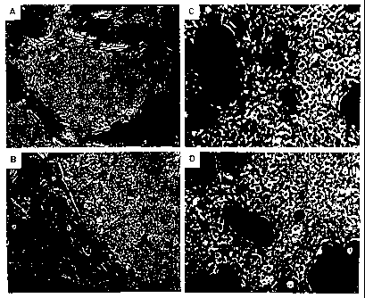
duction of recombinant adenovirus vector (see Fig. 4).
The cell lines N52.E6 and N52.F4 were distinguished by
a rapid growth and particularly good production of ade-
noviral vectors, beneficial properties for the use of
these cell lines for producing gene transfer vectors.
The design of the E1A- and E1B-expressing ex-
pression plasmid used for transforming the amniocytes
precludes the generation of replication-competent ade-
noviruses (RCA) by homologous recombination of an ade-
noviral vector or of an adenoviral helper virus with
the DNA integrated into the transformed amniocytes, in
contrast to 293 cells. As an alternative to this, the
individual El functions can be introduced on various
expression plasmids into the cells to be transfected.
It is, of course, also possible, as for the 293 cell
line, to carry out a transformation of amniocytes and
to test the batches generated in the production of gene
transfer vectors for the RCA content, for example using
a PCR or infection assay. The RCA-containing batches
can then be discarded where appropriate.
Thus a further aspect of the present inventi-
on relates to a permanent amniocytic cell line which
can be obtained by the process proposed herein. In a
specific embodiment, the invention relates to the per-
manent amniocytic cell line N52.E6 which was deposited
on Oct. 26, 1999 at the Deutsche Sammlung von Mikroor-
ganismen und Zellkulturen GmbH (DSMZ) in accordance
17
CA 02391591 2002-05-14
-18-
with the Budapest treaty and has received the accession
number DSM ACC2416.
In a further aspect, the present invention
relates to the use of amniocytes for producing adenovi-
rus-transformed permanent amniocytic cell lines. The
term "adenovirus-transformed" means herein transforma-
tion by one or more transforming adenovirus genes.
"Transformation" refers in this connection to conversi-
on of a eukaryotic cell which is subject to growth con-
trol into a so-called permanent cell line which grows
unrestrictedly. A further aspect is the use of the ade-
noviral gene products of the E1A and E1B regions for
producing permanent amniocytic cell lines.
The present invention further comprises the
use of a permanent amniocytic cell line for producing
gene transfer vectors. "Gene transfer vectors" mean
herein generally all vectors with which one or more
therapeutic genes can be transferred or introduced into
the desired target cells and, in particular, viral vec-
tors having this property. In addition, the permanent
amniocytic cell lines can be used to produce adenovirus
mutants. "Adenovirus mutants" mean adenoviruses which
have at least one mutation in the E1A and/or E1B genes.
In a preferred embodiment, they do not, however, in
contrast to adenoviral gene transfer vectors, harbor
any therapeutic genes. A typical example thereof com-
prises adenovirus mutants in which the E1B 55 kD prote-
in is not expressed (for example the adenovirus mutant
d11520 (Barker et al., Virology 156, 107-121, 1987)).
Adenovirus mutants in which the E1B 55 kD protein is
not expressed are of great interest for the therapy of
oncoses because the virus mutant replicates exclusively
in tumor cells and not or to a negligible extent in
primary normal cells (Bischoff et al., Science 274,
373-376, 1996; Kirn et al., Nature Med. 4, 1341-1342,
1998).
A preferred embodiment is the use of a perma-
nent amniocytic cell line for producing adenovirus vec-
tors, AAV (adeno-associated virus) vectors, retrovirus
vectors, lentivirus vectors, chimeric adenovirus-AAV
vectors, chimeric adenovirus-retrovirus vectors and/or
chimeric adenovirus-lentivirus vectors. A use for pro-
ducing herpes vectors is also possible.
AAV vectors normally comprise only the ITRs
of AAV and some adjacent, noncoding AAV sequences.
Their capacity for uptake of foreign DNA is about
4.5 kb. As described above, various systems exist for
producing recombinant AAV vectors. It is common to all
these systems that the components necessary for repli-
cation, expression and packaging of the recombinant
18
CA 02391591 2002-05-14
-19-
vector are provided. Specifically, these comprise ex-
pression cassettes which code for the AAV rep and cap
proteins, and the adenoviral helper functions. The ade-
noviral helper functions necessary for AAV production
are the E1A, E1B, E2, E4 and VA genes. The E1A and E1B
functions are provided in the E1A- and E1B-expressing
amniocytic cell lines and can therefore be used to pro-
duce AAV vectors. The E2, E4 and VA functions can be
provided by coinfection with adenovirus or by cotrans-
fection with E2-, E4- and VA-expressing plasmids or by
using adenovirus/AAV or herpes simplex virus/AAV hybrid
vectors (Samulski et al., supra; Allen et al., supra;
Tamayose et al., supra; Flotte et al., supra; Conway et
al., supra; Chiorini et al., supra; Ferrari et al., su-
pra; Salvetti et al., supra; Xiao et al., supra; Grimm
et al. , supra; Zhang et al., supra) .
Retrovirus vectors, that is to say vectors
derived from retroviruses, are likewise of great im-
portance as vehicles for transfection within the scope
of gene therapeutic procedures, for example for gene
therapy in the central nervous system (Suhr et al.,
Arch. Neurol. 56, 287-292, 1999). Retroviral vectors
can be produced in stable vector-producing cell lines
or by transient transfection. The individual components
used to produce retroviral vectors normally include one
or more plasmids which express the structural proteins
and the replication and integration proteins, as well
as a plasmid which comprises the vector itself (Miller,
in:. Retroviruses, Coffin, Hughes, Varmus ed., Cold
Spring Harbor Laboratory Press, 1997, pp. 437-473). If
those plasmids which contain an origin of replication,
such as, for example, the SV40 origin of replication,
are used, the amniocytic cell lines are modified so
that proteins which promote replication of the plasmid
are stably expressed. For example, in the case of plas-
mids which contain the SV40 origin of replication, an
amniocytic cell line which expresses the T antigen of
SV40 is used.
Lentivirus vectors are vectors derived from
lentiviruses (Naldini et al., Science 272, 263-267,
1996; Dull et al., J. Virol. 72, 8463-8471, 1998). Len-
tiviral vectors can be produced in stable vector-
producing cell lines or by transient transfection.
"Chimeric vectors" mean vectors which are the
product of a fusion of nucleic acids from two or more
different viral vectors. Permanent amniocytic cell li-
nes can be used according to the present description
for producing chimeric vectors. In this system, for ex-
ample, an adenovirus vector, preferably an adenovirus
vector of large capacity, harbors a DNA fragment which
has the sequence information for an integrating virus
19
CA 02391591 2002-05-14
-20-
which is derived, for example, from a retrovirus or
from AAV. After transcription of a target cell, the in-
tegrating virus harboring a therapeutic gene is relea-
sed from the adenoviral background (for example in the
case of a retroviral insert by producing infectious re-
troviral particles which transduce neighboring cells
and integrate stably as DNA). Examples of chimeric vec-
tors produced in 293 cells have been described in the
past, for example as chimeric adenovirus-retrovirus
vectors (Feng et al., Nature Biotech. 15, 866-870,
1997) and as chimeric adenovirus-AAV vectors (Recchia
et al., Proc. Natl. Acad. Sci. USA 96, 2615-2620,
1999). Production in ElA and E1B-expressing amniocytic
cell lines is to be preferred because, in contrast to
293 cells, replication-competent vectors cannot be ge-
nerated by homologous recombination.
Adenovirus vectors, that is to say vectors
derived from adenoviruses, are of great importance in
particular as vehicles for transfection within the
scope of gene therapeutic procedures. The adenovirus
vectors may be first-generation adenovirus vectors, se-
cond-generation adenovirus vectors, adenovirus vectors
of large DNA capacity and/or deleted adenovirus vec-
tors, which are produced with the aid of a permanent
amniocytic cell line.
a) Production of first-generation adenoviral vectors
First-generation adenoviral vectors are
usually characterized by deletions of the E1A and E1B
genes. Some first-generation adenoviral vectors compri-
se, in addition to the deletion of the ElA and E1B ge-
nes, also deletions of the E3 region. E3 functions are
dispensable for the growth of adenoviral vectors in
cell culture.
First-generation adenoviral vectors can be
produced in E1A- and ElB-expressing amniocytic cell li-
nes. This is done by infecting the E1A- and
E1B-expressing cells preferably with 3-5 infectious
units per cell (3-5 MOI). After about 36 to 72 hours,
the cells show a cytopathic effect. The cells are har-
vested by standard protocols. Adenoviral vector can be
purified from them by CsCl density gradient centrifuga-
tion or by chromatographic processes.
b) Production of second-generation adenoviral vectors
Second-generation adenoviral vectors are cha-
racterized by deletions of ElA and E1B genes. Some se-
cond-generation adenoviral vectors also comprise a de-
letion of the E3 region. In addition to the deletion of
the ElA and E1B genes, second-generation adenoviral
CA 02391591 2002-05-14
-21-
vectors are characterized by inactivation and pre-
ferably deletion of at least one other essential adeno-
viral gene, for example an E2A gene, an E2B gene and/or
a E4 gene, or, for example, by deletions of E2 functi-
ons in combination with deletions of E4 functions.
To produce second-generation adenoviral vec-
tors, the functions which the vector itself does not
express, due to inactivation and/or deletion, must be
provided by the amniocytic cell line. For this purpose,
it is possible for amniocytic cell lines which stably
express E1A and ElB to be stably modified by transfec-
tion of expression cassettes which express the gene
products coding for one or more other adenoviral func-
tions. For example, to produce a second-generation ade-
noviral vector which has, in addition to the deletion
of the E1A and E1B genes, also a deletion of an E2A,
E2B and/or E4 gene, the appropriate gene or genes is
(are) introduced by transfection together with a selec-
tion marker into the E1A- and E1B-expressing amniocytic
cell line. Cell clones which, in addition to the ex-
pression of E1A and E1B functions, also express E2A,
E2B and/or E4 functions can then be used to produce the
particular second-generation vector. The E2 and/or E4
genes are usually under the transcriptional control of
a heterologous promoter, which either is constitutively
active or can be regulated.
c) Production of adenoviral vectors of large DNA ca-
pacity
Adenoviral vectors of large DNA capacity are
characterized by deletion of most or all of the viral
coding sequences. These vectors preferably comprise on-
ly the viral ITRs and the viral packaging signal. The
adenoviral functions are provided by a helper virus in
trans. Various systems for producing adenoviral vectors
of large DNA capacity have been described. It is common
to all the systems described to date and using a helper
virus that the helper virus corresponds to a replicati-
on-deficient, E1A- and ElB-deleted adenovirus. The hel-
per virus comprises either a complete packaging signal
(Mitani et al., Proc. Natl. Acad. Sci. USA 92,
3854-3858, 1995; Fisher et al., Virology 217, 11-22,
1996; Kumar-Singh and Chamberlain, Hum. Mol. Genet. 5,
913-921, 1996) or a mutated packaging signal (Kochanek
et al., Proc. Natl. Acad. Sci. U.S.A. 93, 5731-5736,
1996). In the latter case, the vector is preferably
packaged in viral capsids because the helper virus con-
tains an attenuated packaging signal and therefore is
packaged less efficiently. Alternatively, the packaging
signal of the helper virus can be excised after the in-
21
CA 02391591 2002-05-14
-22-
fection of the producer cell line by using a recombina-
se (Parks et al., Proc. Natl. Acad. Sci. USA 93,
13565-13570, 1996; Hardy et al., J. Virol. 71,
1842-1849, 1997). For example, the packaging signal of
the helper virus can be flanked by loxP recognition se-
quences of bacteriophage Pl. Expression of the Cre re-
combinase of bacteriophage P1 results in excision of
the packaging signal of the helper virus. However, be-
cause of the absence of the packaging signal, no packa-
ging of the helper virus into capsids takes place. The
Cre recombinase gene of bacteriophage Pl is introduced
by transfection together with a selection marker into
the E1A- and ElB-expressing amniocytic cell line. Cell
clones which, in addition to expression of E1A and E1B
functions, also express the Cre function of bacterio-
phage P1 can then be used to produce the particular
vector of large DNA capacity.
d) Production of "deleted" adenoviral vectors
"Deleted" adenoviral vectors have been de-
scribed as first-generation vectors which have loxP re-
cognition sequences of bacteriophage Pl positioned in
the viral genome in such a way that, on infection of
Cre-expressing 293 cells, most of the viral coding se-
quences or all the viral coding sequences are deleted
by recombination between the loxP recognition sequen-
ces. The genome size of these vectors is about 9 kb.
The capacity for uptake of foreign DNA is likewise
about 9 kb (Lieber et al., J. Virol, 70, 8944-8960,
1996). For use in the production of deleted adenoviral
vectors, the Cre recombinase gene of bacteriophage P1
is introduced by transfection together with a selection
marker into the ElA- and E1B-expressing amniocytic cell
line. Cell clones which, in addition to expression of
ElA and E1B functions, also express the Cre function of
bacteriophage Pl can then be used to produce the parti-
cular deleted Ad vector.
e) Production of tropism-modified gene transfer vec-
to r s
In a preferred embodiment, the permanent am-
niocytic cell line is used to produce tropism-modified
gene transfer vectors. The tropism of a virus and of a
viral vector derived from this virus decides whether a
particular cell type can be successfully transduced
with a vector or not. Uptake of a gene transfer vector
into a cell is the first step for successful gene
transfer into this cell. The tropism of a viral vector
is thus an essential factor for efficient in vitro or
22
CA 02391591 2002-05-14
- 23 -
in vivo gene transfer into a particular cell or into a
tissue. Interaction of the surface of a viral vector
(of the capsid in the case of adenoviral or AAV vec-
tors, of the virus envelope in the case of retroviral
or lentiviral vectors) with the cell membrane of a tar-
get cell is necessary for uptake into a particular
cell. Although the exact mechanism of uptake of a viral
vector into a target cell sometimes varies between dif-
ferent vectors, in all cases the interaction of surface
structures of the viral vector (usually protein li-
gands) with structures on the target cell (usually re-
ceptors or adhesion molecules) plays an essential part.
Uptake of adenoviral vectors takes place, for example,
by receptor-mediated endocytosis. This entails parts of
the adenoviral capsid binding to cellular receptors. In
the case of adenoviral vectors derived from Ad2 or Ad5,
according to the current state of knowledge there is
usually binding of part of the knob domain of the fiber
proteins to the coxsackie adenovirus receptor (CAR) and
part of the penton base to av(33 or avP5 integrins. The
binding of the knob domain on CAR is, according to the
current state of knowledge, necessary for adhesion of
the vector to the cell membrane of the target cell,
whereas binding of the penton base to integrins is ne-
cessary for internalization of the vector into the tar-
get cell.
Amniocytic cell lines can be used to produce
tropism-modified vectors. This applies, for example, to
the production of first- and second-generation adenovi-
ral vectors, to adenoviral vectors of large DNA capaci-
ty, to deleted adenoviral vectors, to chimeric adenovi-
ral vectors, to AAV vectors, to retroviral and/or len-
tiviral vectors. Various strategies can be used to pro-
duce tropism-modified vectors in amniocytic cell lines.
The strategy used for the particular tropism modifica-
tion may vary for different vectors (for example adeno-
viral vector, AAV vector, retroviral vector). It is
common to the various strategies that the surface of
the particular vector (virus capsid in the case of ade-
noviral and AAV vectors, virus envelope in the case of
retroviral and lentiviral vectors) is altered so that
the binding of the vector to the target cell is alte-
red. Examples of modifications for adenoviral vectors
are:
a) Exchange of fiber proteins between different sero-
types: this results in adenoviral vectors whose
capsid carries a fiber protein of a different
serotype. Examples thereof are exchange of the na-
tural fiber protein of adenoviral vectoj~s derived
from serotype 2 by a fiber protein derived from
23
CA 02391591 2002-05-14
-24-
serotype 17 (Zabner et al., J. Virol. 73,
8689-8695, 1999) or from serotype 9 (Roelvink et
al., J. Virol. 70, 7614-7621, 1996). Other ex-
amples are exchange of the natural fiber protein
of adenoviral vectors derived from serotype 5 by a
fiber protein derived from serotype 7a (Gall et
al., J. Virol, 70, 2116-2123, 1996) or from sero-
type 3 (Stevenson et al., J. Virol. 71, 4782-4790,
1997; Krasnykh et al., J. Virol. 70, 6839-6846,
1996; Douglas et al., Neuromuscul. Disord. 7,
284-298, 1997).
b) Removal of the fiber protein: the fiber protein
can be removed by processes of genetic manipulati-
on so that uptake of the vector takes place solely
via interaction of the penton base or of the hexon
protein (Falgout et al., J. Virol. 62, 622-625,
1988; Legrand et al., J. Virol. 73, 907-919,
1999).
c) Modification of the C terminus of the fiber prote-
in with a peptide: examples thereof are modifica-
tion of the C terminus with a polylysine peptide
(Yoshida et al., Hum. Gene Ther. 9, 2503-2515,
1998: Wickham et al., Nat. Biotechnol. 14,
1570-1573, 1996; Wickham et al., J. Virol. 71,
8221-8229, 1997), a polyhistidine peptide (Douglas
et al., Nat. Biotechnol. 17, 470-475, 1999) or a
gastrin-releasing peptide (Michael et al., Gene
Ther. 2, 660-668, 1995).
d) Modification of parts of the knob domain of the
fiber protein by insertion of a peptide: examples
thereof are insertion of a FLAG epitope (Krasnykh
et al., J. Virol. 72, 1844-1852, 1998) or inserti-
on of an RGD peptide (Dmitriev et al., J. Virol.
72, 9706-9713, 1998; Kasono et al., Clin Cancer
Res. 5, 2571-2579, 1999).
e) Modification of the penton base: one example the-
reof is replacement of an RGD motif within the
penton base by an LDV motif with the aim of media-
ting binding of the vector to a4(31 integrins
(Wickham et al., Gene Ther. 2, 750-756, 1995).
f) Modification of the hexon protein: one example
thereof is insertion of an epitope derived from
24
CA 02391591 2002-05-14
- 25 -
poliovirus type 3 (Crompton et al., J. Gen. Virol.
75, 133-139, 1994).
An alternative strategy which can be used to
alter the tropism of vectors produced in amniocytic
cell lines is based on the use of ligands which mediate
binding of the vector to cell membrane structures such
as, for example, cellular receptors or adhesion molecu-
les. These ligands may be peptides, proteins or else
antibodies. The ligands can be linked to the surface of
the vectors by various processes. The linkage of the
ligands to the surface of the vectors (of the capsids
in the case of adenoviral or AAV vectors) can be produ-
ced by using antibodies or by a chemical crosslinking
reaction. On use of antibodies it is possible to use
antibodies whose specificity is directed against the
capsid of the vector (for example against the knob do-
main of the fiber protein). Alternatively, it is possi-
ble to use antibodies whose specificity is directed
against an epitope which has been introduced as neoepi-
tope (for example a FLAG epitope or a myc epitope) into
the capsid of the vector. Examples thereof are well
known to the skilled worker. Examples of the use of
bispecific antibodies are described in Wickham et al.,
J. Virol. 70, 6831-6838, 1996 (anti-FLAG/anti-
(x-integrin); in Wickham et al., Cancer Immunol. Im-
munther. 45, 149-151, 1997; Harari et al., Gene Ther.
6, 801-807, 1999 (anti-FLAG/anti-E-selectin) for trans-
duction of endothelial cells; in Miller et al., Cancer
Res. 58, 5738-5748, 1998; Blackwell et al., Arch. Oto-
laryngol. Head Neck Surg. 125, 856-863, 1999 (an-
ti-Ad/anti-EGFR) for transduction of tumor cells; in
Wickham et al., J. Virol. 71, 7663-7669, 1997 (anti-
FLAG/anti-CD3) for transduction of T cells; in Tillman
et al., J. Immunol. 162, 6378-6383, 1999 (anti-
CD40/anti-Ad) for transduction of dendritic cells. Ex-
amples of the use of single-chain antibodies with spe-
cificity for one virus capsid determinant which is cou-
pled to a ligand are described in Watkins et al., Gene
Ther. 4, 1004-1012, 1997; in Goldman et al., Cancer
Res. 57, 1447-1451, 1997; Rancourt et al., Clin. Cancer
Res. 4, 2455-2461, 1998; Gu et al., Cancer Res. 59,
2608-2614, 1999; Rogers et al., Gene Ther. 4,
1387-1392, 1997 (anti-Ad/FGF2) for transduction of
FGF2-receptor-expressing tumor cells; in Douglas et
al., Nat. Biotechnol. 14, 1574-1578, 1996; Douglas et
al., Neuromuscular Disord. 7, 284-298, 1997 (an-
ti-Ad/Folat) for transduction of tumor cells which ex-
press the folic acid receptor on the cell surface.
In the case of gene transfer vectors in which
the natural tropism has been abolished and replaced by
CA 02391591 2002-05-14
-26-
another tropism, for example by introducing a ligand
into the knob domain of the fiber protein of Ad5, it
may be necessary to modify a permanent amniocytic cell
line by the preferably stable expression of a receptor
which recognizes this new ligand (Douglas et al., Nat.
Biotechnol. 17, 470-475, 1999). It is likewise possible
for the permanent amniocytic cell line to be used to
produce gene transfer vectors which have a defect in
the production of one or more structural proteins. This
is done by complementing the particular defects of the
gene transfer vector in the permanent amniocytic cell
line. For example, an adenoviral vector which has a mu-
tation in the gene coding for the fiber protein can be
produced in an amniocytic cell line which complements
the defect in the fiber protein. This is achieved by
introducing a fiber expression cassette into the am-
niocytic cell line and stable or inducible expression
of the fiber protein in this amniocytic cell line (Von
Seggern et al., J. Gen. Virol. 79, 1461-1468, 1998).
The fiber protein expressed in the amniocytic cell line
may be a natural, unmodified fiber protein or else an
altered, for example tropism-modified, fiber protein
(Von Seggern et al., supra). It is also possible to
produce adenoviral vectors completely lacking the fiber
protein in the permanent amniocytic cell line (Legrand
et al., J. Virol., 73, 907-919, 1999; Von Seggern et
al., J. Virol. 73, 1601-1608, 1999).
The use of E1A- and ElB-expressing amniocytic
cell lines is to be preferred because, in contrast to
293 cells, no generation of replication-competent vec-
tors can take place by homologous recombination. In a
particular embodiment of the aspect of the use of an
amniocytic cell line for producing gene transfer vec-
tors, this cell line is the cell line according to the
invention.
Therapeutic genes
The products of the genes, in particular of
the therapeutic genes, which can be encoded and ex-
pressed by vectors produced in transformed amniotic
cells, that is to say a permanent amniotic cell line,
can be, for example, any muscle proteins, coagulation
factors, membrane proteins or cell cycle proteins. Ex-
amples of proteins which can be expressed by vectors
produced in transformed amniocytes are dystrophin
(Hoffman et al., Cell 51, 919, 1987), factor VIII (Wion
et al., Nature 317, 726 1985), cystic fibrosis trans-
membrane regulator protein (CFTR) (Anderson et al.,
Science 251, 679, 1991), ornithine transcarbamylase
(OTC) (Murakami et al., J. Biol. Chem., 263, 18437,
26
CA 02391591 2002-05-14
-27-
1988), alphal-antitrypsin (Fagerhol et al., in: Hum.
Genet., vol. 11, Harris ed., Plenum, New York, p. 1,
1981). The genes coding for proteins are known and can
be cloned from genomic or cDNA banks. Examples of such
genes are the dystrophin gene (Lee et al., Nature 349,
334, 1991), the factor VIII gene (Toole et al., Nature
312, 342 1984), the CFTR gene (Rommens et al., Science
245, 1059, 1989, Riordan et al., Science 245, 1066,
1989), the OTC gene (Horwich et al., Science 224, 1066,
1984), and the alphal-antitrypsin gene (Lemarchand et
al., Proc. Natl. Acad. Sci. USA, 89, 6482, 1992).
Examples of other genes expressed by vectors
which can be produced in transformed amniocytes are the
p53 gene for treating oncoses (Wills et al., Hum. Gene
Ther. 5, 1079, 1994, Clayman et al., Cancer Res. 55, 1,
1995), the Rb gene for treating vascular proliferative
disorders (Chang et al., Science 267, 518, 1995), or
the thymidine kinase gene of herpes simplex virus (HSV)
type 1 for the therapy of oncoses. The gene expressed
by vectors produced in transformed amniocytes does not
necessarily code for a protein. Thus, for example, it
is possible for functional RNAs to be expressed. Ex-
amples of such RNAs are antisense RNAs (Magrath, Ann.
Oncol. 5, Suppl 1), 67-70 1994, Milligan et al., Ann.
NY Acad. Sci. 716, 228-241, 1994, Schreier, Pharma. Ac-
ta. Helv., 68, 145-159 1994), and catalytic RNAs (Cech,
Biochem. Soc. Trans. 21, 229-234, 1993; Cech, Gene 135,
33-36, 1993; Long et al., FASEB J. 7, 25-30, 1993; Rosi
et al., Pharm. Therap. 50, 245-254, 1991).
Vectors produced in transformed amniocytes
may, in addition to the therapeutic gene, comprise any
reporter gene in order to be able to follow expression
of the vector better. Examples of reporter genes are
known in the prior art and include, for example, the
(3-galactosidase gene (Fowler et al., Proc. Natl. Acad.
Sci. USA 74, 1507, 1977).
Vectors which can be produced in transformed
amniocytes may comprise more than a single gene. The
maximum number of genes which can be produced in such
vectors depends on the uptake capacity of the particu-
lar vector and on the size of the genes.
The choice of the promoters which control ex-
pression of the therapeutic genes of vectors produced
in transformed amniocytes is not critical. Viral or
nonviral promoters which show constitutive, tissue-
specific or regulable activity can be used for expres-
sing a protein or a functional RNA. The SV40 or cytome-
galovirus promoter (Andersson et al., J. Biol. Chem.
264, 8222-8229, 1964) can be used, for example, for
constitutive expression of a gene. The use of the mu-
27
CA 02391591 2007-10-22
-28-
scle creatine kinase (MCK) promoter permits tissue-
specific expression of a protein or of a functional RNA
in skeletal muscle and myocardium. Gene expression can
be controlled quantitatively and qualitatively by the
use of a regulable system (Furth et al., Proc. Natl.
Acad. Sci. USA 91, 9302-9306, 1994).
It is possible to include in vectors which
can be produced in transformed amniocytes genetic ele-
ments which influence the behavior of the vector inside
the recipient cell. Examples of such elements are ele-
ments which facilitate nuclear targeting of the vector
DNA (Hodgson, Biotechnoloqy 13, 222-225, 1995).
Vectors produced in this way can be used in
vitro or in vivo. An in v1tro gene transfer takes place
outside the body, for example by adding the vector to
cells in culture or to primary cells which have been
taken from the body for the purpose of gene transfer.
In the case of in vivo gene transfer, vector particles
can be applied in various ways depending on the tissue
which is to be transduced. Examples are injection into
the arterial or venous vascular system, direct injec-
tion into the relevant tissue (for example liver,
brain, muscle), instillation into the relevant organ
(for example lung or gastrointestinal tract) or direct
application onto a surface (for example skin or blad-
der).
The present invention further provides
a permanent amniocytic cell line comprising at least
one nucleic acid which brings about expression of the
gene products of the adenovirus ElA and ElB regions.
The present invention further
provides a process for producing a permanent
amniocytic cell line which comprises the transfection
of amniocytes with at least one nucleic acid which
brings about expression of the adenoviral gene
products of the E1A region and E1B region.
The present invention further
provides a permanent amniocytic ceil line obtainable by
the above-mentioned process.
The present invention further
provides the permanent amniocytic cell line N52.E6 (DSM
ACC2416).
The present invention further
provides a process for producing a gene transfer
vector, the process comprising transfecting the gene
transfer vector into a permanent amniocytic cell line
28
CA 02391591 2007-10-22
-28a-
comprising at least one nucleic acid which brings
about expression of the gene products of the
adenovirus E1A and ElB regions.
The present invention further
provides a process for producing an adenovirus mutant,
the process comprising transfecting the adenovirus
mutant into a permanent amniocytic cell line
comprising at least one nucleic acid which brings
about expression of the gene products of the
adenovirus ElA and E1B regions.
The following figures and example are inten-
ded to illustrate the invE:ntion in detail without re-
stricting it thereto.
DESCRIPTION OF THE FIGURES
Fig. 1 shows a summary of the clonings: Fig 1A depicts
diagrammatically the cloning steps for plasmid
STK146. Fig. 1B depicts the left-terminal ap-
prox. 15% of the qenome of adenovirus type 5,
including the El RNAs, the coding regions, the
starting points of ElA and ElB transcription,
and the splice donor and splice acceptor sites
and polyadenylation sequences which are im-
portant for the cloning. It is important that
the splice donor site at base pair 3511 of Ad5
has been retained in the cloning for plasmid
STK146, but that the splice acceptor site and
the polyadenylation signal have been replaced
by corresponding f`unctions of SV40. In additi-
on, the ElA promoter of Ad5 has been replaced
by the PGK promoter. Thus STK146 contains the
Ad5 sequences from base pair 505-5322 and the
28a
CA 02391591 2002-05-14
-29-
cell lines transformed with this plasmid con-
tain no Ad5 sequences which are present in
first- or second-generation adenoviral vectors
or in loxP helper viruses.
Fig. 2 shows the cell islets (Fig. 2B and Fig. 2B) ob-
tained from amniocytes by transformation by
adenoviral El functions, and the cell lines
N52.E6 (DSMZ ACC2416; Fig. 2C) and N52.F4 (Fig.
2D) cloned from single cells. This time it
should be noted that the cell lines and single
cell clones differ morphologically from the am-
niocytes in that they are usually smaller and
there is no contact inhibition of their growth.
Fig. 3 shows the integration status of STK146 in eight
different El-transformed amniocytic cell lines
by means of a Southern blot.
Fig. 4 shows, listed in a table, the production of a
first-generation adenoviral vector in various
cloned cell lines on the basis of bfu (blue
forming units) per cell and the efficiency of
transfection of the appropriate cell lines on
the basis of plaque formation.
Fig. 5 shows the expression of the ElA and ElB pro-
teins of Ad5 in eleven cloned amniocytic cell
lines (Western blot).
Fig. 6 shows the time course of the synthesis of re-
combinant adenoviral vectors in two cloned cell
lines N52.E6 and N52.F4. Fig. 6A shows the syn-
thesis of a first-generation adenoviral vector,
and Fig. 6B shows the synthesis of an adenovi-
ral vector with large DNA capacity.
EXAMPLES
1. Clonings
Fig. 1 depicts a summary of the clonings.
29
CA 02391591 2002-05-14
-30-
a) Plasmid STK136
Plasmid STK 136 contains the murine phospho-
glycerate kinase promoter (seq. No. 1; Adra et al., Ge-
ne 60, 65-74, 1987) in pBluescript KsII (Stratagene)
and was produced as follows:
3.5 g of plasmid PGK-hAAT (Kay et al., Hepa-
tology 21, 815-819, 1995) were digested with EcoRV and
fractionated by size in a 1.5% agarose gel. The 0.5 kb
band containing the PGK promoter fragment which was
sought was, after staining in ethidium bromide, cut out
and the DNA was electroeluted. At the same time, pBlue-
script KSII was digested with EcoRV and HincIl, and the
free DNA ends were dephosphorylated. After subsequent
phenol/chloroform extraction and ethanol precipitation,
equimolar amounts of these DNA fragments were ligated
and transformed into ultracompetent XL-2 Blue bacteria
(Stratagene). The plasmid clones were characterized by
means of a restriction digestion, and the plasmid re-
sulting therefrom was called STK136 (isolate #6).
b) Plasmid STK137
Plasmid STK137 contains the complete El ex-
pression cassette of Ad5 including the 3' splice and
polyadenylation signals from SV40 and was produced as
follows:
PCR amplification of the Ad5 sequence bp 505-841
(PCR I) (Seq. No. 2)
10 ng of the plasmid pXCl (Microbix) were am-
plified together with 400 ng each of the oligonucleoti-
des 27759 (Seq. No. 3) and 27634 (Seq. No. 4),
0.2 mM dNTPs and 1.25 U Pfu polymerase in 10 mM KC1,
10 mM (NH4) 2S09i 20 mM Tris/HC1, pH 8.75, 2 mM MgSO9,
0.1% Triton X-100, 100 g/ml BSA under the following
conditions:
I 10 minutes at 94 C,
II 1 minute at 94 C,
2 minutes at 50 C,
3 minutes at 72 C,
III 10 minutes at 72 C.
CA 02391591 2002-05-14
-31-
Repeating step II in 15 cycles. The DNA was purified
using the QIAquick PCR purification kit (Qiagen) as
stated by the manufacturer and was precipitated with
ethanol.
To clone the PCR fragment, 2.5 g of pBlue-
script KSII were digested with EcoRV, and the free DNA
ends were dephosphorylated, ligated in equimolar
amounts with the PCR fragment and transformed into XL-2
Blue cells. The plasmid resulting therefrom is referred
to as #1 hereinafter.
PCR amplification of the Ad5 sequence bp 3328-3522
(PCR II) Seq. No. 5):
10 ng of the plasmid pXCl (Microbix) were am-
plified together with 400 ng each of the oligonucleoti-
des 27635 (Seq. No. 6) and 27636 (Seq. No. 7) under the
conditions described above. After the PCR, the DNA was
extracted with phenol/chloroform, precipitated with et-
hanol, digested with EcoRI, again extracted with phe-
nol/chloroform, precipitated and dissolved in 30 1 of
TE.
PCR amplification of the 3' splice and polyadenylation
signal from SV40 with the aid of the plasmid pGL2-Basic
bp 1978-2749 (PCR III) (seq. no. 8) :
20 ng of the plasmid pGL2-Basic (Promega,
GenBank/EMBL Acc. No.: X65323) were amplified together
with 800 ng each of the oligonucleotides 27637 (seq.
No. 9) and 27638 (seq. No. 10), 0.4 mM dNTPS and
2.5 U of Pfu polymerase under the conditions described
above. After the PCR, the DNA was extracted with phe-
nol/chloroform, precipitated with ethanol, digested
with EcoRI, again extracted with phenol/chloroform,
precipitated in ethanol and dissolved in 30 l of TE.
Then 10 l each of DNA from PCR II and III were ligated
in a volume of 50 l, extracted with phenol/chloroform,
precipitated with ethanol and digested with BamHI in a
volume of 100 l. After renewed phenol/chloroform ex-
traction and ethanol precipitation, the DNA was ligated
with equimolar amounts of pBluescript KSII DNA which
had previously been digested with BamHI and dephospho-
rylated. The plasmid resulting therefrom is referred to
as #29 hereinafter. For further cloning, 3.5 g of
plasmid DNA #29 were digested with SacIi and BglII, de-
phosphorylated, extracted with phenol/chloroform and
precipitated in ethanol. At the same time, 3.5 g of
pXCl were digested with BglII and SacII, and the 2.9 kb
fragment was fractionated by electrophoresis and elec-
troeluted. Equimolar amounts of the two DNAs were liga-
31
CA 02391591 2002-05-14
-32-
ted and transformed into XL-2 Blue cells. The plasmid
resulting therefrom is referred to as #5 hereinafter.
For the final cloning of STK137, plasmid #1 was dige-
sted with HinclI and BspEI and fractionated by electro-
phoresis, and an approx. 350 bp fragment was electroe-
luted. As vector DNA, plasmid #5 was digested with KspI
(isoschizomer of SacII), the ends were filled in with
T4 polymerase, and phenol/chloroform extraction and et-
hanol precipitation were carried out. The DNA was then
digested with BspEI, the ends were dephosphorylated,
and phenol/chloroform extraction and ethanol precipita-
tion were again carried out. The two DNAs were ligated
and transformed into XL-2 Blue cells. The plasmid re-
sulting therefrom was called STK137 (isolat #34).
c) Plasmid STK146 (Seq. No. 18)
Plasmid STK146 contains the murine PGK promo-
ter, the complete El region of Ad5 (bp 505-3522) and
the 3' splice and polyadenylation signal of SV40.
For the cloning, 4 gg of STK137 were digested
with EcoRV and BamHI and fractionated by electrophore-
sis, and the 3.7 kb fragment was electroeluted. In ad-
dition, 3.3 g of STK136 were digested with EcoRV and
BamHI, dephosphorylated, phenol/chloroform extracted
and precipitated with ethanol. Equimolar amounts of the
two plasmids were ligated and transformed into XL-2
Blue cells. The plasmid resulting therefrom was called
STK139. Final sequence analysis of STK139 revealed a
mutation at the bp 2613 (the numbering in this connec-
tion refers to the Ad5 DNA sequence), which led to a
tyrosine to asparagine amino acid exchange (2613 TAC ~
GAC). For this reason, the fragment containing the mu-
tation in STK139 was replaced by the BstEII (bp 1915)-
BglII (bp 3328) fragment from pXCl. This was done by
digestion of STK139 with BstEII and BglII, dephosphory-
lation, phenol/chloroform extraction, ethanol precipi-
tation, fractionation by electrophoresis and electroe-
lution of the 5.8 kb fragment. pXCl was likewise dige-
sted with BstEII and BglII and fractionated by electro-
phoresis, and the 1.4 kb fragment was electroeluted.
After ligation and transformation, DNA from 4 plasmid
clones was sequenced; two of them contained the correct
sequence at bp 2613. Isolate number 2 was sequenced
completely and is referred to as STK146 hereafter.
d) Sequence analysis of STK146
32
CA 02391591 2002-05-14
- 33 -
500 ng of STK146 #2 were sequenced with 10 pmol of the
following sequence primers under standard conditions:
Primer 28231 Ad5 nt. 901-920
(Seq. No. 11)
28232 Ad5 nt.
1301-1320 (Seq. No. 12)
28233 Ad5 nt.
1701-1720 (Seq. No. 13)
28234 Ad5 nt.
2100-2119 (Seq. No. 14)
28235 Ad5 nt.
2500-2519 (Seq. No. 15)
28236 Ad5 nt.
2853-2872 (Seq. No. 16)
28237 Ad5 nt..
3249-3268 (Seq. No. 17)
2. Cultivation of primary amniocytes and
cell lines
All the cell culture reagents,
media and sera were purchased from GIBCO Life Technolo-
gies. The cell line 293 which was used as control in
some experiments was cultivated in modified Eagle's me-
dium (MEM) with 10% fetal calf serum (FCS), lx penicil-
lin/streptomycin at 37 C (100x, Cat# 10378-016), 95%
humidity and 5% C02. The new El-transformed cell lines
were produced using primary fetal cells which had been
obtained from amniotic fluid by amniocentesis as part
of prenatal diagnosis. After the biopsy, the cells were
seeded by routine methods in plastic culture bottles
and cultivated in Ham's F10 medium (nutrient mixture
Ham's F10 with L-glutamine, Cat# 31550-023), 10% FCS,
2% Ultroser G, lx antibiotic/antimycotic solution
(100x, Cat# 15254-012), 2.5 g/ml Fungizione (amphote-
ricin B, Cat# 15290-018). Some of the cells adhered to
the base of the cell culture bottle and proliferated.
Sufficient cells for a chromosome analysis were availa-
ble after about 2 weeks. After establishment of the ka-
ryotype, amniocyte cultures with numerically and struc-
turally normal chromosomes were used to produce the
cell line. Cells from three different sources, taken by
amniocentesis either 3, 6 or 7 weeks beforehand, were
used in various experiments. The nutrient medium used
was Ham's F10 medium, 10% FCS, 2% Ultroser Cr, lx anti-
biotic/antimycotic solution, 2.5 g/ml Fungizione . The
culture conditions were 37 C, 95% humidity and 5% C02.
33
CA 02391591 2002-05-14
-34-
Seven days after transfection, the amniocytes were cul-
tivated further in Ham's F10 medium, 10% FCS, lx peni-
cillin/streptomycin. After the generation of single-
cell clones, these were transferred to new dishes and
cultivated further in alpha-MEM with 10% FCS, lx peni-
cillin/streptomycin.
3. Transfection and transformation of am-
niocytes
For the transfection, the amniocytes were seeded on
cell culture dishes (diameter 60 mm, surface area
22.1 cmZ) at a density of 2- 5 x 10⁵ per dish and
transfected the following day. For the transfection,
20 g of plasmid STK146 were digested with ScaI, ex-
tracted with phenol/chloroform, precipitated with etha-
nol and taken up in 20 l of TE, which gave a DNA con-
centration of 0.5 g/ l. In the initial experiments,
amniocytes were transfected 3 or 7 weeks after removal,
in 5 dishes each, with the Effectene transfection kit
as stated by the manufacturer (Qiagen) as follows:
4}tl of STK146 digested with
ScaI were mixed with 146 l of EC buffer. After additi-
on of 8 l of enhancer, the solution was briefly vor-
texed and incubated at room temperature for 5 minutes.
Then 25 l of Effectene were added and, after vortexing
for 10 seconds, incubated at room temperature for a
further 10 minutes. During this, the medium was cau-
tiously aspirated off the cells and replaced by 4 ml of
fresh nutrient medium (see Section 2. above). After the
incubation was complete, the transfection mixture was
mixed with 1 ml of fresh nutrient medium and cautiously
added dropwise to the cells. The cells were cultivated
further as described above. Seven days after the trans-
fection, the cells in each dish were transferred to a
larger dish (diameter 150 mm, surface area 147.8 cm2).
This was done by cautiously aspirating off the medium,
and the cells being washed with PBS, detached in tryp-
sin and transferred to a new dish and cultivated
further as described in Section 2. 18 to 22 days after
the transfection clonal cell islets were clearly to be
seen and were clearly distinguished morphologically
from the amniotic cells (Figs. 2A, 2B). The main pro-
portion in an untransformed amniocyte culture consists
of larger cells which show contact inhibition of their
growth. Cells in transformed single-cell colonies are
very much smaller, grow much more quickly and show no
contact inhibition of their growth. They grow as cell
islets consisting of smaller cells which are crowded
tightly together, and are unambiguous under the light
34
CA 02391591 2002-05-14
-35-
microscope and can be identified without difficulty.
These cell islets were picked and transferred to a new
dish (diameter 60 mm) containing the medium described
above. After further growth, the cell lines were trans-
ferred to 147.8 cm2 cell culture dishes and cultivated
further as described under 2. After the first transfer
to 147.8 cm2 cell culture dishes, the cell passages we-
re counted. Initially, about 40 cell clones from the
cells which had been transfected 3 and 7 weeks after
removal were cultivated further. Subsequently, that is
to say after prolonged cultivation, there was a drastic
change in the morphology of some of the cell clones,
and they showed instability in their growth characteri-
stics. The further experiments were restricted to
further cultivation and analysis of eight morphologi-
cally stable cell lines. These were referred to as fol-
lows: GS.A55 (produced from amniocytes transfected
3 weeks after removal), GS.N21, GS.N24, GS.N27, GS.N49,
GS.N51, GS.N52, GS.N53 (produced from amniocytes trans-
fected seven weeks after removal).
During the first passages all
the cell clones showed a comparable morphology, but
this was changed by subsequent passages. Thus, for ex-
ample, some cell clones changed to assume a highly
rounded shape and, after further passages, they were no
longer adherent. Other cell clones showed extensive
vacuolization, but this did not appear to have any ef-
fect on their growth. After the single-cell cloning,
all the cell lines showed a uniform morphology and, for
example, N52.EG and N52.F4 had an epithelial appearan-
ce. They were comparable to 293 cells in their growth
rate and cell density.
4. Efficiency of the transformation
In order to determine the effi-
ciency of the transformation by the El functions more
accurately, seven new dishes each containing 2-5 x 10⁵
cells were transfected as described in Section 3. The
cells were transferred only 24 hours after the trans-
fection to dishes 147.8 cmz in size and were cultivated
further in Ham's F-10 medium, 10% fetal calf serum, 2%
Ultroser G, 1 x antibiotic/antimycotic solution,
2.5 g/ml Fungizione for 5 days and in Ham's medium,
10% fetal calf serum, lx penicillin/streptomycin solu-
tion for a further 25 days. A dish with untransfected
cells was cultivated under the same conditions as a
control. During this time, the morphologically clearly
distinguishable (see Fig. 2A, B) colonies resulting
from single transformation events were counted. Single-
cell clones could be counted on all the cell culture
CA 02391591 2002-05-14
-36-
dishes apart from the untransfected control dish. On
average there were 4 cell clones per plate, which cor-
responded to a transformation efficiency of 1 in
0.5-1 x 10⁵ cells.
5. Single-cell cloning
As already mentioned, some of
the cell lines showed different morphological characte-
ristics, which is why up to ten single cell lines were
set up from each cell line. The passages for the indi-
vidual cell lines differed in these cases: GS.A55: P17,
GS.N21: P24, GS.N24: P20, GS.N27: P19, GS.N49: P21,
GS.N51: P39, GS.N52: P22, GS.N53: P20. For this purpo-
se, the cells were detached from the cell culture dis-
hes and, at a concentration of 5 x 10⁶ cells/ml, dilu-
ted 1:1000, 1:50,000 and 1:500,000 in nutrient medium.
100 l cells from all the dilutions were seeded onto
96-well plates, and the cell clones which had unambi-
guously resulted from single cells were cultivated
further. Fig. 2C shows the cell line GS.N52.E6 (DSMZ
No.), and Fig. 2D shows cell line GS.N52.F4; both cell
clones are derived from the original cell line GS.N52.
6. Characterization of the El cell lines
a) Southern blot analyses
Southern blot analyses were car-
ried out in order to investigate the integration status
of the El region in the cell lines. This was done by
isolating genomic DNA from all eight El amniotic clones
(see Section 5.), and 5 g of each were digested with
EcoRV, fractionated by electrophoresis and transferred
to a nylon membrane. EcoRV cuts once in the El expres-
sion cassette. Hybridization with radiolabeled STK146
DNA confirmed integration of 1-2 copies of STK146 in
all clones. Fig. 3 shows the integration pattern in the
El cell clones. There are mainly two high molecular
weight bands evident for all the clones, which indica-
tes integration of a single copy. The cell clones
GS.N24 and GS.N52 each showed an additional band with
relatively strong intensity, which might indicate inte-
gration of tandem copies of STK146. For none of the
cell clones were there any bands smaller than STK146
digested with ScaI and EcoRV, which suggested that all
the integrates were present completely and not deleted.
b) Generation of recombinant adenoviruses
36
CA 02391591 2002-05-14
-37-
After the single-cell cloning,
the cell clones were tested for their ability to gene-
rate recombinant adenovirus. This was done once by in-
fecting 3-5 single-cell clones of each cell line in ap-
prox. 70% confluent 24-well plates with about 5 MOI
(multiplicity of infection) of Ad(3gal (recombinant
first-generation adenoviral vector). 48 hours after the
infection, the cells were lysed by freezing and thawing
three times, and the amount of Adpgal produced was ana-
lyzed by infecting 293 cells and subsequently staining
the cells (MacGregor et al., in: Gene Transfer and Ex-
pression Protocols, Murray ed. Humana, Clifton, NJ,
vol. 7, pp. 217-235, (1991)) for detecting
(3-galactosidase production (Fig. 4). This method af-
fords an only approximate production of recombinant
adenovirus because the number of cells and therefore
the amount of virus used may differ in the different
cell clones because of their size and growth rate. The
cell clones which gave the largest yield in this first
test were analyzed more exactly in a further experi-
ment. This was done by seeding about 3 x 10' cells on 3
dishes (diameter 100 mm, surface area 60 cm2). The
cells were counted the next day and were infected with
exactly 5 MOI of Ad(3gal based on the number of cells
found. 48 hours after the infection, the cells were
harvested and lysed by freezing and thawing three
times, and the amount of Ad(3gal produced was analyzed
by infecting 293 cells and subsequently staining. The
result is depicted in Fig. 4.
c) Transfection efficiency
Some of the cloned cell lines
were tested for plaque formation. This was done by
transfecting approx. 70% confluent cell culture dishes
(diameter 60 mm) with 2 g of infectious plasmid GS66
by the calcium phosphate method. Plasmid GS66 contains
the complete adenovirus genome with a deletion in the
El region from nucleotide 440 to nucleotide 3523. The
adenoviral terminal repeat sequences (ITRs) are flanked
in this plasmid by SwaI restriction cleavage sites, so
that infectious virus and plaques can be produced after
transfection of SwaI-digested plasmid. About 24 hours
after transfection, the cells were covered with about
10 ml of MEM, 1% agarose, 0.5x penicillin/streptomycin,
0.05% yeast extract. Plaques were visible after incuba-
tion at 37 C, 95% humidity, 5% CO2 for about 1 week.
Fig. 4 shows the number of counted plaques averaged for
2 independent transfections in each case.
37
CA 02391591 2002-05-14
-38-
d) Expression of E1A and E1B functions of
Ad5
Expression of the Ad5-E1A and of
the E1B-2lkD proteins in the cloned cell lines was de-
tected by Western blot analyses using monoclonal anti-
bodies.
The cells were carefully deta-
ched from a cell culture (diameter 10 cm) in PBS/lmM
EDTA, pelleted and taken up in 150 l of 50 mM tris/HC1
PH 8, 140 mM NaCl, 0.5% NP40, 4 mM EDTA, 2 mM EGTA,
0.5 mM PMSF, 5% glycerol. The cells were lyzed by addi-
tional Dounce grinding and centrifuged down at
13,000 rpm for 10 minutes, and the protein concentrati-
on in the supernatant was determined using a protein
determination kit (BIORAD, Microassay Procedure). 10 g
of protein were fractionated on a 12% SDS polyacrylami-
de gel, transferred to a nitrocellulose membrane (Hy-
bond ECL, Amersham Pharmacia Biotech) and incubated
with an anti-E1A or anti-E1B 2lkD antibody (Calbiochem,
dilution 1:300). The next day, the blot was washed and
hybridized with a second anti-mouse antibody (E1A) or
anti-rat antibody (E1B) to which horseradish peroxidase
was coupled. The horseradish peroxidase reaction was
started by incubation of equal volumes of enhance solu-
tion 1 and 2 (ECL, Amersham Pharmacia Biotech), and the
photochemical reaction which developed thereby was vi-
sualized by brief exposure to an X-ray film. Fig. 5
shows the result of a Western blot analysis.
e) Time-course of the synthesis of recombinant first-
generation adenoviral vectors and adenoviral vec-
tors with large DNA capacity
Two cloned cell lines N52.E6 and
N52.F4 showed the highest yield of recombinant first-
generation adenoviral vectors in the experiments de-
scribed above. Further knowledge about the time course
is important for optimal production of adenoviral vec-
tors, especially when these cells are adapted to sus-
pension cultures and the success of infection cannot be
followed by means of a cytopathic effect. For this ana-
lysis, several dishes (diameter 6 cm) each with 3 x 10⁶
cells were infected with 5 MOI of Ad(3gal and harvested
.at the stated times after the infection. The yield of
recombinant adenovirus was again determined by infec-
ting 293 cells, staining and counting the blue cells
(Fig. 6 A).
It is intended in future also to
use the new cell lines for producing adenoviral vectors
with large DNA capacity (see above). Production of the-
38
CA 02391591 2002-05-14
-39-
se adenoviral vectors requires helper viruses which
supply the deleted functions and proteins for a lytic
infection cycle in trans. The packaging signal of these
helper viruses is deleted with the aid of loxP recogni-
tion sequences and Cre recombinase, which is expressed
by the cell line, on infection. It is therefore inten-
ded in future experiments that the new El-transformed
cell lines will be transfected with a Cre-expressing
plasmid, and the recombinase will be expressed stably.
It has been tested in prelimina-
ry experiments whether the new El amniocytes are also
able to produce adenoviral vectors of large DNA capaci-
ty, and whether production kinetics and the amount of
produced vectors corresponds to that achieved with the
existing Cre-expressing 293 cells. For this purpose,
several dishes each with 3 x 10⁶ cells of the cell li-
nes N52.E6 and N52.F4 were infected with 5 MOI of loxP
helper virus and 10 MOI of AdGS46 ((3-gal-expressing
adenoviral vector with large DNA capacity) and harve-
sted at the stated times after the infection. The yield
of 0-gal-expressing adenoviral vector with large DNA
capacity was again determined by infecting 293 cells,
staining and counting the blue cells (Fig. 6B). The
amount of adenoviral vectors of large DNA capacity syn-
thesized in the amniocytes corresponds to that also
produced in Cre-expressing 293 cells (data not shown).
39
CA 02391591 2008-05-09
-39a-
SEQUENCE LISTING
<110> KOCHANEK, Stefan
<120> PERMANENT AMNIOCYTE CELL LINE, THE PRODUCTION THEREOF
AND ITS USE FOR PRODUCING GENE TRANSFER VECTORS
<130> AML/13642.2
<140> 2,391,591
<141> 2000-11-07
<150> PCT/EP00/10992
<151> 2000-11-07
<150> DE 199 55 558.3
<151> 1999-11-18
<160> 18
<170> PatentIn Ver. 2.1
<210> 1
<211> 513
<212> DNA
<213> Mouse, Phosphoglycerat-Kinase-Promoter
<400> 1
gaattctacc gggtagggga ggcgcttttc ccaaggcagt ctggagcatg cgctttagca 60
gccccgctgg cacttggcgc tacacaagtg gcctctggcc tcgcacacat tccacatcca 120
ccggtaggcg ccaaccggct ccgttctttg gtggcccctt cgcgccacct tctactcctc 180
ccctagtcag gaagttcccc cccgccccgc agctcgcgtc gtgcaggacg tgacaaatgg 240
aagtagcacg tctcactagt ctcgtgcaga tggacagcac cgctgagcaa tggaagcggg 300
taggcctttg gggcagcggc caatagcagc tttgctcctt cgctttctgg gctcagaggc 360
tgggaagggg tgggtccggg ggcgggctca ggggcgggct caggggcggg gcgggcgccc 420
gaaggtcctc cggaggcccg gcattctcgc acgcttcaaa agcgcacgtc tgccgcgctg 480
ttctcctctt cctcatctcc gggcctttcg acc 513
<210> 2
<211> 337
<212> DNA
<213> Ad5
<400> 2
gagtgccagc gagtagagtt ttctcctccg agccgctccg acaccgggac tgaaaatgag 60
acatattatc tgccacggag gtgttattac cgaagaaatg gccgccagtc ttttggacca 120
gctgatcgaa gaggtactgg ctgataatct tccacctcct agccattttg aaccacctac 180
ccttcacgaa ctgtatgatt tagacgtgac ggcccccgaa gatcccaacg aggaggcggt 240
ttcgcagatt tttcccgact ctgtaatgtt ggcggtgcag gaagggattg acttactcac 300
ttttccgccg gcgcccggtt ctccggagcc gcctcac 337
<210> 3
<211> 29
<212> DNA
<213> Artificial Sequence
<220>
CA 02391591 2008-05-09
-39b-
<223> Description of Artificial Sequence:
Oligonucleotide
<400> 3
atcgagtgcc agcgagtaga gttttctcc 29
<210> 4
<211> 22
<212> DNA
<213> Artificial Sequence
<220>
<223> Description of Artificial Sequence:
Oligonucleotide
<400> 4
gtgaggcggc tccggagaac cg 22
<210> 5
<211> 216
<212> DNA
<213> Ad5
<400> 5
ctcgcggatc cagatctgga aggtgctgag gtacgatgag acccgcacca ggtgcagacc 60
ctgcgagtgt ggcggtaaac atattaggaa ccagcctgtg atgctggatg tgaccgagga 120
gctgaggccc gatcacttgg tgctggcctg cacccgcgct gagtttggct ctagcgatga 180
agatacagat tgaggtactg aaatggaatt ccggtc 216
<210> 6
<211> 32
<212> DNA
<213> Artificial Sequence
<220>
<223> Description of Artificial Sequence:
Oligonucleotide
<400> 6
gacgccaatt ccatttcagt acctcaatct gt 32
<210> 7
<211> 31
<212> DNA
<213> Artificial Sequence
<220>
<223> Description of Artificial Sequence:
Oligonucleotide
<400> 7
ctcgcggatc cagatctgga aggtgctgag g 31
<210> 8
CA 02391591 2008-05-09
-39c-
<211> 782
<212> DNA
<213> SV40 (pGL2basic), Genbank X65323
<400> 8
cgactgaatt caatttttaa gtgtataatg tgttaaacta ctgattctaa ttgtttgtgt 60
attttagatt ccaacctatg gaactgatga atgggagcag tggtggaatg cctttaatga 120
ggaaaacctg ttttgctcag aagaaatgcc atctagtgat gatgaggcta ctgctgactc 180
tcaacattct actcctccaa aaaagaagag aaaggtagaa gaccccaagg actttccttc 240
agaattgcta agttttttga gtcatgctgt gtttagtaat agaactcttg cttgctttgc 300
tatttacacc acaaaggaaa aagctgcact gctatacaag aaaattatgg aaaaatattc 360
tgtaaccttt ataagtaggc ataacagtta taatcataac atactgtttt ttcttactcc 420
acacaggcat agagtgtctg ctattaataa ctatgctcaa aaattgtgta cctttagctt 480
tttaatttgt aaaggggtta ataaggaata tttgatgtat agtgccttga ctagagatca 540
taatcagcca taccacattt gtagaggttt tacttgcttt aaaaaacctc ccacacctcc 600
ccctgaacct gaaacataaa atgaatgcaa ttgttgttgt taacttgttt attgcagctt 660
ataatggtta caaataaagc aatagcatca caaatttcac aaataaagca tttttttcac 720
tgcattctag ttgtggtttg tccaaactca tcaatgtatc ttatcatgtc tggatccgtc 780
ga 782
<210> 9
<211> 32
<212> DNA
<213> Artificial Sequence
<220>
<223> Description of Artificial Sequence:
Oligonucleotide
<400> 9
cgactgaatt caatttttaa gtgtataatg tg 32
<210> 10
<211> 27
<212> DNA
<213> Artificial Sequence
<220>
<223> Description of Artificial Sequence:
Oligonucleotide
<400> 10
tcgacggatc cagacatgat aagatac 27
<210> 11
<211> 20
<212> DNA
<213> Artificial Sequence
<220>
<223> Description of Artificial Sequence:
Oligonucleotide
<400> 11
ccttgtaccg gaggtgatcg 20
CA 02391591 2008-05-09
-39d-
<210> 12
<211> 20
<212> DNA
<213> Artificial Sequence
<220>
<223> Description of Artificial Sequence:
Oligonucleotide
<400> 12
tggcgcctgc tatcctgaga 20
<210> 13
<211> 20
<212> DNA
<213> Artificial Sequence
<220>
<223> Description of Artificial Sequence:
Oligonucleotide
<400> 13
tacatctgac ctcatggagg 20
<210> 14
<211> 20
<212> DNA
<213> Artificial Sequence
<220>
<223> Description of Artificial Sequence:
oligonucleotide
<400> 14
caagaatcgc ctgctactgt 20
<210> 15
<211> 20
<212> DNA
<213> Artificial Sequence
<220>
<223> Description of Artificial Sequence:
Oligonucleotide
<400> 15
ggctgcagcc aggggatgat 20
<210> 16
<211> 20
<212> DNA
<213> Artificial Sequence
<220>
CA 02391591 2008-05-09
-39e-
<223> Description of Artificial Sequence:
Oligonucleotide
<400> 16
agggttcggg gctgtgcctt 20
<210> 17
<211> 20
<212> DNA
<213> Artificial Sequence
<220>
<223> Description of Artificial Sequence:
Oligonucleotide
<400> 17
cctgaacggg gtgtttgaca 20
<210> 18
<211> 7090
<212> DNA
<213> Plasmid STK146
<400> 18
gtggcacttt tcggggaaat gtgcgcggaa cccctatttg tttatttttc taaatacatt 60
caaatatgta tccgctcatg agacaataac cctgataaat gcttcaataa tattgaaaaa 120
ggaagagtat gagtattcaa catttccgtg tcgcccttat tccctttttt gcggcatttt 180
gccttcctgt ttttgctcac ccagaaacgc tggtgaaagt aaaagatgct gaagatcagt 240
tgggtgcacg agtgggttac atcgaactgg atctcaacag cggtaagatc cttgagagtt 300
ttcgccccga agaacgtttt ccaatgatga gcacttttaa agttctgcta tgtggcgcgg 360
tattatcccg tattgacgcc gggcaagagc aactcggtcg ccgcatacac tattctcaga 420
atgacttggt tgagtactca ccagtcacag aaaagcatct tacggatggc atgacagtaa 480
gagaattatg cagtgctgcc ataaccatga gtgataacac tgcggccaac ttacttctga 540
caacgatcgg aggaccgaag gagctaaccg cttttttgca caacatgggg gatcatgtaa 600
ctcgccttga tcgttgggaa ccggagctga atgaagccat accaaacgac gagcgtgaca 660
ccacgatgcc tgtagcaatg gcaacaacgt tgcgcaaact attaactggc gaactactta 720
ctctagcttc ccggcaacaa ttaatagact ggatggaggc ggataaagtt gcaggaccac 780
ttctgcgctc ggcccttccg gctggctggt ttattgctga taaatctgga gccggtgagc 840
gtgggtctcg cggtatcatt gcagcactgg ggccagatgg taagccctcc cgtatcgtag 900
ttatctacac gacggggagt caggcaacta tggatgaacg aaatagacag atcgctgaga 960
taggtgcctc actgattaag cattggtaac tgtcagacca agtttactca tatatacttt 1020
agattgattt aaaacttcat ttttaattta aaaggatcta ggtgaagatc ctttttgata 1080
atctcatgac caaaatccct taacgtgagt tttcgttcca ctgagcgtca gaccccgtag 1140
aaaagatcaa aggatcttct tgagatcctt tttttctgcg cgtaatctgc tgcttgcaaa 1200
caaaaaaacc accgctacca gcggtggttt gtttgccgga tcaagagcta ccaactcttt 1260
ttccgaaggt aactggcttc agcagagcgc agataccaaa tactgtcctt ctagtgtagc 1320
cgtagttagg ccaccacttc aagaactctg tagcaccgcc tacatacctc gctctgctaa 1380
tcctgttacc agtggctgct gccagtggcg ataagtcgtg tcttaccggg ttggactcaa 1440
gacgatagtt accggataag gcgcagcggt cgggctgaac ggggggttcg tgcacacagc 1500
ccagcttgga gcgaacgacc tacaccgaac tgagatacct acagcgtgag ctatgagaaa 1560
gcgccacgct tcccgaaggg agaaaggcgg acaggtatcc ggtaagcggc agggtcggaa 1620
caggagagcg cacgagggag cttccagggg gaaacgcctg gtatctttat agtcctgtcg 1680
ggtttcgcca cctctgactt gagcgtcgat ttttgtgatg ctcgtcaggg gggcggagcc 1740
tatggaaaaa cgccagcaac gcggcctttt tacggttcct ggccttttgc tggccttttg 1800
ctcacatgtt ctttcctgcg ttatcccctg attctgtgga taaccgtatt accgcctttg 1860
agtgagctga taccgctcgc cgcagccgaa cgaccgagcg cagcgagtca gtgagcgagg 1920
CA 02391591 2008-05-09
-39f-
aagcggaaga gcgcccaata cgcaaaccgc ctctccccgc gcgttggccg attcattaat 1980
gcagctggca cgacaggttt cccgactgga aagcgggcag tgagcgcaac gcaattaatg 2040
tgagttagct cactcattag gcaccccagg ctttacactt tatgcttccg gctcgtatgt 2100
tgtgtggaat tgtgagcgga taacaatttc acacaggaaa cagctatgac catgattacg 2160
ccaagcgcgc aattaaccct cactaaaggg aacaaaagct gggtaccggg ccccccctcg 2220
aggtcatcga attctaccgg gtaggggagg cgcttttccc aaggcagtct ggagcatgcg 2280
ctttagcagc cccgctggca cttggcgcta cacaagtggc ctctggcctc gcacacattc 2340
cacatccacc ggtaggcgcc aaccggctcc gttctttggt ggccccttcg cgccaccttc 2400
tactcctccc ctagtcagga agttcccccc cgccccgcag ctcgcgtcgt gcaggacgtg 2460
acaaatggaa gtagcacgtc tcactagtct cgtgcagatg gacagcaccg ctgagcaatg 2520
gaagcgggta ggcctttggg gcagcggcca atagcagctt tgctccttcg ctttctgggc 2580
tcagaggctg ggaaggggtg ggtccggggg cgggctcagg ggcgggctca ggggcggggc 2640
gggcgcccga aggtcctccg gaggcccggc attctcgcac gcttcaaaag cgcacgtctg 2700
ccgcgctgtt ctcctcttcc tcatctccgg gcctttcgac cagcttgata tcgagtgcca 2760
gcgagtagag ttttctcctc cgagccgctc cgacaccggg actgaaaatg agacatatta 2820
tctgccacgg aggtgttatt accgaagaaa tggccgccag tcttttggac cagctgatcg 2880
aagaggtact ggctgataat cttccacctc ctagccattt tgaaccacct acccttcacg 2940
aactgtatga tttagacgtg acggcccccg aagatcccaa cgaggaggcg gtttcgcaga 3000
tttttcccga ctctgtaatg ttggcggtgc aggaagggat tgacttactc acttttccgc 3060
cggcgcccgg ttctccggag ccgcctcacc tttcccggca gcccgagcag ccggagcaga 3120
gagccttggg tccggtttct atgccaaacc ttgtaccgga ggtgatcgat cttacctgcc 3180
acgaggctgg ctttccaccc agtgacgacg aggatgaaga gggtgaggag tttgtgttag 3240
attatgtgga gcaccccggg cacggttgca ggtcttgtca ttatcaccgg aggaatacgg 3300
gggacccaga tattatgtgt tcgctttgct atatgaggac ctgtggcatg tttgtctaca 3360
gtaagtgaaa attatgggca gtgggtgata gagtggtggg tttggtgtgg taattttttt 3420
tttaattttt acagttttgt ggtttaaaga attttgtatt gtgatttttt taaaaggtcc 3480
tgtgtctgaa cctgagcctg agcccgagcc agaaccggag cctgcaagac ctacccgccg 3540
tcctaaaatg gcgcctgcta tcctgagacg cccgacatca cctgtgtcta gagaatgcaa 3600
tagtagtacg gatagctgtg actccggtcc ttctaacaca cctcctgaga tacacccggt 3660
ggtcccgctg tgccccatta aaccagttgc cgtgagagtt ggtgggcgtc gccaggctgt 3720
ggaatgtatc gaggacttgc ttaacgagcc tgggcaacct ttggacttga gctgtaaacg 3780
ccccaggcca taaggtgtaa acctgtgatt gcgtgtgtgg ttaacgcctt tgtttgctga 3840
atgagttgat gtaagtttaa taaagggtga gataatgttt aacttgcatg gcgtgttaaa 3900
tggggcgggg cttaaagggt atataatgcg ccgtgggcta atcttggtta catctgacct 3960
catggaggct tgggagtgtt tggaagattt ttctgctgtg cgtaacttgc tggaacagag 4020
ctctaacagt acctcttggt tttggaggtt tctgtggggc tcatcccagg caaagttagt 4080
ctgcagaatt aaggaggatt acaagtggga atttgaagag cttttgaaat cctgtggtga 4140
gctgtttgat tctttgaatc tgggtcacca ggcgcttttc caagagaagg tcatcaagac 4200
tttggatttt tccacaccgg ggcgcgctgc ggctgctgtt gcttttttga gttttataaa 4260
ggataaatgg agcgaagaaa cccatctgag cggggggtac ctgctggatt ttctggccat 4320
gcatctgtgg agagcggttg tgagacacaa gaatcgcctg ctactgttgt cttccgtccg 4380
cccggcgata ataccgacgg aggagcagca gcagcagcag gaggaagcca ggcggcggcg 4440
gcaggagcag agcccatgga acccgagagc cggcctggac cctcgggaat gaatgttgta 4500
caggtggctg aactgtatcc agaactgaga cgcattttga caattacaga ggatgggcag 4560
gggctaaagg gggtaaagag ggagcggggg gcttgtgagg ctacagagga ggctaggaat 4620
ctagctttta gcttaatgac cagacaccgt cctgagtgta ttacttttca acagatcaag 4680
gataattgcg ctaatgagct tgatctgctg gcgcagaagt attccataga gcagctgacc 4740
acttactggc tgcagccagg ggatgatttt gaggaggcta ttagggtata tgcaaaggtg 4800
gcacttaggc cagattgcaa gtacaagatc agcaaacttg taaatatcag gaattgttgc 4860
tacatttctg ggaacggggc cgaggtggag atagatacgg aggatagggt ggcctttaga 4920
tgtagcatga taaatatgtg gccgggggtg cttggcatgg acggggtggt tattatgaat 4980
gtaaggttta ctggccccaa ttttagcggt acggttttcc tggccaatac caaccttatc 5040
ctacacggtg taagcttcta tgggtttaac aatacctgtg tggaagcctg gaccgatgta 5100
agggttcggg gctgtgcctt ttactgctgc tggaaggggg tggtgtgtcg ccccaaaagc 5160
agggcttcaa ttaagaaatg cctctttgaa aggtgtacct tgggtatcct gtctgagggt 5220
aactccaggg tgcgccacaa tgtggcctcc gactgtggtt gcttcatgct agtgaaaagc 5280
gtggctgtga ttaagcataa catggtatgt ggcaactgcg aggacagggc ctctcagatg 5340
ctgacctgct cggacggcaa ctgtcacctg ctgaagacca ttcacgtagc cagccactct 5400
cgcaaggcct ggccagtgtt tgagcataac atactgaccc gctgttcctt gcatttgggt 5460
CA 02391591 2008-05-09
-39g-
aacaggaggg gggtgttcct accttaccaa tgcaatttga gtcacactaa gatattgctt 5520
gagcccgaga gcatgtccaa ggtgaacctg aacggggtgt ttgacatgac catgaagatc 5580
tggaaggtgc tgaggtacga tgagacccgc accaggtgca gaccctgcga gtgtggcggt 5640
aaacatatta ggaaccagcc tgtgatgctg gatgtgaccg aggagctgag gcccgatcac 5700
ttggtgctgg cctgcacccg cgctgagttt ggctctagcg atgaagatac agattgaggt 5760
actgaaatgg aattcctcta gtgatgatga ggctactgct gactctcaac attctactcc 5820
tccaaaaaag aagagaaagg tagaagaccc caaggacttt ccttcagaat tgctaagttt 5880
tttgagtcat gctgtgttta gtaatagaac tcttgcttgc tttgctattt acaccacaaa 5940
ggaaaaagct gcactgctat acaagaaaat tatggaaaaa tattctgtaa cctttataag 6000
taggcataac agttataatc ataacatact gttttttctt actccacaca ggcatagagt 6060
gtctgctatt aataactatg ctcaaaaatt gtgtaccttt agctttttaa tttgtaaagg 6120
ggttaataag gaatatttga tgtatagtgc cttgactaga gatcataatc agccatacca 6180
catttgtaga ggttttactt gctttaaaaa acctcccaca cctccccctg aacctgaaac 6240
ataaaatgaa tgcaattgtt gttgttaact tgtttattgc agcttataat ggttacaaat 6300
aaagcaatag catcacaaat ttcacaaata aagcattttt ttcactgcat tctagttgtg 6360
gtttgtccaa actcatcaat gtatcttatc atgtctggat ccactagttc tagagcggcc 6420
gccaccgcgg tggagctcca attcgcccta tagtgagtcg tattacgcgc gctcactggc 6480
cgtcgtttta caacgtcgtg actgggaaaa ccctggcgtt acccaactta atcgccttgc 6540
agcacatccc cctttcgcca gctggcgtaa tagcgaagag gcccgcaccg atcgcccttc 6600
ccaacagttg cgcagcctga atggcgaatg ggacgcgccc tgtagcggcg cattaagcgc 6660
ggcgggtgtg gtggttacgc gcagcgtgac cgctacactt gccagcgccc tagcgcccgc 6720
tcctttcgct ttcttccctt cctttctcgc cacgttcgcc ggctttcccc gtcaagctct 6780
aaatcggggg ctccctttag ggttccgatt tagtgcttta cggcacctcg accccaaaaa 6840
acttgattag ggtgatggtt cacgtagtgg gccatcgccc tgatagacgg tttttcgccc 6900
tttgacgttg gagtccacgt tctttaatag tggactcttg ttccaaactg gaacaacact 6960
caaccctatc tcggtctatt cttttgattt ataagggatt ttgccgattt cggcctattg 7020
gttaaaaaat gagctgattt aacaaaaatt taacgcgaat tttaacaaaa tattaacgct 7080
tacaatttag 7090