Note : Les descriptions sont présentées dans la langue officielle dans laquelle elles ont été soumises.
CA 02403779 2002-09-20
WO 01/75452 PCT/GBO1/01442
Detection of Nerve Tissue Damage
This invention relates to diagnostics in the fields of neurotoxicology and
neuropathology and more particularly to the visualisation of areas of damage
to nerve
cells and/or tissue.
The detection of damage to nerve cells and/or tissue is important when testing
for the
toxicity of drugs (i.e. determining the neurotoxicology of drugs) and when
determining
the presence of a neuropathology.
In the toxicity testing of drugs it is necessary to determine whether the test
compound
has any adverse effects on the central nervous system. This determination has
a number
of components: First is the question whether the compound crosses the blood-
brain
burner and, if so, whether it has any toxic effects; second it must be
determined where
in the brain or central nervous system any toxic effects are localised; third,
what doses
of the compound give the effects and what doses are safe?
Studies on nerve cells in culture can give some generalised data on toxicity
and dose
effects, but conventionally these questions are addressed using behavioural
studies,
often in the form of an Irwin profile. Ascending doses of the compound are
injected
into animals, which are then observed and assessed over a range of parameters
relating
to feeding, sleep, movement, etc. These assays have the disadvantage of being
slow,
resource-intensive, and difficult to interpret.
The problem of determining the presence of a neuropathology is how to
recognise areas
of brain damage or disease where specific markers of damage may not be
available.
Some disorders are characterised by very specific pathological features.
Examples are
the phosphorylated Tau and neurofibrillary tangles of Alzheimer's disease, and
the
depleted dopaminergic neurons of Parkinson's disease. Many disorders, however,
have
no such markers, and consequently have been difficult to define. An example of
this
type of disorder is Frontal lobe dementia, which is responsible for probably
10% of all
CA 02403779 2002-09-20
WO 01/75452 PCT/GBO1/01442
2
demential (compared to 40% for Alzheimer's disease) but hardly registers as a
disorder
because the pathology is ill-defined. Another example is that of
Schizophrenia, where
there is almost certainly some neuropathology, but it is too ill-defined and
difficult to
recognise to be a useful criterion.
Schizophrenia is a brain disease whose aetiology is largely unknown, but one
current
hypothesis is that the origins of the disorder lie early in life, possibly
during prenatal
brain development. This 'neurodevelopmental hypothesis' suggests that a brain
abnormality is present early in life but does not fully manifest itself
clinically until late
adolescence or early adulthood. This hypothesis has grown from studies of the
neuropathology and epidemiology of the disease, and has been supported by more
recent imaging studies. These latter studies have demonstrated an enlargement
of the
cerebral ventricles in schizophrenic patients as well as structural
abnormalities in the
frontal and temporal lobes. This agrees, in general, with neuropathological
reports of
temporal and frontal lobe abnormalities of the schizophrenic brain.
Pathological
studies also indicate that subtle abnormalities of cortical development may be
present.
The findings of cytoarchitectural abnormalities, along with a lack of gliosis,
have been
taken as evidence that schizophrenia is a developmental disorder. Nonetheless,
the
pathological findings have been distinguished mostly by their variability, and
by the
subtlety of the changes observed in schizophrenic patients in the markers that
have been
described.
In studies of the expression of POU domain transcription factors during brain
development it has been found that a particular transcription factor, called
SCIP
(suppressed cAMP inducible POU) and also known as Oct-6 and Tst-1, is
expressed in
certain populations of brain cells during development. SCIP appears to have a
predominant developmental role being expressed in embryonic stem (ES) cells,
and the
mouse inner cell mass (Suzuki et al., EMBO, 1990; 9: 3723-3732 and Meijer et
al.,
Nucleic Acids Res., 1990; 18: 7357-65), but its best-characterised role is in
Schwann
cell development in the peripheral nervous system where it regulates the
timely onset of
myelination (Bermingham et al., Genes Dev., 1996;15:1751-62).
CA 02403779 2002-09-20
WO 01/75452 PCT/GBO1/01442
3
In the developing rodent telencephalon, SCIP expression is turned on as
neurons
become post-mitotic and migrate to their final positions in the cortical
plate, the
embryonic cortical grey matter. This means that SCIP is expressed during the
period in
which neurons first begin to establish their neuronal identity and axonal
projection, and
while they find their definitive cortical layer. In the postnatal brain, SCIP
expression is
mostly lost, but is retained by certain specific sub-populations of neurons in
layer 5 and
2/3 of the cerebral cortex, and CA1 field of the hippocampus (Frantz et al.,
J. Neurosci.,
1994; 14: 472-485). The role of SCIP in neuronal development is unknown, but
the
timing of its expression suggests that it may play a role in establishing
neuronal
sub-type identity.
It has now been discovered that normal adult brain expresses minimal levels of
SCIP
protein, but if the brain has been damaged, then SCIP is expressed at
significant levels
by nerve cells at the sites of damage. This appears to be true whatever the
nature of the
damaging agent. This phenomenon has been demonstrated, for example, in human
brain damaged by focal cortical dysplasia and schizophrenia, and in rodent
brain
damaged by physical injury, epileptic electrical activity, or by ischaemia.
SCIP can
therefore be use as a marker of nerve tissue damage. Moreover, SCIP expression
appears to be stable. Once SCIP is turned on in response to damage, it remains
expressed for many months or even years.
There is a need in the art for a method for quickly and easily determining the
neurotoxicity of drugs and for determining the presence of neurological
damage,
especially neurological damage for which no marker has been defined.
The present invention provides the use of SCIP as a marker of neurological
damage.
The present invention provides a method of detecting neurological damage
comprising
assaying for the expression of a SCIP gene in nerve cells and/or tissue in
which
expression of increased levels of SCIP indicates neurological damage.
CA 02403779 2002-09-20
WO 01/75452 PCT/GBO1/01442
4
It has been found that adult nerve cells and/or tissue, especially brain,
expresses
minimal levels of SCIP protein, but if the nerve cells and/or tissue has been
damaged,
then SCIP is expressed at increased levels by nerve cells at the site of
damage
irrespective of the nature of the damaging agent. Increased levels are levels
which
result in the easy detection of SCIP encoding mRNA or SCIP protein using
standard
assay techniques such as in situ hybridisation using a labelled polynucleotide
or
immunohistochemistry using labelled antibody molecules. Preferably, the level
of
SCIP expression, as measured by the level of mRNA or SCIP protein is increased
at
least SO%, more preferably at least 100% compared to the level in
corresponding nerve
cells and/or tissue that has not been damaged. Accordingly, by assaying for
the
expression of the SCIP gene in nerve tissue it is possible to determine
whether there has
been any neurological damage.
The term "neurological damage" refers to any damage of the nervous system
including
the brain and the central nervous system. Preferably the term means any damage
to the
brain. The damage may be caused by accident or by a disease including damage
generated by physical injury, ischaemic insult, developmental injury, or acute
neurotoxic insult. Examples of neurological damage include cytotoxic damage of
neurones leading to neuronal loss; damage to axons or dendritic processes
leading to
loss of neuronal projections and demyelination; inflammation of the nervous
system
leading to glial proliferation, scarring, and cytotoxic responses. Further
examples of
neurological damage include psychiatric or neurodegenerative disorders such as
schizophrenia or frontal lobe dementia and epilepsy. The neurological damage
may
also be within an animal wherein the damage has been purposefully induced, for
example in a toxicology study involving injection of a potentially toxic drug.
The term "SCIP gene" refers to the human, mouse, rat, or any other
functionally
equivalent homolog or mutant of the SCIP gene. The sequence of the human SCIP
gene has accession number NM 002699 (Genebank) and is described in Monuki et
al,
Science, 249. 1300-1309, 1990. The rat SCIP gene has accession number M72711
(Genebank) and is described in Kuhn et al, Mol.Cell. Biol, 11, 4642-4650,
1991. The
CA 02403779 2002-09-20
WO 01/75452 PCT/GBO1/01442
sequence of the mouse SCIP gene has accession number M88302 (Genebank) and is
described in Hara et al, PNAS USA, 89 3280-3284, 1992. There is great homology
between the SCIP genes of human and rodents, with the human sequence being
98.8%
homologous to the mouse and rat sequence.
The term "functionally equivalent homologs and mutants of a native SCIP gene"
refers
to any nucleotide sequence which has at least 80% sequence homology with the
sequence of the human SCIP gene and which is expressed at sites of
neurological
damage. Preferably the SCIP gene has at least 90% sequence homology with the
human SCIP gene and is expressed at sites of neurological damage.
The term "SCIP protein" as used herein refers to any polypeptide encoded by
SCIP
gene as defined above and includes proteins which have post-translation
modifications
such as the addition of carbohydrate groups.
The term "SCIP mRNA" as used herein refers to any mRNA transcribed from the
SCIP
gene as defined above and includes truncated mRNA transcripts and
alternatively
spliced mRNA transcript.
The expression of the SCIP gene may be assayed by using any suitable assay
procedure.
Preferably, expression of the SCIP gene is assayed using an antibody molecule
having
affinity for the SCIP protein encoded by the SCIP gene. Alternatively, a
probe, such as
a labelled polynucleotide probe, can be used to identify the presence of SCIP
encoding
mRNA. As will be apparent to those skilled in the art, there are numerous
other
methods such as RT PCR which can be used to detect SCIP mRNA.
The nerve tissue can be any nerve tissue including the brain and central
nervous system
and the nerve cells can be derived from any nerve tissue. Preferably the nerve
tissue is
brain, more preferably the nerve tissue is the cerebral cortex of a brain.
In a particular preferred embodiment, the method of the present invention
comprises
obtaining a sample of nerve cells and/or tissue from a subject and contacting
the nerve
CA 02403779 2002-09-20
WO 01/75452 PCT/GBO1/01442
6
cells and/or tissue with an antibody molecule having affinity for SCIP protein
in order
to determine if SCIP protein is present.
The antibody molecule may be any antibody molecule which is capable of
specifically
binding the SCIP protein. The antibody molecule may be a polyclonal antibody
or a
monoclonal antibody. Fragments of antibodies capable of specifically binding
the SCIP
protein may also be used, such as Fv, Fab, F(ab')Z fragments and single chain
Fv
fragments. The antibody molecule may be a recombinant antibody molecule such
as a
chimeric antibody molecule. Methods for producing such antibody molecules are
well
known to those skilled in the art.
The antibody molecule is preferably labelled. Suitable labels include
horseradish
peroxidase (HRP), chloramphenicoltransferase (CAT), digoxygenin (DIG),
fluorescein
and radioisotopes such as'ZSI, 3H and'4C.
Depending on the label used, the amount of labelled antibody molecule
immobilised
can be determined using standard methods well known to those skilled in the
art. For
example, if the label is HRP, the degradation of luminol by the enzyme and the
associated emission of chemiluminescence can be measured. However, if a
radioactive
label is used, the presence of the label is measured by detecting the emitted
radiation.
It is also possible to provide a first antibody molecule having affinity for
SCIP protein
and a second labelled antibody molecule having affinity for the first antibody
molecule.
The use of such combinations of antibody molecules is well known to those
skilled in
the art.
The method of the present invention may also be performed wherein a sample of
nerve
cells and/or tissue is obtained from a subject and contacted with a probe that
recognises
SCIP mRNA.
Preferably the probe is labelled. Suitable labels include any one of the
labels referred to
above with respect to the antibody molecule. Preferably the probe is labelled
with
CA 02403779 2002-09-20
WO 01/75452 PCT/GBO1/01442
digoxygenin and is detected by using an anti-dioxygenin antibody conjugated to
alkaline phosphatase. Such antibodies are available from Boehringer Mannheim.
Preferably the probe is a nucleic acid probe such as an RNA probe or DNA
probe.
The probe is preferably a nucleic acid probe having a sequence corresponding
to that of
at least part of the SCIP mRNA. The probe may be of any size; however,
preferably the
probe is about 10 to 500, more preferably about 20 to 300 and most preferably
about 30
to 200 nucleotides in length.
It is preferred that the sequence of the probe corresponds to any part of the
SCIP mRNA
which is unique to the SCIP gene. Accordingly, it is preferred that the probe
does not
have a sequence corresponding to the POU homeo-domain or the POU-domain. The
POU homeo-domain and the POU-domain are well defined and known to those
skilled
in the art. For example, the POU homeo-domain of the mouse SCIP gene encodes
amino acids 335 to 396 of the mouse SCIP protein and the POU-domain of the
mouse
SCIP gene encodes amino acids 240 to 319 of the mouse SCIP protein. The POU
homeo-domain and POU-domain of the human and rat SCIP gene are in
substantially
the same positions as in the mouse SCIP gene.
Preferably the probe is a nucleic acid probe corresponding to part of the SCIP
mRNA
encoding the N-terminal region of the SCIP protein.
Preferably the probe is an RNA probe produced by transcribing the following
sequence
using T3 and T7 polymerases.
5'ggaggcggcggcgcgggacccggcctgcaccacgcactgcacgaggacggccacgaggcacagctggagccgtcg
ccaccaccgcacctgggcgcacacggacacgcacggacatgcacacgcgggcggcctgcacgcggcggcggcggcgc
acctgcaccggg3'
The invention provides a means of identifying areas of nerve cell and/or
tissue damage
by using a reagent that recognises either the SCIP protein or the mRNA
transcribed
from the SCIP gene.
CA 02403779 2002-09-20
WO 01/75452 PCT/GBO1/01442
g
The nerve cells and/or tissue under consideration may be removed from a
subject
suspected of harbouring neurological damage. The nerve cells and/or tissue may
be
removed post-mortem or removed while the subject is alive as a biopsy. The
subject
may be a human or a non-human animal such as a mouse or a rat.
Nerve tissue can be prepared for conventional immunohistochemistry, using
standard
procedures known to those practiced in the art. For example, when the nerve
tissue is
brain, the brain is fixed in a standard fixative, such as formalin, then
embedded in
paraffin and sectioned on a microtome. Alternatively, the brain can be frozen,
then
sectioned on a cryostat. Brain sections prepared thus can then be analysed for
the
expression of the SCIP gene, e.g. by staining immunohistochemically, or by in
situ
hybridisation.
The present invention also provides a kit for detecting SCIP expression
comprising a
first antibody molecule having affinity for SCIP protein, a second labelled
antibody
molecule having affinity for the first antibody molecule, development reagents
to
develop a colour reaction when in combination with the label of the second
antibody,
appropriate buffer diluents and a counterstain to stain the cells and/or
tissue and provide
contrast to SCIP containing material labelled using the antibody molecules.
The present invention also provides a further kit for detecting SCIP
expression by in
situ hybridisation (ISH), wherein the kit comprises a labelled nucleic acid
probe
encoding a sequence complimentary to SCIP mRNA, buffered solutions for
preincubation and incubation steps, a labelled antibody molecule having
affinity for the
labelled nucleic acid probe, development reagents which develop a colour
reaction on
contact with the labelled antibody molecule, appropriate buffered diluents and
a
counterstain to stain the cells and/or tissue and provide contrast to SCIP
containing
material which is labelled using the labelled nucleic acid probe and antibody
molecule.
It is further preferred that the kits of the present invention comprises
suitable
components for performing a negative and/or a positive result. The components
for
performing a positive results are used to detect a gene expressed in the
tissue of interest.
CA 02403779 2002-09-20
WO 01/75452 PCT/GBO1/01442
9
It could be a constitutively expressed gene, such as GAPDH, or a tissue-
specific gene,
which in the nervous system could be neurofilament, tau, or glial fibrillary
acidic
protein. The negative results is preferably obtained by using a nucleotide
probe having
the sequence of the SCIP gene itself. This is a standard approach known by
those
practiced in the art.
As indicated above the kit for detecting SCIP expression using an antibody
molecule
comprises:
~ A first antibody molecule having affinity for SCIP protein.
~ A second antibody molecule having affinity for the first antibody molecule.
Usually the second antibody molecule is an antibody raised in a second species
that
specifically reacts to immunoglobulins of the species in which the first
antibody
molecule was raised. The second antibody molecule preferably has conjugated to
it
either a fluorescent or enzyme label, as is conventional for indirect
immunohistochemistry. Examples of fluorescent labels are FITC or RITC:
examples of enzyme labels are a HRP or alkaline phosphatase.
~ Development reagents. These are used to develop a colour reaction when in
contact with the label of the second antibody molecule. Examples are
diamino-benzidine and hydrogen peroxide for peroxidase-linked conjugates.
These
are provided with appropriate buffered diluents.
~ Diluents for both the first and second antibody molecules typically comprise
a
buffered saline solution plus a source of protein, e.g. bovine serum albumin,
plus a
detergent, e.g. Triton-X100.
~ Counterstains, to stain the cells and/or tissue and provide contrast to the
SCIP-stained material are well known to those skilled in the art.
As indicated above the kit for detecting SCIP expression by ISH comprises:
~ a nucleic acid probe encoding sequences identical to and complimentary with
SCIP
mRNA. These probes will typically carry a label such as a hapten,
e.g.digoxygenin,
for subsequent detection.
CA 02403779 2002-09-20
WO 01/75452 PCT/GBO1/01442
~ A number of buffered solutions for the various pre-incubation and incubation
steps
in the procedure.
~ An labelled antibody molecule having affinity for the labelled nucleic acid,
e.g.an
anti-digoxygenin antibody, conjugated to a label, such as alkaline phosphate.
A
diluent for this antibody molecule is also preferably included.
~ Development Reagents. Enzyme reagents are generally used which develop a
colour reaction, on which the detection is based. Examples are NBT (4-vitro-
blue
tetrazolium chloride) and BCIP (5-bromo-4-chloro-3-indolyl phosphate)
diamino-benzidine and hydrogen peroxide for peroxidase-linked conjugates.
These
are provided with appropriate buffered diluents.
~ A counterstain, to stain the cells and/or tissue and provide contrast to the
SCIP-stained material.
The present invention allows any nerve cells and/or tissue that are expressing
SCIP to
be visualised by standard microscopy. The pattern of expression can then be
compared
with control animals (e.g. adult rats or mice of over 40 weeks of age) or
humans, and
areas of the tissue identified where SCIP is being expressed specifically in
the areas of
damage. By virtue of this identified SCIP expression, practitioners will be
readily able
to determine whether the subject has neurological damage. They will also be
able to
ascertain which precise area of the nervous system has been adversely
affected. This
allows conclusions to be drawn concerning the damage to the nerve cells and/or
tissue
by the disease or the experimental manipulation to which the subject has been
subjected.
In neurotoxicology, the present invention provides a quick and accurate means
of
identifying neurotoxic agents. It is useful for the assessment of novel drugs
or in
toxicological screens of other compounds, such as assessments of potentially
toxic
environmental agents or bacterial toxins.
In neuropathology, the present invention provides a quick and accurate means
of
identifying the nature and location of neuropathology associated with those
diseases
where specific markers of neuropathology are not available. This invention can
be used
CA 02403779 2002-09-20
WO 01/75452 PCT/GBO1/01442
11
as a diagnostic for subjects that are alive or post-mortem or to investigate
the pathology
of different neurological disorders.
The present invention is now illustrated in the appended examples with
reference to the
following figures.
Figure 1 shows SCIP staining in the CA4 region of the hippocampus. Scale bar:
SO~.m.
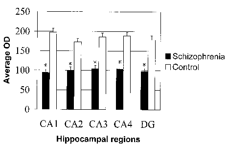
Figure 2 shows the mean optical density of SCIP stained neurons in the CA1,
CA2,
CA3, CA4 and dentate gyrus regions in schizophrenic and control groups.
Figure 3 shows Western blot analysis. Brain extracts from the frontal (Fs) and
temporal
lobe (Ts) of three schizophrenics were compared with similar brain regions (Fc
and Tc)
of matched controls using a polyclonal antiserum against SCIP. SCIP was
recognised as
a 45 KDa product.
EXAMPLES
MATERIALS AND METHODS
TISSUE PREPARATION
Human Tissue
Surgical samples were collected either from MRC Brain Bank, Institute of
Psychiatry,
King's College London, or acutely from surgical specimens. The demographic
characteristics of the samples used in Example 1 are described in Tables 1 and
2. There
were no significant differences in age, gender or post-mortem interval between
groups
(Table 3). Exclusion criteria covered any central nervous system related
disorders such
as head injury, alcohol dependence or Alzheimer's disease. Tissue was obtained
from
patients with a clinical diagnosis of schizophrenia according to DSM-ITI-R
criteria.
Mean neuroleptic exposure in the month prior to death was estimated for
schizophrenic
subjects and expressed in chlorpromazine equivalents (CPZE).
CA 02403779 2002-09-20
WO 01/75452 PCT/GBO1/01442
12
Separate tissue specimens were also obtained from patients with a pathological
diagnosis of either focal cortical dysplasia or Alzheimer's disease.
The tissue preparation was standard for histopathological specimens. The
specimens
were fixed in 10% formalin for between 24-48 hours, cut into between 4 and 20
slices
depending on the size of the specimen, then embedded in paraffin blocks and
sectioned
at 7 p.m.
Rodent Tissue
Tissue specimens were taken from BalbC mice over 40 weeks of age that had
undergone unilateral brain injury in the hippocampal region, and from Wistar
rats with
induced global ischaemia. The tissue specimens were fixed in 4%
paraformaldehyde
overnight at 4°C, embedded in paraffin wax and sectioned at 7 pm.
Neurotoxic Iniury
Adult rats or mice were injected infra-peritoneally with a compound known to
cause
neurotoxic effects, for example, phenytoin (75mg/kg) or 3-nitropropanoic acid
(120mg/kg). One day following this injection, the animals were killed using
standard
approved techniques, and their brains were removed and processed for
immunocytochemistry. This preparation is a standard procedure for those
knowledgable in the art. It involves fixation of the tissue with 4%
paraformaldehyde,
cryoprotecting the tissue by immersion overnight in 30% sucrose solution, then
freezing
of the tissue in liquid nitrogen. The tissue is then cut on a cryostat at a
thickness of
lOpM. The tissue sections are then processed for immunocytochemistry using
standard
procedures.
Preparation of antibody
The tissue sections are stained using an antibody that reacts specifically
with the
protein, SCIP. The antibody can be prepared according to the method of Meijer
et al.,
Nucleic Acids Res., 18. 7357-7365 (1990); Meijer et al., Nucleic Acids Res.,
20.
2241-2247 (1992). Typically, such an antibody can be raised against a purified
preparation of the protein prepared by over-expression of the protein in E.
coli, into
CA 02403779 2002-09-20
WO 01/75452 PCT/GBO1/01442
13
which has been introduced an expression plasmid encoding SCIP. This can be
achieved
by cloning the BamHI-BglII fragment from pNISCIP behind the Isopropyl
(3-D-Thiogalactopyranoside (IPTG) inducible T7 promoter in the BamHI site of
the
pETllA expression vector (Novagen). See Meijer et al., Nucleic Acids Res., 18,
7357-7365 (1990); Meijer et al., Nucleic Acids Res., 20 2241-2247 (1992). This
construct can then be transfected into the BL21 strain of E. coli. An
overnight culture is
diluted 1 in 10 and cultured at room temperature to an OD6oo = 0.8. Over-
expression is
induced by adding IPTG to a final concentration of 0.4 mM and the culture is
incubated
for 4 hours.
For large scale purification, a 500 ml IPTG induced bacteria culture is
pelleted, washed
once with Phosphate-Buffered Saline (PBS), resuspended in 10 ml 6M urea/PBS
and
sonicated. The cell lysate is cleared by centrifugation at 12000 rev./min for
5 min at
4°C.
Imidazole is added to the cell lysate to a final concentration of 0.8 mM and
incubated
overnight at 4°C with 300 p1 Ni-NTA beads (Qiagen). The following day,
the Ni-NTA
is washed twice with 10 ml of a 6 M urea/PBS/80 mM imidazole solution for 15
min
and three times with 6 M urea/PBS/8 mM imidazole solution. SCIP protein is
eluted
from the matrix in 500 p1 6 M urea/PBS/0.8 mM imidazole solution. This
purification
procedure produces high levels of pure (>95%) and intact SCIP protein as
judged by
Coomassie stained polyacrylamide gel electrophoresis (SDS-PAGE). See Zwart et
al.,
Mech. Dev., 54, 185-194 (1996).
Generation of anti-SCIP antiserum
Following over-expression and purification of the SCIP protein, antibodies can
be
raised in rabbits (White New Zealand) by three consecutive injections of 0.5-
1.0 mg
SCIP protein resuspended in Freund's adjuvant with a 4 weeks interval between
each
injection. See Zwart et al., Mech. Dev., 54, 185-194 (1996).
SCIP antibodies are then affinity purified by binding to the SCIP protein
immobilised
on nitrocellulose. After preincubation with 1% BSA/3% powdered milk/0.05%
CA 02403779 2002-09-20
WO 01/75452 PCT/GBO1/01442
14
Tween-20/PBS for 2 h at 4°C, the nitrocellulose is incubated overnight
with the
antiserum that has been precleared with BL21 cell lysate at room temperature
for 3 h.
After extensive washing with PBS the SCIP antibodies are eluted from the
nitrocellulose by 3 M KSCN/0.1 M NaPO~/500 pg/ml BSA solution. To remove the
KSCN the antibody solution is passed over a 0.1 M NaPOa (pH 7.5) equilibrated
Sephadex G-50 column. See Zwart et al., (supra).
The SCIP polyclonal antiserum raised by this method is highly specific since
it does not
cross react with other POU proteins such as Oct-1/3/4, Brn-1/3/4. In addition
to this,
there is great homology of isolated SCIP cDNA between human and rodents with
the
human sequence (Tobler et al., Nucleic Acids Res., 21, 1043 (1993) being 98.8%
homologous to the sequence of mice (Zimmerman et al., Nucleic Acids Res., 19,
956
(1991) and rats (He et al., Nature, 340, 6228 (1989); Monuki et al., Science,
249,
1300-1303, (1990)). The antibody can be used to detect rodent and human SCIP
protein in immunohistochemical applications.
Immunohistochemistry
The sectioned brain material was stained immunohistochemically to reveal the
presence
and location of immunoreactive SCIP in the tissue section. This was done using
standard immunohistochemical procedures.
Wax-imbedded sections were dewaxed and rehydrated in methanol. Frozen sections
were kept at -20°C, and brought to room temperature immediately before
use.
Thereafter the procedure for both types of material was the same. To block
non-endogenous peroxidase activity, the sections are incubated with
methanol/3% HZOz
solution for 20 min. After extensive washes first with distilled water and
then with
Tris-Buffered Saline (TBS), the sections are blocked with normal swine serum
(Dako),
diluted 1:10 in TBS, for 30 min at room temperature and then incubated in the
primary
anti-SCIP (1:250) antibody in TBS overnight at 4°C.
For bright-field microscopy, sections are incubated for 45 min with a
biotinylated
Swine anti-Rabbit secondary antibody at 1:200 (Dako) and then for 45 min with
an
CA 02403779 2002-09-20
WO 01/75452 PCT/GBO1/01442
avidin-biotin-peroxidase complex (Vector Laboratories), followed by a 5 min
reaction
with a diamino benzidine (DAB) /0.03% hydrogen peroxide in PBS kit (Vector
Laboratories). The samples are then dehydrated in an ethanol series, followed
by three
rinses in xylene, and then permanently mounted with DPX mounting medium and
coverslipped.
For fluorescence microscopy, immunolabelled sections are incubated for 1 h at
room
temperature with rabbit conjugated fluorescent markers at 1:200 (Vector).
Sections are
then embedded in anti-fade media (Vectashield) and coverslipped for storage.
Following the staining procedure, SCIP expression can be detected by light
and/or
fluorescent microscopy. Cells in the tissue sections that were expressing SCIP
will be
labelled by the antibody staining procedures. In normal undamaged adult brain
material, such cells are rare. This is an indication that the damage induced
SCIP
expression, and that the SCIP immunoreactivity is diagnostic of the damage,
and that
the sites of SCIP immunoreactivity are indicative of the sites of damage.
In situ Hybridisation
SCIP expression can be detected using in situ hybridisation (ISH) rather than
immunohistochemistry. In this case, the presence of mRNA encoding the SCIP
protein
is detected rather than the protein itself. ISH is a standard technique
familiar to those
practiced in the art (Wilkinson, D.G., In Situ Hybridisation: A Practical
Approach, 1st
Edn, 87-106, 1992). The sections from damaged brain material are dewaxed in
Histoclear three times for 10 min each, followed by 2 washes in methanol for 5
min
each. Then, sections are rehydrated through a graded series (100%, 75%, 50%
and 25%)
of methanols made up in PBT for 5 min each and washed twice with PBT for 5 min
each. After rehydration, sections are treated with 10 pg/ml proteinase K
(Boehringer
Mannheim) in PBT for 10 minutes at 37 °C; refixed in 4%
paraformaldehyde in PBS for
min and acetylated with 0.1 M triethanolamin acetate. Slides are then
dehydrated via
25%, 50%, 75% and 100% series of methanol for 5 min in each. To block non-
specific
binding of RNA probes, sections are prehybridized with a buffer containing 5 x
SSC
(0.3 M NaCI, 0.03 M sodium citrate, pH 7.4), 50% deionized formamide (BDH), lm
<IMG>
CA 02403779 2002-09-20
WO 01/75452 PCT/GBO1/01442
17
As with the immunohistochemical detection, the expression of SCIP can be
detected by
this method. Thus it will be apparent that SCIP expression has been
upregulated at
sites of neurological damage, and is thus a marker of those sites of damage.
EXA1VIPLE 1
ANALYSIS OF BRAIN TISSUE FROM PATIENTS WITH ALZHEIMER'S
DISEASE
Based on the methods described above blocks of temporal lobe were taken at the
level
of the lateral geniculate body and included the parahippocampal gyrus and
hippocampus. Blocks of the frontal lobe were taken at the level of the sharp
ventral
curve at the anterior end of the corpus callosum trunk. The subjects from
which the
samples are taken are shown in Table 2. All blocks used for
immunohistochemistry
were fixed in 10% formalin and subsequently coronally sliced before being
embedded
in paraffin wax.
Seven pm thick sections were stained using standard immunohistochemical
procedures
to reveal the presence and location of SCIP protein. Briefly, sections were
dewaxed,
rehydrated in methanol and pre-treated with 1% HzOz for 30 minutes. Sections
were
then microwaved at 800 W for eight minutes in a 0.001 % solution of citric
acid/phosphate buffer (pH 6.0). After extensive washes with Tris-Buffered
Saline
(TBS), the sections were blocked with normal swine serum (Dako), diluted 1:10
in
TBS, for 30 min and then incubated in the primary rabbit polyclonal anti-
SCIP(1:250)
antibody in TBS overnight at 4°C. The SCIP polyclonal antiserum used in
this study
was raised against the N-terminal region of SCIP, a region of least homology
with other
POU proteins such as Oct-1/3/4 and Brn-1/3/4. The three-step
avidin-biotin-horse-radish peroxidase complex system was used (Dako, Ltd) and
the
antibody was visualised using the chromogen diaminobenzidine (Vector).
Negative
controls consisted of duplicate sections that were processed in parallel and
consisted of
adjacent tissue sections in which the primary antibody was replaced by TBS.
CA 02403779 2002-09-20
WO 01/75452 PCT/GBO1/01442
18
Western blot
Protein extracts were prepared from the temporal and frontal lobes of three
schizophrenic and three control cases. Each extract was washed twice with PBS
and
lysed by the addition of 1 % Nonidet P-40 lysis buffer (0.5 M Tris-HCl pH 8.0,
3 M
NaCI, O.SM EDTA plus protease inhibitors: 2 pg of pepstatin per ml, 2 ~g of
leupeptin
per ml, 1 pg of peprotonin per ml) and vortexing. Solubilised samples were
then
centrifuged at 13,000 rpm at 4°C, for 10 min. The protein concentration
from each
extract was estimated by performing a DC protein assay (BioRad). After protein
quantification, samples were solubilised in standard sodium dodecyl sulfate
(SDS)
sample buffer (0.25M Tris-HCl pH 6.8, 0.2 % bromophenol blue, 40% glycerol,
20%
2-mercaptoethanol and 8% SDS), denatured, loaded on 10%Tris-Polyacrylamide
gels
(BioRad) and run at a constant 200 Volts for 35 minutes. The proteins were
then
transferred to 0.2 pm nitrocellulose paper (Sigma) using a semidry blotting
apparatus
(BioRad) and run at 10 Volts for 30 minutes. The blots were blocked with 10 %
casein
solution (Sigma) for 30 min and they were then treated with avidin C/ biotin
kit
according to the manufacturer's instructions (Sigma). Next, the membranes were
washed with TBS-T (25mM Tris-HCl pH 7.5, 0.5 M NaCI and 0.3% Tween 20) and
incubated with primary polyclonal antibody anti-SCIP(1:3500) in TBS-T for 30
minutes. Blots were washed with TBS-T and incubated with secondary
biotinylated
goat anti-rabbit antibody (Vector) for 30 minutes. Finally, a Vectastain ABC
complex
system was used (Vector) and the blots were treated with the chromogen
diaminobenzidine (Vector) until bands could be clearly seen. Negative controls
consisted of duplicate blots that were processed in parallel in which the
primary
antibody was replaced by TBS-T.
Image Analysis
All sections were analysed using a Leica light microscope with image analysis
software
(Image Pro-plus) and motorised stage. This system enabled us to tie together
separate
microscopic fields, viewed individually at high magnification to form single
composite
images of large strips encompassing the hippocampal formation. The boundaries
of the
hippocampal formation were drawn at low magnification and each subregion
delineated
using standard criteria described previously (Lorento de No, J. Psychiatry
Neurol.,
CA 02403779 2002-09-20
WO 01/75452 PCT/GBO1/01442
19
1934; 46: 113-177; Amaral DG, Insausti R: Hippocampal formation. In Paxinos G.
(Ed.), The Human Nervous System. Academic Press 1990; 711-756). In order to
randomly select neurons for each of the five regions, the image of the
hippocampal
composite was captured and a grid of crosses was placed on top of it.
The optical density of SCIP stained neurons was quantified in the CA1, CA2,
CA3,
CA4 and dentate gyros (DG) regions for both schizophrenic and control cases
using a
256-point grey scale. For the schizophrenic cases, the cytoplasmic staining of
neurons
whose nuclei were visible in section were analysed. For the control cases,
there was
sufficient background staining to enable us to identify the cytoarchitecture
of the
hippocampus and make comparable cytoplasmic analysis of neurons. Optical
density
readings were estimated only for neurons that were intersecting with the
crosses of the
grid. The mean optical density values across the fields of each region were
then
calculated.
Data were analysed using the Mann Whitney U rank sum test (SPSS 10.0). To
adjust
for multiple comparisons the Bonferroni correction factor was applied, and a p
value of
0.01 was considered significant.
Results
Immunohistochemical staining
SCIP was widely expressed in the hippocampus of all schizophrenic specimens
whilst
there was little or no staining above background in the control cases. SCIP
staining was
predominantly cytosolic and it was seen in the pyramidal cell layer of the
hippocampus
and in the granule cell layer of the dentate gyros (Fig 1). In the temporal
lobe of
schizophrenic samples, SCIP staining was more prominent in the CA2, CA3, CA4,
and
in the granule cell layer of the dentate than staining in the CA1. No similar
conclusions
could be drawn for the matched control sections as there was no or very little
SCIP
immunoreactivity present.
To assess the intensity of SCIP staining in the schizophrenic and control
samples, the
optical density patterns were quantified in the CA1, CA2, CA3, CA4, and in the
dentate
CA 02403779 2002-09-20
WO 01/75452 PCT/GBO1/01442
gyrus. Figure 2 shows mean optical density estimates per hippocampal subregion
for
control and schizophrenic samples. Mann Whitney U rank tests revealed that
there were
significant reductions in optical density measurements in the schizophrenic
group in all
5 hippocampal regions examined, with p values being less than 0.001 in all
cases
between schizophrenics and controls. This shows that the intensity of SCIP
staining
was significantly higher in the schizophrenic subjects than in the controls.
To explore the possibility that neuroleptic medication, age of subjects and/or
post-mortem delay may affect the expression of SCIP, the correlation of each
of the
above factors with the mean optical density values obtained for each of the 5
regions
using Spearman's rank correlation test was analysed. In the schizophrenic
group, there
was no significant correlation between SCIP staining and mean neuroleptic
exposure
(CPZE) (p>0.1 in all regions), neither was a significant relationship found
between
SCIP staining and age or post-mortem delay in any regions (p> 0.1 in all
cases).
Western Analvsis
Protein levels of SCIP were examined in extracts from the frontal and temporal
cortex
of three schizophrenics and three matched controls. Immunoblots confirmed that
the
SCIP antibody recognises a single protein of about 45 KDa, as expected. There
were
high levels of SCIP in the frontal and temporal lobe of the schizophrenic
specimens
whilst there was no or very little SCIP expression in the same regions of the
matched
controls (Fig 3).
The results demonstrates that extensive SCIP immunoreactivity is present in
the frontal
and temporal lobes of schizophrenic specimens, whilst there is limited
expression of
SCIP in matched controls. The findings indicate that SCIP is useful as a
neuropathological marker in schizophrenia as well as a marker of any
neurological
damage.
Testing of compounds for neurotoxici
The neurotoxicity of compounds can be tested according to the invention by
contacting
cells, tissues or animals with test compounds and testing for the expression
of SCIP by
CA 02403779 2002-09-20
WO 01/75452 PCT/GBO1/01442
21
methods described above. Increased levels of SCIP expression are indicative of
neurotoxicity and therefore compounds which do not lead to neurotoxicity are
selected.
Methods of contacting cells, tissue or animals are well known to those skilled
in the art.
All references referred to herein are hereby incorporated by reference.
CA 02403779 2002-09-20
WO 01/75452 PCT/GBO1/01442
22
Table 1: Cases used for the temporal lobe immunohistochemical study
Case Age Gender Diagnosis CPZE PM Cause of death
delay
1 24 M S 200 29 Renal failure
2 34 M S 4000 21 Myocarditis
3 46 F S 600 96 Cardiac arrest (OD)
4 49 M S 700 25 Ruptured aneurysm
62 M S 350 31 Peritonitis
6 68 M S 200 45 Myocardial Infarction
7 73 M S 0 25 Pneumonia
8 74 M S 3500 23 Myocardial Infarction
9 75 M S 500 94 Pneumonia
88 F S 0 20 Pneumonia
11 20 M C - 26 Multiple injuries
12 33 F C - 96 Pulmonary embolus
13 44 M C - 70 Myocardial infarction
14 51 M C - 15 Pneumonia
63 M C - 26 Coronary artery
occlusion
16 64 M C - 47 Myocardial infarction
17 76 M C - 41 Bronchopneumonia
18 80 F C - 31 Pulmonary embolus
19 80 M C - 35 Left ventricular
failure
86 M C - 6 Myocardial infarction
CPZE: mean daily neuroleptic exposure a month prior to death, in
chlorpromazine
equivalents; S: schizophrenia; C: control; PM post-mortem
CA 02403779 2002-09-20
WO 01/75452 PCT/GBO1/01442
23
Table 2: Cases used for frontal and temporal lobe Western analysis.
Case Age Gender Diagnosis CPZE PM Cause of death
delay
21 32 F S 500 46 Pulmonary
embolus
22 S 1 M S 800 44 Myocardial
Infraction
23 62 M S 300 36 Pulmonary
tuberculosis
24 33 F C - 56 Pulmonary
embolus
25 51 M C - 52 Chronic
cardiomyopathy
26 67 M C - 41 Myocardial
Infraction
CPZE: mean daily antipsychotic exposure a month prior to death, in
chlorpromazine
equivalents; S: schizophrenia; C: control; PM: post-mortem
CA 02403779 2002-09-20
WO 01/75452 PCT/GBO1/01442
24
Table 3: Comparison of demographic factors in schizophrenia and control groups
used
in the temporal lobe study and in the frontal versus temporal lobe study
Temporal lobe Frontal & Tem poral lobe
study study
Schizophrenia Control Schizophrenia Control
Age(years) 59.3 (20.3) 59.7 (22.2)48.3 (15.2) 50.3 (17.1)
Gender (M/F) 8M/2F 8M/2F 2M/1F 2M/1F
Post-mortem 40.9 (29.3) 39.3 (26.6)42 (5.3) 49.6 (7.8)
delay
Values are mean and (standard deviation)