Note : Les descriptions sont présentées dans la langue officielle dans laquelle elles ont été soumises.
CA 02403911 2002-09-27
WO 01/72766 PCT/USO1/10328
QUALITY CONTROL REAGENTS FOR NUCLEIC ACID MICROARRAYS
TECHNICAL FIELD
The present invention relates to nucleic acid
hybridization methodologies, and more particularly to quality
control reagents used in the course of conducting such
methods.
BACKGROUND OF THE INVENTION
Technology relating to genetic analysis has
substantially evolved over the past two decades, and
particularly during the last 10 years. The state of the art
entails the preparation of microarrays of hundreds, thousands
or in some cases, hundreds of thousands of oligonucleotides or
clones of DNA sequences of interest e.g., genes or portions
thereof implicated in human disease such as cancers,
Alzheimer's, etc. Formerly, the DNA molecules were cloned in
cells such as bacteria to generate sufficient quantities to
prepare the microarray. The advent of PCR technology provided
a much easier way to generate a large quantity of DNA. Thus,
rather than making copies of an entire vector, the DNA of
interest is flanked by primer sequences such as T7, T3, M13
forward, M13 reverse and SP6. The PCR reaction results in
amplification of the DNA and the flanking sequences. Once the
DNA is amplified or the oligonucleotides synthesized, it is
spotted onto the microarray. Microarrays are available
commercially or may be customized by an individual laboratory,
depending upon the specific DNAs or diagnostic application of
interest.
A DNA microarray can function properly only if each
DNA probe spotted onto the coated slide is firmly attached to
the slide and available for hybridization to the labeled
sample. Verification that each feature is functioning
CA 02403911 2002-09-27
WO 01/72766 PCT/USO1/10328
2
properly is vital to the subsequent quantitation and analysis
of the data. Thus, to enhance the precision and reliability
of diagnoses made based upon nucleic acid hybridization, the
microarrays are typically subjected to one or more types of
quality control. In general, these involve staining with a
fluorescent dye such as ethidium bromide or using single
fluroescently-labeled oligonucleotides. Quality control
specific to the microarray is a two-pronged issue, namely:
(1) has DNA been placed on the position on the microarray; and
(2) is it the DNA that was intended. Current quality control
methods are regarded as deficient in one or more respects
because there is a lack of functional testing for
hybridization and limited sensitivity.
Accordingly, there is a need for quality control
reagents to test DNA microarrays from these standpoints.
SUMMARY OF THE INVENTION
A first aspect of the present invention is directed
to a kit for conducting quality control reactions on a
microarray of nucleic acids. The kits contains the following
elements:
a container containing a first buffer solution
comprising a first reagent containing a nucleic acid matrix
carrying a detectable label, the matrix having attached
thereto an oligonucleotide probe that binds nucleic acid
contained on the microarray; and
directions for conducting the quality control
reactions with said first reagent and the nucleic acids on the
microarray.
In preferred embodiments, the matrix contains a polynucleotide
monomer having an intermediate region containing a linear,
double stranded waist region having a first end and a second
end, wherein the first end terminates with two single stranded
hybridization regions, each from one strand of the waist
region, and the second end terminates with one or two single
stranded hybridization regions, each from one strand of the
waist region. More preferably, each of the hybridization
CA 02403911 2002-09-27
WO 01/72766 PCT/USO1/10328
3
regions and the waist region of the monomer contains sequences
obtained from a master sequence containing no repeats of
subsequences having from 2 to 6 nucleotides. In other
preferred embodiments, the matrix contains a plurality of such
polynucleotide monomers bonded together by hybridization at at
least one such hybridization region.
The oligonucleotide probe is attached to the matrix
via ligation or hybridization and cross-linking. It may be
designed with a random sequence, in which case, it is
advantageously used as a qualitative reagent in the case that
it will detect the presence of nucleic acid on the microarray.
In other embodiments, the oligonucleotide has a sequence
substantially complementary to a known nucleic acid sequence
that is supposed to be present on the microarray. Thus, in
preferred embodiments, the oligonucleotide binds a primer
sequence such as T7, T3, M13 forward, M13 reverse or SP6.
Preferred detectable labels are fluorescent dyes
such as Cy3'a', CyS~'~', AlexaT''' 488 and AlexaTM 594.
In yet other preferred embodiments, the kit includes
a second container containing a second buffer solution for
conducting the quality control reactions. The kit may also
contain another container containing a second buffer solution
containing a second reagent. The second reagent differs from
the first reagent in that the detectable label is resolvable
from the detectable label on the first reagent and/or the
oligonucleotide binds different nucleic acid contained on the
microarray. Thus, many different quality control reactions
may be conducted substantially simultaneously.
The oligonucleotide probe does not have to be part
of the kit . It can be synthesized and attached to the matrix
by the end user. Accordingly, a second aspect of the present
invention is directed to a kit for conducting quality control
reactions on a microarray of nucleic acids, containing a first
container containing a first buffer solution containing a
nucleic acid matrix carrying a detectable label. The kit also
contains directions for (a) producing a reagent by attaching
CA 02403911 2002-09-27
WO 01/72766 PCT/USO1/10328
4
to said matrix an oligonucleotide having a first end portion
that attaches to the matrix and a second end portion that
binds nucleic acid on the microarray, (which preferably
includes the sequence of the outer arm or branch of the matrix
S to which the first end portion binds) and (b) conducting the
quality control reactions with the reagent and the nucleic
acids on the microarray. In preferred embodiments, the kit
also contains a second container containing a second buffer
solution in which to conduct the quality control reactions
between the reagent and the nucleic acid on the microarray.
As an alternative to the second aspect, the kit may
contain the oligonucleotide probe in a separate container.
Thus, in a third aspect, the present invention provides a kit
for conducting quality control reactions on a microarray of
nucleic acids, including:
a first container containing a first buffer solution
containing a nucleic acid matrix carrying a detectable label;
a second container containing a second buffer
solution containing an oligonucleotide having a first end
portion that attaches to the matrix and a second end portion
that binds nucleic acid on the microarray; and
directions for attaching the oligonucleotide to the
matrix to prepare the first reagent and for conducting the
quality control reactions with the first reagent and the
nucleic acids on the microarray.
In preferred embodiments, the oligonucleotide is
attached to the matrix indirectly, e.g., via hybridization and
cross-linking to a complement capture oligonucleotide that is
directly attached to the matrix. The complement capture
oligonucleotide may be provided already attached to the
matrix, in a separate container of the kit, or synthesized and
attached to the matrix by the end user. The kit may further
include a third container containing a third buffer solution
in which to attach the oligonucleotide probe to the matrix.
Methods for preparing the kits are also provided.
CA 02403911 2002-09-27
WO 01/72766 PCT/USO1/10328
A further aspect of the present invention is
directed to a method for conducting quality control reactions
on a microarray of nucleic acids. The method entails:
providing the microarray of nucleic acids;
5 providing a reagent comprising a nucleic acid matrix
carrying a detectable label, said matrix having attached
thereto an oligonucleotide that binds nucleic acid contained
on the microarray;
contacting the reagent with the microarray; and
detecting the label as an indication of the presence
or type of nucleic acid on the microarray. This aspect of the
invention pertains to the kits described above in connection
with the first aspect of the present invention.
Yet a further aspect of the present invention is
directed to a method for conducting quality control reactions
on a microarray of nucleic acids. This method entails:
providing the microarray of nucleic acids;
providing a nucleic acid matrix carrying a
detectable label, and attaching to the matrix an
oligonucleotide having a first end portion that attaches to
the matrix and a second end portion that binds nucleic acid on
the microarray;
contacting the reagent with the microarray; and
detecting the label as an indication of the presence
or type of nucleic acid on the microarray. This aspect
pertains to the use of the kits described in the second and
third aspects of the present invention. In preferred
embodiments, directions for conducting the reactions are also
provided.
BRIEF DESCRIPTION OF THE DRAWINGS
Figs. 1A and 1B are schematic representation of
elements of the quality control reagents useful in the present
invention;
Figs. 2 and 3 are schematic representations of
quality control reagents of the present invention; and
CA 02403911 2002-09-27
WO 01/72766 PCT/USO1/10328
6
Fig. 4 is a flow diagram that schematically
illustrates a method for conducting quality control reactions
of the present invention.
BEST MODE OF CARRYING OUT THE INVENTION
The present invention is directed to quality control
reagents for use with nucleic acid microarrays, kits
containing the reagents, and methods for preparing and using
the quality control reagents and kits.
Thus, one aspect of the present invention is
directed to the quality control reagents and their
intermediates. One element of the reagent is a labeled moiety
that contains a branched matrix composed of individual
oligonucleotides or nucleic acid-like molecules (a
polynucleotide matrix) that carries a plurality of detectable
labels . In one embodiment, the labeled moiety is attached to
a randomer. In another embodiment, it is attached to a DNA
sequence that hybridizes with a specific primer sequence. Yet
another embodiment is directed to an intermediate for the
preparation of a quality control reagent, and contains the
labeled moiety attached to a bridging oligonucleotide. A free
end of the bridging oligonucleotide serves as a point of
attachment for an oligonucleotide i.e., a probe that binds the
primer sequence and/or any portion of the arrayed DNA under
the conditions in which the products are used. The probe
oligonucleotide may be provided by or for the end user.
The Polynucleotide Matrix
A variety of branched nucleic acid matrices designed
to carry a plurality of labels are known in the art. See,
e.g., U.S. Patents 5,124,246 and 5,656,731 to Urdea, et al.
Preferred matrices exhibit a relatively highly ordered and
symmetrical architecture and are commonly referred to as
"nucleic acid matrices". Dendritic molecules, per se, are
highly-branched arborescent structures that were originally
assembled from organic polymers. They have found industrial
applications as chemical reagents, lubricants, contrast media
for magnetic resonance and the like. See, e.g., Barth et al.,
CA 02403911 2002-09-27
WO 01/72766 PCT/USO1/10328
7
Bioconjugate Chemistry 5:58-66 (1994); Gitsov & Frechet,
Macromolecules 26:6536-6546 (1993); Hawker & Frechet, J. Amer.
Chem. Soc. 112:7638-7647 (1990a); Hawker & Frechet,
Macromolecules 23:4726-4729 (1990b); Hawker et al., J. Chem.
Soc. Perkin Trans. 1:1287-1297 (1993); Lochmann et al. J.
Amer. Chem. Soc. 115:7043-7044 (1993); Miller et al., J. Amer.
Chem. Soc. 114:1018-1025 (1992); Mousy et al., Macromolecules
25:2401-2406 (1992); Naylor et al., J. Amer. Chem. Soc.
111:2339-2341 (1989); Spindler & Frechet, Macromolecules
26:4809-4813 (1993); Turner et al., Macromolecules 26:4617-
4623 (1993); Wiener,. et al., Magnetic Resonance Med. 31(1):1-8
(1994) and U.S. Patents 4,558,120; 4,507,466; 4,568,737;
4,587,329; 4,857,599; 5,527,524; and 5,338,532 to Tomalia.
Matrices offer several advantages over other molecular
architectures. First, they contact the maximum volume or area
with a minimum of structural elements. Second, the growth of
matrices can be highly controlled to yield molecules of ideal
size and molecular weight. Finally, the large number of
defined "ends" can be derivatized to yield highly labeled
molecules with defined spacing between the labels. Nucleic
acid matrices have been constructed following the technology
that was originally applied to conventional organic polymers.
See Hudson et al., "Nucleic Acid Dendrimers: Novel Biopolymer
Structures," Am. Chem. Soc. 115:2119-2124 (1993); and U.S.
Patent 5,561,043 to Cantor.
More preferred are nucleic acid matrices that have
some overall similarity to the aforementioned purely dendritic
structures but yet are structurally distinct therefrom. These
nucleic acid matrices are taught in U.S. Patents 5,175,270;
5,484,904 and 5,487,973 to Nilsen et al. The unique molecular
design of Nilsen's matrices accommodates a large number of
labels, in the order of several hundred, resulting in more
than a 100-fold amplification of the signal compared to
various prior art methods. Target nucleic acids can be
detected even when present in the sample in extremely small
(e. g., femptogram (1015)) amounts.
CA 02403911 2002-09-27
WO 01/72766 PCT/USO1/10328
8
These polynucleotides are defined in terms of a
plurality of polynucleotide monomers bonded together by
hybridization; each polynucleotide monomer having an
intermediate region comprising a linear, double stranded waist
region having a first end and a second end, the first end
terminating with two single stranded hybridization regions,
each from one strand of the waist region, and the second end
terminating with one or two single stranded hybridization
regions, each from one strand of the waist region; and in the
dendritic polynucleotide each polynucleotide monomer is
hybridization bonded to at least one other polynucleotide
monomer at at least one such hybridization region. Due to the
way in which these matrices are assembled, the outer layer of
monomers of the polynucleotide contains a plurality of free
hybridization arms. The number of such arms varies depending
upon the structure of the individual monomers and the number
of monomer layers contained in the polynucleotide. The
assembly via hybridization may begin with an initiator nucleic
acid molecule having three or more single stranded regions.
In these cases, hybridization of nucleic acid molecules to the
free single stranded ends of the initiator generates the first
layer product. In the case of hybridization of an initiator
with three arms with three-armed matrix monomers, a first
layer having six arms is produced. The more preferred seven
strand dendritic structure utilizes monomers with four arms;
consequently, the first layer possesses twelve arms.
Subsequent layers of hybridization lead to a geometric
expansion of the single-stranded ends and a three-dimensional
dendritic organization of nucleic acids.
In even more preferred embodiments, the
polynucleotides exhibit maximal self-assembly. In these
embodiments, each of said hybridization regions and said waist
regions of said plurality of monomers comprise sequences
containing no repeats of subsequences having X nucleotides,
wherein X is an integer of at least 2. In preferred
embodiments, X is an integer from 2 to 6 or 7; in more
CA 02403911 2002-09-27
WO 01/72766 PCT/USO1/10328
9
preferred embodiments, X is 3, 4, 5 or 6. These more
preferred matrices are assemblies of several layers of
monomers. The labeled moiety may contain just a single
monomer, however. See WO 99/06595.
As disclosed herein, the matrices per se may be
"nucleic acid-like" in the sense that their composition is not
limited strictly to the use of individual nucleotides and
nucleic acids. For example, the matrices may be assemblies
using peptide-nucleic acids (PNAs) or nucleic acid analogs
prepared in accordance with standard techniques.
In its broadest sense, the detectable label is any
compound employed as a means for detecting an oligonucleotide.
Examples of labels include fluorescent dyes, biotin,
digoxigenin, radionucleotides, antibodies, enzymes and
receptors such that detection of the labeled polynucleotide
(the labeled moiety) is by fluorescence, conjugation to
streptavidin and/or avidin, antigen-antibody and/or antibody-
antibody interactions, quantitation of radioactivity, and
catalytic and/or ligand-receptor interactions. Fluorescent
dyes are preferred. Examples include Cy3T'° and Cy5T°" (both
available from Amersham Pharmacia Biotech), fluorescein,
FluorX, Oregon GreenTM, the AlexasM series dyes ( e. g. , Alexa'~"
488 and 594), and the BODIPY1'°" series dyes, all of which are
commercially available from various sources including NEN,
Molecular Probes, Boehringer Mannheim and Amersham Life
Sciences.
The individual label molecules may be attached to
the polynucleotide matrix in several ways. Figs. 1A and 1B
are schematic illustrations of such, wherein the matrix
contains a single monomer. As shown in Fig. 1A, label
molecules 10 are attached to individual nucleotides of free
outer arms 12 and 12' of matrix 14. Fig. 1B illustrates a
preferred embodiment wherein label molecules 10 are attached
to individual nucleotide bases of oligonucleotides 16 and 16'
which are hybridized with free, single stranded outer arms 12
and 12' respectively, of polynucleotide monomeric matrix 18.
CA 02403911 2002-09-27
WO 01/72766 PCT/USO1/10328
The oligonucleotide has one end portion that hybridizes with a
branch or in the case of the more preferred embodiments, a
free outer arm, of the matrix. Such labeled oligonucleotides
are described in U.S. Patent 6,046,038. These embodiments
5 allow for enhanced detection capabilities that may or may not
be needed in the case of quality control, depending upon the
sensitivity of the instrumentation.
In a first preferred embodiment of the present
invention, the labeled polynucleotide matrix is directly
10 attached to an oligonucleotide that binds a target on the
microarray. The oligo can be attached to a branch or free
outer arm of the matrix by direct ligation or via
hybridization and cross-linking (for purposes of enhanced
stability). The sequence of the target complementary oligo
can be relatively random or specific in nature. An oligo
containing a random sequence is referred to as a randomer.
Generally, the sequence is from about 8 to about 20 bases.
Due to the random nature of the sequence, the product serves
well as a general quality control reagent because it
hybridizes with virtually any DNA molecule under the
conditions in which it is used. A binding event between the
quality control reagent and a position on the microarray
indicates that DNA has been spotted onto a specific position
thereon.
Alternatively, the product is relatively
"customized" and the target complementary sequence is referred
to as a specific complementary sequence. It is attached to
the matrix instead of a randomer. It is preferred that the
sequence is complementary to the primer sequences) that
flanks the DNA molecules contained in each of the wells which
more often than not, is the same for all positions on the
microarray. Examples of oligonucleotides complementary to
commonly used primer sequences are set forth below.
Sp6-7B0 Oliao
5'-ATT TAg gTg ACA CTA TAT TTT TCg -3' (SEQ ID N0:1)
CA 02403911 2002-09-27
WO 01/72766 PCT/USO1/10328
11
T7-7B0 Oliao
5'-TAA TAC gAC TCA CTA TAg ggT TTT TCg-3' (SEQ ID
N0:2)
T3-7B0 Oliao
5'- TAA CCC TCA CTA AAg ggA TTT TTC g-3' (SEQ ID N0:
3)
M13F-7B0 Oliao (M13 FORWARD)
5'- gTT gTA AAA CgA CCA gTg ttt ttc G-3' (SEQ ID
N0:4)
M13R-7B0 Oliao (M13 REVERSE)
5' CAC ACA ggA AAC AgC TAT gTT TTT Cg -3' (SEQ ID
N0:5)
Perfect complementarity for known primers (and other
nucleic acids) is the case if for no other reason than the
primer sequences are known. In general, however, perfect
complementarity is not required. Base mismatches can be
accommodated provided that the sequence binds the primer under
conditions in which the quality-control reagent is used (in
which case the oligonucleotide is said to have a sequence
substantially complementary to a nucleic acid believed to be
present on the microarray).
The product will be sold in the form of a kit . The
quality control reagent is separately contained in an
appropriate buffer solution, preferably a neutral buffer. The
kit may also contain a hybridization buffer to be used along
with the quality control reagent to actually conduct the
quality control hybridization reactions with the microarrayed
DNA. Other suitable buffers are commercially available e.g.,
ExpressHybT°z (Clontech), Ultrahyb~" (Ambion). Otherwise, they
may be prepared on an individual basis. The kit further
contains manufacturer's protocols or directions for use. Two
specific protocols, the first directed to a quality-control
reagent with a "randomer" sequence and the second directed to
a reagent having a sequence specific to a known primer, are
set forth below.
CA 02403911 2002-09-27
WO 01/72766 PCT/USO1/10328
12
In a second preferred embodiment of the present
invention, a free branch or outer arm of the polynucleotide
matrix serves as a complement capture oligonucleotide or is
attached to a complement capture oligonucleotide (e.g., by
S direct ligation or by hybridization and cross-linking). Fig.
2 schematically illustrates one such example wherein
complement capture oligonucleotide 21 is hybridized with a
portion of outer free arm 22 of matrix 23. Oligonucleotide 24
is bifunctional and contains one end portion 25 that functions
as a matrix capture sequence and binds to complement capture
oligonucleotide 21 or a portion thereof, and another end
portion or subsequence 26 that is a randomer or a specific
complementary sequence as described above. The matrix-capture
sequence is attached to the complement capture sequence or the
outer free arm of the matrix, preferably by hybridization
and/or cross-linking. Oligonucleotide 24 may be provided
along with a suitable buffer in a separate vial of the kit, in
which case the kit further contains a buffer in which to
conduct the attachment of oligonucleotide 24 to complement
capture oligonucleotide 21. Alternatively, the end user may
prepare oligonucleotide 24 and attach it to the matrix, in
which case, the protocol or directions further contain the
sequence of at least the portion or subsequence of complement
capture oligonucleotide 21 to which end portion 25 attaches.
Thus, use of this embodiment of the present invention entails
attaching the bifunctional oligonucleotide to the matrix
(e. g., via an outer free arm or indirectly via a complement
capture oligonucleotide) and then contacting the fully
assembled labeled matrix with the target sequences present on
the microarray. In Fig. 2, labels 27 are attached to the
matrix by oligonucleotide 28 that hybridizes with free outer
arm 29.
Plainly, modifications with respect to the
components in the kit and the procedures for using the
components are well within the skill of the routineer in the
art. For instance, the kit may contain, in separate
CA 02403911 2002-09-27
WO 01/72766 PCT/USO1/10328
13
containers, two or more quality control agents each of which
carries a label resolvable from the other label(s). The
differently labeled reagents may carry the same or different
target complementary oligonucleotide. Each individual reagent
may have specificity for more than one nucleic acid sequence
believed to be present on the microarray (e. g., by having
attached oligos that bind nucleic acids having different
sequences). Likewise, in the second preferred embodiment, the
kit may contain two or more types of B oligonucleotides that
contain subsequences that bind different primers.
Invariably, there is some precipitation or settling
of components in a hybridization buffer during storage. Thus,
in those embodiments of the present invention that include a
hybridization buffer, its components are re-suspended,
typically by heating and mixing, prior to use. The reagent is
assembled (if not supplied as such in the kit) then added to
the hybridization buffer. The resultant mixture is added to
the microarray, which is then covered and incubated under
suitable conditions to allow the nucleic acid binding events
to occur. In those cases wherein the detectable label is a
fluorescent dye, it is important that the array is stored in
the dark until scanned. The fluorescence of dyes,
particularly CyS, diminishes rapidly even in ambient light.
Following incubation, the microarray is washed with another
buffer solution to remove non-bound reagents. The microarray
is then scanned in accordance with standard procedures. In
preferred embodiments of the present invention wherein the
detectable labels are the fluorescent dyes Cy3T°" and Cy5T°",
which are scanned via dual channel analysis, it is preferred
that both channels are scanned simultaneously or that the Cy5T°"
channel is scanned first, followed by the Cy3a'°° channel.
Standard procedures e.g., for preparing the nucleic
acids, spotting the nucleic acids onto the microarrays, and
scanning the microarrays, typically entail on or more of the
following: preparation of total RNA from cultured human
cells; preparation of polyA+ mRNA from total human RNA;
CA 02403911 2002-09-27
WO 01/72766 PCT/USO1/10328
14
amplification and purification of cDNAs for microarray
manufacture; microarray manufacture and processing; generating
control mRNAs by in vitro transcription; , generating
fluorescent cDNA controls by linear PCR; preparation of
S fluorescent probes from total human mRNA; cDNA microarray
hybridization and washing; gene expression analysis with
microarrays; and mutation detection with oligonucleotide
microarrays. These procedures are described in M. Schena and
R.W. Davis (1998). Genes, Genomes and Chips. In DNA
Microarrays: A Practical Approach (ed. M. Schena), Oxford
University Press, Oxford, UK, in press; Schena, M. and R.W.
Davis (1998). Parallel Analysis with Biological Chips. in PCR
Methods Manual (eds. M. Innis, D. Gelfand, J. Sninsky),
Academic Press, San Diego, in press; Lemieux, B., Aharoni, A.,
and M. Schena (1998). Overview of DNA Chip Technology.
Molecular Breeding 4, 277-289; Schena, M., Heller, R.A.,
Theriault, T.P., Konrad, K., Lachenmeier, E., and R.W. Davis
(1998). Microarrays: biotechnology's discovery platform for
functional genomics. Trends in Biotechnology 16:301-306;
Heller, R.A., Schena, M., Chai, A., Shalom D., Bedilion, T.,
Gilmore, J., Woolley, D.E., and Davis, R.W. (1997); Discovery,
and analysis of inflammatory disease-related genes using cDNA
microarrays. Proceedings of the National Academy of Sciences
USA 94:2150-2155; Schena, M., Shalom D., Heller, R., Chai,
A., Brown, P.O., and R.W. Davis. (1996). Parallel Human Genome
Analysis: Microarray-Based Expression Monitoring of 1,000
Genes. Proceedings of the National Academy of Sciences USA 93:
10614-10619; Schena, M. (1996). Genome analysis with gene
expression microarrays. BioEssays 18:427-431; Schena, M.,
Shalom D., Davis, R.W. and Brown, P.O. (1995). Quantitative
monitoring of gene expression patterns with a complementary
DNA microarray. Science 270:467-470; Vishwanath, et al.,
Science 283:83-87 (1999); Nilsen, et al., J. Theor. Biol.
187:273-284 (1997); Sambrook, et al., (Eds.), Molecular
Cloning, A Laboratory Manual (2nd Ed.), Cold Spring Harbor
Laboratory Press (1989); and Ausubel, et al., (Eds.), Current
CA 02403911 2002-09-27
WO 01/72766 PCT/USO1/10328
Protocols in Molecular Biology, John Wiley & Sons, Inc.
(1998).
The following examples are intended to further illustrate
certain preferred embodiments of the invention and are not
5 limiting in nature. Unless indicated otherwise, all parts and
percentages are by weight.
EXAMPLE 1
Method for Detection and Quality Control using a Random
Oligonucleotide labeled DNA Matrix
10 A Detection Kit for cDNA Arrays
Kit Contents:
Vial 1 Random Sequence Cy3~ 3DNA~ Reagent
(Genisphere, Montvale, NJ). Use at 2.5 uL per
uL assay.
15 Vial 2 Hybridization buffer- 0.25 M NaP04, 4.5~
SDS, 1 mM EDTA, and 1X SSC. (Stored at -20sC in
the dark.)
Microarray preparation:
A microarray was prepared as directed by the
20 manufacturer or by customary protocol procedures. The nucleic
acid sequences containing the DNA or gene probes were
amplified using known techniques in polymerase chain reaction
(PCR), then spotted onto glass slides, and processed according
to conventional procedures.
3DNA~ Reagent preparation:
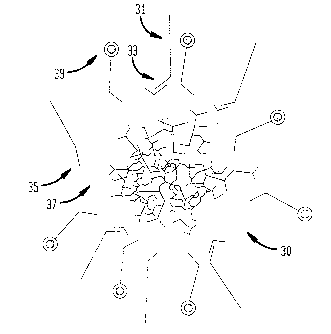
The Cy3~ 3DNA~ reagent is schematically illustrated in Fig. 3.
The reagent 30 was prepared as follows. Oligonucleotide 31
having the general structure outlined below was synthesized.
5'- -Matrix Sequence Complement-3', wherein N
represents a random nucleotide.
Matrix Sequence Complement 33 is an oligonucleotide sequence
that hybridizes to outer surface arms 35 of matrix 37. This
oligonucleotide was hybridized and cross-linked to DNA
matrices that were also labeled with about 250 Cy3
oligonucleotides 39.
3DNA~ Array Hybridization:
CA 02403911 2002-09-27
WO 01/72766 PCT/USO1/10328
16
The hybridization buffer of Vial 2 was thawed and
resuspended by heating to 65$C for 10 minutes. The buffer was
mixed by inversion to ensure that the components were
resuspended evenly. If necessary, the heating and mixing were
repeated until all the components were resuspended. Two- and
one-half (2.5) uL of 3DNA~ reagent of Vial 1 were added to
17.5 ~.zL of hybridization buffer to yield a hybridization
mixture. As schematically illustrated in Fig. 4, the
hybridization mixture including Cy3~ 3DNA~ reagent 42 was
added to microarray 44. The microarray was covered and
incubated at a temperature of from about 37-°C to 42°-C for
about 2-6 hours to overnight in a humidified chamber.
Post-Hybridization Wash:
The microarray was washed for 10 minutes at 42°-C
with 2X SSC buffer containing 0.2$ SDS. The microarray was
then washed for 10 minutes at room temperature with 2X SSC
buffer. The microarray was then washed for 10 minutes at room
temperature with 0.2X SSC buffer.
Signal Detection:
The microarray was then scanned as directed by the
scanner's manufacturer for detecting, analyzing, and assaying
the hybridization pattern.
EXAMPLE 2
Method for Detection and Quality Control using a Primer-
Specific Binding DNA Matrix
A Detection Kit for cDNA Arrays
Kit Contents:
Vial 1 Primer Specific Binding Cy3~ 3DNA~ Reagent
(Genisphere, Montvale, NJ). Use at 2.5 uL per
20 uL assay.
Vial 2 Hybridization buffer- 0.25 M NaPOa, 4.5~
SDS, 1 mM EDTA, and 1X SSC. (Stored at -20°-C in
the dark.)
Microarray preparation:
A microarray was prepared as directed by the
manufacturer or by customary protocol procedures. The nucleic
CA 02403911 2002-09-27
WO 01/72766 PCT/USO1/10328
17
acid sequences comprising the DNA or gene probes were
amplified using known techniques in PCR, then spotted onto
glass slides, and processed according to conventional
procedures.
3DNA~ Reagent preparation:
Oligonucleotides,
5' ATT TAG GTG ACA CTA TAT TTT CG -3' (SEQ ID NO:1) - SP6-7B0
5' TAA TAC GAC TCA CTA TAG GGT TTT TCG -3' (SEQ ID N0:2) - T7-
7B0
5' TAA CCC TCA CTA AAG GGA TTT TTC -3' (SEQ ID N0:3) - T3-7B0
5' GTT GTA AAA CGA CCA GTG TTT TTCG -3' (SEQ ID N0:4) -
Ml3Forward -7B0
5' CAC ACA GGA AAC AGC TAT GTT TTT CG -3' (SEQ ID N0:5) -
Ml3Reverse-7B0,
were synthesized by an outside vendor (Oligos Etc, Inc.
Wilsonville, OR), and ligated to the outer arms of a Cy3
labeled DNA matrix.
3DNA~ Array Hybridization:
The hybridization buffer of Vial 2 was thawed and
re-suspended by heating to 65°-C for 10 minutes. The buffer
was mixed by inversion to ensure that the components were re
suspended evenly. If necessary, the heating and mixing were
repeated until all the components were re-suspended. Two and
one-half (2.5) uL of 3DNA~ reagent of Vial 1 were added to
17.5 uL of hybridization buffer to yield a hybridization
mixture. The hybridization mixture was added to the
microarray. The microarray was covered and incubated at a
temperature of from about 37 to 42QC for about 2-6 hours to
overnight in a humidified chamber.
Post-Hybridization Wash:
The microarray was washed for 10 minutes at 42°-C
with 2X SSC buffer containing 0.2~ SDS. The microarray was
then washed for 10 minutes at room temperature with 2X SSC
buffer. The microarray was then washed for 10 minutes at room
temperature with 0.2X SSC buffer.
Signal Detection:
CA 02403911 2002-09-27
WO 01/72766 PCT/USO1/10328
18
The microarray was then scanned as directed by. the
scanner's manufacturer for detecting, analyzing, and assaying
the hybridization pattern.
Example #3
Method for Detection and Quality Control using a Random
Oligonucleotide with a Capture Sequence and a Cy3 Labeled DNA
Matrix
A Detection Kit for cDNA Arrays
Kit Contents:
Vial 1 Cy3~ 3DNA~ Reagent (Genisphere, Montvale,
NJ). Use at 2.5 uL per 20 uL assay.
Vial 2 Random Sequence Oligonucleotide with 3DNA
capture sequence
Vial 2 Hybridization buffer- 0.25 M NaP04, 4.5$ SDS, 1
mM EDTA, and 1X SSC. (Stored at -20°-C in the dark.)
Microarray preparation:
A microarray was prepared as directed by the
manufacturer or by customary protocol procedures. The nucleic
acid sequences containing the DNA or gene probes were
amplified using known techniques in PCR, then spotted onto
glass slides, and processed according to conventional
procedures.
3DNA~ Reagent preparation:
An oligonucleotide having the general structure
outlined below was synthesized.
5' - GGC CTC ACT GCG CGT CTT CTG TCC CGC CTT TTT CG -3' (SEQ
ID N0:6)
---Matrix Capture Sequence Complement
This oligonucleotide was ligated to a Cy3 labeled matrix. The
matrix capture sequence complement is an oligonucleotide
sequence that hybridizes to the 5' end of a bifunctional
oligonucleotide (contained in vial #2), one end of which binds
to sequences spotted on a microarray, in this case random
sequences, and a second end which hybridizes to the
complementary sequence attached to the matrix.
CA 02403911 2002-09-27
WO 01/72766 PCT/USO1/10328
19
Random sequence oligonucleotide with 3DNA capture sequence:
An oligonucleotide having the general structure
outlined below was synthesized.
5'- nfNNNNNNNN-Matrix Capture Sequence-3'
3DNA~ Array Hybridization:
The hybridization buffer of Vial 2 was thawed and
re-suspended by heating to 65qC for 10 minutes. The buffer
was mixed by inversion to ensure that the components were re-
suspended evenly. If necessary, the heating and mixing were
repeated until all the components were re-suspended. Two and
one-half (2.5) uL of 3DNA~ reagent of Vial 1 were added to
17.5 uL of hybridization buffer to yield a hybridization
mixture. The hybridization mixture was added to the
microarray. The microarray was covered and incubated at a
temperature of from about 37 to 42°-C for about 2-6 hours to
overnight in a humidified chamber.
Post-Hybridization Wash:
The microarray was washed for 10 minutes at 42qC
with 2X SSC buffer containing 0.2$ SDS. The microarray was
then washed for 10 minutes at room temperature with 2X SSC
buffer. The microarray was then washed for 10 minutes at room
temperature with 0.2X SSC buffer.
Signal Detection:
The microarray was then scanned as directed by the
scanner's manufacturer for detecting, analyzing, and assaying
the hybridization pattern.
INDUSTRIAL APPLICABILITY
The invention is useful in the field of diagnostics,
particularly as it pertains to screening individuals
All patent and non-patent publications cited in this
specification are indicative of the level of skill of those
skilled in the art to which this invention pertains. All
these publications and patent applications are herein
incorporated by reference to the same extent as if each
individual publication or patent application was specifically
CA 02403911 2002-09-27
WO 01/72766 PCT/USO1/10328
and individually indicated as being incorporated by reference
herein.
Those skilled in the art will recognize, or be able
to ascertain, using no more than routine experimentation,
5 numerous equivalents to the specific substances and procedures
described herein. Such equivalents are considered to be
within the scope of this invention, and are covered by the
following claims.