Note : Les descriptions sont présentées dans la langue officielle dans laquelle elles ont été soumises.
CA 02409717 2002-11-05
WO 01/85992 PCT/GBO1/02099
1
ASSAY FOR CELL CYCLE MODULATORS BASED ON THE MODULATION OF
CYCLIN D1 DEGRADATION IN RESPONSE TO IONISING RADIATION
Field of the Invention.
The present invention relates to the finding that cyclin D1 is
targeted for destruction in cells which have been exposed to
ionising radiation (IR). The finding gives rise to novel
targets for the control of the cell cycle and the treatment of
diseases such as cancer.
Background to the Invention.
Cyclins are essential components of the cell cycle machinery.
They function to bind and activate their specific cyclin
dependent kinase (CDK) partners. During progression through
the G1 phase of the cell cycle two major types of cyclins are
required: D-type cyclins and cyclin E. Together they cause
phosphorylation of the retinoblastoma family of tumor
suppressor proteins (pRb, p107, and p130) in G1 and abrogate
their inhibitory activity (Lipinski and Jacks, 1999). The
three D type cyclins are very similar (more than 70% identity)
but share very little homology with cyclin E. The D cyclins
activate primarily CDK4 and 6 whereas cyclin E activates CDK2.
Furthermore, during cell cycle progression D cyclins are
active at mid-G1 whereas cyclin E appears later just prior to
the G1/S transition (Draetta, 1994; Sherr, 1994; Sherr and
Roberts, 1995). Therefore, progression through Gl depends
initially on D cyclin-CDK4/6 protein complexes and later on
cyclin E-CDK2. Given the crucial part that D type cyclins
play in progression through the cell cycle, it is perhaps not
surprising that their expression is frequently deregulated in
cancer (Sherr, 1995).
CA 02409717 2002-11-05
WO 01/85992 PCT/GBO1/02099
2
Cell cycle arrest in response to either mitogen deprivation or
genotoxic stress requires CDK inhibitors (CKIs) of the CIP/KTP
family which includes p21°1p1, p27xipl and p57kipz (Morgan, 1995;
Sherr, 1995). Members of this family bind both CDK2 and CDK4
complexes, but act as potent inhibitors of cyclii~. E-CDK2
protein complexes and as positive regulators in the case of D
cyclins-CDK4/6 (Sherr and Roberts, 1999). D type cyclins
connect extracellular signalling pathways to the cell cycle
machinery as their promoters respond to a variety of mitogenic
signals, such as those transduced by the Ras and APC-~i-
catenin-Tcf/Lef pathways (Morin, 1999; Tetsu and McCormick,
1999). Furthermore, mitogen deprivation accelerates cyclin D1
proteolysis via the PI3K-PKB/Akt-GSK3-~3 pathway. GSK3-(3
phosphorylates cyclin D1 at threonine 286, which triggers its
nuclear export, ubiquitination and degradation (Diehl et al.,
1998; Diehl et al., 1997). Mitogenic signals activate the
PI3K-PKB/Akt pathway, which in turn inhibit GSK3-~i kinase
activity and stabilize cyclin D1 protein. Expression of C-Myc
also causes activation of the cyclin D1 and D2 promoters.
Increased protein levels of D cyclins results in complex
formation with their CDK partners, which function to sequester
p21°lPl and p27kipl away from cyclin E-CDK2 complexes, allowing
G1-S progression (Bouchard et al., 1999; Perez-Roger et al.,
1999) .
DNA damage checkpoints control the timing of cell cycle
progression in response to genotoxic stress (reviewed in
(Weinert, 1998)). Arrest in G1 is thought to prevent aberrant
replication of damaged DNA and arrest in G2 allows cells to
avoid segregation of defective chromosomes. Primary among
mammalian checkpoint genes is the tumor suppressor p53. In
response to DNA damage, such as IR, p53 is required for G1
arrest (Kastan et al., 1991; Kastan et al., 1992; Kuerbitz et
CA 02409717 2002-11-05
WO 01/85992 PCT/GBO1/02099
3
al., 1992; Livingstone et al., 1992; Yin et al., 1992),
apoptosis (last reviewed in Sionov and Haupt, 1999) and to
sustain arrest of cells prior to M phase (Bunt et al., 1998;
Chan et al., 1999). In response to IR, rapid phosphorylation
of p53 by the ATM-CHK2 pathway on serines 15 and 20, leads to
release of Mdm2 and stabilization of p53 (Meek, 1999 and
references therein).
Since p53 acts primarily as a transcription factor,
stabilization of p53 activates transcription of target genes
required for various aspects of the genotoxic stress response.
In particular, p53 transactivation is required to induce an
efficient G1 arrest (el-Deiry et al., 1993; Waldman et al.,
1995). An essential transcriptional target of p53 in
induction of G1 arrest is p21°lpl (Waldman et al., 1995).
Accumulation of p21°ipl inhibits cyclin-E/CDK2 activity and
therefore G1-S transition. However, as this p53 response
depends on transcriptional activation, the time required to
execute this type of cell cycle arrest is rather long and
exceeds in most cases eight hours.
Disclosure of the Invention.
We have now found that cells initiate a fast and efficient,
p53-independent, G1 arrest after DNA damage caused by IR. We
have identified a p53-independent mechanism that implements an
efficient G1 arrest immediately after exposure to genotoxic
stress. In particular, we have found that IR, an inducer of
DNA damage, induces a rapid degradation of cyclin D1 in cells,
and that this inhibits progression of cells through the G1
phase of the cell cycle. Degradation of cyclin D1 is mediated
through a motif "RXXL" found in the N-terminal region of
cyclin D1.
CA 02409717 2002-11-05
WO 01/85992 PCT/GBO1/02099
4
We have also found that in tumour cells which express cyclin
D1 appear to retain this rapid response. This finding has
potential relevance in the treatment of cancer by irradiation,
where problems may be encountered in overcoming the resistance
of cells to irradiation. Because irradiation induces a G1
arrest in tumour cells, this may provide the cells with an
opportunity to initiate DNA repair prior to replication, thus
ensuring survival of the tumour. By blocking this protective
mechanism, the efficacy of therapy in which DNA damage is
induced in target cells may be enhanced.
Accordingly, the present invention provides an assay for a
modulator of cell cycle control, which assay comprises:
(a) providing a cell in culture together with a
potential modulator compound, said cell expressing a
cyclin D1 which is undergoes degradation in response
to DNA damage;
(b) exposing said cell to a DNA damaging agent; and
(c) determining the extent to which the presence of the
potential modulator compound inhibits the
degradation of said cyclin D1.
The potential modulator compound may be a cellular protein,
which can be introduced into the cell by providing for its
expression from a cDNA. Accordingly, another aspect of the
invention provides a method to discover genes whose protein
products participate in the same signalling pathways as cyclin
Dl degradation. Thus the invention provides an assay which
comprises:
(a) providing a cell in culture, said cell expressing a
cyclin D1 which undergoes degradation in response to
DNA damage;
(b) introducing into said cell a member of a cDNA
CA 02409717 2002-11-05
WO 01/85992 PCT/GBO1/02099
library operably linked to a promoter which
expresses said cDNA in said cell;
(c) exposing such cell to a DNA-damaging agent and
determining the extent to which the expression of
said cDNA modulates the degradation of said cyclin
Dl; and optionally
(d) isolating said cDNA.
In a further aspect, we have found that the "RXXL" motif, when
transplanted to a different protein (in the examples below,
cyclin D2), acts as a destruction box which directs the
protein for degradation in response to IR. Thus in a further
embodiment of the invention, there is provided an assay which
comprises:
(a) providing a cell in culture together with a
potential modulator compound, said cell expressing a
reporter protein having an RXXL destruction box and
which protein undergoes degradation in response DNA
damage;
(b) exposing said cell to a DNA damaging agent; and
(c) determining the extent to which the presence of the
potential modulator compound inhibits the
degradation of said reporter protein.
In another aspect, our experiments suggest that the cyclin D1-
derived RXXL motif targets cyclin D1 (or a protein comprising
this motif) to the anaphase promoting complex (APC) of a cell.
The APC is a complex of about a dozen proteins which regulate
various aspects of the cell cycle. While not wishing to be
bound by any one particular theory, it is believed that the
APC marks cyclin D1 for proteolysis. The interaction between
the APC and the cyclin D1 provides a further target for
therapeutic intervention. Thus in this aspect, the invention
CA 02409717 2002-11-05
WO 01/85992 PCT/GBO1/02099
6
provides an assay for a modulator of the cell cycle which
assay comprises:
(a) providing a cyclin D1, the APC or a component
thereof which interacts with cyclin D1, together
with a potential modulator compound; and
(b) determining the extent to which the presence of the
potential modulator compound inhibits the
interaction of said cyclin D1 and APC or component
thereof.
The data provided herein indicate that the interaction between
cyclin D1 and the APC may be mediated by CDK4. Thus in the
abovementioned aspect of the invention, the assay may be
performed in the presence of a CDK4.
Our experiments suggest that in the interaction between cyclin
D1 and the APC, the protein to which cyclin Dl binds is Cdc20,
an activator of the APC. It is believed that Cdc20 is a
crucial component for the degradation of cyclin D1 in response
to DNA damage, by this pathway. Thus the abovementioned
aspect of the invention further provides an assay wherein the
component of the APC which interacts with cyclin D1 is a Cdc20
protein.
These and other aspects of the invention are set out below.
Brief Description of the Figures
Figure 1. Initiation and maintenance of G1 arrest induced by IR.
The percentage increase in G1 is shown as the
difference in o G1 content between irradiated and
control cells.
CA 02409717 2002-11-05
WO 01/85992 PCT/GBO1/02099
7
Figure 2. Genotoxic stresses induce rapid and specific
degradation of cyclin D1 protein.
The estimated half-life of cyclin D1 protein is shown.
Figure 3. Cyclin D1 degradation after genotoxic stress is
independent of GSK3-(3.
GSK3-~i activity in response to IR is shown.
Figure 4. A destruction motif in cyclin D1 is required for
degradation by genotoxic stress.
(A) Sequence comparison of the cyclin Dl RxxL motif
and neighboring amino acids to cyclin D2, D3, E, Ume3p
and cyclins A and B.
(B) Half life of wild type and L32A mutant cyclin D1
Figure 5. Degradation of cyclin D1 is required for initiation of
G1 arrest by IR.
(A) Expression of a histone H2B-GFP fusion construct.
(B) Ability of mutants of cyclin Dl to block the
initiation of a Gl arrest.
(C) Incorporation of BrdU in MCF-7/E6 cells was used
to measure effects on S phase in response to TR.
(D) Examination of the requirement for cyclin D1
degradation in the presence of p53 activity.
(E) S-phase response to IR of primary MEFs lackinge
cyclin D1.
Figure 6. Abrogation of cyclin D1 degradation sensitizes to IR.
(A) Survival of cells rendered unable to degrade
cyclin D1 in response to IR.
(B) Effect of IR on immortalised MEFs derived from
cyclin D1 knockout mice (D1-~-), cyclin E knockin mice
(D1-~--E) and wild type MEFs.
CA 02409717 2002-11-05
WO 01/85992 PCT/GBO1/02099
8
Detailed Description of the Invention.
DNA damage inducing agents include ionizing radiation as well
as other DNA damaging agents used in chemotherapy, such as
cis-platin or anthracyclins such as doxorubicin or its
hydrochloride salt, adriamycin. Such agents are widely used
in cancer therapy and doses, routes and modes of
administration are well understood by the skilled
practitioner.
In assays of the invention, the cyclin D1 may be any suitable
mammalian cyclin D1, particularly human cyclin D1. Human D1
Cyclin has been cloned and sources of the gene can be readily
identified by those of skill in the art. See for example,
Xiong et al, 1991, Cell 65; 691-699 and Xiong et al, 1992,
Genomics 13; 575-84. Murine D1 cyclin has also been cloned.
Other mammalian cyclins can be obtained using routine cloning
methods analogous to those described in the aforementioned
references.
Although wild-type cyclin D1 is preferred, mutants of D1 which
still retain the ability to target the cyclin for destruction
in response to DNA damage may also be used. Examples of
cyclin D1 mutants are well known in the art and a particular
mutant is illustrated in the accompanying Examples. For
example, the mutant may the cyclin D1-T286A mutant.
It is not necessary to use the entire cyclin D1 proteins for
assays of the invention. Fragments of the cyclin may be used
provided such fragments retain the RXXL motif described herein
and retain the ability to be targeted for destruction in a
cell in response to DNA damage. Fragments include N-terminal
fragments which retain the CDK4 binding domain as well as the
CA 02409717 2002-11-05
WO 01/85992 PCT/GBO1/02099
9
RXXL motif.
Fragments of cyclin D1 may be generated in any suitable way
known to those of skill in the art. Suitable ways include,
but are not limited to, recombinant expression of a fragment
of the DNA encoding the cyclin. Such fragments may be
generated by taking DNA encoding the cyclin, identifying
suitable restriction enzyme recognition sites either side of
the portion to be expressed, and cutting out said portion from
the DNA. The portion may then be operably linked to a
suitable promoter in a standard commercially available
expression system. Another recombinant approach is to amplify
the relevant portion of the DNA with suitable PCR primers.
Small fragments of the cyclin (up to about 20 or 30 amino
acids) may also be generated using peptide synthesis methods
which are well known in the art.
The ability of suitable fragments to be targeted for
destruction in response to DNA damage may be tested using
routine procedures such as those described in the accompanying
examples. Reference herein to cyclin D1 includes the above
mentioned mutants and fragments which are functionally able to
retain this property, and desirably also retain the ability to
bind to activate CDK4 and/or CDK6.
The cyclin D1 may be expressed as a fusion with a marker
protein, for example a protein which can be detected via its
enzymatic or colourimetric (e.g. fluorescent, luminescent or
the like) properties. Fox example, the cyclin D1 may be fused
with green fluorescent protein (GFP) in order to provide a
visual marker within a cell. Other marker proteins include
chloramphenicol acetyl transferase, luciferase, beta-
galactosidase, horseradish peroxidase, and the like.
CA 02409717 2002-11-05
WO 01/85992 PCT/GBO1/02099
In a further embodiment, the RXXL motif of the cyclin D1 may
be inserted into such a marker protein in order that the
marker protein itself is targeted for destruction by a cell in
response to DNA damage. The motif may be inserted into the
protein in a location so as to retain the activity of the
protein, e.g. fluorescence. Those of skill in the art will be
able to determine suitable sites, for example between regions
of secondary structure or folded domains, as well as the N-
and C- termini. One or more of these motifs (e.g. from 2 to
10, such as 2, 3, 4 or 5), which may be the same or different,
may be inserted into such proteins, for example at different
locations or in tandem.
It will be understood that the identity of the second and
third amino acids, "XX" of the motif may be the same or
different and may each be any amino acid. Examples of RXXL
motifs include RAML, RQKL, RAAL and RTAL. These or other
variations may be used. Preferably, the amino acid side chain
is non-aromatic and non-cyclic, for example selected from A,
G, T, M, S, C, V, L and I.
The motif may be inserted into the marker protein with
flanking sequences found in a naturally occurring cyclin D1,
for example up to 5, 10 or 20 contiguous residues found N-
and/or C-terminal to the motif.
The cyclin D1 or reporter protein will generally be generated
within a cell by means of recombinant expression. Vectors for
the production of these proteins are illustrated in the
accompanying examples, and analogous techniques, which are
well known in themselves, may be used by those of skill in the
art in providing analogous vectors to produce proteins for
CA 02409717 2002-11-05
WO 01/85992 PCT/GBO1/02099
11
assays within the scope of the present invention. Recombinant
expression in a cell may be via transient or stable
transfection of the cell.
In the abovementioned aspect of the invention which comprises
introducing an expressible cDNA into a cell, the cDNA will
usually be a member of a cDNA library. Conveniently, the cDNA
will be carried by a vector such as a retroviral or adenoviral
vector which allows introduction of the cDNA into the cell by
infection with a viral particle. In a preferred aspect, the
method of the invention will be practised on a multiplicity of
members of the cDNA library simultaneously, for example by
infecting cells at a multiplicity of infection of 1 virus per
cell, and plating said cells into separate wells of microtitre
plates, e.g. one or more 96-well plates. The cells will be
allowed to grow to provide clonal populations in each well
which may then be assayed in~accordance with the invention.
cDNA libraries may be provided from a range of species, though
most preferably of the species corresponding to the Cell type
in which the assay of this embodiment of the invention is
performed. Mammalian, particularly human, cDNA libraries are
preferred. The cDNA libraries may be obtained from a range of
tissue sources, including liver, lung, muscle, nerve, brain
cells. The cells may be fetal, normal human or tumour cells.
An example of the production and use of a retroviral cDNA
library may be found in Whitehead et al, 1995, Mol. Cell.
Biol., 15; 704-710.
Where the assay of the invention is conducted within a cell,
the effect on the degradation of the cyclin D1 or reporter
protein (reference henceforth to cyclin D1 in assays of the
invention will be understood to include reporter proteins
CA 02409717 2002-11-05
WO 01/85992 PCT/GBO1/02099
12
unless specifically indicated to the contrary) may be
determined by any suitable means. For example, the amount of
the protein may be measured directly, e.g. in the case of a
fluorescent reporter by measuring the fluorescence with the
cell (or generally a culture of cells), or by immunoassay
techniques which determine in a quantitative or qualitative
manner the amount of that protein in the cell.
Alternatively, the status of the cell cycle may be observed,
for example the cell cycle distribution of cells may be
observed, to determine whether the presence of the potential
modulator compound has reduced the amount of cells in G1 phase
due to the inhibition of cyclin D1 destruction.
It will be appreciated that the above-described assays of the
invention will be conducted by reference to suitable controls,
which may be either run in parallel with any of the assays, or
conducted under a set of reference conditions which are
reproduced in the assay, apart from the presence of a
potential modulator compound.
In another aspect of the invention, there is provided an assay
which relates to the interaction of cyclin D1 protein and the
APC or component thereof which interacts with said protein.
It is known in the art that the progression of eukaryotic
cells through the cell cycle is controlled by a number of
events, including the regulated association of specific
cyclins with a CDK (cyclin-dependent-kinase). At the end of
mitosis, mitotic cyclin degradation is required. In
eukaryotic cells which have been studied, including yeast,
Xenopus oocytes and clam oocytes, degradation of cyclin B is
mediated by a complex of proteins called the anaphase
CA 02409717 2002-11-05
WO 01/85992 PCT/GBO1/02099
13
promoting complex (APC) which functions as a cell cycle-
regulated ubiquitin-protein ligase (Zachariae et al, Science,
1996, 274;1201-1204). The APC is part of the essential cell
cycle machinery whose components are evolutionarily conserved
(Irniger et al., 1995 Cell, 81, 269-78; King et al., 1995
Cell, 81, 279-88; Tugendreich et al., 1995 Cell, 81, 261-268;
Peters et al., 1996 Science, 274, 1199-1201; Zachariae et al.,
1996 Science, 274, 1201-4) . In yeast CDC16, CDC23, CDC26,
CDC27 and APCI have been identified as genes coding for some
of these components (Lamb et al., 1994 EMBO J., 13, 4321-
4328; Irniger et al., 1995 ibid; Zachariae et al., 1996,
ibid). WO 98/21326 describes the APC complex and methods for
analysing components thereof.
Members of the APC include Cdcl6 (also referred to as APC6),
Cdc23 (also referred to as APCB), Cdc26, Cdc27 (also referred
to as APC3), APC1 and APC2.
Such polypeptides may be obtained from a wide variety of
sources, including fungi, such as S.cerevisiae or S.pombe,
Aspergillus spp and Candida spps, invertebrates such as
Drosophila, vertebrates including amphibians such as .Xenopus
and mammals such as mice and other rodents or primates
including humans. The sequences of these proteins are widely
available from a number of sources, and vectors encoding these
proteins are also available. For example, Sikorski et al,
(1993) Mol. Cell Biol., 13, 1212-1221 and (1990) Cell 60, 307-
317) disclose Cdc23 from S.cerevisiae and a number of variants
thereof, including thermolabile variants. Human cdc23 (APC8)
is found on Genbank accession number 3283051 and C.albicans
APC8 on pla~Ce 396132:A03 Forward of the Candida genome
project. Lamb et al (EMBO J., ibid) describe Cdcl6, Cdc23 and
Cdc27 from S.cerevisiae and their interaction by two-hybrid
CA 02409717 2002-11-05
WO 01/85992 PCT/GBO1/02099
14
assay and co-immunoprecipitation. Reference is also made by
these authors to sources of Cdc27 from S.pombe, Aspergillus
nidulans, Drosophila melanogaster and humans, and to Cdcl6
from S.pombe. Cdc27 and Cdcl6 activity in Xenopus eggs has
been analysed by King et al (Cell, 1995, 81;279-288). Human
Cdc27 and Cdcl6 cDNAs are described by Tudendreich et al
(Cell, 1995, 81;261-268). The Cdcl6 cDNA was obtained by
analysis of an EST database with a known Cdcl6 sequence to
identify a partial human Cdcl6 cDNA sequence, which was then
used to construct a full length cDNA. This technique may be
used to identify other members of the APC from sources, where
such sources are not presently available in the art. Human
cdc27 and cdcl6 sequences are also identified in US Patent
5,726,025.
APC8 is one of three APC components which comprise multiple
copies of a 34 amino acid repeat motif, termed TPR (Hirano et
a1, 1990 Cell 60, 319-328; Sikorski et a1, 1990, ibid),
arranged as a block of tandem TPRs in the C-terminus, with one
or two additional TPRs in the N-terminus. It has been
proposed that TPRs mediate protein-protein interactions (Lamb
et al, 1994, ibid) and thus in addition to APC8, cdcl6,and
cdc27 polypeptides are also of interest as second components
in the assay of the invention.
Polypeptides which are fragments, variants and fragments
thereof of the APC members may also be used, provided that
such polypeptides retain the ability to interact with a cyclin
D1 protein, particularly a cyclin D1 protein of the same
species as the APC member. Variants and fragments may be made
by routine recombinant DNA techniques, as discussed above for
the production of cyclin D1.
CA 02409717 2002-11-05
WO 01/85992 PCT/GBO1/02099
Thus assays of the present invention include assays in which
the interaction between cyclin D1 and the APC is examined
within a cell in which the APC has been produced by the cell,
as well as assays in which one or more components of the APC
are provided as isolated proteins and brought into contact
with an isolated cyclin D1 protein, under conditions in which
the two proteins, in the absence of a potential modulator,
interact.
In the case of the former, the interaction of the cyclin Dl
and APC may be determined by means such as detecting one of
the two components, for example by immunological means,
followed by detecting whether or not the second of these
components is associated with the first. For example, as
illustrated herein, the interaction is determined by
immunoprecipitation of a cell extract using an antibody
against the APC subunit Cdc27 followed by immunoblotting the
precipitated material to confirm the presence of cyclin D1.
In the case of the latter, the interaction may be determined
by providing an isolated component of the APC and the cyclin
D1 protein, and directly observing the interaction between the
two. Those of skill in the art may select any APC component
using routine methodology to determine which, in the absence
of a potential modulator compound, provides an interaction
which. is suitable for detection by the particular assay format
selected. For example, the APC component may be selected from
any of those mentioned above, such as Cdcl6 (also referred to
as APC6), Cdc23 (also referred to as APC8), Cdc26, Cdc27 (also
referred to as APC3), APCl and APC2. The component may also
be, either alternatively or in addition, an activator of the
APC such as a fizzy-related protein, e.g. such as Cdc20 and
Hctl.
CA 02409717 2002-11-05
WO 01/85992 PCT/GBO1/02099
16
As indicated above, our experiments have shown that the
component of the APC which binds cyclin Dl is a Cdc20 protein.
Thus in a preferred embodiment of the assay the APC component
is a Cdc20. p55Cdc20 has been sequenced in mammalian cells
(Weinstein et a1.,1994, Moll Cell Biol, 14(5), 3350-63). Cdc20
is available from humans (GenBank accession number AAH01088),
mice (GenBank ref. NP-075712), s.pombe (GenBank ref. T41719),
s.cerevisiae (GenBank ref. NP 001246), Atlantic surf clam
(GenBank ref. AAC06232, and Tritrichomonas (GenBank ref.
AAB5112), and is a homologue of the Xe-fzy, and dm#2-fzy
proteins.
The assay may also be performed in the presence of a CDK4.
Any suitable CDK4 protein may be used, e.g. a human CDK4 or
any other available homologue, e.g. a mammalian, vertebrate or
yeast homologue. The CDK4 protein may be an entire wild type
CDK4 or a fragment or variant thereof which retains the
ability to facilitate the degradation of cyclin D1 via the APC
in response to DNA damage.
A variety of assay formats may be used. For example, the
interaction between the cyclin D1 polypeptide and the poly-
peptide member of the APC may be assayed most directly by
tagging one or both of the polypeptides, either in vivo or in
vitro, and using the tag as a handle to retrieve the tagged
component from a mixture comprising both polypeptides and a
putative modulator compound, followed by measuring the amount
of other polypeptide which is associated with the retrieved
polypeptide.
For example, the interaction between a cyclin D1 polypeptide
and an APC polypeptide may be studied by labeling one with a
CA 02409717 2002-11-05
WO 01/85992 PCT/GBO1/02099
17
detectable label and bringing it into contact with the other
which has been immobilized on a solid support. Suitable
detectable labels include 35S-methionine which may be
incorporated into recombinantly produced polypeptides. The
recombinantly produced polypeptides may also be expressed as a
fusion protein containing an epitope which can be labeled with
an antibody, such as an antibody immobilized on a solid
support.
The protein which is immobilized on a solid support may be
immobilized using an antibody against that protein bound to a
solid support or via other technologies which are known per
se. A preferred in vitro interaction may utilize a fusion
protein including glutathione-S-transferase (GST). may be
immobilized on glutathione agarose beads. An alternative is
to use a histidine tag (e.g. a His6 tag) which may be used to
immobilize a polypeptide on Ni++ beads. In an in vitro assay
format of the type described above the putative modulator
compound can be assayed by determining its ability to modulate
the amount of labeled first polypeptide which binds to the
immobilized GST- or Ni++-second polypeptide. This may be
determined by fractionating the glutathione-agarose or Ni++
beads by SDS-polyacrylamide gel electrophoresis.
Alternatively, the beads may be rinsed to remove unbound
protein and the amount of protein which has bound can be
determined by counting the amount of label present in, for
example, a suitable scintillation counter.
Alternatively an antibody attached to a solid support and
directed against one of the polypeptides may be used in place
of GST to attach the molecule to the solid support.
Antibodies against the cyclin D1 and APC polypeptides may be
obtained in a variety of ways known as such in the art.
CA 02409717 2002-11-05
WO 01/85992 PCT/GBO1/02099
1. 8
Alternatively, these polypeptides may be in the form of fusion
proteins comprising a epitope unrelated to these polypeptides,
such as an HA or myc tag. Such antibodies and nucleic acid
encoding such epitopes are commercially available.
Other tags may include enzymes, such as horse radish
peroxidase, or luciferase, or biotin, avidin or streptavadin.
The interaction between cyclin D1 and an APC polypeptide may
be examined by two-hybrid assays (e. g. Fields and Song, 1989,
Nature 340; 245-246). In such an assay the DNA binding domain
(DBD) and the transcriptional activation domain (TAD) of the
yeast GAL4 transcription factor are fused to the first and
second molecules respectively whose interaction is to be
investigated. Other transcriptional activator domains may be
used in place of the GAL4 TAD, for example the viral VP16
activation domain. In general, fusion proteins comprising DNA
binding domains and activation domains may be made.
In an alternative mode, one of the cyclin D1 polypeptide and
APC polypeptide may be labelled with a fluorescent donor
moiety and the other labelled with an acceptor which is
capable of reducing the emission from the donor. This allows
an assay according to the invention to be conducted by
fluorescence resonance energy transfer (FRET). In this mode,
the fluorescence signal of the donor will be altered when the
polypeptides interact. The presence to a candidate modulator
compound which modulates the interaction will increase the
amount of unaltered fluorescence signal of the donor.
FRET is a technique known per se in the art and thus the
precise donor and acceptor molecules and the means by which
they are linked to their respective polypeptides may be
CA 02409717 2002-11-05
WO 01/85992 PCT/GBO1/02099
19
accomplished by reference to the literature.
Suitable fluorescent donor moieties are those capable of
transferring fluorogenic energy to another fluorogenic
molecule or part of a compound and include, but are not
limited to, coumarins and related dyes such as fluoresceins,
rhodols and rhodamines, resorufins, cyanine dyes, bimanes,
acridines, isoindoles, dansyl dyes, aminophthalic hydrazines
such as luminol and isoluminol derivatives, aminophthalimides,
aminonaphthalimides, aminobenzofurans, aminoquinolines,
dicyanohydroquinones, and europium and terbium complexes and
related compounds.
Suitable acceptors include, but are not limited to, coumarins
and related fluorophores, xanthenes such as fluoresceins,
rhodols and rhodamines, resorufins, cyanines,
difluoroboradiazaindacenes, and phthalocyanines.
A preferred donor is fluorescein and preferred acceptors
include rhodamine and carbocyanine. The isothiocyanate
derivatives of these fluorescein and rhodamine, available
from Aldrich Chemical Company Ltd, Gillingham, Dorset, UK, may
be used to label the polypeptides. For attachment of
carbocyanine, see for example Guo et al, J. Biol. Chem., 270;
27562-8, 1995.
Another assay format is dissociation enhanced lanthanide
fluorescent immunoassay (DELFIA) (Ogata et a1,(1992) J.
Immunol. Methods 148(1-2)i 15-22). This is a solid phase
based system for measuring the interaction of two
macromolecules. Typically one molecule (e.g. the cyclin D1
protein) is immobilised to the surface of a multi-well plate
and the other molecule (e.g. the APC component) is added in
CA 02409717 2002-11-05
WO 01/85992 PCT/GBO1/02099
solution to this. Detection of the bound partner is achieved
by using a label consisting of a chelate of a rare earth
metal. This label can be directly attached to the interacting
molecule or may be introduced to the complex via an antibody
to the molecule or to the molecules epitope tag.
Alternatively, the molecule may be attached to biotin and a
streptavidin-rare earth chelate used as the label. The rare
earth used in the label may be europium, samarium, terbium or
dysprosium. After washing to remove unbound label, a
detergent containing low pH buffer is added to dissociate the
rare earth metal from the chelate. The highly fluorescent
metal ions are then quantitated by time resolved fluorimetry.
A number of labelled reagents are commercially available for
this technique, including streptavidin, antibodies against
glutathione-S-transferase and against hexahistidine.
Modulator compounds are those which cause the various
interactions described herein which form the basis of the
present invention to be altered, e.g. agonised or antagonised.
The preferred assays of the invention will be designed for
antagonists, i.e. inhibitors, of the interactions.
The amount of putative modulator compound which may be added
to an assay of the invention will normally be determined by
trial and error depending upon the type of. compound used.
Typically, from about 10 to 200 uM concentrations of putative
modulator compound may be used, for example from 50 to 100 pM.
Modulator compounds which may be used may be natural or
synthetic chemical compounds used in drug screening,
programmes. Extracts of plants which contain several
characterised or uncharacterised components may also be used.
Inhibitor compounds may be provided by way of libraries of
CA 02409717 2002-11-05
WO 01/85992 PCT/GBO1/02099
21
commercially available compounds. Such libraries, including
libraries made by combinatorial chemical means, are available
from companies such as Oxford Asymmetry, Oxford, UK; Arqule
Inc, MA, USA; Maybridge Limited, Cornwall, UK, and Tripos UK
Limited, Bucks, UK.
A particular class of modulator compounds which may be used
are peptides or peptide-mimetics which are based upon the
cyclin D1-derived RXXL motif. Thus such peptides, which form
a further aspect of the present invention, may comprise at
least 4 amino acids, and preferably no more than 50, such as
no more than 40, for example no more than 30, or no more than
20 amino acids, e.g. from 4 to 10 amino acids, in which the
motif RXXL is present. The two central XX residues may be
those exemplified herein above. Such peptides will present
the RXXL motif to compete with cyclin D1 in a cell, such that
the peptide is capable of down-regulating the response of the
cell to DNA damage. Such peptides are preferably based upon
the cyclin D1 sequence itself, e.g. are peptide which
correspond to a cyclin D1 sequence or have high homology
thereto, such as more than 70%, more than 800, more than 90%
or more than 95% amino acid identity. Amino acid identity may
be determined by computer based alignment programs, such as
BLAST, using default parameters.
A further class of modulator compounds are antibodies which
bind to the RxxL motif of cyclin D1, thus interfering with the
ability of the APC to initiate destruction of this protein.
By "antibodies", this is meant whole antibodies as well as
fragments thereof comprising the variable domains, such as
single chain Fvs, Fabs and the like.
A yet further class of modulators are peptides which may be
CA 02409717 2002-11-05
WO 01/85992 PCT/GBO1/02099
22
selected, e.g. from peptide display libraries on phage, which
bind to the RXXL motif. Such peptides are typically short,
e.g. around 5 to 15 amino acids, and have high affinity, being
selected from highly diverse libraries.
Modulators such as the peptides and antibodies mentioned above
may be used in the course of IR or other therapy in which DNA
damage is induced wherein the peptides inhibit cell cycle
arrest.
Such a therapy provides for the ability to reduce doses of
radiation or chemical agents which cause DNA damage and thus a
reduction in potential damage to non-target cells.
Modulators of the invention may be formulated in the form of a
salt. Salts of modulators of the invention which may be
conveniently used in therapy include physiologically
acceptable base salts, eg derived from an appropriate base,
such as alkali metal (e. g. sodium), alkaline earth metal (e. g.
magnesium) salts, ammonium and NR4 (wherein R is Cl_4 alkyl)
salts. Salts also include physiologically acceptable acid
addition salts, including the hydrochloride and acetate salts.
Modulators which are peptides or antibodies may be made
synthetically or recombinantly, using techniques which are
widely available in the art. Synthetic production generally
involves step-wise addition of individual amino acid residues
to a reaction vessel in which a peptide of a desired sequence
is being made.
Modulators of the invention may be in a substantially isolated
form. It will be understood that the modulator may be mixed
with carriers or diluents which will not interfere with the
CA 02409717 2002-11-05
WO 01/85992 PCT/GBO1/02099
23
intended purpose of the modulator and still be regarded as
substantially isolated.
The invention also extends to fusion peptides comprising the
peptides described above linked at the N- or C- terminus, or
both, to further sequence(s). These further sequences) may
be selected to provide particular additional functions to the
resulting fusion peptide. The further sequences do no include
sequences which are naturally contiguous to the cyclin D1
peptides.
In general the further sequences) will not comprise more than
a total of 500 amino acids, optionally split between the N-
and C- terminus in any proportion. More desirably the
sequences will be much shorter, for example not more than 200,
preferably not more than 100, for example not more than 50 or
even not more than 20 amino acids in total. The further
sequences) may be selected by those of skill in the art for a
variety of purposes, such as tags (e.g. an HA or myc tag), or
membrane translocation sequences capable of directing the
fusion peptide through the membrane of a eukaryotic cell.
Modulators may be formulated into pharmaceutical compositions.
The compositions comprise the modulator together with a
pharmaceutically acceptable carrier or diluent.
Pharmaceutically acceptable carriers or diluents include those
used in formulations suitable for oral, topical, or parenteral
(e.g. intramuscular or intravenous) administration. The
formulations may conveniently be presented in unit dosage form
and may be prepared by any of the methods well known in the
art of pharmacy. Such methods include the step of bringing
into association the active ingredient with the carrier which
CA 02409717 2002-11-05
WO 01/85992 PCT/GBO1/02099
24
constitutes one or more accessory ingredients. In general the
formulations are prepared by uniformly and intimately bringing
into association the active ingredient with liquid carriers or
finely divided solid carriers or both, and then, if necessary,
shaping the product.
For example, formulations suitable for parenteral
administration include aqueous and non-aqueous sterile
injection solutions which may contain anti-oxidants, buffers,
bacteriostats and solutes which render the formulation
isotonic with the blood of the intended recipient; and aqueous
and non-aqueous sterile suspensions which may include
suspending agents and thickening agents, and liposomes or
other microparticulate systems which are designed to target
the modulator to blood components or one or more organs.
The composition may comprise a mixture of more than one, for
example two or three, peptides of different sequences having
the RXXL motif.
The invention also provides a modulator of the invention and a
cytotoxic or cytostatic agent for separate or simultaneous use
in the treatment of proliferating cells, for example tumour
cells, either in vitro or in vivo.
The invention further provides the use of a modulator of the
invention for the manufacture of a medicament for the
treatment of proliferating cells wherein said cells are also
treated, separately or simultaneously, with a DNA damaging
therapy such a chemotherapy or IR.
In a further aspect, the finding that cyclin D1 with a mutant
RXXL motif is not destroyed via the APC in response to DNA
CA 02409717 2002-11-05
WO 01/85992 PCT/GBO1/02099
damage provides a target for gene therapy, e.g. to enhance the
response of target cells to DNA damage. Nucleic acids
encoding a cyclin D1 in which the RXXL motif has been altered
to be non-functional (e.g. by substitution of R or L),
particularly when in the form of a recombinant vector, may be
used in methods of gene therapy. A construct capable of
expressing such nucleic acid may be introduced into cells of a
recipient by any suitable means, such that the altered D1 is
expressed in the cells.
The construct may be introduced in the form of naked DNA,
which is taken up by some cells of animal subjects, including
muscle cells of mammalians. In this aspect of the invention
the construct will generally be carried by a pharmaceutically
acceptable carrier alone. The construct may also formulated
in a liposome particle, as described above.
Such methods of gene therapy further include the use of
recombinant viral vectors such as adenoviral or retroviral
vectors which comprise a construct capable of expressing a
polypeptide of the invention. Such viral vectors may be
delivered to the body in the form of packaged viral particles.
Constructs of the invention, however formulated and delivered,
will be for use in treating tumours in conjunction with
therapy. The construct will comprise nucleic acid encoding
the altered cyclin D1 linked to a promoter capable of
expressing it in the target cells. The constructs may be
introduced into cells of a human or non-human mammalian
recipient either in situ or ex-vivo and reimplanted into the
body. Where delivered in situ, this may be by for example
injection into target tissues) or in the case of liposomes,
inhalation.
CA 02409717 2002-11-05
WO 01/85992 PCT/GBO1/02099
26
Gene therapy methods are widely documented in the art and may
be adapted for use in the expression of the altered cyclin D1.
The invention is illustrated by the following examples.
DNA damage causes stabilization of p53, leading to cell cycle
arrest through induction of the CDK inhibitor p21°1P1. As
accumulation of p21°ipl by p53 requires transcription, several
hours are required to exert this cell cycle inhibitory
response. We demonstrate in these examples that in response to
ionizing irradiation (IR) cells initiate an immediate and p53-
independent G1 arrest, which is caused by proteolysis of
cyclin D1. This is mediated through a destruction box in the
amino terminus of cyclin D1. The Anaphase Promoting Complex
(APC), a genetic link between destruction box-containing
proteins and proteolysis in yeast, is potentially involved in.
IR-induced degradation of cyclin D1, as it is physically
associated with the cyclin D1/CDK4 complex. Functionally,
destruction of cyclin Dl leads to a release of p21°iPl from
CDK4 complexes to inhibit CDK2 activity. Interference with
cyclin D1 degradation prevents cells from initiating a rapid
G1 arrest and renders cells more susceptible to DNA damage.
Our results demonstrate that induction of G1-arrest in
response to IR is minimally a two step process: a fast
induction of G1 arrest mediated by cyclin D1 proteolysis and a
slower maintenance of arrest resulting from increased p53
stability.
p53-independent initiation of G1 arrest induced by IR.
Since the transcriptional response by p53 is a relatively slow
process, we asked whether initiation of a G1 arrest following
CA 02409717 2002-11-05
WO 01/85992 PCT/GBO1/02099
27
genotoxic stress requires p53. We generated stable MCF-7
clones containing either pCDNA3.1-E6 or pCDNA3.1 (Neo).MCF-
7/pCDNA3.1-E6 expresses the HPV16 E6 protein, which mediates
degradation of p53 (Scheffner et al., 1990). The MCF-7 clones
were irradiated (20Gy) and cellular protein extracts were made
two hours later, separated on 10% SDS PAGE, and immunoblotted
to detect p53 and cyclin D1 proteins. In the presence of E6,
p53 stabilization in response to IR was almost completely
prevented in MCF-7 cells. Consistent with this, no induction
of p21°ipl by IR was seen in the E6-expressing MCF-7 cells.
To better visualize the cell cycle effects, we treated
irradiated cells with nocodazole, which arrests cells in M
phase unless they are arrested in G1 as a result of IR. Close
examination of the cellular response of both parental and E6
cells to IR by flow activated cell sorter (FACS) analysis
revealed that both exhibited an approximately 15% increase in
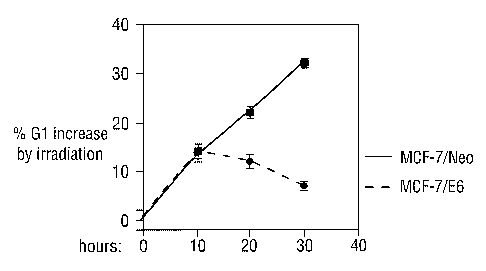
G1 ten hours after the induction of genotoxic stress (Fig. 1).
At twenty and thirty hours after IR, the fraction of parental
MCF-7 cells in G1 increased steadily, whereas the E6 cells
gradually lost their initial Gl arrest (Fig. 1). This result
suggests that cells undergo an initial G1 arrest within 10
hours after exposure to IR and that this initial response does
not require p53 activity.
Specific induction of cyclin D1 proteolysis by genotoxic
stress.
In contrast to p53, we noticed that the cyclin D1 protein
level is downregulated both in parental MCF-7 cells and in E6-
expressing derivatives within two hours following IR.
Downregulation of cyclin D1 was maintained over a period of 24
hours and was not seen both with another G1 cyclin (cyclin E)
and the G2/M cyclins A and B1. To study the effects of
CA 02409717 2002-11-05
WO 01/85992 PCT/GBO1/02099
28
genotoxic stress on the kinetics of cyclin Dl protein
downregulation we exposed U2-OS cells to varying amounts of IR
and harvested cells at different time points. Total lysates
were analysed by immunoblotting against cyclin D1 and p53
proteins. Exposure to 6 to 20 grays (Gy) resulted in a clear
downregulation of cyclin Dl protein levels as early as 10
minutes after IR and a similar effect was seen with 2 Gy after
60 minutes. Compared to the degradation of cyclin D1, the
upregulation of p53 was slow following IR. This result shows
that in U2-OS cells rapid downregulation of cyclin D1 occurs
after IR, which precedes p53 stabilization. Cyclin D1
downregulation occurred with similar kinetics in MCF-7 cells.
We next examined the mechanism underlying the rapid decrease
in cyclin D1 protein by genotoxic stress. Northern analysis
was carried out on RNA extracted from non-treated and
irradiated (20 Gy) MCF-7. At the mRNA level cyclin D1 was
slightly elevated at 2 and 4 hours after IR. The effect of IR
on cyclin D1 protein expressed from a heterologous CMV
promoter was studied. MCF-7 cells were transfected with 2 mg
total DNA containing either vector or 0.5 mg CMV promoter
based cyclin D1 expression plasmid. Co-transfected GFP
construct (0.03 mg) was used to control transfection
efficiency. After 48 hours cells were irradiated (20 Gy) and 2
hours later cellular proteins were extracted, separated on 10%
SDS PAGE and immunoblotted to detect cyclin D1 and GFP
proteins. When expressed from a heterologous CMV promoter,
cyclin Dl protein was also downregulated by IR to a similar
extent as the endogenous protein. We therefore conclude that
transcriptional regulation is not responsible for the cyclin
D1 downregulation following IR.
We then asked whether cyclin D1 protein stability was affected
CA 02409717 2002-11-05
WO 01/85992 PCT/GBO1/02099
29
in response to IR using a pulse-chase experiment. MCF-7 cells
were pulse-labelled with [35S]-methionine and after IR chased
with excess cold methionine for the indicated periods of time.
Cyclin D1 protein was immunoprecipitated, separated on SDS-
PAGE and detected by PhosphorImager. Cyclin D1 was
destabilized immediately after IR; its half-life decreased
from 40 minutes to less then 20 (Fig. 2). To ask whether the
IR-induced degradation of cyclin D1 is mediated by the
proteasome, MCF-7 cells were exposed to IR and subsequently
the proteasome inhibitor cbz-LLL was added at increasing
concentrations for two hours. After two hours, protein lysates
were made, separated on 10% SDS PAGE, and Western blotted
sequentially with antibodies against cyclin D1, p53 and cyclin
E proteins. Even though it was added after exposure to IR, 5
mM cbz-LLL was sufficient to completely block cyclin D1
downregulation without any effect on cyclin E protein levels.
Cyclin D1 was also rapidly degraded in response to other
genotoxic agents such as cis-platin. Collectively, these
results indicate strongly that accelerated proteolysis induced
by genotoxic stress is the main mechanism responsible for the
rapid downregulation of cyclin D1 protein.
We then asked if cyclin D1 degradation after genotoxic stress
is common to many cell types and is uncoupled from cell cycle
progression.HeLa, HPV16-containing cervical carcinoma; CAPAN,
SEK1-mutated pancreas carcinoma; SW1417, SEK1 mutated colon
carcinoma; AT-1BR, primary fibroblasts from AT patient; MEF,
p19~~--mouse embryo fibroblasts; T47D and ZR75-1, breast
carcinoma with low and high level of cyclin D1, respectively;
U2-OS, osteosarcoma cells were subjected to treatments with 20
Gy IR and 10 mM proteasome inhibitor as above. SaOS-2
osteosarcoma were either transfected with 0.1 and 0.5mg cyclin
D1 construct. Co-transfected GFP construct was used to
CA 02409717 2002-11-05
WO 01/85992 PCT/GBO1/02099
control transfection efficiency. After 48 hours cells were IR
(20 Gy)and two hours later cellular proteins were extracted,
separated on 10% SDS PAGE and immunoblotted to detect cyclin
D1 and GFP proteins. Genotoxic stress-induced cyclin D1
degradation was seen in a variety of cell line's, with SaOS-2
osteosarcoma cells being the only exception to date.. Since
transfected cyclin D1 protein did not degrade following IR
either, it is clear that the inability of SaOS-2 cells to
degrade cyclin D1 does not involve alterations in the cyclin
D1 itself. Cyclin D1 degradation also occurred both in HeLa
cells that do not arrest in G1 following IR due to the
presence of the HPV E6 and E7 proteins and in U2-OS cells
which were growth arrested artificially by the induction of
ARF
p19 with muristerone-A. We therefore conclude that
mechanistically, cyclin D1 degradation after genotoxic stress
is uncoupled from cell cycle progression. Moreover, cyclin D1
degradation could occur in cell lines that lack functional
pl6INIC4A' pl9~xF~ pRb and p53 proteins and the ATM and SEK1
kinases and does not depend on these proteins.
Remarkably, exposure to IR of cells which express apart from
cyclin D1 also the closely related cyclins D2 or D3 (Mouse
Embryo Fibroblasts (MEFs) and HeLa), revealed that IR-induced
degradation was unique to cyclin Dl.
Cyclin D1 degradation by genotoxic stress is independent of
the GSK-3~ pathway.
Activation of the PI3K-PKB/Akt-GSK-3(3 pathway leads to cyclin
D1 degradation through phosphorylation of threonine 286 of
cyclin D1 by GSK3-~i (Diehl et al., 1998). We therefore asked
whether this pathway is also activated by IR and is involved
CA 02409717 2002-11-05
WO 01/85992 PCT/GBO1/02099
31
in stress-induced degradation of cyclin D1. To investigate the
co-immunoprecipitation of GSK3-~i with CDK4-cyclin D1 complex,
MCF-7 cells were subjected to treatment with proteasome
inhibitor cbz-LLL and IR. 5% of the cell lysates or the
immunoprecipitated protein complexes were separated on 10%
SDS-PAGE and immunoblotted against cyclin-D1, CDK4, GSK3-~3 and
control JNK1 proteins. GSK3-~i was found to be specifically
associated with the CDK4/cyclin D1 complex in the co-
immunoprecipitation experiments. However, the amount of GSK3-~i
bound to CDK4jcyclin D1 was not significantly increased in
response to IR. We used proteasome inhibitors to protect
cyclin D1 from degradation thereby making a direct comparison
between the different treatments possible. Fig. 3 shows that
neither the total cellular activity of GSK3-(3 kinase nor the
GSK3-~i activity associated with CDK4 was elevated by IR. To
further investigate whether the GSK3-(3 pathway is involved in
the degradation of cyclin D1 by IR we treated irradiated cells
with Li+ ions, as Lip has been shown to inhibit all GSK3
activity in cells (Stambolic et al., 1996). MCF-7 cells were
treated with increasing concentrations of LiCl or control KCl
and subsequently IR (20 Gy). Lysates were prepared after 2
hours, separated on 10% SDS-PAGE and immunoblotted
sequentially with anti-cyclin D1 and anti-p53 antibodies. If
this pathway is involved, Li+ ions should inhibit cyclin D1
degradation. Results showed that Li+ ions had no detectable
effect on cyclin D1 degradation by IR although, as expected,
an increase in cyclin D1 levels was seen in non-irradiated
cells due to inhibition of basal GSK3-~3 activity (Diehl et
al., 1998). Finally, a mutant of cyclin D1 in which the GSK3-[3
phosphorylation site was mutated (T286A), which is completely
refractory to GSK3-~3 induced degradation (Diehl et al., 1998),
was fully responsive to IR-induced degradation. Collectively,
CA 02409717 2002-11-05
WO 01/85992 PCT/GBO1/02099
32
these results strongly suggest that cyclin Dl degradation
induced by genotoxic stress is independent of the PI3K-
PKB/Akt-GSK3(3 pathway.
Cyclin D1 degradation by genotoxic stress requires a RxxL
destruction motif.
To map the motif in cyclin D1 that mediates its degradation by
genotoxic stress we analyzed several mutants of D1 by
expression in MCF-7 cells. In all these experiments a co-
transfected GFP construct was used to confirm equal
transfection efficiencies between irradiated- and control
cells. When cyclin D1 was mutated at a site within the cyclin
box that is essential for activation of CDK4/6 (mutant K112E),
D1 degradation by IR remained. The same result was obtained
when the pRb family binding site in cyclin D1 was mutated
(LxCxE mutant). We therefore conclude that D1 induced
degradation by genotoxic stress is independent of both CDK4/6
kinase and pRb binding.
In the yeast Saccharomyces Cerevisiae, degradation of the
cyclin C homologue Ume3p can be induced by various stress
signals such as heat, oxidative stress and ethanol shock
(Cooper et al., 1997; Cooper et al., 1999). Three regions in
Ume3p are required for stress-induced degradation, including a
destruction box at the amino terminus (RxxL motif), the amino
terminal region of the cyclin box and a PEST domain. Close
inspection of the cyclin D1 protein sequence revealed that
cyclin D1, but not cyclin D2 and D3, harbors a destruction
box-like motif in its N-terminus (Fig. 4A). Since cyclin D2 is
not degraded by the genotoxic.stress response we mutated
cyclin D1 to the corresponding amino acid in cyclin D2. We.
found that point mutations within the amino terminal region of
CA 02409717 2002-11-05
WO 01/85992 PCT/GBO1/02099
33
the cyclin box (amino acids 87 to 99) had no effect on the
degradation by IR. However, two independent point mutations
within the putative destruction box of cyclin D1 (either R29Q
or L32A) completely abolished degradation by IR. Combining
each of these mutations in the destruction box with a mutation
in the GSK3-(3 phosphorylation site (R29Q;T286A and L32A~T286A
mutants) gave rise to a higher level of protein expression in
non-irradiated cells that was fully resistant to the IR
effect, in sharp contrast to the T286A single mutant. These
data suggest that the RxxL destruction box in cyclin D1 is the
major motif that renders cyclin D1 susceptible to degradation
by IR. To further investigate this, we performed a pulse-chase
experiment with the cyclin D1 L32A destruction box mutant to
determine its half-life. MCF-7 cells were transfected by
electroporation with wild type or L32A mutant cyclin Dl
expression vector, pulse-labelled with [35S]-methionine and
chased for varying periods of time with excess cold
methionine. Fig. 4B shows a graphic representation of the
results of this experiment, which indicates that the wild type
and L32A mutant cyclin D1 have a comparable half-life in non-
irradiated cells of about 50 minutes. This is comparable to
that of endogenous cyclin D1 protein (Fig. 2). Significantly,
the L32A mutant cyclin D1 protein was not destabilized in
response to IR, whereas the wild type protein was (Fig. 4B).
Taken together, these results define the destruction motif at
amino acids 29 to 32 as necessary for cyclin D1 degradation by
genotoxic stress, but not for its normal metabolic turnover.
To ask whether this motif is sufficient to mediate degradation
in response to IR we transplanted it to the non-responsive
cyclin D2 protein. MCF cells were transfected with either
wild type or mutant D2 expression plasmids. The effect of
irradiation on cyclin D2-RAMLK mutant, in which the amino
CA 02409717 2002-11-05
WO 01/85992 PCT/GBO1/02099
34
acids at positions 29-33 were changed to resemble the cyclin
D1 RXXL motif, was studied. After 48 hours cells were IR (20
Gy)and two hours later cellular proteins were extracted,
separated on 10% SDS PAGE and immunoblotted to detect cyclin
D2, cyclin D2-RAMLK and GFP proteins. Co-transfected GFP
construct was used to control transfection efficiency.
Remarkably, changing four amino acids in cyclin D2, thereby
creating the cyclin D1 RxxL motif, converted it to a genotoxic
stress degradable cyclin. This result demonstrates that the
RxxL motif of cyclin D1 is necessary and, when placed in the
context of a D-type cyclin, also sufficient to mediate
degradation in response to genotoxic stress.
The role of the motif was further investigated by expression
of a fusion protein in which GFP was expressed in a fusion
with cyclin DI. It was found that this fusion protein was
also targeted for degradation. Such a fusion protein provided
an efficient and simple read out of the degradation of the
protein which contains the Dl-derived destruction box.
Specific interaction of cyclin-D1/CDK4 complex with the APC.
Destruction boxes are conserved motifs (consensus: RxxL) found
in mitotic cyclins subject to proteolytic cleavage by a multi-
component ubiquitin protein ligase, named the Anaphase-
Promoting Complex (APC). Since cyclin D1 harbors a destruction
box-like motif, we searched for an association of endogenous
cyclin D1/CDK4 complexes with Cdc27, a conserved component of
the APC (King et al., 1995).
In a first experiment, whole cell extracts of MCF-7 cells were
immunoprecipitated with either an antiserum raised against the
APC subunit Cdc27 or a control anti-p38 antibody. The presence
CA 02409717 2002-11-05
WO 01/85992 PCT/GBO1/02099
of CDK4, cyclin D1 and Cdc27 proteins was detected by
immunoblotting. In non-transfected MCF-7 cells we clearly and
specifically detected both endogenous CDK4 and cyclin D1
proteins in Cdc27 immunoprecipitates.
In a second experiment, MCF-7 cells were irradiated (20 Gy),
and one hour later, cells were harvested and protein lysates
were prepared. Subsequently, extracts were immunoprecipitated
with either anti-cyclin D1 or control antibodies and subjected
to immunoblotting against cdc27, cyclin D1 and CDK4 proteins.
Cdc27 was found to be present in cyclin Dl inmmoprecipitates.
In a third experiment, MCF-7 cells were treated with 20 Gy IR
and 10 mM proteasome inhibitor cb~-LLL and harvested one hour
later. Immunoprecipitation and immunoblotting were carried out
as above. Cdc27 was found to be present in anti-CDK4, but not
anti-CDK2, immunoprecipitates. Significantly, the interaction
between CDK4 and Cdc27 was not affected by IR, whereas the
amount of Cdc27 bound to cyclin D1 decreased, most likely due
to degradation of cyclin D1 by IR. These results indicate that
the APC is constitutively associated with the cyclin D1/CDK4
complex and are consistent with a model in which the APC is
responsible for cyclin D1 proteolysis in response to IR.
Cyclin D1 degradation is required to initiate G1 arrest
induced by IR.
We wished to address the role of cyclin D1 degradation in the
initiation of G1 arrest by genotoxic stress. Our strategy was
to abolish IR-induced cyclin D1 degradation using transient
over-expression of the IR-non-degradable mutant (D1-L32A). In
transient transfections, the cyclin Dl-T286A (TA) mutant was
reproducibly expressed at higher levels than wild type cyclin
CA 02409717 2002-11-05
WO 01/85992 PCT/GBO1/02099
36
D1. Therefore, to compete more efficiently with the relatively
high. level of endogenous cyclin D1 in MCF-7 cells, we
performed most of the next experiments using the double mutant
T286A;L32A as a genotoxic stress-resistant protein and the D1-
T286A mutant as a degradable control. In these experiments we
transiently introduced expression vectors into cells using
electroporation (see experimental procedures). The advantage
of this method is that we reproducibly obtained more than 90%
transient transfection efficiencies with very homogeneous
expression of the introduced vectors. This is demonstrated by
expression of a histone H2B-GFP fusion construct (Fig. 5A).
Here MCF-7 cells were transfected by electroporation with 2 mg
DNA containing either vector or 0.5 mg histone H2B-GFP
expression construct. After 17 hours cells were washed, to
clear dead cells, and after additional 48 hours collected and
analyzed by FACE ). This allowed us to perform experiments
without selection of the transfected population.
To assess the ability of mutants of cyclin Dl to block the
initiation of a G1 arrest, we focused first on MCF-7/E6 cells
since they initiate a G1 response to IR, which is
indistinguishable from parental MCF-7 cells, but have no
effects originating from p53. We electroporated MCF-7/E6 cells
with wild type or mutant cyclin D1 expression vectors and
after 48 hours, cells were irradiated, treated with nocodazole
and 10 hours later the cell cycle distribution was analyzed by
FACS. Fig. 5B shows that the initiation of a G1 arrest of
control GFP-transfected MCF-7/E6 cells to IR was similar to
non-transfected population (induction of 15% G1 increase,
Figs.SB). Cells transiently transfected with the IR-non-
degradable mutants D1-L32A and D1-T286A;L32A had only an
increase of 4% and 2% in G1 phase cells in response to IR,
respectively. The double mutant D1-T286A;L32A was most
CA 02409717 2002-11-05
WO 01/85992 PCT/GBO1/02099
37
efficient in blocking the IR induced G1 arrest, most likely
because of its efficient accumulation in cells. The residual
2% G1 increase in the D1TA-L32A transfected population may be
the result of the fact that we did not transfect 100% of the
population (Fig. 5A). Over-expression of the IR-degradable D1
and D1TA mutant proteins gave a partial effect on Gl increase
(Fig. 5B), probably because not all of the overexpressed
protein was degraded.
In a second experiment, to measure effects on S phase in
response to IR, MCF-7/E6 cells were transfected as in B and 48
hrs later were IR (5 Gy). After additional 9 hours 7.5 mg/ml
BrdU was added and cells were harvested 1 hour later, fixed,
stained with anti-BrdU and FITC conjugated goat-anti-mouse
antibodies and analyzed by FACS. We observed.approximately a
10% reduction of cells in S-phase ten hours after IR (Fig.
5C). Over-expression of D1TA-L32A gave complete resistance to
the IR-induced S phase decrease, but did not affect the
initial G2/M arrest (Fig. 7C). These results suggest strongly
that in the absence of a functional p53 DNA damage checkpoint,
the initial G1 arrest in response to IR is the result of rapid
cyclin D1 degradation.
We then examined the requirement for cyclin D1 degradation in
the presence of p53 activity. Parental MCF-7 and MCF-7/E6
Cells were transfected with 1 mg of the plasmid cyclin D1TA-
L32A, or mock-transfected with GFP as described above. Similar
to untreated parental MCF-7 cells, mock-transfected cells
induced about 15% and 35% Gl arrest in response to 10 Gy IR
after 10 and 24 hours, respectively (Figs. 1 and 5D). MCF-7
cells, transiently transfected with cyclin D1TA-L32A were
unable to efficiently initiate G1 arrest at 10 hours (4-5% G1
increase). However, between 10 and 24 hours, these cells
CA 02409717 2002-11-05
WO 01/85992 PCT/GBO1/02099
38
induced a G1 arrest with comparable kinetics as the mock-
transfected cells, indicating that the slow response was to a
large extent unaffected. The opposite effect was seen in the
E6-expressing cells: the initiation of G1 arrest was normal
but the slower response (after 10 hours) was affected (Figs. 1
and 7D). Consistent with these data, transient over-expression
of D1TA-L32A in MCF-7/E6 abrogated both the initial and the
slower G1 arrest functions (Fig. 7D). These results indicate
that MCF-7 cells respond to IR by activating two distinct and
independent pathways. They initiate G1 arrest through a
process that depends on the ability of cells to degrade cyclin
Dl and later on they maintain and further strengthen it by
stabilizing p53.
In a further experiment primary wild type and cyclin D1-~- MEFs
were irradiated (l0 Gy) and harvested after 2 hours. Whole
cell extracts were prepared and analyzed by SDS-PAGE
immunoblotting procedure using antibodies against cyclin D1.
In agreement with a role for cyclin D1 in the initiation of G1
arrest following IR, results showed that the S-phase response
to IR of primary MEFs lacking cyclin D1 is defective when
compared to wild type MEFs. Wild type and D1-~- cells were
irradiated (10 Gy) and harvested at between 0, 2, 4 and 6
hours. 1 hour before harvesting, 7.5 mg/ml BrdU was added and
cells were analyzed by FACS (Fig. 5E). Cyclin Dl knockout MEFs
consistently had higher fraction of S phase cells in the first
hours after IR than control wild type MEFs, whereas no effect
was observed on the induction of G2/M block immediately after
stress (Fig. 5E) .
CA 02409717 2002-11-05
WO 01/85992 PCT/GBO1/02099
39
Cyclin D1 degradation by genotoxic stress induces a rapid
redistribution of p21°ipl from CDK4 to CDK2.
One mechanistic explanation as to how cyclin D1 degradation
can cause a fast G1 cell cycle arrest is by release of CKIs
from CDK4 to inhibit CDK2 complexes. To investigate this,
parental MCF-7 and MCF-7/E6 cells were irradiated and
harvested one hour later. Whole cell extracts were immuno-
precipitated with anti-CDK4, anti-CDK2 or control anti-p38
antibodies. 100 of the total extracts and the
immunoprecipitates were separated on 12% SDS-PAGE. To
distinguish between mechanisms involving proteolytic cleavage
and others we examined IR effects also in the presence of 10
mM of the proteasome inhibitory agent cbz-LLL. Analysis of
extracts of both cell types by sequential immunoblotting, with
anti-cyclin D1, anti-p21°ipl, anti-p27klpl, anti-CDK4, anti-CDK2
and control anti-p38 antibodies, revealed that the level of
p2l~lpi in MCF-7/E6 was only somewhat reduced compared to
parental cells. This observation is in line with previous
observations that p53 has a limited effect on basal p21°ipl
levels in cells (Macleod et al., 1995; Parker et al., 1995).
In co-immunoprecipitation experiments using both cell types,
we observed that in non-irradiated cells, more cyclin D1 was
associated to CDK4 than to CDK2. Upon exposure to IR, cyclin
D1 level was reduced both in CDK4- and CDK2 protein complexes,
a process that could be blocked by proteasome inhibitor. This
indicates that genotoxic stress-induced cyclin D1 degradation
is the main mechanism to initiate its disappearance from CDK2
and CDK4 complexes. Most importantly, we could clearly detect
that p21°ipl dissociated from CDK4 and started to accumulate in
CDK2 complexes, even at this early time point, a process that
CA 02409717 2002-11-05
WO 01/85992 PCT/GBO1/02099
was also dependent on proteolysis. In contrast, p27k1p1 was
associated with CDK4 in non-irradiated cells and it did not
redistribute to CDK2 complexes upon IR. We therefore detect an
early p53-independent and proteasome-dependent, redistribution
of p21°iPi, but not of p27k1p1, from CDK4 complexes to CDK2.
We next determined the CDK2 activity precipitated from MCF-
7/E6 cells treated with IR. Cells were treated as above,
except that cells were harvested after 2 hours. Using histone
Hl as a substrate we found that IR markedly reduced CDK2
activity after two hours, which could be blocked by treatment
with proteasome inhibitor. Identical results were obtained
with parental MCF-7 cells. Therefore, protein degradation
seems to be necessary for fast CDK2 kinase inhibition after
genotoxic stress.
To examine the role of cyclin D1 degradation in the process of
p21°ipl redistribution and CDK2 inhibition we analyzed CDK4
complexes from cells transfected, by electroporation, with the
IR-non-degradable D1-TA-L32A mutant. MCF-7/E6 cells were mock-
tranfected or tranfected with with 1 mg of H2B-GFP, D1-TA, or
D1-TA-L32A as described in the previous example. After 48
hours cells were irradiated (20 Gy) and 1 hour later whole
cell extracts were prepared and subjected to co-
immunoprecipitation with anti-CDK4 and control anti-p38
antibodies. 5% of each extract and the immunoprecipitated
complexes were separated on 12% SDS-PAGE and immunoblotted
against p21°iPl, cyclin D1 and CDK4. Consistent with the
results described above, already one hour after IR we detected
efficient removal of cyclin D1 from CDK4 complexes. Over-
expression of the Dl-TA in cells increased the amount of
cyclin D1 bound to CDK4 in non-irradiated cells, which was
CA 02409717 2002-11-05
WO 01/85992 PCT/GBO1/02099
41
reduced in irradiated cells. However, due to the higher pre-IR
levels, more cyclin D1 remained bound to CDK4 after IR as
compared to either mock or H2B-GFP-transfected cells. In sharp
contrast, the IR-non-degradable D1 mutant (TA-L32A) remained
associated with CDK4 after IR and almost no p21°lpl was
released from CDK4 complexes by DNA damage. This result
demonstrates that p21~1p1 dissociation from CDK4 complexes in
response to IR requires cyclin D1 degradation.
We then examined the CDK2 activity in response to IR of cells
transiently over-expressing either D1TA or D1TA-L32A proteins.
MCF-7/E6 cells were electroporated as above, irradiated (20 Gy)
and harvested 2 hours later. CDK2 protein was immunoprecipitated
and its kinase activity was examined using Histone 1 as a
substrate (H1). CDK2 protein level was determined by
immunoblotting (IB) of the same membrane with an antibody against
CDK2. Two hours after IR inhibition of CDK2 activity in mock-
transfected cells was comparable to non-transfected cells. In
contrast, in response to IR CDK2 activity remained unchanged in
cells expressing the IR-non degradable D1TA-L32A.
Collectively, these results demonstrate that initiation of G1
arrest by IR is a result of the ability of cells to degrade
cyclin D1. Degradation of cyclin Dl is required to inhibit
CDK2 activity by redistribution of p21°ipl from CDK4 complexes
to CDK2. However, we can not rule out that other processes
that are influenced by cyclin D1 degradation, are involved as
well.
Cyclin D1 degradation is required for cellular resistance to
genotoxic stress
Next, we determined the survival of cells that were rendered
CA 02409717 2002-11-05
WO 01/85992 PCT/GBO1/02099
42
unable to degrade D1 in response to IR. MCF-7 cells were
transiently transfected with the IR-non-degradable cyclin
D1TA-L32A construct at increasing concentrations. Cells were
washed 17 hours after transfection and exposed to IR (20 Gy)
after an additional 24 hours. Five days after irradiation,
floating and adherent cells were harvested and analyzed for
their sub-G1 content by FACS. Fig. 6A shows that expression of
cyclin D1TA-L32A significantly increased cell death in
response to IR in a concentration dependent fashion (up to
22% more cell death). This occurred with very limited toxicity
of cyclin D1TA-L32A on untreated cells (5o more cell death).
In a second experiment immortalized MEFs of either wild type
(wt), cyclin Dl-knockout (D1-~-) or cyclin E knockin into the
cyclin Dl locus (D1-~--E) origins were exposed to IR (10 Gy)
and harvested 6 days later for FACS analysis (Fig.6B).
Consistent with a critical role for cyclin D1 in DNA damage
response, immortalized MEFs derived from cyclin Dl knockout
mice (D1-~-) were more sensitive to IR as compared to wild type
immortalized MEFs (10 % more cell death, Fig. 6B).
Significantly, immortalized MEFs derived from D1-~- mice which
express cyclin E under the control of the cyclin D1 promoter
(cyclin E knockin mice, (Geng et al., 1999)), were also more
sensitive to IR (D1-~--E, Fig. 6B). Collectively, these data
indicate that cyclin D1 degradation is an essential component
of the cellular response to genotoxic stress, in the absence
of which the cell's ability to deal with DNA damage is
compromised.
Initiation and maintenance of G1 arrest by genotoxic stress.
Genotoxic stresses, such as IR, induce a fast and strong G1
arrest that is sustained over a prolonged period of time. We
report here that this type of G1 arrest builds up in two
CA 02409717 2002-11-05
WO 01/85992 PCT/GBO1/02099
43
different and mechanistically distinct phases: initiation and
maintenance. The initial process is fast (accomplished in a
period of less than ten hours), strong (more than 15% increase
in G1 in an asynchronous population) and is mediated by cyclin
D1 degradation. p53 activity is dispensable for G1 arrest in
this initial period. At a later stage, p53 activity is
required to maintain and further strengthen the initial p53-
independent G1 arrest. These distinct mechanisms collaborate
to allow the cell to achieve a fast and sustained G1 arrest in
response to IR.
Judging from the speed at which cyclin Dl is degraded by
genotoxic stress (Fig. 2), it appears that all factors
required to mediate cyclin Dl degradation are pre-existing in
the cell. Such pre-existing machinery is well-suited to carry
out a quick response to genotoxic stress. In contrast, the G1
arrest established by activation of the p53 pathway is
indirect and involves p53 protein accumulation by de novo
protein synthesis, its translocation to the nucleus,
transcriptional activation of p53 target genes such as p21°1p1,
translation of p53-induced transcripts, and accumulation of
the induced proteins to sufficiently high levels that they
affect the cell cycle. This p53 response depends on several
time-consuming processes and is therefore inherently slow.
Therefore, the p53 response appears more suited to maintain
and further strengthen an already established G1 arrest,
rather than to initiate it. This notion is supported by the
present data, which show that p53 hardly contributes to G1
arrest in the first 10 hours after exposure to IR.
Our results suggest strongly that the initial phase of G1
arrest following IR relies primarily on downregulation of
cyclin D1 protein levels. Several lines of experimental
CA 02409717 2002-11-05
WO 01/85992 PCT/GBO1/02099
44
evidence support the notion that induced proteolysis is the
main mechanism used by irradiated cells to reduce cyclin Dl
protein levels. First, treatment of cells with IR caused a
significant decrease in cyclin D1 protein stability (Fig. 2).
Second, treatment of cells with specific inhibitors of the
proteasome completely blocked cyclin D1 downregulation by IR.
Third, downregulation of cyclin D1 is mediated through a
destruction box, a motif that is involved in proteolytic
destruction of mitotic cyclins (Fig. 4A). Fourth, mutation of
the cyclin Dl destruction box rendered the protein non-
degradable by IR, whereas transplantation of the cyclin D1
destruction box to the IR-non-degradable cyclin D2 protein,
rendered cyclin D2 unstable in response to IR (Fig. 4B).
Finally, in cells treated with both IR and proteasome
inhibitor, cyclin D1 accumulated to higher levels than non-
treated cells. Together, these results indicate that exposure
to IR triggers a rapid proteolysis of cyclin D1 and virtually
exclude the possibility that IR also controls cyclin Dl other
levels, such protein translation.
Genotoxic stress versus mitogen deprivation.
Cyclin D1 plays a role in relaying mitogenic signals to the
cell cycle machinery. When cells are deprived of mitogens,
cyclin D1 is phosphorylated at threonine 286 by GSK3-b and
targeted for nuclear export and proteolysis (Diehl et al.,
1998; Diehl et al., 1997). Stimulation of cell cycle entry by
mitogens activates the PI3K-PKB/Akt pathway, which inhibits
GSK3-b activity, leading to accumulation of cyclin D1 in the
nucleus. Similar to mitogen deprivation, genotoxic stresses
induce cyclin D1 degradation. However, this is accomplished
through a different and independent pathway. First, genotoxic
stress-induced cyclin D1 degradation occurs both in cycling
CA 02409717 2002-11-05
WO 01/85992 PCT/GBO1/02099
cells and in arrested cells with similar efficiencies (Figure
3). Second, GSK3-b is neither activated by IR nor involved in
genotoxic stress-mediated cyclin D1 degradation (Fig. 3).
Third, both signals converge on different protein motifs in
Cyclin D1. Whereas the mitogenic signals are mediated by
phosphorylation of cyclin D1 at threonine 286, genotoxic
stress requires an intact RxxL destruction box motif (amino
acids 29-32) within cyclin D1.
It is noteworthy that the three D-type cyclins differ in their
sensitivity to genotoxic stress-induced degradation.
Proteolytic degradation by genotoxic stress was specific to
cyclin D1 and was not observed with its homologues cyclin D2
or D3 (Figs. 4). Consistent with this, the RxxL motif is not
conserved in these cyclins. This may suggest that under
physiological conditions the D type cyclin family can modulate
cellular response to the various external signals. Whereas
cyclin D1 will mediate efficient responses to both mitogen
deprivation and genotoxic stress, cyclin D2 will respond only
to the former.
Our data by no means rule out the possibility that the
specific degradation machinery responsible for cyclin D1
degradation by genotoxic stress also targets other proteins
that may function in other genotoxic stress responses such as
apoptosis, repair or G2-M arrest. It will therefore be
important to identify the requirements and essential consensus
amino acid motif that mediates the specific interaction with
the relevant proteolytic machinery.
Rapid p21°ip'' redistribution and inhibition of CDK2 activity.
Genetic experiments with mice in which the cyclin E gene was
CA 02409717 2002-11-05
WO 01/85992 PCT/GBO1/02099
46
placed under the control of the cyclin Dl promoter have
suggested that cyclin E is a downstream target of cyclin D1
(Geng et al., 1999; Roberts, 1999; Sicinski et al., 1995). Our
data are consistent with such a model in that degradation of
cyclin D1 in G1 by IR immediately affects cyclin E-associated
kinase activity. IR significantly inhibits cyclin E-CDK2
activity within two hours . Remarkably, we find that the
initial inhibition of CDK2 activity depends almost exclusively
on the cellular proteolytic activity and more specifically on
the ability to degrade cyclin D1. Blocking either proteasome-
mediated proteolysis or specifically cyclin D1 proteolysis was
sufficient to abrogate completely CDK2 inhibition by IR.
Moreover, we demonstrate that cyclin D1 degradation initiates
A specific release of p21°ipl from CDK4 complexes immediately
after IR, a process that culminates in a rapid increase of
p21°lpl associated with cyclin E/CDK2 and inhibition of its
kinase activity. However, in the absence of p53 this effect
was not sufficient to maintain cells in G1 20 to 30 hours
after IR (Fig. 1), even though low levels of eyclfin D1 protein
were maintained at that time. The escape of cells with non-
functional p53 from the initial G1 arrest probably stems from
the fact that the reservoir of p21°iPl held by cyclin D1/CDK4
complex is quickly exhausted in response to IR. Consequently,
newly synthesized CDK2/cyclin E complexes will be active and
able to drive cells into S phase. In cells harboring wild type
p53, activation of newly synthesized cyclin E/CDK2 will be
prevented through induction of p21°1P1 expression by p53.
In contrast to p21°ipl, which was rapidly released from CDK4
upon exposure to IR, p27kip1 remained bound to CDK4. We do not
know what the molecular basis is for this specific
CA 02409717 2002-11-05
WO 01/85992 PCT/GBO1/02099
47
redistribution of inhibitors by genotoxic stress. This
situation is clearly different from the redistribution of CKI
family proteins from CDK4/6 induced by TGF-b. In this case
induction of one of the INK4 family members efficiently
competes for binding of both p21°lpl and p27k1p1 to cyclin D-
CDK4j6 complexes.
The RxxL destruction motif and the APC.
Induction of cyclin D1 degradation by genotoxic stress
requires a RxxL motif at the amino terminus of cyclin Dl. RxxL
motifs, also known as destruction boxes, have been studied
most extensively in mitotic cyclins. The sea urchin cyclin B
must be degraded for cells to exit mitosis, which is dependent
on a nine amino acid motif including the RxxL box (Glotzer et
al., 1991). Likewise, the Anaphase-Promoting Complex (APC), a
multimeric ubiquitin ligase complex of 1.5 MDa, is essential
for mitotic cyclin degradation through their destruction box
(Irniger et al., 1995; King et al., 1996). The specificity and
timing of proteolysis by the APC is determined by
phosphorylation and association with activating proteins of
the fizzy protein family such as Cdc20 and Hct1 (Lukas et al.,
1999; Schwab et al., 1997; Sigrist and Lehner, 1997; Visintin
et al., 1997). Which components of the APC direct the
specificity of binding to RxxL motifs is unknown.
Interestingly, during cell cycle progression, APC carries out
its major role in exit from M phase, but remains active in Gl
and GO when mitotic kinases are no longer active (Amon et al.,
1994; Brandeis and Hunt, 1996). This suggests possible roles
for APC in G1 and GO phases of the cell cycle as well. Our
identification of the RxxL destruction motif as a necessary
element for cyclin D1 degradation points to the involvement of
CA 02409717 2002-11-05
WO 01/85992 PCT/GBO1/02099
48
APC in this process. Strongly supporting this view is the fact
that the cyclin D1/CDK4 complex specifically associates with
the APC in cycling cells. Whereas the interaction of APC with
CDK4 remains intact in cells exposed to IR, the interaction
with cyclin Dl decreases rapidly. Therefore, it seems that
CDK4 serves as a bridging factor between cyclin D1 and the
APC. This suggests a model in which the APC marks cyclin D1
for proteolysis and is subsequently free to bind another
cyclin D1 molecule via CDK4.
The APC in response to DNA damage
To identify which proteins within the APC complex are required
for cyclin D1 destruction following genotoxic stress we looked
at the human p55Cdc20 protein, an activator of the APC member
of the fizzy protein family. We looked at Cdc20 protein in
MCF-7 cells two hours after exposure to IR and found that its
mobility was slightly shifted, an indication for modification
and possibly regulation.
Furthermore, as discussed above, cyclin D1 degradation by IR
occurs in many cell lines and cell types, except for human
Saos-2 osteosarcoma cells. Since exogenously introduced cyclin
D1 was also not subject to degradation by IR in Saos-2 cells,
a likely explanation is that this cell type lacks an upstream
component in the pathway. We therefore monitored the Cdc20
protein level in Saos-2 cells, and found that Cdc20 is hardly
or not at all expressed in Saos-2 cells when compared to MCF-7
or a number of other cell-types. This effect was specific to
Cdc20 as Cdc27, a core component of the APC, is equally
expressed in both cell types. The level at which Cdc20
expression is hampered is yet to be determined.
CA 02409717 2002-11-05
WO 01/85992 PCT/GBO1/02099
49
To test the functional requirement of Cdc20 for cyclin D1
destruction by IR we cloned Cdc20 that was amplified by PCR
from a human cDNA library into a mammalian expression vector
and re-introduced it into Saos-2 cells. We then selected
stable clones and examined Cdc20 and cyclin D1 protein levels
in response to IR. Expression of Cdc20 was clearly detected in
two clones, 8 with low expression and 9 with expression
similar to the levels seen in MCF-7 cells. Importantly, re-
expression of Cdc20 restored significant cyclin D1 degradation
in response to IR.
To investigate the possible interaction between Cdc20 and
cyclin Dl we employed in vitro GST pull-down assays. We
labelled either the full-length Cdc20 protein or a truncated
form, containing only the seven WD40 motifs, with 35S
Methionine using the reticulocyte lysate system. These
proteins were then incubated with purified GST-cyclin D1
protein produced in bacteria and immobilized on beads. A
clear and specific interaction of both Cdc20 proteins with
cyclin D1 was detected. Furthermore, this specific interaction
was retained when only the first 85 amino acids of cyclin Dl,
containing its RxxL destruction box fused to GST, were used.
Taken together, these results indicate that the human
p55Cdc20, an activator of the APC, is a crucial component
responsible for conducting the response from DNA damage to
destruction of cyclin D1 via the APC and direct interaction.
Induction of cyclin D1 degradation by genotoxic stress and
cancer.
The p16=NK4A-cyclin D1-pRb pathway is disrupted in most, if not
all, human tumors. In a substantial number of tumors cyclin D1
is over-expressed by mechanisms involving gene amplification,
CA 02409717 2002-11-05
WO 01/85992 PCT/GBO1/02099
chromosomal translocations, transcriptional activation or
defects in proteolysis (Hanahan and Weinberg, 2000).
Interestingly, we find that cyclin D1-induced degradation by
genotoxic stress is intact in the vast majority of cell lines
examined. Moreover, it occurs both in the presence and absence
of the main genes involved in tumorigenesis (p53, pRb, pl6INx4A
and pl9ARF). The finding that the genotoxic stress-induced
cyclin D1 degradation pathway is intact in most tumor cells
may be related to the fact that disruption of this pathway
will not elevate cyclin Dl protein levels in non-stressed
cells and therefore does not confer a selective advantage to
tumor cells.
We show that activation of cyclin D1 proteolytic cleavage by
genotoxic stresses occurs in a broad range of cell types and
is conserved from man to mouse. Our findings also have
potential relevance for treatment of cancer. We demonstrate
that abrogation of genotoxic stress-induced cyclin D1
degradation sensitizes cells to genotoxic stress with no
significant effect on survival of non-irradiated cells (Fig.
6). This result suggests that specific inhibition of genotoxic
stress induced-cyclin D1 degradation could make chemotherapy
and radiotherapy more effective and selective as tumor cells
often express much higher levels of cyclin Dl than the
surrounding normal tissue.
Experimental procedures
Materials, antibodies and plasmids construction
Cis-platin was purchased from Teva. Histone Hl and the proteasome
inhibitor cbz-LLL were purchased from Sigma. IR was done with a
2x415 Ci 13'Cs source.
CA 02409717 2002-11-05
WO 01/85992 PCT/GBO1/02099
51
For Western blot and co-immunoprecipitation analyses the
antibodies used in this study were anti-human p53 (Do-1), anti-
mouse p53 (FL-393), anti-cyclin Dl (H-295 and M-20), anti-human
cyclin D2 (C-17), anti-mouse cyclin D2 (M-20), anti-cyclin D3 (C-
16), anti-cyclin E (M-20), anti-CDK4 (H-22), anti-CDK2 (M-
2) , anti-p21°ipl (C_19) , anti-JNK1 (FL) and anti-p38 (C-20) from
Santa Cruz. Other antibodies used were anti-GSK3-b mAb
(Transduction lab.), anti-Kip1/p27 mAb (Transduction lab.), anti-
Cdc27 mAb (Transduction lab.), rabbit anti-pl9~F (ABCAM) and
rabbit-anti-GFP (made in house).
The plasmids pRC-CMV-cyclin D1 and the mutants K112E and LxCxE
were described (Zwijsen et al., 1997). pRC-CMV cyclin D2 clone
was described (Dowdy et al., 1993). Cyclin D1 mutants, T286A,
E92V, R98H, R29Q, L32A and cyclin D2-RAMLK were generated by site
directed mutagenesis using polymerase chain reactor (PCR) and
were cloned in the pCDNA3.1 vector (Clontech). The double mutants
R29Q-T286A and L32A-T286A were generated by conventional cloning
using an internal unique BssHII site in cyclin D1 cDNA. All
constructs and mutants were verified by DNA sequence analysis.
The plasmid used for green florescent protein (GFP) expression
was pEGFP (Clontech). H2B-GFP has also been described (Kanda et
al., 1998). For pIND-pl9~F construct the mouse pl9~F cDNA tagged
with HA (Quelle et al., 1995) was cloned into the pIND vector
(Invitrogen).
Cell transfection
Cell transfection was carried out in two ways. In Figures 1 to 5,
MCF-7 cells were either transiently or stably transfected with
DOTAP (Boehringer Mannheim). Transient transfection experiments
presented in Figures 5 and 6 were done using electroporation.
Here, 3x10⁵ MCF-7 cells were resuspended in 100 ml of
CA 02409717 2002-11-05
WO 01/85992 PCT/GBO1/02099
52
electroporation buffer containing 2 mM Hepes pH: 7.2, 15 mM
K2HP04/KH2P04, 250 mM manitol and 1mM MgCl2 at a final pH of 7.2.
Either one or two mg of DNA was added and the cells and DNA were
transferred to a 0.1 cm electroporation cuvette (BioRad) and
electroporated with Gene Pulser II apparatus and Gene Pulser II
RF module (BioRad) at 140 volts, 15 times 1.5 msec burst duration
and 1.5 sec intervals. Five minutes after electroporation, cells
were seeded in a 10 cm dish. Cells were washed 16 hours after
transfection and the experiment was preformed either 24 or 48
hours later.
To generate the MCF-7/Neo and MCF-7/E6 stable clones, cells were
transfected with either pCDNA3.1 or the HPV16 E6 construct and
selection with 750 mg/ml of 6418 was carried out for 2 weeks.
Selected clones were tested by immunoblot analysis.
The pIND-pl9~FStable inducible U2-OS clone was generated using
the Ecdysone system (Invitrogen) and will be described in more
detail elsewhere. Gene induction was done with 1 mM Muristerone-A
(Invitrogen) for 20 hours.
Immortalization of primary MEFs
Primary MEFs were immortalized using infection with a LZRS virus
caring the Bmi-1 cDNA which downregulates expression of the INK4a
locus (Jacobs et al., 1999).
Cell cycle profile analysis
For FACS analysis cells were trypsinized and resuspended in 600
m1 solution containing 0.6o NP-40, 50 mg/ml RNaseA and 50 mg/ml
propidium iodide in PBS. In each assay ten thousand cells were
collected by FACScan (Becton Dickinson) and analyzed with the
CellQuest program (Becton Dickinson). For Bromodeoxyuridine
CA 02409717 2002-11-05
WO 01/85992 PCT/GBO1/02099
53
(BrdU) labelling, cells were incubated 1 hour prior to the
harvest with 7.5 mg/ml BrdU. After harvest, cells were fixed in
ethanol and stained sequentially with mouse anti-BrdU antibodies
(DAKO) and FITC-conjugated goat-anti-mouse- antibodies (MONOSAN)
according to a standard protocol (Boehringer Mannheim).
For determination of sub-G1 population MCF-7 cells were
transfected by electroporation, as described above, and
irradiated (20 Gy) after 24 hours. Five days later, floating and
adherent cells were harvested and analyzed by FACScan.
Determination of sub-G1 population in wt and D1-~- MEFs was done
similarly only that cells were irradiated (10 Gy) and analyzed
six days later.
Pulse-chase experiments
MCF-7 cells were starved in Dulbecco's modified Eagle's medium
(DMEM) without methionine and cysteine containing 5% dialyzed
serum for 1 hour and then were metabolically labelled with L-
[ssS] methionine and L- [3sS] cysteine for 2 hours. Subsequently
cells were treated with IR (20 Gy) and chased in DMEM containing
% serum for the indicated time periods . Cells were lysed in
lysis buffer containing 50 mM Hepes pH: 7.4,Ø1% NP-40, 250 mM
NaCl, 10 mM b-glycerophosphate, 0.5 mM sodium vanadate, 0.5 mM
DTT and protease inhibitor cocktail (Complete, Boehringer
Mannheim) for 20 min at 4 C and centrifuged for 15 min at 4 C.
Protein samples were pre-cleared with protein A-sepharose beads
for 20 min at 4 C, immunoprecipitated with the anti-cyclin D1 (H-
295) antibody for 1 hr at 4 C and washed three times with RIPA
buffer (150 mM NaCl, 1% NP-40, 0.5% DOC, 0.1% SDS and 50 mM Tris:
pH 8.0). Fifty ml SDS-sample buffer was added, samples were
boiled for 5 min and 20 ml were resolved on 10% SDS-PAGE. The gel
was dried, treated with fixation solution for 30 min and protein
CA 02409717 2002-11-05
WO 01/85992 PCT/GBO1/02099
54
amounts were quantified with PhosphorImager (BAS-2000, Fuji).
Co-immunoprecipitation experiments
Cells (two 80% confluent 10 cm dishes per treatment) were
collected and lysed in 500 ml lysis buffer for 30 minutes on ice
and then 500 ml of lysis buffer without NaCl was added. Extracts
were centrifuged at 14,000 rpm for 15 minutes at 4 C and
immunoprecipitated for 1 hour at 4 C in total volume of 800 ml
with 200 ml of 10o slurry protein A-sepharose beads (Pharmacia
Tech.) pre-conjugated to 2 mg of the specific antibody. The beads
were washed five times and the bound proteins were eluted by
boiling in SDS-sample buffer and resolved by 12o SDS-PAGE.
For Co-immunoprecipitation of cyclin D1 and CDK4 with APC, MCF-7
cells (80o confluent 10 cm dish per treatment) were extracted and
immunoprecipitated as described previously (Agami et al., 1999).
Immunoprecipitations were carried out using rabbit antiserum
against Cdc27 (Kramer et al., 1998), anti-CDK4 (H-22), anti-
cyclin D1 (M-20) and the controls anti-Abl (K-12), Anti-CDK2(M-2)
and anti-p38 antibodies. Immunoblotting was done using the mouse
monoclonal anti-Cdc27 (Transduction lab.) and rabbit polyclonals
anti-cyclin D1 (H-295) and anti-CDK4 (H-22).
In-vitro immunoprecipitation-kinase assays.
To determine CDK2 activity, specific complexes from either MCF-
7/Neo or MCF-7/E6 cells were immunoprecipitated from extracts
using anti-CDK2 antibody (M-2). The beads were washed two
additional times with kinase buffer (20 mM Tris HCl pH:7.4, 4 mM
MgCl2 and 0.5 mM DTT) and kinase reaction was carried out in 50
ml volume kinase buffer containing 10 mg histone-H1 as a specific
substrate, 10 mCi ~Y-32pJ _ATP (5000 mCi/mmol, Amersham) and 30 mM
CA 02409717 2002-11-05
WO 01/85992 PCT/GBO1/02099
ATP at 37 C for 30 minutes. GSK3-(3 activity, was determined
exactly as described in (van Weeren et al., 1998) using peptide
PG-S1 as a substrate.
Legends to figures
Figure 1. Initiation and maintenance of G1 arrest induced by IR.
Stable MCF-7 clones containing either pCDNA3.1 (Neo) or pCDNA3.1-
E6 were irradiated (10 Gy) and after 30 min 1 mg/ml nocodazole
was added. At the indicated time points after IR cells were
harvested and analyzed by flow activated cell sorter (FACS).
Untreated cells (nt) were harvested at the 10 hour time point.
Each experiment was carried out in duplicate. The percentage
increase in G1 is the difference in % G1 content between
irradiated and control cells.
Figure 2. Genotoxic stresses induce rapid and specific
degradation of cyclin D1 protein.
Endogenous cyclin Dl was immunoprecipitated from MCF-7 cells that
were metabolically labelled, IR (20 Gy) and chased for the
indicated time points. Cyclin D1 was visualized with
PhosphoImager and quantified. The estimated half-life of cyclin
D1 protein is shown.
Figure 3. Cyclin D1 degradation after genotoxic stress is
independent o f GSIC3 - ~ .
GSK3-~3 activity in response to IR. MCF-7 cells were IR (20 Gy)
and treated with 10 mM proteasome inhibitor cbz-LLL, as
indicated. Lysates were prepared and subjected to co-
immunoprecipitation with either anti-CDK4, anti-GSK3-(3 or control
anti-JNK1. GSK3-~i kinase activity was determined as described
(van Weeren et al., 1998).
Figure 4. A destruction motif in cyclin D1 is required for
CA 02409717 2002-11-05
WO 01/85992 PCT/GBO1/02099
56
degradation by genotoxic stress.
(A) Sequence comparison of the cyclin D1 RxxL motif and
neighboring amino acids to cyclin D2, D3, E, Ume3p and cyclins A
and B. (B) Half life of wild type and L32A mutant cyclin D1. MCF-
7 cells were transfected by electroporation (see Fig. 5A) with 4
mg of wild type cyclin D1 or 6 mg of the L32A mutant and divided
into five 3 cm dishes. After 60 hrs cells were pulse-labelled.
Typically, 3-4 folds cyclin D1 expression over endogenous protein
was obtained.
Figure 5. Degradation of cyclin D1 is required for initiation of
G1 arrest by IR.
(A) Expression of a histone H2B-GFP fusion construct. Transfected
population is indicated and reproducibly was higher than 90%. (B)
Ability of mutants of cyclin D1 to block the initiation of a G1
arrest. MCF-7/E6 cells were electroporated with 1 mg of the
indicated constructs. After 48 cells were irradiated (10 Gy),
treated with nocodazole and 10 hours later the cell cycle
distribution was analyzed by FACS. A summary of the observed
percentage G1 increase on irradiation, from three independent
experiments, is shown. (C) Incorporation of BrdU in MCF-7/E6
cells was used to measure effects on S phase in response to IR.
Bars represent two independent experiments in duplicates. (D)
Examination of the requirement for cyclin D1 degradation in the
presence of p53 activity. Parental MCF-7 and MCF-7/E6 cells were
transfected with 1 mg of the indicated plasmids as described in
5A and the experiment was done as described in 5B. A summary of
two independent experiments in duplicates is shown. (E) S-phase
response to IR of primary MEFs lacking cyclin D1. Wild type and
D1-~- cells were irradiated (10 Gy) and harvested at the indicated
time points. 1 hour before harvesting, 7.5 mg/ml BrdU was added
and cells were analyzed by FACS. Bars represent two independent
experiments in duplicates.
CA 02409717 2002-11-05
WO 01/85992 PCT/GBO1/02099
57
Figure 6. Abrogation of cyclin D1 degradation sensitizes to IR.
(A) Survival of cells rendered unable to degrade cyclin D1 in
response to IR. Parental MCF-7 cells were electroporated with
increasing amounts of cyclin D1TA or D1TA-L32A mutant constructs
as described above. Apoptic cell death was scored as the sub-G1
fraction in a FACS analysis.(B) Effect of IR on immortalised MEFs
derived from cyclin D1 knockout mice (D1-~-), cyclin E knockin
mice (D1-~--E) and wild type MEFs. Cell death was scored as
above.
CA 02409717 2002-11-05
WO 01/85992 PCT/GBO1/02099
58
References
Agami, R., Blandino, G., Oren, M., and Shaul, Y. (1999).
Interaction of c-Abl and p73alpha and their collaboration to
induce apoptosis. Nature 399, 809-13.
Agami, R. and Bernards, R. (2000). Distinct initiation
and maintenance mechanisms cooperate to induce G1 cell cycle
arrest in response to DNA damage. Cell, 102(1), 55-66.
Adams, P. D., Sellers, W. R., Sharma, S. K., Wu, A. D.,
Nalin, C. M. and Kaelin, W. G. (1996). Identification of a
cyclin-cdk2 recognition motif present in substrates and p21-
like cyclin-dependent kinase inhibitors. Mol Cell Biol,
16(12), 6623-33.
Amon, A., Irniger, S., and Nasmyth, K. (1994). Closing
the cell cycle circle in yeast: G2 cyclin proteolysis
initiated at mitosis persists until the activation of G1
cyclins in the next cycle. Cell 77, 1037-50.
Bouchard, C., Thieke, K., Maier, A., Saffrich, R.,
Hanley-Hyde, J., Ansorge, W., Reed, S., Sicinski, P., Bartek,
J., and Eilers, M. (1999). Direct induction of cyclin D2 by
Myc contributes to cell cycle progression and sequestration of
p27. Embo J 18, 5321-33.
Brandeis, M., and Hunt, T. (1996). The proteolysis of
mitotic cyclins in mammalian cells persists from the end of
mitosis until the onset of S phase. Embo J 15, 5280-9.
Bunz, F., Dutriaux, A., Lengauer, C., Waldman, T., Zhou,
S., Brown, J. P., Sedivy, J. M., Kinzler, K. W., and
Vogelstein, B. (1998). Requirement for p53 and p21 to sustain
G2 arrest after DNA damage. Science 282, 1497-501.
Chan, T. A., Hermeking, H., Lengauer, C., Kinzler, K. W.,
and Vogelstein, B. (1999). 14-3-3Sigma is required to prevent
mitotic catastrophe after DNA damage. Nature 401, 616-20.
Cooper, K. F., Mallory, M. J., Smith, J. B., and Strich,
R. (1997). Stress and developmental regulation of the yeast C-
CA 02409717 2002-11-05
WO 01/85992 PCT/GBO1/02099
59
type cyclin Ume3p (Srbllp/SsnBp). Embo J 16, 4665-75.
Cooper, K. F., Mallory, M. J.,.and Strich, R. (1999).
Oxidative stress-induced destruction of the yeast C-type
CyClln Ume3p-requires phosphatidylinositol-specific
phospholipase C and the 26S proteasome. Mol Cell Biol 19,
3338-48.
Diehl, J. A., Cheng, M., Roussel, M. F., and Sherr, C. J.
(1998). Glycogen synthase kinase-3beta regulates cyclin D1
proteolysis and subcellular localisation. Genes Dev 12, 3499-
511.
Diehl, J. A., Zindy, F., and Sherr, C. J. (1997).
Inhibition of cyclin D1 phosphorylation on threonine-286
prevents its rapid degradation via the ubiquitin-proteasome
pathway. Genes Dev 11, 957-72.
Dowdy, S. F., Hinds, P. W., Louie, K., Reed, S. I.,
Arnold, A., and Weinberg, R. A. (1993). Physical interaction
of the retinoblastoma protein with human D cyclins. Cell 73,
499-511.
Draetta, G. F. (1994). Mammalian Gl cyclins. Curr Opin
Cell Biol 6, 842-6.
el-Deiry, W. S., Tokino,.T., Velculescu, V. E., Levy, D.
B., Parsons, R., Trent, J. M., Lin, D., Mercer, W. E.,
Kinzler, K. W., and Vogelstein, B. (1993). WAF1, a potential
mediator of p53 tumor suppression. Cell 75, 817-25.
Geng, Y., Whoriskey, W., Park, M. Y., Bronson, R. T.,
Medema, R. H., Li, T., Weinberg, R. A., and Sicinski, P.
(1999). Rescue of cyclin D1 deficiency by knockin cyclin E.
Cell 97, 767-77.
Glotzer, M., Murray, A. W., and Kirschner, M. W. (1991).
Cyclin is degraded by the ubiquitin pathway. Nature 349, 132-
8.
Hanahan, D., and Weinberg, R. A. (2000). The hallmarks of
cancer. Cell 100, 57-70.
CA 02409717 2002-11-05
WO 01/85992 PCT/GBO1/02099
Irniger, S., Piatti, S., Michaelis, C., and Nasmyth, K.
(1995). Genes involved in sister chromatid separation are
needed for B-type cyclin proteolysis in budding yeast. Cell
81, 269-78.
Jacobs, J. J., Kieboom, K., Marino, S., DePinho, R. A.,
and van Lohuizen, M. (1999). The oncogene and Polycomb-group
gene bmi-1 regulates cell proliferation and senescence through
the ink4a locus. Nature 397, 164-8.
Kanda, T., Sullivan, K. F., and Wahl, G. M. (1998).
Histone-GFP fusion protein enables sensitive analysis of
chromosome dynamics in living mammalian cells. Curr Biol 8,
377-85.
Kastan, M. B., Onyekwere, O., Sidransky, D., Vogelstein,
B., and Craig, R. W. (1991). Participation of p53 protein in
the cellular response to DNA damage. Cancer Res 51, 6304-11.
Kastan, M. B., Zhan, Q., el-Deiry, W. S., Carrier, F.,
Jacks, T., Walsh, W. V., Plunkett, B. S., Vogelstein, B., and
Fornace, A. J., Jr. (1992). A mammalian cell cycle checkpoint
pathway utilizing p53 and GADD45 is defective in ataxia-
telangiectasia. Cell 71, 587-97.
King, R. W., Deshaies, R, J., Peters, J. M., and
Kirschner, M. W. (1996). How proteolysis drives the cell
cycle. Science 274, 1652-9.
King, R. W., Peters, J. M., Tugendreich, S., Rolfe, M.,
Hieter, P., and Kirschner, M. W. (1995). A 20S complex
containing CDC27 and CDC16 catalyzes the mitosis-specific
conjugation of ubiquitin to cyclin B. Cell 81, 279-88.
Kramer, E. R., Gieffers, C., Holzl, G., Hengstschlager,
M., and Peters, J. M. (1998). Activation of the human
anaphase-promoting complex by proteins of the CDC20/Fizzy
family. Curr Biol 8, 1207-10.
Kuerbitz, S. J., Plunkett, B. S., Walsh, W. V., and
Kastan, M. B. (1992). Wild-type p53 is a cell cycle checkpoint
CA 02409717 2002-11-05
WO 01/85992 PCT/GBO1/02099
61
determinant following irradiation. Proc Natl Acad Sci U S A
89, 7491-5.
Lipinski, M. M., and Jacks, T. (1999). The retinoblastoma
gene family in differentiation and development. Oncogene 18,
7873-82.
Livingstone, L. R., White, A., Sprouse, J., Livanos, E.,
Jacks, T., and Tlsty, T. D. (1992). Altered cell cycle arrest
and gene amplification potential accompany loss of wild-type
p53. Cell 70, 923-35.
Lukas, C., Sorensen, C. S., Kramer, E., Santoni-Rugiu,
E., Lindeneg, C., Peters, J. M., Bartek, J., and Lukas, J.
(1999). Accumulation of cyclin Bl requires E2F and cyclin-A-
dependent rearrangement of the anaphase-promoting complex.
Nature 401, 815-8.
Macleod, K. F., Sherry, N., Hannon, G., Beach, D.,
Tokino, T., Kinzler, K., Vogelstein, B., and Jacks, T. (1995).
p53-dependent and independent expression of p21 during cell
growth, differentiationy and DNA damage. Genes Dev 9, 935-44.
Morgan, D. O. (1995). Principles of CDK regulation.
Nature 374, 131-4.
Morin, P. J. (1999). beta-catenin signaling and cancer.
Bioessays 21, 1021-1030.
O'Neill, T., Dwyer, A. J., Ziv, Y., Chan, D. W., Lees-
Miller, S. P., Abraham, R. H., Lai, J. H., Hill, D., Shiloh,
Y., Cantley, L. C. and Rathbun, G. A. (2000). Utilization of
oriented peptide libraries to identify substrate motifs
selected by ATM. J Biol Chem, 275(30), 22719-27.Parker, S. B.,
Eichele, G., Zhang, P., Rawls, A., Sands, A. T., Bradley, A.,
Olsan, E. N., Harper, J. W., and Elledge, S. J. (1995). p53-
independent expression of p21Cip1 in muscle and other
terminally differentiating cells. Science 267, 1024-7.
Perez-Roger, I., Kim, S. H., Griffiths, B., Sewing, A.,
and Land, H. (1999). Cyclins D1 and D2 mediate myc-induced
CA 02409717 2002-11-05
WO 01/85992 PCT/GBO1/02099
62
proliferation via sequestration of p27(Kip1) and p21(Cip1).
Embo J 18, 5310-20.
Quelle, D. E., Zindy, F., Ashmun, R. A., and Sherr, C. J.
(1995). Alternative reading frames of the INK4a tumor
suppressor gene encode two unrelated proteins capable of
inducing cell cycle arrest. Cell 83, 993-1000.
Roberts, J. M. (1999). Evolving ideas about cyclins. Cell
98, 129-32.
Scheffner, M., Werness, B. A., Huibregtse, J. M., Levine,
A. J., and Howley, P. M. (1990). The E6 oncoprotein encoded by
human papillomavirus types 16 and 18 promotes the degradation
of p53. Cell 63, 1129-36.
Schwab, M., Lutum, A. S., and Seufert, W. (1997). Yeast
Hctl is a regulator of Clb2 cyclin proteolysis. Cell 90, 683-
93 .
Sherr, C. J. (1994). G1 phase progression: cycling on cue
[see comments]. Cell 79, 551-5.
Sherr, C. J. (1995). Mammalian G1 cyclins and cell cycle
progression. Proc Assoc Am Physicians 107, 181-6.
Sherr, C. J., and Roberts, J. M. (1999). CDK inhibitors:
positive and negative regulators of Gl-phase progression.
Genes Dev 13, 1501-12.
Sherr, C. J., and Roberts, J. M. (1995). Inhibitors of
mammalian G1 cyclin-dependent kinases. Genes Dev 9, 1149-63.
Sicinski, P., Donaher, J. L., Parker, S. B., Li, T.,
Fazeli, A., Gardner, H., Haslam, S. Z., Bronson, R. T.,
Elledge, S. J., and Weinberg, R. A. (1995). Cyclin D1 provides
a link between development and oncogenesis in the retina and
breast. Cell 82, 621-30.
Sigrist, S. J., and Lehner, C. F. (1997). Drosophila
fizzy-related down-regulates mitotic cyclins and is required
for cell proliferation arrest and entry into endocycles. Cell
90, 671-81.
CA 02409717 2002-11-05
WO 01/85992 PCT/GBO1/02099
63
Sionov, R. V., and Haupt, Y. (1999). The cellular
response to p53: the decision between life and death. Oncogene
18, 6145-57.
Stambolic, V., Ruel, L., and Woodgett, J. R. (1996).
Lithium inhibits glycogen synthase kinase-3 activity and
mimics wingless signalling in intact cells [published erratum
appears in Curr Biol 1997 Mar 1;7(3):196]. Curr Biol 6, 1664-
8.
Tetsu, O., and McCormick, F. (1999). Beta-catenin
regulates expression of cyclin D1 in colon carcinoma cells.
Nature 398, 422-6.
van Weeren, P. C., de Bruyn, K. M., de Vries-Smits, A.
M., van Lint, J., and Burgering, B. M. (1998). Essential role
for protein kinase B (PKB) in insulin-induced glycogen
synthase kinase 3 inactivation. Characterization of dominant-
negative mutant of PKB. J Biol Chem 273, 13150-6.
Visintin, R., Prinz, S., and Amon, A. (1997). CDC20 and
CDH1: a family of substrate-specific activators of APC-
dependent proteolysis. Science 278, 460-3.
Waldman, T., Kinzler, K. W., and Vogelstein, B. (1995).
p21 is necessary for the p53-mediated G1 arrest in human
cancer cell-s. Cancer Res 55, 5187-90.
Weinert, T. (1998). DNA damage and checkpoint pathways:
molecular anatomy'and interactions with repair. Cell 94, 555-
8.
Weinstein, J., Jacobsen, FW., Hsu-Chen, J., Wu, T., Baum,
LG., (1994). A novel mammalian protein p55CDC, present in
dividing cells is associated with protein Kinase activity and
has homology to the Saccharomyces cerevisiae cell division
cylce proteins Cdc20 and Cdc4. Mol Cell Biol 14(5), 3350-63.
Yin, Y., Tainsky, M. A., Bischoff, F. Z., Strong, L. C.,
and Wahl, G. M. (1992). Wild-type p53 restores cell cycle
control and inhibits gene amplification in cells with mutant
CA 02409717 2002-11-05
WO 01/85992 PCT/GBO1/02099
64
p53 alleles. Cell 70, 937-48.
Zwijsen, R. M., Wientjens, E., Klompmaker, R., van der
Sman, J., Bernards, R., and Michalides, R. J. (1997). CDK-
independent activation of estrogen receptor by cyclin D1. Cell
88, 405-15.
Zwijsen, R. M., Buckle, R. S., Hijmans, E. M., Loomans,
C. J. and Bernards, R. (1998). Ligand-independent recruitment
of steroid receptor coactivators to estrogen receptor by
cyclin D1. Genes Dev, 12(22), 3488-98.