Note : Les descriptions sont présentées dans la langue officielle dans laquelle elles ont été soumises.
CA 02411095 2002-12-06
WO 01/97718 PCT/GBO1/02771
1
TRANSCUTANEOUS PROSTHESIS
This invention relates to transcutaneous prosthesis and includes a method of
fitting a prosthesis having a transcutaneous component to a patient.
Amputation of limbs or digits can occur due to trauma or because of surgical
removal. Examples of trauma. include loss of fingers in machinery accidents,
loss of
limbs in car accidents or as a result of land mine explosions. Surgical
removal can also
be indicated as a result of cardio-vascular disease, diabetes and cancerous
tumours to
the bone or soft tissues.
After amputation, it is common to fit an external endo-prosthetic device that
is
attacked to the body via by a skin interface. This commonly involves the
manufacture
of a custom-made socket which is secured to the stump using straps or clamps.
A
number of disadvantages arise from the use of such endo-prosthetic devices.
For
example:-
(1) Skin is not a satisfactory high Ioad bearing structure and often breaks
down under load, becoming inflamed and uncomfortable and, in severe cases,
pressure
sores are formed which are diffcult to heal.
(2) Changes in the shape of the stump may mean that a new custom-made
socket is required.
(3) The use of sockets for receiving the stump are commonly sweaty and
uncomfortable.
(4) Where a joint is involved, the external prosthesis is usually moved by
muscle groups situated at a distance from the attached prosthesis and
therefore motion
is ineffcient and unnatural.
A major object of the present invention is to provide a prosthesis which
overcomes some or all of the above disadvantages.
According to one aspect of the present invention there is provided a
transcutaneous prosthesis which comprises a first component shaped for
implantation
into a bone, a second component intended for location between the bone and the
skin,
the second component having a surface treatment for stimulation of
fibroblastic cell
CA 02411095 2002-12-06
WO 01/97718 PCT/GBO1/02771
2
proliferation and attachment of epithelial cells and a third component
intended for
location exterior to the skin surface having a low surface energy which deters
bacterial
adhesion.
The prosthesis provided by the present invention is thus an intra-osseous
transcutaneous prosthesis (ITAP) and has a number of advantages. For example,
the
first component is attached directly to load-bearing parts of the bony
skeleton such
that load is transmitted through bone. This means that the patient is able to
apply much
more power to the prosthesis. Also, motion and perception of movement is more
natural because of the bone attachment. Moreover, because the skin takes no
part in
transmitting the load from the bone to the external part of the prosthesis,
there is no
pressure on the skin surface which would cause inflammation or discomfort.
The first component is formed with some suitable means for preventing
rotation of the component in the bone which may comprise flutes or grooves or
functionally similar shaped surfaces. These surfaces may be shaped to fit the
profile of
the intramedullary cavity, where present. Also, the first component is
preferably
provided with a surface treatment which encourages osseous integration.
Suitable
surface treatments include hydroxyapatite which is a hydrated calcium
phosphate. The
surface may also be formed with small apertures or pits to encourage
integration
between the bone and the first component. Where micro pits are formed in the
surface, these may be of the order of 20 to 500 microns in size, preferably 20
to 100
microns.
The second component extends between the bone and the epithelial surface.
This component is provided with a surface treatment for stimulating fibrous
tissue
ingrowth. Again, this component may be treated with an hydroxyapatite or
aluminium
oxide coating and the coating treated with materials which encourage the
adhesion of
epithelial cells to the second component. This component may also have a
coating
which is porous to encourage soft tissue ingrowth. Materials which encourage
such
growth include adhesion promoting proteins such as fibronectin or laminin. In
order
to aid adhesion of the fibrous tissue to the second component, the hypodermic
is
CA 02411095 2002-12-06
WO 01/97718 PCT/GBO1/02771
3
preferably surgically removed during the procedure of installing the
prosthesis. The
goal is to attach the skin to the implant to prevent movement of the skin and
shear
forces separating epithelial cells at the interface and underlying dermis and
thereby
permitting infection to enter between the skin and the prosthesis.
The third component comprises the exterior part of the prosthesis and this has
a low surface energy which deters bacterial adhesion. A low surface energy can
be
achieved by coating this part of the prosthesis with a non-stick material such
as a
diamond-like carbon, a fluorinated polymer or a silicone polymer.
The prosthesis may be made up from separate components connected together,
or two or more of the components may be formed integrally and given
appropriate
surface treatments.
The external component will preferably include a safety device comprising a
linkage which breaks under an unusual load such as, for example, one caused by
the
patient falling. This will allow the external component to detach from the
skeletal and
transcutaneous component without causing damage to the bone or to the skin. An
additional feature which will protect the fixation of an intramedullary post
is an
external device which limits torque transmitted to the adjustable fixation.
The torque
transmitted may be adjustable so that with time, the transmitted torque can be
increased, as the internal component integrates with the bone.
In a further preferred embodiment of the present invention the second
component may be provided so as to extend outwardly from the first and third
components in a manner that increases the external surface area of the second
component. The second component may also be provided with through holes which
further increase the external surface area and allow growth of tissue through
the
second component. This has been found to advantageously facilitate the
integration of
the component with fibrous tissue growth.
The invention is illustrated by the accompanying drawings in which:-
Figures l and 2 are diagrammatic views through part of a deer's antler and
Skull;
CA 02411095 2002-12-06
WO 01/97718 PCT/GBO1/02771
4
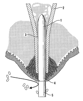
Figure 3 is a diagrammatic part section showing a transcutaneous prosthesis in
accordance with the invention, fitted to a patient.
Figure 4 is a perspective view of a preferred embodiment of a prosthesis in
accordance with the present invention drawn on a larger scale compared with
Figure 3.
Referring to the drawings, the present invention was in part stimulated by
study of the skin-bone interface around red deer antlers. This is a unique
structure and
may be thought of as a biological model for a transcutaneous implant. The deer
antler
at periods of the year is very heavily loaded during the rut. Histological
examination
indicates that the layer of skin epithelial cells become thinner as the
epithelial layer
approaches the antler, such that at the antler-skin interface an epithelial
skin layer is
only about one cell thick. The dermis is intimately attached to the bone
(pedicle)
interface. The attachment is achieved through a series of "Sharpeys fibres"
which
attach to the dermis and to the bone and prevent differential skin movement.
Antlers
do not normally become infected and the bone structure is invaginated with
small
pores measuring 18 to 40 microns in diameter. This helps the interface between
the
dermis and the bone to resist shear stresses. These features are shown in
Figures 1
and 2 of the accompanying drawings.
The prosthesis of the present invention is shown in Figure 3 and may be
considered an artificial analogue of the deer's antler. The prosthesis
comprises a first
component (1) which is inserted into the intramedullary canal of a bone (2).
Component (1) is formed with longitudinally extending cutting flutes which
engage in
the bone as the prosthesis is inserted into the intramedullary canal and
resists rotation.
The surface of the component (1) may be coated with a material to encourage
osseous
integration such as a hydroxy apatite material and/or be micro-pitted. The
second
component (4) extends from the end of the bone to the surface of the skin.
This
component may be cylindrical as drawn, or could be flattened to a mushroom
shape,
thereby increasing the surface area over which the soft tissue can be
attached.
Component (4) is given a surface treatment to encourage attachment of the
epithelial
to the implant. Such surface treatments include giving the surface a micro-
pitted
CA 02411095 2002-12-06
WO 01/97718 PCT/GBO1/02771
structure and/or coating the surface with adhesion proteins such as laminin or
fibronectin which encourage fibrous growth into the surface of the component
(4) of
the prosthesis.
Prior to installing the prosthesis, the hypodermic is preferably surgically
removed. Further, a surface is provided on the second component which is
porous and
promotes fibrous tissue ingrowth. Suitable materials for coating the surface
include
alumina oxide ceramics and hydroxy apatite. This surface, preferably after
being given
a porous surface treatment, is coated with an adhesion promoting protein, e.g.
by
spraying the prosthesis with a solution of the adhesion-promoting protein, by
dipping
the prosthesis in a concentrated solution of the protein and freeze drying, or
by dipping
into a sterile solution of the adhesion-promoting protein prior to
implantation.
The removal of the hypodermic surgically during the amputation and
installation procedure assists in stimulating attachment of the skin to the
implant and
thereby prevents shear forces on the skin separating the epithelial cells at
the interface.
The third component (5) of the prosthesis extends through the skin and is
given a non-stick surface on its exterior portion. Suitable materials include
fluorinated
polymers such as polytetrafluoroethylene, siliconised polymers and diamond
like
carbon. The presence of a non-stick surface discourages bacteria from
attaching to
the prosthesis and helps to prevent infection. The non-stick surface may be
applied to
the exterior portion of the third component (5) using the technique of
chemical vapour
deposition (CVD). The use of CVD is well known in the art for applying a
surface of
diamond-like carbon. When applying a surface layer of diamond, as disclosed in
EP-
B-0545 542 the method generally involves providing a mixture of hydrogen or
oxygen
gas and a suitable gaseous carbon compound such as a hydrocarbon, applying
energy
to that gas to dissociate the hydrogen into atomic hydrogen or the oxygen into
atomic
oxygen and the carbon into active carbon ions, atoms or CH radicals and
allowing
such active species to deposit on the substrate to form diamond. The energy to
cause
dissociation may be provided in a number of ways common to the art, for
example by
hot filament or by microwave source. A non-stick surface of fluorinated
polymer or
CA 02411095 2002-12-06
WO 01/97718 PCT/GBO1/02771
6
silicone polymer may be applied to the third component by polymerising a
monomer or
prepolymer in contact with the component.
It may be convenient to apply the low energy surface treatment to the third
component while masking the remaining components of the prosthesis. Also, the
second component of the prosthesis may be treated with the adhesion-promoting
protein after applying the low energy surface to the third component, and it
may be
desirable to mask the third component while applying the adhesion-promoting
protein.
The third component may be connected to an artificial limb or digit. For
example, in the case of a replacement finger or part finger, the first
component may be
implanted into the remaining bone with the second component instituting the
transcutaneous portion, and the third component extending beyond the severed
stump.
An artificial digit or part digit can then be attached to the third component.
The prosthesis may be implanted either in a one-stage procedure or in a two-
stage procedure where the first component is implanted into the bone and
allowed to
integrate before the transcutaneous part is attached.
There is shown in figure 4 a further preferred embodiment of the present
invention wherein the second component (4) is extended in an outward direction
perpendicular to the first and third components in a plate like form. This
feature
provides the second component (4) with a large surface area which
advantageously
facilitates the integration of the second component (4) with fibrous tissue
growth. As
also shown in Figure 4, through holes (6) may be provided in the plate like
extension
of the second component (4), which further increase the external surface area
and also
allowing tissue to grow through the second component further facilitating
integration.
Although the above description refers to a series of components, it will be
appreciated
that each component may be a portion of an integral element manufactured from
a
single piece of material. It is, however, preferred that a frangible linkage
is provided
between the third and second components or between the second and first
component,
so that in the event that a high load is applied to the third component, or to
a member
attached thereto, the linkage will fair so as to protect the implanted bone
from injury.
CA 02411095 2002-12-06
WO 01/97718 PCT/GBO1/02771
7
While the present invention has been described with particular reference to
the
provision of a prosthesis for replacement of lost digits or limbs, the
invention is also
applicable to other prosthesis which extend through the skin, e.g. dental
implants.