Note : Les descriptions sont présentées dans la langue officielle dans laquelle elles ont été soumises.
CA 02429615 2010-05-28
DETERMINATION OF RISK AND TREATMENT OF
COMPLICATIONS OF PREMATURITY
FIELD OF THE INVENTION
[002] The present invention relates generally to determining the risk of
developing
complications of premature birth and low birth weight, and particularly to
complications
associated with IGF-L The present invention further relates to methods for
treating such
conditions.
BACKGROUND OF THE INVENTION
[003] Of an estimated 4.2 million live births in the United States each year,
approximately
383,000 (about 9%) occur prematurely. Preterm labor and its complications are
major perinatal
public health issues in developed societies today. Low birth-weight infants or
infants born
prematurely miss a major part of the critical period of in utero growth. They
account for half of
all infant deaths and three-quarters of long-term morbidity. They impose a
heavy burden on the
national economy, because of the high costs of special care in both the
neonatal period and over
the life-span of survivors. Many survivors also have diminished quality of
life because of
physical damage resulting directly from prematurity.
[004] The length of a normal pregnancy or gestation is considered to be 40
weeks (280
days) from the date of conception. Jnfants born before 37 weeks gestation are
considered
premature and may be at risk for complications. Advances in medical technology
have made it
possible for infants born as young as 23 weeks gestational age (17 weeks
premature) to survive.
Infants born prematurely are at higher risk for death or serious complications
due to their low
birth weight and the immaturity of their body systems. Low birthweight, del*
by a cut-off of
1
CA 02429615 2003-05-20
WO 02/43578 PCT/US01/47285
2,500 g, serves as a marker for high risk newborns, as it is correlated with
prenatal risk factors,
intrapartum complications and neonatal disease, and is composed largely of
preterm births.
Studies on very low birthweight, defined as less than 1,500 g or less than
1,000 g cut-offs that
identify infants at highest risk, those with high rates of severe respiratory
and neurological
complications associated with extreme prematurity. (See, Hack, M., Klein, N.
K., & Taylor, H.
G., Long-term developmental outcomes of low birth weight infants. The Future
of Children,
5,176-196 (1995)).
[005] The lungs, digestive system, and nervous system (including the brain)
are not fully
developed in premature babies, and are particularly vulnerable to
complications. The most
prevalent medical problems encountered in preterm infants are retinopathy of
prematurity,
developmental delay, mental retardation, bronchopulmonary dysplasia,
necrotizing enterocolitis,
and intraventricular hemorrhage.
[006] Retinopathy of prematurity (ROP) is a potentially blinding disease,
initiated by lack
of retinal vascular growth after premature birth. The greatest risk factor for
development of ROP
is low birth weight and gestational age. ROP occurs in two phases. (Simons, B.
D. & Flynn, J.
T. (1999) International Ophthalmology Clinics 39, 29-48). When infants are
born prematurely
the retina is incompletely vascularized. In infants who develop ROP, growth of
vessels slows or
ceases at birth leaving maturing but avascular and therefore hypoxic
peripheral retina. (Ashton,
N. (1966) Am .1 Ophthalmol 62, 412-35; Flynn, J. T., O'Grady, G. E., Herrera,
J., Kushner, B. J.,
Cantolino, S. & Milam, W. (1977) Arch Ophthalmol 95, 217-23). This is the
first phase of ROP.
[007] The extent of non-perfusion of the retina in the initial phase of ROP
appears to
determine the subsequent degree of neovascularization, the late destructive
stage of ROP, with
the attendant risk of retinal detachment and blindness. (Penn, J. S., Tolman,
B. L. & Henry, M.
M. (1994) Invest Ophthalmol Vis Sci 35, 3429-35). If it were possible to allow
blood vessels to
grow normally in all premature infants, as they do in utero, the second
damaging neovascular
phase of ROP would not occur. When ROP was first described in 1942, the
etiology was
unknown. However, the liberal use of high supplemental oxygen in premature
infants was soon
associated with the disease and hyperoxia was shown to induce ROP-like
retinopathy in neonatal
animals with incompletely vascularized retinas. This suggested that an oxygen-
regulated factor
was involved. Expression of vascular endothelial growth factor (VEGF), which
is necessary for
normal vascular development, is oxygen-regulated and was found to be important
for both
phases of ROP. (Aiello, L. P., Pierce, E. A., Foley, E. D., Takagi, H., Chen,
H., Riddle, L.,
Ferrara, N., King, G. L. & Smith, L. E. (1995) Proc Natl Acad Sci USA 92,
10457-61;
Robinson, G. S., Pierce, E. A., Rook, S. L., Foley, E., Webb, R. & Smith, L.
E. (1996) Proc Natl
2
CA 02429615 2003-05-20
WO 02/43578 PCT/US01/47285
Acad Sci USA 93, 4851-6; Pierce, E. A., Foley, E. D. & Smith, L. E. (1996)
Arch Ophthalmol
114, 1219-28; Stone, J., Itin, A., Alon, T., Peer, J., Gnessin, H., Chan-Ling,
T. & Keshet, E.
(1995) J Neurosci 15, 4738-47; Alon, T., Hemo, I., Itin, A., Pe'er, J., Stone,
J. & Keshet, E.
(1995) Nature Medicine 1, 1024-8; Ozaki, H., Seo, M. S., Ozaki, K., Yamada,
H., Yamada, E.,
Okamoto, N., Hofmann, F., Wood, J. M. & Campochiaro, P. A. (2000) American
Journal of
Pathology 156, 697-707). High supplemental oxygen affects the first phase of
vascular growth
in ROP animal models through suppression of VEGF expression. However, with
current careful
use of moderate oxygen supplementation, the oxygen level in patients is not a
significant risk
factor for ROP, yet the disease persists, suggesting that other factors are
also involved. (Kinsey,
V. E., Arnold, H. J., Kalina, R. E., Stern, L., Stahlman, M., Odell, G.,
Driscoll, J. M., Jr., Elliott,
J. H., Payne, J. & Patz, A. (1977) Pediatrics 60, 655-68; Lucey, J. F. &
Dangman, B. (1984)
Pediatrics 73, 82-96).
[008] A premature infant has an incompletely developed brain. Because the
breathing
center in the brain may be immature, many premature infants are vulnerable to
neurologic injury
caused by bleeding or low oxygen supply in the brain. The neurologic injury
(e.g.,
intraventricular or periventricular hemorrhage, hypoxic injury around the time
of birth) and
various early infections of premature birth pose risks of developmental delay,
i.e., slowed
progression in achieving developmental milestones. Children with early
developmental delay
are considered "at risk" for mental retardation. Mental retardation refers to
an impairment in
general intellectual functioning, together with global deficits in other life
skills, which must
develop before age 18. Children born extremely premature are much more likely
to develop
mental retardation than children born healthy at term. Neurologic injury can
be detected by, for
example, an electroencephalogram (EEG). EEG provides useful information that
reflects the
function of the neonatal brain. The EEG may assist in determining brain
maturation, focal or
generalized abnormalities. EEG tests brain activity in the outer layer of the
brain by measuring
electrical current from brain nerve cells. Electrodes are attached to various
parts of the head and
a graph is made of electrical activity. Brain waves can be interpreted
according to their
frequency (the number of waves per second) and according to their morphology
(shape of single
waves or of wave groups).
[009] Intraventricular hemorrhage (IVH) is currently the best known cause of
central
nervous system morbidity in preterm neonates. Virtually all major IVH occurs
at gestational age
of 28-30 weeks or less. 90% of significant IVH occurs within the first days to
week of life in
approximately 15 - 40% of high risk neonates. IVH is a condition in which
immature and fragile
blood vessels within the brain burst and bleed into the hollow chambers
(ventricles) normally
3
CA 02429615 2003-05-20
WO 02/43578 PCT/US01/47285
reserved for cerebrospinal fluid and into the tissue surrounding them. The
severity of IVH is
graded according to a scale of I-IV, with I being bleeding confined to a small
area around the
burst vessels and IV being an extensive collection of blood not only in the
ventricles, but in the
brain tissue itself. Grades I and II are not uncommon, and the baby's body
usually reabsorbs the
blood with no ill effects. However, more severe IVH can result in
hydrocephalus, a potentially
fatal condition in which too much fluid collects in the ventricles, exerting
increased pressure on
the brain and causing the baby's head to expand abnounally. To drain fluid and
relieve pressure
on the brain, doctors will either perform lumbar punctures, a procedure in
which a needle is
inserted into the spinal canal to drain fluids; install a reservoir, a tube
that drains fluid from a
ventricle and into an artificial chamber under or on top of the scalp; or
install a ventricular shunt,
a tube that drains fluid from the ventricles into the abdomen, where it is
reabsorbed by the body.
Infants who are at high risk for IVH usually have an ultrasound examination of
the brain in the
first week after birth, followed by others if bleeding is detected. Presently,
IVH cannot be
prevented; however, close monitoring ensures that procedures to reduce fluid
in the brain are
implemented quickly to minimize possible damage.
[0010] Approximately 1% of all infants develop respiratory distress syndrome
reflecting
pulmonary immaturity. Among infants treated for respiratory distress syndrome
in neonatal
intensive care units (ICUs), approximately 20 to 30% will develop the most
common form of
chronic infant lung disease, bronchopulmonary dysplasia (BPD). (Northway WH.
Bronchopulmonary dysplasia: twenty-five years later. Pediatrics 1992; 89:969-
973).
Approximately 7,000 new cases of BPD are diagnosed every year. (Davis JM,
Rosenfeld WN.
Chronic lung disease. In: Avery GB, Fletcher MA, MacDonald MG, eds.
Neonatology:
pathophysiology and management of the newborn. Philadelphia, PA: JIB
Lippincott, 1994; 453-
477). Among infants with BPD, there is a high rate of hospital readmission (up
to 60%) and
subsequent death (up to 20%), mainly from cardiopulmonary failure. (Southall
DP, Samuels
MP. Bronchopulmonary dysplasia: a new look at management. Arch Dis Child 1990;
65:1089-
1095). Although survival has improved, advances in therapy have not
significantly decreased
the incidence of BPD. (Frank L. Antioxidants, nutrition and bronchopulmonary
dysplasia. Clin
Perinatol 1992; 19:541-562; Rush MG, Hazinski TA. Current therapy of
bronchopulmonary
dysplasia. Clin Perinatol 1992; 19:563-590). Prematurity, barotrauma, and
oxygen toxicity
contribute to the pathogenesis of BPD, but the exact mechanisms by which the
neonatal lung
undergoes such severe disruption in structure and function are incompletely
understood.
[0011] Insulin growth factor I (IGF-I) is a well-known regulator of postnatal
growth and
metabolism. See, Baker J, Liu JP, Robertson EJ, Efstratiadis A. Role of
insulin-like growth
4
CA 02429615 2003-05-20
WO 02/43578 PCT/US01/47285
factors in embryonic and postnatal growth. Cell 1993; 75:73-82. It has a
molecular weight of
approximately 7.5 kilodaltons (Kd). IGF-I has been implicated in the actions
of various other
growth factors, since treatment of tissues with such growth factors leads to
increased production
of IGF-I. However, its role in prenatal growth and development has only
recently been
recognized. See, Gluckman PD, Harding YE. The physiology and pathophysiology
of
intrauterine growth retardation. Hormone Research 1997; 48:11-6. Experimental
data obtained
in IGF-I 4" mice suggest that IGF-I play an important role in the third
trimester of embryonic
growth and development of several tissues. See, Baker J, Liu JP, Robertson EJ,
Efstratiadis A.
Role of insulin-like growth factors in embryonic and postnatal growth. Cell
1993; 75:73-82. In
support of the IGF-14" data in mice, two patients with genetic defects of the
IGF-I system were
shown to display impaired prenatal growth and development of the central
nervous system. One
girl had single allele deletion of the IGF-I receptor gene and one boy had
partial deletion of the
IGF-I receptor gene. See, Woods KA, Camacho-Hubner C, Savage MO, Clark AJ.
Intrauterine
growth retardation and postnatal growth failure associated with deletion of
the insulin-like
growth factor I gene. New England Journal of Medicine 1996; 335:1363-7; and de
Lacerda L,
Carvalho JA, Stannard B, et al., 1999 In vitro and in vivo responses to short-
term recombinant
human insulin-like growth factor-I (IGF-I) in a severely growth-retarded girl
with ring
chromosome 15 and deletion of a single allele for the type I IGF receptor
gene. Clin.
Endocrinol. 51(5): 541-50.
[0012] IGF-I has insulin-like activities and is mitogenic (stimulate cell
division) and/or is
trophic (promote recovery/survival) for cells in neural, muscular,
reproductive, skeletal and other
tissues. Unlike most growth factors, IGF is present in substantial quantity in
the circulation, but
only a very small fraction of this IGF is free in the circulation or in other
body fluids. Most
circulating IGF is bound to the IGF-binding protein, and more particularly to
the IGFBP-3. IGF-
I may be measured in blood serum to diagnose abnormal growth-related
conditions, e.g.,
pituitary gigantism, acromegaly, dwarfism, various growth hormone
deficiencies, and the like.
Although IGF-I is produced in many tissues, most circulating IGF-I is believed
to be synthesized
in the liver.
[0013] Almost all IGF circulates in a non-covalently associated ternary
complex composed
of IGF-I, IGFBP-3, and a larger protein subunit termed the acid labile subunit
(ALS). The IGF-
I/IGFBP-3/ALS ternary complex is composed of equimolar amounts of each of the
three
components. ALS has no direct IGF binding activity and appears to bind only to
the IGF-
I/IGFBP-3 binary complex. The IGF-I/IGFBP-3/ALS ternary complex has a
molecular weight
of approximately 150 Kd. This ternary complex is thought to function in the
circulation "as a
CA 02429615 2003-05-20
WO 02/43578 PCT/US01/47285
reservoir and a buffer for IGF-I preventing rapid changes in the concentration
of free IGF"
(Blum et al., pp. 381-393, Modern Concepts In Insulin-Like Growth Factors (E.
M. Spencer, ed.,
Elsevier, New York, 1991).
[0014] IGFBP-3 is the most abundant IGF binding protein in the circulation,
but at least five
other distinct IGF binding proteins (IGFBPs) have been identified in various
tissues and body
fluids. Although these proteins bind IGFs, they each originate from separate
genes and have
unique amino acid sequences. Thus, the binding proteins are not merely analogs
or derivatives
of a common precursor.
[0015] IGF-I and IGF-I binding proteins such as IGFBP-3 may be purified from
natural
sources or produced by recombinant means. For instance, purification of IGF-I
from human
serum is well known in the art (Rinderknecht et al. (1976) Proc. Natl. Acad.
Sci. USA 73:2365-
2369). Production of IGF-I by recombinant processes is shown in EP 0 128 733,
published in
December of 1984. IGFBP-3 may be purified from natural sources using a process
such as that
shown by Baxter et al. (1986, Biochem. Biophys. Res. Comm. 139:1256-1261).
Alternatively,
IGFBP-3 may be synthesized recombinantly as discussed by Sommer et al., pp.
715-728,
Modern Concepts Of Insulin-Like Growth Factors (E. M. Spencer, ed., Elsevier,
New York,
1991). Recombinant IGFBP-3 binds IGF-I in a 1:1 molar ratio.
[0016] Despite the increasing advances in the understanding of complications
of
prematurity, there are no presently available effective treatments or methods
of determining the
risk of developing these life-threatening conditions, as premature morbidity
and death is very
prevalent.
SUMMARY OF THE INVENTION
[0017] In one aspect of the present invention there is provided a method for
determining the
risk of developing a complication of preterm birth in a patient born before 40
weeks of gestation
or weighing 10% less than the average for the patient's gestational age. The
method involves
measuring serum IGF-I and/or IGF-I binding protein levels after birth of the
patient to obtain an
IGF-I or IGF-I binding protein level; and correlating said IGF-I or IGF-I
binding protein level
with an in utero baseline level of IGF-I or IGF-I binding protein based on
gestational age
matched mean levels in utero, wherein an IGF-I or IGF-I binding protein level
below the mean
gestational age in utero level indicates the patient is at an increased risk
of developing a
complication of preterm birth. The complications of preterm birth include
retinopathy of
prematurity, developmental delay, mental retardation, bronchopulmonary
dysplasia, and
intraventricular hemorrhage.
6
CA 02429615 2011-10-05
It is provided the use of IGF-1, a IGF-1 agonist or an analog thereof in the
manufacture of a medicament
for treating retinopathy of prematurity, necrotizing enterocolitis,
bronchopulmonary dysplasia, or
intraventricular hemorrhage in an infant born before 40 weeks of gestation,
the IGF-1, IGF-1 agonist or
analog thereof elevating the infant's serum IGF-1 level so that the level
before 40 weeks postmenstrual
age is not below an in utero baseline level of IGF-1 based on gestational age
matched mean IGF-1 level
in utero.
It is provided the use of IGF-1, a IGF-1 agonist or an analog thereof for
treating retinopathy of
prematurity, necrotizing enterocolitis, bronchopulmonary dysplasia, or
intraventricular hemorrhage in an
infant born before 40 weeks of gestation, the IGF-1, IGF-1 agonist or analog
thereof elevating the infant's
serum IGF-1 level so that the level before 40 weeks postmenstrual age is not
below an in utero baseline
level of IGF-1 based on gestational age matched mean IGF-1 level in utero.
It is provided a method for diagnosing the risk of developing a complication
of preterm birth in a patient
born before 40 weeks of gestation or weighing 10% less than the average for
the patient's gestational age
comprising the steps of:
(a) measuring postnatal serum levels of at least one of IGF-1 and IGF binding
protein in a blood
sample of the patient, wherein the IGF binding protein binds IGF-1, to obtain
an IGF-1 or IGF binding
protein level; and
(b) comparing the postnatal IGF-1 or IGF binding protein levels with an in
utero baseline level of
IGF-1 or IGF binding protein based on gestational age matched mean levels in
utero,
wherein a postnatal IGF-1 or IGF binding protein level below the 90%
confidence interval for the
matched gestational age in utero level indicates the patient is at an
increased risk of developing a
complication of preterm birth,
wherein the complication of preterm birth is selected from the group
consisting of retinopathy of
prematurity, developmental delay, mental retardation, bronchopulmonary
dysplasia, necrotizing
enterocolitis and intraventricular hemorrhage.
It is provided a method for diagnosing the risk of developing a complication
of preterm birth in a patient
born before 40 weeks of gestation or weighing 10% less than the average for
the patient's gestational age
comprising the steps of:
(a) measuring postnatal serum levels of at least one of IGF-1 and IGF binding
protein in a blood
sample of the patient, wherein the IGF binding protein binds IGF-1, to obtain
an IGF-1 or IGF binding
protein level; and
(b) comparing the postnatal IGF-1 or IGF binding protein levels with a
baseline level of IGF-1 or
IGF binding protein based on gestational age matched mean levels of patients
born before 40 weeks of
gestation or weighing 10% less than the average for the patient's gestational
age that do not develop
complications of preterm birth,
wherein a postnatal IGF-1 or IGF binding protein level below the mean
gestational age level of
patients that do not develop complications of preterm birth indicates that the
patient is at an increased risk
of developing a complication of preterm birth,
wherein the complication of preterm birth is selected from the group
consisting of retinopathy of
prematurity, developmental delay, mental retardation, bronchopulmonary
dysplasia, necrotizing
enterocolitis and intraventricular hemorrhage.
It is provided an IGF-1, an IGF-1 agonist or an analog thereof for use in the
treatment of retinopathy of
prematurity, necrotizing enterocolitis, bronchopulmonary dysplasia, or
intraventricular hemorrhage in an
infant born before 40 weeks of gestation, the IGF-1, IGF-1 agonist or analog
thereof elevating the infant's
serum IGF-1 level so that the level before 40 weeks postmenstrual age is not
below an in utero baseline
level of IGF-1 based on gestational age matched mean IGF-1 level in utero.
6a
CA 02429615 2003-05-20
WO 02/43578 PCT/US01/47285
[0018] In
another aspect of the invention, there is provided a method for treating a
patient
suffering from a complication of preterm birth or preventing a patient from
developing a
complication of preterm birth. The method involves administering to a patient
having a serum
level IGF-I below the norm for in utero, an effective amount of IGF-I, an
analog, or an agonist
thereof to elevate the patient's IGF-I level to an in utero baseline level.
The in utero baseline
level is preferably elevated to a concentration from 10 g/L to 150 ,g/L. In
one embodiment of
the invention, IGF-I or an analog thereof is administered in combination with
an IGF binding
protein capable of binding IGF-I. In the preferred embodiment, the IGF binding
protein capable
of binding IGF-I is
biding protein 3 (IGFBP-3). The IGF-I or an analog thereof (with or
without the IGF binding protein capable of binding IGF-I), or an agonist
thereof is administered
subcutaneously, intravenously, intramuscularly, or orally. Oral administration
is preferred.
[0019] In yet another aspect of the invention there is provided use of an IGF-
I, an analog or
an agonist thereof in the manufacture of a medicament for treating a
complication of preterm
birth.
[0020] Finally, there is also provided an article of manufacture comprising
packaging
material and a pharmaceutical agent contained within the packaging material.
The packaging
material comprises a label which indicates that the pharmaceutical may be
administered, for a
sufficient term at an effective dose, for treating and/or preventing
complications associated with
preterm birth. The pharmaceutical agent comprises IGF-I or an analog, or an
agonist thereof
together with a pharmaceutically acceptable carrier.
[0021] It is to be understood that while the invention has been described in
conjunction with
the preferred specific embodiments thereof that the foregoing description as
well as the examples
that follow are intended to illustrate and not limit the scope of the
invention. Other aspects,
advantages and modifications within the scope of the invention will be
apparent to those skilled
in the art to which the invention pertains.
BRIEF DESCRIPTION OF THE DRAWINGS
[0022] The accompanying drawings, which are incorporated in and constitute a
part of this
specification, illustrate embodiments of the invention and, together with the
description, serve to
explain the objects, advantages, and principles of the invention. In the
drawings, Figure 1
represents inclusion of study subjects. The scheme illustrates 99 very preterm
infants eligible for
study of growth factors and postnatal morbidity. All children with a
gestational age <27 weeks
belonged to this group.
7
CA 02429615 2003-05-20
WO 02/43578 PCT/US01/47285
[0023] Figures 2A and 2B illustrate individual longitudinal pattern of IGF-
I levels in
premature infants (Fig. 2A) without retinopathy of prematurity (ROP) (n=31)
and (Fig. 2B) with
ROP (n=17). The gray area depicts the 90 % confidence interval for IGF-I
values using the
technique of cordocentesis and a similar IGF-I assay as used in the present
study 18. Dotted lines
indicate individual longitudinal IGF-I values of a twin-pair.
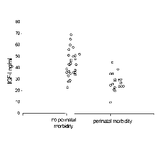
[0024] Figure 3 shows serum IGF-I levels at 33 weeks of gestation in 29
children without
perinatal morbidity and in 19 children with perinatal morbidity. The
horizontal line indicates an
IGF-I concentration of 30 jig/L. 25 of 29 children without postnatal morbidity
but only 4 of 19
children with perinatal morbidity had values above 30 jig/L.
[0025] Figure 4 shows relative impact of serum levels of IGF-I and post-
menstrual GA on
the risk for retinopathy of prematurity as estimated by multiple logistic
regression analysis.
Post-menstrual age at birth (24-32 weeks) indicated in the graph. The
regression analysis shows
that if post-menstrual age is 24 weeks at birth a mean IGF-I level at 31-35
weeks of 40 i.tg/L
carries a risk of developing ROP of 50% (dashed line). However, if post-
menstrual age is 32
weeks at birth, an IGF-I level of 12 g/L carries a risk of developing ROP of
50%.
[0026] Figures 5A-B illustrate effect of IGF-1 inhibition on vascular growth.
Flat-mounted
whole retina shows that in IGF-14" mice (Fig. 5A) there is less progression of
vascular
development (bright area) compared to IGF-1+/+ littermate controls (Fig. 5B).
[0027] Figures 6A-B show a laser microdissection of retina anterior to growing
vessels. In
Fig. 6A, VEGF mRNA is visualized anterior to the growing vessels in flat-
mounted retina. Fig.
6B shows the area containing VEGF (insert) removed by laser microdissection in
both IGF-14-
mice and control IGF-144+ retinal cross sections, and VEGF mRNA analyzed by
qRT-PCR
relative to cyclophilin control.
[0028] Figure 7 illustrates mean serum IGF-I at matched gestational ages in
infants with
and without ROP. The mean IGF-1 level for infants with ROP (white circles) and
without ROP
(dark circles) is shown versus gestational age. Error bars indicate standard
error of the mean.
[0029] Figure 8 shows replicate blots prepared from total cell lysates and
stained either
with phospho-AKT (Ser 473) antibody or antibody which recognizes AKT
irrespective of
phosphorylation status (total-AKT). Following serum starvation to reduce
baseline AKT
phosphorylation, cells were stimulated with VEGF, IGF-1, or both for times
indicated.
[0030] Figures 9A-D are a schematic representation of IGF-1 / VEGF control of
blood
vessel development in ROP. Fig. 9A shows that in utero, VEGF is found at the
growing front of
vessels. IGF-1 is sufficient to allow vessel growth. Fig. 9B shows that with
premature birth,
IGF-1 is not maintained at in utero levels and vascular growth ceases, despite
the presence of
8
CA 02429615 2003-05-20
WO 02/43578 PCT/US01/47285
VEGF at the growing front of vessels. Both endothelial cell survival (AKT) and
proliferation
(MAPK) pathways are compromised. With low IGF-1 and cessation of vessel
growth, a
demarcation line forms at the vascular front. High oxygen exposure (as occurs
in animal models
and in some premature infants) may also suppress VEGF, further contributing to
inhibition of
vessel growth. Fig. 9C shows that as the premature infant matures, the
developing but non-
vascularized retina becomes hypoxic. VEGF increases in retina and vitreous.
With maturation,
the IGF-1 level slowly increases. Fig. 9D shows that when the IGF-1 level
reaches a threshold at
¨ 34 weeks gestation, with high VEGF levels in the vitreous, endothelial cell
survival and
proliferation driven by VEGF may proceed. Neovascularization ensues at the
demarcation line,
growing into the vitreous. If VEGF vitreal levels fall, normal retinal vessel
growth can proceed.
With normal vascular growth and blood flow, oxygen suppresses VEGF expression,
so it will no
longer be overproduced. If hypoxia (and elevated levels of VEGF) persist,
further
neovascularization and fibrosis leading to retinal detachment can occur.
[0031] Figure 10 illustrates the concentration of serum IGFBP-3 and IGF in
retinopathy of
prematurity.
DETAILED DESCRIPTION
[0032] We demonstrated in a mouse model that insulin-like growth factor 1 (IGF-
I) is
necessary for normal development of retinal blood vessels. See, Hellstrom A,
Perruzzi C, Ju M,
et al. Low IGF-I suppresses VEGF-survival signaling in retinal endothelial
cells: direct
correlation with clinical retinopathy of prematurity. Proc Natl Acad Sci U S
A. 2001; 98:5804-8.
See also Example #2 infra. Retinopathy of prematurity (ROP) is associated with
abnormal
retinal development in which the retinal vessel growth lags behind development
in utero. We
conducted a prospective longitudinal study measuring serum IGF-I levels weekly
in premature
infants from birth (post-menstrual age 24 to 32 weeks) until discharge from
the hospital. Infants
were evaluated for ROP and other morbidity of prematurity: bronchopulmonary
dysplasia
(BPD), intraventricular hemorrhage (IVH) and necrotizing enterocolitis (NEC).
We have found
that persistent low serum levels of IGF-I after premature birth are associated
with complications
of prematurity such as ROP. Therefore, we have devised methods of determining
the risk and
treating complications associated with preterm birth.
[0033] In the third trimester of pregnancy, fetal IGF-I levels rise rapidly
in utero and this
increase is associated with development of fetal tissue. See, Gluckman PD,
Harding JE. The
physiology and pathophysiology of intrauterine growth retardation. Hormone
Research 1997;
48:11-6. IGF-I levels after premature birth are lower than post-menstrual-age-
matched fetal
9
CA 02429615 2003-05-20
WO 02/43578 PCT/US01/47285
levels in utero, particularly at post-menstrual ages corresponding to the
third trimester. See,
Lineham JD, Smith RM, Dahlenburg GW, et al. Circulating insulin-like growth
factor I levels in
newborn premature and full-term infants followed longitudinally. Early Hum Dev
1986; 13:37-
46. In IGF-14" mice, absence of IGF-I prevents normal retinal vascular growth
See, Hellstrom A,
Perruzzi C, Ju M, et al. Low IGF-I suppresses VEGF-survival signaling in
retinal endothelial
cells: direct correlation with clinical retinopathy of prematurity. Proc Natl
Acad Sci U S A. 2001;
98:5804-8. In premature infants who develop ROP, cessation of normal retinal
vascular growth
precedes proliferative retinopathy. We hypothesized that in premature babies,
ROP and other
postnatal morbidity might be a result of abnormal tissue maturation associated
with an inability
of some prematurely born infants to attain serum IGF-I levels comparable to
those normally
found in utero.
[0034] The relative risk for ROP and other morbidity was increased 5.7-fold
(95%
confidence interval 2.2-14.6) if IGF-I was 301.1g/L at 33 weeks post-menstrual
age. After
adjustment for post-menstrual age, each increase of 5 pg/L mean IGF-I during
post-menstrual
age 31-35 weeks decreased the risk of ROP by 59%. The median level of IGF-I at
31-35 weeks
of gestation was 26 ILtg/L (range 17-49) for infants with ROP and other
morbidity (n=19),
compared to 38 [tg/L (range 20-59) in the group without postnatal morbidity
(n=29), p<0.0001.
[0035] Preterm infants who develop ROP and other postnatal morbidities (BPD,
IVH and
NEC) have low serum levels of IGF-I after birth compared to infants without
ROP and other
complications. The serum levels of IGF-I in infants with ROP displayed a slow
relatively linear
rise during gestational weeks 31-36. In contrast, serum levels of IGF-I in
infants without ROP or
other postnatal morbidities tended to have a different pattern and increased
more rapidly,
reaching levels close to those seen in utero, with a maximum IGF-I value at an
age
corresponding to gestational weeks 31-35 (Figure 2). Therefore, serum IGF-I
levels predict
complications of preterm birth, such as ROP. Prematurity per se (gestational
or post-menstrual
age and birth weight) has historically been by far the strongest risk factor
for ROP. See, Simons
BD, Flynn JT. Retinopathy of prematurity and associated factors. International
Ophthalmology
Clinics 1999; 39:29-48. However, we found that the mean IGF-I level at post-
menstrual weeks
31-35 was as important as the degree of prematurity per se (post-menstrual age
at43irth) as a
predictive factor for ROP and other complications of prematurity.
[0036] The peak level of IGF-I seen in premature infants without morbidity
occurred during
a critical developmental period in utero when significant maturation of the
eyes, lungs, kidneys
and brain normally takes place. See, O'Rahilly R, Muller F. Human Embryology
and
Teratology. New York: Wiley-Liss, 1996. It was recently shown experimentally
that IGF-I is
CA 02429615 2003-05-20
WO 02/43578 PCT/US01/47285
important to the action of vascular endothelial growth factor (VEGF) in
regulating retinal
vascular growth. In retinal vascular endothelial cells, minimum levels of IGF-
I are necessary for
maximum VEGF activation of the MAPK and Akt pathways, important for
endothelial cell
survival and proliferation. See, Hellstrom A, Perruzzi C, Ju M, et al. Low IGF-
I suppresses
VEGF-survival signaling in retinal endothelial cells: direct correlation with
clinical retinopathy
of prematurity. Proc Natl Acad Sci U S A. 2001; 98:5804-8; and Smith LE, Shen
W, Perruzzi C,
et al. Regulation of vascular endothelial growth factor-dependent retinal
neovascularization by
insulin-like growth factor-I receptor. Nature Medicine 1999; 5:1390-5. The
level of IGF-I
required for maximum VEGF activation of the Akt pathway corresponded to the
level seen in
premature infants who did not develop ROP. The critical role of the IGF-I
system in retinal
vascular development has been supported in a clinical study where patients
with genetic defects
in the IGF-I or IGF-I receptor were found to have a reduced number of retinal
vascular
branching points (Hellstrom, personal observation). Thus, the reduced serum
levels of IGF-I
seen in these infants may cause some of the morbidity associated with
prematurity.
[0037] The major fetal source of IGF-I is the placenta, although ingested
amniotic fluid may
also provide IGF-I to the fetus. See, Bauer MK, Harding JE, Bassett NS, et al.
Fetal growth and
placental function. Molecular & Cellular Endocrinology 1998; 140:115-20.
Several studies have
shown that, in utero, umbilical cord levels of IGF-I are higher than postnatal
serum levels in
post-menstrual age-matched preterm infants. See, Lineham JD, Smith RM,
Dahlenburg GW, et
al. Circulating insulin-like growth factor I levels in newborn premature and
full-term infants
followed longitudinally. Early Hum Dev 1986; 13:37-46. In a preterm baby, the
gastrointestinal
development is not fully completed at birth and thus enteral nutrition may not
be tolerated. As
IGF-I is a nutrition-dependent factor, the low serum levels found among some
preterm infants
might be explained by deficient general nutrition. See, Smith WJ, Underwood
LE, Keyes L,
Clemmons DR. Use of insulin-like growth factor I (IGF-I) and IGF-binding
protein
measurements to monitor feeding of premature infants. J Clin Endocrinol Metab
1997; 82:3982-
8. However, as it has been shown that enteral IGF-I administration enhances
gastrointestinal
development in fetal sheep, a combination of exogenous IGF-I and adequate
general nutrition
may be necessary in order to obtain optimal development after premature birth.
See, Kimble
RM, Breier BH, Gluckman PD, Harding JE. Enteral IGF-I enhances fetal growth
and
gastrointestinal development in oesophageal ligated fetal sheep. Journal of
Endocrinology 1999;
162:227-35.
11
CA 02429615 2003-05-20
WO 02/43578 PCT/US01/47285
Definitions
[0038] "Preterm" or "preterm birth" or "prematurity" refers to birth of a
patient prior to 40
weeks of gestation or weighing 10% less than the average for the patient's
gestational age.
[0039] "IGF-I" refers to insulin-like growth factor I from any species,
including bovine,
ovine, porcine, equine, and human, preferably human, and, if referring to
exogenous
administration, from any source, whether natural, synthetic, or recombinant,
provided that it will
bind IGF binding protein at the appropriate site.. IGF-I can be produced
recombinantly, for
example, as described in PCT publication WO 95/04076.
[0040] An "IGFBP" or an "IGF binding protein" refers to a protein or
polypeptide from the
insulin-like growth factor binding protein family and normally associated with
or bound or
complexed to IGF-I whether or not it is circulatory (i.e., in serum or
tissue). Such binding
proteins do not include receptors. This definition includes IGFBP-1, IGFBP-2,
IGFBP-3,
IGFBP-4, IGFBP-5, IGFBP-6, Mac 25 (IGFBP-7), and prostacyclin-stimulating
factor (PSF) or
endothelial cell-specific molecule (ESM-1), as well as other proteins with
high homology to
IGFBPs. Mac 25 is described, for example, in Swisshelm et al., Proc. Natl.
Acad. Sci. USA, 92:
4472-4476 (1995) and Oh et al., J. Biol. Chem., 271: 30322-30325 (1996). PSF
is described in
Yamauchi et al., Biochemical Journal, 303: 591-598 (1994). ESM-1 is described
in Lassalle et '
al., J. Biol. Chem., 271: 20458-20464 (1996). For other identified IGFBPs,
see, e.g., EP 375,438
published Jun. 27, 1990; EP 369,943 published May 23, 1990; WO 89/09268
published Oct. 5,
1989; Wood et al., Molecular Endocrinology, 2: 1176-1185 (1988); Brinkman et
al., The
EMBO J., 7: 2417-2423 (1988); Lee et al., Mol. Endocrinol., 2: 404-411(1988);
Brewer et al.,
BBRC, 152: 1289-1297 (1988); EP 294,021 published Dec. 7, 1988; Baxter etal.,
BBRC, 147:
408-415 (1987); Leung et al., Nature, 330: 537-543 (1987); Martin et al., J.
Biol. Chem., 261:
8754-8760 (1986); Baxter et al., Comp. Biochem. Physiol., 91B: 229-235 (1988);
WO 89/08667
published Sep. 21, 1989; WO 89/09792 published Oct. 19, 1989; and Binkert et
al., EMBO J.,
8: 2497-2502 (1989).
[0041] "IGFBP-3" refers to insulin-like growth factor binding protein 3.
IGFBP-3 is a
member of the insulin-like growth factor binding protein family. IGFBP-3 may
be from any
species, including bovine, ovine, porcine and human, in native-sequence or
variant form,
including but not limited to naturally-occurring allelic variants. IGFBP-3 may
be from any
source, whether natural, synthetic or recombinant, provided that it will bind
IGF-I at the
appropriate sites. IGFBP-3 can be produced recombinantly, as described in PCT
publication
WO 95/04076.
12
CA 02429615 2003-05-20
WO 02/43578 PCT/US01/47285
[0042] A "therapeutic composition," as used herein, is defined as
comprising IGF-I, an
analog thereof, or IGF-I in combination with its binding protein, IGFBP-3 (IGF-
I/IGFBP-3
complex). The therapeutic composition may also contain other substances such
as water,
minerals, carriers such as proteins, and other excipients known to one skilled
in the art.
[0043] "Analogs" of IGF-I are compounds having the same therapeutic effect as
IGF-I in
humans or animals. These can be naturally occurring analogs of IGF-I (e.g.,
truncated IGF-I) or
any of the known synthetic analogs of IGF-I. See, for example, US Pat. No.
5,473,054 for
analog compounds of IGF-I.
[0044] "Agonists" of IGF-I are compounds, including peptides, which are
capable of
increasing serum and tissue levels of IGF, especially IGF-I, in a mammal and
particularly in a
human. See, for example, US Pat. No. 6,251,865 for IGF agonist molecules.
[0045] "Developmental delay" as used herein shall mean abnormal neurogenesis
which has
the potential of leading to slowed mental progression in achieving
developmental milestones.
Developmental delay can, in some cases, be determined by means of
electroencephalogram.
[0046] The present invention provides, in one aspect, a method for determining
the risk of
developing a complication of preterm birth in a patient born before 40 weeks
of gestation or
weighing 10% less than the average for the patient's gestational age. The
method involves
measuring serum IGF-I and/or IGF binding protein levels after birth of the
patient to obtain an
IGF-I level or a level of IGF binding protein capable of binding IGF-I; and
correlating said
levels of IGF-I or IGF binding protein capable of binding IGF-I with an in
utero baseline level of
IGF-I or IGF binding protein based on gestational age matched mean levels in
utero, wherein an
IGF-I level or a level of IGF binding protein capable of binding IGF-I below
the mean
gestational age in utero level indicates the patient is at an increased risk
of developing a
complication of preterm birth. The complications of preterm birth suitable for
the methods of
the present invention include retinopathy of prematurity, developmental delay,
mental
retardation, bronchopulmonary dysplasia, necrotizing enterocolitis, and
intraventricular
hemorrhage.
[0047] The level of IGF and IGF binding protein capable of binding IGF-I can
also be
measured via a method which uses antibodies, called the ligand-mediated
immunofunctional
method (LIFA). This method is disclosed in US Patent No. 5,593,844, the
disclosure of which,
regarding antibodies and other materials and conditions that can be used in
the assay, is
incorporated herein by reference.
[0048] Suitable commercially-available IGF antibodies include Nos. 5345-
0329 and 5345-
0209 of Biogenesis Ltd., Poole, Dorset, UK; GF006 of Chemicon International
Inc., Temecula,
13
CA 02429615 2003-05-20
WO 02/43578 PCT/US01/47285
CA, USA; SC-7144 and SC-1422 of Santa Cruz Biotechnology Inc., Santa Cruz, CA,
USA; and
MAS 974p of Harlan Sera-Lab Ltd., Loughborough, Leicestershire, UK.
[0049] In another aspect of the invention, there is provided a method for
treating a patient
suffering from a complication of preterm birth or preventing a patient from
developing a
complication of preterm birth. The method involves administering to a patient
having a serum
level IGF-I below the norm for in utero, an effective amount of IGF-I or an
analog, or an agonist
thereof to elevate the patient's IGF-I level to an in utero baseline level.
The in utero baseline
level is preferably elevated to a concentration from 10 Rg/L to 150 pg/L. In
one embodiment of
the invention, IGF-I or an analog thereof is administered in combination with
IGF binding
protein capable of binding IGF-I. In the preferred embodiment, the IGF binding
protein capable
of binding IGF-I is IGF binding protein 3 (IGFBP-3). The IGF-I or analog or an
agonist thereof
may be administered subcutaneously, intramuscularly, intravenously or orally.
Oral
administration is preferred.
[0050] It is preferred that the methods of the present invention be
initiated soon after birth in
order to effectively prevent complications of prematurity and to promote
normal vascular
development. This is especially critical for the treatment of ROP, wherein
increasing IGF-I late
in the course of the disease may promote the late neovascular, destructive
phase of ROP. See,
O'Rahilly R, Muller F. Human Embryology and Teratology. New York: Wiley-Liss,
1996; and
Smith LE, Kopchick JJ, Chen W, et al. Essential role of growth hormone in
ischemia-induced
retinal neovascularization. Science 1997; 276:1706-9. The treatment which is
delayed until after
the non-vascularized retina becomes hypoxic might trigger abnormal retinal
neovascularization.
[0051] Administration of IGF-I or an analog or an agonist thereof, or IGF-I of
an analog
thereof in combination with IGF binding protein results in increases in
circulating levels of IGF-
I. Accordingly, administration of IGF-I or IGF-I in combination with IGF
binding protein is
useful for the treatment or prevention of symptoms, disorders, and conditions
associated with
low circulating levels of IGF-I.
[0052] The inventive methods disclosed herein provide for the parenteral an
oral
administration of IGF-I, an analog or an agonist thereof, or IGF-I or an
analog in combination
with IGF binding protein complex to infants in need of such treatment.
Parenteral administration
includes, but is not limited to, intravenous (IV), intramuscular (IM),
subcutaneous (SC),
intraperitoneal (IP), intranasal, and inhalant routes. In the method of the
present invention, IGF-
I, an agonist or an analog thereof are preferably administered orally. IV, IM,
SC, and IP
administration may be by bolus or infusion, and may also be by slow release
implantable device,
including, but not limited to pumps, slow release formulations, and mechanical
devices. The
14
CA 02429615 2003-05-20
WO 02/43578 PCT/US01/47285
formulation, route and method of administration, and dosage will depend on the
disorder to be
treated and the medical history of the patient. In general, a dose that is
administered by
subcutaneous injection will be greater than the therapeutically-equivalent
dose given
intravenously or intramuscularly. Preferably, the dose of IGF-I or an analog
thereof
administered will be from about 25 g/kg to about 2 mg/kg of body weight. More
preferably,
the dose of IGF-I, an agonist, or an analog thereof will be from about 50
g/kg to about 1 mg/kg.
[0053] A composition comprising equimolar amounts of IGF-I and IGF binding
protein may
be used. Preferably the IGF-I and IGF binding protein are complexed prior to
administration.
Preferably, the complex is formed by mixing approximately equimolar amounts of
IGF-I and
IGF binding protein dissolved in physiologically compatible carriers such as
normal saline, or
phosphate buffered saline solution. More preferably, a concentrated solution
of recombinant
human IGF-I and a concentrated solution of recombinant human IGF binding
protein are mixed
together for a sufficient time to form an equimolar complex. Most preferably,
recombinant
human IGF-I and recombinant human IGF binding protein are combined to form a
complex
during purification, as described in International Patent Application No. WO
96/40736.
[0054] For parenteral or oral administration, compositions of the complex may
be semi-
solid or liquid preparations, such as liquids, suspensions, and the like.
Physiologically
compatible carriers are those that are non-toxic to recipients at the dosages
and concentrations
employed and are compatible with other ingredients of the formulation. For
example, the
formulation preferably does not include oxidizing agents and other compounds
that are known to
be deleterious to polypeptides. Hence, physiologically compatible carriers
include, but are not
limited to, normal saline, serum albumin, 5% dextrose, plasma preparations,
and other protein-
containing solutions. Optionally, the carrier may also include detergents or
surfactants.
[0055] In yet another aspect of the invention there is provided use of an IGF-
I, an agonist or
analog thereof in the manufacture of a therapeutic composition for treating a
complication of
preterm birth.
[0056] Finally, there is also provided an article of manufacture comprising
packaging
material and a pharmaceutical agent contained within the packaging material.
The packaging
material comprises a label which indicates that the pharmaceutical may be
administered, for a
sufficient term at an effective dose, for treating and/or preventing
complications associated with
preterm birth. The pharmaceutical agent comprises IGF-I, an agonist or an
analog thereof
together with a pharmaceutically acceptable carrier.
[0057] For therapeutic applications, IGF-I or an analog thereof may be
suitably
administered to a patient, alone or as part of a pharmaceutical composition,
comprising the IGF-I
CA 02429615 2003-05-20
WO 02/43578 PCT/US01/47285
or an analog thereof together with one or more acceptable carriers thereof and
optionally other
therapeutic ingredients. The carrier(s) must be "acceptable" in the sense of
being compatible
with the other ingredients of the formulation and not deleterious to the
recipient thereof.
[0058] The pharmaceutical compositions of the invention include those
suitable for oral,
nasal, topical (including buccal and sublingual), or parenteral (including
subcutaneous,
intramuscular, intravenous and intradermal) administration. The formulations
may conveniently
be presented in unit dosage form, e.g., tablets and sustained release
capsules, and in liposomes,
and may be prepared by any methods well know in the art of pharmacy. See, for
example,
Remington's Pharmaceutical Sciences, Mack Publishing Company, Philadelphia, PA
(17th ed.
1985).
[0059] Such preparative methods include the step of bringing into
association with the
molecule to be administered ingredients such as the carrier which constitutes
one or more
accessory ingredients. In general, the compositions are prepared by uniformly
and intimately
bringing into association the active ingredients with liquid carriers,
liposomes or finely divided
solid carriers or both, and then if necessary shaping the product.
[0060] Compositions of the present invention suitable for oral
administration may be
presented as discrete units such as capsules, cachets or tablets each
containing a predetermined
amount of the active ingredient; as a powder or granules; as a solution or a
suspension in an
aqueous liquid or a non-aqueous liquid; or as an oil-in-water liquid emulsion
or a water-in-oil
liquid emulsion, or packed in liposomes and as a bolus, etc.
[0061] A tablet may be made by compression or molding, optionally with one or
more
accessory ingredients. Compressed tablets may be prepared by compressing in a
suitable
machine the active ingredient in a free-flowing form such as a powder or
granules, optionally
mixed with a binder, lubricant, inert diluent, preservative, surface-active or
dispersing agent.
Molded tablets may be made by molding in a suitable machine a mixture of the
powdered
compound moistened with an inert liquid diluent. The tablets optionally may be
coated or scored
and may be formulated so as to provide slow or controlled release of the
active ingredient
therein.
[0062] Compositions suitable for topical administration include lozenges
comprising the
ingredients in a flavored basis, usually sucrose and acacia or tragacanth; and
pastilles comprising
the active ingredient in an inert basis such as gelatin and glycerin, or
sucrose and acacia.
[0063] Compositions suitable for parenteral administration include aqueous
and non-
aqueous sterile injection solutions which may contain anti-oxidants, buffers,
bacteriostats and
solutes which render the formulation isotonic with the blood of the intended
recipient; and
16
CA 02429615 2003-05-20
WO 02/43578 PCT/US01/47285
aqueous and non-aqueous sterile suspensions which may include suspending
agents and
thickening agents. The formulations may be presented in unit-dose or multi-
dose containers, for
example, sealed ampules and vials, and may be stored in a freeze dried
(lyophilized) condition
requiring only the addition of the sterile liquid carrier, for example water
for injections,
immediately prior to use. Extemporaneous injection solutions and suspensions
may be prepared
from sterile powders, granules and tablets.
[0064] The invention will be further characterized by the following examples
which are
intended to be exemplary of the invention.
EXAMPLE 1
Study Subjects
[0065] All eligible patients were at high risk of developing ROP and other
morbidity on the
basis of their postmenstrual ages at birth. All infants born at a post-
menstrual age of less than 32
weeks at The Queen Silvia Children's Hospital in Goteborg between December
1999 and May
2001 were eligible for the study. Exclusion criteria were inability to
complete postnatal clinical
follow-up until an age corresponding to 40 post-menstrual weeks and any
conspicuous
congenital anomaly.
[0066] Ninety-nine eligible babies were born at The Queen Silvia Children's
Hospital,
Goteborg between December 1999 and May 2001. Forty-eight infants were excluded
because
the investigator was unable to contact the parents in time to initiate the
study (Figure 1). The
mean post-menstrual age at birth among the excluded children was 30 weeks; no
child in this
group had a post-menstrual age at birth of less than 27 weeks. Fifty-one
infants were identified
as potential participants in the study. The parents of these 51 patients all
gave permission for
participation of their child. After data collection was completed, permission
to publish the data
was withdrawn by the parents of one baby, who consequently was excluded. In
the first 20 days
of life two infants died.
[0067] In total, 48 babies, including 6 twin pairs, with a median post-
menstrual age at birth
of 27.0 weeks (range 24.0 ¨31.8 weeks) were included. All children were
hospitalized in a
neonatal intensive care unit. Gestational age at birth was based on fetal
ultrasonography,
performed at week 16 post-menstruation. Twenty-seven of the children were
included in a
previously reported study on cross-sectional IGF-I values and ROP. See,
Hellstrom A, Perruzzi
C, Ju M, et al. Low IGF-I suppresses VEGF-survival signaling in retinal
endothelial cells: direct
correlation with clinical retinopathy of prematurity. Proc Natl Acad Sci U S
A. 2001; 98:5804-8.
17
CA 02429615 2003-05-20
WO 02/43578 PCT/US01/47285
Nutrition
[0068] All infants were nourished according to the routines for premature
babies at the
neonatal unit. Oral feeding with increasing amounts of human/maternal breast-
milk was
introduced during the first or second day of life. At three days of age,
parenteral nutrition was
introduced if the child could not tolerate oral feeding with at least half the
amount of the
scheduled 24-hours requirement. The breast-milk given to children with a birth-
weight below
1500 grams was fortified with 0.8 g protein per 100 ml breast-milk (gradually
introduced over
one week) from 10 days of age until the baby weighed 2000 grams.
IGF-I Analysis
[0069] Without knowledge of ROP status, intravenous blood-samples (0.5 ml)
were taken
weekly, stored at ¨20 to ¨80 C, from birth until discharge of the infants
from the hospital. All
blood samples of each baby were analyzed at the same time. Serum was diluted
1:50 and IGF-I
was measured in duplicate by an IGFBP-blocked RIA, without extraction and in
the presence of
¨ 250-fold excess of IGF-II (Mediagnost GmbH, Tubingen, Germany). See. Blum
WF, Breier
BH. Radioimmunoassays for IGFs and IGFBPs. Growth Regulation 1994; 4:11-9. The
intra-
assay coefficient of variation (CV) at 10.2 g/L and 34.5 p,g/L was 15.7% and
9.6%,
respectively. The interassay CV at 10.2 p,g/L and 34.5 p,g/L was 23.9% and
12.1%, respectively.
IGFBP-3 Analyses
[0070] The native concentrations of serum IGFBP-3 were diluted 1:300 and
measured in
duplicate, and determined using a RIA See. Blum WF, Breier BH.
Radioimmunoassays for IGFs
and IGFBPs. Growth Regulation 1994; 4:11-9. The intra-assay and interassay CV
at 1773 ng/ml
was 6.1% and 10.6%, respectively.
Morbidity Evaluation
[0071] ROP was classified according to the International Classification
(Anonymous. An
international classification of retinopathy of prematurity. Prepared by an
international
committee. British Journal of Ophthalmology 1984; 68:690-7) and subdivided
into Stage 1
(demarcation line), Stage 2 (ridge), Stage 3 (ridge with extraretinal
fibrovascular proliferations),
stage 4 (subtotal retinal detachment) and Stage 5 (total retinal detachment).
The presence of
dilatation of the posterior retinal vessels was referred to as "plus" disease.
For the purpose of
18
CA 02429615 2003-05-20
WO 02/43578 PCT/US01/47285
this study, ROP was defined as the presence of any stage higher than Stage 1
of the disease. The
severity of ROP was classified according to its most advanced stage. The
infants were examined
according to a routine protocol, which consisted of dilated eye fundus
examinations from the
chronological age of 5 to 6 weeks until the eyes were fully vascularized, if
no ROP or Stage 1
ROP was found. If ROP Stage 2 or more was diagnosed, examinations were
performed once or
twice a week, depending on the severity of the disease, until the condition
was considered stable
with or without treatment. The infants eyes were examined by indirect
ophthalmoscopy after
pupillary dilatation with 1% cyclogyl. Care was taken to minimize pain and
stress during the
examinations.
Other Morbidity Evaluation
[0072] The diagnosis bronchopulmonary dysplasia (BPD) was based on the typical
appearance of BPD on serial chest x-rays and the need for oxygen
supplementation at gestational
week 36. See, Shennan AT, Dunn MS, Ohlsson A, Lennox K, Hoskins EM. Abnormal
pulmonary outcomes in premature infants: prediction from oxygen requirement in
the neonatal
period. Pediatrics 1988; 82:527-32. The hospital file of each child was also
reviewed for
intracranial hemorrhage (NH) (grade 2-4; diagnosed by perinatal cerebral
ultrasonography
(Burstein J, Papile LA, Burstein R. Intraventricular hemorrhage and
hydrocephalus in premature
newborns: a prospective study with CT. AJR. American Journal of Roentgenology
1979;
132:631-5)) and necrotizing enterocolitis (NEC) with gut perforation leading
to surgery.
Statistical Analysis
[0073] In comparison of children with ROP Stage 0-1 and children with ROP
Stage 2-3, the
length of time from birth to reach IGF-I >30 g/L and the mean level of
available measurements
of IGF-I at post-menstrual weeks 31-35 were analyzed with the Wilcoxon-Mann-
Whitney U-test.
A multiple logistic regression analysis was performed for ROP 8. The potential
explanatory
variables in the model were post-menstrual or gestational age (GA), birth
weight (BW) and the
individual mean level of IGF-I during post-menstrual weeks 31-35. The model
used was logit
(ROP stage >1=1, else ROP=0) = a +13ix GA (weeks) + 132 x Mean IGF-I week 31-
35 (j.tg/L).
Individual longitudinal serum IGF-I levels were used in the evaluation of the
IGF-I pattern.
[0074] The postnatal morbidity was dichotomized as no morbidity (ROP Stage 0-
1, no BPD,
IVH Stage 0-1 and no NEC) or postnatal morbidity (ROP Stage 2-4, BPD, IVH
Stage 2-4 or
NEC). P-values less than 0.05 were considered significant.
19
CA 02429615 2003-05-20
WO 02/43578 PCT/US01/47285
Demographics of Participating Infants
[0075] The baseline characteristics of the infants with ROP (n=17) compared to
those with
no ROP (n=31) demonstrated that the children with ROP had lower gestational
age and weight at
birth, (Table 1).
IGF-I and ROP and Other Perinatal Morbidity
[0076] Nineteen of the 48 infants had postnatal morbidity (ROP, IVH, BPD or
NEC)
associated with preterm birth. Seventeen of the 19 infants with morbidity
developed ROP, and
13 of the 17 with ROP had other morbidities in addition. In total 11 had BPD,
4 had NEC
leading to surgery and 4 had IVH. Only 2 children had postnatal morbidity
(IVH) without also
having ROP (Table 2). A different longitudinal IGF-I pattern was found in the
preterm infants
with no or minimal ROP as compared to the group with ROP (Figure 2). Preterm
children with
ROP Stage 0-1 (n=31) had a peak level of IGF-I at a gestational age of 31-35
weeks while
preterm children with ROP Stage 2-3 (n=17) had a slow rise of IGF-I level
without a peak
(Figure 2). The median duration of time from birth to IGF-I reaching 30 pg/L
was 16 days (range
0-53 days) in infants with ROP Stage 0-1 (n=31), compared to 59 days (range 1-
100 days) for
those that developed ROP Stage 2-3 (n=17), (P<0.0001), Figure 2. The median
level of IGF-1 at
31-35 weeks of gestation was 26 g/L (range, 17-49 pg/L) for infants with ROP
or other
postnatal morbidity (n=19), compared to 38 pg/L (range 20-59 pg/L) in the
group without
postnatal morbidity (n=29), P<0.0001. At 33 gestational weeks, 4 of the 19
children with ROP
or other postnatal morbidity had IGF-I values above 30 p,g/L, while 15
children had IGF-I values
30 p,g/L. Among the 29 children without postnatal morbidity, 25 children had
IGF-I values
above 30 g/L while 4 children had values below 30 g/L, Figure 3. Thus, preterm
children with
IGF-I 30 g/L at 33 weeks of gestation had a relative risk of 5.7 (95%
confidence interval 2.2-
14.6) to develop ROP or other postnatal morbidity. Among the 6 twin pairs in
the study, the
twin with more morbidity had the lowest IGF-I values (data not shown).
Mean IGF-I Compared with Post-Menstrual Age and Birth Weight
[0077] The results of the multiple regression analysis, taking IGF-I and post-
menstrual GA
into account, was logit (ROP Stage 2-3) = 23 ¨0.18 (mean IGF-I week 31-35/
g/L) - 0.65
(GA/weeks). The relative risk of ROP associated with a 5 g/L increase of mean
IGF-I during
post-menstrual weeks 31-35 was e ¨0.9 =0.41 when adjusting for post-menstrual
age. Thus, an
increase of 5 pbg/L, in mean IGF-I during post-menstrual weeks 31-35 decreased
the risk of
CA 02429615 2003-05-20
WO 02/43578 PCT/US01/47285
having ROP stage 2-3 by 59 %, while an increase of 1 gestational week
decreased the risk by 48
% (Figure 4). The results of the multiple regression analysis, taking IGF-I
and BW into
account, was logit (ROP Stage 2-3) = 10 ¨ 0.16 (mean IGF-I week 31-35/ g/L) -
0.62 (BW/100
grams).
EXAMPLE 2
Measurement of Vessel Growth in IGF-1 Knockout Mice
[0078] These studies adhered to the ARVO Statement for the Use of Animals in
Ophthalmic
and Vision Research. IGF-I null mice (IGF-I4-) were generated through
inbreeding mice carrying
heterozygous IGF-I-flox+/- (L/-) on a mixed C57/129sv background. See, Liu, J.
L. & LeRoith,
D. (1999) Endocrinology 140, 5178-84. Born as dwarfs with severe developmental
deficiency,
only 40% of the few born survived postnatal life. Their littermates, L/L or L/-
were virtually
identical and normal. Genotyping using PCR and Southern blot analysis on tail
DNA samples
were performed as previously reported. See, Liu, J. L., Grinberg, A.,
Westphal, H., Sauer, B.,
Accili, D., Karas, M. & LeRoith, D. (1998) Mol Endocrinol 12, 1452-62. At post-
natal day 5, 5
IGF14- and 6 IGF1+1+ sibling mice were sacrificed and eyes were isolated, then
fresh frozen in
OCT and serially sectioned (8 lam). Thirty sections were made through the
pupil and optic nerve
and blood vessels stained with fluoresceinated Griffonia Bandereiraea
Simplicifolia Isolectin B4
(Vector Laboratories, Burlingame, California). The length of vascularized
retina was measured
from the optic nerve along the surface of the ganglion layer to the edge of
the vascular front, and
represented as a percentage of the total length of the retina, from the optic
nerve to the ora
serrata.
Retinal Flat Mount
[0079] Eyes from 5 IGF-14" and 5 IGF-1+/+ littermate control mice were
enucleated at P5
following intracardiac perfusion with fluorescein-dextran in 4%
paraformaldehyde. See,
D'Amato, R., Wesolowski, E. & Smith, L. E. (1993) Microvasc Res 46, 135-42.
Retinas were
isolated, flat-mounted with glycerol-gelatin and photographed with a
fluorescence microscope.
VEGF mRNA was visualized according to standard protocol. See, Pierce, E. A.,
Foley, E. D. &
Smith, L. E. (1996) Arch Ophthalmol 114, 1219-28.
Laser Capture Microdissection
[0080] OCT embedded eyes from 5 IGF-14- mice and 6 IGF-144+ littermate
controls were
sectioned at 8 m in a cryostat, mounted on uncoated glass slides and
immediately stored at ¨80
21
CA 02429615 2003-05-20
WO 02/43578 PCT/US01/47285
C. Slides containing frozen sections were immediately fixed in 70% ethanol for
30 sec, stained
with hematoxylin (Meyers) and eosin (H/E), followed by 5 second dehydration
steps in 70%,
95% and 100% ethanol and a final 10 minute dehydration step in xylene. Once
air-dried, the
anterior avascular third of retinal sections were microdissected, without RPE
contamination, with
a PixCell II LCM system (Arcturus Engineering, Mountain View, California).
Each population
was estimated to be greater than 95% 'homogeneous' as determined by
microscopic visualization
of the captured cells. Material from 40 sections from > 4 mice was combined,
RNA isolated,
converted to cDNA as described, and specific cDNA was quantified using qRT-
PCR.
RNA /cDNA Isolation
[0081] Total RNA was isolated from pooled microdissected retina from IG-F-e-
and control
IGF-1+/+mice. See, Chirgwin, J. M., Przybyla, A. E., MacDonald, R. J. &
Rutter, W. J. (1979)
Biochemistry 18, 5294-9. All cDNA samples were aliquoted and stored at -80 C.
The VEGF
mRNA compared to cyclophilin was measured for IGF-14- and control IGF-1+/+
retina.
Analysis of VEGF Expression
[0082] PCR primers targeting VEGF and two unchanging control genes
(cyclophilin and
18S) were designed using Primer Express software (Perkin Elmer, Norwalk,
Connecticut) and
synthesized (Oligo Therapeutics, Wilsonville, Oregon). Amplicons generated
during the PCR
reaction were gel purified and sequenced to confirm the selection of the
desired sequence.
Quantitative analysis of gene expression were generated using an ABI Prism
7700 Sequence
Detection System (TaqMan ) and the SYBR Green master mix kit (Perkin Elmer,
Norwalk,
Connecticut). VEGF: Forward 5'-GGAGATCCTTCGAGGAGCACTT-3' (SEQ ID NO:1),
Reverse 5'-GGCGATTTAGCAGCAGATATAAGAA-3' (SEQ ID 'A-0:2); Cyclophilin:
Forward 5'-CAGACGCCACTGTCGCTTT-3' (SEQ ID NO:3), Reverse 5'-
TGTCTTTGGAACTTTGTCTGCAA-3' (SEQ ID NO:4); 18S ribosomal RNA: Forward 5'-
CGGCTACCACATCCAAGGAA-3' (SEQ ID NO:5), Reverse 5'-
GCTGGAATTACCGCGGCT-3' (SEQ ID NO:6).
Clinical Studies
[0083] On an IRB-approved protocol, all children with a gestational age less
than 32 weeks
at birth and without any obvious abnormalities born at The Queen Silvia
Children's Hospital,
Goteborg between December 15, 1999 and March 15, 2000 were invited to
participate in the
present study. With written consent, 0.5 ml blood was collected weekly from
birth to hospital
22
CA 02429615 2003-05-20
WO 02/43578 PCT/US01/47285
discharge. Serum IGF-I was measured in duplicate by an IGFBP-blocked RIA,
without
extraction and in the presence of 250-fold excess IGF-II (Blum, W. F. &
Breier, B. H. (1994)
Growth Regulation 4, 11-9) (Mediagnost GmbH, Tabingen, Germany). The intra-as
say CV were
8.1, 4.4, and 4.5% at concentrations of 55, 219 and 479 lug/L, respectively,
and the interassay CV
were 10.4, 7.7, 5.3% at concentrations 55, 219, 479 pg/L, respectively.
ROP Examinations
[0084] Dilated retinal examinations with indirect ophthalmoscopy were
performed weekly
or biweekly from the age of 5 to 6 weeks until the retina was fully
vascularized or the condition
considered stable. Children with plus disease and/or Stage 3 ROP had more
frequent
examinations. ROP changes were classified according to the International
Classification of ROP.
Retinal Endothelial Cells and Analyses of AKT Phosphorylation
[0085] Experiments with bovine retinal endothelial cells (VEC Technologies,
Rensselaer,
New York) were performed four times with similar results. Moreover, similar
results were
obtained with separate bovine retinal endothelial cell populations isolated as
described
previously. See, Smith, L. E., Shen, W., Perruzzi, C., Soker, S., Kinose, F.,
Xu, X., Robinson,
G., Driver, S., Bischoff, J., Zhang, B., Schaeffer, J. M. & Senger, D. R.
(1999) Nature Medicine
5, 1390-5. For analyses of AKT phosphorylation, cells were grown in complete
culture medium
(MCDB-131 Complete) (VEC Technologies, Rensselaer, New York) to confluence in
24 well
plates coated with bovine collagen type 1 (50 ilg/mlVitrogen, (Cohesion Co.,
Palo Alto,
California). At confluence, cells were shifted for several days to endothelial
basal medium
(EBM) (Clonetics, San Diego, California) containing 2% fetal bovine serum to
reduce baseline
phosphorylation of AKT. On the day of assay, cells were shifted to serum-free
EBM for four
hours to reduce baseline further and then stimulated with VEGF, IGF-1, or both
(R&D Systems,
Minneapolis, Minnesota) as indicated. Cells were lysed in electrophoresis
sample buffer and
subjected to electrophoresis in 10% polyacrylamide gels followed by electro-
blotting as
described (Id.). Blots were stained with phospho-AKT antibody (Ser-473,
Pharmingen, San
Diego, California), secondary antibody, and chemiluminescent substrate also as
described (Id.).
To visualize total AKT, replicate blots were prepared and stained with an
antibody, which binds
both phosphorylated and non-phosphorylated AKT (H-136, Santa Cruz
Biotechnology, San
Diego, California).
23
CA 02429615 2003-05-20
WO 02/43578 PCT/US01/47285
IGF-1 is Critical for Normal Retinal Vascular Growth
[0086] To test whether IGF-1 is critical for normal retinal vascular
development and
therefore critical to the development of ROP (Flynn, J. T., O'Grady, G. E.,
Herrera, J., Kushner,
B. J., Cantolino, S. & Milam, W. (1977) Arch Ophthalmol 95, 217-23; and Penn,
J. S., Tolman,
B. L. & Henry, M. M. (1994) Invest Ophthalmol Vis Sci 35, 3429-35), we
examined retinal
vessels in IGF-14" mice (which lack both circulating and local IGF-1) and
their normal littermate
controls (IGF-1+/+). The systemic level of IGF-1 (versus local production)
contributes most
significantly to retinopathy. See, Spranger, J., Buhnen, J., Jansen, V.,
Krieg, M., Meyer-
Schwickerath, R., Blum, W. F., Schatz, H. & Pfeiffer, A. F. H. (2000) Hormone
& Metabolic
Research 32, 196-200.
[0087] Mice were perfused with FITC dextran at postnatal day 5 (P5), eyes
enucleated and
retinas examined in cross section and flat mount. There was significantly
retarded vascular
growth in the eyes of the IGF-14- mice (Fig. 1A) compared to IGF-1+/+ controls
with normal IGF-
1 levels (Fig. 1B). At P5 the percent distance of the vessels from optic nerve
to periphery was 58
4.8 % for IGF-14- retinas versus 70.3 5.8% for IGF-14-i+ controls (P<0.001)
indicating that
IGF-1 is critical for normal vascular development and that low IGF-1 in the
neonatal period
could cause retardation of vascular growth.
[0088] VEGF is an important factor in normal vessel development and is found
anterior to
the growing vascular front. See, Pierce, E. A., Foley, E. D. & Smith, L. E.
(1996) Arch
Ophthalmol 114, 1219-28; Stone, J., Itin, A., Alon, T., Pe'er, J., Gnessin,
H., Chan-Ling, T. &
Keshet, E. (1995) J Neurosci 15, 4738-47; and Alon, T., Hemo, I., Itin, A.,
Pe'er, J., Stone, J. &
Keshet, E. (1995) Nature Medicine 1, 1024-8. Vessels grow towards the moving
wave of
VEGF, which is induced as unvascularized retina matures anteriorly
(physiological hypoxia) and
is then suppressed posteriorly as vessels supply oxygen (Fig. 2A). Inhibition
of VEGF can cause
retardation of vascular growth. See, Aiello, L. P., Pierce, E. A., Foley, E.
D., Takagi, H., Chen,
H., Riddle, L., Ferrara, N., King, G. L. & Smith, L. E. (1995) Proc Nall Acad
Sci USA 92,
10457-61; Robinson, G. S., Pierce, E. A., Rook, S. L., Foley, E., Webb, R. &
Smith, L. E. (1996)
Proc Natl Acad Sci USA 93, 4851-6; and Ozaki, H., Seo, M. S., Ozaki, K.,
Yamada, H.,
Yamada, E., Okamoto, N., Hofmann, F., Wood, J. M. & Campochiaro, P. A. (2000)
American
Journal of Pathology 156, 697-707. To test if the effect of low IGF-1 on
inhibition of vascular
growth was due to absence of VEGF, we laser microdissected the area of retina
anterior to blood
vessels in P5 IGF-14- and control IGF-1+/+ retinal cross sections to detect
VEGF mRNA using
qRT-PCR (Fig. 2B). Anterior to the vessels in both IGF-14- and IGF-1+/+
control retinas, VEGF
mRNA was present in comparable amounts relative to cyclophilin control as
measured by qRT
24
CA 02429615 2003-05-20
WO 02/43578 PCT/US01/47285
PCR. Thus low IGF-1 does not inhibit vascular growth through suppression of
VEGF. See,
Smith, L. E., Shen, W., Perruzzi, C., Soker, S., Kinose, F., Xu, X., Robinson,
G., Driver, S.,
Bischoff, J., Zhang, B., Schaeffer, J. M. & Senger, D. R. (1999) Nature
Medicine 5, 1390-5 and
Smith, L. E., Kopchick, J. J., Chen, W., Knapp, J., Kinose, F., Daley, D.,
Foley, E., Smith, R. G.
& Schaeffer, J. M. (1997) Science 276, 1706-9. IGF-I control is either
downstream of VEGF or
permissive to its action in vascular development. This data also supports the
hypothesis that
VEGF, in the absence of IGF-1, cannot stimulate normal retinal vascular
development.
Prolonged Low Level of IGF-1 is Associated with both Suppression of Vascular
Growth
and Proliferative ROP
[0089] To test the hypothesis that a prolonged period of low IGF-1 levels
after birth was
associated with lack of vascular growth followed by ROP in premature infants,
we prospectively
measured IGF-1 plasma levels weekly after birth and coordinately examined
retinas in all
premature infants born at gestational ages 26 to 30 weeks at high risk for ROP
(n = 31). ROP
stages 0-4 were defined according to the International Classification (Flynn,
J. T. (1985)
Ophthalmology 92, 987-94) and for our studies ROP stages 2-5 was defined as
ROP and ROP
stages 0-1 as no ROP.
[0090] We first confirmed that lack of vascular growth is associated with
proliferative ROP.
See. Flynn, J. T., O'Grady, G. E., Herrera, J., Kushner, B. J., Cantolino, S.
& Milam, W. (1977)
Arch Ophthalmol 95, 217-23. The normal immature retina has a gradual
transition from
translucent vascularized retina into gray non-vascularized retina without a
distinct border
between the two. In ROP, a sharp observable stationary border consisting of a
line or ridge
between vascularized and non-vascularized retina becomes apparent. In all
patients with ROP (n
= 10) there was a demarcation line anterior to which no vessels were seen. In
all infants without
ROP (n = 19) there was no ridge and no demarcation line indicating more normal
growth of the
vascular front (data not shown).
[0091] The mean duration of time from birth to IGF-1 reaching 30 ng/ml was 19
days
(range 1-79) in infants who developed no ROP (n = 19) compared to 58 days
(range 29-120) for
those that developed ROP (n = 10), (13. 0.0001) confirming the hypothesis that
prolonged low
levels of IGF-1 were associated with ROP. IGF-1 might be lower in utero in
younger fetuses
and therefore related simply to gestational age. However, the mean IGF-1 level
at the same
gestational age was consistently lower in infants who developed ROP than those
who did not
develop ROP with a difference at 34 weeks of 25 ng/ml for ROP (range 21-35)
versus 43 ng/ml
for no ROP (range 11-58) (P -0.002). Maximum IGF-I in the period gestational
age 30 ¨ 35
CA 02429615 2003-05-20
WO 02/43578 PCT/US01/47285
weeks was significantly lower among the children with ROP (38 ng/ml (range 28-
54 ng/ml))
than the children without ROP (52 ng/ml (range 29-90 ng/ml)) P <0.04. In all
infants who
developed ROP, the onset of the proliferative phase of ROP did not occur
before IGF-1 levels
increased to > 3Ong/ml. In summary, the development of ROP was strongly
associated with a
prolonged period of low IGF-1 (<30 ng/ml) followed by rise to "threshold" (>30
ng/ml) at ¨34-
35 weeks gestation, the mean onset of proliferative ROP in our cohort. Infants
with early higher
IGF-1 levels had more normal vascular development and did not develop ROP
(Fig. 3).
IGF-1 Supports VEGF-Activation of the AKT Survival Pathway in Retinal
Endothelial
Cells
[0092] Late stage ROP is characterized by initial cessation of vascular growth
followed by a
sudden proliferation of neovascularization at ¨34 weeks post conceptual age,
whatever the
chronological age of the infant. We postulated that low IGF-1 prevented
maximum VEGF-
induced endothelial cell function because there was a supporting effect of IGF-
1 on VEGF-
regulated retinal vascular endothelial cell survival and proliferation. We
have previously shown
that IGF-1 is required for maximum VEGF stimulation of the MAPK pathway,
important to cell
proliferation. See, Smith, L. E., Shen, W., Perruzzi, C., Soker, S., Kinose,
F., Xu, X., Robinson,
G., Driver, S., Bischoff, J., Zhang, B., Schaeffer, J. M. & Senger, D. R.
(1999) Nature Medicine
5, 1390-5.
[0093] Cell survival, which is also critical to both phases of ROP, is
associated with
activation of the AKT pathway, which can be accomplished in endothelial cells
by stimulation
with sufficient concentrations of VEGF (Carmeliet, P., Lampugnani, M. G.,
Moons, L.,
Breviario, F., Compernolle, V., Bono, F., Balconi, G., Spagnuolo, R.,
Oostuyse, B., Dewerchin,
M., Zanetti, A., Angellilo, A., Mattot, V., Nuyens, D., Lutgens, E., Clotman;
F., de Ruiter, M. C.,
Gittenberger-de Groot, A., Poelmann, R., Lupu, F., Herbert, J. M., Cohen, D. &
Dejana, E.
(1999) Cell 98, 147-57; Fujio, Y. & Walsh, K. (1999) Journal of Biological
Chemistry 274,
16349-54; and Gerber, H. P., McMurtrey, A., Kowalski, J., Yan, M., Keyt, B.
A., Dixit, V. &
Ferrara, N. (1998) Journal of Biological Chemistry 273, 30336-43.) or IGF-1
(Michell, B. J.,
Griffiths, J. E., Mitchelhill, K. I., Rodriguez-Crespo, I., Tiganis, T.,
Bozinovski, S., de
Montellano, P. R., Kemp, B. E. & Pearson, R. B. (1999) Current Biology 9, 845-
8.). However,
the possibility that these two cytokines exert complementary effects towards
AKT activation had
not been explored. Therefore, we tested the effects of IGF-1 on VEGF
activation of AKT in
retinal endothelial cells. We found that VEGF (10 ng/ml) and IGF-1 (50 ng/ml)
individually
stimulated modest increases in AKT phosphorylation (2.5-fold), but that both
together stimulated
26
CA 02429615 2003-05-20
WO 02/43578 PCT/US01/47285
a 5-fold increase (Fig. 4). However, the complementary action of VEGF and IGF-
1 towards
stimulation of AKT phosphorylation was not observed when IGF-1 was reduced to
10 ng/ml.
Thus, these data indicate that 50 ng/ml IGF-1, which approximates a more
normal physiological
circulating concentration in newborns, acts together with VEGF to activate AKT
(as indicated by
phosphorylation of senile 473), and therefore supports endothelial cell
survival in retina. By
contrast, when IGF-1 is reduced to 10 ng/ml, comparable to the serum level
present in premature
infants likely to develop ROP, no such complementarity with VEGF is observed.
Consequently,
in such patients, lower than normal levels of IGF-1 likely translate into
reduced AKT activation
and reduced endothelial cell survival, despite the presence of a constant
level of VEGF.
Discussion
[0094] These studies demonstrate that IGF-1 is necessary for vascular growth
and
rationalize the disease process of ROP, which begins with cessation of the
growth of retinal
vessels after premature birth. A key difference between vascular growth in
utero and after birth
is that IGF-1 falls in premature infants after birth. See, Lineham, J. D.,
Smith, R. M.,
Dahlenburg, G. W., King, R. A., Haslam, R. R., Stuart, M. C. & Faull, L.
(1986) Early Hum Dev
13, 37-46. Our findings suggest that if IGF-1 increases quickly in premature
infants after
delivery, allowing normal vascular development, ROP does not occur.
[0095] VEGF has been shown to play a significant role in the development of
blood vessels
but is insufficient in the presence of low IGF-1 levels to allow blood vessel
growth. See, Smith,
L. E., Shen, W., Perruzzi, C., Soker, S., Kinose, F., Xu, X., Robinson, G.,
Driver, S., Bischoff, J.,
Zhang, B., Schaeffer, J. M. & Senger, D. R. (1999) Nature Medicine 5, 1390-5;
and Smith, L. E.,
Kopchick, J. J., Chen, W., Knapp, J., Kinose, F., Daley, D., Foley, E., Smith,
R. G. & Schaeffer,
J. M. (1997) Science 276, 1706-9. VEGF is produced in the increasingly hypoxic
avascular
retina as metabolic demands increase with development and VEGF levels rise in
the vitreous.
See, Aiello, L. P., Avery, R. L., Arrigg, P. G., Keyt, B. A., Jampel, H. D.,
Shah, S. T., Pasquale,
L. R., Thieme, H., Iwamoto, M. A., Park, J. E. & et al. (1994) N Engl J Med
331, 1480-7; and
Miller, J. W., Adamis, A. P. & Aiello, L. P. (1997) Diabetes Metab Rev 13, 37-
50. When IGF-1
rises more quickly after birth as occurs in the non-ROP infants, VEGF does not
accumulate since
vascular growth can occur which provides oxygen to the maturing retina and
controls VEGF
production. See, Pierce, E. A., Foley, E. D. & Smith, L. E. (1996) Arch
Ophthalmol 114, 1219-
28; and Stone, J., Itin, A., Alon, T., Pe'er, J., Gnessin, H., Chan-Ling, T. &
Keshet, E. (1995) J
Neurosci 15, 4738-47. When IGF-1 is low for an extended period, vessels cease
to grow,
maturing avascular retina becomes hypoxic and VEGF accumulates in the
vitreous. As IGF-1
27
CA 02429615 2003-05-20
WO 02/43578 PCT/US01/47285
rises to a threshold level with high levels of VEGF present, a rapid growth of
new blood vessels
(retinal neovascularization) is triggered (Fig. 5). This rapid vascular growth
is likely based on
increased survival and proliferation of vascular endothelial cells since IGF-1
and VEGF are
complementary for endothelial cell function through the MAPK and AKT signal
transduction
pathways. In particular, our data indicate that IGF-1 (and perhaps other
cytokines) is necessary
at minimal levels to promote maximum function of VEGF.
[0096] This work has direct clinical implications for diagnosis and treatment
of ROP. These
findings suggest that IGF-1 levels can be used to predict which babies will
develop ROP. The
differences in pattern of IGF-1 levels between patients that do and do not
develop ROP suggest
that increasing serum IGF-1 early after birth may prevent this disease. After
premature birth
potential sources of IGF-1 are lost, including ingestion of amniotic fluid,
which contains high
levels of IGF-1. IGF-1 may be increased to the levels found in infants without
ROP through
increased caloric intake (17), oral ingestion of IGF-1 to mimic ingestion of
amniotic fluid (34),
or an intravenous supply to raise IGF-1 to a more normal level. Since ROP is
correlated with
other developmental problems, increasing IGF-1 levels to the level of infants
without ROP may
also improve neurological development (Johnston, B. M., Mallard, E. C.,
Williams, C. E. &
Gluckman, P. D. (1996) J Clin Invest 97, 300-8) and somatic growth (Kimble, R.
M., Breier, B.
H., Gluckman, P. D. & Harding, J. E. (1999) Journal of Endocrinology 162, 227-
35).
[0097] Both IGF-1 and VEGF are also important in the second or neovascular
phase of
ROP. See, Anonymous. An international classification of retinopathy of
prematurity. Prepared
by an international committee. British Journal of Ophthalmology 1984; 68:690-
7; Shennan AT,
Dunn MS, Ohlsson A, Lennox K, Hoskins EM. Abnormal pulmonary outcomes in
premature
infants: prediction from oxygen requirement in the neonatal period. Pediatrics
1988; 82:527-32;
Burstein J, Papile LA, Burstein R. Intraventricular hemorrhage and
hydrocephalus in premature
newborns: a prospective study with CT. AJR. American Journal of Roentgenology
1979;
132:631-5; Smith, L. E., Shen, W., Perruzzi, C., Soker, S., Kinose, F., Xu,
X., Robinson, G.,
Driver, S., Bischoff, J., Zhang, B., Schaeffer, J. M. & Senger, D. R. (1999)
Nature Medicine 5,
1390-5; and Smith, L. E., Kopchick, J. J., Chen, W., Knapp, J., Kinose, F.,
Daley, D., Foley, E.,
Smith, R. G. & Schaeffer, J. M. (1997) Science 276, 1706-9. IGF-1 is critical
for retinal
neovascularization. See, Smith, L. E., Shen, W., Perruzzi, C., Soker, S.,
Kinose, F., Xu, X.,
Robinson, G., Driver, S., Bischoff, J., Zhang, B., Schaeffer, J. M. & Senger,
D. R. (1999) Nature
Medicine 5, 1390-5. Thus although we would predict that early intervention to
increase IGF-1
would allow normal vascular growth and prevent the development of the second
potentially
28
CA 02429615 2010-05-28
destructive phase of ROP, late intervention after accumulation of VEGF might
trigger or
exacerbate retinal neovascularization.
[0098] It will be apparent to those skilled in the art that various
modifications and variations
can be made to the present invention without departing from the spirit and
scope of the
invention. Thus, it is intended that the present invention cover the
modifications and variations
of this invention provided they come within the scope of the appended claims
and their
equivalents.
TABLE L
BASE-LINE CHARACTERISTICS OF INFANTS WITH AVAILABLE OUTCOME DATA*
CHARACTERISTICS NON ROP ROP
(N=31) (N=17)
Birth Weight (g)
750g 2 7
750- 999 g 9 8
>1000 g 20 2
Mean (g) 1195.81353.6 780.61164.1
Gestational age (wk)
<-27wk 10 13
28-31 wk 21 4
Mean (wk) 28.211.9 25.911.6
Males (% total) 14(45%) 6(55%)
Singletons (% of total 22(76%) 12(71%)
*Plus-Minus values are means SD. Because of rounding, percentages may not
total 100.
29
CA 02429615 2003-05-20
WO 02/43578
PCT/US01/47285
TABLE II.
INCIDENCE OF ROP AND OTHER PERINATAL MORBIDITY IN 48 CHILDREN BORN VERY
PRETERM
MORBIDITY NUMBER OF INFANTS
(% of total)
Any ROP 17(35%)
ROP without other complications 4(8%)
ROP, BPD & IVH 2(4%)
ROP, BPD & NEC 2(4%)
ROP & BPD 7(15%)
ROP & NEC 2(4%)
IVH 2(4%)
Any Morbidity 19(40%)
*ROP; retinopathy of prematurity (stage 2-3), BPD; bronchopulmonary dysplasia,
NEC;
necrotizing enterocolitis, IVH; intraventricular hemorrhage (grade2-3).
Because of rounding,
percentages may not total 100.
ak 02429615 2003-11-27
30a
SEQUENCE LISTING
<110> CHILDREN'S MEDICAL CENTER CORPORATION
<120> DETERMINATION OF RISK AND TREATMENT OF COMPLICATIONS OF PREMATURITY
<130> 13297-62CA CC/gc
<140> CA 2,429,615
4141> 2001-11-13
<150> 60/274,252
<151> 2001-03-09
<150> SE 0004405-7
<151> 2000-11-28
<160> 6
<170> PatentIn Ver. 2.1
<210> 1
e211 22
<212> DNA
<213> Artificial Sequence
=
<220>
<223> Description of Artificial Sequence: Primer
<400> 1
ggagatcctt cgaggagcac tt 22
<210> 2
<211> 25
<212> DNA
<213> Artificial Sequence
<220>
<223> Description of Artificial Sequence: Primer
<400> 2
ggcgatttag cagcagatat aagaa 25
<210> 3
19
<212> DNA
<213> Artificial Sequence
<220>
<223> Description of Artificial Sequence: Primer
<400> 3
cagacgccac tgtcgcttt 19
CA 02429615 2003-11-27
30b
<210> 4
<211> 23
<A2> DNA
-:-213> Artificial Sequence
<220>
<223> Description of Artificial Sequence: Primer
<400> 4
tgtctttgga actttgtctg caa 23
<210> 5
<211> 20
<212> DNA
<-213> Artificial Sequence
<220>
<223> Description of Artificial Sequence: Primer
<400> 5
cggctaccac atccaaggaa 20
<210> 6
<211> 18
<212> DNA
<213> Artificial Sequence
<220>
<223> Description of Artificial Sequence: Primer
<400> 6
gctggaatta ccgcggct 18