Note : Les descriptions sont présentées dans la langue officielle dans laquelle elles ont été soumises.
CA 02436043 2003-08-14
-1-
FOCUSABLE VIRTUAL MICROSCOPY APPARATUS AND METHOD
'technical Field
This invention relates to a method of, and an
apparatus for, acquiring and consc=ructing virtual
microscope slides that include a Z-axis image dimension
across the entire virtual slide, from a specimen on a
support, such as a glass microscope slide, such Z-axis
image content being relative to multiple individual
principal, or reference, image focal positions across the
glass microscope slide; and for storing, and
transferring the virtual microscope slide images
including a coordinated and seamless in the X, Y-plane of
a Z-axis dimension, for viewing by another to allow
virtual focusing at a local or remote location.
Backgro~aand of the :Invention
Magnification cf small objects using a microscope is
well known. Mi~,~roscopes facilitate magnification of
small objects to thereby allow details of the small
objects to be rendered visible. At any given
magnification, a microscope has a corresponding field of
view. In general, the greater the amount of
magnification the smaller the corresponding field of view
relative to the object. Similarly, and as represented in
FIG. 1, at any given focal distance, a microscope
CA 02436043 2003-08-14
-2-
objective lens(10) has a corresponding focal plane with a
depth of field (11) (that is, a Z-axis range within which
objects will appear to be in focus). In general also,
the greater amount of magnification the smaller the
corresponding depth of field relative to the object. The
capture of single digital images of these microscope
fields of view is well known, and the art is experienced
with the capture and display of stack: of images at a
single object position to record depth of field image
content. Such images are used for example in confocal
microscopy instruments to image through objects by
varying the Z-axis focal position of each image in the
image stack at a single X, Y planar location.
In the early microscope technology, around 1750,
microscope specimens were placed between 2 small, thin
circular glass plates, and mounted on long ivory
"sliders" that could be pulled back and forth in a slot
under the microscope objective lens. With today's
technology the sliders have been replaced by rectangular
glass "slides" as a mounting structure, the object
specimen is placed on the slide and sometimes covered by
a thinner glass "coverslip". These glass slide mounting
structures are not flat over their entire surface area,
i.e within the tolerances of the depth of field of a
common 40x to 100x microscope objective lens. They are
thicker in some portions than in others and sometimes
CA 02436043 2003-08-14
-3-
have a warp or curvature. This creates a significant
problem in the construction of a virtual microscope slide
in contrast to taking a single field of view image. This
is because in most instances the Z-axis focal plane of
the objective will not be positioned in the same cross
sectioned portion of the specimen, and. thus not be "in
focus" across the entire surface of the slide, i.e in
adjacent planar X, Y field of views, without adjusting
the specimen in the Z-axis dimension in some manner. For
example, in the simple case of one end of the slide being
thicker than the other end, all other factors being
equal, and assuming the stage support is flat, this
produces a slope across the slide with regard to
positioning the same portion of the cross section of the
specimen in the objectives focal plane. This is not a
problem for single field of view multiple Z dimension
images because the slope is not apparent in the small
field of view. Another aspect of this problem relates to
the stage support. Stages commercially available are
often not parallel and flat across the complete working
distance of the commonly used glass microscope slides.
Also microtome sectioning does not produce uniformly
thick sections. So in cross section the thickness of the
specimen object varies. Thus the proper focal plane can
vary from place to place on the slide from a multitude of
factors. The fecal distance position is determined by
CA 02436043 2005-06-16
-4-
the microscope objective lens, and although the lens
could move to adjust the focal plane position, it is
common to move the stage platform that holds the glass
slide structure up and down in the Z direction to obtain
the optimal focal plane for a given specimen location and
single field of view, or image tile. Thus, as is well
known in the art, the focal plane position in the Z-axis,
relative to the microscope slide planar surface and
deposited specimen thereon varies substantially from
point to point for accurate focus in a give specimen.
Virtual microscope slides are also known. U.S.
Patent No. 6,272,235 B1 (entitled Method and Apparatus
for creating a virtual microscope slide) teaches the
creation, storage and Internet or intranet display of
virtual microscope slides. As taught therein, a virtual
microscope slide typically comprises a digitized
magnified view of part or all of a microscope slide and
an object (such as a biological specimen) disposed
thereon. Virtual microscope slides; when created,
overcome limitations of the microscope optical field of
view restrictions; they have a data structure for storing
the digital images from different parts of the slide to
enable the reconstruction of an X, Y planar view from
composite image parts; and when viewed, overcome the
limitations of the finite size of computer terminal
CA 02436043 2003-08-14
-5-
display screens, with Internet or int;ranet viewer
software that seamlessly and rapidly allows the user to
navigate from place to place in the virtual image, and to
zoom the virtual image to mimic changing of magnification
with different microscope objecti~Ves. Prior art virtual
slides allow computer viewing to mimic the viewing and
inspection process obtained by looking through a real
microscope with respect to viewing abutted, aligned X, Y
planar image views.
As taught in the aforesaid patent, the area of the
object digitized is comprs.sed of multiple, adjacent,
microscope objective optical fields of view captured at a
single Z-plane focal distance. In some cases thousands
of microscope objective optical fields of view are
recorded to rep~~esert the virtual microscope slide. As
taught in the aforesaid patent, tr.e individual digitized
fields of view are referred to as tiles. The chosen
Z-plane object position varies f.or a given tile with the
X, Y location on the microscope slide, and as taught in
the aforesaid patent, is obtained as a representative,
reasonably optimum, focal position choice by an automatic
focusing determination on individual image fields, or by
extension from previously determined focal positions of
nearby image fields. The object is digitized and the
resulting images stored in a data structure that allows
for subsequent retrieval for review or image processing.
CA 02436043 2003-08-14
-6-
Because of the limitations of the microscope
objective lens optics field of view, the capture event of
virtual microscope slide tiles is always restricted to
only a small part of the object in at least one planar
S dimension. As further taught in the aforesaid patent,
the digital capture was with a 3 color chip CCD sensor,
which enabled the same object area sarnpled pixel point in
and individual tile to be captured as 3 identical color
pixels, in register with each other. In an alternative
embodiment of ~: scanning method not taught in the
aforesaid pater..t, a line sensor, e.g ~~ith dimensions of 3
x 2098 pixels, could be used and moved in one direction
at a constant speed, and the sampling could be performed
to acquire a series of tiles of dimension 3 x 2098 stored
in computer memory to form a larger image segment.
However, this image segment is still limited in one
direction by the optical field of view, and subsequent
adjacent tiled image segments are acquired to construct
the virtual microscope slide. In this case the 3 pixels
at each given position along the line sensor provide
different color sensing, thus there is a small loss of
color and spatial information with. this method. As is
kr_own in the art, other combinations of_ sensor sampling
can be obtained. However to construct a truly virtual
microscope slide image capture that can be reconstructed
to abut captured image portions, the method must overcome
CA 02436043 2003-08-14
_7_
the limitation of the very small optical field of view in
at least one dimension of the object plane of the
microscope objective lens at high magnifications.
Typically this is accomplished by either moving the stage
or the microscope objective to cover the object area and
construct the digitized image data structure.
It may be further appreciated that the digitized
image data structure may be stored in numerous ways to
facilitate future viewing. One method may be to simply
store each capture event in a very large contiguous
digital memory or storage. In this case the subsequent
viewing may be accomplished by simply indexing this
memory and displaying standard 2 dimensional images, e.g
of X by Y pixel size, on a computer screen. However,
with this method the virtual slide Internet server memory
requirements become very large. As described in the
aforesaid patent a tiled data structure is more efficient
of server memor=~ and Internet transmission speed.
It is additionally taught in the aforesaid patent,
that the standard computer video display will also only
display a small portion of the total virtual slide at the
original capture resolution, or highest magnification.
To overcome this, various methods of image data structure
and storage have been described, and typically the viewer
program can zoom in and out to display high and low
magnification fields, and can cache portions of the
CA 02436043 2003-08-14
_g_
virtual slide image data that have been previously
transmitted from digital storage or an Internet server
and viewed. The viewer display programs must handle the
indexing and addressing to bring in only the user
requested image portions. Also, the virtual microscope
slide can be scrolled in various directions and thereby
mimic movement of the object/slide with respect to the
microscope objective lens. Such virtual microscope slides
can be used for a variety of purposes, including
education, training, and quantitative and qualitative
analysis.
For many applications, such virtual microscope
slides work well, and especially with specimens that are
of relatively uniform thickness and with features of
interest that tend to be within a single depth of field.
Such virtual microscope slides solve the first of two
significant technological issues of virtual microscopy;
the first being the issue of aligning small adjacent
image segment views and displaying them seamlessly in X,
Y registration. For any given level of magnification,
the microscope can be automatically focused on such a
specimen and the corresponding single focal plane image
digitally captured and stored for later retrieval and
use.
When the specimen exhibits significantly varying
depth, however, and/or where features of interest are
CA 02436043 2003-08-14
_g_
more widely spaced with respect to depth, prior art
virtual microscope slides may contain images that are not
fully focused with respect to one or more desired
elements. This is the second major technical issue with
virtual microscopy; the issue being finding the proper
focal plane to represent the image in 'the first place, or
alternatively including the Z-axis dimension across the
entire slide and in so doing in either case, overcoming
the problem of a non-flat microscope glass slide support
and the problem of tissue sectioning and deposition
irregularities that change the position of the optimum
focal plane relative to the planar X, Y surface of the
glass slide. Consistent with the inherent problems of
this second issue, obtaining stacks of Z-plane images in
an uncoordinated fashion from many different non-abutting
object positions, without an integrated virtual slide
data structure is both difficult to adequately store and
retrieve, and to view in a coherent fashion in an
Internet or Internet environment. For example, and with
reference to FIG. 2, a microscope slide (21) can bear a
specimen having portions (22) of relatively even depth,
or Z-axis position, and/or portions (23) that vary
significantly with respect to depth. Whale some portions
(22) may reside within the depth of field (11), other
portions (26 and 27) that extend above or below the depth
of field (11) will likely appear unfocused in the
CA 02436043 2003-08-14
-10-
resultant image. Similarly, features of interest (24)
that occur within the depth of field (11) will appear
focused but features of interest (25) that are outside
the depth of f:~eld range may appear unfocused.
Regardless of whether such a virtual microscope slide is
being used academically, for tissue microarray imaging,
as in patent US 6,466,690 B2, or with diagnostic intent,
unfocused elements often .render such an image unsuitable
for the desired activity.
SLU~nary of the Invention
In accordance with the present invention, there is
provided a new and improved method and apparatus for
constructing, storing, and then viewing virtual
microscope slides from a microscope specimen. that
includes the capture of multiple Z-plane images to
preserve depth of field image content. The improved
method and apparatus also includes storing the data
structure of the individual tiled, or captured images in
a format that includes the Z-plane images but is relative
to a chosen optimal image tile, allowing for full
reconstruction of adjacent areas in multiple Z-planes,
and enabling an Internet virtual microscope server to
efficiently transfer the virtual slide images with
multiple Z-planes for viewing by another at a remote
location. This is achieved in the preferred embodiment
as a multiple Z,-axis sequence of image captures,
CA 02436043 2005-06-16
-11-
referenced by an automatically obtained chosen z-axis
focus position of a single tile at a given X, Y position,
as such scanning is taught in the aforementioned patent.
Multiple Z-plane images are captured above and below the
given reference tile, and associated with it in the data
structure.
The preferred data structure is also provided with a
proprietary virtual slide Internet/intranet Browser and
rM
generic component panel viewing programs, e.g. an ActiveX
rM
component and Java Applet, all of which allow the remote
user to manipulate the Z-axis image dimension when
viewing virtual slide images, either in the proprietary
Internet/intranet Browser, or in the users own
application programs or general purpose Internet/intranet
Browsers. The data structure may be transmitted over the
Internet or intranet so that users may focus up or down
at a given object position to view the virtual slide
specimen throughout a Z-axis depth, and thus bring
objects and detail into focus that cannot be seen with
just one recorded Z depth of focus tile. In the
preferred embodiment of this invention such viewing can
be accomplished by moving a computer mouse wheel back and
forth, or by moving through different Z-axis images with
computer keyboard up or down arrows. Further the viewing
programs allow the user to scroll and to view neighboring
CA 02436043 2003-08-14
-12-
image areas of neighboring tiles and view the associated
Z-axis images.
Turning now in greater detail to aspects of the
invention, problems with achieving the additional Z-axis
image content relative to the principal image focal plane
are overcome by the system of the invention. The system
includes a microscope stage which holds and supports the
glass slide (21) at a certain fixed distance below the
microscope objective (10), so that the specimen on the
glass slide has an appropriate object within the depth of
focus (11) for the given microscope objective. The
microscope stage is computer controlled by precision.
stepping motors in the X, Y plane and also irl the Z-axis
dimension. Scanning in the X, Y plane with the preferred
method of this invention occurs by moving the stage with
the X, Y stepping motors precisely frorn one image field
of view to another to acquire image ti=Les. The step
sizes for each x or y movement occur in predetermined
incremental step sizes so that the tiles abut and align
with one another. Since the glass slide is held and
supported firmly by the stage, and the specimen is held
firmly on the glass slide, the effect is to move the
glass slide and thus new specimen parts into the field of
a view of the microscope objective. however, the content
of the image is subject to the given depth of focus (11)
of the objective. Specimen parts in the field of view,
CA 02436043 2003-08-14
-13-
but outside of the depth of focus region are not included
in the image content. The microscope stage which
supports the microscope slide is also controlled in the
Z-axis direction so that it can move the specimen parts
in a field of view on the slide that are not in the Z-
axis depth of focus region, into the Z-axis depth of
focus region as desired. Movement of the microscope
stage in the Z-axis is computer controlled in digital
increments of Z-axis step size. Each digital unit
represents the smallest incremental step possible. For
example, in one automated microscope system, the Olympus
BX61 (sold by Olympus America Inc. 2 Corporate Center
Drive, Melville, NY 11747, USA) with the internal
motorized Z-drive, one increment represents .0lum.
During the setup phase, prior to scan initiation certain
Z-axis step size parameters are defined for automatic
focus, and for a subsequer_t Z Stack image tile save
procedure. For any given tile the Z Stack save procedure
saves a set of 4 image tiles above a given reference Z-
axis position and 4 image tiles below what Z-axis
reference position. Each image tile i.n the set is
separated from the next in the Z-axis dimension by the Z-
axis step size parameter. The relative reference
position for each new field of view tile is obtained by
an iterative automatic focus procedure as follows. Upon
moving to the next tile, the Z-axis focus position is
CA 02436043 2003-08-14
-14-
incrementally changed to go up 4 times ._n automatic focus
step sizes and acquire an image at each step and then to
go down in automatic focus step sizes and acquire an
image at each step. A focus contrast parameter is
computed on each image. The automatic focus position is
then determined by choosing the Z-axis position
associated with the largest value of the focus parameter
from the reference image and the set of 8 image tiles.
If the largest value is at one end of the sequence, the
procedure is recursively repeated until the largest value
is found in the middle range of the seouence of tiles.
This becomes the reference tile image. At that point the
system proceeds to use the Z-axis step size and execute
the Z Stack save procedure. These Z stack image planes
are added to the tiled image data structure, and
associated with the reference tile so that they can be
easily accessed for later retrieval and display. The
same series of events is repeated for all field of views
associated with the capture of the virtual microscope
slide.
The step sizes chosen as input parameters for the
scan relate to the Z-axis incremental resolution of the
microscope system, to the chosen microscope objective
lens, and to the requirements of the specimen, determined
2~ primarily by the sectioning thickness of tissue sections
or the smear thickness of blood or cell smears. Tor
CA 02436043 2003-08-14
-15-
example, an incremental Z-axis size of .o2um, with an
automatic focus step size of 40 units wc>u1d provide a
travel range of l.6um up and l.6um down, for a total
travel in one sequence of 3.6um in a tissue section.
This can be compared for example to a commonly used
tissue section thickness of 5 um. A Z stack step size of
20 would then similarly result in a focus range of l.8um
that could be examined virtually in 9 discreet and
different depth of field focal planes, according to the
apparatus and method of the invention.
It should be appreciated that the 2 step procedure
of first determining a next relative focus position, and
then recording the full chosen Z stack range allows for
the compensation of irregularities caused by non-flatness
of the glass slide substrate, and by uneven tissue
sectioning and deposition of clumped cells in blood and
in smear preparations. This preferred method including
the recursive aspect, and different adjustable Z-axis
step sizes for the automatic focus, and then for the Z
Stack capture, also enable a robust tracking up and down
reference focal depth of field slopes :in the specimen.
The preferred method also allows for an efficient storage
of image information that effectively increases the
usable image content in the Z-axis dimension. This is
especially true for very thick specimens, such as plant
material mounted on a glass slide, or thick sections
CA 02436043 2003-08-14
-16-
including whole mounts of small organisms and insects.
When used with the virtual slide Internet server and
viewer software the preferred method allows for efficient
user visual inspection and viewing of the additional Z-
axis image content.
Brief Description of the Drawings
FIG. 1 comprises a depiction of a prior art
microscope objective and a corresponding depth of field
in the focal plane of the objective;
FIG. 2 comprises a prior art depiction of a specimen
on a microscope slide showing object detail in the
specimen inside of ar~d outside of the depth of field;
FIG. 3 comprises a block diagram depiction of an
embodiment configured in accordance with the inver:tion;
Fig. 3A comprises a window that allows the automatic
focus setup step size parameter to be input.
Fig 3B comprises a window that allows the Z Stack
setup step size parameter to be input.
Fia 3C comprises an example of a virtual slide
folder with part of the data structure showing the
correspondence between the reference data structure image
tile, the Z-axis dimension focus data structure image
tiles, and the .ini data file for the given virtual
slide.
CA 02436043 2003-08-14
-17-
FIG. 4 comprises a side elevation view of a specimen
on a microscope slide in an embodiment configured in
accordance with the invention;
FIG. 5 comprises a depiction of overlapping depth of
fields in an embodiment configured in accordance with the
invention;
FIG. 6 comprises a depiction of non-overlapping
depth of fields ir_ an embodiment configured in accordance
with the invention;
20 FIG. 7 comprises a side elevationa:L view of another
embodiment configured in accordance with the invention;
FIG. 8 comprises a top plan view of a composite
virtual microscope slide;
FIG. 9 comprises a perspective view of a symbolic
I5 model of a virtual microscope slide as configured in
accordance with the invent~_on;
FIG. 10 comprises a flow diagram configured in
accordance with an embodiment of tie invention: and
FIG. 11 comprises a flow diagram detail as
20 configured in accordance with another embodiment of the
inver~ t i on .
Skilled artisans will appreciate that elements in
the figures are illustrated for simplicity and clarity
and have not necessarily been drawn to scale. For
25 example, the dimensions of same of the elements in the
figures may be exaggerates relative to other elements to
CA 02436043 2003-08-14
-28-
help to improve understanding of various embodiments of
the present invention. Some features may also be depicted
in limited numbe~_s and common elements may be omitted for
purposes of brevity and clarity.
Detailed Descri~at3on of the Preferred Embodiment
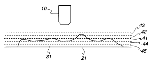
Fig. 3 is a block diagram of a system according to
the invention for acquiring a virtual microscope slide,
that includes a Z-axis image dimension across the entire
virtual slide. The system includes a microscope
subsystem 15 with a digitally controlled stage platform
28 for supporting the glass microscope slide 21. The
digital stage platform 28 can operate over a large number
of increments to position the stage in the horizontal x
and y plane with high precision. .A glass microscope
slide or other substrate 22 is placed on the stage 28.
The system also includes a controlling computer system 32
with a keyboard 37, a mouse 38 with a mouse wheel control
39, and a display monitor 40. The controlling computer
system keyboard and mouse are used via the automatic
focus step size setup window 12 to input the automatic
focus step size parameters and the Z Stack step size
setup window 13 is used to input the Z Stack step size
parameters.
Fig. 3A shows the focus setup step size 55 input
control. Also in Fig. 3A are shown the associated setup
controls for the frequency of focus 56, a threshold for
CA 02436043 2003-08-14
-19-
controlling whether automatic focus should be performed
on a specific field of view, and a control 58 to manually
increment the Z-axis dimension to move the microscope
stage 28 up or down vertically in incremental units. As
may be appreciated by the foregoing description, and the
following descriptions, and as is well :known in the art,
the control of focus at high magnification and small
depth of field is complicated and involves many
variables. It is also time consum_ng if performed on
every specimen image field of view in constructing or
capturing a virtual microscope slide image data set.
Therefore, in the preferred embodiment and in the
subsequently described alternative embodiments there are
additional control and setup parameters to overcome major
variables and to enable a faster overall virtual slide
scan capture time. Some of these are seen in Fig 3A.
For example, the frequency of focus 56 parameter allows
automatic focus to occur on every adjacent field of view
if it is set to 2, or on every other field if it is set
to 2, etc. In the following detailed description the
assumption is that the frequency of focus is set to 1.
However, if it is set to a higher number the reference
tiles of field of views not focused take the default
focus contrast value of_the last previous focused image
tile. In the a:Lternate embodiments of the invention, the
reference tile position is sometimes obtained in a
CA 02436043 2003-08-14
-20-
different fashion as described for those embodiments.
There is a significant speed of scan advantage related to
not focusing on every field of view. However, on many
specimens the disadvantage is that the resulting scan may
not have an optimum depth of field position for the
reference image. It may also be appreciated that one of
the Z Stack images may then offer a more optimum image
for the final remote viewer of the images. Also, often
there is not enough image structure in a field of view to
obtain an automatic focus, for example in the case of a
substantially blank or empty field of view. In that case
the control 57 allows for a focus contrast threshold
value input parameter that can be checked to allow
skipping such fields. Also, in that instance the
default reference 2-axis position for the next image
requiring focus is the last previous focused image tile.
Fig. 3B shows the Z Stack step size ~0 input
control. It may be appreciated that there are a
multitude of factors that would require this parameter to
be changed for a specific specimen. However, the most
important of these is usually the magnification of the
specific microscope objective lens being used, since each
lens has a different depth of field specification, in
combination wits the type of specimen and estimated
thickness of the specimen preparation. Also, shown in
Fig. 3B is a checkbox control 59 to either enable or
CA 02436043 2003-08-14
-21-
disable the Z Stack image save for a specific virtual
microscope data capture scan.
According to the teachings of the aforesaid patent
the computer controlled microscope is moved to start a
scan of the entire specimen object 31 using the stage
controller 14 to move the precision stage 28 to a new
objective lens 10 field of view to acquire an initial
image at that position and compute a focus contrast
parameter on that image. According to the present
invention the relative Z-axis reference position for the
first new field of view image tile is obtained by an
iterative automatic focus procedure. The controlling
computer system 32 sends the microscope subsystem 15 Z-
axis control signals to change the Z-axis position
i5 control to move the stage incrementally to go up 4 times
in the automatic focus step size and then to go down 4
times in automatic focus step size. At each incremental
change in the Z-axis position the image acquisition
electronics 27 are controlled to acquire an image. A
focus contrast parameter is computed on each image. The
automatic focus Z-axis position is then determined by
choosing the Z-axis position associated with the largest
value of the focus parameter from the initial reference
image and the set of 8 images. If the largest value is
at one end of the sequence, i.e the 4''' image down or the
4t image up from the reference image, that image becomes
CA 02436043 2005-06-16
-22-
the reference image, and the procedure is recursively
repeated until the largest focus contrast value is found
in the middle range of the sequence of images, i.e. not
at either end image. This becomes the relative Z-axis
reference position for the new field of view image. As
explained more fully in the following, the image tile
associated with this relative Z-axis position is then
stored in the virtual slide data structure.
In the preferred embodiment of the invention the
TM
controlling computer system is operated under a Windows
Operating System (Microsoft Corporation, Redmond,
Washington, USA). Referring to Fig. 3A, the virtual
microscope slide data structure is stored as a Windows
Operating System file folder where each tiled image is a
.jpg image file with an incremental image name
automatically assigned by the controlling computer
systems software program. The .jpg image names are
numbered so that the first acquired image is called
DAO.jpg, the second DAl.jpg, the third DA3, etc. In Fig.
3C there is depicted a virtual slide data folder 42 with
portions of the data structure 43,44,45,46, and 47 also
depicted. The set of 9 image tiles 43,45 and 46 named
DA98 are associated with a specific X, Y specimen image
plane position, and an adjoining set 44 and 48, named
DA99 are associated with another abutted specific X, Y
specimen image plane position. The two reference tiles
CA 02436043 2003-08-14
-23-
are depicted as 43 and 44 for the data structure at the
two X, Y locations. These tiles are in the automatic
focus determined Z-axis position, and the recorded .jpg
image contains the depth of field image structure
associated with that Z-axis position and the field of
view at the respective X, Y location.
During the system program operation to produce a
virtual microscope slide, the controlling computer system
also creates an additional text information file of the
Windows Operating System format .ini. As depicted in
Fig. 3C, this file 47 is named FinalScan.ini. Among
other things this file contains a list of names
corresponding to each reference tile in the virtual
microscope data structure. For each reference tile in
the list, that tiles X, Y, and Z digital location is
tabulated. As taught in the aforementioned patent this
ir_formation is then used by the virtual Internet server
and virtual microscope visual display programs to abut
and reconstruct the various tiled images to allow a
remote viewer to view contiguous regions of virtual slide
images. It may be appreciated that the components of the
data structure shown by example in Fig 3C may be stored
in a database or any other form allowing rapid sequential
access to the reference image and the full Z Stack
components. A novel aspect of this data structure is the
close association of these image components. This
CA 02436043 2003-08-14
-24-
greatly facilitates client server interactions in remote
Internet viewing. Since the this subsequent viewing is
through the limited X, Y planar view of an image display
device, only a few reference tiles (and in certain
limited situations only one reference tile) may be in the
available user view for a focus request to the server.
As will be appreciated in the following description, this
type of data structure facilitates rapid transmission of
Z-axis image content to the client computer. Some
Internet server computers facilitate serving very large
images requiring zooming, by using a pyramid data
structure where different levels of image zoom are pre-
constructed from the original image and kept in memory or
virtual memory at one time. This requires a very large
amount of memory when considering the requirement of
keeping multiple planes of such image structures, such as
shown conceptually it Fig 9. The data structure of this
invention is much more efficient when used specifically
for virtual microscope slides with special viewing
programs, since it in essence is pre-constructed to serve
reference and z Stack images rapidly from memory or
digital disk storage in these small reference and Z Stack
image units.
After capturing a relative tile for the Z-axis
position at a given X, Y specimen plane position the
system of the invention proceeds to use the Z-axis step
CA 02436043 2003-08-14
-25-
size and execute the Z Stack save procedure. To
accomplish this, the controlling computer system 32 is
directed to control the Z-axis positioning control 16 of
the microscope subsystem 15 first to move down the Z-axis
in incremental Z-axis step sizes, and at each step to
acquire an image tile. These image tiles 45 are stored
in the data structure depicted in Fig. 3 by example for
the data structure set DA98. Secondly, the controlling
computer system 32 is directed to control the Z-axis
1C positioning control 16 of the microscope subsystem 15 to
move up the Z-axis in incremental Z-axis step sizes from
the reference Z-axis position, and at each step to
acquire an image tile. These image tiles 46 are stored in
the data structure depicted in Fig. 3 by example for the
data structure set Da98.
The same series of events described above for the
data structure capture of the tile set Da98 is repeated
for all field of views associated with the capture of the
virtual microscope slide. For example, in Fig. 3 as the
data structure set Da99, and so forth.
The result of the above described preferred
embodiment of the system of the invention is in effect to
first factor out, or neutralize, the Z-axis irregularities
in optimum focus position over the X, Y surface of the
slide for the initial relative captured image tile, and
then, secondly to create a set of cohesive Z-axis
CA 02436043 2003-08-14
-26-
dimensioned captured image planes, where each plane
relates to a different, real, physical depth of field
position in the specimen. The first relative Z-axis
positioning has brought into parallel positioning capture,
the optimum depth of field portions of each specimen, and
the Z Stack capture has resulted in image planes above and
below that. This image sequence sampling can be
reconstructed from the data structure storage elements 43
thru 43, 44, 44, 45, 46, and 48 when used with the X, Y
location information stored in data structure element 47.
This reconstruction is depicted in an idealized fashion as
shown in Fig. 4 in cross section and in Fig. 9 in
perspective schematic, as an aid in visualizing the
resulting complete virtual microscope slide data
structure. As described below, in fact only a small
portion of this is seen by the remote viewer at one time,
because of the limitations of the image display of
commonly used computer screens. However, it is all
available for viewing by scrolling and requesting
additional image tiles from a virtual microscope slide
server.
It will be appreciated by those familiar with the art
that the above preferred description of the embodiment of
the invention may be modified in other ways to enable the
creation of a virtual microscope slide with Z-axis image
dimension information. In this regard, an alternative
CA 02436043 2003-08-14
-27-
method of practicing the invention is described. This
method is more applicable for specimen objects that don't
cover a large area, or in those instances where the stage
platform 28 and microscope slide 21 are positioned to
present the specimen 31 in a reasonably flat plane, or
where a lower power objective is used that has a larger
depth of field. For a given level of magnification (such
as 10X for example) the microscope objective 10 with
associated video camera is adjusted up or down, or as in
the preferred embodiment, the stage is adjusted up or
down, either adjustment to bring into view an initial
reference image into the focal plane depth of field of the
microscope objective 10 and used to create magnified
images of the specimen 31 for a given X, Y position in the
specimen plane. A first series of planar abutted image
tiles are obtained as described in the preferred
embodiment as the reference tile set, and stored in the
data structure previously described, and as shown by
example in Fig. 3C, wherein the example reference tiles 43
and 44 are depicted. The reference tiles Z-axis position
in this embodiment are computed using the results of a
prior setup procedure where the Z-axis positions at three
separate places on a specimen are determined and a
mathematical Z-axis plane is determined across the X, Y
plane of the specimen. by computations involving fitting a
plane in the Z-axis by using three X, Y points with known
CA 02436043 2003-08-14
-28-
Z-axis values. In this case during the scanning process
this computed position is used instead of the iterative,
recursive, automated focus procedure described in the
first preferred method. This results in a faster scan and
image capture process.
By way of illustration the capture of the complete
set of tiles in this plane may be visualized in cross
section as the depth of field 41 in Fig. 4. This scan
captures the upper surface of much of the specimen 31.
Then, in accordance with this embodiment, the stage Z-axis
position is changed according to one Z Stack increment and
another series of images are captured and stored in the
data structure shown in Fig. 3. For example, if the first
series of images used the focal distance corresponding
i5 depth of field represented by reference numeral 42, then
by decreasing the stage Z-axis position relative to the
microscope objective, the next series of images would be
represented by reference numeral 42. Conversely, by
increasing the stage Z-axis position relative to the
2U microscope objective the next series of images would be
represented by reference numeral 44. Subsequent series of
images can likewise be captured by positioning different
Z-axis planes in the depth of field region of the
objective. In the embodiment depicted in Fig 4, in
25 addition to the original image series captured 41, two
other series represented by reference numerals 42 and 43
CA 02436043 2003-08-14
-29-
and two additional series represented by reference
numerals 44 and 45 are also captured. By capturing and
storing these additional images from different regions of
the specimen, a virtual focusing capability can be
realized as described below in more detail. It may also
be realized that this method of scanning may be more
suited to a type of triple pixel line sensor described
above as a 3 by 2098 sensor. Sometimes this is referred
to as a single Line sensor. In this case since small
discrete individual tiles are not available, the
prediction of a reference plane by computation, or simple
assumption of a completely flat ar_d parallel X, Y-plane
may be preferred. This type of scan results in saving
images of longer strip tiles, with a width of 2098 pixels
1S inside the field of view of the microscope objective 10 in
one direction, but the saved images extending beyond the
field of view by continuous scanning and storing in the
other direction. The abutted 2098 pixel wide strip tiles
taken together side by side form a virtual microscope
slide.
Also as illustrated in Fig. 4, there are two focal
depth of fields above and two focal depth of fields below,
both provided with respect to the initial reference
setting. In a given application, it may be appropriate to
2S provide only a single additional set of images using only
one slightly different focal depth of field for focal
CA 02436043 2003-08-14
-30-
plane) above and below. For most purposes, however,
images captured at a plurality of differing focal planes
are appropriate. In the preferred embodiment, four focal
planes above and four focal planes below are used in
addition to the original reference focal plane to provide
a total of nine sets of focal plane images. Wherein each
set of focal plane images corresponds to a given focal
distance from the reference setting and all of the sets
share the same level of magnification. By providing this
many sets both above and below the reference focal plane,
relatively smooth and detailed virtual focusing can be
realized that well mimics the look and feel of focusing
with an actual microscope within a useful range of
focusing.
As described in the above, the various depths of
field substantially abut one another. In an alternative
embodiment, and as illustrated in FIG. 5, a given depth of
field 51 for a given series of images can partially
overlap with another depth of field 52 for a different
series of images. Or, if desired and as illustrated in
FIG. 6, different depths of field 61 and 62 as
corresponding to different image series can neither
overlap nor abut one another. Instead, a small gap can
exist between the two fields. In general, adjusting the
focal distances such that the fields are substantially
adjacent one another with little or no overlap probably
CA 02436043 2003-08-14
-31-
represents an optimum configuration, but the other
alternatives may be useful for some purposes depending
upon user requirements.
With reference to FIG. 7, an initial focal plane 71
(as initially determined or predetermined manually or
automatically) having a corresponding depth of field 41A
may be appropriately used when imaging a particular
section of a specimen (not shown) that is within the field
of view when the microscope is located in a first position
20 10A. That is, when the microscope is so positioned, this
initial focal distance 71 represents an optimum focus by
whatever standard the user applies. In acCOrdance with the
various embodiments above, one or more additional images
are also taken o~ this same field of view with slightly
different focal distances. At another portion of the
slide, however, when the microscope is positioned at a
second position 10B, it may be that a different initial
focal plane 72 will yield an optimum focus when using the
same standard as was applied earlier. This different
initial focal distance 72 will have a corresponding depth
of field 41B that is substantially iden~ical in size to
the depth of field 41A for the first position's initial
focal distance 71 but that is positioned a different
distance from the slide 21. This is often the case when
imaging tissue microarray (TMA)cores as described in US
patsnt No. 6,466,690 B2 (entitled Method and Apparatus for
CA 02436043 2003-08-14
-32-
Processing an Image of a Tissue Sample Microarray). There
the image capture is from a great many different objects,
TMA cores, arranged over essentially the entire surface of
the glass microscope slide. Therefore, while the resulting
images still comprise a abutted composite representations
of the object, they refer to different reference image
focal planes. And, according to this embodiment,
regardless of differences as may exist between the initial
focal reference focal plane from object to object, each
resulting image will nevertheless have an identical number
of 2 Stack focal planes available for fine focusing by a
user.
As discussed above, virtual microscope slides,
whether created 'From many small tiles as in the preferred
1S embodiment, or whether created in strips of line segments,
and whether they are stored. in a tiled data structure or
whether they are stored as one large reconstructed image
in memory, such as one focal plane from the set of 5 focal
planes 91 in Fig. 9, cannot usually be 'Viewed in their
entirety at the original captured resolution because of
the finite size and pixel dimensions of a remote viewers
computer display screen. As depicted in FIG. 8, one prior
art approach that is useful. in this regard utilizes a
plurality of individual images 83, referred to as tiles,
to form a larger composite image of the slide 81 and the
specimen 82. US patent no. 6,396,941 B1 (entitled Method
CA 02436043 2005-06-16
-33-
and Apparatus for Internet, Intranet and Local Viewing of
Virtual Microscope Slides) teaches the Internet or
intranet display of virtual microscope slides. As taught
therein, a virtual microscope slide typically comprises a
digitized magnified view of part or all of a microscope
slide and an object (such as a biological specimen)
disposed thereon. The aforementioned patent also teaches
various Internet server and thin client, and other JavaTM
Applet and ActiveXTM viewer methods enabling the
reconstruction of the virtual microscope image content
for a remote viewer. It will be appreciated that the
viewing of a single focal plane depth of view is
accomplished whether the image is stored as a tiled
database structure or as a complete single image plane in
computer core memory. In the preferred embodiment of
this invention however, when the remote viewer sends a
request to the server for a reference image tile focus
for a defined region of interest, the server also sends
the associated Z Stack images all in sequence for that
region of interest. The associated Z Stack images are
cached by the local computer so that a smooth and rapid
local viewing can simulate the analog optical focusing
operation of a real microscope.
Referring now to FIG. 10, in one embodiment, a user
can employ a standard computing platform to interface to
CA 02436043 2003-08-14
-34-
the virtual slide server and data storage facility that
retains the virtual microscope slide information as
described above for a given specimen. A standard
client/server model works well to facilitate such a
relationship, but other data transfer mechanisms can be
used as well as appropriate to a given application.
The relevant process begins with a user platform
retrieving 101 a desired image at a particular
magnification X (such as, for example, 40X). As described
in the aforementioned patent, all images for the object
need not be immediately retrieved and made available
locally. To minimize network transactions, in fact, only
the data required to display a single field of view need
to be immediately retrieved and displayed. In the system
and method of the current invention and the various
embodiments above, each field of view has a corresponding
plurality of images with each image representing a
different focal plane. Therefore, when retrieving and
displaying the first image, one of these images must be
2C selected first. In the preferred embodiment the selection
is the reference image set corresponding to those tiles
that will fill the view window of the remote viewers image
display screen. Also, the associated Z Stack images for
each reference tile are transmitted and cached in the
local computer. In one embodiment, where the initial
automatically determined optimum focal plane image is
CA 02436043 2003-08-14
-35-
flanked on each side by four different focal plane images,
the initial image itself can be automatically selected for
initial retrieval 101 and display 102. The process then
monitors 103 for instructions from the user to modify the
focus. When no such instruction appears, the process
continues 104 in accordance with whatever other functions
are supported (for example, input from the user indicating
a desire to scroll the image in a particular direction can
be received and used to cause retrieval and display of
corresponding images). When a focus modification
instruction is received, however, the process retrieves
105 the image from the local memory cache for that field
of view that corresponds to the instruction and displays
106 it. Pursuant to one embodiment, the user can be
limited to moving the focus in a step by step process with
a mouse wheel 93 or keyboard 37 up or down arrow keys,
such that each increment causes retrieval and display of
the next adjacer_t image in the Z-axis dimension. In the
preferred embodiment the user, or remote viewer, can move
about and focus on the virtual microscope slide with a
wheeled mouse control, essentially as one moves about and
focuses with a physical microscope and slide. With this
capability, a wide variety of specimens can be readily
viewed with good results. Not only can the resultant
virtual microscope slides be used for educational and
training purposes, but also for both qualitative and
CA 02436043 2003-08-14
-36-
quantitative analysis purposes in support of various
diagnostic processes. With reference to FIG. 11, pursuant
to one optional embodiment, when a user seeks to modify
103 the focus as described, the process can determine 111
whether a focusing limit has been reached. For example, if
the user platform has already retrieved and displayed the
image that was captured using the focal plane at the
furthest Z-axis dimension from the reference tile and the
user is now instructing the platform to focus on an even
further distance, the present display can be maintained
112. Optionally, a text message or other indicator can be
provided 113 to the user to alert the user that the focus
limit has been reached. In another embodiment, a visual
indicator can be provided to the user to indicate a
present focusing position. within a range of focusing
possibilities, such that the user can ascertain for
themselves such a condit~Ori.
Those skilled in the art will recognize that a wide
variety of modifications, alterations, and combinations
can be made with respect to the above described
embodiments without departing from the spirit and scope of
the invention, and that such modifications, alterations,
and combinations are to be viewed as being within the
ambit of the inventive concept. It is intended in the
appended claims to cover all those changes and
CA 02436043 2003-08-14
-37-
modifications which fall within the true spirit and scope
of the present irwention.