Note : Les descriptions sont présentées dans la langue officielle dans laquelle elles ont été soumises.
CA 02441626 2003-09-23
WO 02/081655 PCT/US02/09353
FUSION PROTEIN CONSTRUCT AND METHOD
FOR INDUCING HIV-SPECIFIC SERUM IgG
AND SECRETORY IgA ANTIBODIES IN-VIVO
RESEARCH SUPPORT
The research for the present invention was supported in part by NIH grants
GM39589, HD-17557, and AI-34757. The U.S. government has certain rights in the
invention.
FIELD OF THE JNVENTION
The present invention is concerned generally with humoral antibodies
specific against epitopes of human immunodeficiency virus (HIV). It is
particularly
directed to the synthesis and use of gp41 fusion protein constructs as
ixnmunogens
and vaccines effective for inducing HIV-specific serum IgG and secretory IgA
antibodies in vivo.
BACKGROUND OF THE INVENTION
There is presently a worldwide demand for an efficacious vaccine that
reduces the risk of sexual transmission of the human immunodeficiency virus
type 1
(HIV-1) across cervicovaginal and rectal mucosae. In the female genital tract,
it is
thought that HIV-1 is initially "sampled" by motile intraepithelial or
subepithelial
dendritic cells and may initially infect mucosal T cells [Hussain et al.,
Immunolo~y
85: 474-484 (9995); Parr et al., Biol. R_ eprod. 45: 261-265 (1991); Pope et
al., J.
Infect. Dis. 1?9: 5427-5430 (1999); Spira et al., 3. Exp Med. 183: 215-225
(1996)].
In the rectum HIV-1 may enter via damaged epithelium or may cross an intact
epithelial barrier via colonocytes or via specialized antigen transporting
epithelial
cells known as M cells [Amerongen et al., J. ACQ. Immun Def. Synd. 4: 760-765
CA 02441626 2003-09-23
WO 02/081655 PCT/US02/09353
2
(1991); Bomsel, M., Nature Med. 3: 42-47 (1997)]. Once within the mucosa H1V-1
replicates in resident CD4+ T lymphocytes and/or macrophages and may be
carried
by these cells, as well as dendritic cells, to draining lymphoid organs within
days
after initial exposure [Ignatus et al., J. Med. Pathol. 27: 121-128 (1998);
Miller et al.,
J. Med Primatol. 21: 64-68 (1992); Pope et al., Cell 78: 389-398 (1994); Stahl-
Henning et al., Science 285: 1261-1265 (1999)].
Humoral Immunity:
Humoral immunity plays a critical role in preventing and/or modulating
infection with the primate lentiviruses, including HIV, simian
immunodeficiency
virus (SIV), and the HIV-SIV chimeric virus SHIV [Moore & Burton, Nature
Medicine 5: 142-144 (1999)]. For example, experiments in chimpanzees
demonstrated that immunoglobulin (Ig) from the serum of HIV-infected
individuals
(HIVIG), monoclonal Ab (mAb), chimeric mAb, and anti-CD4-immunoglobulin
IgG can all prevent infection with HIV; and that a human mAb to gp41 can
significantly delay signs of infection [Prince et al., AIDS Res. Hum.
Retrovir. 7:
971-973 (1991); Emini et al., Nature 355: 728-730 (1992); Emini et al., J.
Virol. 64:
3674-3678 (1990); Conley et al., J. Virol. 70: 6751-6758 (1996)].
These studies of protection of chimpanzees by passive immunization suggest
that the best correlates of immunoprophylaxis within in vivo studies are
effective
virus neutralizing activity in vitro and a slow Ab dissociation rate constant
[Van Cott
et al., J. lmmunol. 153: 449-459 (1994)]. Similarly, most studies in mice
reconstituted with human peripheral blood mononuclear cells exhibiting severe
combined immunodeficiency syndrome (hu-PBL-SCID) have also demonstrated that
pre- and postexposure protection against HIV infection can be mediated by
murine
mAb, human mAb, and mouse-human chimeric mAb [Safrit et al., A117S 7: 17-21
(1992); Gauduin et al., J. Infect. Dis. 171: 1203-1209 (1995); Parren et al.,
All~S 9:
F1-F6 (1995); Gauduin et al., Nature Med. 3: 1389-1390 (1997)]. All of these
studies suggest that Ab of appropriate specificities can prevent HiV and SIV
' infection with cell-free virions and of slowing viral replication and
disease
progression.
CA 02441626 2003-09-23
WO 02/081655 PCT/US02/09353
3
Active immunization studies:
Vaccine studies in primate models have increased our understanding of the
interplay of viral replication and host immunity. Conjectured for a number of
years,
and now documented in several primate studies, is the observation that
infection
with live-attenuated viral vaccines induces strong cellular and humoral
immunity,
including neutralizing Ab effective against the macaque-grown challenge virus
stocks, which can be considered primary isolates in this system.
The induction of these humoral responses is dependent upon a threshold of
replication of the attenuated virus during primary viremia [Ruprecht et al.,
AIDS 10:
S33-S40 (1996)j. Below this threshold, immune responses are weak and full
protection is not seen except with very weak virus challenges; above the
threshold,
strong host immunity is observed in most animals and protection from infection
with
highly pathogenic SIV challenges ensues. These data and those obtained in
vaccine
studies with live-attenuated SIV, summarized by Table A below, support the
notion
that the level of attenuated virus replication during primary infection
predicts
whether the immune response is sufficient to block infection upon subsequent
challenge with wild type virus.
30
CA 02441626 2003-09-23
WO 02/081655 PCT/US02/09353
4
Unfortunately, several examples of pathogenic effects from highly
attenuated live viral vaccines were documented in five laboratories during the
1998
year, as summarized in a recent editorial [Cohen, J., Science 278: 24-25
(1997)].
Thus, it remains the difficult goal of vaccinologists either: (1) to construct
live-
attenuated viruses that are both effective and safe, or (2) to mimic the
presentation of
viral proteins observed in infection with recombinant antigens or with
replicating or
non-replicating vectors carrying appropriate genes or antigens.
20
CA 02441626 2003-09-23
WO 02/081655 PCT/US02/09353
o ~ ~ ~ ~ i o i i i i o i i o 0 0 0 0 0 0 o i i
oy ~~~~~z ~~~ ~z~~ zzzzz zzz~~
0
0 0
-I- + + + -f -F -I- + + i +
-I- + r + i f -t- -(-
-I- + -I-
+
'T~ N N N
'd
U A ~ N >
O ~
~
o
b
D b
p b
D
CCl m ~ ,-n icd. VJ ~ > > >
~7 v0
. V
" ~ ~ z ~d
~ ~
x ~ ~
.
w N x a. w w w w '
. ~ x Q7 U7 N '~
w ~3 w w x ~ N N ~ '
~ ''x ~ ~ V U U V
~ x
~ x
N
~~~
1
I--1 ~ !--1
r N I--I
F-i Y-1 I--i
I-1 h--1
O ~
N
4~
0o N
~
n
c'Ud ~ N . ~ ~ Ov
,-r N
N >,
~ V~
~ ,~ N ~-~ ~ U U ~ N N
"' ~ 'v
R
~ ~
'
d. ~-; ' R .-:
'~ ~.,
,
'
o . y ~, ~, x
o o ,~ >,
~ ~ '
N
U ~ ~ ~ O
~ U ~ "'
b U '
6
~
o a-. ~ cc! .--i pr C=, 4 .~
N O d~ N + cd .,
Ov ~ .~' p --
fx ~ ~
.~ M
c
O U N ~ _
R~ .fl ~' O'' ~ Cf7
N ~
~
M N ~ -w N iy V1
"' oo ~'' _ > N N
j ~ ~ ~
O ~ ~ .~ -~ ~Y W ~' '~ ~
,~ ~ rx N ~ ~'
N ~ ~ N ~ O ~ ~ ~ ..p
~ O V ~' U
:.'
v~
w a ~4.i ~ ~ ~'~~~ ~Qi ~w'.w ~'.=
~~" ~
O A ~ O
? ~
S ' r+-i O ~ ~ O ~
- ~G ~ "W
a
~ d ~ ~ ~' '~ b~A ~ . ~
> ' + O ~ ~ p ~ V1
c ~, ~
~
a.o ~nN ~ O-oho "' yC7
N ?
~
O ~ '2t t+ ''' v0 ~ C/~ N ~o + T3 ~ ~ O
+ O O N ~ N ~ ~
~ ~ WO ~
~ ~ +
-our n-. O ~V7'+ O=~ ~~NCi~ O
+ ~ P. ~ ~ ~ O ' N
R~ ~ ~ O '
p r. ~ i i ~~ -n p. O O Z3
O by by p. O .1 Z3
p b b0 . ~D n ~ N
i TJ ~ b
y N by N N >,
cd cd ~ a' p., p" m, y., r"
00 N ~ p4 i pa N
~O
.b bA bA 'ZS ~ ~ ~ CO 0~'p ~ ~ p
~ ~ cd cd ~ ~
pa b
O O ~ ~ Ct ~ , ~ ~ N .~ ,
~ ~ , ~ v~
~ OU ~a
~
y
. ~' ~ .
~ .
, ~
O
'
'
~ N 0 ~ ~ p~ O N ~ ~ ~ O ~
~ O ~ ~ ~
.~ ;~ ~
N ~
.U ~ F'r ~ ~ b U V V V ? ? ~ ~O
~ ~ b ~ V N Vj
~ ~
U
~
~zb ~ ~ r~ x ~~~ ~ ~ ~NN~ ~ ~w+
~'' ~~ ~
~z
~'
'
n~ !~ ~ ...'.~ ,~ ~'.~ ~ ~.~'.~.,.a o
U U ~
~.1
n
~ 0
on
o
~
Z
.
' '~ 3 3 -d .c o
~
o
a a
O
O y n cd
N
,~ ~
y N
V ~
N N W N
~ ~ ~ ~
~, w o o ~ a N ,~
~. Q, O" ~" ~ ~ ~ ~ ~ G
7 cd N ~ F-1 1-1 ~r
N N
x U ~ ~ v~ v~ ~ o 0
CA 02441626 2003-09-23
WO 02/081655 PCT/US02/09353
o~
U
N
.O
O~
O
z o
z
0
~.
Ab
.v ~ ~: " o
~t ~ ~:.~: °,~ a~
O~ N.~~ v ~0~1 m yN
. ~ ~ D\ Ov N
U ~' N ~ O ~
Pa n
O ~ ~ ~ ~ Ov
OMO ~ O
l~ 0~1 ~ ~ ~ '~, ~ s~ l~ ~ O ~ n
~o~~~°'~~ o ~Mp~~l~~°p~~~~z~~
oy ~ ,-m ~ ~ W~ o ~' a\ ,-W °' a\ ov ow,
v _,.~1 p v oo
N ~ ~ M ~ ~ v~ ~ ,~ > ,~ ~ r~ 'oN O p O
NN' ~n ' Off' ..G ~' ON"''~.OVV7.b ~.yt~ "'' y0N
~ ~ t/7 ~O V7 ~ ~ ~ .v.~ O ~ O~ 00 U ~ M U N M y d' M ~~ M .~,
i Q\ 4w ~ ~ ~ ~
N N v0 ~j~ ~ O p ~ p i ,_,., d. V1 ~ ~ ~ ~ ~i ~ ~' Q. M ~
N 'a
M vo ~ ~ f~I ~ ~ v~ p c~ ~ a\ oo ~ '~ ~ ~ ~ M ~ ~ M ~ MI ,N-~
G\I ~M1 a~!. ~I U ~ '~~ ~N ~ x ~-i ~~ ~ ~7~ O v~ x ~~ ~ °~ OO
C/~ N ~ U ~ ~ N ~ ~ ~ '~ l\ . ~ .~ l\ N U ~ ~ . ~ ~ N
U f-i ~ U ,-i O y ' ~ ~.,~ O ici~ t~~ ~'., N
~ ~ : o~~~.~~ ~ ~Za ~~~~ ~ o ~~'~' ~ ~
.~ >,~ ~ ,~
~z
z ~~aa~ ~~~;~w ~~~ : ~>~~z~
x
.~.~ iv ~ ~ G '~ ~ oy ~ .~, ~ r-~ ~t at ~ o ~
~ i1 of ~ a~~ o .~ ~ ~I °' ~ " ~I ~ i1 ~ ~ ,N a~~ .~ a~~ ~
NNC~~~.N~Uyc~''~d.~~V~~~'OU~,~,,~~.~.N
.:',..I' O ~ O ~~~-a~ O OI N ~ '~' ,.O N OI v ~ ~c~ ~ O ~ >' ~ >'
wr~c~aaaNC~xxxA~~axx~~a~aaa~a~A
U
y ~ ~ O ,--i CV M d' i ~D l~ oo ~ O ,-i N c~i ~t V 7
~, ~ N M d' V'7 v0 l~ o0 01 ,1 ,1 ,~-m~-i .-i ,-i .-~ ,-~ ,-i ,~ N N N N N N
4~ ~+~i 4-i 4-i 4.: v~~ 4-i W chi 4-i 4.-~ 5~; 4~ 4-.i 4~: e+.~ 4.; W 4-i 9~:
4~: 4-i 4-i v-S 4-: 4~:
CA 02441626 2003-09-23
WO 02/081655 PCT/US02/09353
7
'PrimeBoost' and subunit vaccines tested by challenge with SHIV and SIV:
It has been shown that immunization with HIV-1LA1 gp160 vaccines, in a
recombinant vaccinia virus priming and subunit boosting regimen, protected
macaques against SIV HXBc2 challenge [Haigwood, N.L. and S. Zolla-Pazner,
AIDS 12: S 121-S 132 (1998)]. Using the same challenge model, it was found
subsequently that subunits alone were not protective (gp 120; none out of
three
protected) or partially protective (gp160; two out of four protected).
Complete
protection was observed in all six macaques that received vaccinia virus-
expressing
HIV-1 gp160 and boosts of either gp120 (three out of three protected) or gp160
(three out of three protected). More complex immunogens including Env-bearing
pseudovirion particles were partially effective in providing protection
against SHIV
challenge (three out of five protected). These data underline the importance
of
providing sufficient Env protein in vaccine preparations.
The HIV envelope glycoprotein:
An overview of the scientific reports shows that the envelope glycoprotein
(env) of human immunodeficiency virus-1 (HIV-1) is synthesized as a precursor
molecule gp160 and subsequently processed into its subunits gp120 and gp4l.
Gp120 is non-covalently associated with gp41 and contains the binding sites
for
CD4 molecules, i.e., the cellular receptors of HIV-1, and the chemokine
receptors
such as CCR4 and CXCRS. The gp41 subunit is anchored in the membrane and has
a non-polar fusion peptide at its N-terminus. The gp120-gp41 molecule forms
oligomers on the infected cell surface and on virions. Strong evidence for
trimeric
oligomers states has been reported at length in the published scientific
literature.
The binding of gp120 to CD4 is thought to result in activation of the
membrane fusion activity of gp4l, leading to entry of the viral nucleocapsid
into a
cell. Evidence for a conformational change in the viral glycoprotein upon
binding
CD4 includes alterations in antibody reactivity, increased protease
sensitivity and
the dissociation of gp120.
Recent publications which factually support this summary overview include
the following: Allan et al., Science 228: 1091-1094 (1985); Veronese et al.,
Science
229: 1402-1405 (1985); Dagleish et al., Nature 312: 763-767 (1984); Klatzman
et
CA 02441626 2003-09-23
WO 02/081655 PCT/US02/09353
al., Nature 312: 767-768 (1984); Madden et al., Cell 47: 333-348 (1986); Bosch
et
al., Science 244: 694-697 (1989); Kowalski et al., Science 237: 1351-1355
(1987);
Gelderblom et al., Virolo~y 156: 171-176 (1987); Pinter et al., Virolo~y ~3:
417-422
(1977); Schawaller et al., Virology 172: 367-369 (1989); Earl et al., J.
Virol. 68:
3015-3026 (1994); Weiss et al., J. Virol. 64: 5674-5677 (1990); and Sattentau
Q.
and J.P. Moore, J. Exp. Med. 174: 407-415 (1991); Weissenhorn et al., PNAS 94:
6065-6069 (1997); Weissenhorn et al., EMBO J. 15: 1507-1514 (1997);
Weissenhorn et al., Molecular Membrane Biolo~s 16: 3-9 (1998); and Weissenhorn
et al., Nature 3~7: 426-430 (1997).
Antigen structures which induce Ab responses:
Since the form of immunogen affects the type and specificity of the immune
response, the nature of the immunogens found in natural infection that elicit
Ab
becomes a pivotal issue which impacts on vaccine design. Anti-HIV envelope
polyclonal and monoclonal antibody preparations react with HIV-infected cells,
implying that infected cells express envelope antigens that serve to both
induce Ab
and act as their targets. Thus, HIV+ sera and mAb to gp41 and the V3 and C5
regions of gp120 have been shown to stain cells infected with primary isolates
and
to mediate neutralization and/or Ab-dependent cell-mediated cytolysis (ADCC)
[Zolla-Pazner et al., J. Virol. 69: 3807-3815 (1995); Tyler et al., J.
Immunol. 145:
3276-3282 (1990); Alsmadi et al., J. Virol. 71: 925-933 (1997); Bauir et al.,
J.
Immunol. 157: 2168-2173 (1996). This demonstrates that infected cells express
virus-derived antigens. Oligomeric envelope proteins also are immunogenic.
As summarized in a recent paper [Haigwood, N.L. and S. Zolla-Pazner,
AIDS 12: S 121-S 132 (1998)], while oligomer-specific mAb have only been
described in immunized mice and rabbits, several human mAb have been described
which show better reactivity with polymeric than with monomeric HIV envelope
molecules. Amongst the first of these were human mAb to gp41 which
preferentially react with oligomeric forms of gp41 on Western blot [Zolla-
Pazner et
al., N. Engl. J. Med. 320: 1280-1281 (1989); Pinter et al., J. Virol. 63: 2674-
2679
(1989)]. Later studies suggested that mAb IgGlbl2, specific for the CD4
binding
domain preferentially binds to structures exposed on oligomeric envelope
protein
CA 02441626 2003-09-23
WO 02/081655 PCT/US02/09353
9
[Fouts et al., J. Virol. 71: 2779-2785 (1997)]; and mAb 2F5, specific for an
epitope
near the transmembrane region of gp4l, binds to the oligomeric structure of
gp41 in
the virion envelope, resulting in neutralization [Muster et al., J. Virol. 68:
4031-4034
(1994)]. That all of these mAb also recognize structures on the monomeric
forms of
gp120 or gp41 is shown by the fact that the hybridoma cell lines producing
these
mAb were each selected using monomeric forms of these envelope glycoproteins.
Immune responses to gp4l:
Recently there has been a renewed interest in the immune response to gp4l.
The potential importance of Ab to gp41 is well demonstrated by the human mAb
2F5 which is specific for the ELDKW epitope near the transmembrane domain of
gp41 and has broad neutralizing activity for laboratory-adapted strains and
primary
isolates of HIV [Muster et al., J. Virol. 68: 4031-40343 (1994)]. Other anti-
gp41
mAb also have been shown to neutralize both laboratory-adapted and primary
isolates of HIV [Hioe et al., Int. Immunol. 9: 1281-1290 (1997); Cotropia et
al.,
AIDS Hum. Retrovir. 12: 221-232 (1996)]; and it was recently suggested that Ab
to
gp41 epitopes in the serum of HIV-infected individuals may play an important
role
in virus neutralization [McKeating et al., Virolo~y 220: 450-460 (1996)].
Additional interest comes from research on the structure of gp41 and its role
in infectivity. Thus, gp4l, which mediates fusion between viral and cellular
membranes, has been shown to consist of a rod-like molecule with a high alpha-
helical content [Weissenhorn et al., EMBO J. 15: 1507-1514 (1996)]; and the
structure of the fusogenic form appears to be composed of a six-helical bundle
of
two regions of the gp41 molecule. The core of the gp41 structure forms an
extended, triple stranded coiled coil derived from a predicted leucine zipper
domain
approximately 30 residues from the N-terminal fusion peptide. A C-terminal a-
helix
packs in the reverse direction against the outside of the coiled coil
following the
groove between two core helices [Weissenhorn et aL, Nature 387: 426-430
(I997);
Chan et al., Cell 89: 263-273 (1997)]. The soluble forms of gp41 visualized by
two
crystal structures contain gp41 residues 30 to 79 and 113 to 153 [Weissenhorn
et al.,
Nature 387: 426-430 (1997)] and a smaller construct contains residues 35 to 70
and
117 to 150 [Chan et al., Cell 89: 263-273 (1997)]. The conformational and
linear
CA 02441626 2003-09-23
WO 02/081655 PCT/US02/09353
5 epitopes exposed on gp41 appear to be different in gp41/gp120 nonfusogenic
configuration and in the fusion active conformation [Sattentau et al., 1995;
Weissenhorn et al., EMBO J. 15: 1507-1514 (1996)].
It has been suggested that the conformational structure of gp41 provides the
fusion-active capability for gp4l. A general model was presented where the
10 complex of gp120/gp41 undergoes major conformational changes after
interaction
with cellular receptors CD4 and chemokine receptors [Berger et al., Annu Rev
Tinmunol 17: 886-900 (1999)]. The conformational changes occurring in- gp41
are
thought to open up intermediary conformational states and the complete
refolding of
the molecule results in the helical hairpin structure observed by
crystallography.
This process is thought to pull two membranes into close proximity and induce
fusion of viral and cellular membranes [see Fig. 3 in Weissenhorn et al.,
Nature 3~7:
426-430 (1970]. It is conceivable that monoclonal antibodies that either block
the
formation of the helical hairpin, like gp41 specific peptides [Kilby et al.,
Nat. Med.
4: 1302-1307], or block the aggregation of gp41 helical hairpin structures (a
number
of trimers are necessary at the site of fusion [Danilei et al., J. Cell Biol.
133: 559-
569 (1996)]) at the site of fusion may inhibit membrane fusion and thus
infection.
HIV envelope glycoprotein variants, synthetic chimeras, and gp41 structure:
In recognition of the fact that the HIV envelope subunit gp41 plays such a
critical role in viral entry by initiating fusion of viral and cellular
membranes,
Weissenhorn and colleagues have synthesized new construct variants of the
ectodomain of HIV-1 and the env gp41 subunit in particular. Thus it has been
shown that the env subunit gp41 forms a slightly soluble, (alpha)-helical, rod-
like
oligomer in the absence of gp120 and the N-terminal fusion peptide
[Weissenhorn et
al., EMBO J. 15: 1507-1514 (1996)]; and also that a rod shaped chimera of a
trimeric GCNA zipper and the HIV-1 gp41 ectodomain can be synthesized and
expressed in E. coli and solubilized by proteolysis [Weissenhorn et al., Proc.
Natl.
Acad Sci. USA 94: 6065-6069 (1997)]; and that the atomic structure of the
ectodomain from HIV gp41 is an extended, triple stranded alpha-helical coil
with the
N-terminus at its tip [Weissenhorn et al., Nature 387: 426-430 (1997)]. The
core of
the molecule forms an extended, triple-stranded alpha-helical coiled coil with
the N-
CA 02441626 2003-09-23
WO 02/081655 PCT/US02/09353
11
terminus at its tip. A C-terminal alpha-helix packs in the reverse direction
against
the outside of the coiled coil following the groove between two core helices.
This
arrangement places the N-terminal fusion peptide and the C-terminal
transmembrane
region at the same end of the rod-shaped molecule [Weissenhorn et al., 1997].
These reported investigations and published papers centered in particular
upon finding new synthetic chimeras which might substantially increase the
solubility of gp41 - and thus possibly increase the number of epitopes exposed
as
well as the potential antigenicity of the gp41 amino acid sequences. As noted
in
these recently published papers, the crystal structures were derived from
different
sources. Core fragments of gp41 were either assembled from synthetic peptides
[Chan et al., 1997], or expressed in E. coli and solubilized with a trimeric
GCN4
zipper fused to the predicted N-terminal coiled coil and trimmed by
proteolysis
[Weissenhorn et al., 1997]. Alternatively, E. coli expressed N-terminal and C-
terminal helical regions were connected by a synthetic linker [Tan et al.,
1997].
All three gp41 structures constructed in this manner (as described in the
published papers) are missing the N-terminal region containing the hydrophobic
fusion peptide and the loop that connects a N-terminal core helix with a C-
terminal
helix. The HIV gp4llGCN4 chimera is missing 39 linker residues, which would
contain a short disulphide linked loop and two carbohydrate sites [Weissenhorn
et
al., Nature (1997)]. Although the disulphide linked loop C-terminal of the
coiled
coil region is characteristic for all retroviral and filoviral fusion
proteins, its function
is not yet known. The disulphide linked loop in HIV might play a role in the
change
of conformation as determined by differential antibody reactivity [Weissenhorn
et
al., EMBO J. (1996)].
Gp41 sequences of different HIV subtypes show a remarkable conservation
for the N-terminal coiled coil as well as for the C-terminal residues that
interact with
the N-terminal core structure [Weissenhorn et al., Nature (1997); Chan et al.,
Cell
(1997)]. Indeed, there are only conservative changes within interfaces of two
N-
terminal helices and one C-terminal helix, and most of the differences are on
the
outside of the C-terminal helix, exposed to the solvent. This reveals that the
C-
terminal helix packs into a highly conserved groove along the core coiled
coil,
CA 02441626 2003-09-23
WO 02/081655 PCT/US02/09353
12
which is remarkable considering the sequence variability in HIV [Myers et al.,
1995].
In addition, there are several lines of evidence that the gp41 membrane
fusion protein exists in two conformations: a native conformation in complex
with
gp120; and a fusion-conformation. First, receptor binding was shown to
increase the
exposure of gp41 epitopes [Sattentau and Moore, 1992] as well as to stimulate
the
dissociation of gp120 from gp41 [Kirsh et al., 1990; Moore et al., 1990; Hart
et al.,
1991]. Antibodies raised against native gp41 (in complex with gp120) [Earl et
al.,
1994] showed a differential reactivity with gp41 expressed (without gp120) in
insect
cells. Some of the antibodies were mapped to the short disulphide linked loop
and
recognized native gp41 but not the fusion conformation [Weissenhorn et al.,
1996].
Second, direct evidence arises from a number of mutagenesis studies, which
showed that residue changes especially within the heptad positions of the
central
coiled coil affect infectivity and membrane fusion, but not processing and
cell
surface expression of gp41/gp120 complexes [Dubay et al., 1992; Cao et al.,
1993;
Chen et al., 1993; Chen 1994]. This indicates that these changes are tolerated
in the
native conformation but not in the fusion conformation.
Third, peptides derived from the gp41 sequence, like DP-107 (part of the N-
terminal coiled coil) and DP-178 (C-terminal helix, with an expression towards
the
transmembrane region), have potent anti-viral activity [Jiang et al., 1992;
Wild et al.,
1992; 1994; Lawless et al., 1996]. The structure of gp41 confirms the view
that
these derived peptides expert their effect by interacting with gp41 during the
receptor induced conformational change. This is also consistent with the
finding
that the assembled complex (N- and C-terminal helices) has no anti-viral
activity
[Lu et al., 1995]. The conformation of gp4l, as observed in the crystal
structure,
shows a temperature dependent denaturation at approximately 80°C
[Blacklow et al.,
1995; Lu et al., 1995; Weissenhorn et al., 1996]; which makes it unlikely that
the
complex comes apart and interacts with individual peptides. Kinetic
measurements
of receptor-activated conformational changes showed that these changes are
initiated
within a few minutes and completed after 20 min [Jones et al., 1998]. It is
also
remarkable that the C-terminal peptide (DP-107) remains active even when added
after mixing of the target cells [Munoz-Barroso et al., 1998]. The C-terminal
CA 02441626 2003-09-23
WO 02/081655 PCT/US02/09353
13
peptide DP-178 does not interact with native gp4l, but binds to gp41 after
induction
of receptor mediated conformational changes, an event which confirms the
structural
changes in gp41 upon receptor binding [Furuta et al., 1998].
Immunization:
It is generally agreed that multiple immune effectors participate in
prevention, containment and clearance of HIV infection. To prevent infection
of
host target cells, antibodies are required. After the first target cells have
been
infected with virus, it is important to have cytotoxic T lymphocytes (CTLs) as
well
as antibodies to reduce cell-to-cell spread and kill infected cells. The exact
amounts
of specific antibodies or CTLs required for mucosal or systemic protection
against
HIV are not known. However, it seems clear that an effective HIV vaccine
should
evoke antibodies that can bind to virus and prevent attachment of virus to
target
cells, as well as CTLs that can eliminate any cells that become infected.
If virus is transmitted directly into the body as through injection,
accidental
needle stick or damaged skin or mucosa, then antibodies and CTLs in the
bloodstream, both of which can readily enter tissues, are most important for
protection. Vaccines that are injected intramuscularly or intradermally are
generally
most efficient for inducing these immune effectors in the blood. However, a
large
proportion of HIV infections are the result of mucosal transmission. This most
often
occurs via the cervical-vaginal mucosa and the rectal mucosa, but may also
occur
via the oral mucosa and nasopharyngeal mucosa. The extent to which antibodies
and CTLs from blood can prevent, contain or clear mucosal infections at a very
early
stage, before virus has spread systemically, is not yet clear. Mucosal
surfaces have
an additional immune protection mechanism: transport of antibodies into
secretions.
Secretory antibodies can provide the first line of defense, preventing contact
of
viruses with the mucosal surface and thereby preventing entry into the body
and
target cell infection altogether (see below). Secretory antibodies are
generally not
induced by systemic immunization. Immunization via mucosal surfaces is usually
required to evoke secretory antibodies and local CTLs and antibodies in
mucosal
tissues. In experimental animals and humans, these effectors are induced most
efficiently at the mucosal site where the vaccine was administered [Haneberg
et al.,
CA 02441626 2003-09-23
WO 02/081655 PCT/US02/09353
14
Infect. Immun. 62: 15-23 (1994); Kozlowski et al., Infect. Immun. 65: 1387-
1394
(1997)]. In addition, vaccines administered mucosally may induce antibodies in
the
bloodstream.
The exact composition of an optimal HIV vaccine, or the protocols or routes
by which it should be administered, are not yet established. One type of
protocol
currently being tested is a combination prime-boost approach in which a live
vaccine
vector (such as fowlpox) carrying HIV genes is given by injection to prime the
immune system, followed by booster doses consisting of subunit antigens
(usually
the HIV envelope proteins gp120 or gp160). The subunit boost appears to be
essential for induction of immune responses in serum. As expected, mucosal
secretory antibodies have not been detected in animal experiments and human
trials
using such protocols. Alternative protocols for induction of secretory
antibodies are
currently being considered. For example, one possibility is administration by
injection of a prime consisting of live HIV vaccine vector or DNA encoding HIV
antigens, followed by boosts consisting of HIV envelope antigens, administered
via
a mucasal surface. The exact form or composition of envelope antigens most
appropriate for mucosal administration are not yet established.
Secretory IgA Antibodies:
There is mounting epidemiological and experimental evidence that the
presence of secretory immunoglobulin A (S-IgA) antibodies directed against the
HIV envelope protein gp41 may reduce or prevent sexual transmission of HIV-1
[Lehner et al., Nature Med. 2: 767-775 (1996)]. For example, studies in Kenya
and
Thailand demonstrated a positive correlation in female sex workers between
resistance to HIV-1 infection and the presence of anti-gp160 S-IgA antibodies
in
cervico-vaginal secretions [Beyer et al., J. Infect. Dis. 179: 59-67 (1999);
Kaul et al.,
AIDS 13: 23-29 (1999)]. A similar correlation was observed in HIV-seronegative
women with HIV-seropositive partners in Italy [Mazzoli et al., Nature Med. 3:
1250-
1257 (1997)]. IgA isolated from secretions of exposed-uninfected women in both
Kenya and Italy inhibited transcytosis of HIV across cultured epithelial
monolayers
in vitro [Devito et al., J. Immunol. 165: 5170-5176 (2000)]. However, Beyrer
et al.
fJ. Inf. Dis. 179: 59-67 (1999)] found that anti-gp160 IgA antibodies in
cervico-
CA 02441626 2003-09-23
WO 02/081655 PCT/US02/09353
5 vaginal secretions of HIV-resistant sex workers failed to react with gp120,
suggesting the antibodies may recognize epitopes located on gp4l. Indeed, a
recent
study has mapped the epitopes recognized by anti-gp160 S-IgA antibodies from
cervico-vaginal secretions of exposed-serononegative sex workers to amino
acids
65-68 (LQAR) of the gp41 ectodomain [Pastori et al., J. Biol. Re~ul. Homeo.
Agts.
10 14: 15-21 (2000)]. In vitro, anti-gp41 IgA antibodies purified from
colostra of HIV-
infected women prevented viral transmission across intestinal epithelial cell
monolayers [Bomsel et al., Immunity 9: 277-287 (1998)].
Thus, an important goal of an effective HIV vaccine strategy should be to
induce anti-gp41 antibodies in secretions of uninfected individuals. However,
only
15 two reports have examined the mucosal immunogenicity in mice of peptides
representing epitopes of gp41 expressed via live recombinant viral vectors
[Durrani
et al., J. Immunol. Meth. 220: 93-103 (1998); Muster et al., J. Virol. 69:
6678-6686
(1995)]. Nevertheless, some additional epitopes that might be useful for
mucosal
protection immunologically are present in the gp41 ectodomain.
The Continuing Problems:
Induction of antigen-specific IgA on mucosal surfaces poses several
challenges. First, mucosal delivery of antigens is required because S-IgA
antibodies
are induced after mucosal but not parenteral immunization [Mestecky et al.,
FEMS
Imm. Med. Micro. 27: 351-355 (2000)]. Vaccines taken up at mucosal sites evoke
proliferation of IgA-committed, antigen-sensitized lymphoblasts in organized
mucosa-associated lymphoid tissue (O-MALT) that eventually seed local and
distant
mucosal and glandular tissues with IgA-producing plasma cells [Brandtzaeg et
al., in
Mucosal Immunology, Acad. Press, 1999, pp. 439-468]. Intranasal immunization
of
humans, for example, can lead to the appearance of antigen-specific IgA in the
secretions of the airways, small intestine, rectum, and female genital tract
[Bergquist
et al., Infect. Imm. 65: 2676-2684 (1997); Kozlowski et al., Immunol. Lett.
69: 98
[Abst. 23.8] (1999)]. However, one major recognized difficulty in mucosal
immunization is that many antigens fail to cross epithelial barriers and gain
access to
the O-MALT. A second major problem is that large doses of protein antigen are
typically required to achieve sufficient sampling by the MALT due to the
presence
CA 02441626 2003-09-23
WO 02/081655 PCT/US02/09353
16
of mucus, proteases and natural clearance mechanisms on mucosal surfaces
[McGhee et al., in Mucosal Immunology, Acad. Press, 1999, pp. 741-757]. A
third
major difficulty is the current absence of identifiable antigens that can be
sampled
by the MALT after mucosal immunization and evoke anti-gp41 S-IgA antibodies
that recognize clinically relevant isolates of HIV-1.
SUM1VIARY OF THE INVENTION
The present invention has multiple aspects and functional forms. A first
aspect of the invention provides a fusion protein construct which is soluble
at
physiological pH and is useful as an immunogen for the induction of HIV-
antigen
specific serum TgG and secretory TgA antibodies in vivo, said fusion protein
construct comprising:
a first amino acid residue fragment at the N-terminal end of the construct
which represents a majority portion of the amino acid sequence for the
ectodomain
of the HIV envelope glycoprotein gp4l; and
a second amino acid residue fragment at the COOH-terminal end of the
construct which represents a part of the amino acid sequence constituting the
influenza virus hemagglutinin protein.
A second aspect of the invention is an immunogen useful in a vaccine for the
induction of HIV-antigen specific serum IgG and secretory IgA antibodies in-
vivo,
said immunogen comprising:
a fusion protein construct which is soluble at physiological pH and is
comprised of:
a first amino acid residue fragment at the N-terminal end of the
construct which represents a majority portion of the amino acid sequence for
the
ectodomain of the HIV envelope glycoprotein gp4l, and
a second amino acid residue fragment at the COOH-terminal end of
the construct which represents a part of the amino acid sequence for the
influenza
virus hemagglutinin protein; and
CA 02441626 2003-09-23
WO 02/081655 PCT/US02/09353
17
a biocompatible carrier fluid suitable for carrying and delivering a
predetermined aliquot of said fusion protein construct to a prechosen site in
a living
subject.
A third aspect of the invention presents a vaccine for the induction of HIV-
antigen specific serum IgG and secretory IgA antibodies in-vivo, said vaccine
comprising:
a fusion protein construct which is soluble at physiological pH and is
comprised of:
a first amino acid residue fragment representing a majority portion of
the amino acid sequence for the ectodomain of the HIV envelope glycoprotein
gp4l,
and
a second amino acid residue fragment representing a part of the
amino acid sequence constituting the influenza virus hemagglutinin protein;
a biocompatible carrier fluid suitable for carrying and delivering a
predetermined aliquot of said fusion protein construct to a prechosen site in
a living
subject; and
at least one adjuvant composition dispersed in said carrier fluid.
A fourth aspect of the invention is a method of immunization for the
induction of HIV-antigen specific serum IgG and secretory IgA antibodies in-
vivo,
said immunization method comprising the steps of:
obtaining an immunogen comprising:
a fusion protein construct which is soluble at physiological pH and is
comprised of:
a first amino acid residue fragment at the N-terminal end of the
construct which represents a majority portion of the amino acid sequence for
the ectodomain of the HIV envelope glycoprotein gp4l, and
a second amino acid residue fragment at the COOH-terminal end of
the construct which represents a part of the amino acid sequence constituting
the
influenza virus hemagglutinin protein, and
CA 02441626 2003-09-23
WO 02/081655 PCT/US02/09353
18
a biocompatible carrier fluid suitable for carrying and delivering a
predetermined aliquot of said fusion protein construct to a prechosen anatomic
site
in the living subject;
systemically administering an aliquot of said immunogen on at least one
occasion to the body of the living subject as a primary immunization; and
mucosally administering an aliquot of said immunogen on at least one
occasion to a prechosen mucosal tissue site in the body of the living subject
as a
secondary immunization.
BRIEF DESCRIPTION OF THE FIGURES
The present invention may be more easily understood and better appreciated
when taken in conjunction with the accompanying drawing, in which:
Fig. 1 is a simplified illustration of the fusion protein construct comprising
part of the present invention;
Figs. 2A and 2B are graphs empirically demonstrating the presence of HIV-1
specific IgG antibodies in the serum of systemically and mucosally immunized
mice;
Fig. 3 is a graph presenting the levels of anti-HIV specific IgA antibodies in
fecal extracts;
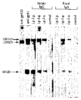
Fig. 4 is a photograph showing the Western blot analysis of serum IgG and
fecal IgA antibodies;
Figs. 5A-5F are photographs showing immunofluorescent reactions which
empirically demonstrate that serum IgG and fecal IgA antibodies from immunized
mice react with PBMCs infected with an HIV-1 NSI primary isolate; and
Figs. 6A-6D are photographs showing immunofluorescent reactions which
empirically demonstrate that fecal IgA antibodies from immunized mice react
with
PBMCs infected with an HIV-1 SI primary isolate.
CA 02441626 2003-09-23
WO 02/081655 PCT/US02/09353
19
DETAILED DESCRIPTION OF THE INVENTION
The present invention, in its most essential and fundamental form, is a
unique fusion protein construct which is prepared for in-vivo use both as an
immunogen and as a vaccine; and is effective for inducing a range of specific
anti-
HIV systemic IgG antibodies and secretory IgA antibodies within the body of
the
living recipient. An efficacious methodology for the immunization of a living
subject using this fusion protein construct as an immunogen and vaccine such
that
both systemic IgG antibodies and secretory IgA antibodies specific against at
least
one epitope of human immunodeficiency virus (HIV) are raised in-vivo is also
an
integral part of the present invention. Accordingly, the present invention
provides a
number of different unique benefits and major advantages, some of which
include
the following:
1. The fusion protein construct comprising a part of the invention is a
composition constituted of two different amino acid sequence fragments joined
linearly in tandem. If desired, the entire fusion protein construct may be
synthesized
chemically using long-established organic compound synthesis techniques as a
complete molecule by joining individual polypeptide fragments together in
fixed
sequence. It is preferred, however, that the fusion protein construct be a
recombinant protein molecule expressed by a genetically modified host cell
(such as
E. coli) cultured in-vitro, which intracellularly carries an introduced
expression
vector bearing specified recombinant DNA sequences encoding the entirety of
the
amino acids residues in proper sequence. The manner in which the fusion
protein
construct is generated is thus merely a question of personal choice and/or
convenience.
2. The fusion protein construct is an integrated dipeptide structure, an
oligopeptide molecule formed of two distinctly different, polypeptide
fragments: a
first polypeptide fragment positioned at the N-terminal end which comprises a
major
portion of the ectodomain of the HIV envelope glycoprotein gp4l; and a second
polypeptide fragment positioned at the COOH-terminal end which comprises a
CA 02441626 2003-09-23
WO 02/081655 PCT/US02/09353
5 meaningful part of the influenza virus hemagglutinin protein. Recognizing
that a
number of different HIV species, subspecies, and strains are currently known
to
exist - each of which presents a slightly different and individual amino acid
residue
sequence as its gp41 glycoprotein content and each of which presents a set of
both
HIV commonly conserved epitopes as well as individually unique epitopes as
gp41
10 antigenic determinants - the fusion protein construct can be formulated and
reformulated at will to contain either (or both) a specific HIV epitope,
customized
construct; or a more generalized, commonly shared and conserved HIV epitope
bearing construct. The broader scope of and particular choices for the amino
acid
residue sequence formulations representing the gp41 peptide fragment of the
15 dipeptide construct allows the manufacturer or intended user to decide in
advance
what the diversity of epitopes and what the range of antigenic specificities
for the
IgG and IgA antibodies induced in-vivo shall be.
3. The fusion protein construct when used as an immunogen andlor
20 vaccine can be used, if desired, to induce only IgG antibodies systemically
in the
recipient host; or, alternatively, can be used to induce both secretory IgA
antibodies
and systemic IgG antibodies concurrently in the recipient. The mode and manner
of
administering the fusion protein construct to the recipient will dictate and
control the
antibody types) actually produced in-vivo as the host's humoral immune
response.
4. The present invention as a whole is clearly intended for the use and
treatment of the homo Sapiens species, humans, as the primary beneficiaries.
However, the fusion protein construct and its medical value as an immunogen
and/or
vaccine is also available for use with all mammals generally regardless of
genus and
species. Accordingly, both human medical/clinical applications and veterinary
mammalian animal immunizations are envisioned and expected.
5. The fusion protein is expressed within insoluble inclusion bodies in
E. coli hosts; and it can be refolded in vitro using a physiological buffer.
The final
yield of refolded protein can be as high as 80 mg from a 1 liter quantity of
E. coli
culture. Successful refolding can be tested by reaction with gp41 specific
antibodies
CA 02441626 2003-09-23
WO 02/081655 PCT/US02/09353
21
and circular dichroism. The addition of the influenza virus HA sequence
renders the
gp41 polypeptide soluble or causes formation of soluble aggregates. It is
envisioned
that gp41 sequence fragments from other HIV Glades will be also solubilized by
this
method. A prospective vaccine cocktail will thus potentially include a mixture
of
gp41 fusion proteins derived from commonly found strains.
6. In the preferred embodiments, the short triple stranded coiled coil
sequence derived from the influenza virus hemagglutinin subunit 2 (HA2) is
engineered to be a substitute in place of the transmembrane region; and will
thus
present the gp41 polypeptide in a native way similar to the situation of
membrane-
anchored gp41 mediated by its own transmembrane region. A similar strategy can
be employed to solubilize other HIV specific proteins or unrelated proteins of
any
nature which form oligopeptides through their transmembrane anchors. The
influenza virus HA2 sequence can be therefore seen as a potential soluble
transmembrane anchor, which will help to present membrane anchored proteins in
a
"native-like" conformation in solution. The length of the triple stranded HA2
part
can be also varied to potentially achieve better solubilization.
7. A range of different embodiments can be generated as longer-length
gp41 variants by including more gp41 residues at the N-terminus as well as at
the C-
terminus, thus covering close to 100 percent of extracellular gp41 residues.
This
will improve the immunogenecity of the gp4lHA construct, by adding potential
additional epitopes.
I. The Parameters Of The Fusion Protein Construct
The fusion protein construct is an integrated dipeptide composition and
structure, as illustrated in Fig. 1. The fusion protein construct is
constituted of two
different peptide fragments which are covalently linked together and linearly
(axially) joined in tandem sequence to form a unitary polypeptide fusion
molecule.
As shown by Fig. l, the construct is formed of two distinctly different,
peptide monomer units: a first peptide fragment which begins at and represents
the
N-terminal end of the construct and comprises a majority [greater than 50% and
CA 02441626 2003-09-23
WO 02/081655 PCT/US02/09353
22
preferably 90% or more] portion of the ectodomain for the HIV envelope
glycoprotein gp4l; and a second peptide fragment located at and representing
the
COOH-terminal end of the construct and comprises a substantive part
(approximately 20%) of the influenza virus hemagglutinin protein.
The ectodomain of the HIV enveloped l~protein gp41
It is recognized that a number of different HIV species, subspecies, and
strains are currently known to exist. For example, HIV-1, HIV-2, and HIV-3
species
of human immunodeficiency virus have been identified (as reported in the
medical
and scientific literature). Similarly, a number of different subspecies or
Glades have
been isolated for each major type of HIV species. Thus, the HXB2 strain is
merely
one example illustrative of the HIV-1 species as a whole. As a point of
information,
a non-exhaustive listing of strains representative of the HIV-1 family is
given by
Table 1 below.
Each strain and species of HIV is recognized as having a slightly different
and individual amino acid residue sequence formulation for the ectodomain of
the
envelope glycoprotein gp4l. For example, the ectodomain of the HIV-laiB
envelope
glycoprotein gp41 in the HXB2 strain has a specified amino acid residue
sequence
which is individual and unique in its residue formulation. The HXB2 strain
gp41
protein also represents and presents a set of HIV commonly conserved and HXB2
unique amino acid residues in sequence as gp41 antigenic determinants
(epitopes).
In this manner, depending upon how much of the native ectodomain of the HXB2
(or other strain of HIV-1) envelope glycoprotein gp41 is utilized as the first
fragment, the fusion protein construct can be formulated towards either a HXB2
epitope specific, customized construct or towards a more general, commonly
conserved HIV-1 epitope bearing construct.
The broader scope of and particular choices for the amino acid residue
sequence formulations as the gp41 first peptide fragment of the construct thus
allows the maker or intended user to choose in advance what degree of
specificity
shall exist in the range of antigenic specificities for the IgG and IgA
antibodies to be
induced in-vivo as the humoral immune response.
CA 02441626 2003-09-23
WO 02/081655 PCT/US02/09353
23
Table 1: HIV-1 species and strains suitable for gp41 fragments
A. HXB2 strain Fisher et al., Nature (London) 316: 262-265
(1985)
B. All known HIV sequence which are available in the database or referenced
by Myers et al., 1995, theoretical biology and biophysics group, Los Alamos,
NM. Human retroviruses and ASS. See also Weissenhorn et al., Nature
387: 426-430. Figure 1e - sequence comparison of different classes of HIV
strains. Each of these publications is expressly incorporated by reference
herein.
C. All HIV envelope sequences found at: http://www.ncbi.nlm.nih.gov/
retroviruses/ using "env" as a search word. All these gp41 sequences can be
synthesized and used to make gp4lHA fusion proteins. All of these gp41
sequences to be found and identified at this web site are expressly
incorporated by reference herein.
CA 02441626 2003-09-23
WO 02/081655 PCT/US02/09353
24
The influenza virus hemag lutinin protein
It is also recognized that a number of subunits coexist as peptide chains in
the influenza virus hemagglutinin protein [Bullough et al., Nature 371: 37
(1994)].
Each of these is distinguishable from the other subunits; and has an
individual amino
acid residue sequence which is identifiably different from the others. Thus
distinct
subunits can be isolated from the overall general structure and composition of
influenza virus hemagglutinin protein; and subunit 2 of the influenza virus
hemagglutin protein represents a unique amino acid sequence formulation. As a
point of information, a listing of the different subunits constituting
influenza virus
hemagglutinin protein is given by Table 2 below.
Subunit 2 of this hemagglutinin protein is the preferred residue sequence
formulation and source for the second polypeptide fragment in making the
fusion
protein construct of the present invention. Here also, because the subunit 2
amino
acid sequence represents and presents a set of influenza virus commonly
conserved
and subunit 2 unique amino acid residues in sequence as gp41 antigenic
determinants (epitopes); and because the maker can choose how much of the
complete native subunit 2 amino acid residue sequence to employ as the second
peptide fragment, the fusion protein construct can be formulated either as a
subunit 2
epitope specific, customized construct or as a more general, commonly
conserved
hemagglutinin protein construct.
The broader scope for and particular choices of the amino acid residue
sequence formulations as the influenza virus hemagglutinin protein second
fragment
of the dipeptide construct thus allows the maker or intended user a second
mode of
choice to determine in advance what degree of specificity shall exist in the
range of
antigenic specificities for the IgG and IgA antibodies to be induced in-vivo
as the
humoral immune response.
CA 02441626 2003-09-23
WO 02/081655 PCT/US02/09353
5
Table 2:
Subunits of influenza virus hemagglutinin protein
suitable as a fragment in a fusion protein construct
Unit/Subunit References)
Subunit 1 Wiley, D.C. and J.J. Skehel,
Annu. Rev. Biochem 56: 365-394.
(I987);
Subunit 2 Stegmann, T. and A. Helenius,
Virus Fusion Mechanisms,
CRC Press, 1993, pp. 89-111
See also: Skehel, J.J. and D.C. Wiley, "Receptor binding and membrane fusion
in
virus entry: The influenza hemagglutinin." Annu. Rev. Biochem. _69: 531-569
(2000) for both subunits of influenza virus HA.
All of these publications are individually incorporated by reference herein.
CA 02441626 2003-09-23
WO 02/081655 PCT/US02/09353
26
II. A Preferred Fusion Protein Construct
A preferred integrated fusion protein construct is made based upon the
_H_XR2 strain of HIV-1 and the subunit 2 of influenza virus hemagglutinin
protein.
The first peptide fragment of the construct thus desirably has a 138 amino
acid
residue length and is a modified version of the native amino acid sequence
found at
residue position nos. 29-167 in the ectodomain of the HIVa~ envelope
glycoprotein
gp41 in the HXB2 strain.
The native amino acid residue sequence for positions nos. 29-167 in the gp41
ectodomain is given by Table 3 below. The native sequence contains a cysteine
residue at each of position nos. 88 and 94. In the present invention, each of
these
cysteine residues at position nos. 88 and 94 respectively have been replaced
and
substituted by serine residues. In this manner, the disulfide bond existing
between
these two cysteine residues in the original native gp41 ectodomain sequence
between the no. 88 and 94 residues has been eliminated.
A second major point of difference from the native original sequence in the
ectodomain of the HXB2 strain original, is that a number of the residues
existing in
the I3XB2 strain at native position nos. 29-167 are glycosylated. In the
present
invention, none of the amino acid residues employed in the first peptide
fragment
are glycosylated.
The second peptide fragment in the preferred fusion protein construct of the
present invention utilizes the subunit 2 of the influenza virus hemagglutinin
protein
as the native source for the amino acid residue sequence; and desirably
employs only
the residues found at position nos. 43-88 respectively. The native amino acid
residue sequence at position nos. 43-88 is given by Table 4 below.
CA 02441626 2003-09-23
WO 02/081655 PCT/US02/09353
27
Table 3
Native gp41 amino acid seq. (nos. 29-167)
Gln-Ala-Arg-Gln-Leu-Leu-Ser-Gly-Ile-Val-Gln-Gln-Gln-Asn-Asn-
Leu-Leu-Arg-Ala-Ile-Glu-Ala-Gln-Gln-His-Leu-Leu-Gln-Leu-Thr
Val-Trp-G1y-Tle-Lys-Gln-Leu-Gln-Ala-Arg-Ile-Leu-Ala-Val-Glu
Arg-Tyr-Leu-Lys-Asp-Gln-Gln-Leu-Leu-Gly-Ile-Trp-Gly-Cys-Ser
Gly-Lys-Leu-Tle-Cys-Thr-Thr-Ala-Val-Pro-Trp-Asn-Ala-Ser-Trp
Ser-Asn-Lys-Ser-Leu-Glu-Gln-Ile-Trp-Asn-His-Thr-Thr-Trp-Met
Glu-Trp-Asp-Arg-GIu-Ile-Asn-Asn-Tyr-Thr-Ser-Leu-Ile-His-Ser-
Leu-Ile-Glu-Glu-Ser-Gln-Asn-Gln-Gln-Glu-Lys-Asn-Glu-Gln-Glu-
Leu-Leu-Glu-Leu-Arp-Lys-Trp-Ala-Ser-Leu-Trp-Asn-Trp-Phe-Asn
Ile-Thr-Asn-Trp
CA 02441626 2003-09-23
WO 02/081655 PCT/US02/09353
28
Table 4:
Native influenza virus hemagglutinin subunit 2, nos. 43-88
Ala- Ile-Asp-Gln-Ile-Asn-Gly-Lys-Leu-Asn-Arg-Val-Ile-Glu-Lys-Thr-Asn-Glu-
Lys-Phe-His-Gln-Ile-Glu-Lys-Glu-Phe-Ser-Glu-Val-Glu-Gly-Arg-Ile-Gln-Asp-Leu-
Glu-Lys-Tyr- Val-Glu-Asp-Thr-Lys
CA 02441626 2003-09-23
WO 02/081655 PCT/US02/09353
29
Also, the invention prefers to utilize the amino acid residues found at nos.
43-88 of subunit 2 in a non-glycosylated form, rather than the glycosylated
residues
existing in the native original. The absence of glycosylated residues serves
to
increase epitope recognition and antibody specificity.
A preferred embodiment of the fusion protein construct therefore is a unified
molecule formed of two polypeptide fragments and having a length of 185 amino
acid residues in sequence. The first residue is a Met - a start/allow
expression in E.
coli. The precise amino acid residue sequence formulation for this 185 residue
length construct is given by Table 5; and the recombinant DNA sequence
encoding
this specific amino acid residue sequence is given by Table 6 below.
Note that within the amino acid sequence of Table 5, the two cysteines are
changed to serine; and there is an extra isoleucine at position 47 in the HA2
residue
sequence which is not present in the native HA2 fragment; and there is a short
Leu-
Asp-Gly sequence inserted between the HIV gp41 and HA2 fragments. This
preferred formulation for the fusion protein construct shares significant
primary,
secondary, and tertiary structural similarities and parallels with the
ectodomain of
gp4l; and is effective as both a systemic and mucosal antigen in-vivo.
Moreover, an
analysis of its crystalline structure (as described in the empirical results
hereinafter)
reveals the central portion or "core" for the first fragment to be alpha-
helical in
appearance and a rod-like oligomer.
CA 02441626 2003-09-23
WO 02/081655 PCT/US02/09353
Table 5:
Modified, gp41 HA fusion protein construct sequence
Met-Gln-Ala-Arg-Gln-Leu-Leu-Ser-Gly-Ile-Val-Gln-Gln-G1n-
Asn-Asn-Leu-Leu-Arg-Ala-Ile-Glu-Ala-Gln-Gln-His-Leu-Leu-
10 Gln-Leu-Thr-Val-Trp-Gly-Ile-Lys-Gln-Leu-Gln-Ala-Arg-Ile-
Leu-Ala-Val-Glu-Arg-Tyr-Leu-Lys-Asp-Gln-Gln-Leu-Leu-G1y-
Ile-Trp-Gly-Ser-Ser-Gly-Lys-Leu-Ile-Ser-Thr-Thr-Ala-Val-
Pro-Trp-Asn-Ala-Ser-Trp-Ser-Asn-Lys-Ser-Leu-Glu-Gln-Ile-
Trp-Asn-His-Thr-Thr-Trp-Met-Glu-Trp-Asp-Arg-Glu-Ile-Asn-
15 Asn-Tyr-Thr-Ser-Leu-Ile-His-Ser-Leu-Ile-Glu-Glu-Ser-G1n-
Asn-Gln-Gln-Glu-Lys-Asn-Glu-Gln-Glu-Leu-Leu-Glu-Leu-Asp-
Lys-Trp-Ala-Ser-Leu-Trp-Asn-Trp-Phe-Asn-Ile -Leu-Asp-
Gly- Ala-Ile-Asp-Gln-Ile-Asn-Gly-Lys-Leu-Asn-Arg-Val-
Ile-Glu-Lys-Thr-Asn-Glu-Lys-Phe-His-Gln-Ile-Glu-Lys-Glu-
20 Phe-Ser-Glu-Val-Glu-G1y-Arg-Ile-Gln-Asp-Leu-Glu-Lys-Tyr-
Val-Glu-Asp-Thr-Lys
CA 02441626 2003-09-23
WO 02/081655 PCT/US02/09353
31
Table 6: Recombinant DNA Sequence
ATGCAAGCACGCCAATTATTGTCTGGTATAGTGCAGCAGCAGAACAATTTGCTGAG
GGCTATTGAGGCGCAACAGCATCTGTTGCAACTCACAGTCTGGGGCATCAAGCAGC
TCCAGGCAAGAATCCTGGCTGTGGAAAGATACCTAAAGGATCAACAGCTCCTGGGG
ATTTGGGGTAGCTCTGGTAAACTGATCAGCACCACTGCTGTGCCTTGGAATGCTAG
TTGGAGTAATAA.ATCTCTGGAACAGATTTGGAATCACACGACCTGGATGGAGTGGG
ACAGAGAAATTAACAATTACACAAGCTTAATACACTCCTTAATTGAAGA.ATCGCAA
AACCAGCAAGAAAAGAATGAACAAGAATTATTGGAATTAGATAAATGGGCAAGTTT
GTGGAATTGGTTTAACATTCTAGATGGAGCCATCGACCAAATCATCAATGGGAAAT
TGAACAGGGTAATCGAGAAGACGAACGAGAAATTCCATCAAATCGAAAAGGAATTC
TCAGAAGTAGAAGGGAGAATTCAGGACCTCGAGAAATACGTTGAAGACACTAAA
CA 02441626 2003-09-23
WO 02/081655 PCT/US02/09353
32
Another major advantage evidenced by this preferred embodiment and
shared by all embodiments of the fusion protein construct as a whole is the
appreciable solubility in water and aqueous liquids generally in comparison to
earlier used and conventionally known forms of the gp41 protein. The
solubility of
the present fusion protein constructs is unusually large, even in comparison
to its
immediate predecessors; and thereby renders this construct most suitable for
use as
an antigen in-vivo.
The appreciable solubility at physiological pH of the present fusion protein
construct is demonstrated by the following evidence: (i) gp41 stays in
solution after
centrifugation; (ii) it forms soluble aggregates as judged by gel filtration
chromatography and dynamic light scattering; (iii) gp4lHA can be concentrated
to
at least 13 mg/ml or potentially higher; (iv) gp41 forms complexes with gp41
specific monoclonal antibodies, and specific binding to Fab fragments derived
from
monoclonal antibodies D31 and 2A2 can be observed (by gel filtration
chromatography as well as by native gel electrophoresis) and gp4lHA can be
separated on native gel electrophoresis when complexed to Fabs derived from
these
gp41 specific monoclonal antibodies; (v) gp4lHA is mostly alpha-helical in
solution
which is consistent with the structure of a core fragment of HIV-1 gp4l. The
replacement of the transmembrane region of gp41 by the influenza virus HA2
sequence induces a native-like structure of the amino acid sequence linking
the outer
core helix from residue 154 to residue 167. Together, these data indicate that
gp4lHA folds into a native-like structure which is therefore suitable for
inducing
conformation specific monoclonal antibodies, either IgG or IgA subtypes to
neutralize HIV strains.
The major differences from other chimeric constructs previously reported in
the scientific literature is therefore apparent. A prior art construct
comprising
extracellular residues of gp41 without the HA fusion part has been described;
however this prior art construct is only soluble at low pH (< pH 4.0).
Moreover, the
earlier construct precipitates out of solution at physiological pH values and
is thus
not suitable or useful for immunization purposes [Caffrey et al., EMBO J.
17(16):
4572-84 (1998); Wingfield et al., Protein Sci. 6(8): 1653-60 (1997)].
CA 02441626 2003-09-23
WO 02/081655 PCT/US02/09353
33
Also, although a similar construct containing residues of gp41 and HA2 was
published before [Weissenhorn et al., PNAS 94: 6065-6069 (1997)], the
constructs
described therein contained an additional sequence at the N-terminus (31
residues
derived from an oligopeptide form of the GCN4 leucine zipper region) and
proteolytic products thereof were then characterized. The basic construction
of
gp4lHA as described herein and its biochemical and biophysical properties have
therefore never existed before. Moreover, the construct used in the PNAS paper
(named pIIgp4IHA) was far less soluble than gp4lHA and only produced soluble
gp41 core fragments after proteolysis. These were also smaller fragments than
gp4lHA and contained less gp41 specific residues. In addition, the gp41
produced
(as described in the PNAS paper) is monodispersed in solution and does not
form
soluble aggregates which are preferable to induce mucosal immunity.
Preferred Manner of Manufacture
A most desirable manner of making the preferred fusion protein construct of
185 amino acid residues in sequence is via recombinant DNA methods and
systems.
One preferred technique is summarized below.
A DNA fragment encoding an N-terminal methionine followed by residues
29 to 167 of HIV-1 gp41 (HXB2 strain) and residues 43 to 88 of influenza virus
hemagglutinin subunit 2 was amplified by polymerase chain reaction using the
plasmid pII4IHA as a template. The nucleotide residues encoding cysteines at
positions 88 and 94 of the gp41 protein had been previously mutated to encode
serine residues to avoid intramolecular disulfide bond formation as described
in
Weissenhorn et al., Proc. Natl. Acad. Sci. USA 94: 6065-6069 (1997). The DNA
fragment was cloned into expression vector pRset (Invitrogen) and introduced
into
Esclzerichia coli BL21/pUBS. The preferred fusion protein construct, referred
to as
gp4lHA, was over-expressed in E. coli BL21/pUBS and purified from inclusion
bodies with a final yield of 100 mg per liter of E. coli culture. GP41HA
protein was
solubilized in 8 M urea and frozen at -80°C. In vitro refolding was
accomplished by
dilution to a protein concentration of 50 M in 20 mM Hepes (N-[hydroxyethyl]
piperazine-N'][2-ethanesulfonic acid]; Hepes; Sigma, Co.) [pH 8.0], which
yielded
soluble aggregates as judged by gel filtration chromatography. After refolding
CA 02441626 2003-09-23
WO 02/081655 PCT/US02/09353
34
gp4lHA could be concentrated to 13 mg/ml or higher and was stored at -
80°C.
Aliquots were thawed and diluted to I mg/ml in Hepes buffer (20 mM Hepes, pH
8.0) immediately prior to use.
The inclusion body preparation - using standard methods - yielded 99
percent purity as judged by SDS PAGE. Refolding can be accomplished at room
temperature or at 4°C resulting in approximately up to 80 percent
yields. After
refolding by dilution and concentration, gp4lHA can be further purified by gel
filtration chromatography; if necessary further purification on an ion
exchange
column can be achieved.
III. Immunogens And Vaccines
The essential component for the immunogens and vaccines of the present
invention is the presence of at least one embodiment of the fusion protein
construct
as an active ingredient within the prepared fluid mixtures serving as
immunogens
and the adjuvant containing preparations serving as vaccines.
Immuno
To be an immunogen, the formulation need only be a mixture of a fusion
protein construct as described herein and a biocompatible carrier fluid
suitable for
carrying and delivering a predetermined aliquot of the fusion protein
construct to a
prechosen site in the body of a living subject.
Immunogens embodying the invention can be administered in any
appropriate carrier for intradermal, subcutaneous, intramuscular, parenteral,
intranasal, intravaginal, intrarectal, oral or intragastric administration.
They can be
introduced by any means that effect antigenicity in humans. The dosage
administered will vary and be dependent upon the age, health, and weight of
the
recipient; the kind of concurrent treatment, if any; the frequency of
treatment; and
the nature of the humoral antibody response desired.
If the fusion protein construct is to be applied to a mucosal site (orally,
intravaginally, intrarectally, intranasally or intragastrically), it can be
admixed in a
concentration from about 0.001 to 1,000.0 ug per gram of a pharmacologically
inert
carrier such as saline, and dextrose solutions. Other possible carriers are
CA 02441626 2003-09-23
WO 02/081655 PCT/US02/09353
5 polyoxyethylene monolaurate 5% in water, sodium lauryl sulfate 5% in water,
gastric acid inhibitors, protease inhibitors, pH neutralizers, and the like.
Materials
such as anti-oxidants, bumectants, viscosity stabilizers, and the like may be
added, if
necessary.
Similarly, if the immunogens are to be given intradermally, subcutaneously,
10 intramuscularly, intravenously or parenterally, they will be prepared in
sterile form;
in multiple or single dose formats; and dispersed in a fluid carrier such as
sterile
physiological saline or 5% dextrose solutions commonly used with injectables.
In
addition, other methods of administration can be advantageously employed as
well.
15 Vaccines
To be a prepared vaccine, the minimal formulation comprises a
predetermined quantity of a fusion protein construct as described herein; a
biocompatible carrier suitable for carrying and delivering a predetermined
aliquot of
a fusion protein construct to a prechosen site in the body of a living
subject; and at
20 least one adjuvant composition dispersed in the carrier fluid or coupled to
the fusion
protein construct. The vaccine, by definition, incorporates an immunogen and
includes one or more adjuvants to facilitate or stimulate the immune response
and to
prolong the antigenic effect in-vivo over time.
Among the useful adjuvant substances conventionally known are those
25 compositions approved by the FDA (currently or pending for systemic and/or
mucosal immunizations). Some are preferred for mucosally-administered vaccines
and others are preferred for intragastric administered vaccines.
In addition, for mucosal administration it is often desirable to also include
one or more protease inhibitors in the overall formulation and recipe for a
vaccine.
30 Among the known protease inhibitor compounds deemed useful in a vaccine
preparation are: aprotinin, leupeptin, AEBSF and bestatin. Any or all of these
may
be used at will with the present invention.
CA 02441626 2003-09-23
WO 02/081655 PCT/US02/09353
36
IV. Modes Of Administration
It is a particular goal of the present invention to induce mucosal IgA
antibodies in-vivo which are specific against one or more epitopes of HIV-1.
The
objective of mucosal antibodies conforms to the mucosal vaccination strategies
for
women as recently published [Kozlowski et al., J. Infect. Dis. 179: 5493-549
(1999], a strategic approach which serves men equally well with regard to
potential
HIV infections.
Multiple modes of inoculation, the manner of introducing an immunogen or
vaccine, are conventionally known and used. The systemic or parenteral forms
of
administration (introduction by injection or perfusion) typically include
intraperitoneal, intravenous, intramuscular, subcutaneous, and subdermal
inoculations. In contrast, mucosal modes of administration may include not
only the
intranasal and intragastric forms of introduction, but also oral,
intravaginal, and
intrarectal introductions.
It has long been recognized that systemic administrations often produce
different results from mucosal administrations of similar or identical
substances.
One major difference between the modes of administration is that in-vivo
induction
of IgA antibodies, especially secretory IgA antibodies, usually demands and
requires
using one or more forms of mucosal administration on at least one occasion;
and
typically requires multiple repeat inoculations over time using the same
mucosal
administration to be clinically effective. In comparison, if the same
innoculum is
systemically administered on orie or multiple occasions, primarily serum IgG
antibodies are produced in-vivo by the recipient of the immunogen or vaccine.
As evidenced by the experiments and empirical data described hereinafter,
the present invention may be employed in the alternative to induce either
serum IgG
antibodies alone; or to induce both secretory IgA and serum IgG antibodies
concurrently. The preferred mode of administration using the fusion protein
construct as the immunogen or vaccine is to induce both anti-HIV IgA and IgG
antibodies concurrently in the living host.
CA 02441626 2003-09-23
WO 02/081655 PCT/US02/09353
37
Method For Immunization
Although three different methods of immunization were tested in mice [as
described in the experiments hereinafter], the focus of the mouse study was
centered
upon a method of immunization for the induction of both HIV-antigen specific
IgA
antibodies in mucosal secretions and IgG antibodies in serum in-vivo. This
method
comprises the steps of obtaining an immunogen (or vaccine) comprising a fusion
protein construct and a biocompatible carrier fluid suitable for carrying and
delivering a predetermined aliquot of the fusion protein construct to a
prechosen
anatomic site in the living subject; systemically administering an aliquot of
the
immunogen (or vaccine) on at least one occasion (and preferably on multiple
occasions) to the body of the living subject as a primary immunization; and
mucosally administering an aliquot of the immunogen (or vaccine) on at least
one
occasion (and preferably on multiple occasions) to a prechosen mucosal tissue
site in
the body of the living subject as a second immunization.
Illustrative Protocol
The following is presented as merely one example illustrative and
representative of a clinical immunization protocol; and also serves as a basis
for
malting the many different variants and procedural alternatives conventionally
known and medically employed as immunization procedures to induce specific
humoral antibodies in-vivo within the body of the recipient.
Preferences
It is most preferred that the immunogens and vaccines embodying the present
invention be used as a systemic or mucosal boost by following a systemic
prime/mucosal andlor boost regimen. The living recipient is first primed by a
systemic injection since a systemic prime was shown previously to augment
intestinal secretory IgA responses following subsequent mucosal boosts. A
systemic
prime followed by three intranasal (i.n.) or intragastric (i.g.) boosts will
successfully
induce serum IgG and fecal IgA antibodies that will recognize gp41 protein in
both
laboratory-adapted and primary isolates of HIV-1. In this manner, recombinant
gp41 ectodomain is most useful as a mucosal antigen in prime-boost vaccine
CA 02441626 2003-09-23
WO 02/081655 PCT/US02/09353
3S
strategies to stimulate protective anti-HIV-1 S-IgA antibodies in humans.
However,
recombinant gp4lHA may also be useful in protocols designed to induce systemic
IgG antibodies primarily, as in systemic prime/systemic boost strategies.
Examplary Procedures For Administration By
Parenteral And Mucosal Immunization Routes:
Immunization for induction of anti=g~p4lHA ~A antibodies:
Although a combination of systemic and mucosal immunization routes were
used in mice to test immunogenicity of gp4lHA, immunization strategies
utilizing
mucosal administration routes alone (intranasal, peroral, intrarectal, and
intravaginal) are more likely to generate greatest levels of anti-gp41
secretory IgA
(S-IgA) antibodies in mucosal secretions of humans.
Based on results of previous human studies with other antigens, it is
expected that intravaginal or intranasal immunization would produce greatest
concentrations of anti-gp41 S-IgA (S-IgA) antibodies in genital tract
secretions.
Intranasal or intrarectal immunization, on the other hand, would likely prove
most
effective for generating anti-gp41 S-IgA antibodies in rectal secretions.
Though
peroral immunization has been found less effective for induction of specific
IgA in
the rectum and genital tract, peroral administration of gp4lHA would be
expected to
induce greater levels of anti-gp41 IgA antibodies in small intestinal and
salivary
secretions, the latter of which could reduce or prevent oral transmission of
HIV.
Studies in both mice and humans also suggest that intranasal immunization
could be as effective as systemic immunization for induction of gp41-specific
IgG
antibody in the circulation. Hence, a combination of intranasal and rectal or
vaginal
immunization routes may be optimal for induction of anti-gp41 IgG in the
bloodstream and anti-gp41 S-IgA in both genital tract and rectal secretions.
Nevertheless, gp4lHA IgA and IgG antibodies could be induced in humans using
combinations of mucosal and systemic immunization routes.
CA 02441626 2003-09-23
WO 02/081655 PCT/US02/09353
39
Gp4lHA construct of choice for immunization:
If immunization is to be performed with a single gp4lHA construct in the
United States, it would be preferable to use gp4lHA having a Clade B sequence
(strains MN, HXB2, etc) since HIV-1 Clade B viruses predominate in North
America. However, HIV-1 Clade A and E viruses are being detected more
frequently in the U.S. population and, in the future, immunization with a
combination of Clade A, B, and E sequence gp4lHAs may be ideal.
Gp4lHA Immunization Doses:
Based on the dosages of other recombinant HIV proteins administered in
humans, it is likely that intramuscular, intradermal, and intranasal
immunization
with gp4lHA would require a dose ranging from 50 - 500 ~,g. Oral, rectal, and
vaginal immunizations would likely require greater gp4lHA doses ranging from
250 - 1000 ~,g.
Number of Doses:
To obtain optimal immune responses, it is likely that gp4lHA should be
administered a minimum of 3 times within the first year. One annual booster
immunization may be required for several years to maintain levels of
circulating and
secretory anti-gp41 antibodies.
Administration schedule:
Further testing will be required to establish optimal immunization schedules.
However, based on results of previous human mucosal immunization studies, it
is
anticipated that best results may be achieved if intranasal, intrarectal,
peroral, or
intravaginal immunization is performed a total of 3 times at biweekly or
monthly
intervals.
Detection of antibody res op nses:
Subjects are expected to demonstrate peak levels of circulating IgG and
secretory IgA two weeks after the 3rd immunization. However, detectable
CA 02441626 2003-09-23
WO 02/081655 PCT/US02/09353
5 antibodies should be present in sera and secretions two weeks after the 2nd
immunization.
Procedures for systemic or mucosal immunizations:
Form:
10 The following procedures are based on the assumption that gp4lHA will be
manufactured as a lyophilized powder in vials that must be reconstituted with
0.5 ml - 1 ml water or sodium chloride diluent.
For intramuscular immunization:
15 1) The reconstituted contents of a vial will be drawn into a 1 cc syringe
using a 11/z
inch or 1 inch (for thin subjects) 22 gauge needle.
2) The solution will be injected into either the deltoid, quadriceps, or
gluteal muscle.
For intradermal immunization:
20 1) The reconstituted contents of a vial will be drawn into a I cc syringe
using a 1/2
inch 26-gauge needle.
2) The contents will then be injected into the arm above the deltoid or into
the thigh
above the quadriceps.
25 For intranasal immunization:
1) The reconstituted contents of a vial will be transferred into a sterile 1.5
ml vial
using a 1 cc syringe equipped with a 11/2 inch 22 gauge needle.
2) The syringe and needle will be discarded.
3) The solution of gp41I3A will be drawn from the 1.5 ml tube into a sterile
eye
30 dropper.
4) The vaccine recipient will be asked to sit upright in a chair with head
tilted back
at an approximate 45 degree angle.
5) One drop of the gp4lHA solution will be dispensed inside the left nostril
near the
opening of the passageway. A second drop will be similarly be added inside the
35 right nostril. Immediately, the nostrils will gently be pinched shut to
ensure coating
of the entire nasal passage with solution.
CA 02441626 2003-09-23
WO 02/081655 PCT/US02/09353
41
6) Step 5 will be repeated until the entire contents of the gp4lHA solution
have been
administered to the vaccine recipient.
For peroral immunization:
1) The reconstituted contents of a vial will be withdrawn using a syringe and
11/2
inch 22 gauge needle.
2) The syringe contents will be ejected into a paper cup containing 150 mls of
a
stomach acid neutralizing sodium bicarbonate/water solution (e.g. alka
seltzer).
3) The contents of the cup will be mixed using a sterile tongue depressor.
4) The subject will then ingest the contents of the paper cup.
5) The subject will refrain from eating or drinking (with the exception of
water) for
2 hours.
For intrarectal immunization:
1) The reconstituted contents of a vial will be drawn through a 11/2 inch 22
gauge
needle into a 1 cc tuberculin syringe.
2) The needle will be discarded.
3) The subject will be placed on the exam table such that they are laying flat
on their
stomach or on their side.
4) The anus of the subject will be lubricated slightly with KY jelly.
5) The syringe will be inserted through the anus 6 cm into the rectum.
6) The subject will remain prone for 10 minutes.
For intrava~inal immunization:
1) The contents of a vial will be reconstituted with 3 ml of diluent, then
drawn
slowly through a 11/2 inch 22 gauge needle into the barrel of a 5 cc syringe
previously loaded with 0.5 g of the inert powder Eldexomer (Perstorp, Sweden).
2) The needle will be discarded.
3) The subject will be placed on the exam table in supine position with feet
in
stirrups.
4) A vaginal speculum will be inserted.
5) The contents of the syringe will be deposited in the posterior fornix of
the vagina.
CA 02441626 2003-09-23
WO 02/081655 PCT/US02/09353
42
6) The speculum will then be removed.
7) The subject will then remain prone for 10 minutes.
V. Experiments and Empirical Data
To demonstrate the merits and value of the present invention, a series of
planned experiments and empirical data are presented below. It will be
expressly
understood, however, that the experiments described and the results provided
are
merely the best evidence of the subject matter as a whole which is the
invention; and
that the empirical data, while limited in content, is only illustrative of the
scope of
the invention envisioned and claimed.
Materials and Methods:
Preparation of recombinant gp4l.
A DNA fragment encoding an N-terminal methionine followed by residues
29 to 167 of HIV-1 gp41 (HXB2 strain) and residues 43 to 88 of influenza virus
hemagglutinin subunit 2 was amplified by polymerise chain reaction using the
plasmid pII4IHA as a template. The nucleotide residues encoding cysteines at
positions 88 and 94 of the gp41 protein had been previously mutated to encode
serine residues to avoid intramolecular disulfide bond formation.
The DNA fragment was cloned into expression vector pRset (Invitrogen) and
introduced into Escherichia coli BL21/pUBS. The gp41 fusion protein, referred
to
as gp4lHA, was over-expressed in E. coli BL21/pUBS and purified from inclusion
bodies with a final yield of 100 mg per liter of E. coli culture. GP41HA
protein was
solubilized in 8 M urea and frozen at -80°C. In vitro refolding was
accomplished by
dilution to a protein concentration of 50 M in 20 mM Hepes (N-[hydroxyethyl]
piperazine-N'-[2-ethanesulfonic acid]; Hepes, Sigma Co.) [pH 8.0], which
yielded
soluble aggregates as judged by gel filtration chromatography. After refolding
gp4lHA could be concentrated to 13 mg/ml or higher and was stored at -
8°C.
Aliquots were thawed and diluted to 1 mg/ml in Hepes buffer (20 mM Hepes, pH
8.0) immediately prior to use.
CA 02441626 2003-09-23
WO 02/081655 PCT/US02/09353
43
Arzimals and immunization protocols.
Female BALB/c (H-2d) mice 6-8 weeks of age were purchased from Charles
River Laboratories (Wilmington, MA). They were housed in the Children's
Hospital animal facility on standard rodent diet and allowed to acclimate for
at least
one week prior to this study. All experiments involving mice were done under
strict
compliance with the guidelines established by the NIH, Children's Hospital and
Harvard Medical School.
There were three experimental groups and one control group of mice in this
study. The three experimental groups were immunized ("primed") by a single
intraperitoneal (i.p.) injection on day zero of 0.5 ml PBS (pH 7.4) containing
gp4lHA (50 g) and the systemic adjuvant N-acetylmuramyl-L-alanyl-D-
isoglutamine (MDP; Calbiochem, La Jolla, CA) (50 g). Group one (n=4) was
boosted systemically with the same dose given i.p. on days 7, 21 and 35. Mice
in
group two (n=6) were boosted intranasally (i.n.) on days 7, 21 and 35 with 401
of
PBS (pH 7.4) containing gp4lHP (50 g), the mucosal adjuvant cholera toxin (1
g)
(List Biological Laboratories, Campbell, CA), and protease inhibitors. The
final
concentrations of the protease inhibitors (Calbiochem, La Jolla, CA) were:
aprotinin
(50 U/ml), leupeptin (5 g/ml), AEBSF (48 g/ml), and bestatin (1 g/ml).
Intranasal
immunization was performed by lightly anesthetizing the mice with
methoxyflurane
(Pitmann-Moore, Mundelein, IL), then spotting 101 of the gp4lHA solution into
each nare. The mice were allowed to recover and this procedure was repeated 2
hr
later. Mice in group three (n-6) were boosted intragastrically (i.g.) on days
7, 21,
and 35 with a sodium biocarbonate (0.1 M) solution (0.4 ml) containing gp4lHA
(250 g), cholera toxin (5 g) and protease inhibitors. Mice were deprived of
food for
2 hrs before and 1 hr after i.g. immunization. Lg. immunization was performed
on
mice under light methoxyflurane anesthesia using a 1 cc syringe and a
disposable
20G x 1.5 inch blunt-ended feeding needle (Popper and Sons, New Hyde Park,
NY).
Group four (n=4) was not immunized.
CA 02441626 2003-09-23
WO 02/081655 PCT/US02/09353
44
Serum and feces collection
Blood samples (0.2-0.4 ml) were collected via retro-orbital bleed from mice
under avertin anesthesia seven days before the first immunization and 10 days
after
the final immunization. Avertin was prepared by dissolving 5.0 g
tribromoethanol
(Sigma Co.) into 10 ml of tertamyl alcohol (Fisher Co.), then diluting this
solution
1:80 into pre-warmed (37°C) PBS just prior to use. Aliquots of serum
samples were
stored at -80°C. Feces were collected six days before the first
immunization and 7
and 14 days after the final immunization. Five or six freshly voided fecal
pellets
were collected from each mouse and placed into a pre-weighed Eppendorf tube
containing 0.5 ml of PBS, 1% goat serum as blocking agent, and protease
inhibitors.
The tubes were then re-weighed and the weight of the feces m grams (average
0.1g
per mouse) was determined. The fecal suspensions were vortexed fox 30 s,
incubated on ice for 20 min, and the insoluble material was removed by
centrifugation for 10 min at 12,000 x g. The resulting fecal extract was
passed
through a 0.45 m filter (Millipore Co), collected into a 1.5 ml microfuge
tube, and
aliquots (0.1 ml) were stored at -80°C.
Enzyme-lif2ked itn munosorbent assays (ELISA)
For anti-gp4lHA ELISAs, 0.1 ml of gp4lHA (1 g/ml) in 20 mM HEPES
buffer (pH 8.2) was applied to each well of a 96 well of Nunc-Immuno plate
(Maxisorp F96; A/S Nunc, Roskilde, Denmark). Plates were incubated overnight
at
4°C in a humidified chamber, washed with PBS containing Tween-20 (0.05%
v/v),
then blocked with PBS containing Tween-20 (0.05%) and goat serum (1% v/v) for
1
hr at 37°C. Serum or fecal samples were serially diluted into blocking
buffer then
applied to each well (1001/well) and incubated for 2 hr at room temperature
(23°C)
in a humidified chamber. The plates were washed, overlaid with affinity-
purified,
peroxidase-conjugated, goat polyclonal antibodies (Southern Biotechnology
Associates, Inc. (SBA), Birmingham, AL) specific for either the alpha chain of
mouse IgA (1 g/ml) or the gamma chain of mouse IgG (0.5 g/ml). Plates were
developed using the one-component TMB substrate as suggested by the
manufacturer (I~irkegaard & Perry Laboratories, Gaithersburg, MD). For all
assays
an anti-gg41 mouse monoclonal IgG antibody which recognizes an epitope located
CA 02441626 2003-09-23
WO 02/081655 PCT/US02/09353
5 between amino acids 730 and 750 (ImmunoDiagnostics, Inc., Bedford MA) was
used as a positive control.
HIV-specific and total IgA and IgG in feces and serum was determined by
ELISA essentially as previously described [Kozlowski et al., AIDS Res. Hum.
Retroviruses 10: 813-822 (1994)]. Nunc MaxiStop microtiter plates were coated
10 with 3.3 glrnl HIV-lInB viral lysate (Cambridge Scientific, Rockville, MD),
3.3 g/ml
HIV-1MN viral lysate (Advanced Biotechnologies Inc., Columbia, MD), or 1 g/ml
affinity-purified goat anti-mouse IgA antibodies (ICN, Aurora, OH). Purified
mouse
myeloma IgA (SBA) was used as a standard in total IgA assays, and the anti-
gp41
monoclonal IgG antibody (described above) was used as the standard for all HIV-
15 1MN and HIV-1IIIB assays.
Westen2 blot a~zalysis
HIV-1 lysate Western blot strips were from Calyptebiomedical (Alameda,
CA). Nitrocellulose strips were incubated for 2 h in blocking buffer (PBS
20 containing goat serum (2% v/v) and Tween-20 (0.1% v/v) at room temperature,
then
overnight in serum samples (diluted 1:250 into blocking buffer) or fecal
samples
(diluted 1:50 in blocking buffer). The strips were then washed and incubated
with
biotin-conjugated, goat-polyclonal antibodies specific for the gamma chain of
mouse
IgG (0.5 g/ml; SBA) or the alpha chain of mouse IgA (0.5 g/ml; SBA), followed
by
25 ~ peroxidase-conjugated avidin (2 g/ml). The strips were developed using
the ECL
chemiluminescent detection system (Amersham-Pharmacia Biotech, Piscataway,
NJ) and Kodak X-GMAT film.
Itzfectioh of cells with IIIV 1.
30 H9 T cells (American Type Culture Collection, Rockville, MD) chronically-
infected with the HIV-IIm clone, HIV-l~BZ (provided by Dr. Anna Aldovini,
Children's Hospital, Boston, MA), were established after inoculating 1 x 10⁶
cells
with 2.5 x 10⁴ TCID5o viral stock. Cells surviving acute infection were
maintained
at > 90% viability by splitting cultures 1/10 every 3 d in RPMI 1640
containing 25
35 mM HEPES, 600 g/ml L-glutamine, 100 units/ml penicillin, 100 g/ml
streptomycin
(Life Technologies, Grand Island, NY) and 20% fetal bovine serum (FBS;
CA 02441626 2003-09-23
WO 02/081655 PCT/US02/09353
46
BioWhittaker, Walkersville, MD). The TCID50/ml of the HIV-182 stock was
determined by endpoint titration in H9 cells using methods described for
peripheral
blood mononuclear cells (PBMC) [40] except that 5 x 10⁴ H9 cells were placed
in
each 96 well on day 0 and cells were split 1/10 on day 4. For neutralization
assays a
fixed viral inoculum (75 TCll~5o/well) was preincubated with mouse sera, fecal
extracts, or monoclonal control antibodies prior to being mixed with H9 cells.
HIV-
1 treated cells were assayed for p24 production as described previously
[Kozlowski
et al., All~S Res. Hum. Retroviruses 10: 813-822 (1999)].
To obtain cells infected with HIV-1 primary isolates, PBMC were isolated
from normal blood by standard density gradient centrifugation on Ficoll-Paque
(Amercham-Pharmacia) and 1 x 10' cells were cultured for 3 d with 2.5 g/ml
phytohemagglutinin (Sigma Co.) in 10 ml of complete RPMI 25 cm2 flask. The
cells were then washed, adjusted to 1 x 10⁶/m1 in 10 ml medium containing 5%
IL-2
(Hemagen, Columbia, MD), and returned to their original flask. The PBMC were
then inoculated with 1 ml of culture supernatant containing an HIV primary
isolate
(all 1-5 x 10⁴ TCID50/ml). Cultures of uninfected control PBMC from the same
donor were established in parallel and maintained in flasks for 2-3 wk with'/a
splitting at 3-4 d intervals in IL-2 supplemented medium.
HIV primary isolates were the gift of Dr. Robert Husson (Department of
Medicine, Children's Hospital, Boston, MA). These isolates were obtained from
infected women by co-culture technique and were previously characterized as
syncytium-inducing (SI) or non-SI (NSI) using the MT-2 T cell line [Husson et
al.,
J. Pediatr. 126: 865-871 (1995)]. "Macrophage tropic" NSI isolates replicate
in
activated primary T cells but not T cell lines [Unutmaz et al., Proc. Natl.
Acad. Sci.
USA 94: 1615-1618 (1997)]. A highly cytopathic SI isolate that produced
syncytia
in PBMC cultures was selected for use in this study. An NSI isolate that
consistently produced no syncytia when propagated in PBMCs was chosen.
Infection of NSI-inoculated PBMC cultures was confirmed by screening culture
fluid for HIV p24 antigen using a commercially available ELISA kit (NEN,
Boston,
MA).
CA 02441626 2003-09-23
WO 02/081655 PCT/US02/09353
47
Immunocytochemistry of HIV 1 infected cells
Slides of NSI- or SI-infected PBMC were prepared on post-infection days 10
to 14. Slides of control uninfected cultured PBMC from the same donor were
prepared in parallel. Suspensions of PBMC were first subjected to density
gradient
centrifugation on Ficoll-Paque to remove dead cells, and then washed 3 times
with
Dulbecco's PBS (DPBS) containing 5% goat serum and adjusted to 2 x 10⁶
cells/ml.
Aliquots of cells in suspension (1501) were then centrifuged for 4 min at 750
rpm
onto glass slides using a Cytospin 3 (Shandon-Lipshaw, Pittsburgh, PA). After
centrifugation slides were transferred to staining jars containing 100%
acetone,
which were placed at -20°C overnight. Slides were then rinsed 3 times
with PBS
and stored at 4°C immersed in PBS with 0.1% azide. Cytocentrifuge
slides of H9
cells or H9 cells infected with HIV-1~2 were prepared as described above
except
that cells were suspended at 1 x 10⁶ cells/ml prior to centrifugation.
NSI and SI cytocentrifuge slides were screened by immunostaining using a
rabbit anti-p24 polyclonal antibody (Advanced Biotechnology Incorporated,
Columbia, MD) to determine which preparations contained greatest numbers of
infected cells. To evaluate mouse sera and fecal extracts for the presence of
IgG and
IgA antibodies that recognize native viral gp41 on the surfaces or within the
cytoplasm of infected cells, cytospin slides were washed three times in PBS,
blocked
with PBS containing Tween-20 (0.05% v/v) and goat serum (1% v/v), then
overlaid
with serum samples (diluted 1:50 into blocking solution) or fecal extracts
(diluted
1:10 into blocking solution) for 2 hr at room temperature. After washing,
slides
were treated with biotin-conjugated, goat polyclonal antibodies specific for
either
the alpha chain of mouse IgA (20 g/mI) or the gamma chain of IgG (20 g/ml) for
1
hr, washed, and then stained with fluorescein-conjugated streptavidin (SA-
FITC; 2
g/ml, Pierce Chemical Co., Rockford, IL). Slides were post-fixed for 5 min in
2%
paraformaldehyde (v/v) in PBS, then mounted with Moviol [0.5 g/ml glycerol,
0.1
g/ml Mowiol 4-88 (Calbiochem, San Diego, CA), 10 mglml diazbicylo[2.2.2]octane
(Sigma Co.) in O.1M Tris-HCl (pH 8.5)]. Slides were viewed using a BioRad MRC
1024 confocal laser-scanning microscope at the Harvard Digestive Disease
Center
Core Facility. Images were collected using the BioRad imaging software and
edited
using Abode Photoshop 5Ø
CA 02441626 2003-09-23
WO 02/081655 PCT/US02/09353
48
Experimental Series I
Experiment 1: Systemic and mucosal immunogenicity of gp4lHA
To test whether the recombinant gp41 protein gp4lHA is immunogenic in
mice when used in a systemic prime/mucosal boost regimen, groups of mice were
IO primed intraperitoneally (i.p.), then boosted either i.p., intranasally
(i.n.) or
intragastrically (i.g.) three times at two-week intervals, as described in the
Materials
and Methods. Serum and fecal extracts were collected 7 and 10 days after the
last
immunization and screened by ELISA for reactivity to gp4lHA antigen.
Antibody levels in fecal extract can serve as an indicator of secretory
antibodies in the gastrointestinal tract of mice. The average reciprocal
endpoint titer
of gp4lHA-specific IgG in sera of mice in all immunized groups exceeded
600,000
on day 7, whereas reciprocal endpoint titers in pre-immune sera or
unirnmunized
controls were less than 100 (data not shown). High levels of anti-gp4lHA IgA
antibodies were detected in fecal extracts collected on day 10 from i.n, and
i.g.
immunized mice (mean reciprocal endpoint titer >10,000) - but not in extracts
from
i.p. immunized mice (<160) or non-immunized controls (<80). These data
demonstrate that the gp4lHA fusion protein was immunogenic in mice; and that
mucosal boosts were necessary to evoke secretory IgA antibodies.
Experiment 2: HIV-1 specific IgG in serum of systemically
and mucosally immunized mice
Because gp4lHA is a recombinant fusion protein containing portions of both
HIV-1 gp41 and influenza HA, the gp4lHA ELISA data of Experiment 1 did not
indicate whether gp41-specific antibodies were induced in these animals.
Therefore,
in this experiment, the serum samples were analyzed by ELISA for reactivity to
gp41 in HIV-IIIIB viral lysate. The empirical results are shown graphically by
Figs.
2A and 2B respectively.
Fig. 2 as a whole demonstrates that HIV-specific IgG antibodies are induced
in serum. Fig. 2A shows geometric mean (bars) and individual concentrations of
anti-HIV-lIi~ IgG antibody quantitated by ELISA in serum of mice before
immunization and 10 days after the last immunization with gp4lHA. Fig. 2B
shows
CA 02441626 2003-09-23
WO 02/081655 PCT/US02/09353
49
the concentrations of anti-HIV-1IIIB and anti-HIV-1MN IgG antibodies measured
in
day 10 serum of all immunized mice. Geometric mean concentrations of anti-HIV-
lIIIB IgG in preimmune serum of mice in the i.n., i.g., and i.p. immunization
groups
were 0.064, 0.066, and 0.068 g/ml, respectively. For each group, postimmune
antibody concentrations were determined to be significantly greater than those
in
corresponding preimmune serum using the two-tailed paired t-test.
As shown in Fig. 2A, an i.p. prime followed by three i.n., i.g., or i.p.
boosts
with gp4lHA induced significant concentrations of serum anti-HIV-laIB IgG
antibodies. The geometric mean reciprocal endpoint titers measured in these
sera
were 86,000 (i.p.li.n.), 24,000 (i.p.li.g.), 31,000 (i.p./i.p.), as compared
to less than
50 for all pre-immune samples. Although the i.p./i.n. immunized mice tended to
have the highest levels of HIV-lIIIB-specific serum IgG antibodies, there were
no
statistical differences between the three groups of immunized mice. Thus, each
prime/boost regimen was equally effective at inducing anti-gp41 specific
systemic
IgG antibodies.
Serum samples were also tested by ELISA for reactivity to gp41 in viral
lysate of HIV-1~, another laboratory-adapted T cell tropic HIV isolate.
Regardless
of immunization route, post-immune serum from all mice contained significant
concentrations of anti-HIV-1MN IgG antibodies as revealed by Fig. 2B. Analysis
of
individual samples showed a strong correlation (p<0.0001) between the
concentration of anti-HIV-1MN IgG antibodies and anti-HIV-laIB IgG antibodies.
The extent of cross-reactivity to HIV-1MN, estimated by dividing
concentrations of
anti-HIV-1MN IgG by anti- HIV-lii~ IgG antibody, was determined to average
94%.
Experiment 3: Levels of anti-HIV-1 specific IgA antibodies in fecal extracts
To determine whether systemic prime followed by mucosal boosts with
gp4lHA induced mucosal IgA antibodies to gp4l, fecal; extracts from immunized
mice were examined by ELISA for IgA antibodies to HIV-laiB viral lysate.
Because
an anti-gp41 monoclonal IgA standard was not available, the concentrations of
anti-
HIV-lII~ IgA antibodies were estimated using as a standard the anti-gp41 IgG
monoclonal antibody described above. In interpreting these results, one is
aware of
the fact that intestinal IgA antibodies are generally in dimeric (or
oligomeric) form;
CA 02441626 2003-09-23
WO 02/081655 PCT/US02/09353
5 and consequently, it is likely that concentrations of specific IgA were
underestimated by a factor of 2.5 when using an IgG standard.
Postimmune fecal extracts from mice in the i.p./i.n. and i.p./i.g.
immunization groups both demonstrated mean reciprocal endpoint titers of 220
and
mean concentrations of 6 g/ml anti- HIV-lIIIB IgA antibody. It is known that
total
10 IgA immunoglobulin concentrations can vary widely in fecal samples
[Haneberg et
al., Infect. Immun. 62: 15-23 (1999)]. Therefore, to more accurately compare
the
levels of specific intestinal IgA in fecal extracts, the HIV specific activity
in each
extract was calculated and is presented in Fig. 2.
Fig. 3 is a graph demonstrating the presence of HIV-specific IgA antibody in
15 fecal extracts from gp4lHA immunized mice. As seen therein, the
concentrations
(g/ml) of anti- HIV-1mB IgA antibody were divided by total IgA concentration
(g/ml) in fecal extracts to obtain HIV-specific activity for IgA. The specific
activity
x 100 measured in extracts prepared from feces of non-immunized mice and those
collected from gp4lHA immunized mice is shown 7 days after the last
20 immunization. The bars represent geometric means. The fecal extracts from
mice in
the i.n. and i.g. groups were determined by ANOVA to contain significantly
greater
HIV-specific IgA activity than those from non-immunized and i.p. immunized
mice.
The empirical data reveals that fecal extracts from mice boosted by either
i.g.
or i.n. routes demonstrated significantly greater HIV specific activity than
those
25 from mice in the i.p. or non-immunized control groups (p<0.0001 by ANOVA).
There was no statistical difference between the average specific activity of
IgA in
feces from i.g. boosted mice (mean 1.6) and from i.n. mice (mean 1.1); and
both of
these values were greater than the respective mean specific activities (i.g.
0.3; i.n.
0.8) determined for IgG in serum of these animals (data not shown). Like anti-
HIV-
3O lms IgG serum antibodies, anti- HIV-lIIIB IgA in i.n. and i.g. fecal
extracts cross-
reacted with HIV-1MN viral lysate (not shown), and concentrations of specific
IgA
measured in these two ELISAs were highly correlated (p<0.0001). Thus, these
results show that systemic prime followed by mucosal boosting with gp4lHA can
induce anti-gp41 specific IgA antibodies in mucosal secretions and anti-gp41
35 specific IgG antibodies in serum.
CA 02441626 2003-09-23
WO 02/081655 PCT/US02/09353
51
Experiment 4: Western blot analysis of serum IgG and fecal IgA antibodies
To confirm the gp41 specificity of serum IgG and fecal IgA antibodies, we
examined the reaction patterns of sera and fecal extracts on HIV-laIB Western
blot
strips. The results of these analyses are presented by Fig. 4.
Fig. 4 shows that anti-gp4lHA serum IgG and fecal IgA antibodies react
with monomeric and oligomeric gp4l. HIV-lms lysate Western blot strips
(Calypte
Biomedical, Alameda, CA) were probed with monoclonal anti-HIV envelope
antibodies, anti-p4lHA antisera, or fecal extracts from gp4lHA immunized mice.
As described in the Materials and Methods, anti-sera or fecal extracts from
groups of
mice were pooled and diluted 1:250 or 1:50, respectively, before being used
for
Western blot analysis. Monoclonal IgG antibodies and serum IgG antibodies were
detected using affinity purified goat IgG antibodies for the Fc fragment of
mouse
IgG. Fecal IgA antibodies were detected using affinity purified goat
antibodies
specific for the alpha chain of mouse IgA. A single protein band of
approximately
120 kD corresponding to gp120 was present on strips probed with a mouse
monoclonal anti-gp120 IgG (ImmunoDiagnostics) antibody. The mouse
monoclonal anti-gp41 antibody 2A2 revealed three bands at apparent molecular
weights of 40 kD, 120 kD and 160 kD, corresponding to monomeric and oligomeric
forms of gp4l.
In particular, Fig. 4, lane a shows an anti- HIV-lIIIB V3 loop monoclonal
antibody (hnmunoDiagnostics) that recognizes both gp120 and gpI60 labeled a
single band of approximately 120 kD on these strips. An anti-gp41 monoclonal
IgG
antibody (ImmunoDiagnostics) which reacted strongly with proteins of
approximately 40 kD and 160 kD is shown by Fig. 4, lane b, and appears weakly
with a protein of approximately 120 kD.
The diffuse band at 40 kD presumably corresponds to monomeric
glycosylated gp4l. The band of approximately 160 kD was not gp160 since it was
not labeled with anti-gp120 monoclonal antibodies (Fig. 4, lane a). Rather,
this 160
kD band and the faint band of 120 kD in lane b is believed to reflect the
presence of
disulfide-linked gp41 oligomers present in certain commercial Western blot
strip
preparations [Zolla-Pazner et al., New Eng. J. Med. 320: 1280-1281 (I989)].
CA 02441626 2003-09-23
WO 02/081655 PCT/US02/09353
52
By Western blot analysis serum, IgG antibodies from i.p., i.p./i.n. and
i.p./i.g.
gp4lHA immunized mice reacted with proteins of 401cD and 160 kD (Fig. 4, lanes
c-e) with a pattern identical to that which was obtained with the commercial
monoclonal anti-gp41 antibody. Pre-immune mouse serum did not react with any
proteins present in HIV-1 lysate (Fig. 4, lane f), in agreement with data
obtained
previously by ELISA.
To determine whether the fecal IgA antibodies were specific for gp4l,
Western blot strips were incubated in fecal extracts, then developed using
goat anti-
mouse IgA antibodies. IgA antibodies in fecal extracts from i.p./i.g. and
i.p./i.n.
immunized mice reacted with proteins at 160 and 40 kD, corresponding to
monomeric and oligomeric gp41 (Fig. 4, lanes h-i). There were no detectable
anti-
gp41 IgA antibodies present in fecal extracts from i.p. immunized mice (Fig.
4, lane
g) or unimmunized controls (Fig. 4, lane j).
Experiment 5: Neutralization of HIV-1 infection in-vitro
Serum and fecal extracts from gp4lHA immunized mice which contained the
highest concentrations of anti- HIV-lIIIB antibody by ELISA were then tested
in
vitro for their ability to neutralize HIV-lms infections of H9 T cells.
Preincubation
of virus with 10 g/ml of neutralizing anti-gp120 IgG monoclonal antibody used
as a
positive control reduced viral infection of these cells by 99% whereas an
identical
concentration of isotype-matched anti-Epstein Barn virus monoclonal antibody
had
no effect (data not shown). Similarly, pooled human HIV-positive sera (diluted
1/40) was found to reduce infection by 63% compared to the same dilution of
pooled
HIV-negative sera. However, viral levels in cell cultures containing post-
immune
mouse serum diluted 1/20-1/80 did not differ significantly from those in
cultures
with corresponding preimmune serum dilutions. Potential neutralization by IgA
in
fecal extracts could not be assessed because both preimmune and postimmune
fecal
extracts caused cell death after 3 days of culture, even at dilutions > 1/100.
CA 02441626 2003-09-23
WO 02/081655 PCT/US02/09353
53
Experiment 6: Serum IgG and fecal IgA antibodies recognize peripheral
blood mononuclear cells infected with HIV-1 primary isolates
Although mouse anti-gp4lHA serum antibodies were unable to protect a T
cell line from infection by cell-free HIV-lIIIB in vitro, this tissue culture
assay does
not reflect the mechanism of protection that is most important on mucosal
surfaces
in vivo. Attachment of SIgA antibodies to viral surface proteins could induce
viral
aggregation and entrapment in mucus layers on vaginal and rectal surfaces in
an
environment devoid of target cells such as T cells [Lamm, M.E., Ann. Rev.
Microbiol. 51: 311-340 (1997)]. IgA in mucosal secretions that can bind virus
or
virus-infected cells may be sufficient to reduce HIV infection of mucosal
tissues in
vivo. Thus this experiment sought to determine whether the anti-gp4lHA IgA
antibodies in mouse fecal samples recognized gp41 from a primary, clinically
relevant "T cell-tropic" SI isolate and a "macrophage-tropic" NSI isolate.
Peripheral blood mononuclear cells (PBMC) were infected with primary SI
or NSI HIV-1 isolates and probed by indirect immunofluorescence with serum and
fecal extracts from preimmune or gp4lHA immunized mice that showed the highest
anti-gp41 titers by ELISA. The results are shown by Figs. 5A-5F respectively;
and
should be compared to Figs. 6A-6D as well.
Fig. 5 as a whole demonstrates that serum IgG and fecal IgA from gp4lHA
immunized mice react with PBMCs infected with NSI isolate. Fig. 5A shows mouse
monoclonal IgG and anti-gp41 antibody 2A2. Fig. 5B shows unimmunized mouse
serum IgG. Fig. 5C shows i.p./i.n. serum IgG. Fig. 5D shows i:p./i.g. serum
IgG.
Fig. 5E shows i.p./i.n. fecal extracts. Fig. 5F shows unimmunized mouse fecal
extracts.
In particular, Fig. 5A reveals cytospin preparations of PBMCs infected with
a primary NSI isolate which were probed with monoclonal anti-HIV envelope
antibodies; while Figs. 5B-5D show anti-gp4lHA antisera and Figs. 5E-5F show
fecal extracts from gp4lHA immunized mice. Monoclonal IgG antibodies and
serum IgG antibodies were detected using biotinylated goat IgG antibodies
specific
for the Fc fragment of mouse IgG and SA-FITC. Fecal IgA antibodies were
detected using affinity purified goat antibodies specific for the alpha chain
of mouse
IgA and SA-FITC.
CA 02441626 2003-09-23
WO 02/081655 PCT/US02/09353
54
In contrast, Fig. 6 as a whole demonstrates that fecal IgA antibodies from
gp4lHA immunized mice react with PBMCs infected with an HIV-1 SI primary
isolate. Fig. 6A shows anti-gp41 monoclonal antibody 2A2; Fig. 6B shows
unimmunized mouse fecal extract; Fig. 6C shows i.p./i.g. fecal extract; and
Fig. 6D
shows i.p./i.g. fecal extract.
In particular, cytospin preparations of PBMCs infected with a primary SI
isolate were probed with a monoclonal anti-HIV gp41 antibody (Fig. 6A) or
fecal
extracts from gp4lHA immunized mice (Figs. 6C-6D). Immunocytochemistry was
performed as described in the legend to Fig. 5.
These empirical results reveal and demonstrate that infection of PBMC with
primary HIV-1 isolates gives rise to an asynchronous, heterogeneous population
of
cells, some which are heavily infected with virus and some which remain
uninfected. The monoclonal anti-gp41 antibody 2A2 strongly labeled a sub-
population of cells in both NSI- (Fig. 5A) and SI-infected (Fig. 6A) PBMC
acetone-
fixed cytospin preparations, but did not label uninfected PBMC controls.
Because
acetone fixation permeabilizes cells, we were unable to determine whether the
monoclonal antibody was staining cell surface-associated gp4l, intracellular
gp41/gp160, or both. Serum IgG from mice boosted with gp4lHA i.p. (data not
shown), i.n. (Fig. 5C) and i.g. (Fig. 5D) labeled a sub-population of cells in
the NSI-
infected (Fig. 5) and SI-infected (data not shown) PBMC cultures with a
frequency
similar to that observed with the gp41 monoclonal antibody control. Serum IgG
from unimmunized control mice did not stain NSI- or SI-infected PBMCs (Fig.
5B).
To determine whether anti-gp4lHA IgA antibodies in feces reacted with
NSI- or SI-infected PBMCs, cytospin preparations were overlaid with fecal
extracts
(diluted 1:10), then developed using biotinylated goat anti-mouse IgA
antiserum and
streptavidin fluorescein. Fecal IgA from mice mucosally boosted with gp4lHA
labeled a sub-population of cells within the NSI-infected (Fig. 5) and SI-
infected
(Fig. 6) PBMCs cytospin preparations with a pattern and frequency similar to
that
obtained with the gp41 control IgG antibody. Fecal extracts from i.p. boosted
mice
(data not shown) or unimmunized control mice (Figs. 5 and 6) did not react
with
infected cells. These data indicate that anti-gp4lHA serum IgG and fecal IgA
antibodies recognize gp41 from clinically relevant HIV-1 isolates.
CA 02441626 2003-09-23
WO 02/081655 PCT/US02/09353
5
Conclusions Supported By The Data Of Experimental Series I:
1. The immunogenicity of the preferred construct, gp4lHA, a recombinant
protein containing the ectodomain of gp41 from HIV-laIB is empirically
10 demonstrated in-vivo. The systemic prime-mucosal boost regimens with gp4lHA
induced anti-gp41 IgG antibodies in serum and IgA antibodies in secretions
that
recognized laboratory adapted and primary isolates of HIV-1. Although
performed
in mice, these data are significant because they demonstrate that a
recombinant form
of gp41 is immunogenic when given mucosally and is capable of stimulating S-
IgA
15 antibodies against clinically relevant HIV-1 isolates.
2. The fusion protein construct, gp4lHA, shares significant primary,
secondary,
and tertiary structure with the ectodomain of gp4l; and this accounts for its
observed
effectiveness as both a systemic and mucosal antigen. The gp4lHA protein
contains
20 13~ amino acids - a sequence representing 90% of the native gp41
ectodomain. In
contrast to gp120 (which contains multiple hypervariable domains), the primary
amino acid sequence of the gp41 ectodomain is relatively conserved among HIV-1
isolates from different Glades worldwide. Indeed, the amino acid sequence of
the
ectodomain of gp41 from HIV-lIIIB differs from HIV-1MN by only 5 amino acids.
3. Analysis of the crystal structure of the central portion or "core" of the
gp41
ectodomain indicates that it forms a-helical, rod-like oligomers. The fusion
protein
construct gp4lHA assumes a folded conformation and has an a-helical content
similar to the core of gp4l. Moreover, because native gp41 is believed to
assemble
in the viral membrane as a trimer, the gp4lHA protein is believed also to
assume a
similar tertiary structure - since two mouse monoclonal antibodies D31 and 2A2
(each of which recognizes conformational dependent epitopes on oligomeric
gp160)
bind gp4lHA.
4. Two notable differences exist between the fusion protein construct gp4lHA
and native gp4l. The first is: due to site-directed mutations in the highly
conserved
CA 02441626 2003-09-23
WO 02/081655 PCT/US02/09353
56
cysteine residues at positions 88 and 94, gp4lHA cannot and does not make the
intramolecular disulfide bond necessary for the formation of the
immunodominant
loop. The second is: gp4lHA was produced in E. coli hosts and consequently
lacks
the four N-linked carbohydrate side chain modifications normally present on
the
gp41 ectodomain. This has substantial irnmunological consequences since
immunization of macaques with non~l~ylated gp120, as compared to
glycosylated gp120, resulted in a broadened humoral immune response and
enhanced neutralizing antibody titers against wild-type, glycosylated virus.
The present invention (and the empirical data described hereinafter which
factually evidence and support the invention) are a reaffirmation of the
differences
in the specific mode of administration: the induction of antigen specific S-
IgA
antibodies in mucosal secretions occurs after mucosal but not after systemic
immunization. Whereas the three prime-boost immunization strategies
empirically
tested (i.e., i.p./i.p.; i.p./i.n.; i.p./i.g.) were each equally effective at
inducing anti-
HIV-1 serum IgG antibodies in mice, only the i.p./i.n. and i.p./i.g. modes of
administration gave rise to anti-go41 IgA antibodies in feces. Thus, the
present
invention demonstrates that secretory antibodies are initiated only after
antigens are
delivered via transepithelial transport into organized lymphoid tissue located
within
the mucosa or in nearby lymph nodes, where antigen specific mucosal B cells
are
generated. The presence of anti-gp4lHA antibodies following i.g. and i.n.
immunization are ample evidence to indicate that gp4lHA was sampled by the
mucosa of both the gut-associated and nasal-associated lymphoid tissues.
6. The present invention also is supporting evidence that soluble non-adherent
protein antigens are weak mucosal antigens because they are inefficiently
sampled
by the MALT and/or are rapidly degraded by proteases present in secretions.
The
efficacy of gp4lHA as a mucosal antigen is believed to be primarily due to the
fact
that the recombinant protein aggregates in solution, as determined by gel
filtration
chromatography and native gel electrophoresis (Weissenhorn et al., unpublished
results). Aggregated proteins are believed to be better mucosal antigens than
soluble
CA 02441626 2003-09-23
WO 02/081655 PCT/US02/09353
57
proteins because they are more resistant to mucosal proteases and are more
effectively sampled by the follicle-associated epithelium.
7. Tntranasal immunization is an especially appealing route for delivery of
vaccines against sexually transmitted diseases like HIV-1 because of its
ability to
stimulate S-IgA antibodies in both local and distant mucosal secretions. The
appearance of antigen specific IgA at distant mucosal sites following i.n.
immunization is believed due to the emigration of antigen specific B cells
from the
nasal associated lymphoid tissue. In addition, intranasal immunization
stimulates
the cellular immune responses. For example, i.n. (but not i.p.) immunization
of mice
with recombinant proteins induced antigen specific cytotoxic T lymphocytes in
the
female genital tract, spleen and cervical lymph nodes.
8. It is essential to recognize and appreciate that systemic immunization
alone
with the fusion protein construct gp4lHA failed to induce detectable
neutralizing
serum IgG antibodies, as determined by the T cell protection assay. This fact
is not
surprising since neutralizing epitopes on gp41 are rare and/or poorly
presented to the
immune system.
9. As empirically described hereinafter, the fact that secretory anti-gp4lHA
IgA antibodies recognized PBMCs infected with primary HIV-1 isolates shows
that
secretory antibodies evoked by gp4lHA immunization in humans will likely have
a
protective capacity in vivo. Whereas serum antibodies generally provide
protection
in vivo by blocking the interaction of virus with specific target cells, SIgA
antibodies function by intercepting microbial pathogens before they enter the
body.
On mucosal surfaces in vivo (anti-gp4lHA) S-IgA protect epithelia by cross-
linking
and agglutinating microorganisms in mucosal secretions, enhancing their
entrapment
and clearance in mucus, and in some cases by blocking or sterically hindering
the
microbial surface molecules that mediate epithelial attachment. In addition, S-
IgA
in the epithelial export pathway may even intercept incoming viral particles.
Anti-
gp41 IgA antibodies reduce HIV-1 transmission across epithelial monolayers
apparently by arresting viral transepitheIial transport within apical
recycling
CA 02441626 2003-09-23
WO 02/081655 PCT/US02/09353
58
endosomes. Therefore, binding of S-IgA antibodies to cell-free and cell-
associated
HIV-1, both of which are present in semen, is expected to reduce the effective
infectious viral inoculum at mucosal surfaces.
Experimental Series II
It has been previously showed that systemic priming followed by mucosal
boosting of mice with gp4lHA could induce gp41-specific IgG in sera and
specific
IgA in intestinal secretions of mice. In two studies, it has been determined
that
systemic priming is not required for induction of gp41 antibodies in mucosal
secretions or in the circulation of these animals. In these studies, the
administration of gp4lHA by the nasal route alone was found to generate very
high levels of gp41-specific IgG in sera and IgA in secretions. It has also
been
determined that gp4lHA can induce anti-gp41 IgA antibodies in vaginal
secretions. The latter finding is particularly important because the presence
of
anti-gp41 IgA antibodies in vaginal secretions of HIV exposed but uninfected
women has been associated with resistance. Thus, the inconclusion of gp4lHA in
HIV vaccine formulations should be highly effective for induction of gp-41
specific IgA in secretions of both the rectum and the female genital tract,
the
primary sites of HIV exposures.
Experiment 7: Nasal administration of gp4lHA for induction of gp41-specific
IgA in vaginal secretions
Objective:
To determine whether administration of gp4lHA by the nasal immunization
route alone can induce gp41-specific IgA antibodies in sera and intestinal and
vaginal secretions.
CA 02441626 2003-09-23
WO 02/081655 PCT/US02/09353
59
Materials Arad Methods:
Two female Balb/c mice were immunized by the nasal route a total of 3
times, at biweekly intervals, with 50 ,ug gp4lHA plus 1 ,ug CT. Nasal
immunization
was performed after sedation of mice by administering these proteins in a
total
volume of 10 ~,1 (5 ~,l per naris) using a pipetman. Blood, feces, and vaginal
secretions were collected before the first immunization and 10 days after each
immunization. Fecal extracts were prepared as described [Mantis et al.].
Vaginal
secretions were collected by instilling 50 ,u1 of PBS in the vagina with a
pipetman,
mixing gently three times, then removing the fluid. The specimens collected
were
analyzed for the presence of anti-gp41 antibodies by ELISA using recombinant
gp4lMN (rgp4lMN; Immunodiagnostics, Woburn, MA) as a coating reagent.
Antibody levels were considered significant if they were 2-fold greater than
those
measured in pre-immunization samples.
Results:
As shown below in Table E1, a nasal prime/boost vaccination strategy with
gp4lHA was able to induce significant concentrations of gp41-specific IgG
antibodies in sera and gp41-specific IgA antibodies in both intestinal and
vaginal
secretions of mice. In serum, anti-gp41 IgG antibody concentrations were found
significantly increased as early as 10 days after the 1St immunization.
Induction of
anti-gp41 IgA antibodies in mucosal secretions, on the other hand, required 2
nasal
immunizations.
35
CA 02441626 2003-09-23
WO 02/081655 PCT/US02/09353
5 Table El. Concentrations of anti-gp41 antibodies in sera and secretions
after nasal
immunization with gp41 HAa
anti-gp41 IgG or IgA antibodies
10 IgG in sera IgA in fecal extracts IgA in vaginal washes
after immunization after immunization after immunization
Mouse #1 #2 #3 #1 #2 #3 #1 #2 #3
15 A 102.2 378.6 425.3 0.1 0.2 1.8 ndb 0.5 2.0
B 50.8 234.3 263.7 nd 0.1 0.7 nd 0.2 1.3
aConcentrations shown are ,ug/ml and significantly greater than those in
preimmune
specimens.
20 bnd=not detectable (less than 0.02 ,ug/ml).
30
CA 02441626 2003-09-23
WO 02/081655 PCT/US02/09353
s1
Conclusions drawn from Experiment 7:
Systemic and rnucosal immune responses to gp41 can be generated in mice
by administering gp4lHA with adjuvant solely in the nasal cavity. Gp4lHA can
be
used as an immunogen for generating gp41-specific IgA antibodies in vaginal
secretions.
Experiment ~: A gp4lHA nasal boost in HIV-vaccinated mice increases
the frequency of vaginal anti-gp41 IgA antibody responses
Objective:
To determine whether gp4lHA can boost gp41-specific antibody responses
in mice previously immunized with aldrithiol-inactivated HIV particles (ALD-
HIV).
Materials And Methods:
Female Balb/c mice were nasally-immunized 3 times, at biweekly intervals,
with 1 ,ug CT plus 20 ,ug ALD-HIV (n=6) or a non-HIV containing aldrithiol-
inactivated mock preparation (ALD-mock; n=6). Thirty days after the 3=a
immunization, all nuce were boosted by the nasal route with 50 ,ug gp4lHA plus
1
~,g CT. Sera, fecal extracts, and vaginal secretions collected at intervals
after each
immunization were analyzed by ELISA for the presence of antibodies reactive
with
recombinant HIVgp120MN, gp4lMN, and p24mB.
Results:
Mice immunized with ALD-mock did not demonstrate HIV antibodies at any
time prior to the nasal boost with gp4lHA, as anticipated (data not shown).
Mice
immunized with ALD-HIV developed p24-specific antibodies in sera and
secretions.
However, ALD-HIV did not induce anti-gp 120 or gp41 antibodies in mice (not
shown). This suggests that the doses of ALD-HIV administered may have been too
low for generation of antibodies to the less immunogenic gp120 and gp41
proteins
compared to p24.
CA 02441626 2003-09-23
WO 02/081655 PCT/US02/09353
62
After boosting all mice with gp4lHA, IgA antibodies to gp41 were detected
in vaginal secretions of 4/6 ALD-HIV immunized mice but only 1/6 ALD-mock
immunized mice (Table 2). This suggests that previous immunization with ALD-
HIV did establish gp41-specific memory T helper cells or B cells in mice.
Interestingly, previous ALD-HIV nasal immunization did not appear to have
primed
mice for recall responses to gp41 in the systemic compartment or
gastrointestinal
tract. As shown in Table E2, a similar number of mice in both the HIV naive
and
ALD-HIV immunization groups were found to develop serum IgG antibodies to
gp41 and intestinal IgA antibodies to gp41 after receiving the gp4lHA nasal
boost.
20
30
CA 02441626 2003-09-23
WO 02/081655 PCT/US02/09353
63
Table E2. Induction of gp41 antibodies in sera and secretions of ALD-mock
versus
ALD-HIV-immunized mice after nasal boosting with gp4lHA
post gp4lHA nasal boost fold increases in antibody
gp41-specific gp41-specific gp41-specific
IgA IgA
Mouse IgG in serum in fecal extract in vaginal
a v wash v
M1~ a 2.9 -
M2 2.1 - -
M3 11.5
M4 - - -
M5 6.2 3.5 2.6
M6 20.8 - -
Nl 12.8 2.1 -
N2 - - 3.1
N3 13.7 - 2.4
N4 2.0 2.9 2.7
N5 2.3 2.8 2.9
N6 - -
aTen days after the gp4lHA boost; no gp41 antibodies were found before
boosting.
bTwenty four days after gp4lHA boost; no gp41 antibodies present before
boosting.
~M1-M6 are ALD-mock immunized mice; N1-N6 are ALD-HIV immunized mice.
ono significant change when compared to pre-boost levels.
CA 02441626 2003-09-23
WO 02/081655 PCT/US02/09353
64
Conclusion drawn from Experiment 8:
A single gp4lHA nasal boost can produce vaginal anti-gp41 IgA antibodies
in mice previously immunized with an HIV vaccine candidate containing native
gp4l. One gp4lHA nasal immunization in naive mice does not appear sufficient
for
induction of vaginal or intestinal IgA antibodies to gp4l. However, the
finding that
a single nasal dose of gp4lHA can induce serum anti-gp41 IgG antibodies in 67%
of
naive mice suggests that gp4lHA is highly immunogenic because multiple mucosal
immunizations with other antigens are typically required for induction of both
systemic and mucosal immune responses in mice.
Conclusions Supported By The Data Of Experimental Series II:
Taken together, the data from experiments 7 and 8 indicate that:
1) Systemic priming with gp4lHA is not required for induction of serum or
mucosal gp41-specific antibodies. The nasal immunization route alone can be
used to generate anti-gp41 IgG antibodies in the circulation and anti-gp41 IgA
antibodies in intestinal and vaginal secretions.
2) Gp4lHA can be used to generate anti-gp41 IgA antibodies in vaginal
secretions.
3) Although two nasal immunizations with gp4lHA may be required for
induction of vaginal and intestinal IgA antibodies to gp4l, a single nasal
dose of gp4lHA can induce serum anti-gp41 IgG antibodies in the majority
of vaccinated animals.
4) Gp4lHA could be highly effective as a boosting preparation for induction of
vaginal anti-gp41 IgA antibodies in HIV vaccine recipients.
The present invention is not to be limited in scope nor restricted in form
except
by the claims appended hereto.