Note : Les descriptions sont présentées dans la langue officielle dans laquelle elles ont été soumises.
CA 02441986 2003-09-23
WO 02/077199 PCT/US02/09517
-1-
METHODS AND PRODUCTS RELATED TO FGF DIMERIZATION
BACKGROUND OF THE INVENTION
Fibroblast growth factors (FGFs) are involved in a wide range of physiological
processes including morphogenesis as well as disease processes such as tumor
angiogenesis (Ornitz, D.M. (2000) Bioessays 22(2), 108-12; Taipale, J. et al.
(1997)
Faseb J 11(1), 51-9; Hanahan, D. et al. (1996) Cell 86(3), 353-64). The FGF
family
consists of at least 20 members including the well-characterized acidic FGF
(FGFl) and
basic FGF (FGF2), both of which are potent mitogens of many cell types. FGF
signaling
to is mediated primarily through high-affinity interaction with cell-surface
FGF receptors
(FGFRs), transmembrane polypeptides composed of immunoglobulin-lilce and
tyrosine
lcinase domains. FGF binding to different isoforms of FGFR is believed to
trigger
receptor dimerization followed by transphosphorylation of specific tyrosine
residues
(Schlessinger, J. et al. (1995) Cell 83(3), 357-60). Phosphorylated tyrosine
residues in
turn activate other signaling proteins, leading to cell proliferation,
migration and
survival.
For proper presentation to its cogent FGFR, FGF2, and other members of the
FGF family, interact with heparin/heparan sulfate-like glycosaminoglycans
(HLGAGs).
Consisting of a disaccharide repeat of glucosamine and uronic acid, HLGAGs are
2o heterogeneous in length (10 to 100 disaccharide units) and chemical
composition
(including differential sulfation, acetylation and epimerization of each
disaccharide unit)
(Guimond, S. et al. (1993) J Biol Chem 268(32), 23906-14). Found in the
extracellulax
matrix and on cell surface as paxt of proteoglycans, HLGAGs modulate FGF2
activity by
low-affinity interactions with specific FGF2 and FGFR binding sites (Faham, S.
et al.
(1996) Scie~tce271(5252), 1116-20; Ornitz et al. (1995) Science 268(5209), 432-
6; Kan, M. et
al. (1993) ScieNCe 259(5103), 1918021) facilitating FGF2 binding to FGFR.
HLGAGs
promote FGF2-induced activation of FGFR through a number of mechanisms,
including
regulating diffusion rate of FGF2 (Dowd, C.J. et al. (1999) JBiol Chem 274(8),
5236-44;
Flaumenhaft, R. et al. (1990) J Cell Biol 111(4), 1651-9) and possibly
dictating the
3o specificity of FGF2-FGFR binding through interactions with both FGF2 and
FGFR
(Guimond, S.E. et al. (1999) Cu~~ Biol 9(22), 1343-6; Kan, M. et al. (1999)
JBiol Chem
274(22), 15947-52).
CA 02441986 2003-09-23
WO 02/077199 PCT/US02/09517
-2_
Confusion exists in the prior art concerning the status of FGF when it
interacts
with the FGF receptor and initiates signal transduction. Examination of apo-
FGF and
FGF-HLGAG crystal structures has led to the proposal of preferential FGF2 self
association in a cis mode, with substantial protein-protein interactions
between the
adjacent molecules (Venkataraman, G. et al. (1996) Proc Natl Acad Sci USA
93(2), 845-
50). However, NMR studies predict a different mode of FGF oligomerization,
viz., a
symmetrical FGF2 dimer with possible disulfide bond formation between two
surface
cysteines (Moy, F.J. et al. (1997) Biochemistry 36(16), 4782-91. Furthermore,
the
recently solved FGF1-decasaccharide co-crystal points to a FGF tr°a~rs
dimer involving
to no FGF-FGF contacts (DiGabriele, A.D. et al. (1998) Nature 393(6687), 812-
7, a
mechanism for dimerization which may or may not extend to other members of the
FGF
family, viz., FGF2. More recently, several crystallographic studies of FGF-
FGFR and
FGF-FGFR-HLGAG complexes, including FGF2:FGFR1 (Plotnikov, A.N. et al. (1999)
Cell 98(5), 641-50) FGF1:FGFR2 (Plotnilcov, A.N. et al. (2000) Cell 101(4),
413-24),
FGF2:FGFR2 ( Plotnikov, A.N. et al. (2000) Cell 101(4), 413-24), FGF1:FGFR2
(Stauber, D.J. et al. (2000) Ps°oc Natl Acad Sci USA 97(1), 49-54),
reveal assemblages of
two FGFs bound to two FGFRs with no FGF-FGF contacts in the complex. Thus,
conflicting biochemical and biophysical evidence makes it unclear whether FGF
oligomerization is important for signaling through FGFR and, if so, which
dimerization
2o mode of FGF, involving either protein contact or no protein contact,
mediates FGF
signaling. This problem is compounded when one considers that the two recent
crystal
structures of the ternaay complex between FGF, FGFR, and HLGAG (Schlessinger,
J. et
al. (2000) Mol Cell 6(3), 743-50; Pellegrini, L. et al. (2000) Nature
407(6807), 1029-34)
reveal different stoichiometries for the complex with markedly divergent
geometries.
SUMMARY OF THE INVENTION
It was discovered according to some aspects of the invention that FGF dimers
are
biologically active and result in transphosphorylation of FGFR. Prior art
studies have
demonstrated that HLGAGs facilitate FGF oligomerization (Ornitz, D.M. et al.
(1992)
3o Mol Cell Biol 12(1), 240-7; Herr, A.B. et al. (1997) J Biol Chefn 272(26),
16382-9;
Spivak-I~xoizman, T. et al. (1994) Cell 79(6), 1015-24) in vitro. Due to a
lack of direct
evidence, however, it was unclear whether this biochemical phenomenon was
important
CA 02441986 2003-09-23
WO 02/077199 PCT/US02/09517
-3-
for FGF2 signaling. Furthermore, different modes of FGF-FGF interactions have
been
observed in various studies, drawing into question what modes of FGF
oligomerization,
if any, are biologically relevant. Using conformational studies and molecular
engineering techniques to systematically explore proposed modes of FGF2
oligomerization and to evaluate the importance of FGF-FGF interactions in
signaling, it
was discovered according to the invention that dimerization of FGF is
important for the
biological activity of FGF. The data described herein demonstrates that a FGF
dimer
involving substantial non-covalent protein-protein contact is readily formed
and it is able
to mediate signaling.
In some aspects the invention provides a pharmaceutical composition of a
modified FGF dimer comprising two FGF monomers linked to one another, wherein
the
dimer includes at least one modification from a native FGF dimer, and a
pharmaceutically acceptable carrier. In other aspects the invention is a
composition of a
stabilized modified FGF dimer comprising two FGF monomers linlced to one
another,
wherein the dimer includes at least one modification from a native FGF dimer.
In some embodiments the FGF dimer of the pharmaceutical composition is
stabilized. In other embodiments the pharmaceutical composition is sterile.
In some embodiments the two FGF monomers are FGF2. In preferred
embodiments the modification is a linker molecule connecting the two monomers
and
2o more preferably the linker molecule is a peptide. The FGF dimer in some
embodiments
is a protein produced by recombinant DNA technology, e.g., by expression of a
nucleic
acid having the sequence of SEQ ID NO.: 5 or a functional equivalent. In other
embodiments at least one FGF monomer has an amino acid sequence corresponding
to
SEQ ID NO.: 1 or a functional variant thereof. Optionally the peptide linker
is GAL,
GAR, or GARG. In some embodiments the peptide linker includes a protease site
or an
integrin binding sequence, such as RGD.
In other embodiments the modification is in at least one of the FGF monomers
and is a cysteine residue that does not occur in the native FGF monomer. For
instance, at
least one FGF monomer may have an amino acid sequence corresponding to SEQ ID
3o NO.: 7 or a functionally equivalent variant thereof, but wherein the FGF
monomer
includes at least one cysteine residue at amino acid number 81 (SEQ ID NO.:
2).
Alternatively, the pharmaceutical composition includes at least one FGF
monomer has
CA 02441986 2003-09-23
WO 02/077199 PCT/US02/09517
-4-
an amino acid sequence corresponding to SEQ ID NO.: 7 or a functionally
equivalent
variant thereof, but wherein the FGF monomer includes at least one cysteine
residue at
amino acid number 100 (SEQ ID NO.: 3). In some embodiments the pharmaceutical
composition includes both FGF monomers having an amino acid sequence
corresponding to SEQ ID NO.: 7 or a functionally equivalent variant thereof,
but wherein
the FGF monomers include at least one cysteine residue at each of amino acid
numbers
81 and 100 (SEQ ID NO.: 4). Optionally at least one of the naturally occurring
cysteines
includes a conservative or non-conservative substitution. In yet other
embodiments both
of the FGF monomers include a cysteine residue that does not occur in the
native FGF
l0 monomer.
Thus, the composition may include an FGF dimer having at least one FGF
monomer with an amino acid sequence corresponding to SEQ ID NO.: 2, SEQ ID
NO.:
3, or SEQ. ID NO.: 4.
The two FGF monomers are linked to one another by a chemical linkage such as
for example a disulfide bond.
In other embodiments the modification of the FGF dimer is in at least one of
the
FGF monomers and is a deletion of at least one or all of the 9 N-terminal
amino acid
residues of the monomer. This deletion may be in one or both of the monomers.
The N-
terminal end of the monomer may also be substituted with a protease site or an
integrin
2o binding sequence.
Optionally the dimer may be complexed with an HLGAG and or the FGF dimer
may be formulated in a microparticle.
In another aspect the invention is a FGF dimer composed of two FGF monomers
linked to one another via a peptide linker, optionally formulated in a
pharmaceutically
acceptable carrier. In some embodiments the dimer is complexed with an HLGAG.
In
other embodiments at least one FGF monomer has an amino acid sequence
corresponding to SEQ ID NO.: 1 or a functionally equivalent variant thereof.
The peptide linker may be of a variety of lengths or sequences. Some preferred
linkers include but are not limited to GAL, GAR, and GARG. Optionally the
peptide
linker includes a protease site or an integrin binding sequence, such as RGD.
CA 02441986 2003-09-23
WO 02/077199 PCT/US02/09517
-5-
The invention in other aspects is a method for promoting signal transduction,
by
contacting a cell with an FGF dimer of any one of claims 1-25 or 28-34 in an
effective
amount for promoting signal transduction.
In other aspects the invention relates to therapeutic methods, such as a
method for
treating stroke, promoting angiogenesis, promoting collateral blood vessel
formation,
promoting nerve regeneration, promoting wound healing, treating or preventing
a
nervous system disease, i.e. a central nervous system disease or a peripheral
nervous
system disease, or preventing myocardial damage in heart disease and surgery.
The
methods are performed by administering to a subject in need thereof, a
stabilized FGF
l0 dimer composed of two FGF monomers linked to one another or other FGF dimer
of the
invention, and a pharmaceutically acceptable carrier in an effective amount
for treating
the disorder or obtaining the desired biological effect. Preferably the FGF
dimer is in
the form of any of the pharmaceutical compositions described herein. In some
embodiments the subject is a human. In other embodiments the FGF dimer is pre-
incubated with an HLGAG prior to administering it to the subject.
In other aspects, the invention is a method for treating or preventing an FGF
sensistive disorder by administering to a subject in need thereof, an
effective amount for
activating an FGFR a stabilized FGF dimer composed of two FGF monomers linlced
to
one another or other FGF dimer of the invention.
In yet other aspects the invention is a screening assay for identifying an FGF
dimer binding compound, by contacting a library of compounds with the FGF
dimer of
any one of the invention, and identifying a compound that binds the FGF diner
to
identify the FGF diner binding compound. Optionally the method includes the
step of
determining whether the FGF binding compound is an FGF inhibitor by
determining
whether the FGF binding compound can block FGF diner interaction with an FGF
receptor.
In other aspects the invention relates to compositions of the FGF diner
binding
compound or the FGF inhibitor identified according to the assay and methods
for
inhibiting FGF activity in a subject by administering to the subject an FGF
inhibitor.
3o In other aspects the invention relates to therapeutic methods using an FGF
inhibitor, such as a method for treating cancer, inhibiting angiogenesis, or
treating
chronic inflammation. These methods are also performed by administering to a
subject
CA 02441986 2003-09-23
WO 02/077199 PCT/US02/09517
-6-
in need thereof, the FGF inhibitor of the invention, and a pharmaceutically
acceptable
carrier in an effective amount for treating the disorder or obtaining the
desired biological
effect. In some embodiments the subject is a human.
Each of the limitations of the invention can encompass various embodiments of
the invention. It is, therefore, anticipated that each of the limitations of
the invention
involving any one element or combinations of elements can be included in each
aspect of
the invention.
BRIEF DESCRIPTION OF THE DRAWINGS
to Figure 1 depicts the analysis of various binding sites on FGF2. The surface
of a
FGF2 molecule can be approximated as the faces of a parallelepiped. Of the six
faces,
two opposite faces represent the receptor binding sites (pointing into and out
of the plane
of the paper), while the other four (denoted as oligomerizing and heparin
binding)
represent directions about which FGF can associate. Two of the three
oligomerizing
directions are aligned along the same plane. Translation of FGF2 molecules
along these
two directions forms the basis of FGF2 oligomerization.
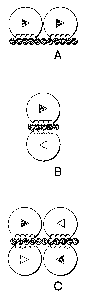
Figure 2 illustrates the proposed modes of FGF dimerization. Either a closed
or
an open triangle is drawn inside each FGF molecule to distinguish different
orientations.
The round indentation within FGF represents the heparin-binding domain. HLGAG
is
depicted as a chain of beads. (A) Two FGF molecules, oriented asymmetrically
in cis,
bind to the same side of HLGAG in a "side-by-side" fashion (Herr, A.B. et al.
(1997) J
Biol Chem 272(26), 16382-9; Venkataraman, G. et al. (1996) P~oc Natl Acad Sci
USA
93(2), 845-50; Venkataraman, G. et al. (1999) P~oc Natl Acad Sci USA 96(5),
1892-7).
(B) Two FGF molecules are oriented in traps to the axis of HLGAG in a "head-to-
head"
fashion (DiGabriele, A.D. et al. (1998) Nature 393(6687), 812-7). (C) Four FGF
molecules interact both in cis and traps with HLGAG (Moy, F.J. et al. (1997)
Biochemistry 36(16), 4782-91). Note that, for the cis interaction, the two FGF
molecules
are symmetrically related as opposed to the dimer in (A).
Figure 3 details the oxidative crosslinking studies. (A) Oxidative
c~osslihkin~ of
3o wild type FGF2 avcd cysteihe mutant. Wild-type FGF2 was oxidized with (lane
1) or
without (lane 2) heparin. A minor amount of dimer was detected, which likely
resulted
from the crosslinking reaction between unfolded protein. Cysteine mutant,
which was
CA 02441986 2003-09-23
WO 02/077199 PCT/US02/09517
_7_
designed based on the model of FGF2 dimerization (Venkataraman, G. et al.
(1996) P~oc
Natl Acad Sci ZISA 93(2), 845-50), was oxidized with (lane 3) or without (lane
4) heparin
under the same conditions as the wild-type. All reaction products were
separated using
non-reducing SDS-PAGE (15%) followed by silver staining. The extent of
oligomerization achieved by the cysteine mutant was compared to wild-type. (B)
Schematic rep~esehtation of the p~~oteivc p~~otein aid protein-HLGAG
ihte~actio~cs in
cysteine mutants. Two cysteine mutant molecules are shown, each with two dimer
interfaces as represented by striped (site p) and open (site p') rectangles.
Two solvent-
exposed cysteines (C81 and C 100 as shown near site p' and p, respectively)
were
to engineered such that they would position in close proximity with each other
at the
interface. (C) Dime~ization aad oligome~izatio~c of cysteiv~e mutant
wef°e mediated by the
native structure of the p~~otein. Lane 1, cysteine mutant alone; lane 2,
cysteine mutant
oxidized without heparin; lane 3, same as Iane 2 but protein was heat/SDS-
denatured
prior to oxidative crosslinking and lane 4, same as lane 2 but treated with 1
mM DTT.
Oxidative crosslinking of cysteine mutant was abolished by either denaturing
or reducing
treatments.
Figure 4 illustrates the engineering, cloning and purification of dFGF2. (A) A
scheme is shown for linking two FGF2 genes and subcloning them into an
expression
vector for protein expression. Restriction sites (Ndel, Sacl and Spel) were
introduced to
2o the 5' and 3' ends of FGF2 cDNA by PCR. (B) Restriction digest of the
expression
vector with two tandemly-linked FGF2 cDNAs is shown. Lane l, NdellSpel digest
of the
expression vector; lane 2, NdellSacl digest and lane 3, SacllSpel digest. (C)
Schematic
of the protein product obtained upon expression of the genetic construct of
(A). An N-
terminus His tag, a C-terminus T7 tag and two thrombin cleavage sequences
(gray
rectangles) are present to facilitate protein purification. The arrows
indicate the positions
of thrombin cleavage. (D) Wild-type mFGF2 (lane 1) and dFGF2 (lane 2) are
separated
by SDS-PAGE under reducing condition. The molecular size is shown on the side.
Figure 5 shows the structural properties of dFGF2. The near UV CD spectrum of
dFGF2 is shown. dFGF2 was concentrated to 1 ~,M and buffer-exchanged into 10
mM
3o sodium phosphate, pH 7.2. Data were recorded in an average of 20 scans
between 195
nm and 260 nm. The characteristic intense negative CD signals observed near
200 nm is
indicative of properly folded FGF2.
CA 02441986 2003-09-23
WO 02/077199 PCT/US02/09517
_g_
Figure 6 describes the competitive binding of dFGF2 for FGFR2. (A) MALDI
MS profile of a mixtu~~e of mild type FGF2 and the ectodomaih of FGFR2.
Observed in
the mass spectrum are (M+H)+ ion for an FGF2 dimer (mlz 30,214) and trimer
(mlz
45,132), FGFR2 monomer (mlz 24,888) and dimer (mlz 49,572), and a 1:l FGF2-
FGFR2
complex (m/z 39,896). The theoretical molecular masses for FGF2 and FGFR2 are
15114 and 24864, respectively. (B) Mass spectrum of the FGF2/FGFR2 mixture i~
the
pr~esehce of a homogenous HLGAG decasaccharide. Addition of a decasacchaxide
(Decay to FGF2/FGFR2 promotes the formation of a 2:2 FGF2:FGFR2 complex with
an
observed (M+H)+ ion at nZlz 82,650 (with Decay or mlz 79,872 (without Decay.
The
(M+H)+ ion for two dimeric FGFR2 species are also observed, the first at m/z
49,692
represents the apo complex and the second at m/z 52,474 is a 2:1 FGFR2:Deca
complex.
l~set, mass spectrum of dFGF2 added to the mixture of Deca/FGF2/FGFR2 shown
above. Three high molecular weight complexes are observed: 2:2 FGF2:FGFR2,
complexes with or without Deca and a 1:2 dFGF2:FGFR2 complex without Deca.
Figure 7 illustrates the SMC proliferation assay. Serum-starved SMC were
stimulated with the indicated molar concentrations of wild-type (~) and dFGF2
(~).
SMC were grown (A) in the absence of chlorate or (B) upon addition of 75 mM
chlorate.
After 21 h at 37°C, [3H] thymidine was added for 3 h. Cells were
harvested, washed and
measured [3H] thymidine incorporation was counted. Maximal count/min for wild-
type
2o and dFGF2 were about 6000 and 5000, respectively. The proliferation curve
of dFGF2
is shifted towards the left of wild-type. The molar concentrations for half
maximal
proliferation by wild-type and dFGF2 are 270 pM and 60 pM, respectively.
Figure 8 describes the HUVEC survival assay. Serum-starved HUVEC were
stimulated with the indicated concentrations of wild-type and dFGF2, or
without any
growth factor. Cells supplemented with 10% FCS served as positive control.
After 18 h,
cell viability was determined colorimetrically using MTS reagent. Both wild-
type and
dFGF2 restored HUVEC viability following serum starvation and dFGF2 achieved
the
same levels of cell viability at a lower molar concentration than wild-type.
Figure 9 details the in vivo potency of dFGF2. Slit lamp photographs of rat
3o corneas on day 6 after implantation with Hydron pellets containing (A) no
bFGF as
control, (B) 1.5 pmole mFGF2, (C) 6.0 pmole mFGF2, or (D) 0.7 pmole dFGF2.
Area of
pellet implantation is designated with an arrow. The control pellet did not
induce a
CA 02441986 2003-09-23
WO 02/077199 PCT/US02/09517
-9-
significant angiogenic response, while pellets containing dFGF2 induced an
intense
neovascular response originating from the limbal vessels and reaching the
pellet on day 6
after the implantation. Pellets containing mFGF2 (B, C) induced a less
vigorous, but still
detectable, angiogenic response on day 6 after implantation. In the Table, the
extent of
corneal angiogenic response was expressed as linear length and circumferential
cloclc
hours. '~ indicates Standard Error.
DETAILED DESCRIPTION
The invention relates to biologically active FGF dimers and uses thereof. It
has
to been discovered according the invention that FGF dimers are biologically
active. The
FGF dimers have, in some aspects, greatly enhanced biological activities. Most
of the
prior art studies describing the therapeutic use of FGF have described the use
of FGF
monomers. In addition, prior art studies have suggested that monomer forms of
FGF2
may form active signaling complexes (Pantoliano, M.W. et al. (1994)
Biochemistry
33(34), 10229-48; Pye, D.A. et al. (1999) JBiol Chem 274(19), 13456-61). For
instance
in a recent study, it was found that covalently linked complexes of monomer
FGF with a
pool of heparin dodecasaccharides were able to promote cell proliferation in
vitro (Pye,
D.A. et al. (1999) JBiol Chem 274(19), 13456-61). However, as observed herein
(data
presented in Examples section), this complex was less active than uncomplexed
FGF in
2o promoting 3H-thymidine incorporation. In contrast, the dimeric FGF (dFGF)
construct
presented in this study is several times mope potent in biological assays than
is wild-type
FGF, with reduced dependence on exogenous HLGAGs for activity. The invention
is
based at least in part on the fording that dimers of FGF have significantly
improved
biological activities as compared to the monomer.
Several signaling pathways mediated by growth factors and cytokines involve
binding of ligands to their cell surface receptors to facilitate receptor
dimerization
(Heldin, C.H. (1995) Cell 80(2), 213-23), a key step leading towards
activation of
intracellular signaling cascade. The structure, conformation, and
oligomerization status
of FGF as it interacts with FGFR to produce a biological signal are unknown.
The
3o studies of the invention have identified important characteristics of the
FGF-FGFR
interaction that have led to the development of a therapeutically important
class of
compounds. In general, it was discovered that FGF2 does have a preference to
CA 02441986 2003-09-23
WO 02/077199 PCT/US02/09517
- 10-
oligomerize, and the studies described herein point to the fact that this
oligomerization
interface involves protein-protein contact. Additionally, dimeric FGF (dFGF)
constructs
based on these biochemical findings were found to have potent biological
activity. Thus,
FGF dimers are potent mediators of FGFR dimerization and concomitant
signaling.
, Through rational design of a disulfide-mediated sequential dimer (cysteine
mutant) based on extensive analysis of FGF2 crystal structures we demonstrated
(A) a
marked increase in the amount of oligomers formed compared with wild-type
FGF2,
which has the same number of surface cysteines but at different positions, (B)
higher
extent of oligomerization by pre-incubating cysteine mutant with heparin, and
(C) that
to the observed oligomers involve specific protein contacts and are disulfide-
mediated. The
above findings strongly support a model in which FGF2 molecules self associate
through
specific FGF-FGF interactions in a sequential fashion and that HLGAG may serve
to
provide a "platform" to stabilize the intermolecular interactions between FGF2
molecules.
To determine whether the active FGF2 dimer involves protein-protein contact in
contrast to the FGF2 dimer observed in the FGF-FGFR co-crystal structures that
laelc
protein-protein contact, a tandemly-linked dimeric FGF2 (dFGF2) molecule was
constructed using conformational studies and genetic engineering tools. dFGF2
was
designed such that the short distance between the two FGF2 molecules within
the
dimeric protein would allow for substantial FGF-FGF interactions while making
the non-
contacting dimer mode less favorable and therefore enable us to dissect
whether a
contacting FGF2 dimer can elicit biological activity. We showed though mass
spectrometry that dFGF2 interacts with FGFR in a ratio of 1:2 suggesting that
dFGF2
can bind to a dimer of FGFR. Furthermore, these results indicate that one
mode,
involving substantial protein contact, by which FGF2 and its receptor may
interact is
through the binding of FGFR to a FGF2 dimer. These biochemical findings were
supported by the biological activity of the dFGF2 molecule, described in the
Examples.
To test whether a contacting FGF2 dimer can elicit biological activity, dFGF2
was subjected to two independent cell culture assays. From both the SMC
proliferation
3o and HUVEC survival assays, dFGF2 exhibited elevated biological activity
compared
with wild-type FGF2. This effect was especially pronounced in the SMC assays
where
dFGF2 was several fold more active than wild-type and only 30% less active in
the
CA 02441986 2003-09-23
WO 02/077199 PCT/US02/09517
-11-
absence of HLGAGs as in their presence (as opposed to wild type FGF2 wherein
activity
was significantly reduced in the absence of cell surface HLGAGs). These
findings
demonstrated that dFGF2, in which FGF-FGF interactions are predicted to be
substantial,
forms an active signaling complex with the receptor. In addition,
proliferation of
chlorate-treated SMC demonstrated that dFGF2 was less HLGAG-dependent for
signaling. These data suggest that one mechanism by which HLGAGs modulate FGF2
activity is by stabilizing two FGF2 molecules in a diner mode to facilitate
receptor
dimerization. Because dFGF2 is already dimeric, its dependency on HLGAGs for
proper
presentation to the receptor was lower compared to wild-type FGF2. The dFGF
to construct was also found to be a potent pro-angiogenic agent ih vivo, much
more so than
wild-type FGF, thus providing compelling evidence that the dFGF construct,
involving
substantial protein-protein contact, forms an active signaling complex at the
cell surface.
Thus the biochemical, cell culture, and in vivo assays demonstrate that a FGF2
diner is involved in the active signaling complex and axe inconsistent with
prior art data
on the different FGF2-FGFR crystal structures, which show no FGF-FGF
interactions.
Such an inconsistency may reflect the inherent complexity and the multifaceted
nature of
the FGF system. One possible explanation is that the different structural
configurations
of FGF-FGFR may reflect the different states, viz., "on" or "off' states of
the signaling
complex. Thus, a mode of FGFZ dimerization involving protein-protein
interactions
2o could lead to a cooperative FGF2,-FGFR interaction by promoting subsequent
oligomerization and signaling whereas the non-contacting FGF2 dimerization may
lead
to an inactive complex.
Thus in some aspects, the invention relates to compositions of FGF diners. An
"FGF diner" as used herein is an FGF diner composed of two FGF monomers linked
to
one another. An FGF diner is also referred to herein as dFGF. FGF diners
include
modified FGF diners and native FGF diners that have been stabilized to
maintain the
dimeric state.
Fibroblast growth factor (FGF) was first described by its activity derived
from
bovine brain or pituitary tissue which was mitogenic for fibroblasts and
endothelial cells.
3o It was later noted that the primary mitogen from brain was different from
that isolated
from pituitary. These two factors were named acidic and basic FGF (now known
as
CA 02441986 2003-09-23
WO 02/077199 PCT/US02/09517
-12-
FGFl and FGF2), respectively, because they had similar biological activities
but differed
in their isoelectric points.
It is now known that a laxge family of proteins exist, which are considered to
be
FGF. The fibroblast growth factor (FGF) family consists of at least twenty
three distinct
members which generally act as mitogens for a broad spectrum of cell types.
For
example, FGF2 is mitogenic ih vitro for endothelial cells, vascular smooth
muscle cells,
fibroblasts, and generally for cells of mesoderm or neuroectoderm origin,
including
caxdiac and skeletal myocytes (Gospoda~owicz et al., J. Cell. Biol. 70: 395-
405, 1976;
Gospoda~owicz et al., J. Cell. Biol. 89: 568-578, 1981 acrd Ka~dami, J. Nlol.
Cell.
to Biochem. 92:124-134, 1990). In vivo, FGF2 has been shown to play a role in
avian
cardiac development (Sugi et al., Dev. Biol. 168:567-574, 1995 ahd Mima et
al., Proc.
Nat'l. Acad. Sci. 92: 467-471, 1995), and to induce coronary collateral
development in
dogs (Laza~ous et al., Ci~culatio~c 94:1074-1082, 199. In addition to
eliciting a
mitogenic response that stimulates cell growth, fibroblast growth factors can
stimulate a
large number of cell types to respond in a non-mitogenic manner. These
activities
include promotion of cell migration into wound areas (chemotaxis), initiation
of new
blood vessel formulation (angiogenesis), modulation of nerve regeneration and
survival
(neurotrophism), modulation of endocrine functions, stimulation or suppression
of
specific cellulax protein expression, extracellular matrix production and cell
survival
(Bard, A., ahd Bohlen, P., handbook ofExp. Pha~macol. 95(1): 369-418,
Sprihge~,
1990). These properties provide a basis for using fibroblast growth factors in
therapeutic
approaches to accelerate wound healing, nerve repair, collateral blood vessel
formation,
and the like. For example, fibroblast growth factors have been suggested to
minimize
myocardium damage in heart disease and surgery (U.S Pat. No. 4,378,347 to
F~av~co).
All the members of the FGF family bind heparin and retain structural homology
across species, suggesting a conservation of their structure/function
relationship (O~nitz
et al., .I Biol. Chem. 271 (25):15292-15297, 1996.). A protein is a member of
the FGF
family, as used herein, if it shows significant sequence and three-dimensional
structural
homology to other members of the FGF family, FGF-like activity in in vitro or
in vivo
3o assays and binds to heparin or heparin-lilce substances.
FGF signaling is mediated primarily through high-affinity interaction with
cell
surface FGF receptors (FGFRs), transmembrane polypeptides composed of
CA 02441986 2003-09-23
WO 02/077199 PCT/US02/09517
-13-
immunoglobulin-like and tyrosine lcinase domains. FGF binding to different
isoforms of
FGFl~ is believed to trigger receptor dimerization followed by
transphosphorylation of
specific tyrosine residues. Phosphorylated tyrosine residues in turn activate
other
signaling proteins, leading to cell proliferation, migration and survival. We
have
analyzed various crystal structures of FGF extensively and have proposed a
model of
FGF signaling. In this model, two molecules of FGF2 are associated
preferentially along
the 31A axis and heparin saccharide can bind to the FGF2 to stabilize the
diner.
Another mode of dimerization (along the 33A axis) is also proposed.
A preferred FGF according to the invention is FGF2, and in some embodiments
to human FGF2 is preferred. The term "FGF2" as used herein refers to any
fibroblast
growth factor-2 exhibiting biologic activity. FGF2 include but are not limited
to the 155
amino acid protein recognized as native FGF2 (SEQ ID NO.: 1), truncated forms
exhibiting activity, extended forms such as placental FGF, higher molecular
weight N-
terminally extended forms and functionally equivalent FGF2 derivatives of any
of these.
The term specifically includes natural FGF2 extracted from mammalian tissue as
well as
recombinant polypeptides expressed from DNA from any species.
The three-dimensional structures of FGF2 has been determined (E~iksso~c, E.
A.,
et al., P~ac. Nat. Acad. Sci. U.S.A. 88: 3441-3445 (1991), Zhahg, J., et al.,
Proc. Nat.
Acad. Sci. U.SA. 88: 3446-3450 (1991), avcd Zhu, H., et al., Science 251: 90-
93 (1991)).
The overall structure of FGF2 can be described as a trigonal pyramid where
each of the
three sides are built of two (3-strands together forming a (3-sheet barrel of
six antiparallel
strands (ET°iksson, E. A., et al., P~oc. Nat. Acad. Sci. U.SA. 88: 3441-
3445 (1991)). The
base of the pyramid is built of six additional [3strands extending from the
three sides of
the pyramid to close one end of the barrel for a total of twelve -strands.
Thus, a
threefold repeat is observed in the folding of the polypeptide chain and a
pseudo-three-
fold axis passes through the center of the base of the molecule and extends
through the
apex of the pyramid. Of the amino acids conserved within the FGF family of
proteins,
most are located within the core (3-strand regions of FGF2.
A "modified FGF diner" as used herein is an FGF diner composed of two FGF
3o monomers linked to one another, wherein the diner includes at least one
modification
from a native FGF diner. The modification may be within the amino acid
sequence of
one or both the FGF monomers or it may be the linkage itself. For instance,
the modified
CA 02441986 2003-09-23
WO 02/077199 PCT/US02/09517
-14-
FGF dimer may be composed of two naturally occurring FGF monomers which are
linked by a linker molecule.
In some embodiments the modified FGF dimer is stabilized. A stabilized dimer
is one in which the monomers have a higher probability of remaining in a
dimeric
complex than monomeric FGF ordinarily would remain in a dimeric complex. The ,
stabilized dimer may be accomplished through a variety of mechanisms. For
example a
linker molecule may be used to stabilize the dimeric structure of FGF.
Covalent or other
non-covalent interactions may also be used to stabilize the dimer, as long as
the
interactions form a more stable dimeric form of FGF than the non-covalent
interactions
between native FGF monomers.
It was surprisingly discovered according to the invention that the stabilized
FGF
dimers have improved activity over FGF monomers or native dimers.
As used herein, "linked" or "linkage" means two entities are bound to one
another by any physiochemical means. It is important that the linlcage be of
such a
nature that it does not impair substantially the effectiveness of the FGF
monomers or the
binding specificity of the dimer with the FGFR. Keeping these parameters in
mind, ably
linkage known to those of ordinary skill in the art may be employed, covalent
or
noncovalent. Linkages according to the invention include linker molecules and
chemical
linkages. Such means and methods of linkage are well known to those of
ordinary skill
in the art.
Linked monomers of FGF in an FGF dimer, when used with respect to a
pharmaceutical composition of an FGF dimer refers to the fact that at least
greater than
50% of the FGF monomers in the composition are in a dimeric state. Preferably
at least
55%, 60%, 65%, 70%, 75%, 80%, 85%, 90%, 95%, or 100% of the FGF monomers are
in a dimeric form.
A "linker molecule" as used herein is a molecule which forms an indirect
linlcage
between the two monomers. In some embodiments the linker molecule is a spacer
molecule that is attached to each of the monomers, either covalently or non-
covalently.
One method for attaching a spacer to the monomers is with the use of
functionalized
3o groups on the monomer to facilitate linkage and/or linker groups interposed
between the
monomers to facilitate their linkage. Another method involves the synthesis in
a single
process of both monomers and the linker, whereby the components of the dimer
could be
CA 02441986 2003-09-23
WO 02/077199 PCT/US02/09517
-15-
regarded as one in the same entity. For example, using recombinant DNA
methodology
a nucleic acid construct encoding both monomers and a linking peptide,
oriented such
that when the protein is expressed the linking peptide connects the two
monomers, can
be used to. generate the dimer. These and other methods for indirect linkage
axe intended
to be embraced by the present invention.
Specific examples of covalent bonds include those wherein bifunctional cross-
linlcer molecules axe used. The cross-linker molecules may be homobifunctional
or
heterobifunctional, depending upon the nature of the molecules to be
conjugated.
Homobifunctional cross-linlcers have two identical reactive groups.
Heterobifunctional
to cross-linlcers have two different reactive groups that allow sequential
conjugation
reaction. Various types of commercially available cross-linkers are reactive
with one or
more of the following groups: primary amines, secondary amines, sulfhydriles,
carboxyls, carbonyls and carbohydrates. The linker molecule may also be
attached to the
monomer using non-covalent bonds. Non-covalent conjugation may be accomplished
by
direct or indirect means including hydrophobic interaction, ionic interaction,
and other
affinity interactions. The linking molecules may also be modified such that
they are
noncleavable in physiological environments or cleavable in physiological
environments.
Such molecules may resist degradation.
In a preferred embodiment the linker molecule is a peptide which is produced
using recombinant technology along with the FGF monomers. An example of an FGF
dimer produced by this method is set forth in the Examples section. The
exemplary FGF
dimer has the amino acid sequence of SEQ ID NO.: 6. The FGF dimer was
expressed
from the DNA having the sequence of SEQ ID NO.: 5. Briefly, an expression
vector
which will express the FGF dimer is generated. The expression vector includes
the
sequence for two FGF monomers and a linker peptide, operably arranged to
produce a
functional fusion protein. This is depicted schematically in Figure 4. One
example of a
linker useful for generating the diners is GAL. Other linkers include but axe
not limited
to GAR and GARG. The distance of the GAL linker between the N terminus of one
monomer and the C terminus of the other monomer is 271. The distance between
the 2
3o monomers of the FGF observed in crystal structures in ~421~. The distance
between
monomers in an FGF1 diner in transform is ~70~. For FGF2 271 is preferred.
CA 02441986 2003-09-23
WO 02/077199 PCT/US02/09517
-16-
Thus, one of ordinary skill in the art, in light of the present disclosure, is
enabled
to produce the FGF dimers by standard technology, including recombinant
technology,
direct synthesis, mutagenesis, etc. For instance, using recombinant technology
one may
substitute appropriate codons in SEQ ID NO: 5 to produce the desired amino
acid
substitutions by standard site-directed mutagenesis techniques. Obviously, one
may also
use any sequence which differs from SEQ ID NO: 5 only due to the degeneracy of
the
genetic code as the starting point for site directed mutagenesis. The mutated
nucleic acid
sequence may then be ligated into an appropriate expression vector and
expressed in a
host such as E. coli. The resultant modified FGF dimer may then be purified by
to techniques well known in the art, including those disclosed below in the
Examples.
Preferably the FGF dimers are substantially pure. As used herein, the term
"substantially
pure" means that the proteins are essentially free of other substances to an
extent
practical and appropriate for their intended use. In particular, the proteins
are
sufficiently pure and are sufficiently free from other biological constituents
of their hosts
cells so as to be useful in, for example, protein sequencing, or producing
pharmaceutical
preparations.
In another set of embodiments an isolated nucleic acid encoding the modified
FGF dimer of the invention is provided. As used herein with respect to nucleic
acids, the
term "isolated" means: (i) amplified in vitro by, for example, polymerase
chain reaction
(PCR); (ii) recombinantly produced by cloning; (iii) purified, as by cleavage
and gel
separation; or (iv) synthesized by, for example, chemical synthesis. An
isolated nucleic
acid is one which is readily manipulable by recombinant DNA techniques well
knomn in
the art. Thus, a nucleotide sequence contained in a vector in which 5' and 3'
restriction
sites are known or for which polymerase chain reaction (PCR) primer sequences
have
been disclosed is considered isolated but a nucleic acid sequence existing in
its native
state in its natural host is not. An isolated nucleic acid may be
substantially purified, but
need not be. For example, a nucleic acid that is isolated within a cloning or
expression
vector is not pure in that it may comprise only a tiny percentage of the
material in the
cell in which it resides. Such a nucleic acid is isolated, however, as the
term is used
3o herein because it is readily manipulable by standard techniques known to
those of
ordinary slcill in the art.
CA 02441986 2003-09-23
WO 02/077199 PCT/US02/09517
- 17-
As used herein, a coding sequence and regulatory sequences are said to be
"operably joined" when they are covalently linked in such a way as to place
the
expression or transcription of the coding sequence under the influence or
control of the
regulatory sequences. In order that the coding sequences be translated into a
functional
protein the coding sequences axe operably joined to regulatory sequences. Two
DNA
sequences are said to be operably joined if induction of a promoter in the 5'
regulatory
sequences results in the transcription of the coding sequence and if the
nature of the
linkage between the two DNA sequences does not (1) result in the introduction
of a
frame-shift mutation, (2) interfere with the ability of the promoter region to
direct the
to transcription of the coding sequences, or (3) interfere with the ability of
the
corresponding RNA transcript to be translated into a protein. Thus, a promoter
region
would be operably joined to a coding sequence if the promoter region were
capable of
effecting transcription of that DNA sequence such that the resulting
transcript might be
translated into the desired protein or polypeptide.
The precise nature of the regulatory sequences needed for gene expression may
vary between species or cell types, but shall in general include, as
necessary, 5'
non-transcribing and 5' non-translating sequences involved with initiation of
transcription and translation respectively, such as a TATA box, capping
sequence,
CAAT sequence, and the like. Especially, such 5' non-transcribing regulatory
sequences
2o will include a promoter region which includes a promoter sequence for
transcriptional
control of the operably joined gene. Promoters may be constitutive or
inducible.
Regulatory sequences may also include enhancer sequences or upstream activator
sequences, as desired.
As used herein, a "vector" may be any of a number of nucleic acids into which
a
desired sequence may be inserted by restriction and ligation for transport
between
different genetic environments or for expression in a host cell. Vectors are
typically
composed of DNA although RNA vectors are also available. Vectors include, but
are
not limited to, plasmids and phagemids. A cloning vector is one which is able
to
replicate in a host cell, and which is further characterized by one or more
endonuclease
3o restriction sites at which the vector may be cut in a determinable fashion
and into which
a desired DNA sequence may be ligated such that the new recombinant vector
retains its
ability to replicate in the host cell. In the case of plasmids, replication of
the desired
CA 02441986 2003-09-23
WO 02/077199 PCT/US02/09517
-18-
sequence may occur many times as the plasmid increases in copy number within
the host
bacterium, or just a single time per host as the host reproduces by mitosis.
In the case of
phage, replication may occur actively during a lytic phase or passively during
a
lysogenic phase. An expression vector is one into which a desired DNA sequence
may
be inserted by restriction and ligation such that it is operably joined to
regulatory
sequences and may be expressed as an RNA transcript. Vectors may further
contain one
or more marker sequences suitable for use in the identification of cells which
have or
have not been transformed or transfected with the vector. Markers include, for
example,
genes encoding proteins which increase or decrease either resistance or
sensitivity to
to antibiotics or other compounds, genes which encode enzymes whose activities
are
detectable by standard assays known in the art (e.g., !3-galactosidase or
allcaline
phosphatase), and genes which visibly affect the phenotype of transformed or
transfected
cells, hosts, colonies or plaques. Preferred vectors are those capable of
autonomous
replication and expression of the structural gene products present in the DNA
segments
to which they are operably joined.
As used herein, the term "stringent conditions" refers to parameters known to
those skilled in the art. One example of stringent conditions is hybridization
at 65°C in
hybridization buffer (3.5 x SSC, 0.02% Ficoll, 0.02% polyvinyl pyrolidone,
0.02%
bovine serum albumin (BSA), 25mM NaH2PO4 (pH7), 0.5% SDS, 2mM EDTA). SSC is
O.15M sodium chloride/O.15M sodium citrate, pH7; SDS is sodium
dodecylsulphate; and
EDTA is ethylene diamine tetra acetic acid. There are other conditions,
reagents, and so
forth which can be used, which result in the same degree of stringency. A
skilled artisan
will be familiax with such conditions, and thus they are not given here. The
skilled
artisan also is familiar with the methodology for screening cells for
expression of such
molecules, which then are routinely isolated, followed by isolation of the
pertinent
nucleic acid. Thus, homologs and alleles of the modified FGF dimer of the
invention, as
well as nucleic acids encoding the same, may be obtained routinely, and the
invention is
not intended to be limited to the specific sequences disclosed.
For prokaryotic systems, plasmid vectors that contain replication sites and
control
3o sequences derived from a species compatible with the host may be used.
Examples of
suitable plasmid vectors include pBR322, pUCl8, pUCl9 and the like; suitable
phage or
bacteriophage vectors include 7~gt10, ~,gtl l and the like; and suitable virus
vectors
CA 02441986 2003-09-23
WO 02/077199 PCT/US02/09517
-19-
include pMAM-neo, pKRC and the like. Preferably, the selected vector of the
present
invention has the capacity to autonomously replicate in the selected host
cell. Useful
prokaryotic hosts include bacteria such as E. coli, Flavobacte~ium heparinum,
Bacillus,
Str~eptomyces, Pseudomohas, Salmonella, Se~~atia, and the like.
To express the modified FGF dimer of the invention in a prokaryotic cell, it
is
necessary to operably join the nucleic acid sequences of the monomers and the
linker to a
functional prokaryotic promoter. Such promoter may be either constitutive or,
more
preferably, regulatable (i.e., inducible or derepressible). Examples of
constitutive
promoters include the int promoter of bacteriophage 7~, the bla promoter of
the (3-
to lactamase gene sequence of pBR322, and the CAT promoter of the
chloramphenicol
acetyl transferase gene sequence of pPR325, and the like. Examples of
inducible
prokaryotic promoters include the major right and left promoters of
bacteriophage 7~ (P~,
and PR), the trp, ~ecA, lacZ, lacl, and gal promoters of E coli, the oc-
amylase (Ulmanen
et al., J. Bactef°iol. 162:176-182 (1985)) and the ~-28-specific
promoters of B. subtilis
15 (Gilman et al., Gene sequence 32:11-20 (1984)), the pramoters of the
bacteriophages of
Bacillus (Gryczan, In: The Molecular Biology of the Bacilli, Academic Press,
Inc., NY
(1982)), and St~eptomyces promoters (Ward et al., Mol. Gen. Genet. 203:468-478
(1986)).
Prokaryotic promoters are reviewed by Glick (J. Ind. Micf°obiol.
1:277-282
20 (1987)); Cenatiempo (Biochimie 68:505-516 (1986)); and Gottesman (Ann. Rev.
Genet.
18:415-442 (1984)).
Proper expression in a prokaryotic cell also requires the presence of a
ribosome
binding site upstream of the encoding sequence. Such ribosome binding sites
are
disclosed, for example, by Gold et al. (Anu. Rev. Mic~obiol. 35:365-404
(1981)).
25 Because prokaryotic cells will not produce the modified FGF dimer of the
invention with normal eukaryotic glycosylation, expression of the modified FGF
dimer
of the invention by eulcaryotic hosts is possible when glycosylation is
desired. Preferred
eukaryotic hosts include, for example, yeast, fungi, insect cells, and
mammalian cells,
either in vivo or in tissue culture. Mammalian cells which may be useful as
hosts include
3o HeLa cells, cells of fibroblast origin such as VERO or CHO-Kl, or cells of
lymphoid
origin, such as the hybridoma SP2/0-AG14 or the myeloma P3x63Sg8, and their
derivatives. Preferred mammalian host cells include SP2/0 and J558L, as well
as
CA 02441986 2003-09-23
WO 02/077199 PCT/US02/09517
-20-
neuroblastoma cell lines such as IMR 332 that may provide better capacities
for correct
post-translational processing. Embryonic cells and mature cells of a
transplantable organ
also are useful according to some aspects of the invention.
In addition, plant cells are also available as hosts, and control sequences
compatible with plant cells are available, such as the nopaline synthase
promoter and
polyadenylation signal sequences.
Another preferred host is an insect cell, for example in D~osophila larvae.
When
using insect cells as hosts, the D~osophila alcohol dehydrogenase promoter can
be used
(Rubin, Science 240:1453-1459 (1988)). Alternatively, baculovirus vectors can
be
to engineered to express large amounts of the modified FGF dimer of the
invention in
insects cells (Jasny, Science 238:1653 (1987); Miller et al., In: Genetic
Engihee~i~g
(1986), Setlow, J.K., et al., eds., Plenum, Vol. 8, pp. 277-297).
Any of a series of yeast gene sequence expression systems which incorporate
promoter and termination elements from the genes coding for glycolytic enzymes
and
which are produced in large quantities when the yeast are grown in media rich
in glucose
may also be utilized. Known glycolytic gene sequences can also provide very
efficient
transcriptional control signals. Yeast provide substantial advantages in that
they can also
carry out post-translational peptide modifications. A number of recombinant
DNA
strategies exist wluch utilize strong promoter sequences and high copy number
plasmids
2o which can be utilized for production of the desired proteins in yeast.
Yeast recognize
leader sequences on cloned mammalian gene sequence products and secrete
peptides
bearing leader sequences (i.e., pre-peptides).
A wide variety of transcriptional and translational regulatory sequences may
be
employed, depending upon the nature of the host. The transcriptional and
translational
regulatory signals may be derived from viral sources, such as adenovirus,
bovine
papilloma virus, simian virus, or the like, where the regulatory signals are
associated
with a particular gene sequence which has a high level of expression.
Alternatively,
promoters from mammalian expression products, such as actin, collagen, myosin,
and the
like, may be employed. Transcriptional initiation regulatory signals may be
selected
3o which allow for repression or activation, so that expression of the gene
sequences can be
modulated. Of interest are regulatory signals which are temperature-sensitive
so that by
CA 02441986 2003-09-23
WO 02/077199 PCT/US02/09517
-21 -
varying the temperature, expression can be repressed or initiated, or which
are subject to
chemical (such as metabolite) regulation.
As discussed above, expression of the modified FGF dimer of the invention in
eukaryotic hosts requires the use of eukaryotic regulatory regions. Such
regions will, in
general, include a promoter region sufficient to direct the initiation of RNA
synthesis.
Preferred eukaryotic promoters include, for example, the promoter of the mouse
metallothionein I gene sequence (Hamer et al., J. Mol. Appl. Geh. 1:273-288
(1982)); the
TK promoter of Herpes virus (McKnight, Cell 31:355-365 (1982)); the SV40 early
promoter (Benoist et al., Natuf°e (London) 290:304-310 (1981)); the
yeast gal4 gene
to sequence promoter (Johnston et al., P~oc. Natl. Acad. Sci. (USA) 79:6971-
6975 (1982);
Silver et al., P~oc. Natl. Acad. Sci. (USA) 81:5951-5955 (1984)).
As is widely known, translation of eulcaryotic mRNA is initiated at the codon
which encodes the first methionine. For this reason, it is preferable to
ensure that the
linkage between a eukaryotic promoter and the DNA sequences which encode the
modified FGF dimer of the invention does not contain any intervening codons
which are
capable of encoding a methionine (i.e., AUG). The presence of such codons
results
either in the formation of a fusion protein (if the AUG codon is in the same
reading
frame as the modified FGF dimer coding sequence) or a frame-shift mutation (if
the
AUG codon is not in the same reading frame as the modified FGF dimer coding
2o sequence).
In one embodiment, a vector is employed which is capable of integrating the
desired gene sequences into the host cell chromosome. Cells which have stably
integrated the introduced DNA into their chromosomes can be selected by also
introducing one or more markers wluch allow for selection of host cells which
contain
the expression vector. The marker may, for example, provide for prototrophy to
an
auxotrophic host or may confer biocide resistance to, e.g., antibiotics, heavy
metals, or
the like. The selectable marker gene sequence can either be directly linlced
to the DNA
gene sequences to be expressed or introduced into the same cell by co-
transfection.
Additional elements may also be needed for optimal synthesis of the FGF mRNA.
These
elements may include splice signals, as well as transcription promoters,
enhancers, and
termination signals. cDNA expression vectors incorporating such elements
include those
described by Okayama, Molec. Cell. Biol. 3:280 (1983).
CA 02441986 2003-09-23
WO 02/077199 PCT/US02/09517
-22-
In a preferred embodiment, the introduced sequence will be incorporated into a
plasmid or viral vector capable of autonomous replication in the recipient
host. Any of a
wide variety of vectors may be employed for this purpose. Factors of
importance in
selecting a particular plasmid or viral vector include the following: the ease
with which
recipient cells that contain the vector may be recognized and selected from
those
recipient cells which do not contain the vector, the number of copies of the
vector which
are desired in a particular host and whether it is desirable to be able to
"shuttle" the
vector between host cells of different species. PrefeiTed prolcaryotic vectors
include
plasmids such as those capable of replication in E. eoli (such as, for
example, pBR322,
to ColEl, pSC101, pACYC 184, and ~VX. Such plasmids are, for example,
disclosed by
Sambrook, et al. (Molecular- Cloning: A Labo~ato~y .tl~lanual, second edition,
edited by
Sambroolc, Fritsch, ~ Maniatis, Cold Spring Harbor Laboratory, 1989)).
Bacillus
plasmids include pC194, pC221, pT127 and the like. Such plasmids are disclosed
by
Gryczan (In: The Molecular Biology of the Bacilli, Academic Press, NY (1982),
pp. 307-
329). Suitable St~eptomyces plasmids include pIJ101 (Kendall et al., J.
Bacte~iol.
169:4177-4183 (1987)), and streptomyces bacteriophages such as c~C31 (Chater
et al., In:
Sixth Inte~hatio~cal Symposium oyi Actinomycetales Biology, Alcademiai Kaido,
Budapest,
Hungary (1986), pp. 45-54). Pseudomonas plasmids are reviewed by John et al.
(Rev.
Infect. Dis. 8:693-704 (1986)), and Izaki (Jpv~. J. Bacte~iol. 33:729-742
(1978)).
2o Preferred eukaryotic plasmids include, for example, BPV, EBV, SV40, 2-
micron
circle, and the like, or their derivatives. Such plasmids are well known in
the art
(Botstein et al., .Miami Yij~ct~. Symp. 19:265-274 (1982); Broach, In: The
Moleculaf°
Biology of the Yeast Saccha~omyees: Life Cycle a~ca' I~che~itance, Cold Spring
Harbor
Laboratory, Cold Spring Harbor, NY, p. 445-470 (1981); Broach, Cell 28:203-204
(1982); Bollon et al., J. Cliv~. Hematol. Oncol. 10:39-48 (1980); Maniatis,
In: Cell
Biology: A Comprehensive Treatise, Vol. 3, Gene Sequence Expression, Academic
Press, NY, pp. 563-608 (1980)). Other preferred eukaryotic vectors are viral
vectors.
For example, and not by way of limitation, the pox virus, herpes virus,
adenovirus and
various retroviruses may be employed. The viral vectors may include either DNA
or
3o RNA viruses to cause expression of the insert DNA or insert RNA.
Additionally, DNA
or RNA encoding the modified FGF dimer polypeptides may be directly injected
into
cells or may be impelled through cell membranes after being adhered to
microparticles.
CA 02441986 2003-09-23
WO 02/077199 PCT/US02/09517
- 23 -
Once the vector or DNA sequence containing the constructs) has been prepared
for expression, the DNA constructs) may be introduced into an appropriate host
cell by
any of a variety of suitable means, i.e., transformation, transfection,
conjugation,
protoplast fusion, electroporation, calcium phosphate-precipitation, direct
microinjection,
and the like. After the introduction of the vector, recipient cells are grown
in a selective
medium, which selects for the growth of vector-containing cells. Expression of
the
cloned gene sequences) results in the production of the modified FGF diner.
This can
take place in the transformed cells as such, or following the induction of
these cells to
differentiate (for example, by administration of bromodeoxyuracil to
neuroblastoma cells
to or the like).
In some embodiments the modified FGF diners are composed of truncated FGF
monomers. For instance one or more amino acids may be removed from the N-
terminal
end of the protein without altering the protein folding or activity of the
protein. A
detailed analysis of specific sites and regions within the FGF monomers that
can be
15 manipulated is presented in Table 1. Based on the information presented in
Table 1 it is
possible to construct mutants of the monomers that are used for generating the
dimeric
FGF. The mutants can have altered biological activity, stabilization, etc.
Table 1: Manipulable Sites and Regions within FGF
Name of FGF mutants _ Functions
del 9 _ 1 S 9 N-terminal as truncation
del 28 1 28 N-terminal as truncation
N102R Promote dimerization (31A
axis)
L98E "
L98E/N102R "
R60I "
L98E/N 102R/R60I "
Y124R Inhibit dimerization (31A
axis)
L52E Promote dimerization (33A
axis)
P49E "
V68R Inhibit dimerization (33A
axis)
N71 R "
Q134C disulfide diner (33A axis)
Q134C/C87S exclusive disulfide diner
(33A axis)
R81 C/S 1 OOC disulfide diner (3 1A axis)
R81C/S100C/C87S/C69S exclusive disulfide diner
(31A axis)
R81 C/S 100C/C87S/C69S/C25S/C92Sdisulfide diner w/o internal
cys
C87S/C69S/C25S/C92S no cys
CA 02441986 2003-09-23
WO 02/077199 PCT/US02/09517
-24-
C87S/C69S/C25S/C92S/R81C disulfide dimes w% 1 cys
(81C)
C87S/C69S/C25S/C92S/S100C disulfide dimes w/ 1 cys
(100C)
N102R/R60I Promote dimerization (31A
axis)
N 102R/K86A
K26A Reduce heparin binding
K26S
K125A "
K125D "
K119E "
R120T "
Kl 19A/R120A "
Y103A Reduce receptor binding
Y111A/W114A "
For example it is possible to promote dimerization through non-covalent
interactions using N102R, L98E etc. mutants. These mutants are designed to
form non-
covalent dimers stabilized by ionic interaction between adjacent proteins. The
mutated
residues are positioned at the °dimerization interface' for stabilizing
the dimes.
Additionally dimerization may be promoted using covalent disulfide linlcages
e.g.,
R81C/S100C/C87S/C69S or cys mutant which is designed to form covalent dimers
stabilized by di-sulfide bond (under oxidative conditions). Both of these
types of FGF
modifications fall within the definition of chemical linkages described below.
to Other mutations that can be made result in reduced heparin binding, e.g.,
these
mutants have mutations at the heparin-binding sites such that the mutated
residues (e.g.
K-->A) would not interact with heparin; reduced receptor binding, e.g. these
mutants
have mutations at the receptor binding site of FGF such that the mutated
residues do not
interact with FGFR. In some aspects it may also be desirable to modify the FGF
15 monomers to prevent dimerization, e.g. for controls or competitors, or to
prevent FGF
activity. Dimerization (non-covalent) may be inhibited with e.g. Y124R which
is
designed to disrupt dimerization by introducing the mutated residue to block
the
interface between the two proteins.
In the description herein, reference is made to the amino acid residues and
2o residue positions of native FGF2 with 9 N-terminal residues deleted
disclosed in SEQ ID
NO.: 7. In particular, residues and residue positions are referred to as
"corresponding to"
a particular residue or residue position of FGF. As will be obvious to one of
ordinary
skill in the art, these positions are relative and, therefore, insertions or
deletions of one or
CA 02441986 2003-09-23
WO 02/077199 PCT/US02/09517
-25-
more residues would have the effect of altering the numbering of downstream
residues.
In particular, N-terminal insertions or deletions would alter the numbering of
all
subsequent residues. Therefore, as used herein, a residue in a recombinant
modified
FGF2 dimer will be referred to as "corresponding to" a residue of the full
FGF2 if, using
standard sequence comparison programs, they would be aligned. Many such
sequence
alignment programs are now available to one of ordinary shill in the art and
their use in
sequence comparisons has become standard. As used herein, this convention of
referring
to the positions of residues of the recombinant modified FGF dimers by their
corresponding native FGF residues shall extend not only to embodiments
including N-
1o terminal insertions or deletions but also to internal insertions or
deletions.
In addition, in the description herein, certain substitutions of one amino
acid
residue for another in a recombinant FGF or FGF dimer are referred to as
"conservative
substitutions." As used herein, a "conservative amino acid substitution" or
"conservative
substitution" refers to an amino acid substitution in which the substituted
amino acid
residue is of similar charge as the replaced residue and is of similar or
smaller size than
the replaced residue. Conservative substitutions of amino acids include
substitutions
made amongst amino acids within the following groups: (a) the small non-polar
amino
acids, A, M, I, L, and V; (b) the small polar amino acids, G, S, T and C; (c)
the amido
amino acids, ~ and N; (d) the aromatic amino acids, F, Y and W; (e) the basic
amino
2o acids, I~, 1Z and H; and (f) the acidic amino acids, E and D. Substitutions
which are
charge neutral and which replace a residue with a smaller residue may also be
considered
"conservative substitutions" even if the residues axe in different groups
(e.g.,
replacement of phenylalanine with the smaller isoleucine). The term
"conservative
amino acid substitution" also refers to the use of amino acid analogs or
variants.
Methods for making amino acid substitutions, additions or deletions are well
known in the art and are described in detail in the Examples below. The terms
"conservative substitution", "non-conservative substitutions", "non-polar
amino acids",
"polar amino acids", and "acidic amino acids" are all used consistently with
the prior art
terminology. Each of these terms is well-known in the art and has been
extensively
3o described in numerous publications, including standard biochemistry text
books, such as
"Biochemistry" by Geoffrey Zubay, Addison-Wesley Publishing Co., 1986 edition,
CA 02441986 2003-09-23
WO 02/077199 PCT/US02/09517
-26-
which describes conservative and non-conservative substitutions and properties
of amino
acids which lead to their definition as polar, non-polar or acidic.
Even when it is difficult to predict the exact effect of a substitution in
advance of
doing so, one skilled in the art will appreciate that the effect can be
evaluated by routine
screening assays, preferably the biological assays described herein.
Modifications of
peptide properties including thermal stability, hydrophobicity, susceptibility
to
proteolytic degradation or the ability to interact with the receptor axe
assayed by methods
well known to the ordinarily skilled artisan. For additional detailed
description of
protein chemistry and structure, see Schulz, G. E. et al., Principles of
Protein Structure,
to Sprihger-Tlerlag, New York, 1979, and Creighton, T. E., Ps oteihs:
Structure and
Molecular Principles, 1~h H. Freeman & Co., San Francisco, 1984.
Additionally, some of the amino acid substitutions are non-conservative
substitutions. In certain embodiments where the substitution is remote from
the active or
binding sites, the non-conservative substitutions are easily tolerated
provided that they
preserve the tertiary structure characteristic of native FGF, thereby
preserving the active
and binding sites. Non-conservative substitutions, such as between, rather
than within,
the above groups (or two other amino acid groups not shown above), which will
differ
more significantly in their effect on maintaining (a) the structure of the
peptide backbone
in the area of the substitution (b) the charge or hydrophobicity of the
molecule at the
2o target site or (c) the bulk of the side chain.
The proteins of the present invention can also comprise, in addition to the 20
standard amino acids, non-naturally occurring amino acid residues. Non-
naturally
occurring amino acids include, without limitation, trans-3-methylproline, 2,4-
methanoproline, cis-4-hydroxyproline, trans-4-hydroxyproline, N-methyl-
glycine, allo-
threonine, methylthreonine, hydroxyethyl-cysteine, hydroxyethylhomocysteine,
nitroglutamine, homoglutamine, pipecolic acid, tent-leucine, norvaline, 2-
azaphenylalanine, 3-azaphenylalanine, 4-azaphenyl-alanine, 4-
fluorophenylalanine, 4-
hydroxyproline, 6-N-methyl lysine, 2-aminoisobutyric acid, isovaline and
.alpha.-methyl
serine.
3o Several methods are known in the art for incorporating non-naturally
occurring
amino acid residues into proteins. For example, an in vitro system can be
employed
wherein nonsense mutations are suppressed using chemically aminoacylated
suppressor
CA 02441986 2003-09-23
WO 02/077199 PCT/US02/09517
tRNAs. Methods for synthesizing amino acids and aminoacylating tRNA are known
in
the art. Transcription and translation of plasmids containing nonsense
mutations are
carried out in a cell free system comprising an E. coli S30 extract and
commercially
available enzymes and other reagents. Proteins are purified by chromatography.
See, for
example, Robertson et al., J. Am. Chem. Soc. 113:2722, 1991; Ellmav~ et al.,
Meth.
E~~ymol. 202: 301, 1991; Chuhg et al., Science 259: 806-09, 1993; and Chuhg et
al.,
Py~oc. Natl. Acad. Sci. USA 90:10145-49, 1993. In a second method, translation
is
carried out in Xenopus oocytes by microinjection of mutated mRNA and
chemically
aminoacylated suppressor tRNAs (Tu~catti et al., J. Biol. Chem. 271:19991-98,
199b~.
l0 In a third method, E. coli cells are cultured in the absence of a natural
amino acid that is
to be replaced (e.g., phenylalanine) and in the presence of the desired non-
naturally
occurring amino acids) (e.g., 2-azaphenylalanine, 3-azaphenylalanine, 4-
azaphenylalanine, or 4-fluorophenylalanine). The non-naturally occurring amino
acid is
incorporated into the protein in place of its natural counterpart. See, e.g.,
Koide et al.,
15 Biochem. 33: 7470-76, 1994. Naturally occurring amino acid residues can be
converted
to non-naturally occurring species by in vitf°o chemical modification.
Chemical
modification can be combined with site-directed mutagenesis to further expand
the range
of substitutions (Wyn~z and Richa~ds, Protein Sei. 2: 395-403, 1993).
Additionally, the linker sequences, and the N/C terminal tags can be
substituted
2o with other sequences for defined purposes, such as integrin binding
sequences, protease
sites (e.g., in the linker to manipulate cleavage), epitopes, etc.
The FGF DNA used in generating the FGF diners may be natural, recombinant
or synthetic. Thus, DNA starting material is isolated from tissue or tissue
culture,
constructed from oligonucleotides using conventional methods, obtained
commercially,
25 or'p'repared by isolating RNA coding for FGF from fibroblasts, and using
this RNA to
synthesize single-stranded cDNA wk~ich is used as a template to synthesize the
corresponding double stranded DNA.
The term "chemical linkage" as used herein refers to a direct linkage between
the
two monomers. The direct linkage may be covalent or non-covalent. In some
preferred
3o embodiments the chemical linkage is a covalent disulfide linkage, arising
from the
interaction between two cysteine residues that have been incorporated into the
monomers. Examples of monomers having cysteines incorporated therein that can
CA 02441986 2003-09-23
WO 02/077199 PCT/US02/09517
-28-
produce disulfide bonds include those having sequences set forth in SEQ. ID
NOs.: 2-4.
Exemplary methods for generating these types of FGF dimers having a chemical
linkage
is set forth in the Examples section.
In addition to the modified FGF dimers of the invention, some embodiments and
aspects of the invention utilize naturally occurring FGF dimers. Preferably
the naturally
occurring FGF dimers are stabilized. Stabilizing agents include, but are not
limited to,
glycosaminoglycans, such as heparin, heparin fragments, heparan sulfate and
dermatan
sulfate, or glucan sulfates, such as dextran sulfate, Tri 3 oligosacchaxides,
and
cyclodextrin sulfate. Stabilized FGF monomers of this type are described, for
example,
to in EP 251 806, EP 267 015, EP 312 208, EP 345 660, EP 406 856, EP 408 146,
WO 89-
12464, WO 90-01941 and WO 90-03797.
HLGAGs that enhance 'natural' dimerization of FGF are in general
oligosaccharides of 8-10 monosaccharide units long with 2-O and N- sulfation.
The
HLGAG can also be obtained from heparin or its fragments. Tri 3 is a unique
HLGAG
15 that promotes dimerization in that it is both short (three saccharides
long) and
undersulfated. It has previously been described in Ornitz et al in Science
268:432.
The FGF dimers of the invention have important therapeutic and diagnostic
utilities. For instance, the FGF dimers can promote vasculaxization, cell
growth, and/or
cell survival, and thus have application in tissue repair such as healing of
wounds, burns,
2o bone fractures, surgical abrasions, gastrointestinal ulcers, and the like
as well as tissue
repair during ischemia and myocardial infarction via neovasculaxization of
ischemic
tissue. FGF2 is also effective in maintaining certain hematopoietic lineages
in long term
primary bone marrow culture and for the survival and possible differentiation
of
hematopoietic progenitor cells.
25 The FGF dimers of the invention may be used for any of the same purposes as
native FGF. For instance, the FGF dimers can be used to promote angiogenesis.
The
invention is useful in a variety of in vita°o, ivy vivo and ex vivo
methods.
The FGF dimers may be used, for instance, in a method for promoting
angiogenesis. In this method an effective amount for promoting angiogenesis of
the FGF
30 dimer is administered to a subject in need of treatment thereof.
Angiogenesis as used
herein is the formation of new blood vessels in tissue in response to stimuli.
The
methods for promoting angiogenesis are particularly useful in the treatment of
ischemic
CA 02441986 2003-09-23
WO 02/077199 PCT/US02/09517
-29-
tissues which are deprived of blood perfusion for any reason, such as the
result of
coronary or peripheral artery disease which deprives the tissue of adequate
blood flow.
Neovascularization, or angiogenesis, is the growth and development of new
arteries. It is
critical to the normal development of the vascular system, including injury
repair.
Disorders in which angiogenesis is desirable include, for example, various
ulcerating diseases of the gastrointestinal tract such as regional ileitis,
ulcerative colitis
and peptic ulcer (either duodenal or gastric); tissue injuries such as burns,
wounds,
postoperative tissues, thrombosis, arteriosclerosis; musculo-skeletal
conditions such as
bone fractures, ligament and tendon repair, tendonitis and bursitis; skin
conditions such
to as minor burns, cuts, lacerations, bed sores; slow-healing and chronic
ulcers such as
those seen in diabetics; and in tissue repair during ischaemia and myocardial
infarction.
Thus the methods of the invention are useful for treating cerebral ischemia.
Cerebral ischemia may result in either transient or permanent deficits and the
seriousness
of the neurological damage in a patient who has experienced cerebral ischemia
depends
15 on the intensity and duration of the ischemic event. A transient ischemic
attaclc is one in
which the blood flow to the brain is interrupted only briefly and causes
temporary
neurological deficits, which often are clear in less than 24 hours. Symptoms
of TIA
include numbness or weakness of face or limbs, loss of the ability to speak
cleaxly and/or
to understand the speech of others, a loss of vision or dimness of vision, and
a feeling of
2o dizziness. Permanent cerebral ischemic attacks, also called stroke, are
caused by a
longer inten-uption in blood flow to the brain resulting from either a
thromboembolism.
A stroke causes a loss of neurons typically resulting in a neurologic deficit
that may
improve but that does not entirely resolve. Thromboembolic strolce is due to
the
occlusion of an extracranial or intracranial blood vessel by a thrombus or
embolus.
25 Because it is often difficult to discern whether a strolce is caused by a
thrombosis or an
embolism, the term "thromboembolism" is used to cover strokes caused by either
of
these mechanisms.
The methods of the invention in some embodiments are directed to the treatment
of acute thromboembolic stroke using FGF dimers. An acute stroke is a medical
30 syndrome involving neurological injury resulting from an ischemic event,
which is an
interruption in the blood supply to the brain.
CA 02441986 2003-09-23
WO 02/077199 PCT/US02/09517
-30-
An effective amount of an FGF dimer alone or in combination with another
therapeutic for the treatment of stroke is that amount sufficient to reduce ih
vivo brain
injury resulting from the stroke. A reduction of brain injury is any
prevention of injury
to the brain which otherwise would have occurred in a subject experiencing a
thromboembolic stroke absent the treatment of the invention. Several
physiological
parameters may be used to assess reduction of brain injury, including smaller
infarct size,
improved regional cerebral blood flow, and decreased intracranial pressure,
for example,
as compared to pretreatment patient parameters, untreated stroke patients or
stroke
patients treated with thrombolytic agents alone.
to The FGF dimers may be used alone or in combination with a therapeutic agent
for treating stroke. Examples of therapeutics useful in the treatment of
stroke include
anticoagulation agents, antiplatelet agents, and thrombolytic agents.
Anticoagulation agents prevent the coagulation of blood components and thus
prevent clot formation. Anticoagulants include, but axe not limited to,
heparin, warfarin,
coumadin, dicumarol, phenprocoumon, acenocoumarol, ethyl biscoumacetate, and
indandione derivatives.
Antiplatelet agents inhibit platelet aggregation and are often used to prevent
thromboembolic stroke in patients who have experienced a transient ischemic
attack or
stroke. Antiplatelet agents include, but axe not limited to, aspirin,
thienopyridine
2o derivatives such as ticlopodine and clopidogrel, dipyridamole arid
sulfmpyrazone, as
well as RGD mimetics and also antithrombin agents such as, but not limited to,
hirudin.
Thrombolytic agents lyse clots which cause the thromboembolic stroke.
Thrombolytic agents that have been used in the treatment of acute venous
thromboembolism and pulmonary emboli and are well known in the art (e.g. see
Hehhekehs et al, JAm Coll Ca~diol; v. ~5 (7 supp), p. 1 ~S-22S (1995); Holmes,
et al, J
Am Coll Ca~diol; v.25 (7 supply, p. IOS-17S(1995)). Thrombolytic agents
include, but
are not limited to, plasminogen, aa-antiplasmin, streptolcinase,
antistreplase, tissue
plasminogen activator (tPA), and urolcinase. "tPA" as used herein includes
native tPA
and recombinant tPA, as well as modified forms of tPA that retain the
enzymatic or
3o fibrinolytic activities of native tPA. The enzymatic activity of tPA can be
measured by
assessing the ability of the molecule to convert plasminogen to plasmin. The
fibrinolytic
activity of tPA may be determined by any ih vit~~o clot lysis activity known
in the art,
CA 02441986 2003-09-23
WO 02/077199 PCT/US02/09517
-31 -
such as the purified clot lysis assay described by Ca~lsov~, et. al., Ahal.
Biochem. 168,
428-435 (1988) and its modified form.
The FGF dimers are also useful for treating and preventing neurodegenerative
disease and for promoting nerve regeneration and spinal chord repair. FGF is
involved
in regulating dopaminergic neuron survival and metabolism, either directly, or
indirectly
by effecting adjacent cells (Dal Toso et al. J. Neu~osci., 8(3): 733-745
(1988)). The
degeneration of the substantia nigra dopaminergic neurons which characterizes
Parkinson°s Disease is normally treated using pharmacological
interventions to augment
the declining natural dopamine supply to the striatum. Neuronal grafts, using
embryonic
to substantia nigral tissue also have shown some potential for relieving
experimentally
. induced Parkinsonism in rodents and primates and in some human Parlcinsonian
patients.
The FGF dimers may be used to treat neural cells to produce differentiating or
differentiated dopaminergic cells prior to transplant of the dopaminergic
cells into the
patient. The term "dopaminergic neural tissue" refers to the tissue from
regions of the
CNS that are known, in the mature state, to contain significant numbers of
dopaminergic
cell bodies.
Purified populations of differentiated dopaminergic cells, derived from
primary
culture or from the proliferated precursor progeny of neural stem cells, may
be implanted
into dopamine deficient regions of the brain of a recipient. Alternatively,
cells that have
been cultured in a culture medium that induces the formation of dopaminergic
cells may
be implanted into the brain prior to the completion of the differentiation
process.
Following implantation, the differentiation of dopaminergic cells may be
completed in
vivo. Any suitable method for purifying the cells may be used, or the cells
could be
implanted together with other neural cells. Any suitable method for the
implantation of
dopaminergic cells or precursor cells near the region of dopamine depletion
may be used.
Methods taught in U.S. Pat. No. 5,082,670 to Gage et al. for the injection of
cell
suspensions, such as fibroblasts, into the CNS may be employed for the
injection of the
differentiated dopaminergic cells prepared using the FGF dimers. Additional
approaches
and methods may be found in Neural Grafting in the Mammalian CNS, Bjorklund
and
3o Stenevi, eds., (1987). Xeno and/or allografts may require the application
of
immunosuppressive techniques or induction of host tolerance to enhance the
survival of
the implanted cells.
CA 02441986 2003-09-23
WO 02/077199 PCT/US02/09517
-32-
The FGF dimers may be used in treatment of disorders associated with
myocardial infarction, congestive heart failure, hypertrophic cardiomyopathy
and dilated
caxdiomyopathy. FGF dimers of the present invention may also be useful for
limiting
infarct size following a heart attack, for promoting angiogenesis and wound
healing
following angioplasty or endarterectomy, for developing coronary collateral
circulation,
for revascularization in the eye, for complications related to poor
circulation such as
diabetic foot ulcers, for stroke (as described above), following coronary
reperfusion
using pharmacologic methods and other indications where angiogenesis is of
benefit.
FGF dimers may be useful for improving cardiac function, either by inducing
cardiac
to myocyte neogenesis and/or hyperplasia, by inducing coronary collateral
formation, or by
inducing remodeling of necrotic myocardial axes.
Additionally a role for FGF in osteogenesis has recently been reported in
individual cases, for example in Bio~zate~ials 1l, 38-40 (1990). It is
reported in Acta
O~thop. Scand. 60, (4) 473-476 (1989) that an increased content of mineralized
tissue
was found in implants of demineralized bone matrix (DBM) which had been
charged
with recombinant human FGF and implanted intramuscularly into rats. Thus the
FGF
dimers also find use in bone remodeling and repair. Bone remodeling is the
dynamic
process by which tissue mass and skeletal architecture are maintained. The
process is a
balance between bone resorption and bone formation, with two cell types
thought to be
2o the major players. These cells axe the osteoblast and osteoclast.
Osteoblasts synthesize
and deposit matrix to become new bone.
The FGF dimers may also be useful for the treatment of nervous system
diseases.
A nervous system disease is a disease involving one or more nerve cells, which
may be a
disease of the central nervous system or of the peripheral nervous system.
Diseases or
disorders of the central nervous system include but are not limited to
Pathophysiologic
complications such as herniations and cerebral edema; Malformations and
developmental
diseases such as neural tube defects and syringomyelia and hydromyelia;
Perinatal brain
injury such as cerebral palsy; Trauma such as parenchymal injuries
(concussion, etc.),
traumatic vasculax injury ( e.g., hematoma and traumatic subarachnoid
hemorrhage and
3o traumatic intraparenchymal hematoma), and spinal cord injury;
Cerebrovascular Disease
such as hypoxia, ischemia and infarction, nontraumatic intracranial
hemorrhage,
vascular malformations, hypertensive cerebrovascular disease; Infections such
as
CA 02441986 2003-09-23
WO 02/077199 PCT/US02/09517
-33-
meningitis, chronic meningoencephalitis (e.g., tuberculous and chronic
meningitis,
neurosyphilis, lyme disease), viral encephalitis, spongiform encephalopathies,
fungal
infection; Demyelinating diseases such as multiple sclerosis and acute
disseminated
encephalomyelitis; Degenerative diseases such as Alzheimer's disease, Piclc's
disease,
Paxlcinsonism, Huntington's disease, Friedreich's ataxia; Genetic diseases of
metabolism
(affects CNS); Toxic and acquired metabolic diseases such as vitamin
deficiencies; and
Neurocutaneous syndromes such as neurofibromatosis (NFl, NF2), tuberous
sclerosis
and Von Hippel-Lindau disease.
Diseases or disorders of the peripheral nervous system include but are not
limited
to to Inflammatory neuropathy such as Guillain-Barre syndrome and chronic
inflammatory
demyelinating polyradiculoneuropathy; Infectious neuropathy such as leprosy,
diptheric
neuropathy, and varicella-zoster virus (can also affect CNS); Hereditary
neuropathy such
as hereditary motor and sensory neuropathy I (HMSN I), HMSN II, HMSN III,
hereditary sensory and autonomic neuropathy I (HSAN I), HSAN II, HSAN III,
adrenoleukodystrophy, familial amyloid polyneuropathies, porphyria,
Refsum°s disease;
and Acquired metabolic and toxic neuropathies such as peripheral neuropathy
induce by
adult-onset diabetes mellitus, from metabolic and nutritional causes, toxic
causes or
induced by trauma.
The FGF dimers are also useful for any other indication that FGF is otherwise
2o useful for. Since the compositions have a mechanism of action similar to
native FGF,
but with a higher efficacy, these compounds are useful for any of the same
uses as native
FGF. These include the diseases described above as well as any other
indications that
FGF is useful for.
In general, when administered for therapeutic purposes, the formulations of
the
invention are applied in pharmaceutically acceptable solutions. Such
preparations may
routinely contain pharmaceutically acceptable concentrations of salt,
buffering agents,
preservatives, compatible carriers, adjuvants, and optionally other
therapeutic
ingredients.
In some aspects the compositions are formulated for delivery to a subject. A
3o composition is formulated for delivery to a subject if it is in a material
that is non-toxic
to the subject. For instance, a material that is formulated in an SDS buffer
is not
formulated for delivery to a subject. In some embodiments the FGF dimers are
also
CA 02441986 2003-09-23
WO 02/077199 PCT/US02/09517
-34-
included in delivery vehicles that promote more efficient or sustained release
delivery.
These vehicles are described in more detail below.
The compositions of the invention may be administered per se (neat) or in the
form of a pharmaceutically acceptable salt. When used in medicine the salts
should be
pharmaceutically acceptable, but non-pharmaceutically acceptable salts may
conveniently be used to prepare pharmaceutically acceptable salts thereof and
are not
excluded from the scope of the invention. Such pharmacologically and
pharmaceutically
acceptable salts include, but are not limited to, those prepared from the
following acids:
hydrochloric, hydrobromic, sulphuric, nitric, phosphoric, malefic, acetic,
salicylic,
to p-toluene sulphonic, tartaric, citric, methane sulphonic, formic, malonic,
succinic,
naphthalene-2-sulphonic, and benzene sulphonic. Also, pharmaceutically
acceptable
salts can be prepared as alkaline metal or allcaline earth salts, such as
sodium, potassium
or calcium salts of the carboxylic acid group.
Suitable buffering agents include: acetic acid and a salt (1-2% W/V); citric
acid
15 and a salt (1-3% W/V); boric acid and a salt (0.5-2.5% W/V); and phosphoric
acid and a
salt (0.8-2% W/V). Suitable preservatives include benzalkonium chloride (0.003-
0.03%
W/V); chlorobutanol (0.3-0.9% W/V); parabens (0.01-0.25% W/V) and thimerosal
(0.004-0.02% W/V).
The present invention provides pharmaceutical compositions, for medical use,
2o which comprise FGF dimers together with one or more pharmaceutically
acceptable
carriers and optionally other therapeutic ingredients. In some embodiments the
pharmaceutical compositions are formulated for in vivo delivery. A preferred
mode of
delivery includes the use of sustained release carriers. The term
"pharmaceutically-acceptable carrier" as used herein, and described more fully
below,
25 means one or more compatible solid or liquid filler, dilutants or
encapsulating substances
which are suitable for administration to a human or other animal. In the
present
invention, the term "carrier" denotes an organic or inorganic ingredient,
natural or
synthetic, with which the active ingredient is combined to facilitate the
application. The
components of the pharmaceutical compositions also are capable of being
commingled
3o with the FGF dimers, and with each other, in a manner such that there is no
interaction
which would substantially impair the desired pharmaceutical efficiency.
CA 02441986 2003-09-23
WO 02/077199 PCT/US02/09517
-35-
A variety of administration routes are available. The particular mode selected
will depend, of course, upon the particular FGF dimer selected, the particular
condition
being treated and the dosage required for therapeutic efficacy. The methods of
this
invention, generally speaking, may be practiced using any mode of
administration that is
medically acceptable, meaning any mode that produces effective levels of FGF
activity
without causing clinically unacceptable adverse effects. A preferred mode of
administration is a parenteral route. The term "parenteral" includes
subcutaneous
injections, intravenous, intramuscular, intraperitoneal, intra sternal
injection or infusion
techniques. Other modes of administration include oral, mucosal, rectal,
vaginal,
to sublingual, intranasal, intratracheal, inhalation, ocular, transdermal,
etc.
For oral administration, the compounds can be formulated readily by combining
the active compounds) with pharmaceutically acceptable carriers well known in
the art.
Such carriers enable the compounds of the invention to be formulated as
tablets, pills,
dragees, capsules, liquids, gels, syrups, slurries, suspensions and the like,
for oral
15 ingestion by a subject to be treated. Pharmaceutical preparations for oral
use can be
obtained as solid excipient, optionally grinding a resulting mixture, and
processing the
mixture of granules, after adding suitable auxiliaries, if desired, to obtain
tablets or
dragee cores. Suitable excipients are, in particular, fillers such as sugars,
including
lactose, sucrose, mannitol, or sorbitol; cellulose preparations such as, for
example, maize
20 staxch, wheat starch, rice starch, potato starch, gelatin, gum tragacanth,
methyl cellulose,
hydroxypropylmethyl-cellulose, sodium carboxymethylcellulose, and/or
polyvinylpyrrolidone (PVP). If desired, disintegrating agents may be added,
such as the
cross-linked polyvinyl pyrrolidone, agar, or alginic acid or a salt thereof
such as sodium
alginate. Optionally the oral formulations may also be formulated in saline or
buffers for
25 neutralizing internal acid conditions or may be administered without any
carriers.
Dragee cores are provided with suitable coatings. For this purpose,
concentrated
sugar solutions may be used, which may optionally contain gum arabic, talc,
polyvinyl
pyrrolidone, carbopol gel, polyethylene glycol, and/or titanium dioxide,
lacquer
solutions, and suitable organic solvents or solvent mixtures. Dyestuffs or
pigments may
3o be added to the tablets or dragee coatings for identification or to
characterize different
combinations of active compound doses.
CA 02441986 2003-09-23
WO 02/077199 PCT/US02/09517
-36-
Pharmaceutical preparations which can be used orally include push-fit capsules
made of gelatin, as well as soft, sealed capsules made of gelatin and a
plasticizer, such as
glycerol or sorbitol. The push-fit capsules can contain the active ingredients
in
admixture with filler such as lactose, binders such as starches, and/or
lubricants such as
talc or magnesium stearate and, optionally, stabilizers. In soft capsules, the
active
compounds may be dissolved or suspended in suitable liquids, such as fatty
oils, liquid
paraffin, or liquid polyethylene glycols. In addition, stabilizers may be
added.
Microspheres formulated for oral administration may also be used. Such
microspheres
have been well defined in the art. All formulations for oral administration
should be in
1o dosages suitable for such administration.
For buccal administration, the compositions may take the form of tablets or
lozenges formulated in conventional manner.
For administration by inhalation, the compomids for use according to the
present
invention may be conveiuently delivered in the form of an aerosol spray
presentation
15 from pressurized packs or a nebulizer, with the use of a suitable
propellant, e.g.,
dichlorodifluoromethane, trichlorofluoromethane, dichlorotetrafluoroethane,
carbon
dioxide or other suitable gas. In the case of a pressurized aerosol the dosage
unit may be
determined by providing a valve to deliver a metered amount. Capsules and
cartridges of
e.g. gelatin for use in an inhaler or insufflator may be formulated containing
a powder
20 mix of the compound and a suitable powder base such as lactose or starch.
The compounds, when it is desirable to deliver them systemically, may be
formulated for parenteral administration by injection, e.g., by bolus
injection or
continuous infusion. Formulations for injection may be presented in unit
dosage form,
e.g., in ampoules or in mufti-dose containers, with an added preservative. The
25 compositions may take such forms as suspensions, solutions or emulsions in
oily or
aqueous vehicles, and may contain formulatory agents such as suspending,
stabilizing
and/or dispersing agents.
Pharmaceutical formulations for parenteral administration include aqueous
solutions of the active compounds in water-soluble form. Additionally,
suspensions of
3o the active compounds may be prepared as appropriate oily injection
suspensions.
Suitable lipophilic solvents or vehicles include fatty oils such as sesame
oil, or synthetic
fatty acid esters, such as ethyl oleate or triglycerides, or liposomes.
Aqueous injection
CA 02441986 2003-09-23
WO 02/077199 PCT/US02/09517
-37-
suspensions may contain substances which increase the viscosity of the
suspension, such
as sodium carboxymethyl cellulose, sorbitol, or dextran. Optionally, the
suspension may
also contain suitable stabilizers or agents which increase the solubility of
the compounds
to allow for the preparation of highly concentrated solutions.
Alternatively, the active compounds may be in powder form for constitution
with
a suitable vehicle, e.g., sterile pyrogen-free water, before use.
The compounds may also be formulated in rectal or vaginal compositions such as
suppositories or retention enemas, e.g., containing conventional suppository
bases such
as cocoa butter or other glycerides.
to In addition to the formulations described previously, the compounds may
also be
4
formulated as a depot preparation. Such long acting formulations may be
formulated
with suitable polymeric or hydrophobic materials (for example as an emulsion
in an
acceptable oil) or ion exchange resins, or as sparingly soluble derivatives,
for example,
as a sparingly soluble salt.
15 The pharmaceutical compositions also may comprise suitable solid or gel
phase
carriers or excipients. Examples of such carriers or excipients include but
are not limited
to calcium carbonate, calcium phosphate, various sugars, starches, cellulose
derivatives,
gelatin, and polymers such as polyethylene glycols.
Suitable liquid or solid pharmaceutical preparation forms are, for example,
2o aqueous or saline solutions for inhalation, microencapsulated,
encochleated, coated onto
microscopic gold particles, contained in liposomes, nebulized, aerosols,
pellets for
implantation into the skin, or dried onto a sharp object to be scratched into
the skin. The
pharmaceutical compositions also include granules, powders, tablets, coated
tablets,
(micro)capsules, suppositories, syrups, emulsions, suspensions, creams, drops
or
25 preparations with protracted release of active compounds, in whose
preparation
excipients and additives and/or auxiliaries such as disintegrants, binders,
coating agents,
swelling agents, lubricants, flavorings, sweeteners or solubilizers are
customarily used as
described above. The pharmaceutical compositions are suitable for use in a
variety of
drug delivery systems. For a brief review of methods for drug delivery, see
Lange~,
3o Science 249:1527-1533, 1990.
The compositions may conveniently be presented in unit dosage form and may be
prepared by any of the methods well known in the art of pharmacy. All methods
include
CA 02441986 2003-09-23
WO 02/077199 PCT/US02/09517
-38-
the step of bringing the active FGF dimers into association with a carrier
which
constitutes one or more accessory ingredients. In general, the compositions
are prepared
by uniformly and intimately bringing the polymer into association with a
liquid carrier, a
finely divided solid carrier, or both, and then, if necessary, shaping the
product. The
polymer may be stored lyophilized.
Other delivery systems can include time-release, delayed release or sustained
release delivery systems. Such systems can avoid repeated administrations of
the FGF
dimers of the invention, increasing convenience to the subject and the
physician. Many
types of release delivery systems are available and known to those of ordinary
slcill in the
to art. They include polymer based systems such as polylactic and polyglycolic
acid,
polyanhydrides and polycaprolactone; nonpolymer systems that are lipids
including
sterols such as cholesterol, cholesterol esters and fatty acids or neutral
fats such as
mono-, di and triglycerides; hydrogel release systems; silastic systems;
peptide based
systems; wax coatings, compressed tablets using conventional binders and
excipients,
15 partially fused implants and the like. Specific examples include, but are
not limited to:
(a) erosional systems in which the polysaccharide is contained in a form
within a matrix,
found in U.S. Patent Nos. 4,452,775 (Dent); 4,667,014 (Nestor et al.); and
4,748,034 and
5,239,660 (Leonard) and (b) diffusional systems in which an active component
permeates at a controlled rate through a polymer, found in U.S. Patent Nos.
3,832,253
20 (Higuchi et al.) and 3,854,480 (Zaffaroni). In addition, a pump-based
hardware delivery
system can be used, some of which are adapted for implantation.
A subject is any human or non-human vertebrate, e.g., dog, cat, horse, cow,
pig,
goat, rabbit, mouse, rat.
The invention also encompasses screening assays. One screening assay of the
25 invention is useful for identifying an FGF dimer binding compound. The
assay involves
contacting a library of compounds with the FGF dimer of the invention and
identifying a
compound that binds the FGF dimer to identify the FGF dimer binding compound.
The
assay may optionally include the further step of determining whether the FGF
binding
compound is an FGF inhibitor by determining whether the FGF binding compound
can
3o block FGF dimer interaction with an FGF receptor. These types of assays are
routine in
the art. One of skill in the art is now enabled to perform these assays based
on the
teachings disclosed in the instant invention. Thus, the FGF dimers can be used
to screen
CA 02441986 2003-09-23
WO 02/077199 PCT/US02/09517
-39-
libraries of compounds, such as small molecules or peptide libraries. The
invention also
includes compositions of the molecules identified in these assays, e.g., the
FGF dimer
binding compound and the FGF inhibitor. The FGF inhibitor can be used for
inhibiting
FGF activity in a subject by administering to the subject. These inhibitor
compounds are
particularly useful for inhibiting angiogenesis and thus are potent anti-
cancer agents.
The inhibitors are also useful for treating chronc inflammation.
The following examples are provided to illustrate specific instances of the
practice of the present invention and are not to be construed as limiting the
present
invention to these examples. As will be apparent to one of ordinary skill in
the art, the
l0 present invention will find application in a vaxiety of compositions and
methods.
Examples
Materials-Ampicillin, isopropyl 13-D-thiogalactopyranoside (IPTG), 1,10-
phenthroline, sodium chlorate and dithiothreitol (DTT) were from Sigma (St.
Louis,
' MO). Recombinant human wild-type FGF2 was a gift from Scios Nova (Mountain
View, CA). The expression vector pETl4b variant was a generous gift from D.
Ornitz of
Washington University. Heparin sodium USP porcine intestinal mucosa was from
Kabi
Pharmacia (Franklin, OH). Ready-Gel (15% polyacrylamide gel), Bradford assay
lcit,
immunoblot assay lcit and silver staining kit were from Bio-Rad (Hercules,
CA).
Site-directed Mutagehesis, P~oteih Expr~essiou and Pu~ificatioh of Cysteine
Mutant Site-directed mutagenesis was carried out through a two-step PCR
procedure as
described previously (Higuchi, R. (1990) PCR Protocols: A guide to Methods ahd
Applications (Innis, M.A. et al. Eds), Academic Press, San Diego). PCR
products were
subcloned into pCR2.1-TOPO vector (Invitrogen, Carlsbad, CA). Inserts were
subcloned into a variant of pETl4b expression vector through the NdellSpef
sites. To
express recombinant protein, overnight culture of BL21 cells was transferred
to two 500
ml LB medium supplemented with ampicillin (400 mg/L) and allowed to grow with
shaking at 37°C until cell density reached OD6oo of ~0.5. IPTG (1 mM)
was added to
induce protein expression for 2 h. Protein purification by Ni-chromatography
was
performed as previously described (Ernst, S. et al. (1996) Bioche~z J 315(Pt
2), 589-97;
Padera, R. et al. (1999) Faseb J 13(13), 1677-87). Purity of the protein was
assessed by
CA 02441986 2003-09-23
WO 02/077199 PCT/US02/09517
-40-
SDS-PAGE under non-reducing conditions and concentration was determined by
Bradford assay using recombinant wild-type FGF2 as control.
Oxidative C~osslihkiug-Purified protein was buffer-exchanged into HEPES
buffer with 10 kDa molecular mass cut-off membranes (Millipore, Beverly, MA).
Oxidative crosslinlcing was performed by incubating 50 ~.g protein (30 ~,M
final) with
750 p,M Cu2+-phenanthroline (made from a 1:1 mixture of 25 mM CuS04 and 130 mM
phenanthroline) in 100 ~,1 reaction volume at room temperature for 10 min.
Longer
incubation time (up to 2 h) did not significantly increase the amount of
oligomer formed.
For heparin treatment, protein was incubated with 3 ~,M heparin for 1 h prior
to
l0 crosslinking. The protein to heparin ratio was 10:1, which was previously
shown to be
optimal for FGF2 dimer formation (Davis, J. et al. (1999) Biochem J341 (Pt 3),
613-20).
Other reaction conditions are indicated in the legend to Figure 3. The
reaction was
terminated with 0.1 M EDTA and 10 mM iodoacetic acid. Crosslinked products
were
analyzed by electrophoresis in 15% non-reducing SDS-PAGE gels followed by
silver
staining.
Cohfo~mational Studies-Conformational studies were performed with the
Insight II package (Molecular Simulations, Burlington, MA) on a Silicon
Graphics
workstation (Mountain View, CA). The coordinates of FGF2 dimer in the FGF2-
FGFRl
crystal structure (entry: 1CVS) and that of free FGF2 (entry: 4FGF) were
obtained from
2o the Protein Data Bank. The sequential dimer was constructed from 4FGF by
translating
the coordinates along the 31A axis.
The linker used in the experiment contained a tripeptide with the sequence of
GAL. However, since the N- and C-termini of FGF2 in most of the crystal
structures axe
disordered, the modeled linker included the tripeptide sequence and the
disordered
residues of FGF2. The sequence of the linker was of the form Cterm-GAL-Nterm~
where
term ~d Nterm are the disordered C- and N-termini of FGF2, respectively. By
deleting
residues from the disordered N-terminus, linlcers of different lengths could
be obtained.
The most optimal structure for each of the linkers was obtained as follows.
Combinations
of structures for the linker were generated from the C-terminus of one of the
FGF2
3o monomers to the N-terminus of the other monomer in both the receptor-bound
and
sequential dimer using the homology modeling of Insight II. A good starting
structure
from the randomly generated linker structures in each FGF2 dimer was subject
to energy
CA 02441986 2003-09-23
WO 02/077199 PCT/US02/09517
-41 -
minimization with the Newton-Raphson method until convergence. Potentials were
assigned using the Consistent Valence forcefield. Interchanging the N- and C-
termini
among monomers did not lead to significant changes in the model of the
crosslinlced
dimer, and therefore it did not affect the interpretation of the results.
Const~~uction of dime~ic o~ dFGF2-Based on the results from the
conformational studies, the two DNA sequences of FGF2 were ligated and
subcloned
into an expression vector as outlined in Fig. 4(A). NdellSacl sites were
introduced by
PCR to the 5' and 3' ends of the first sequence while SacllSpel sites were
introduced to
the second. Both the first and the second sequences encode for the FGF2 with
the first 9
l0 N-terminal residues removed. To facilitate purification of dFGF2, a 6x His
tag and a
thrombin cleavage site were introduced by PCR to the 5' end of the first
sequence, and a
T7 tag and another thrombin cleavage site were introduced similarly to the 3'
end of the
second sequence. Upon subcloning of the PCR product of the first sequence into
pCR2.1-TOPO (which carries an internal Spel site), a SacllSpel double digest
was
performed to linearize the vector. The PCR product from the second sequence
was
subcloned similarly and the insert was excised by a SacllSpel double digest.
Ligation
between the linearized vector and the insert from the second sequence resulted
in a fused
DNA of two tandemly-linked FGF2 DNA sequences. DNA sequencing was performed
to confirm the identities of the fused DNA sequences. Protein expression and
2o purification were performed as above except that a T7-affinity column was
used as
described by the manufacturer (Novagen, Madison, WI) after Ni-chromatography.
Biochemical studies were performed to ensure that dFGF2 was folded properly.
Immunoblot analysis using a monoclonal antibody against the native form of
wild-type
FGF2 showed that the elutants from Ni and T7-affinity chromatograplues were
recognized by the antibody in a concentration dependent fashion.
CD spectroscopy-dFGF2 was concentrated to 1 p,M and buffer-exchanged into
10 mM sodium phosphate, pH 7.2. CD spectroscopy of dFGF2 was performed in a
quartz cell with a 1 mm pathlength (Starnz, Atascadero, CA) at room
temperature. Data
were recorded in an average of 20 scans between 195 nm and 260 iun on an Aviv
62SD
spectropolarimeter.
Pt otein Mass Spectrometry-MALDI-MS was completed by diluting a solution
of FGF2, FGFRl, and an HLGAG decasaccharide to 20 ~,M in 10 mM sodium
phosphate
CA 02441986 2003-09-23
WO 02/077199 PCT/US02/09517
-42-
pH 7Ø To 1 p,L of this sample was added an equimolar amount of the dFGF2
from
which the 6x His tag and the T7 tag had been removed by thrombin cleavage as
described by the manufacturer (Novagen, Madison, WI). The sample was allowed
to
come to equilibrium for 30 minutes at 4°C. 1 ~,L of the sample was then
immediately
spotted on the MALDI target with l~,L of a saturated sinapinic acid solution
in 50%
acetonitrile. After drying, the sample was washed with water, dried under a
stream of
nitrogen, and subjected to mass spectral analysis. MALDI-MS spectra were
acquired in
the linear mode using a Voyager Elite reflectron time-of flight instrument
(PerSeptive
Biosystems, Framingham, MA) fitted with a 337-mn nitrogen laser. Delayed
extraction
was used to increase resolution (25 lcV, grid at 91%, guide wire at 0.25%,
pulse delay
350ns, low mass gate at 2000). As indicated in the text, all species were
within 0.1% of
their theoretical values.
SMC P~olife~ation Assay--Smooth muscle cells (SMC) isolated from bovine
aorta were maintained in propagation media supplemented with 10% bovine calf
serum
(BCS), 2 mM L-glutamine and antibiotics. Proliferation assay of SMC, as
measured by
tritium incorporation, was performed as follows. Cells were split at 95%
confluence and
seeded onto 24 well plates at 1 ml per well. After 24 h, cells were serum-
starved in
media supplemented with 0.1% BCS for another 24 h. An appropriate amount of
growth
factors was added to 8 wells for each protein concentration tested. 75 mM
sodium
2o chlorate was added to half of the wells for each condition. After 21 h,
[3H] thymidine
(l~,Cilm1) was applied to each well and incubated for 3 h. Cells were washed
with PBS
and 0.5 ml 1M NaOH was subsequently added. The contents of each well were
transferred to scintillation vials filled with 5 ml ScintiSafe Plus 50%
(Fischer, NJ)
scintillation fluid. Total [3H] thymidine incorporation was measured by liquid
scintillation counting.
HUTlEC Survival Assay-Human umbilical vein endothelial cells (HUVEC) in
passage three or four were cultured on 1 % gelatin-coated tissue culture
plates in medium
M199 (BioWhittaker, Walkerville, MD) supplemented with 20% fetal bovine serum
(FBS). After 24 h, HLJVEC were trypsinized briefly at 37°C, washed
twice with PBS
3o and resuspended in medium containing 0.5% FBS and 1% bovine serum albumin
(BSA). The cells were seeded at a density of approximately 1-2 x 10⁴ per well
onto 96-
well plates coated with fibronectin-like polymer (Sigma, St Louis, MO).
Appropriate
CA 02441986 2003-09-23
WO 02/077199 PCT/US02/09517
- 43 -
amounts of growth factors were added to the wells using a mufti-channel
pipette. Each
experimental condition was tested in six different wells. Cell viability was
assessed after
18 h using a colorimetric MTS assay (Promega, Madison, WI) by measuring
absorbance
at 490 nm.
Angiogenic Assay ih the Rat Cornea-Pellets containing sucralfate with FGF2 or
sucralfate alone were prepared as described by Kenyon et al (Kenyon, B.M. et
al. (1996)
Inves Ophthalmol his Sci 37(8), 1625-32). Briefly, suspensions of sterile FGF2
solution
containing appropriate amounts of mFGF2 (5 pg and 20 ~.g) and dFGF2 (5 ~,g)
were
prepared and speed vacuumed for 5 min. 10 ~l of 12% Hydron in ethanol was
added and
to the suspension was deposited onto an autoclave sterilized nylon mesh. The
mesh was
stacked between two layers of fiber covered with a thin film of Hydron. After
drying on
a sterile petri dish for 30 min, the fibers of the mesh were pulled apart
under a
microscope. iTJith the aid of a dissecting microscope, uniformly sized pellets
were
selected from approximately 200 pellets produced. Each pellet contained
approximately
1.5 pmole and 6 pmole mFGF2 or 0.7 pmole dFGF2. Control pellets containing no
FGF2 were also prepared.
For pellet implantation, Sprague Dawley rats (male, 400-450 g, n= 5) were
anesthetized with Ketamine (80 mg/kg) or Xylazine (10 mg/lcg). Using an
operating
microscope, an intrastromal linear keratomy was performed with a surgical
blade (Bard-
2o Parker no. 15, Becton Dickenson, Franklin Lakes, NY) parallel to and 2 mm
away from
the limbus. A lamellar micropocket was dissected toward the limbus. A single
pellet
was placed to the base of the pocket with jeweler's forceps. On day 6 after
the
implantation, the corneal angiogenesis was photographed with a slit lamp and
the area of
angiogenesis assessed as described (Kenyon, B.M. et al. (1996) Ivcves
Ophthalmol Tlis Sci
37(8), 1625-32).
~ RESULTS
F~°amewo~k fog the pf~eseht study-The three dimensional structure of
FGF2 has
been thoroughly elucidated by a variety of biophysical techniques, including
solution
3o NMR and crystallography (Faham, S. et al. (1996) Science 271(5252), 1116-
20; Moy,
F.J. et al. (1997) Biochemistry 36(16), 4782-91; DiGabriele, A.D. et al.
((1998) Nature
393(6687), 812-7; Plotnikov, A.N. et al. (1999) Cell 98(5) 641-50; Plotnikov,
A.N. et al.
CA 02441986 2003-09-23
WO 02/077199 PCT/US02/09517
-44-
(2000) Cell 101(4), 413-24; Stauber, D.J. et al. (2000) Proc Natl Acad Sci USA
97(1),
49-54; Schlessinger, J. et al. (2000) Mol Cell 6(3), 743-50; Moy, F.J. et al.
(1996)
Biochemistry 35(42), 13552-61; Zhang, J.D. et al. (1991) P~°oc Natl
Acad Sci USA 88(8),
3446-50; Eriksson, A.E. et al. (1993) Protein Sci 2(8), 1274-84). All have
pointed to
roughly the same basic structure for FGF2, whether free, bound to its HLGAG
ligand, or
complexed with the receptor. An analysis of all of these structures, suggests
that three
orthogonal surfaces exist on FGF2 (Fig. 1). As indicated in the figure, the
first surface
has been implicated in binding of FGF2 to its high affinity protein receptor.
Through
rigorous biochemical and site-directed mutagenesis studies, a second,
orthogonal surface
to has been implicated in HLGAG binding. The third surface, orthogonal to both
of the
first two has been implicated in FGF2 oligomerization.
~lVithin the third surface, biochemical and structural studies have suggested
different modes of FGF2 oligomerization both in the presence and absence of
HLGAGs
(Venkataraman, G. et al. (1996) Proc Natl Acad Sci USA 93(2), 845-50; Moy,
F.J. et al.
(1997) Biochemistry 36(16), 4782-91; DiGabriele, A.D. et al. (1998) Nature
393(6687),
812-7). As schematically represented in Fig. 2, three modes of HLGAG-induced
FGF2
dimerization axe possible. Specific protein-protein contacts axe involved in
both the
sequential and symmetrical FGF2 dimers (Fig. 2(A) and (C), respectively) but
not in the
HLGAG-bridged or sandwich dimer (Fig. 2(B)). Earlier it was demonstrated that
FGF2
2o was capable of dimerization and oligomerization in the absence of heparin
using an
amine-specific chemical crosslinker with an 11 ~ spacer (Davis, J.C. et al.
(1999)
Biochem J 341 (Pt 3), 613-20). This composition, however, was never purified
and was
not tested for biological activity. The dimeric composition was manipulated in
biochemical assays and was only formulated in toxic materials that are not
pharmaceutically acceptable. This observation was not consistent with the
proposed
HLGAG-bridged dimer in Fig 2(B) since, in this FGF sandwich model, there axe
no
residues on neighboring FGF2 molecules proximate to one another and thus
available for
covalent crosslinking with an 11 A spacer (additional experiments described
below also
were not consistent with this dimer mode). Therefore, we focused our initial
3o experiments on determining whether either of the dimer models involving
protein
contacts (represented in Fig. 2(A) and (C)) are accurate representations of
FGF2
dimerization mediated by HLGAGs.
CA 02441986 2003-09-23
WO 02/077199 PCT/US02/09517
- 45 -
Strategy to investigate FGF dimey~ization: Oxidative crosslihking though
surface
exposed cystei~e ~~esidues ofz FGF2-To establish the presence of proximal
contacts
between FGF2 molecules and distinguish between different modes of FGF2
dimerization, we performed oxidative crosslinking experiments targeting the
cysteine
residues of FGF2 using copper phenanthroline, an oxidative agent used widely
for
disulfide bond formation (Bisaccia, F. et al. (1996) Biochem Biophys Acta
1292(2) 281-
88). This approach is anticipated to probe for atomic distance interactions
between the
FGF molecules, through the introduction of a disulfide bond between two FGF2
molecules. As discussed below, by taking advantage of the surface exposed
cysteine
to residues in FGF2 and through rationally engineering cysteine residues on
the surface of
FGF2, we systematically explored possible modes of FGF2 dimerization.
Oxidative c~osslinking of wild type FGF2-There are four cysteines in FGF2,
two of which are surface exposed (C69 and C87) and two of which are buried in
the
protein core (C25 and C92). The surface positions of the two exposed cysteines
(C69
and C87) in wild-type FGF2 are related to each other by 90 degrees. Taking
advantage
of the surface exposed cysteine residues in the wild-type structure of FGF2,
we
performed oxidative crosslinl~ing studies to test the proposed symmetrical
mode of FGF2
dimerization of Fig. 2(C), as this model predicts facile crosslinlung between
two FGF2
molecules. Under mild oxidative conditions, wild-type FGF2 showed very little
oligomer formation in the presence and absence of heparin (Fig. 3(A), lane l
and 2,
several control experiments were performed to ensure authenticity of the data,
and are
described below). The absence of significant diners or oligomers suggests that
either
the FGF-FGF interface does not involve molecular contacts or that the contacts
are such
that the two surface exposed cysteines are not at the diner interface. Our
observation is
not consistent with the proposed symmetrical mode of FGF2 dimerization wherein
dimerization is mediated by disulfide bond formation between C69 of each
monomer
(Moy, F.J. et al. (1997) Biochemistry 36(16), 4782-91).
Rational design of the cysteihe mutant In a previous study, we had performed
an extensive analysis on all FGF2 crystal structures available at that time,
and identified
3o protein-protein interfaces (p-p' and q-q') that were conserved along the
two unit cell axes
(Venkataraman, G. et al. (1996) P~oc Natl Acad Sci USA 93(2), 845-50). Based
on our
analysis, we _ had proposed a FGF2 dimerization model in which FGF2 molecules
are
CA 02441986 2003-09-23
WO 02/077199 PCT/US02/09517
-46-
preferentially self associated in a sequential fashion and HLGAG binding
stabilized
FGF2 dimers and oligomers that were subsequently presented to FGFR for
signaling. In
this model, non-covalent FGF-FGF interactions translated along the
oligomerization
direction (Fig. 2(B)) are expected to lead to FGF2 oligomerization. If this
model indeed
describes a mode of FGF oligomerization, then we would predict that by
substituting
cysteine residues near the protein-protein interface between adjacent FGF2
molecules,
intermolecular disulfide bonds could be created under mild oxidative
conditions. The
sequential dimer formed in this fashion would be stabilized by significant
protein-protein
contacts. As a first step towards testing this hypothesis, we searched for
candidate pairs
to of residues in the p-p' interface that when mutated to cysteine residues, a
disulfide
linkage could be generated in a facile manner upon oxidative crosslinlcing.
Through
conformational studies, we found that optimal disulfide bond formation would
be
achieved when R81 and S 100 were mutated into cysteines, as schematically
represented
in Fig. 3(B). The two introduced cysteines are located on the opposite sides
o~ FGF2
such that intramolecular disulfide bond formation would be disfavored. The two
original
cysteines, C69 and C87, were mutated to serines such that the total number of
surface
cysteines within the primary amino acid sequence of FGF2 remained the same.
This
protein, with four mutations (R81C/S100C/C69S/C87S), is hereafter referred to
as the
cysteine mutant. The cysteine mutant was constructed by site-directed
mutagenesis as
2o described under Experimental P~oceduf°es. The protein retained
biological activity to
stimulate cell proliferation as compared to wild-type, suggesting that the
introduced
mutations did not grossly alter protein folding.
~xidative c~osslinkiug of the cysteine mutant Under exactly the same oxidative
conditions as applied to wild-type, the cysteine mutant yielded a markedly
higher amount
of oligomers as compared to wild type FGF2. Notably, the extent of
oligomerization was
elevated by pre-incubating the protein with heparin (Fig. 3(A), lanes 3 and
4). In
addition, crosslinking of a mutant FGF that lacked one of these cysteines at
the interface
(i. e., either the R81 C or S 1000 mutation) resulted in significantly less
oligomer
formation, further suggesting that the covalent dimer was formed through
disulfide bond
3o formation between the designed C81 and C100. Together, these observations
strongly
support the sequential mode of FGF2 dimerization and also suggest that the
extent and
stability of FGF2 oligomers are increased by binding to HLGAGs (Venlcataraman,
G. et
CA 02441986 2003-09-23
WO 02/077199 PCT/US02/09517
-47-
al. (1996) P~oc Natl Acad Sci USA 93(2), 845-50). Several controls were
performed to
ensure the authenticity of specific cysteine-mediated FGF2 oligomerization.
Addition of
a reducing agent such as DTT converted the observed dimers and oligomers into
monomers (Fig. 3(C), lane 4), indicating the original crosslinking pattern was
the result
of disulfide-linked oligomers. Also, oligomerization was abolished when the
cysteine
mutant was denatured prior to crosslinking (Fig. 3(C), lane 3), suggesting
that
oligomerization was mediated through the native structure of the protein and
the
observed oligomers were not formed due to non-specific protein aggregation. In
addition, since two cysteines (C25 and C92) were buried in the protein core,
they could
to potentially contribute to the observed oligomerization if the protein was
unfolded during
crosslinking. To exclude this possibility, the primary amino acid sequence of
the
cysteine mutant was further altered by substituting the two internal cysteines
with serines
(i.e., additional C25S/C92S mutations were introduced). The introduction of
these two
additional mutations did not change the crosslinlcing pattern, further
indicating that only
the surface exposed C81 and C100 contributed to disulfide-induced
oligomerization.
Taken together, these oxidative crosslinl~ing studies support a model wherein
FGF2
monomers form sequential dimers via a substantial protein-protein interface
and this
interaction is fiuther promoted by binding to HLGAGs. These results are
consistent with
other experimental studies including analytical ultracentrifugation of FGF2
with an
octasaccharide, chemical crosslinking and mass spectrometry of FGF2 with or
without
the addition of exogenous HLGAGs (Ornitz, D.M. et al. (1992) Mol Cell Biol
12(1) 240-
7; Herr, A.B. et al. (1997) J Biol Chem 272(26), 16382-9; Davis, J.C. et al.
(1999)
Biochem J 341 (Pt 3), 613-20; Venkataraman, G. et al. (1999) Proc Natl Acad
Sci USA
96(5), 1892-7).
The crosslinked dimers proved to be difficult to purify for further
biochemical
and biological characterizations. Therefore, we adopted an alternative
strategy of
constructing an FGF2 dimer using a combination of conformational studies and
genetic
engineering tools, enabling us to investigate the biological importance of
FGF2 dimers.
This latter point is of special importance since the above biochemical studies
indicate
3o that while a cis FGF dimer does preferentially form in solution it might
only form under
non-physiological conditions (i.e., high protein concentrations,
heparin:protein ratios of
1:10, etc.). However, by constructing a defined FGF2 dimer and testing its
biological
CA 02441986 2003-09-23
WO 02/077199 PCT/US02/09517
-48-
activity, we can determine whether the oligomer mode indicated by the
biochemical
studies, viz., a cis dimer involving substantial protein contact, is able to
form an active
signaling complex at the cell surface.
Engivtee~ing of a tavcdemly lifzked FGF2 dimes though a lihke~ to probe
contact
and non-contact FGF FGF ihte~actiohs: Design of a dimeric FGF2-Conformational
studies of FGF-FGFR interactions led to the proposal that receptor clustering
is
facilitated by receptor binding to a FGF dimer (Venlcataraman, G. et al.
(1999) P~oc Natl
Acad Sci USA 96(7), 3658-63). However, the recently solved structures of 2:2
FGF
FGFR complexes, which are proposed to be active signaling complexes, revealed
no
to contact between the two FGF molecules (Plotnikov, A.N. et al. (1999) Cell
98(5), 641-
50; Plotnikov, A.N. et al. (2000) Cell 101 (4), 413-24; Stauber, D.J. et al.
(2000) Py~oc
Natl Acad Sci USA 97(1), 49-54; Schlessinger, J. et al. (2000) Mol Cell 6(3),
743-S0;
Pellegrini, L. et al. (2000) Nature 407(6807), 1029-34).
To determine whether FGF-FGF interaction is important for FGFR binding and
concomitant signaling, we "forced" FGF2 molecules into a cis dimerization mode
by
engineering a dimeric FGF2 protein containing a tripeptide linker. By deleting
residues
from the N-terminus of the protein we could control the size of the linker
between the
two FGFs. Since there are at least 15 N-terminal residues that are disordered
in all the
FGF2 crystal structures including the proposed active FGF2-FGFR crystal
structures, we
2o expected that these deletions would not significantly affect the folding of
the protein. To
find the optimal linker sequence length that would facilitate the distinction
between the
two modes of FGF-FGF interaction we explored combinations of liucer sequences
with
different lengths that could link the FGF2 monomers in both the FGF-FGF
interaction
modes as outlined in methods section. Our conformational studies showed a
linker with
9 residues deleted from N-terminus would optimally link two FGF2 molecules in
the
sequential dimer, but would form a highly constrained structure when linking
the two
FGF2 molecules observed in the FGF2-FGFRl crystal structure. A dimeric protein
(referred to as dFGF2) containing a tripeptide linker and two FGF2s, linked C
to N, each
with the nine N-terminal residues removed was constructed (Fig. 4). This
engineered
3o dFGF2 dimer is an ideal candidate to discriminate between a contacting FGF2
dimer and
the non-contacting FGF2 dimer as observed in the FGF2-FGFRl structure. The
protein
CA 02441986 2003-09-23
WO 02/077199 PCT/US02/09517
-49-
was expressed in E.Coli and purified by two chromatographic steps as described
in the
Exper~imev~tal P~ocedu~es section.
Prior to investigating the biological activity of dFGF2, we performed
biochemical
studies to ensure that dFGF2 was folded properly. First, as mentioned in the
Experimental Procedure section, we assessed the overall folding of the protein
by
immunoblot. The dFGF construct stained at approximately twice the level of
wild-type
FGF. In addition, when the purified protein was heat-denatured in the presence
of 1
SDS, the intensity in immunoblot was drastically reduced to background level.
The
above results suggested that dFGF2 was properly folded with respect to the
epitope
to recognized by this antibody. To assess the overall secondary structure, the
banding
positions of near LTV circular dichroic (CD) spectroscopy of dFGF2 was
analyzed. The
CD spectrum showed a negative minima neax 200 nm (Fig. 5), which is
characteristic of
the native monomeric FGF2 (mFGF2) (Davis, J.C. et al. (1999) Biochem J 341 (Pt
3),
613-20). In addition, dFGF2 bound to a heparin-POROS colum~i and was eluted
only at
1.8 M NaCI (compared to 1.2 M NaCI for mFGF2). Not only did this latter result
suggest that dFGF2 was properly folded, it also suggested that dFGF2 has a
higher
affinity for HLGAGs than does mFGF2, perhaps through a cooperative binding
interaction between the two linked FGF~ units and the heparin column. If this
is the case,
then dFGF2 might have a reduced dependence on exogenous HLGAGs for activity.
We
explore below the functional attributes, including the effect of HLGAGs on
dFGF2
activity.
Stoichionzet~y of FGF2-FGFR-HLGAG ihte~actiov~s-Mass spectrometry was
used to determine whether dFGF2 could compete with wild-type FGF2 for binding
to
FGFR2. Preliminary MALDI analysis of dFGF2 yielded a species consistent with
the
expected mass for dFGF2 of 37,066 Da. As a next step, we investigated the
nature of
wild-type FGF2-FGFR interactions both in the presence as well as in the
absence of
HLGAGs. These studies indicated that, in the absence of an HLGAG, wild type
FGF2
bound FGFR with a stoichiometry of 1:1 (Fig. 6(A)), consistent with FGF-FGFR
crystal
structures (Plotnikov, A.N. et al. (1999) Cell 98(5), 641-50; Plotnilcov, A.N.
et al. (2000)
3o Cell 101(4), 413-24). However, addition of an HLGAG decasacchaxide
(consisting of a
trisulfated disaccharide repeat unit which is known to bind with high affinity
to FGF2
and support FGF2-mediated signaling), resulted in the formation of a
detectable 2:2:1
CA 02441986 2003-09-23
WO 02/077199 PCT/US02/09517
-50-
FGF:FGFR:HLGAG complex (Fig. 6(B)), again consistent with the ternary complex
observed for FGF1 (Pellegrini, L. et al. (2000) Natu~e407(6807), 1029-34).
Addition of
dFGF2 to this complex resulted in the formation of a new 1:2 complex of
dFGF2:FGFR
(inset in Fig. 6(B)). Notably, we could detect no dFGF:FGFR species with
decasaccharide bound. The absence of this species could be either because the
complex
does not form in solution or that it is not ionized and detected under the
conditions of this
experiment. In addition, since the ionization efficacies of the various
species
undoubtedly differ from one another, with the larger species (especially those
containing
the decasaccharide) being less amenable to ionization than the smaller
species,
to quantitative estimates of the amount of complex formed in this case is not
warranted.
However, detection of a 1:2 dFGF:FGFR complex indicates that this species does
form
at protein levels that approximate those present at the cell surface.
Together, these results indicate that (1) one molecule of dFGF2 having protein
contact is able to support receptor dimerization, (2) one of the roles for
HLGAGs in FGF
binding to FGFR is to support FGF and/or FGFR oligomerization and (3)
biochemically
one mode of FGF oligomerization, and receptor binding involves a dimer with
substantial protein-protein contact. To determine whether the complexes
observed via
mass spectrometry have a biological role, we tested the ability of dFGF2 to
signal in
several cell-based systems.
2o Biological activity of dFGF2-To test if FGF-FGF contacts axe involved in
signaling, dFGF2 was assayed for its biological activity in the following cell
culture
assays. Mitogenicity of dFGF2 was tested on SMC treated with or without
chlorate.
Because chlorate treatment inhibits the biosynthesis of HLGAGs and thereby
depletes
cell surface HLGAGs, the dependency of HLGAG-binding on the activity of dFGF2
for
signaling can be evaluated. With intact cell surface HLGAGs (no chlorate
treatment),
both wild-type and dFGF2 were active in mediating a proliferative response on
S1VIC
(Fig. 7(A)). Importantly, the molar concentrations required to achieve half
maximal
proliferation by wild-type and dFGF2 were 270 pM and 60 pM, respectively.
Hence,
dFGF2 exhibited 4.5 folds more activity as compared to wild-type in promoting
cell
3o proliferation under these culture conditions. In chlorate-treated SMC,
while wild-type
only produced a moderate response in proliferation, a marlced increase in
proliferative
response was exhibited by dFGF2, achieving about 80% of full proliferation in
HLGAG-
CA 02441986 2003-09-23
WO 02/077199 PCT/US02/09517
-51-
depleted cells (Fig. 7(B)). The results from the SMC proliferation assay
suggest a higher
potency in stimulating proliferation and a lower dependence on HLGAG for
signaling by
dFGF2.
In addition to SMC cells, FGF2 is a potent angiogenic factor well known for
its
ability to induce cell survival in endothelial cells. Therefore, we determined
the ability
of dFGF2 to promote cell viability in HUVEC. Using the colorimetric MTS dye
that
reflects the mitochondria) integrity of viable cells, the HUVEC survival assay
provides a
sensitive way to measure endothelial cell viability mediated by the growth
factors added.
In serum deprived HUVEC, cell viability was about 50% of that grown in 10%
serum
to (Fig. 8). Addition of various concentrations of wild-type and dFGF2 can
partially
recover cell viability in a dose-dependent manner. Again, dFGF2 was more
active than
wild-type in stimulating survival in HUVEC on a molar basis, consistent with
its
elevated potency observed in SMC. Together, the biological activity of dFGF2
from two
independent cell types demonstrates that the dimeric construct binds to and
activates
FGFR to elicit various downstream signals as measured by the biological
assays.
In vivo potency of dFGF2. To extend the above in vitro findings, the ability
of
dFGF2 to induce angiogenesis in an experimental in vivo model was
investigated. The
activity of mFGF2 and dFGF2 were compared, side by side, using the rat corneal
poclcet
assay, the results of which are shown in Fig. 8. As anticipated, control
pellets containing
2o no FGFZ (i. e. no angiogenic stimuli) failed to induce appreciable
angiogenesis
(Fig. 9(A)). mFGF2 induced angiogenic response in a dose-dependent manner with
little
angiogensis induced at a protein level of 1.5 pmole (Fig. 9(B)) and more
extensive
angiogenesis induced at 6 pmole (Fig. 9(C)). Thus, the extent of angiogenesis
induced
by mFGF2 is accurately reflected both by the length of induced vessels as well
as the
circumference of those vessels. Compared to mFGF2, dFGF2 induced more
extensive
angiogenesis in the corneas of rats at a lower concentration of 0.7 pmole
(Fig: 9(D)).
With dFGF2, induced blood vessels were longer, of larger circumference, and
more
plentiful, as measured by "clock hours" or the extent of angiogenesis around
the limbus.
In fact 0.7 pmole of dFGF2 was a better angiogenic stimulus than was mFGF2 at
an 8-
3o fold higher level, viz., 6 pmole. Thus, the biological potency of dFGF2, as
measured in
in vitro cell culture experiments, was retained in an ivc vivo animal model,
suggesting that
the dFGF2 construct is a potent biological mediator.
CA 02441986 2003-09-23
WO 02/077199 PCT/US02/09517
-52-
The foregoing written specification is considered to be sufFcient to enable
one
skilled in the art to practice the invention. The present invention is not to
be limited in
scope by examples provided, since the examples are intended as a single
illustration of
one aspect of the invention and other functionally equivalent embodiments are
within the
scope of the invention. Various modifications of the invention in addition to
those
shown and described herein will become apparent to those skilled in the art
from the
foregoing description and fall within the scope of the appended claims. The
advantages
and objects of the invention are not necessarily encompassed by each
embodiment of the
l0 invention.
All references, patents and patent publications that are recited in this
application
are incorporated in their entirety herein by reference.
We claim:
CA 02441986 2003-09-23
WO 02/077199 PCT/US02/09517
-1
SEQUENCE LISTING
<110> Massachusetts Institute of Technology
<120> Methods and Products Related to FGF Dimerization
<130> M00656/70076W0
<140> N/A
<141> 2002-03-27
<150> US 60/279,165
<151> 2001-03-27
<160> 15
<170> PatentIn version 3.1
<210> 1
<211> 155
<212> PRT
<213> Homo sapiens
<400> 1
Met Ala Ala Gly Ser Ile Thr Thr Leu Pro Ala Leu Pro Glu Asp Gly
1 5 10 15
Gly Ser Gly Ala Phe Pro Pro Gly His Phe Lys Asp Pro Lys Arg Leu
20 25 30
Tyr Cys Lys Asn Gly Gly Phe Phe Leu Arg Ile His Pro Asp Gly Arg
35 40 45
Val Asp Gly Val Arg Glu Lys Ser Asp Pro His Ile Lys Leu Gln Leu
50 55 60
Gln Ala Glu Glu Arg Gly Val Val Ser Ile Lys Gly Val Cys Ala Asn
65 70 75 80
Arg Tyr Leu Ala Met Lys Glu Asp Gly Arg Leu Leu Ala Ser Lys Cys
85 90 95
Val Thr Asp Glu Cys Phe Phe Phe Glu Arg Leu Glu Ser Asn Asn Tyr
100- 105 110
Asn Thr Tyr Arg Ser Arg Lys Tyr Thr Ser Trp Tyr Val Ala Leu Lys
115 120 125
Arg Thr Gly Gln Tyr Lys Leu Gly Phe Lys Thr Gly Pro Gly Gln Lys
130 135 140
Ala Ile Leu Phe Leu Pro Met Ser Ala Lys Ser
145 150 155
<210> 2
<211> 146
<212> PRT
<213> Artificial Sequence
CA 02441986 2003-09-23
WO 02/077199 PCT/US02/09517
-2-
<z2o>
<223> Mutant of Native FGF2 with 9 N-terminal Residues Deleted
<400> 2
Pro Ala Leu Pro Glu Asp Gly Gly Ser Gly Ala Phe Pro Pro Gly His
1 5 10 15
Phe Lys Asp Pro Lys Arg Leu Tyr Cys Lys Asn Gly Gly Phe Phe Leu
20 25 30
Arg Ile His Pro Asp Gly Arg Val Asp Gly Val Arg Glu Lys Ser Asp
35 40 45
Pro His Ile Lys Leu Gln Leu Gln Ala Glu Glu Arg Gly Val Val Ser
50 55 60
Ile Lys Gly Val Cys Ala Asn Arg Tyr Leu Ala Met Lys Glu Asp Gly
65 70 75 80
Cys Leu Leu Ala Ser Lys Cys Val Thr Asp Glu Cys Phe Phe Phe Glu
85 90 95
Arg Leu Glu Ser Asn Asn Tyr Asn Thr Tyr Arg Ser Arg Lys Tyr Thr
100 105 110
Ser Trp Tyr Val Ala Leu Lys Arg Thr Gly Gln Tyr Lys Leu Gly Phe
115 120 125
Lys Thr Gly Pro Gly Gln Lys Ala Ile Leu Phe Leu Pro Met Ser Ala
130 135 140
Lys Ser
145
<210> 3
<211> 146
<212> PRT
<213> Artificial Sequence
<220>
<223> Mutant of Native FGF2 with 9 N-terminal Residues Deleted
<400> 3
Pro Ala Leu Pro Glu Asp Gly Gly Ser Gly Ala Phe Pro Pro Gly His
1 5 10 15
Phe Lys Asp Pro Lys Arg Leu Tyr Cys Lys Asn Gly Gly Phe Phe Leu
20 25 30
Arg Ile His Pro Asp Gly Arg Val Asp Gly Val Arg Glu Lys Ser Asp
35 40 45
Pro His Ile Lys Leu Gln Leu Gln Ala Glu Glu Arg Gly Val Val Ser
50 55 60
Ile Lys Gly Val Cys Ala Asn Arg Tyr Leu Ala Met Lys Glu Asp Gly
CA 02441986 2003-09-23
WO 02/077199 PCT/US02/09517
-3-
65 70 75 80
Arg Leu Leu Ala Ser Lys Cys Val Thr Asp Glu Cys Phe Phe Phe Glu
85 90 95
Arg Leu Glu Cys Asn Asn Tyr Asn Thr Tyr Arg Ser Arg Lys Tyr Thr
100 105 110
Ser Trp Tyr Val Ala Leu Lys Arg Thr Gly Gln Tyr Lys Leu Gly Phe
115 120 125
Lys Thr Gly Pro Gly Gln Lys.Ala Ile Leu Phe Leu Pro Met Ser Ala
130 135 140
Lys Ser
145
<210> 4
<211> 146
<212> PRT
<213> Artificial Sequence
<220>
<223> Mutant of Native FGF2 with 9 N-terminal Residues Deleted
<400> 4
Pro Ala Leu Pro Glu Asp Gly Gly Ser Gly Ala Phe Pro Pro Gly His
1 5 10 15
Phe Lys Asp Pro Lys Arg Leu Tyr Cys Lys Asn Gly Gly Phe Phe Leu
20 25 30
Arg Ile His Pro Asp Gly Arg Val Asp Gly Val Arg Glu Lys Ser Asp
35 40 45
Pro His Ile Lys Leu Gln Leu Gln Ala Glu Glu Arg Gly Val Val Ser
50 55 60
Ile Lys Gly Val Cys Ala Asn Arg Tyr Leu Ala Met Lys Glu Asp Gly
65 70 75 80
Cys Leu Leu Ala Ser Lys Cys Val Thr Asp Glu Cys Phe Phe Phe Glu
85 90 95
Arg Leu Glu Cys Asn Asn Tyr Asn Thr Tyr Arg Ser Arg Lys Tyr Thx
100 105 110
Ser Trp Tyr Val Ala Leu Lys Arg Thr Gly Gln Tyr Lys Leu Gly Phe
115 120 125
Lys Thr Gly Pro Gly Gln Lys Ala Ile Leu Phe Leu Pro Met Ser Ala
130 135 140
Lys Ser
145
<210> 5
<211> 1068
CA 02441986 2003-09-23
WO 02/077199 PCT/US02/09517
-4-
<212> DNA
<213> Artificial Sequence
<220>
<223> Recombinant dFGF2
<400> 5
atgggcagcagccatcatcatcatcatcacagcagcggcctggtgccgcgcggcagccat60
atggccgccgggagcatcaccacgctgccagccctgccggaggacggcggcagcggcgct120
ttcccgcccggccacttcaaggaccccaagcggctgtactgcaagaacgggggcttcttc180
ctgcgcatccaccccgacggccgagtggacggggtccgcgagaagagcgacccacacatc240
aaactacaacttcaagcagaagagagaggggtcgtatcgattaaaggagtgtgtgcaaac300
cgttaccttgctatgaaagaagatggaagattactagcttctaaatgtgttacagacgag360
tgtttcttttttgaacgactcgagtctaataactacaatacttaccggtcaaggaaatac420
accagttggtatgtggcactgaaacgaactgggcagtataaacttggattcaaaacagga480
cctgggcagaaagctatactttttcttccaatgtctgctaagagcggagctetgatggcc540
gccgggagcatcaccacgctgccagccctgccggaggacggcggcagcggcgctttcccg600
cccggccacttcaaggaccccaagcggctgtactgcaagaacgggggcttcttcctgcgc660
atccaccccgacggccgagtggacggggtccgcgagaagagcgacccacacatcaaacta720
caacttcaagcagaagagagaggggtcgtatcgattaaaggagtgtgtgcaaaccgttac780
cttgctatgaaagaagatggaagattactagcttctaaatgtgttacagacgagtgtttc840
ttttttgaacgactcgagtctaataactacaatacttaccggtcaaggaaatacaccagt900
tggtatgtggcactgaaacgaactgggcagtataaacttggattcaaaacaggacctggg960
cagaaagctatactttttcttccaatgtctgctaagagcctggtgccgcgcggcagcatg1020
gctagcatgactggtggaactggtggacagcaaatgggttaatagtga 1068
<210>
6
<211>
333
<212>
PRT
<213>
Artificial
Sequence
<220>
<223> RecombinantdFGF2
Amino
Acid
of
<400>
6
Met Gly is His Ser Gly
Ser His His Leu Val
Ser His Ser Pro
His
H
1 5 10 15
Arg Gly ro Ala Gly Gly
Ser Leu Pro Ser Gly
His Glu Asp Ala
Met
P
CA 02441986 2003-09-23
WO 02/077199 PCT/US02/09517
-5-
20 25 30
Phe Pro Pro Gly His Phe Lys Asp Pro Lys Arg Leu Tyr Cys Lys Asn
35 40 45
Gly Gly Phe Phe Leu Arg Ile His Pro Asp Gly Arg Val Asp Gly Val
50 55 60
Arg Glu Lys Ser Asp Pro His Ile Lys Leu Gln Leu Gln Ala Glu Glu
65 70 75 80
Arg Gly Val Val Ser Ile Lys Gly Val Cys Ala Asn Arg Tyr Leu Ala
85 90 95
Met Lys Glu Asp Gly Arg Leu Leu Ala Ser Lys Cys Val Thr Asp Glu
100 105 110
Cys Phe Phe Phe Glu Arg Leu Glu Ser Asn Asn Tyr Asn Thr Tyr Arg
115 120 125
Ser Arg Lys Tyr Thr Ser Trp Tyr Val Ala Leu Lys Arg Thr Gly Gln
130 135 140
Tyr Lys Leu Gly Ser Lys Thr Gly Pro Gly Gln Lys Ala Ile Leu Phe
145 150 155 160
Leu Pro Met Ser Ala Lys Ser Gly Ala Leu Pro Ala Leu Pro Glu Asp'
165 , 170 175
Gly Gly Ser Gly Ala Phe Pro Pro Gly His Phe Lys Asp Pro Lys Arg.
180 185 190
Leu Tyr Cys Lys Asn Gly Gly Phe Phe Leu Arg Ile His Pro Asp Gly
195 200 205
Arg Val Asp Gly Val Arg Glu Lys Ser Asp Pro His Ile Lys Leu Gln
210 215 220
Leu Gln Ala Glu Glu Arg Gly Val Val Ser Ile Lys Gly Val Cys Ala
225 230 235 240
Asn Arg Tyr Leu Ala Met Lys Glu Asp Gly Arg Leu Leu Ala Ser Lys
245 250 255
Cys Val Thr Asp Glu Cys Phe Phe Phe Glu Arg Leu Glu Ser Asn Asn
260 265 270
Tyr Asn Thr Tyr Arg Ser Arg Lys Tyr Thr Ser Trp Tyr Val Ala Leu
275 280 285
Lys Arg Thr Gly Gln Tyr Lys Leu Gly Ser Lys Thr Asn Pro Gly Gln
290 295 300
Lys Ala Ile Leu Phe Leu Pro Met Ser Ala Lys Ser Leu Val Pro Arg
305 310 315 320
Gly Ser Met Ala Ser Met Thr Gly Gly Gln Gln Met Gly
325 330
<210> 7
CA 02441986 2003-09-23
WO 02/077199 PCT/US02/09517
-6-
<211> 146
<212> PRT
<213> Artificial Sequence
<220>
<223> Native FGF2 with 9 N-terminal Residues Deleted
<400> 7
Pro Ala Leu Pro Glu Asp Gly Gly Ser Gly Ala Phe Pro Pro Gly His
1 5 10 15
Phe Lys Asp Pro Lys Arg Leu Tyr Cys Lys Asn Gly Gly Phe Phe Leu
20 25 30
Arg Ile His Pro Asp G1y Arg Val Asp Gly Val Arg Glu Lys Ser Asp
35 40 45
Pro His Ile Lys Leu Gln Leu Gln Ala Glu Glu Arg Gly Val Val Ser
50 55 60
Ile Lys Gly Val Cys A1a Asn Arg Tyr Leu Ala Met Lys Glu Asp Gly
65 70 75 80
Arg Leu Leu Ala Ser Lys Cys Val Thr Asp Glu Cys Phe Phe Phe Glu
85 90 95
Arg Leu Glu Ser Asn Asn Tyr Asn Thr Tyr Arg Ser Arg Lys Tyr Thr
100 105 110
Ser Trp Tyr Val Ala Leu Lys Arg Thr Gly Gln Tyr Lys Leu Gly Ser
115 120 125
Lys Thr Gly Pro Gly Gln Lys Ala Ile Leu Phe Leu Pro Met Ser Ala
130 135 140
Lys Ser
145
<210> 8
<211> 60
<212> DNA
<213> Artificial Sequence
<220>
<223> His tag + Thrombin Site
<400> 8
atgggcagca gccatcatca tcatcatcac agcagcggcc tggtgccgcg cggcagccat 60
<210> 9
<211> 438
<212> DNA
<213> Artificial Sequence
<220>
<223> First Monomer of dFGF2
CA 02441986 2003-09-23
WO 02/077199 PCT/US02/09517
<400>
9
ccagccctgccggaggacggcggcagcggcgetttcccgcccggccacttcaaggacccc 60
aagcggctgtactgcaagaacgggggcttcttcctgcgcatccaccccgacggccgagtg 120
gacggggtccgcgagaagagcgacccacacatcaaactacaacttcaagcagaagagaga 180
ggggtcgtatcgattaaaggagtgtgtgcaaaccgttaccttgctatgaaagaagatgga 240
agattactagcttctaaatgtgttacagacgagtgtttcttttttgaacgactcgagtct 300
aataactacaatacttaccggtcaaggaaatacaccagttggtatgtggcactgaaacga 360
actgggcagtataaacttggattcaaaacaggacctgggcagaaagctatactttttctt 420
ccaatgtctgctaagagc 438
<210>
<211>
9
<212>
DNA
<213>
Artificial
Sequence
<220>
<223>
Linker
<400> 10
ggagctctg 9
<210> 11
<211> 438
<212> DNA
<213> Artificial Sequence
<220>
<223> Second Monomer of dFGF2
<400> 11
ccagccctgc cggaggacgg cggcagcggc gctttcccgc ccggccactt caaggacccc 60
aagcggctgtactgcaagaacgggggcttcttcctgcgcatccaccccgacggccgagtg 120
gacggggtccgcgagaagagcgacccacacatcaaactacaacttcaagcagaagagaga 180
ggggtcgtatcgattaaaggagtgtgtgcaaaccgttaccttgctatgaaagaagatgga 240
agattactagcttctaaatgtgttacagacgagtgtttcttttttgaacgactcgagtct 300
aataactacaatacttaccggtcaaggaaatacaccagttggtatgtggcactgaaacga 360
actgggcagtataaacttggattcaaaacaggacctgggcagaaagctatactttttctt 420
ccaatgtctgctaagagc 438
<210> 12
<211> 69
<212> DNA
<213> Artificial Sequence
CA 02441986 2003-09-23
WO 02/077199 PCT/US02/09517
_g_
<220>
<223> Second Thrombin Site, T7 and 3 Stop Codons of dFGF2
<400> 12
ctggtgccgc gcggcagcat ggctagcatg actggtggaa ctggtggaca gcaaatgggt 60
taatagtga 69
<210> 13
<211> 13
<212> PRT
<213> Artificial Sequence
<220>
<223> His tag
<400> 13
Met Gly Ser Ser His His His His His His Ser Ser Gly
1 5 10
<210> 14
<211> 7
<212> PRT
<213> Artificial Sequence
<220>
<223> Thrombin Cleavage Site
<400> 14
Leu Val Pro Arg Gly Ser His
1 5
<210> 15
<211> 11
<212> PRT
<213> Artificial Sequence
<220>
<223> T7 tag
<400> 15
Met Ala Ser Met Thr Gly Gly Gln Gln Met Gly
1 5 10