Note : Les descriptions sont présentées dans la langue officielle dans laquelle elles ont été soumises.
CA 02453644 2004-01-14
-1-
SIMULTANEOUS MULTIWAVELENGTH TPSF-BASED OPTICAL IMAGING
Field of the Invention
The present invention relates to the field of temporal point spread function
(TPSF)
based imaging in which objects which diffuse light, such as human body tissue,
are imaged using signals resulting from the injection of light into the object
and
detection of the diffusion of the light in the object at a number of positions
while
gathering TPSF-based data to obtain information beyond simple attenuation such
as scattering and absorption. More particularly, the present invention relates
to a
method and apparatus for simultaneous multi-wavelength TPSF-based imaging
which reduces image acquisition time, and also promises to provide enhanced
image information.
Background, of the Invention
Time-domain optical medical images show great promise as a technique for
imaging breast tissue, as well as the brain and other body parts. The
objective is
to analyze at least a part of the temporal point spread function (TPSF) of an
injected light pulse as it is diffused in the tissue, and the information
extracted
from the TPSF is used in constructing a medically useful image.
Two fundamental techniques are known by which the TPSF can be obtained: time
domain and frequency domain. In time domain, a high intensity, short duration
pulse is injected and the diffused light is detected within a much longer time
frame
than the pulse, but nonetheless requiring high-speed detection equipment. In
frequency domain, light is modulated in amplitude at a range of frequencies
from
the kHz range to about 0.5GHz. The light injected is modulated but essentially
continuous, and the information collected is the amplitude and the phase
difference of the light at the detector. Thus, lower intensities are required,
and the
demands of very short, high intensity injection pulse generation and high-
speed
detection are avoided. The acquisition of the data requires the two parameters
of
amplitude and phase shift to be recorded for a large number of modulation
frequencies within the dynamic range provided. The TPSF could be calculated by
inverse Fourier transform, however, the image is typically generated using the
frequency domain data.
CA 02453644 2004-01-14
-2-
In optical imaging of human breast tissue, the breast is immobilized by
stabilizing
plates of the optical head. Although the light injected is not harmful,
prolonged
imaging time is uncomfortable for patients, particularly in the case of female
breast imaging in which the breast is typically secured between support
members
or plates, and is typically immersed in a bath or surrounded by a coupling
medium
contained in a bag. While optical imaging promises a safer and potentially a
more
medically useful technique, imaging time and related patient discomfort
remains a
problem in providing a competitively superior technique.
To maintain the objective of acquiring the best quality images that the
technology
will permit, a minimum acquisition time is required. This acquisition time
required
to generate quality medical images determines the cost efficiency of the
imaging
equipment. Thus a reduction in imaging time will result in greater throughput.
Summary of the Invention
It is an object of the present invention to provide a method and apparatus for
TPSF-based imaging of an object in which acquisition time of an image can be
shortened without sacrificing the effective amount or quality of raw imaging
data
acquired.
It is a further object of the invention to improve the information obtained
from
imaging a turbid medium by collecting multi-wavelength TPSF-based data.
According to one broad aspect of the invention, this object is achieved by
using a
plurality of distinguishable wavelengths simultaneously to acquire
simultaneously
a plurality of TPSF-based imaging data points. Advantageously, different
injection-
detection positions may be used simultaneously to collect imaging data points
simultaneously for the different injection-detection positions, thus covering
more
imaging area faster. Also preferably, the wavelengths may provide
complementary
information about the object being imaged.
According to the invention, there is provided a method of TPSF-based optical
imaging comprising the steps of injecting light at a plurality of wavelengths
into an
object to be imaged at one or more injection positions, and detecting the
injected
light after diffusing in the object at one or more detection positions
simultaneously
for the plurality of wavelengths to obtain separate TPSF-based data for each
of
the wavelengths.
CA 02453644 2004-01-14
-3-
The invention also provides a TPSF-based optical imaging apparatus comprising
at least one source providing light at a plurality of wavelengths, a plurality
of
injection ports and lightguides coupled to the at least one source for
injecting the
light into an object to be imaged at one or more injection positions, a
plurality of
detection ports and lightguides, a wavelength selection device coupled to the
plurality of detection ports and lightguides for separating the plurality of
wavelengths, and a camera detecting the plurality of wavelengths separated by
the device.
Brief Description of the Drawings
The invention will be better understood by way of the following detailed
description
of a preferred embodiment and other embodiments with reference to the
appended drawings, in which:
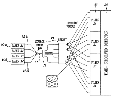
Fig. 1 illustrates schematically the components of the imaging system
according to
the preferred embodiment including the laser sources, injection and detection
port
apparatus, multiwavelength detector, detector signal processor and imaging
computer station;
Fig. 2 illustrates an optical schematic diagram of the imaging system
according to
the preferred embodiment showing the multiple waveguide paths between the
detection ports and the detector, as well as the multiplexed multiple
wavelength
source arrangement;
Fig. 3 illustrates a side view of the detection fibers coupled to a detector
using a
collimating fiber holder; and
Fig. 4 illustrates a plan view of the detector faceplate surface having four
quadrants each provided with a different wavelength selective filter coating.
Detailed Description of the Preferred Embodiment
In the preferred embodiment, the invention is applied to the case of time
domain
optical medical imaging, however, it will be apparent to those skilled in the
art that
the invention is applicable to frequency domain techniques for optical
imaging.
The injected pulses at each of the plurality of wavelengths are preferably
simultaneously injected, however, for the imaging to be "simultaneous", the
time
window reserved for acquiring the TPSF from a single wavelength's injection
CA 02453644 2010-01-08
REPLACEMENT SHEET
-4-
pulse using the chosen detector overlaps between the respective wavelengths
even
if the injected pulses were not simultaneous. In the preferred embodiment, it
is
important to respect the temporal resolution of the detector as better
described
hereinbelow
As illustrated in Fig. 1, the pulsed light source 10 has an output (in
practice, it will
comprise a plurality of laser source outputs at discrete wavelengths, as
described
further hereinbelow) optically coupled via a switch 11 to one of plurality of
waveguides to a number of injection ports of a support 14. The injection ports
are
preferably positioned at a number of fixed positions over the imaging area for
each
wavelength to be used, although the injection port may alternatively be
movable over
the body surface, provided that the body part 16 is immobilized. As is known
in the
art, the injection and detection ports may directly contact the body or a
coupling
medium may be used between the body and the injection/detection ports. The
detection ports and support 18 are arranged in Fig. 1 in transmission mode for
breast
imaging. It also possible to arrange detection ports on the same surface of
the
patient as the injection ports, in which case imaging is achieved by measuring
the
TPSF of the diffused pulse reflected from the tissue.
The light injected is preferably pulses having a duration of about 1 to 100
picoseconds and an average power of about 100 mW. The laser source 10
preferably comprises four laser sources operating at 760 nm, 780 nm, 830 nm,
and
850 nm. These different wavelengths allow for complementary information to be
acquired to build a physiological image of the breast tissue. Referring
concurrently to
Figs. 1 and 2, the output of each wavelength laser source 10a to 10d, for the
four
wavelengths chosen, are coupled to fibers 12a to 12d respectively. The fibers
are
preferably multimode fibers, such as 200/240 micron graded index multimode
fibers.
Although Fig. 2 illustrates for simplicity four injection positions and four
detection
positions, it will be understood that there may be about 10 injection
positions and
typically up to about 50 detector positions. The light from the four sources
is
preferably injected at the same point, and the light is coupled onto a same
fiber 12f,
as shown in Fig. 2, using a coupler 12e, or alternatively the four fibers 12a
to 12d
could be fed as a bundle to the same position within support 14. It will be
appreciated that multiplexing the four wavelengths onto the same fiber 12f
allows a
conventional single wavelength support 14 to be used without taking into
account
different injection positions within the program of the imaging computer or
processor
30. It is of course possible to have injection positions unique to each
wavelength,
CA 02453644 2010-01-08
REPLACEMENT SHEET
-5-
however, to reduce the number of support positions while maintaining the same
number of injection source locations at any chosen wavelength, it is preferred
to
provide multiplexed signals on single fibers or bundled fibers at each
injection/detection site.
While Fig. 2 illustrates a single fiber (or bundle) 12f, there is preferably
10 such fibers
for the 10 injection positions. A fiber switch 11, such as a conventional I by
32 JDS
Uniphase switch is used to switch light from each laser source 10 to a desired
one of
the injection port positions.
The detected optical signals are communicated by waveguides 20, namely 400/440
micron graded index multimode optical fibers, to a spectral channel separator
22,
namely a series of filters in the preferred embodiment. As also shown in
Figures 3
and 4, the filters may comprise band-pass filter coatings 22a, 22b, 22c and
22d on a
faceplate 24 of the detector 26. Each detection fiber 20 is coupled directly
to the
detector 26 without switching, in the preferred embodiment. While it is
possible for
the separator 22 to switch and/or demultiplex the light from fibers 20 onto
lightguides
24, as illustrated in Figure 1, in the preferred embodiment concurrently shown
in
Figures 3 and 4, the fibers 20 are mounted in a collimating fiber holder or
positioner
21 for directing the light from each fiber 20 to the filter 22a to 22d and
then onto the
detector surface 26. The collimating holder has a collimating microlens for
coupling
the light exiting the core of the fiber 20 onto the detector surface with a
small spot
size. In the preferred embodiment, there are 50 detector ports 18, with 200
fibers 20.
Thus an array of 50 fibers is arranged in each quadrant or zone of the
faceplate 22a
to 22d at the detector 26. It will be appreciated that spectral separation may
also be
achieved using an optical spectrometer or a grating device, such as an arrayed
waveguide grating or the like, instead of using a filtering medium or coating.
Preferably, the injection and detection locations are the same for each
wavelength,
however, individual positions for Iightguides for each wavelengths can be
accommodated, e.g. the detector ports could support 200 positions fiber.
CA 02453644 2004-01-14
-6-
The wavelength separated signals are all detected simultaneously by a gated
intensified CCD camera, for example a PicoStar Camera by LaVision. The camera
26 is used to detect the light from each detection port 18 and at each desired
wavelength with picosecond resolution. The injected pulse may spread out over
several picoseconds to several nanoseconds as a result of diffusion through
the
body tissue.
A large number of pulses are injected and their corresponding camera signals
are
processed by imaging computer 30 to determine one data point, i.e. the
temporal
point spread function for a particular wavelength and a particular injection
port and
detection port combination. For a given injection position, the TPSF is
measured
at a number of detector positions at which the detected signal provides good
signal to noise. Such data points are gathered for a large number of
combinations
of wavelengths and port positions to obtain sufficient "raw" data to begin
constructing an image of the tissue. The resulting image can be displayed on
display 32 and printed on a printer 34.
It will also be appreciated that using different detector positions
simultaneously for
a single injector position allows for off-axis information to be used. The
image
processing is thus adapted to take into consideration the geometry related to
the
off-axis data, however, the combination of on-axis and off-axis data is more
accurate and provides 'faster acquisition with better resolution and/or image
robustness.
The imaging computer 30 is also responsible for signaling a laser source 10 to
select a desired wavelength and then switch that wavelength signal to a
desired
output fiber 12. The computer 30 thus progresses through all desired
wavelength
and position combinations to achieve the desired imaging. The laser source 10
synchronizes the camera 26 with each pulse. The laser source 10 may also
comprise a number of fixed wavelength optical sources, as it may also comprise
a
single broadband source.
It will be appreciated that in the case of frequency domain optical imaging,
the
laser 10 will be controlled to be modulated at the desired frequency and
switch its
output onto the desired fiber 12. In this case, the computer 30 will then need
to
sweep through a large number of modulation frequencies at which the amplitude
CA 02453644 2004-01-14
-7-
and phase shift of the detected light is recorded with good accuracy. The TPSF
for
a single data point can be calculated from the amplitude and phase shift data
set
recorded, or typically, the frequency domain data is used directly to
reconstruct
the image.
In the present application, reference is made to a plurality of wavelengths
that can
be separately detected. While these distinct wavelengths can be generated from
a
monochromatic or broadband light source to directly provide the desired
wavelengths, it is alternatively possible to mix a first basic wavelength with
a
second reference wavelength to create a beating of the wavelengths. This can
be
used to tune the basic wavelength to create the desired wavelength at the
plurality
of wavelengths and is another way of providing the light to be injected
according
to the invention. Given that the light source has two parts for the first and
second
wavelengths, it is possible to control or pulse only one to achieve the
desired light
injection.
