Note : Les descriptions sont présentées dans la langue officielle dans laquelle elles ont été soumises.
CA 02475240 2004-07-20
METHOD AND DEVICE TO MEASURE DYNAMIC INTERNAL
CALIBRATION TRUE DOSE RESPONSE CURVES
Field of the Invention
The invention relates to assay devices and methods for constructing assay
devices for
detecting the presence of an analyte in a biological sample and the quantity
of same.
Background of the Invention
Quality standards for immunoassays have traditionally been driven by external
calibration reference standards. Current methods of analysis for typical
immunodiagnostic assays provide diagnostic test results based on generally
accepted
external standard reference measurements. A number of known discrepancies have
become apparent to be quantitation errors induced when assays are carned out,
leading to variations in test results. For example, the concept of assay
sensitivity
attempts to characterize sensitivity by classic statistical analysis based on
repeated
measurement of low concentration samples to confirm that the sample result is
not
statistically different from zero. As the standard error incurred is inversely
proportional to the square root of the number of actual measurements, this
method
does not actually measure the inherent assay sensitivity. Further ref nement
has led to
some improvements. Known in the art as analytical sensitivity, the zero
standard is
measured several times and the limit of sensitivity becomes a concentration
equating
to 2-3 standard deviations (SD) from the mean (M). However, the precision for
this
theoretical determination may be incorrect by an order of magnitude. The
concomitant fitting of any derived external calibration curves) does not
create a true
value dynamic dose response curve that can lead to considerable error in the
actual
sensitivity.
To further measure the accuracy of such analytical measurement, accuracy is
used to
define how close the average measured value is to the true value. The
difference in
measurement is known as the bias or degree of accuracy. Bias may vary over the
range of the assay. It is known in the art that methods for measuring this
true value
need to be developed.
-1-
CA 02475240 2004-07-20
The repeatability of an assay or the estimated error in an analytical assay is
known in
the art as the percentage coefficient of variation (%CV). Automated assay
analysis
machines can be affected by variations in sample concentration, temperature,
heat and
edge effects, incomplete suspension of particles and solid phase
precipitation.
Precision effects also result from fraction separation and counting errors. In
optical
systems error is due to effects of turbidity, presence of fluorophores,
deterioration of
lamps and detectors and the deterioration, over time, of reagents. These
factors
generally lead to significant decreases in signal to noise ratio. Mechanical
manipulation errors can result from poor pipetting and instrument stand-by
periods.
l0 As a direct result, the assessment for precision of any analytical method
requires the
measurement of resulting variability at known and relevant concentrations by
using
defined or standard control solutions to create baseline calibration
standards. Accurate
determination of such calibrators is based on measurement of known
concentrations
in dilution series at predetermined intervals, which are then interpolated.
Commercially available, as well as in-house prepared reference solutions or
reference
standards are available, but are often calibrated with standard or pooled
matrices,
which may vary considerably from actual patient test samples. Part of the
solution to
overcome these errors is to plot the precision against a wide range of
concentrations to
obtain a precision profile, or calibration, of the assay.
Cross reactivity, assay specificity, bias causing interference, alterations in
antigen,
antibody, binding sites, low dose (competitive assay) and high dose (sandwich
assay)
hook effects, heterophilic antibody interference, endogenous interfering auto-
antibodies, complement, rheumatoid factor, interference in solid phase
antibody
binding, endogenous signal generating substances, enzyme inhibitors, catalysts
and
co-factors have also been shown to express confounding activity in assays,
including
cross reactivity, matrix effects and carry over of sample in automated
immunoassay
instruments and samplers.
For clinical applications, the quality control samples may not reflect actual
concentrations in the patient, may not reflect the spectrum of present
analytes and
3o interfere with the sample matrix to no longer reflect the content of the
patient
_2_
CA 02475240 2004-07-20
samples. The quality control samples may measure performance at discrepant
intervals of concentration which may not reflect clinical decision points.
There is therefore a need for an immunoassay that can be reliably calibrated.
Summary of the Invention
The invention is directed to a method for internal dynamic calibration of an
assay
device for determining the concentration of an analyte in a sample where the
assay
device has a substantially planar assay surface. A loading area for receiving
a liquid
sample and a reading area for displaying a detection of analyte are located on
the
surface. A plurality of calibration dots containing pre-determined quantities
of the
1o analyte are printed on the reading portion. A test spot containing a
reagent for binding
said analyte protein is also printed on the reading portion. The analyte is
labeled with
a detectable marker complex prior to introduction onto the assay device. The
analyte -
detectable marker complex will then bind to the reagent in the test dot. The
amount of
antigen in the sample will be proportional to the intensity of detectable
marker in the
test dot. The calibration dots contain differing pre-determined quantities of
the
analyte. Any unlabeled detectable marker will bind to the calibration dots. An
internally calibrated calibration curve can thus be prepared. The intensity of
detectable marker in the test dot can be compared to the calibration curve to
obtain an
absolute value.
2o The method provides a quantitative analysis that is carried out rapidly
using a single
assay device with the ability to contain a known minimum volume of test fluid
and
also to have the ability for flowing fluid through the device in order to meet
a known
concentration of analyte as a function of analyte concentration per tested
volume.
Both the calibration dots and test dots are printed within a single assay
device which
then needs only the application of a single, premixed solution containing the
analyte
and an excess of detecting antibody.
The invention further includes a method for obtaining dynamic true dose
response
curves by printing both calibrator and test samples onto a common test
platform
device. The test platform has a minimum of one test spot. Each test spot has
multiple
3o corresponding calibration spots. The signal obtained from the total number
of
-3-
CA 02475240 2004-07-20
comparative concentration dynamic calibration spots, at indexed X / Y co-
ordinates is
integrated to form the dynamic internal calibration true dose response curve.
In a
similar process, the unknown test spot label response reading is also
integrated over
all obtained readings. The use of multiple test arrays or matrices in platform
format
predicates a confidence limit approaching one hundred percent in having
obtained the
correct test result. The common test platform is exposed to the same test
fluid and
because the calibrator and test samples are exposed simultaneously to the same
test
fluid, accurate measurement of the concentration of analyte present in the
test sample
is determined by the resulting true dose response calibration curve. The
invention
provides the surprising result that the various errors, incurred using known
state of the
art methods, are not reflected when a test sample is processed with the
disclosed
method and device.
According to one aspect of the invention, there is provided a method of
determining
an amount of analyte in a sample solution comprising the following steps:
~ providing an assay device having a substantially planar assay surface having
a plurality of calibration dots printed thereon and a test dot printed
thereon,
the calibration dots containing pre-determined quantities of the analyte, the
test dot including a reagent for binding to said analyte;
~ providing a solution having a reagent for binding to the analyte, said
reagent
being labeled with a detectable marker;
~ introducing the analyte into said solution to form a sample solution;
~ introducing said sample solution onto said assay device;
~ measuring an intensity of detectable marker in said calibration dots;
~ preparing a calibration curve correlating the amount of analyte in said
z5 calibration dots to said intensity of detectable marker;
~ measuring an intensity of detectable marker in said test dot; and
-4-
CA 02475240 2004-07-20
~ calculating an amount of analyte present in said test dot by comparing the
intensity of detectable marker to the amount of analyte corresponding to said
intensity in said calibration curve.
Brief Descriution of the Drawings
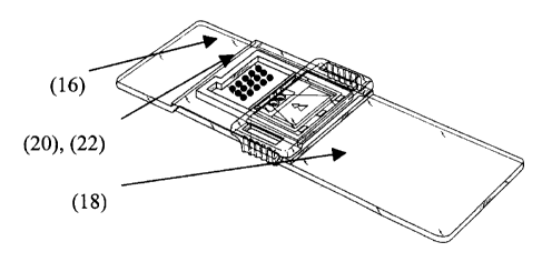
Figure 1 is a top view of an assay device of the present invention for
carrying out a
fixed array test.
Figure 2 is a plot of showing a verification that single and aggregate imrnuno
complexes are quantifiable.
Figure 3 is a plot showing that antigen concentration in the test sample does
not
1o impact fluorescence intensity in the calibration spots.
Figure 4 is an illustration of a PicoTip array printing, spot size and array
matrices.
Figure S is a plot showing a correlation of analyte concentration with
fluorescence
using dynamic true dose response measurement.
Detailed Descrption of the Invention
15 The present method is for calibrating an assay device. A preferred assay
device is
shown in Figure 1. A reading area 16 of the assay device has printed thereon
at least
one and preferably at least two test dots 20. More preferably, a plurality of
dots for
detecting the presence of the analyte are printed on the reading area 16. The
test dots
(20) include a reagent that specifically binds to the protein analyte.
Preferably, the
2o reagent is bound antibodies that specifically bind to the analyte. Other
reagents known
in the art to bind a specific ana.lyte can also be used. For the balance of
the present
discussion the reagent will be referred to as an antibody.
The bound antibodies are preferably spaced apart to make each bound antibody
available for binding to the test antigen free of stearic hindrance from
adjacent antigen
25 complexes. Preferably, a non-reactive protein separates the bound
antibodies in the
test dots.
-5-
CA 02475240 2004-07-20
The reading area 16 has calibration dots 22 printed thereon. The calibration
dots
include a pre-determined amount of said analyte for reacting with un-reacted
reagent
in a vessel, conjugated with a detectable marker. The calibration dots allow
the
intensity of the label to be correlated to the amount of the antigen present.
The
s intensity of label in the test dots can then be used to derive the quantity
of antigen
present.
Many of the problems associated with current methods typical for immunoassays
derive from the assay calibration being determined by introducing external
standard
reference samples for calibration. The present method provides more accurate
results
1o by not using these standard external calibration samples.
In developing a platform device for measuring the quantity of a respective
analyte,
instead of using external standards to generate a calibration or base line,
both
calibration reference spots as well as test spots at unknown concentration are
printed
onto the same test platform. The calibration test spots are printed as known
is concentrations of analyte. The test spots are printed, also at
predetermined X-Y
locations, containing only capture antibody specific for the analyte under
investigation. The test sample is then conjugated with an excess of marker
antibody
and analyte. The marker antibody has previously been conjugated with a
respective
fluorescent label, emitting at a suitable wavelength (e.g. 650 nanometers).
The marker
2o antibody/antigen complex as well as the free, remaining marker antibody is
then
flowed over the test platform using laminar flow. In this fashion, marker
antibody/unknown concentration of antigen complexes are bound by the capture
antibody test spots, whereas free marker antibodies bind to the pre-printed
antigen
spots at known concentrations. Both the test spots (unknown concentration to
be
25 measured) and the calibration spots (known concentrations encompassing the
dynamic range of the analyte) are exposed to the same test fluid sample at the
same
time. The laminar flow effectively places the analyte components within
proximity of
the respective binding sites to promote optimal adhesion kinetics for the
respective
association constants.
3o The test platform is examined in a reader for determination of the
respective
concentrations of fluorescent label attached to the spots on the platform when
-6-
CA 02475240 2004-07-20
activated by suitable wavelength irradiation. The calibration spots,
preferably
originally printed at up to ten different concentrations of analyte, result in
producing a
dynamic internal true dose response curve providing very accurate calibration
reference for the assay. The intensity reading obtained from the test spots
(unknown
concentration) is compared to this calibration Line. The unknown test
concentrations
accurately and efficiently interpolate into the dynamic internal true dose
response
calibration obtained from the known calibration spots.
Each assay device tested obtained similarly accurate results confirming that
the
present method for on-platform dynamic calibration provides an accurate,
enhanced
to and sensitive determination of analyte in both quantitative as well as
qualitative
assays. This immediate and significant benefit demonstrates that these assays,
when
processed using the described method (Dynamic Internal CalibrationTM), do not
reflect
the errors described in association with current other methods for running
these assays
while using externally derived calibration standards. The platform and method
as
described, effectively represent a novel, quantitative and fast method for the
accurate
determination of analyte and or marker concentrations, typically for
diagnostic
clinical markers associated with disease processes. The immediate benefit of
rapid,
accurate measurement of marker concentration allows for rapid dynamic
detection of
marker concentration as an indicator of a disease process (increasing, steady
or
2o decreasing concentration), as well as rapid quantitative monitoring of drug
efficacy in
modifying gene expression for the production of specific marker proteins to
indicate
drug efficacy.
Examples
Example 1: Preferred assay device description.
A preferred assay device is shown in Figure 1. A reading area 16 of the assay
device
18 has printed thereon at least one and preferably at least two test dots 20.
More
preferably, a plurality of dots 22 for detecting the presence of the analyte
are printed
on the reading area 16. The test dots 20 include bound antibodies that
specifically
bind to the protein analyte.
_ '7 _
CA 02475240 2004-07-20
Example 2: Quantitative Fluorescent Imm~uno-Assay.
The sandwich immunoassay matrix incorporates a capture antibody that is
specific for the antigen of interest and a fluorescence conjugated secondary
antibody for detection of analytes and immune complexes. Human chorionic
gonadotropin (HCG), a marker of pregnancy in humans, was used as antigen
for testing of this platform assay. Currently, the dynamic analytical range
for
this test is between 1 to 150 finoUuL (280-37,600mIU/mL) with an assay
volume of S~L. Comparison between the calculated HCG concentration using
the device compared with known HCG concentrations has excellent agreement
to between values (Figure 2, y=1.0717x + 9.9313) and high correlation between
mean values for each concentration tested (r=0.9786). Figure 2 is a graph that
shows confirmation that single and aggregate immuno complexes provide
quantifiable fluorescence when bound to the device platform.
Example 3: Measurement of antigen spot fluorescence at various sample antigen
concentrations.
The assay principle is based on quantitative, non-competitive, heterogeneous
immunoassays. First generation devices were printed with a series of antigen
spots at decreasing concentrations for standard curve auto-calibration;
followed by capture antibody spots. Test sample containing the antigen was
2o processed with lyophilized detecting antibody already conjugated to the
respective indicator or dye. The test sample reacted with label for 2 minutes
and was then dispensed into the chip device to be inserted into the reader.
The
fluorescent intensity of each spot was measured. The reader software
compares the fluorescence of the capture antibody spots (unknown antigen
concentration) with those of the true dose response standard curve (known
concentrations) and calculates the concentration of the test antigen. Figure 3
is
a graph showing data to confirm that antigen concentration in the test sample
does not affect the fluorescence intensities of antigen in the calibration
spots.
_g_
CA 02475240 2004-07-20
Example 4: Advanced Arrav Printing on the Assay Device platform.
Each of four 10 x 10 matrices containing 100 test spots as shown in Figure 4,
used 2.6
mm x 2.6 mm of platform area. The center-to-center separation from spot to
spot was
260 pm. The number of dispensed droplets per spot increments from 1 to 4
droplets)
per spot per array matrix. Total chip device reading area was 8000 Nxn x 10000
Nxn.
This printing pattern allowed 1200 spots to be printed on the platform reading
area.
With a minimum of S spots per test, 3 for calibration and 2 test spots, each
platform
supports 240 tests. This technology advantage allows for fmol/ml antigen
detection
sensitivity and an increasing number of multiplex test arrays for optimal
confidence in
1o diagnostic results by being able to multiplex required test matrices to
optimize ROC
curves. Figure 4 provides an illustration of advanced PicoTip Array printing
technology on the BIOchip platform.
Example 5. Correlation of CK-MB Concentration with Fluorescent Intensity
calibrated using True Dose Response measurement.
As shown in Figure 5, the concentrations of test CK-MB cardiac marker were
measured to determine the dynamic internal true dose response calibration
curve. The
tests confirm that the true dose response accurately plotted as a sigmoid
function over
the dynamic range as tested.
Those skilled in the art will recognize, or be able to ascertain using no more
than
routine experimentation, many equivalents to the embodiments of the invention
described above. Such equivalents are intended to be encompassed in the scope
of the
following claims.
-9-
