Note : Les descriptions sont présentées dans la langue officielle dans laquelle elles ont été soumises.
CA 02478088 2004-08-31
WO 03/077204 PCT/US03/06921
ALGORITHM FOR ACCURATE THREE-DIMENSIONAL
RECONSTRUCTION OF NON-LINEAR IMPLANTED MEDICAL
DEVICES IN VIVO
RELATED APPLICATION
This application claims priority and other benefits from U.S. Provisional
Patent
Application Serial No. 60/362,534, filed March 6, 2002, entitled "ALGORITHM
FOR
ACCURATE THREE-DIMENSIONAL RECONSTRUCTION OF IMPLANTED MEDICAL
DEVICES IN VIVO", incorporated herein by reference in its entirety.
FIELD OF THE INVENTION
The present invention relates to noninvasive implanted medical device motion
and
deformation measurement. More specifically, the invention relates to an
algorithm that enables
three-dimensional reconstruction of the centerline shape and motion of an
implanted non-linear
medical device from biplane radiographic images of the implanted device and a
calibration object.
BACKGROUND OF THE INVENTION
Implantable medical devices, such as leads, vascular devices, heart valves,
annuloplasty
rings or bands, or other prosthetic devices, typically undergo in vitro
testing and structural
modeling to ensure that the device conforms to long-term performance
standards. Although
mechanical failure of such devices is rare, fracture or other forms of
mechanical failure do occur
within the implanted environment following repeated deformation due to cardiac
or other bodily
motion. In vitro tests and structural models are sometimes designed to mimic
or exceed the
deformations that a device will endure once implanted, these testing methods
and structural
models have not been motivated by in vivo measurements of actual device
deformations.
Numerous systems and algorithms have been proposed or are available for
accurate
detection of anatomic surfaces in medical images and for visualizing the
location of a medical
device for surgical navigation. Reference is made, for example, to U.S. Pat.
No. 6,119,033 issued
to Speigelman et al., U.S. Pat. No. 6,236,875 issued to Bucholz et al, and
U.S. Pat. No. 5,983,126
issued to Wittkamp~ Algorithms are also available for performing finite
element analysis for
estimating stress and resultant force distributions along a geometric
structure. However, an
accurate method for reconstruction of an implanted non-linear medical device,
such as a catheter,
CA 02478088 2004-08-31
WO 03/077204 PCT/US03/06921
2
a stmt, or a heart valve device, for example, to measure the repetitive motion
and deformation of
the implanted device is not available.
A method for dynamic three-dimensional reconstruction of an implanted medical
device
shape and motion would be valuable in designing and validating physically
realistic in vitro
mechanical tests and structural models. The inventor of the present invention
previously
developed an algorithm for non-invasive reconstruction of an initially
straight cardiac lead. See
Baxter WW, et al., Medical Image Analysis 2001;5:255-270. However, highly-
curved medical
devices, such as annuloplasty rings or bands, stems, or catheters, for example
cannot be accurately
reconstructed assuming a straight or slightly curved configuration. There
remains a need
therefore, for an algorithm that enables reconstruction of medical devices
such as stems, catheters,
or heart valve devices having non-linear and highly curved geometries.
BRIEF DESCRIPTION OF THE DRAWINGS
Figure 1 is a schematic diagram of a system for acquiring biplane x-ray images
of an
implanted medical device and a calibration object and for incorporating image
data with user-
entered geometry data to generate a four-dimensional reconstruction of the
device centerline.
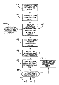
Figure 2 is a flow chart summarizing the steps included in an algorithm, which
may be
implemented using the system of Figure 1, for generating a four-dimensional
reconstruction of an
implanted non-linear medical device centerline.
Figure 3 is a schematic illustration of a user-initialized curved model of an
annuloplasty
band projected on two imaging planes.
Figures 4A-4C are schematic diagrams of a distal end of a non-linear
implantable medical
device for practicing the present invention.
DETAILED DESCRIPTION OF THE INVENTION
The present invention provides a method for determining the centerline shape
and motion
of an implanted medical device as it moves through time due to cardiac,
respiratory, or other
physiological motion. The method includes obtaining biplane radiographic
images of an
implanted non-linear medical device and a calibration object placed in the
imaging volume.
Calibration of biplane images is performed by computing perspective
transformation matrices
using images of the calibration object. The transformation matrices relate the
three-dimensional
CA 02478088 2004-08-31
WO 03/077204 PCT/US03/06921
3
coordinates of the volume occupied by the calibration object to each imaging
plane's local two-
dimensional image coordinates.
The implanted device is reconstructed from the biplane radiographic device
images and a
user-initialized template of the undefonned device. Points on a user-
initialized template of the
non-linear device are projected onto each x-ray image pair using the
transformation perspective
matrices. Through an iterative process using active contours, the device model
translates and
deforms until it matches the biplane image pair, and the resulting device
centerline coordinates are
stored.
This process of determining non-linear medical device centerline position is
repeated for
each time point in an imaging sequence. The reconstructed centerline shape at
instants throughout
a selected imaging sequence can then be displayed to visualize device motion.
Reconstructed
centerline points are output to a text file at each time point for further
analysis or evaluation which
may include in vitro test development, structural model development or
clinical assessment of in
vivo device motion.
The algorithm provided by the present invention can be used to reconstruct a
highly
curved medical device such as a non-linear medical device that has been imaged
using a biplane
x-rays or other imaging techniques producing pairs of conventional planar
images of the
implanted device and a calibration standard. By initializing the algorithm
using a user-specified
curved template, highly curved non-linear medical devices can be accurately
reconstructed.
As indicated above, the present invention is directed toward providing a
method for
reconstructing a dynamic three-dimensional model of an implanted medical
device. Such device
reconstruction is valuable for designing and validating in vitro testing
methods and structural
models and assessing in vivo device motion. The methods included herein are
particularly useful
for reconstructing an implanted medical device having a highly curved geometry
subject to
physiological dynamic motion such as cardiac or respiratory motion. The
present invention is
specifically designed for reconstructing a substantially non-linear medical
device, such as an
annuloplasty ring or band, subjected to cardiac motion, a catheter having a
distal end as illustrated
in FIGS. 4A-4C, or a stmt, for example.
Figure 1 is a schematic diagram of a system for acquiring biplane x-ray
images of an implanted medical device and a calibration object and for
incorporating image data
with user-entered geometry to generate a four-dimensional reconstruction of
the device centerline.
CA 02478088 2004-08-31
WO 03/077204 PCT/US03/06921
4
The system includes an imaging device 14 for generating biplane images of a
medical device 10
implanted in a patient 6 or experimental subject. In a preferred embodiment,
imaging device 14 is
a biplane radiographic imaging device. Biplane views of the imaging field are
simultaneously
recorded, and image data are stored by data storage unit 16 or acquired
directly to a personal
computer or work station 20. Images stored in a desired format by data storage
unit 16, e.g.,
video, film or digital format, are later transferred to work station 20 for
subsequent computer
analysis.
Reconstruction of the implanted device is derived from a biplanar device
image, and device motion can be measured by reconstructing the device at each
point in time
during an imaging sequence. The rate of image acquisition and the duration of
the imaging
sequence are determined according to the application. For analysis of non-
linear medical device
motion, an imaging sequence over one cardiac cycle is typically desired.
Software for processing image data is implemented in a personal computer or
work station
20, with image data transferred from the data storage unit 16 to work station
20. Work station 20
includes a display 21 for displaying acquired biplane images and the evolution
of the implanted
device reconstruction. Work station 20 is also provided with a user interface
22 for receiving
user-entered data regarding device geometry, as will be further described
below.
Figure 2 is a flow chart summarizing the steps included in an algorithm which
may be
implemented using the system of Figure 1 for generating a four-dimensional
reconstruction of an
implanted non-linear medical device centerline. At step 105, biplane
radiographic imaging of the
implanted device is performed, with image data stored andlor transferred as
described above. An
imaging sequence may correspond to one or more cardiac cycles or cardiac cycle
segments or
respiratory cycles or cycle segments or other time duration that captures the
device motion and
deformation of interest. Immediately after biplane images of the implanted non-
linear medical
device are acquired, a calibration object is placed in the imaging volume
without altering the
imaging geometry to record biplane images of the calibration object, as
indicated at step 110.
At step 115, calibration of the biplane imaging volume is performed by
computing
perspective transformation matrices using images of the calibration object.
The transformation
matrices relate the three-dimensional coordinates of the volume occupied by
the calibration object
to each imaging plane's local two-dimensional image coordinates. See Mackay
SA, et al.,
Comput. Biomed. Res. 1982:15: 455-473 for technical details regarding this
transformation,
CA 02478088 2004-08-31
WO 03/077204 PCT/US03/06921
incorporated herein by reference in its entirety. The transformation matrices
are calculated using
estimates based on user-specified image coordinates of landmark points on the
calibration object
from each biplane image and known three-dimensional coordinates of the
landmark points on the
object. User-specified points are entered at step 120 as input to the
calibration process. The
5 resulting transformation relationships can be expressed by:
x;[G]'=k~u~~, and x;[G]2=k2u~2
wherein x; represents the global coordinates with i equaling 1, 2 or 3
corresponding to the three
dimensions of the global volume; [G] ~ and [G]2 represent 4x3 matrices
corresponding to the first
and second planar views, respectively; k' and k2 are scaling factors relating
to the magnification
of a particular view and u~l and u~2 represent the local coordinates of the
first and second planar
views, respectively, with j equaling 1 or 2 corresponding to the two
dimensions of the respective
planar view.
At step 125, a user-specified, curved template of the undeformed implanted
device is
provided as input to initialize the active contour evolution process. The user-
intialized
reconstruction permits an iterative process to begin with a curved non-linear
medical device
template enabling accurate device reconstruction. The curved non-linear device
could include
implantable medical devices such as a catheter, a stmt, or heart valve device,
for example. The
user selects, via user interface 22 (FIG. 1), two or more fiducial points on
or near the images of
the device to orient the three-dimensional curved device template. Points may
correspond to
identifiable landmarks on the medical device such as device endpoints, joints
of dissimilar
materials, recognizable device component locations, or on a starting point not
located directly on
the device, such as a point corresponding to a center of radius 13 (FIG. 1) of
the non-linear
device.
At step 130, the three-dimensional curved template is projected onto the first
of the time-
paired image planes. Figure 3 is a schematic illustration of a user-
initialized curved model of an
annuloplasty band projected on two imaging planes. In order to solve for a
three-dimensional
model point without requiring user intervention, it is necessary to project
spatial positions of a
three-dimensional model onto local image coordinates and iteratively solve for
the three-
dimensional coordinates rather than deriving the three-dimensional global
points from two sets of
local projection coordinates. As shown in Figure 3, active contour model
points 51 along the
centerline of the user-initialized template 50 are projected onto each biplane
image 54 and 56.
CA 02478088 2004-08-31
WO 03/077204 PCT/US03/06921
6
Projection of three-dimensional centerline points onto each two-dimensional
view is achieved
using the corresponding transformation perspective matrices computed
previously at step 11 S.
At step 135, an iterative algorithm is performed to solve for the three-
dimensional
centerline point coordinates for the given time point corresponding to the
first pair of planar
images. The solution algorithm preferably employs an active contour method.
During solution
iterations, the user can interactively prod projected points in each view with
a mouse or other
pointing device.
After finding the centerline coordinates for the current image pair, the next
pair of images
is loaded at step 145 for determining the centerline location of the implanted
non-linear medical
device at the next recorded time point. Steps 130 and 135 are repeated until
the coordinates for
points along the implanted non-linear medical device centerline are calculated
for all instants in
time recorded during a selected imaging sequence as determined at decision
step 140, after which
the algorithm is terminated at step 145. An example of an algorithm for
performing all of the
steps described in FIG. 2, other than the step of generating the user-
specified curved non-linear
medical device template (Step 125) can be found in chapter 2 of Baxter WW, et
al., Medical
Image Analysis 2001, incorporated herein by reference in its entirety
Once the displacements of the device centerline over a given time interval are
known, the
centerline coordinate data may be provided as input for a number of analyses.
Displacement and
shape change measurement data can be used for designing and validating in
vitro testing and
structural modeling methods or for clinical evaluation of in vivo device
motion. Displacement
and deformation data is generated based on clinically realistic device imaging
and known device
geometry thereby providing a powerful framework for device design work and
testing.
Thus, a system and method has been described which allow accurate dynamic
three-
dimensional reconstruction of an implanted non-linear medical device
centerline. Results are
valuable to engineers and scientists in designing new non-linear medical
devices and developing
physically realistic in vitro tests to attempt to ultimately improve overall
device performance. For
example, such results can be used in test developments, structural analysis,
boundary conditions
for generating models, or as an input to implantable stimulation devices.
Specific embodiments
have been described herein to illustrate features of the invention with
respect to a particular
medical device. While the present invention has been described according to
specific
CA 02478088 2004-08-31
WO 03/077204 PCT/US03/06921
7
embodiments in the above disclosure, these embodiments should be considered
exemplary, rather
than limiting, with regard to the following claims.
