Note : Les descriptions sont présentées dans la langue officielle dans laquelle elles ont été soumises.
CA 02497354 2005-03-17
SURGICAL DEVICE FOR REMOVING TISSUE CELLS FROM A BIOLOGICAL STRUCTURE
The invention is directed to a surgical device according to the preamble of
claim 1.
Such devices are used in surgical hospitals for cosmetic purposes and for
treating illnesses, as
well as for harvesting tissue cells that can reproduce.
It is generally known to suction off, for example, excess fatty tissue cells
for cosmetic purposes.
In a first step, a pressurized working fluid is injected into the fatty
tissue, dissolving the fatty tissue
in the working fluid by a chemical reaction. In a second step, a suction
cannula having a reduced
pressure is pushed into the corresponding fatty tissue, whereby the suction
force tears the fatty
tissue completely out of the connective tissue and removes the mixture of
dissolved fatty tissue
and working fluid. The mixture is then collected in a receiving container and
subsequently
disposed of. The suction cannula is formed so as to have several suction
openings that are
uniformly distributed about its periphery.
US-A 5,968,008 discloses a suction device for fatty tissue of this type which
includes an injection
line arranged inside the suction cannula. The injection line terminates in an
outlet opening, from
which a circular liquid jet exits. With his device, the two separate steps of
injecting working fluid
and suctioning off fatty tissue using the working fluid are now performed
simultaneously, so that
the surgical procedure can be performed in less time and continuously. DE 200
09 786 U1
describes a similar suction device for fatty tissue with an injection line
having a slit-like exit
opening, from which the jet of the working fluid exits in a fan-like shape.
This fan-shaped fluid jet
supposedly improves the distribution of the working fluid, so that a larger
volume of fatty tissue
can be uniformly removed.
All the aforedescribed suction devices for fatty tissue are designed to tear
the fatty tissue out of
the connected tissue through the combined effect of the dissolving power of
the working fluid and
the force of the suction flow. However, the combined effect from these two
components causes
CA 02497354 2005-03-17
problems, because the suction force has a constant value, whereas the
dissolution process by
the working fluid is time-dependent. Because suction force and dissolving
power are not
coordinated with each other, the constant suction force is too small at the
beginning of the time-
dependent dissolution process, as not enough tissue cells have been dissolved,
and is too large
at the end of the dissolution process, because the tissue cells have all been
exposed at the end
of the dissolution process. Consequently, the time during which the tissue
cells are exposed to
the working fluid is either too long or the tissue cells are exposed to an
excessively large suction
force. In both situations, the tissue cells to be suctioned off as well as
tissue cells that should be
preserved are destroyed. The human body is subjected to stress which
complicates and
prolongs the healing process.
The suctioned-off fatty tissue cells are destroyed by the suction process and
by the detrimental
influence of the working fluid and can then no longer be used.
DE 100 33 278 A1 of the Applicant describes for the first time a device for
removing tissue cells
from a biological structure. The device is primarily intended to completely
separate the excess
fatty tissue cells from the adjacent tissue cells by a pressurized fluid jet.
The exit opening of the
injection line is shaped to form a flat jet is with a frontal cutting edge
that operates like a scraping
device and therefore effectively peels the fatty tissue cells off. The fluid
is pressurized and
chemically neutral; the pressurized fluid enters in an intelligent manner
between neighboring
smooth and soft fatty tissue cells, urges the tissue cells apart, and thereby
mechanically
separates the strong tendons that hold the tissue together without destroying
the tissue cells.
The carefully separated tissue cells together with the neutral fluid are
suctioned off by a relatively
small suction force and are discharged, or alternatively, are separated again
from the neutral fluid
and reused. This type of surgical devices advantageously separates the fatty
tissue cells solely
by applying the force of the separation jet, whereas the fatty tissue cells
are suctioned off
together with the neutral fluid by the force of the suction flow. Unlike with
prior art devices, the
separation force and the suction force need not be matched and can be selected
independent of
each other, with the respective forces adjusted to provide the least harmful
treatment for the
2
CA 02497354 2005-03-17
patient. Unlike prior art devices, which produce a fluid intermixed with
blood, the novel surgical
tissue removal device produces a milky, white suction flow dominated by fatty
tissue cells.
However, even this surgical removal device still causes stress in the human
body and damage to
a large percentage of fatty tissue cells.
It is the therefore an object of the invention to minimize the required
separation force and the
required suction force of a surgical device of this type.
The object is solved by the characterizing features of claim 1. Additional
embodiments are
recited in the dependent claims 2 to 7. More particularly, the novel surgical
device is designed to
operate with a very small separation force and with a very small suction
force. This is particularly
easy on the human body during a surgical procedure, but also causes less
damage to the tissue
cells, which can then be reused for other purposes.
One explanation for the very small separation force is that one does no longer
operate with a
frontal separation edge, which encounters a significant resistance due to its
width and therefore
has to apply a large force. Instead, with the novel device, the separation tip
initially enters the
space between the tissue cells followed by the inclined separation edges, so
that the tissue
sections to be separated are no longer loosened by a beating motion, but are
instead cut off
along the separation edge. This pure cutting process encounters a very low
resistance, so that
the cutting or separation force can be kept small. Advantageously, the
required cutting force can
be selected by selecting the angle a, i.e., the cutting angle of the
separation edge.
The required suction force can also be kept very small, which is easy to
explain as follows.
Because the separation edge is generally disposed before the suction openings
and because the
separation forces are oriented in the flow direction, i.e., away from the
suction openings, all tissue
parts that are exposed to the separation forces also tend to move away from
the suction
openings. Before the tissue parts that move away can be suctioned off, they
must first be slowed
down, their direction must be reversed, and they must be accelerated again. As
a result, the
suction force has to overcome both the separation forces and the inertia of
the tissue parts, which
CA 02497354 2005-03-17
represents a complex movement inside the human body. Because of the novel
surgical device
requires a smaller separation force due to its novel orientation of the flat
fluid jet, the recovery
process of the tissue parts also requires a smaller force.
Advantageously, the nozzle slit in the novel surgical device can be V-shaped
due to the required
small separation and suction forces, so that very wide separation edges can be
formed, which
increase the effectiveness of the surgical device. Advantageously, more than
one separated flat
fluid jet can be used, which also effectively increases the operating field.
Those skilled in the art will realize that additional embodiments can be
selected without departing
from the scope of the present invention.
The invention will now be described with reference to several embodiments.
It is shown in:
Fig. 1 in a first view, the operating handpiece with a flat jet that is
inclined twice with respect to
the axis,
Fig. 2 the operating handpiece of Fig. 1 in another view,
Fig. 3 the detail X of Figs. 1 and 2,
Fig. 4 a view of the operating handpiece with a flat jet that is inclined once
with respect to the
axis,
Fig. 5 the detail X of Fig. 4,
Fig. 6 the operating handpiece of Fig. 5 with an orientation rotated by
180°,
Fig. 7 the detail X of Fig. 6,
Fig. 8 the operating handpiece with two flat jets that are inclined with
respect to the axis and
angled with respect to each other,
Fig. 9 the detail X of Fig. 8,
Fig. 10 the operating handpiece of Fig. 8 with an orientation rotated by
180°,
Fig. 11 the detail X of Fig. 10,
Fig. 12 a view of the operating handpiece with two flat jets that are inclined
twice with respect to
4
CA 02497354 2005-03-17
the axis and arranged with respect to each other in form of an impeller,
Fig. 13 a different view of the impeller, and
Fig. 14 the detail X of Fig. 12.
The surgical device for removing vital tissue cells from a biological
structure includes a fluid
separation device for the separating a biological structure, as described for
example in EP 0 551
920 B1 of the same Applicant, and a suction device. Both the fluid separation
device and the
suction device are generally known and are therefore not illustrated herein.
The fluid separation
device has a supply container, a pressure pump, and an injection line, whereas
the suction
device has a collection container, a suction pump, and a suction line. The
injection line of the
fluid separation device and the suction line of the suction device both
terminate in an operating
handpiece 1.
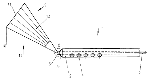
The figures in this application show the distal end of the operating handpiece
1. The distal end of
the operating handpiece 1 includes an outer suction tube 2, whereby the
proximal side of the
suction tube 2 is connected with the suction pump of the suction device, and
the distal end of the
suction tube 2 includes a cone-shaped projection 3 with a center receiving
bore. The suction
tube 2 includes one or more rows of radial suction openings 4 arranged around
the periphery of
the suction tube 2 in a particular pattern.
An injection cannula 5 is disposed inside the suction tube 2 and connected on
the proximal side
by an injection line with the pressure pump of the fluid separation device.
The injection cannula 5
is fitted with clearance into the center receiving bore of the suction tube 2,
whereby the injection
cannula 5 protrudes lengthwise by a certain distance from the through-bore of
the suction tube 2.
The distal end of the injection cannula 5 is formed as an injection nozzle 6
and accordingly has a
conical tip 7 with an apex angle of approximately 90°. One or more
nozzle slits 8 with a particular
shape and arrangement are disposed in the conical surface of the conical tip
7.
The various figures depict different embodiments of these particular nozzles
slits 8 of the injection
nozzle 6 and different arrangements of the suction openings 4 in the suction
tube 2.
Figs. 1 to 3 show a nozzle slit 8 that is inclined by an angle a of maximal
30° with respect to the
CA 02497354 2005-03-17
axial cross-sectional plane of the conical tip 7 and which extends from the
edge of the cone
diameter to the visible edge of the conical tip 7. This results in a flat
fluid jet 9 which is twice
inclined with respect to the conical axis and thereby forms a forward
separation tip 10 with a first
separation edge 11 and a second separation edge 12. Both separation edges 11
and 12 are
arranged adjacent to the separation tip 10. Also formed is an upper peeling
surface 13, on which
the separated tissue parts slide off so as to be carried away towards the
suction openings 4 in the
suction tube 2. The suction openings 4 in the suction tube 2 are hereby
arranged in a single row
in an axial direction of the suction tube 2 and oriented toward the side of
the peeling surface 13
and the location of the separation tip 10.
Figs. 4 and 5 show a V-shaped nozzle slit 8 having to two branches that extend
from a common
tip located on the cone edge of the conical tip 7 to the visible edge of the
cone tip 7. The two
branches of the V-shaped nozzle slit 8 subtend an angle of approximately
90°. This forms an
angled fluid jet 9 with two frontal separation tips 10 and a first separation
edge 11 and a second
separation edge 12. The peeling surface 13 is enclosed by the angle of the
separation jet 9.
Figs. 6 and 7 show another, likewise V-shaped, angled nozzle slit 8 which is
mirror-symmetric to
the V-shaped nozzle slit 8 depicted in Figs. 4 and 5 and forms two outside
peeling surfaces 13.
Another embodiment is shown in Figs. 8 and 9, and Figs. 10 and 11,
respectively. In this
embodiment, two V-shaped nozzle slits 8 are arranged with a spacing
therebetween. In the
embodiment of Figs. 8, 9, two separation tips 10, a first separation edge 11,
a second separation
edge 12, as well as two inside peeling surfaces 13 are formed. Two outside
peeling surfaces 13
are formed on the fluid jet as a result of the different orientation of the
two nozzle slits 8 of Figs.
10, 11.
Another advantageous embodiment is shown in Figs. 12 to 14. Two separate
nozzle jets 8 are
located on either side of the conical tip 7, forming divergent fluid jets 9,
which together have the
shape of an impeller. A forward separation tip 10, a first separation edge 11
and a second
separation edge 12 as well as a peeling surface 13 are associated with each
fluid jet 9. The
suction tube 2 includes two rows of suction openings 4, wherein each of the
row of suction
6
CA 02497354 2005-03-17
openings 4 cooperates vuith the peeling surface 13 of a corresponding fluid
jet 9.
CA 02497354 2005-03-17
List of a reference characters
1 operating handpiece
2 suction tube
3 cone-shaped projection
4 suction opening
injection cannula
6 injection nozzle
7 conical tip
8 nozzle slit
9 fluid jet
separation tip
11 first separation
edge
12 second separation
edge
13 peeling surface
8
